


Case report
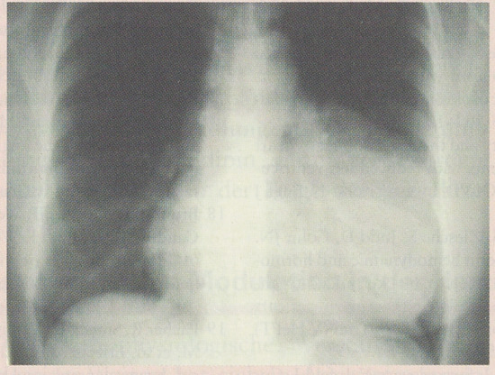
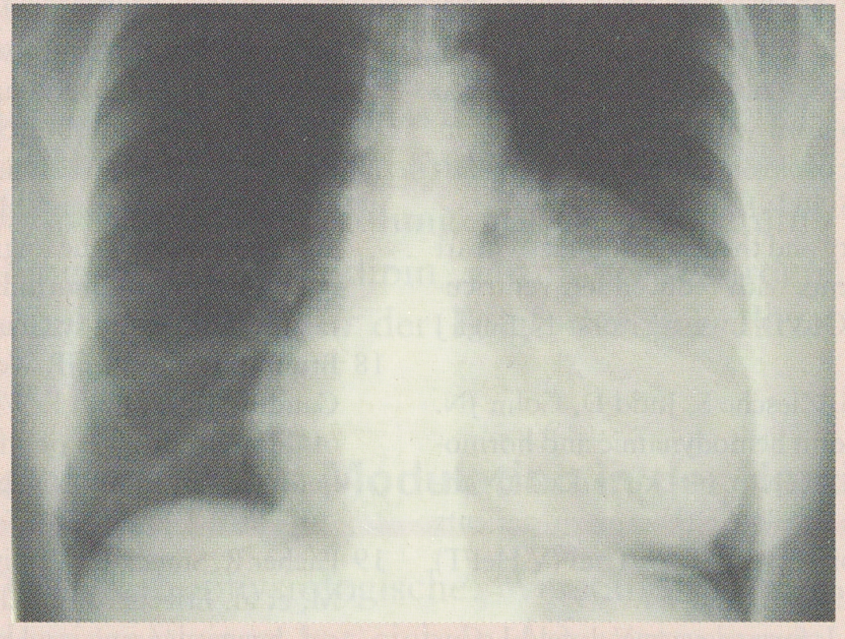
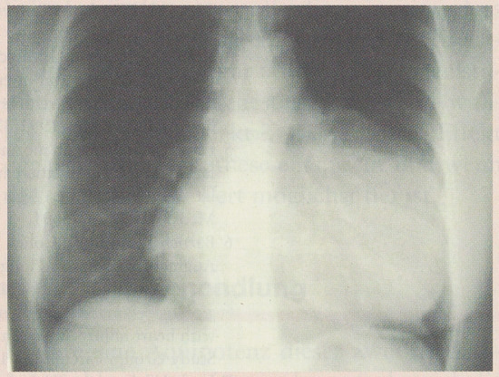
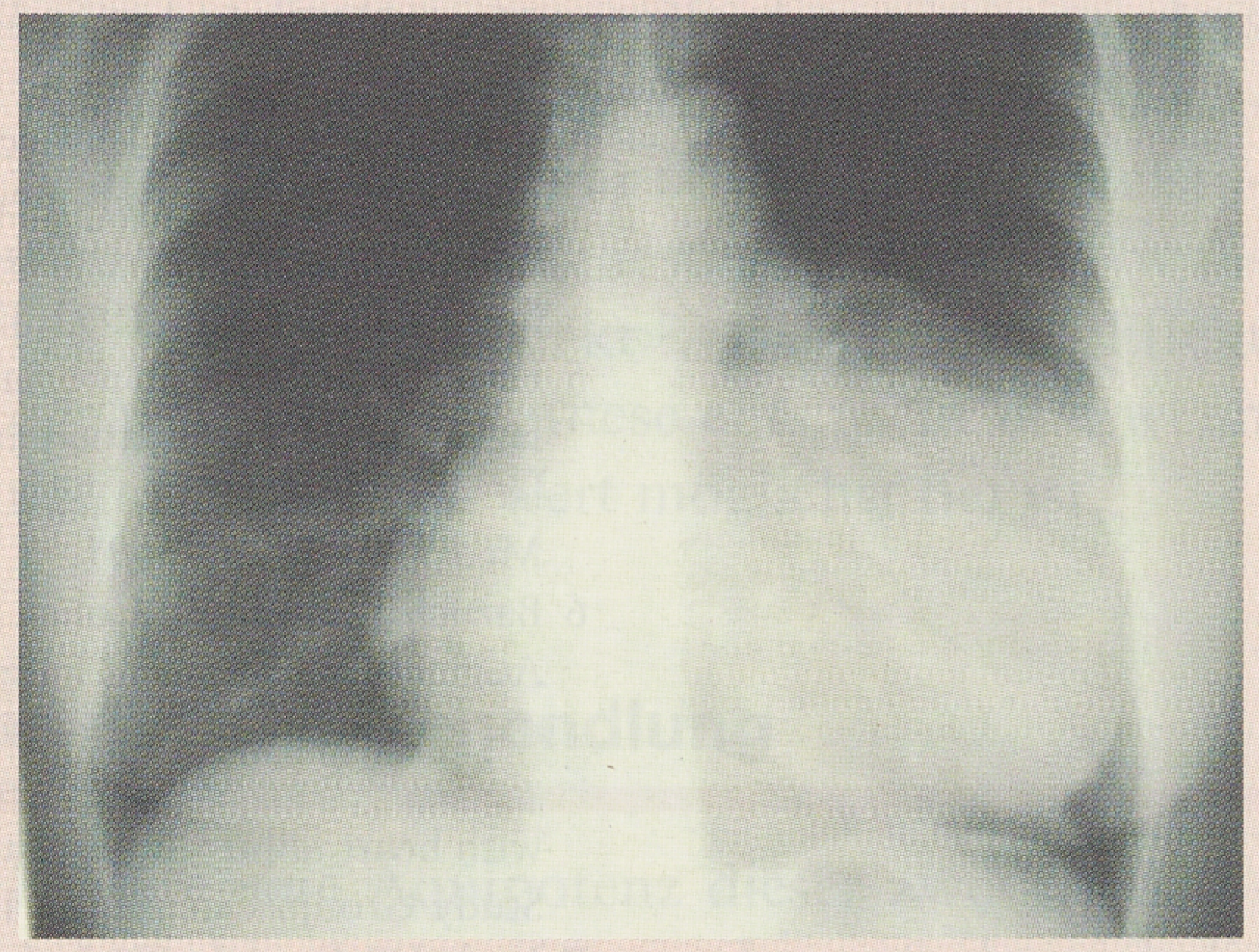
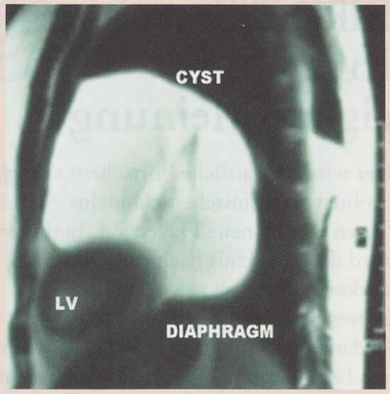
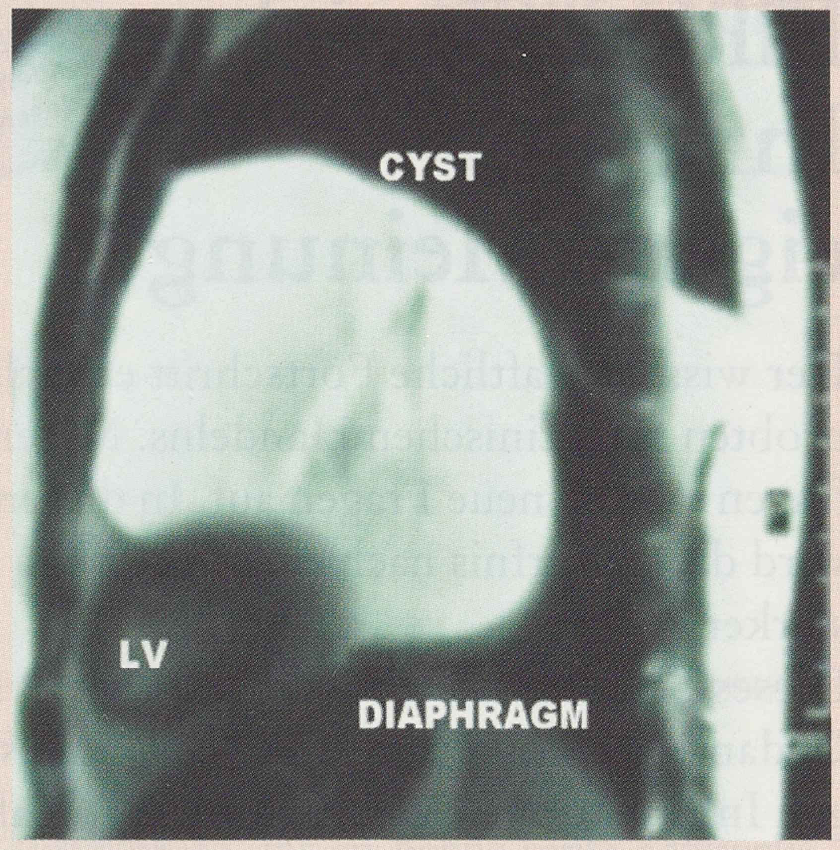
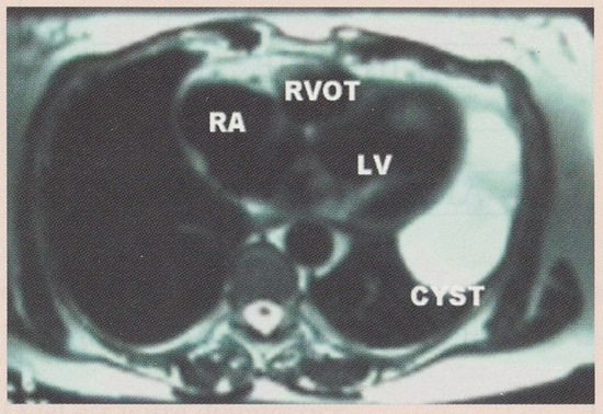
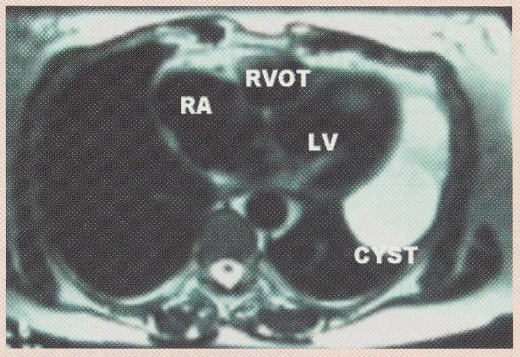
A 54-year-old woman consulted her general practitioner because of general weakness, cough, and left sided chest pain. A radiograph of the chest was obtained, demonstrating a large left sided paracardiac mass resembling left ventricular aneurysm (Figure 1). However, a 12 lead ECG obtained at rest was normal. The patient was referred to echocardiography, where a large paracardiac cyst was found (Figure 2). Echocardiography of the heart was normal. Cardiovascular magnetic resonance (CMR) revealed a signal intense large paracardiac cyst (T2 weighted spin echo imaging; Figure 3, Figure 4) with a low signal intensity on Tl weighted Turbo Spin Echo (not shown), highly consistent with the diagnosis of pericardial cyst, later confirmed by pathology. The cyst was removed by thoracoscopy. This operation was made more difficult by the course of the left phrenic nerve passing along the anterolateral circumference of the pericardial cyst. In the midterm follow-up of 3 months, no recurrence of the cyst was observed on conventional chest radiography neither was chest pain reported by the patient nor left diaphragm paresis noted.
Figure 1.
Plain chest radiograph demonstrating a large left sided paracardial mass.
Figure 2.
Echocardiogram: left ventricle is shown on the left (LV), the paracardiac cyst is visible as a paracardiac signal void large sac located laterally of the left ventricle. Because of the large paracardial cyst, true apical two- and four chamber views could not be obtained.
Figure 3.
Sagittal T2 weighted ECG and respiratory triggered turbo spin echo sequence demonstrating signal intense large, "air bag shaped" cyst in the anterior and mid portion of left hemithorax. LV denotes left ventricle.
Figure 4.
Transverse T2 weighted ECG and respiratory triggered turbo SE sequence demonstrating a signal intense cyst in the anterior and mid portion of the left hemithorax. RVOT = right ventricular outflow tract, RA = right atrium, LV = left ventricle.
Comments
Usually, cysts positioned anterolaterally to the heart are congenital pericardial ("spring water") cysts; however, pericardial cysts are more commonly found at the right costophrenic angle [1] and must be differentiated from bronchogenic cysts, which are usually smaller and located more in the posterior mediastinum. Many other diagnoses have to be considered in a given patient, including cystic teratomas, cystic neurogenic tumors, thymic cysts, aneurysms, and pseudoaneurysms.
In general, congenital pericardial cysts cause no symptoms at all and are only very rarely a cause of serious cardiovascular complications, such as acute right-sided heart failure due to hemorrhage into a pericardial cyst.
If symptoms occur, variate cures are suggested in the literature including punction of the cyst without or with subsequent ethanol sclerosis or with injection of contrast media, open chest surgery, and thoracoscopic resection [2]. The optimal therapeutic approach is dependent on local expertise; intuitively however, given the low risk inherent to the natural course of even large pericardial cysts, one may favour referral to physicians capable to perform minimally invasive surgical interventions.
Usually, clinical examination, a plain chest radiograph and echocardiography are sufficient for clinical decision making. In certain cases however, physicians may prefer to employ more advanced imaging modalities such as MRI, which, by its inherent capabilities to characterize tissues and fluids in the human body, may be helpful in the differential diagnosis of thoracic tumors and cysts [3].
References
- Feigin, D.; Fenoglio; McAllister, H.; et al. Pericardial cysts: A radiologic-pathologic correlation and review. Radiology 1977, 125, 15–19. [Google Scholar] [CrossRef] [Scilit] [PubMed]
- Weder, W.; Klotz, H.P.; von Segesser, L.; Largiader, F. Thoracoscopic resection of a pericardial cyst: a case report. J. Thorac. Cardiovasc Surg. 1994, 107, 313–314. [Google Scholar] [CrossRef] [Scilit] [PubMed]
- Hoffmann, U.; Globits, S.; Frank, H. Cardiac and paracardiac masses. Current opinion on diagnostic evaluation by magnetic resonance imaging. Eur. Heart J. 1998, 19, 553–563. [Google Scholar] [CrossRef] [Scilit] [PubMed]
© 2000 by the author. Attribution - Non-Commercial - NoDerivatives 4.0.