


Abstract
Objective: Gut dysbiosis has been increasingly implicated in the pathophysiology and progression of chronic heart failure. CRT improves cardiac function and promotes reverse cardiac remodelling. However, the impact of CRT on gut microbiota composition remains largely unknown. This study protocol describes a prospective cohort study designed to investigate changes in gut microbiota and microbial-derived metabolites following CRT implantation. Methods: ‘GUT-CRT’ is a single-centre, prospective observational cohort study enrolling 50 adults with chronic heart failure undergoing CRT-P or CRT-D implantation. Stool, urine and serum samples will be collected at two timepoints (pre-CRT and 6 months post-CRT). Gut microbiota composition will be assessed using 16S rRNA gene sequencing or metagenomic analyses. Urinary and serum TMAO concentrations will be quantified using NMR spectroscopy, alongside assessment of heart failure biomarkers, echocardiographic parameters, and cardiopulmonary exercise testing variables. The primary endpoint is the within-participant change in gut microbiota composition following CRT implantation. Secondary analyses will investigate changes in serum and urinary TMAO concentrations and associations between microbiome alterations, and exploratory analyses will investigate associations between microbiome alterations and serum and urinary TMAO concentrations with biomarkers’ responses, echocardiographic parameters, cardiopulmonary exercise performance, and clinical response to CRT. Results: As this is a study protocol, no results are currently available. Planned analyses include alpha-diversity and beta-diversity comparisons, differential taxonomic abundance analyses, metagenomic pathway analyses, multivariable modelling, and exploratory analyses examining associations between microbiome changes and clinical response to CRT. Conclusions: This study may provide novel insights into the interaction between cardiac resynchronization therapy and the gut microbiome in patients with chronic heart failure. Understanding whether improvements in cardiac function and haemodynamics are accompanied by favourable changes in gut microbial composition may contribute to a better understanding of the gut–heart axis and identify potential microbiome-related biomarkers of CRT response.
1. Introduction
1.1. The Human Gut Microbiota: Definition and Overview
All forms of life are believed to have evolved from simple unicellular organisms over billions of years, ultimately giving rise to the highly complex multicellular organisms that exist today. Humans can be considered “superorganisms,” harbouring an estimated 100 trillion microorganisms that coexist with the host and contribute to numerous physiological processes [1,2]. The emergence of microbiome research and the Human Gut Metagenome Project has substantially advanced our understanding of host–microbe interactions and their implications for health and disease.
The gut microbiota refers to the community of microorganisms residing within the gastrointestinal tract, including bacteria, viruses, fungi, and archaea. It is important to distinguish the gut microbiota from the gut metagenome. While the first one describes the microbial communities themselves, the second one includes the complete genetic content of these microorganisms, including all microbial genes and their functional potential [2,3,4].
Through the expression of millions of microbial genes, the gut microbiota produces thousands of bioactive metabolites that play essential roles in maintaining physiological homeostasis. The intestinal microbiota is dominated by seven major bacterial phyla—Firmicutes, Bacteroidetes, Proteobacteria, Fusobacteria, Actinobacteria, Cyanobacteria, and Verrucomicrobia—with Firmicutes and Bacteroidetes accounting for the largest proportion of the microbial population. The relative abundance of these phyla varies substantially among individuals and is influenced by genetic, environmental, and dietary factors [5].
Human gut microbial communities have also been classified into three principal enterotypes: Bacteroides, Prevotella, and Ruminococcus. Enterotype 1 is characterized by the predominance of Bacteroides species, which exhibit both saccharolytic and proteolytic metabolic activities. Enterotype 2 is dominated by Prevotella species, which are involved in the degradation of mucin glycoproteins and complex carbohydrates. Enterotype 3 is characterized by the predominance of Ruminococcus, which participates in mucin degradation and membrane sugar transport [6]. However, the concept of distinct enterotypes remains controversial because of the substantial interindividual variability observed in gut microbial composition [7].
1.2. Functions of the Gut Microbiota
The gut microbiota performs several essential functions that can be broadly categorized into metabolic, protective, structural, and neuroregulatory activities [8,9,10].
1.3. Metabolic Functions
One of the most important functions of the gut microbiota is the production of bioactive metabolites that influence host physiology. Among these, short-chain fatty acids (SCFAs), secondary bile acids, and tryptophan-derived metabolites are of particular importance. SCFAs, including acetate, propionate, and butyrate, are produced through the fermentation of dietary fibres by intestinal bacteria. These metabolites exert anti-inflammatory and antioxidant effects, contribute to the regulation of the renin–angiotensin–aldosterone system, promote vasodilation, and play a critical role in maintaining gut barrier integrity. Furthermore, SCFAs have been implicated in the regulation of the gut–brain axis and cardiovascular homeostasis [8,9].
The gut microbiota also participates in bile acid metabolism by converting primary bile acids into secondary bile acids. These metabolites influence cholesterol metabolism, glucose homeostasis, vascular function, and immune responses. Additionally, microbial metabolism of tryptophan generates a variety of biologically active compounds that regulate inflammatory pathways, neurotransmitter production, intestinal barrier function, and endocrine signalling. Beyond metabolite production, the gut microbiota contributes to the synthesis of essential vitamins, including vitamin K and several B-complex vitamins. It also supports immune homeostasis by regulating both innate and adaptive immune responses and by protecting the host against pathogenic microorganisms [8,9].
1.4. Protective Functions
A substantial proportion of the body’s immune activity is concentrated within the gastrointestinal tract. The intestinal immune system consists primarily of Peyer’s patches, the lamina propria, and the intestinal epithelium, all of which interact closely with the gut microbiota [11].
The intestinal epithelium contains specialized Paneth cells that secrete antimicrobial peptides in response to pathogenic microorganisms. In addition, epithelial cells produce cytokines and chemokines that activate local and systemic immune responses [12].
Peyer’s patches are organized lymphoid structures that facilitate antigen recognition and antibody production. They contain macrophages, dendritic cells, T lymphocytes, and B lymphocytes that coordinate mucosal immune responses. Specialized microfold (M) cells transport luminal antigens to immune cells within Peyer’s patches, thereby promoting antigen presentation and immune surveillance. Subsequently, T and B lymphocytes located within the lamina propria determine whether immune activation or tolerance is required [13].
1.5. The Gut–Brain–Microbiota Axis
The bidirectional communication network linking the gastrointestinal tract and the central nervous system is known as the gut–brain axis. This complex system involves interactions among the enteric nervous system, the autonomic nervous system, and the hypothalamic–pituitary–adrenal axis. In recent years, accumulating evidence has highlighted the central role of the gut microbiota within this communication network, giving rise to the concept of the gut–brain–microbiota axis. The gut microbiota influences both enteric and central nervous system function through multiple mechanisms, including the production of neurotransmitters and neurotrophic factors, regulation of afferent neural pathways, maintenance of intestinal barrier integrity, modulation of mucin production, and generation of microbial metabolites [14,15].
Remarkably, more than 90% of the body’s serotonin and approximately 50% of dopamine are produced within the gastrointestinal tract under the influence of gut microbial activity. Consequently, alterations in microbial composition may influence not only gastrointestinal function but also behaviour, cognition, mood, and systemic physiological processes [16].
1.6. Dysbiosis
Dysbiosis refers to qualitative and quantitative alterations in the composition and function of the gut microbiota that adversely affect the host. This condition is characterized by reduced microbial diversity, loss of beneficial microorganisms, expansion of potentially pathogenic species, and alterations in microbial metabolic activity [17]. Dysbiosis can increase intestinal permeability, facilitating the translocation of bacteria and bacterial products into the systemic circulation. This phenomenon, often referred to as metabolic endotoxemia, contributes to chronic low-grade inflammation and has been implicated in the pathogenesis of numerous cardiometabolic and inflammatory diseases [18].
Multiple factors may contribute to the development of dysbiosis. Dietary patterns rich in animal fats, refined sugars, and processed foods but low in dietary fibre are among the most important determinants. Medications, particularly antibiotics, proton pump inhibitors, non-steroidal anti-inflammatory drugs, and immunosuppressive agents, can also profoundly alter microbial composition. Additional contributing factors include ageing, genetic predisposition, environmental exposures, and a wide range of chronic diseases [19].
1.7. Heart Failure, Cardiac Resynchronization Therapy, and the Gut Microbiota
Heart failure (HF) remains a major global health challenge, and despite significant advances in pharmacological therapies and device-based interventions over the past two decades, HF continues to be associated with frequent hospitalizations, impaired quality of life, and poor long-term prognosis [20]. The growing prevalence of HF, largely driven by population ageing and improved survival following cardiovascular events, highlights the need for a better understanding of the underlying pathophysiological mechanisms and the identification of novel therapeutic targets [21].
Cardiac resynchronization therapy (CRT) is an established treatment for selected patients with symptomatic heart failure with reduced ejection fraction (HFrEF), ventricular dyssynchrony, and prolonged QRS duration who are in optimal guideline-directed medical therapy. Numerous randomized clinical trials have demonstrated that CRT improves symptoms, exercise capacity, left ventricular systolic function, reverse cardiac remodelling, quality of life, and survival while reducing heart failure hospitalizations. Nevertheless, approximately 30–40% of patients do not experience a clinically meaningful response following CRT implantation, and the mechanisms underlying interindividual variability in treatment response remain incompletely understood [22].
Recent evidence suggests that the gut microbiota may play an important role in the pathophysiology and progression of heart failure. Although the complex bidirectional relationship between the gastrointestinal tract and the cardiovascular system is not yet fully understood, growing evidence supports the existence of a dynamic “gut–heart axis,” whereby alterations in gut microbial composition contribute to cardiovascular dysfunction and, conversely, cardiovascular disease influences intestinal physiology and microbial ecology [23].
In patients with HF, reduced cardiac output and elevated central venous pressure contribute to significant haemodynamic alterations within the gastrointestinal tract. Intestinal hypoperfusion, tissue hypoxia, and splanchnic congestion may impair epithelial integrity and promote structural and functional changes in the intestinal barrier. These alterations can lead to increased intestinal permeability, facilitating the translocation of bacteria and bacterial products from the gut lumen into the systemic circulation [24].
One of the hallmark features of advanced HF is chronic intestinal congestion. Reduced cardiac output results in compensatory redistribution of blood flow toward vital organs, often at the expense of splanchnic circulation [25]. Simultaneously, elevated venous pressures contribute to bowel wall oedema and thickening of the intestinal mucosa. Several studies have demonstrated that bowel wall thickness correlates with markers of intestinal permeability, systemic inflammation, elevated white blood cell counts, and increased concentrations of C-reactive protein in patients with HF [26].
Beyond structural alterations, HF is associated with significant impairment of intestinal absorptive function and disruption of epithelial tight junctions. This compromised barrier function facilitates the passage of microorganisms and bacterial endotoxins, particularly lipopolysaccharides (LPS), into the circulation [27]. LPS are biologically active components of the outer membrane of Gram-negative bacteria and are potent activators of innate immune responses through Toll-like receptor 4 (TLR4) signalling pathways. Elevated circulating concentrations of LPS have been reported in patients with HF, particularly during episodes of decompensation, supporting the hypothesis that bacterial translocation contributes to systemic inflammation and disease progression [28].
Importantly, the relationship between intestinal barrier dysfunction and HF appears to be bidirectional. While haemodynamic disturbances promote increased gut permeability, translocated microbial products may further exacerbate myocardial dysfunction through inflammatory and neurohormonal pathways. Experimental studies suggest that LPS-induced activation of inflammatory cascades contributes to adverse cardiac remodelling, endothelial dysfunction, oxidative stress, and progressive deterioration of cardiac function [29].
The absorption of endotoxins and microbial products stimulates the production of pro-inflammatory cytokines, including tumour necrosis factor-α (TNF-α), interleukin-1β (IL-1β), and interleukin-6 (IL-6). Consequently, HF is increasingly recognized as a chronic inflammatory syndrome in which persistent immune activation may contribute to myocardial injury and disease progression. Elevated cytokine concentrations have consistently been associated with greater symptom burden, reduced exercise tolerance, higher hospitalization rates, and worse clinical outcomes in patients with HF. Notably, levels of circulating endotoxins and inflammatory biomarkers have been shown to decrease following successful decongestive therapy, further supporting the role of gut-derived inflammation in HF pathophysiology [30].
Several studies have also reported significant alterations in gut microbiota composition among patients with HF. Compared with healthy individuals, patients with HF often exhibit reduced microbial diversity, depletion of beneficial short-chain fatty acid-producing bacteria, and enrichment of potentially pathogenic microorganisms. These microbial alterations may contribute to chronic inflammation, metabolic dysregulation, and impaired intestinal barrier function, creating a self-perpetuating cycle that accelerates disease progression [31,32,33].
CRT may potentially interrupt this cycle. By improving cardiac output, reducing ventricular filling pressures, promoting reverse remodelling, and alleviating systemic congestion, CRT may restore intestinal perfusion and improve gut barrier integrity. Improved gastrointestinal physiology could subsequently influence microbial composition and function, leading to favourable changes in microbial diversity and metabolite production. However, despite the well-established cardiovascular benefits of CRT, its effects on the gut microbiome have not been systematically investigated. The GUT-CRT study was designed to address this important knowledge gap by prospectively evaluating changes in gut microbiota composition, microbial-derived metabolites, cardiac function, and exercise capacity before and after CRT implantation.
1.8. Gut-Derived Metabolites as Biomarkers in Heart Failure
In addition to alterations in microbial composition, increasing attention has focused on the role of gut microbiota-derived metabolites as mediators and potential biomarkers of cardiovascular disease. The metabolic activity of intestinal microorganisms generates a wide range of bioactive compounds capable of influencing host metabolism, immune responses, vascular function, and myocardial biology [34,35,36].
Among these metabolites, trimethylamine-N-oxide (TMAO) has emerged as one of the most extensively studied biomarkers linking the gut microbiota to cardiovascular disease. TMAO is generated through a two-step metabolic process. Dietary nutrients rich in trimethylamine-containing compounds, including choline, phosphatidylcholine, and L-carnitine, are first metabolized by intestinal bacteria into trimethylamine (TMA). Following absorption, TMA is oxidized in the liver by flavin-containing monooxygenase enzymes to form TMAO [35,36,37].
Numerous clinical studies have demonstrated strong associations between elevated circulating TMAO concentrations and adverse cardiovascular outcomes. Increased TMAO levels have been linked to atherosclerosis, coronary artery disease, stroke, heart failure, and cardiovascular mortality. In patients with HF, elevated TMAO concentrations are associated with increased disease severity, higher natriuretic peptide levels, greater risk of hospitalization, and reduced survival [35,36,37].
Experimental studies suggest several mechanisms through which TMAO may contribute to cardiovascular pathology. TMAO has been shown to promote myocardial fibrosis, enhance inflammatory signalling, induce oxidative stress, impair endothelial function, alter cholesterol metabolism, and increase platelet reactivity. Collectively, these effects may accelerate adverse cardiac remodelling and contribute to progressive ventricular dysfunction [35,36,37,38,39,40].
In contrast, several other gut-derived metabolites appear to exert protective cardiovascular effects. Short-chain fatty acids, particularly acetate, propionate, and butyrate, exhibit anti-inflammatory, antioxidant, and immunomodulatory properties. These metabolites have been associated with improved endothelial function, reduced blood pressure, enhanced intestinal barrier integrity, and attenuation of cardiac hypertrophy in experimental models. Similarly, metabolites derived from amino acid metabolism, including glycine-related pathways, may exert cardioprotective effects through anti-inflammatory and metabolic mechanisms [35,36,37,38,39,40].
Recent metabolomic studies have demonstrated that HF is characterized not only by changes in microbial composition but also by profound alterations in microbial metabolic pathways. Patients with HF often exhibit reduced production of beneficial metabolites and increased generation of pro-inflammatory compounds, reflecting functional dysregulation of the gut microbiome. These observations suggest that metabolomic profiling may provide complementary information beyond taxonomic analysis alone [41,42]. Consequently, gut-derived metabolites are increasingly recognized as promising biomarkers for disease diagnosis, risk stratification, treatment monitoring, and prognosis in HF. Understanding how CRT influences microbial metabolism and TMAO production may therefore provide important mechanistic insights into the relationship between improvements in cardiac function and alterations in the gut microbiome. The integration of microbiome sequencing, metagenomic analyses, serum and urinary TMAO measurements, echocardiographic assessment, and cardiopulmonary exercise testing within the GUT-CRT study offers a unique opportunity to comprehensively investigate the interactions between CRT, the gut microbiota, and clinical outcomes in patients with chronic heart failure [41,42].
2. Methods
2.1. Study Design
‘GUT-CRT’ is a single-centre, prospective observational cohort study enrolling patients with chronic heart failure undergoing implantation of cardiac resynchronization therapy with pacing (CRT-P) or cardiac resynchronization therapy with defibrillator (CRT-D) according to contemporary European Society of Cardiology (ESC) guidelines. The study will be conducted at the First Department of Cardiology, National and Kapodistrian University of Athens, Hippokration General Hospital, Athens, Greece.
The primary objective of the study is to investigate whether improvements in cardiac function and haemodynamic status following CRT implantation are associated with alterations in gut microbiota composition and microbial-derived metabolite production. The study will recruit 50 participants and follow them prospectively from baseline to 6 months after device implantation.
The study has been designed according to the Strengthening the Reporting of Observational Studies in Epidemiology (STROBE) recommendations and follows the EQUATOR Network guidelines for observational research.
2.2. Study Endpoints
2.2.1. Primary Endpoint
The primary endpoint is the within-participant change in gut microbiota composition between baseline (pre-CRT) and 6 months after CRT implantation, assessed through:
- 16S rRNA gene sequencing;
- Shotgun metagenomic analysis.
2.2.2. Secondary Endpoints
Secondary endpoints include:
- Changes in serum and urinary TMAO concentrations.
2.2.3. Exploratory Endpoints
For exploratory analyses, echocardiographic response to CRT will be operationally defined as a relative reduction of at least 15% in left ventricular end-systolic volume (LVESV) at six months compared with baseline, reflecting evidence of left ventricular reverse remodelling. Participants meeting this criterion will be classified as echocardiographic responders, whereas those not meeting the criterion will be classified as non-responders at the six-month assessment.
Recognizing that CRT response is multidimensional and that no universally accepted definition encompasses all clinical outcomes, additional exploratory analyses will evaluate changes in left ventricular ejection fraction, functional capacity assessed by cardiopulmonary exercise testing, natriuretic peptide concentrations, and patient-reported quality of life. Associations between changes in gut microbiota composition, microbial-derived metabolites, and echocardiographic response will be investigated. Changes in LVESV and LVEF will also be evaluated as continuous variables to avoid relying exclusively on a binary responder classification.
Given the exploratory nature and sample size of the study, analyses stratified according to CRT response will be interpreted cautiously.
2.3. Study Population and Sample Size
All consecutive adult patients referred for CRT-P or CRT-D implantation at Hippokration General Hospital between March 2025 and March 2027 will be screened for eligibility and invited to participate.
Written informed consent will be obtained from all participants before enrolment.
The study is designed as a prospective paired cohort study comparing each participant with their own baseline measurements. Given the limited availability of previous data evaluating longitudinal microbiome changes following CRT implantation, formal sample size calculations based on microbiome distance metrics are challenging. The target sample size of 50 participants was determined based on the exploratory nature of the study, feasibility considerations, and previously published cardiovascular microbiome investigations. The primary endpoint concerns within-participant changes in gut microbiota composition between baseline and six months following CRT implantation. Although longitudinal sampling reduces the influence of interindividual microbiome variability, reliable effect-size estimates and within-participant variance parameters specific to CRT-associated microbiome alterations are currently unavailable. Consequently, a robust a priori power calculation for the primary community-level endpoint cannot be performed without introducing potentially unreliable assumptions.
Following completion of the first 10–15 participants, an internal feasibility review will evaluate recruitment rates, paired sample completion rates, laboratory performance, and sequencing quality metrics. No interim efficacy analyses will be performed.
2.3.1. Inclusion Criteria
- Participants must fulfil all of the following criteria:
- Adults undergoing CRT-P or CRT-D implantation according to current ESC guidelines.
- Stable guideline-directed medical therapy for heart failure for at least 6 months before implantation.
- Ability to provide written informed consent.
- Ability to comply with stool and urine sample collection procedures.
2.3.2. Exclusion Criteria
Participants will be excluded if any of the following conditions are present:
- Antibiotic therapy within 1 month prior to stool collection.
- Probiotic supplementation within 1 month prior to stool collection (excluding routine yoghurt consumption).
- Chronic kidney disease stage 4 or stage 5.
- Inflammatory bowel disease.
- Chronic diarrhoeal syndromes.
- Irritable bowel syndrome.
- Active gastrointestinal infection.
- Coeliac disease.
- Previous colectomy.
- Gastrointestinal malignancy.
- Advanced dementia or cognitive impairment preventing compliance with study procedures.
- Active infection within 7 days before biological sample collection.
- Heavy alcohol consumption, defined as more than seven alcoholic drinks per week for women and more than fourteen alcoholic drinks per week for men.
- Development of a major new medical condition during follow-up that may independently affect gut microbiota composition.
2.3.3. Study Procedures
Baseline biological sample collection will be performed between 30 days and one day before CRT implantation. Τhe study investigator will record demographic and anthropometric variables and will provide each participant with a standardized stool collection kit.
Participants will be instructed to collect the stool and urine samples according to the provided instructions and deliver them to the study site on the same day as collection, at least one day before CRT implantation.
Upon receipt, study personnel will label each biological sample with the participant’s unique study identification code, initials, and the date and time of collection. Samples will then be immediately stored at −80 °C until transportation under controlled frozen conditions to the collaborating laboratory for subsequent analyses.
Prior to CRT implantation, all participants will undergo a comprehensive baseline assessment including laboratory testing, ECG, transthoracic echocardiography, and cardiopulmonary exercise testing (CPET).
Laboratory testing will include complete blood count, Urea, Creatinine, Sodium, Potassium, Calcium, Magnesium, Aspartate aminotransferase, Alanine aminotransferase, Alkaline phosphatase, Gamma-glutamyl transferase, High-sensitivity C-reactive protein, ESR, BNP, NT-proBNP, Estimated glomerular filtration rate, TSH, FT3, FT4, HBA1c, FE, Ferritin, and TSAT and a serum sample will stored at −80 °C for NMR analysis.
Echocardiographic evaluation will include measurements of left ventricular ejection fraction (LVEF), left ventricular end-diastolic volume (LVEDV), left ventricular end-systolic volume (LVESV), left ventricular global longitudinal strain (GLS), transmitral inflow velocities and left ventricular filling pressures (E/A ratio and E/e′ ratio), left atrial volume index (LAVI), left and right atrial strain, Pre-Ejection Period (PEP), systolic pulmonary artery pressure (sPAP), right ventricular systolic function (S′ wave), inferior vena cava (IVC) diameter and its respiratory variation.
Cardiopulmonary exercise testing will include assessment of established prognostic parameters, including peak oxygen uptake (VO2peak), oxygen pulse (VO2/HR), 1-min VO2 recovery, carbon dioxide production (VCO2), ventilatory efficiency (VE/VCO2 slope), peak VO2 (PVO2), end-tidal oxygen pressure (PETO2), ventilatory anaerobic threshold (VAT), maximum heart rate (HRmax), heart rate recovery (HRR), dead space-to-tidal volume ratio (VD/VT), oxygen uptake efficiency slope (OUES), and metabolic equivalents (METs). Finally, the Minnesota living with heart failure questionnaire and a dietary questionnaire (14-item Mediterranean Diet Adherence Questionnaire) will be completed by the patient.
The same evaluation protocol will be repeated 6 months following CRT implantation. To minimize potential confounding factors affecting gut microbiome composition and metabolomic profiles, participants must not have received antibiotic therapy within the preceding month, must remain on stable pharmacological treatment before and after implantation, and must not have developed any new major medical condition during the follow-up period. Device interrogation will also be performed to assess CRT function and pacing parameters.
Any clinically necessary modifications in heart failure pharmacotherapy, antibiotic exposure, or other relevant treatments during follow-up will be systematically documented. Dietary adherence and renal function will be assessed at both study timepoints. Given the influence of renal clearance on circulating TMAO concentrations, baseline eGFR and longitudinal changes in renal function will be considered in the interpretation of TMAO measurements and included in relevant adjusted analyses. Where appropriate, sensitivity analyses will be performed to evaluate the potential influence of changes in medication exposure and other microbiome-modifying factors.
2.3.4. Gut Microbiome Analysis
Stool samples will be collected at both study visits using a standardised collection kit provided by the investigators. Following collection, samples will be transported under controlled cooling conditions and stored at −80 °C until analysis. Microbial DNA will be extracted using validated extraction protocols incorporating mechanical bead-beating and chemical lysis procedures.
2.3.5. 16S rRNA Gene Sequencing Analysis
Following DNA extraction and purification, a subset of stool samples will undergo amplification of the V3–V4 hypervariable regions of the bacterial 16S rRNA gene using polymerase chain reaction (PCR). Sequencing libraries will be prepared according to standardized protocols and sequenced on an Illumina next-generation sequencing platform.
Bioinformatic processing will include:
- Quality filtering;
- Sequence trimming;
- Chimera removal;
- Denoising;
- Taxonomic assignment;
- Alpha-diversity analyses;
- Beta-diversity analyses;
- Relative abundance analyses.
Microbiome analyses will be performed using QIIME2 and validated R-based bioinformatics workflows.
2.3.6. Shotgun Metagenomic Sequencing and Analysis
The remaining stool samples will undergo shotgun metagenomic sequencing to provide species-level taxonomic resolution and comprehensive functional characterization of the gut microbiome. Sample allocation will be performed independently of participants’ clinical characteristics, treatment response, and study outcomes. Importantly, paired samples collected from the same participant at baseline and six months following CRT implantation will be analysed using the same sequencing methodology to ensure consistency in longitudinal comparisons. Total microbial DNA will be sequenced on a high-throughput Illumina (Illumina, Inc., San Diego, CA, USA) platform, generating paired-end reads with sufficient sequencing depth for taxonomic and functional profiling.
Raw sequencing data will undergo quality control, including adapter trimming, removal of low-quality reads, and filtering of host (human) DNA contamination. High-quality reads will then be processed using validated bioinformatics pipelines. Taxonomic profiling will be performed to identify bacterial, archaeal, viral, and fungal communities at the species level, while functional annotation will be conducted by mapping sequencing reads to established microbial gene and pathway databases (e.g., KEGG, MetaCyc, UniRef, or EggNOG).
Differential abundance analyses will be performed to identify microbial taxa, genes, and metabolic pathways that differ between study groups. Associations between microbial functional profiles and clinical variables, including dietary intake, anthropometric measurements, biochemical markers, and inflammatory biomarkers, will be evaluated using multivariable regression models while adjusting for potential confounding factors. Where appropriate, pathway enrichment analysis and network analysis will be conducted to identify microbial functions associated with the study outcomes.
2.3.7. Metabolic Phenotyping via Nuclear Magnetic Resonance (1H-NMR) Spectroscopy
Serum and urine samples will be collected at both study visits. To expand upon the gut microbiota characterization and identify the circulating biochemical alterations associated with cardiac resynchronization therapy (CRT), proton Nuclear Magnetic Resonance (1H-NMR) spectroscopy will be conducted on paired, longitudinal serum and urine samples collected within this study cohort. NMR-based metabolomics serves as a non-destructive and highly reproducible profiling platform to capture the bioactive metabolic products resulting from host–microbiome interactions [43]. This methodology enables the concurrent detection and quantification of multiple low-molecular-weight metabolite classes within a single analytical run, generating detailed molecular fingerprints without requiring prior chromatographic separation or chemical derivatization [43,44]. Biofluids collected at baseline (pre-CRT) and at 6 months post-procedure will be thawed on ice and mixed with a phosphate buffer solution prepared in deuterated water (D2O, pH 7.4). The use of this buffer solution standardizes the sample pH against physiological inter-individual variations, minimizes chemical shift fluctuations during spectral acquisition and provides the necessary deuterium lock signal to ensure accurate spectral alignment [45,46]. To ensure analytical reproducibility and monitor instrument stability throughout data acquisition, pooled quality control (QC) samples, prepared from equal aliquots of study specimens, will be analyzed periodically (every 8–10 runs) throughout the acquisition queue. Metabolic features with a coefficient of variation (CV) exceeding 20% across QC runs will be excluded.
The assessment of the gut–heart crosstalk will integrate both untargeted screening and targeted quantification strategies. Untargeted analysis will monitor broad spectral shifts across the chemical shift range, serving as an exploratory tool to identify unpredicted pathway alterations associated with hemodynamic recovery. In parallel, targeted profiling will prioritize the measurement of specific molecular markers linked to gut dysbiosis, barrier permeability and myocardial energetics. To this end, particular attention will be dedicated to the determination of the key gut microbiota-derived metabolite, trimethylamine N-oxide (TMAO), owing to its well-documented prognostic value and utility in the long-term risk stratification of chronic heart failure patients [47]. In 1H-NMR spectra, TMAO yields a well-resolved singlet resonance, ensuring unambiguous identification and absolute quantification within complex biological matrices. Likewise, the analysis will evaluate additional microbial co-metabolites, such as phenylacetylglutamine (PAGln), which is associated with adrenergic receptor hyperactivation, to elucidate microbiome-dependent cardiovascular risk pathways [48]. Targeted identification and absolute quantification of TMAO, PAGln and other key metabolic intermediates will be performed via spectral deconvolution using reference compound libraries within Chenomx NMR Suite, parameterized against experimental acquisition conditions [49]. Absolute quantification will be referenced against internal TSP for urine profiles, whereas serum concentrations will be calibrated to the endogenous anomeric α-d-glucose doublet (δ = 5.23 ppm) to circumvent macromolecular binding artifacts.
A structured multivariate statistical workflow will be implemented to translate the complex spectroscopic profiles into clinically relevant information. Unsupervised Principal Component Analysis (PCA) will be applied first to map the inherent data distribution, detect potential analytical outliers and monitor longitudinal metabolic trajectories over time. Building upon this unsupervised baseline, supervised Orthogonal Partial Least Squares Discriminant Analysis (OPLS-DA) will maximize class separation along the pre- versus post-operative timeline, isolating the key biomarkers altered by cardiac resynchronization therapy (CRT) [50]. To rigorously prevent model overfitting and evaluate statistical validity, OPLS-DA models will be internally validated using 7-fold cross-validation, reporting goodness-of-fit (R2Y) and predictive ability (Q2Y) parameters, complemented by response permutation testing (100–200 random permutations) to confirm model significance and robustness (p < 0.05, negative Q2 intercept).
2.4. Study Timepoints
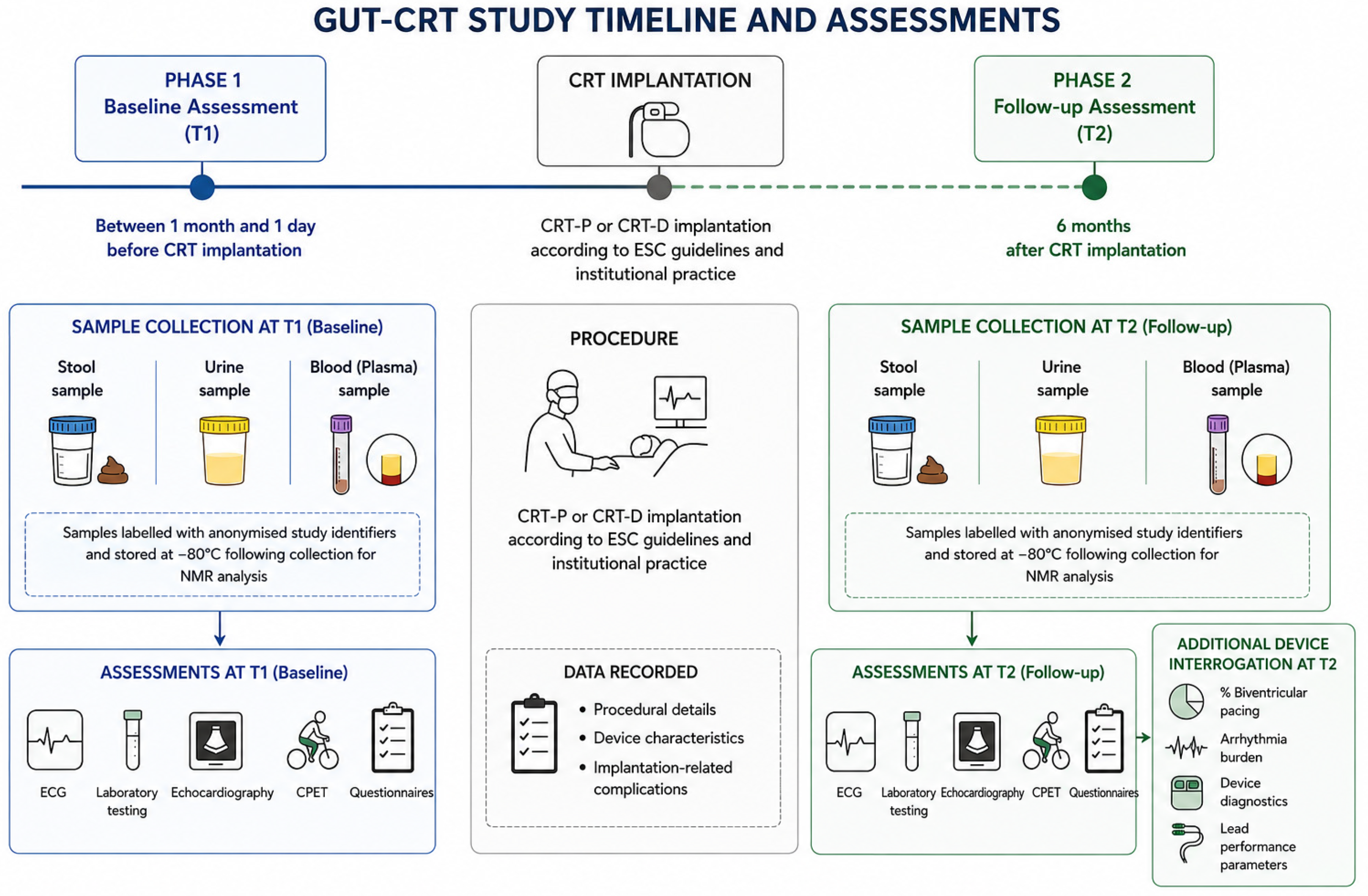
Data will be collected at two predefined timepoints (Figure 1):
Figure 1.
GUT-CRT study timeline and assessments.
2.4.1. Phase 1: Baseline Assessment Before CRT Implantation (T1)
Assessments will occur between one month and one day before CRT implantation.
Participants will receive:
- Stool collection kit;
- Urine collection container;
- Written instructions.
Samples will be labelled using anonymised study identifiers and stored at −80 °C following collection.
ECG, laboratory testing, echocardiography, CPET, and dietary questionnaires will be completed during this phase.
- Cardiac resynchronization therapy implantation
CRT-P or CRT-D implantation will be performed according to contemporary ESC recommendations and institutional practice.
Procedural details, device characteristics, and implantation-related complications will be recorded.
2.4.2. Phase 2: Follow-Up Assessment (T2)
All baseline assessments will be repeated six months after CRT implantation.
Participants will additionally undergo complete device interrogation including:
- Percentage of biventricular pacing;
- Arrhythmia burden;
- Device diagnostics;
- Lead performance parameters.
Participants who receive antibiotics during follow-up will have sample collection postponed for at least one month after completion of therapy whenever clinically feasible.
2.5. Statistical Analysis
Statistical analyses will be performed using appropriate statistical software, including R and validated microbiome analysis packages. Continuous variables will be summarized as mean ± standard deviation or median with interquartile range, depending on their distribution, while categorical variables will be presented as frequencies and percentages. The distribution of continuous variables and paired differences will be assessed using graphical methods and the Shapiro–Wilk test where appropriate.
Longitudinal comparisons between baseline and six months following CRT implantation will account for the paired nature of the study. Changes in continuous clinical, biochemical, echocardiographic and metabolomic variables will be evaluated using paired Student’s t-tests for approximately normally distributed differences or Wilcoxon signed-rank tests for non-normally distributed paired observations.
Gut microbiome analyses will include measures of alpha diversity, such as the Shannon and Simpson indices and observed richness, and beta diversity based on Bray–Curtis dissimilarity. Changes in alpha diversity will be evaluated using paired statistical tests. Differences in microbial community composition between the two study timepoints will be visualized using principal coordinates analysis (PCoA) and evaluated using permutational multivariate analysis of variance (PERMANOVA), with permutations constrained by participant identity to account for repeated measurements. Differences in multivariate dispersion will also be examined.
Differential abundance analyses will be conducted using methods appropriate for compositional microbiome data, including ANCOM-BC2, incorporating repeated-measures structure and relevant covariates. Analyses of 16S rRNA sequencing and shotgun metagenomic datasets will be conducted separately. Metagenomic functional pathway analyses will be considered exploratory.
Multivariable regression analyses will investigate associations between longitudinal microbiome and metabolomic changes and clinical response to CRT. Candidate covariates will include age, sex, baseline renal function, changes in estimated glomerular filtration rate, dietary adherence, relevant medication exposure, antibiotic use and selected clinical comorbidities. Given the modest sample size, the number of covariates included in individual models will be restricted to avoid overfitting, with priority given to variables of established clinical relevance.
Potential outliers will be assessed using graphical and statistical methods. Observations will not be excluded solely because of extreme values unless a documented measurement or processing error is identified. Sensitivity analyses will evaluate the influence of extreme observations.
The primary longitudinal microbiome analysis will include participants with valid paired sequencing data. Missing-data patterns and reasons for incomplete follow-up will be documented. When appropriate and supported by the underlying assumptions, sensitivity analyses using methods suitable for incomplete clinical data will be performed. No imputation of missing microbial abundance profiles will be undertaken routinely.
All statistical tests will be two-sided, with a nominal significance threshold of p < 0.05. For analyses involving multiple microbial taxa, functional pathways or metabolomic features, the Benjamini–Hochberg procedure will be applied to control the false discovery rate. Adjusted p-values will be reported alongside effect estimates wherever applicable. Given the exploratory nature of the investigation, findings will be interpreted with appropriate consideration of effect sizes, uncertainty and the risk of false-positive associations.
3. Discussion
Heart failure is increasingly recognised as a systemic syndrome involving complex interactions among the cardiovascular, immune, metabolic, and gastrointestinal systems. Growing evidence suggests that disruption of the gut–heart axis may contribute to the progression of heart failure through mechanisms involving intestinal congestion, impaired intestinal perfusion, increased intestinal permeability, systemic inflammation, and altered microbial metabolite production. Despite substantial advances in understanding the relationship between heart failure and gut microbiota, little is known regarding the effects of therapeutic interventions on microbial community composition [51,52].
Cardiac resynchronization therapy represents one of the most effective device-based treatments for selected patients with heart failure and electrical dyssynchrony. By improving ventricular synchrony, enhancing cardiac output, and promoting reverse remodelling, CRT may influence gastrointestinal physiology through several mechanisms. Improved intestinal perfusion, reduced venous congestion, restoration of mucosal barrier integrity, and attenuation of systemic inflammation may all contribute to favourable alterations in gut microbiota composition [20,21,22].
Several studies have demonstrated that patients with heart failure exhibit reduced microbial diversity and altered relative abundances of specific bacterial taxa compared with healthy controls. Furthermore, elevated concentrations of gut microbiota-derived metabolites such as trimethylamine-N-oxide (TMAO) have been associated with adverse cardiovascular outcomes, increased mortality, and worsening heart failure severity. Experimental studies suggest that TMAO may promote myocardial fibrosis, endothelial dysfunction, platelet activation, and chronic inflammation, thereby contributing to disease progression [36,42,51,52].
Whether successful CRT implantation can reverse some of these microbiome-related abnormalities remains unknown [53]. The present study seeks to address this knowledge gap by prospectively evaluating microbiome composition before and after CRT implantation using both 16S rRNA sequencing and metagenomic analyses. The combination of taxonomic and functional analyses will provide a comprehensive assessment of microbial changes associated with CRT-mediated improvements in cardiac function.
An additional strength of the GUT-CRT study is the integration of multiple complementary phenotyping approaches. In addition to microbiome analyses, participants will undergo comprehensive echocardiographic assessment, cardiopulmonary exercise testing, biomarker evaluation, and device interrogation. This multimodal approach may facilitate identification of associations between microbial alterations and objective measures of reverse remodelling, functional capacity, and treatment response.
The inclusion of serum and urinary TMAO measurements further strengthens the study design by allowing simultaneous investigation of microbial composition and microbial-derived metabolic activity. Such analyses may provide insights into potential mechanistic pathways linking changes in gut microbiota to clinical outcomes following CRT implantation [51].
The study also incorporates dietary assessment using a validated Mediterranean diet adherence questionnaire. Because dietary patterns represent one of the strongest determinants of gut microbiota composition, adjustment for dietary habits may reduce residual confounding and improve interpretation of observed microbiome changes.
Several limitations should be acknowledged. First, the study is a single-centre observational investigation and therefore may be susceptible to residual confounding and selection bias. Second, the relatively modest sample size reflects the exploratory nature of the study and may limit statistical power for subgroup analyses and taxon-specific investigations. Third, despite efforts to control for factors known to influence microbiota composition, unmeasured environmental or lifestyle factors may contribute to observed microbial variability. Finally, the absence of a non-CRT control group precludes definitive conclusions regarding causality.
Nevertheless, the within-participant longitudinal design represents a major methodological strength because each participant serves as their own control, thereby reducing inter-individual variability that commonly complicates microbiome investigations.
To our knowledge, GUT-CRT will be among the first prospective studies specifically designed to evaluate longitudinal alterations in gut microbiota composition following cardiac resynchronization therapy. The findings may contribute to a better understanding of the biological consequences of CRT beyond conventional cardiac outcomes and provide novel insights into the role of the gut–heart axis in heart failure.
4. Conclusions
The GUT-CRT study is a prospective observational cohort study designed to investigate changes in gut microbiota composition and microbial-derived metabolites following cardiac resynchronization therapy in patients with chronic heart failure.
By combining 16S rRNA sequencing, metagenomic analyses, serum and urinary TMAO measurements, echocardiography, cardiopulmonary exercise testing, and device interrogation, this study aims to provide a comprehensive evaluation of the relationship between CRT-induced clinical improvement and alterations in the gut microbiome.
The results may improve understanding of the gut–heart axis in heart failure and may identify novel microbiome-related biomarkers associated with treatment response and reverse cardiac remodelling following CRT.
Author Contributions
All authors contributed to the conception and design of the study. Data acquisition, analysis, interpretation, manuscript preparation, and final approval of the manuscript will be performed according to individual contributions. All authors have read and agreed to the published version of the manuscript.
Funding
This research received no external funding.
Institutional Review Board Statement
The study has received approval from the Scientific and Ethics Committee of Hippokration General Hospital, Athens (approval code A.Ε.Σ 95/15-03-2025 17317, 15 March 2025). All procedures will be conducted in accordance with the principles of the Declaration of Helsinki and current European data protection regulations.
Informed Consent Statement
Written informed consent will be obtained from all participants before enrolment.
Data Availability Statement
Data generated during the study will be available from the corresponding author upon reasonable request and following institutional approval.
Acknowledgments
Generative AI tools (ChatGPT-6, OpenAI) were used solely for language editing, including improvements in grammar, spelling, punctuation, and clarity. The authors retained full responsibility for the scientific content, accuracy, and integrity of the manuscript
Conflicts of Interest
The authors declare no conflicts of interest.
Abbreviations
| BNP | B-type natriuretic peptide |
| CPET | Cardiopulmonary exercise testing |
| CRT | Cardiac resynchronization therapy |
| CRT-D | Cardiac resynchronization therapy with defibrillator |
| CRT-P | Cardiac resynchronization therapy with pacing |
| ESC | European Society of Cardiology |
| GLS | Global longitudinal strain |
| hsCRP | High-sensitivity C-reactive protein |
| LAVI | Left atrial volume index |
| LVEF | Left ventricular ejection fraction |
| LVEDV | Left ventricular end-diastolic volume |
| LVESV | Left ventricular end-systolic volume |
| METs | Metabolic equivalents |
| NMR | Nuclear magnetic resonance spectroscopy |
| NT-proBNP | N-terminal pro-B-type natriuretic peptide |
| OUES | Oxygen uptake efficiency slope |
| PASP | Pulmonary artery systolic pressure |
| PERMANOVA | Permutational multivariate analysis of variance |
| PREDIMED | Prevención con Dieta Mediterránea |
| TMAO | Trimethylamine-N-oxide |
| VAT | Ventilatory anaerobic threshold |
| VE/VCO2 | Ventilatory efficiency slope |
| VO2peak | Peak oxygen consumption |
| 16SrRNA | 16S ribosomal ribonucleic acid |
References
- Rosenberg, E.; Zilber-Rosenberg, I. Microbes drive evolution of animals and plants: The hologenome concept. mBio 2016, 7, e01395-15. [Google Scholar] [CrossRef] [Scilit] [PubMed]
- Nichols, R.G.; Davenport, E.R. The relationship between the gut microbiome and host gene expression: A review. Hum. Genet. 2021, 140, 747–760. [Google Scholar] [CrossRef] [Scilit] [PubMed] [PubMed Central]
- Qin, J.; Li, R.; Raes, J.; Arumugam, M.; Burgdorf, K.S.; Manichanh, C.; Nielsen, T.; Pons, N.; Levenez, F.; Yamada, T.; et al. A human gut microbial gene catalogue established by metagenomic sequencing. Nature 2010, 464, 59–65. [Google Scholar] [CrossRef] [Scilit] [PubMed]
- Bäckhed, F.; Roswall, J.; Peng, Y.; Feng, Q.; Jia, H.; Kovatcheva-Datchary, P.; Li, Y.; Xia, Y.; Xie, H.; Zhong, H.; et al. Dynamics and stabilization of the human gut microbiome during the first year of life. Cell Host Microbe 2015, 17, 690–703. [Google Scholar] [CrossRef] [Scilit] [PubMed]
- Hou, K.; Wu, Z.X.; Chen, X.Y.; Wang, J.Q.; Zhang, D.; Xiao, C.; Zhu, D.; Koya, J.B.; Wei, L.; Li, J.; et al. Microbiota in health and diseases. Signal Transduct. Target. Ther. 2022, 7, 135. [Google Scholar] [CrossRef] [Scilit] [PubMed]
- Arumugam, M.; Raes, J.; Pelletier, E.; Le Paslier, D.; Yamada, T.; Mende, D.R.; Fernandes, G.R.; Tap, J.; Bruls, T.; Batto, J.M.; et al. Enterotypes of the human gut microbiome. Nature 2011, 473, 174–180. [Google Scholar] [CrossRef] [Scilit] [PubMed]
- Knights, D.; Ward, T.L.; McKinlay, C.E.; Miller, H.; Gonzalez, A.; McDonald, D.; Knight, R. Rethinking “enterotypes”. Cell Host Microbe 2014, 16, 433–437. [Google Scholar] [CrossRef] [Scilit] [PubMed]
- Adak, A.; Khan, M.R. An insight into gut microbiota and its functionalities. Cell. Mol. Life Sci. 2019, 76, 473–493. [Google Scholar] [CrossRef] [Scilit] [PubMed] [PubMed Central]
- Singh, S.; Bastos-Amador, P.; Thompson, J.A.; Truglio, M.; Yilmaz, B.; Cardoso, S.; Sobral, D.; Soares, M.P. Glycan-based shaping of the microbiota during primate evolution. eLife 2021, 10, e67450. [Google Scholar] [CrossRef] [Scilit] [PubMed] [PubMed Central]
- Durack, J.; Lynch, S.V. The gut microbiome: Relationships with disease and opportunities for therapy. J. Exp. Med. 2019, 216, 20–40. [Google Scholar] [CrossRef] [Scilit] [PubMed] [PubMed Central]
- Richards, A.L.; Burns, M.B.; Alazizi, A.; Barreiro, L.B.; Pique-Regi, R.; Blekhman, R.; Luca, F. Genetic and transcriptional analysis of human host response to healthy gut microbiota. mSystems 2016, 1, e00067-16. [Google Scholar] [CrossRef] [Scilit] [PubMed] [PubMed Central]
- Johansson, M.E.; Phillipson, M.; Petersson, J.; Velcich, A.; Holm, L.; Hansson, G.C. The inner of the two Muc2 mucin-dependent mucus layers in colon is devoid of bacteria. Proc. Natl. Acad. Sci. USA 2008, 105, 15064–15069. [Google Scholar] [CrossRef] [Scilit] [PubMed]
- Panneerselvam, D.; Vaqar, S. Peyer Patches. In StatPearls; StatPearls Publishing: Treasure Island, FL, USA, 2023. Available online: https://www.ncbi.nlm.nih.gov/books/NBK557457/ (accessed on 2 January 2023). [PubMed]
- Carabotti, M.; Scirocco, A.; Maselli, M.A.; Severi, C. The gut-brain axis: Interactions between enteric microbiota, central and enteric nervous systems. Ann. Gastroenterol. 2015, 28, 203–209. [Google Scholar] [PubMed]
- Rhee, S.H.; Pothoulakis, C.; Mayer, E.A. Principles and clinical implications of the brain–gut-enteric microbiota axis. Nat. Rev. Gastroenterol. Hepatol. 2009, 6, 306–314. [Google Scholar] [CrossRef] [Scilit] [PubMed]
- Sarkar, A.; Lehto, S.M.; Harty, S.; Dinan, T.G.; Cryan, J.F.; Burnet, P.W. Psychobiotics and the manipulation of bacteria–gut–brain signals. Trends Neurosci. 2016, 39, 763–781. [Google Scholar] [CrossRef] [Scilit] [PubMed]
- Tiffany, C.R.; Bäumler, A.J. Dysbiosis: From fiction to function. Am. J. Physiol. Gastrointest. Liver Physiol. 2019, 317, G602–G608. [Google Scholar] [CrossRef] [Scilit] [PubMed] [PubMed Central]
- Hrncir, T. Gut Microbiota Dysbiosis: Triggers, Consequences, Diagnostic and Therapeutic Options. Microorganisms 2022, 10, 578. [Google Scholar] [CrossRef] [Scilit] [PubMed] [PubMed Central]
- Weiss, G.A.; Hennet, T. Mechanisms and consequences of intestinal dysbiosis. Cell. Mol. Life Sci. 2017, 74, 2959–2977. [Google Scholar] [CrossRef] [Scilit] [PubMed] [PubMed Central]
- Khan, M.S.; Shahid, I.; Bennis, A.; Rakisheva, A.; Metra, M.; Butler, J. Global epidemiology of heart failure. Nat. Rev. Cardiol. 2024, 21, 717–734. [Google Scholar] [CrossRef] [Scilit] [PubMed]
- McDonagh, T.A.; Metra, M.; Adamo, M.; Gardner, R.S.; Baumbach, A.; Böhm, M.; Burri, H.; Butler, J.; Čelutkienė, J.; Chioncel, O.; et al. 2023 Focused Update of the 2021 ESC Guidelines for the diagnosis and treatment of acute and chronic heart failure. Eur. Heart J. 2023, 44, 3627–3639, Erratum in Eur. Heart J. 2024, 45, 53. https://doi.org/10.1093/eurheartj/ehad613. [Google Scholar] [CrossRef] [Scilit] [PubMed]
- Glikson, M.; Nielsen, J.C.; Kronborg, M.B.; Michowitz, Y.; Auricchio, A.; Barbash, I.M.; Barrabés, J.A.; Boriani, G.; Braunschweig, F.; Brignole, M.; et al. 2021 ESC Guidelines on cardiac pacing and cardiac resynchronization therapy. Eur. Heart J. 2021, 42, 3427–3520, Erratum in Eur. Heart J. 2022, 43, 1651. https://doi.org/10.1093/eurheartj/ehac075. [Google Scholar] [CrossRef] [Scilit] [PubMed]
- Mu, F.; Tang, M.; Guan, Y.; Lin, R.; Zhao, M.; Zhao, J.; Huang, S.; Zhang, H.; Wang, J.; Tang, H. Knowledge Mapping of the Links Between the Gut Microbiota and Heart Failure: A Scientometric Investigation (2006–2021). Front. Cardiovasc. Med. 2022, 9, 882660. [Google Scholar] [CrossRef] [Scilit] [PubMed]
- Lupu, V.V.; Adam Raileanu, A.; Mihai, C.M.; Morariu, I.D.; Lupu, A.; Starcea, I.M.; Frasinariu, O.E.; Mocanu, A.; Dragan, F.; Fotea, S. The Implication of the Gut Microbiome in Heart Failure. Cells 2023, 12, 1158. [Google Scholar] [CrossRef] [Scilit] [PubMed] [PubMed Central]
- Gallo, A.; Macerola, N.; Favuzzi, A.M.; Nicolazzi, M.A.; Gasbarrini, A.; Montalto, M. The Gut in Heart Failure: Current Knowledge and Novel Frontiers. Med. Princ. Pract. 2022, 31, 203–214. [Google Scholar] [CrossRef] [Scilit] [PubMed]
- Sandek, A.; Bauditz, J.; Swidsinski, A.; Buhner, S.; Weber-Eibel, J.; von Haehling, S.; Schroedl, W.; Karhausen, T.; Doehner, W.; Rauchhaus, M.; et al. Altered intestinal function in patients with chronic heart failure. J. Am. Coll. Cardiol. 2007, 50, 1561–1569. [Google Scholar] [CrossRef] [Scilit] [PubMed]
- Pasini, E.; Aquilani, R.; Testa, C.; Baiardi, P.; Angioletti, S.; Boschi, F.; Verri, M.; Dioguardi, F. Pathogenic Gut Flora in Patients With Chronic Heart Failure. JACC Heart Fail. 2016, 4, 220–227. [Google Scholar] [CrossRef] [Scilit] [PubMed]
- Lu, Y.C.; Yeh, W.C.; Ohashi, P.S. LPS/TLR4 signal transduction pathway. Cytokine 2008, 42, 145–151. [Google Scholar] [CrossRef] [Scilit] [PubMed]
- Hietbrink, F.; Besselink, M.G.; Renooij, W.; de Smet, M.B.; Draisma, A.; van der Hoeven, H.; Pickkers, P. Systemic inflammation increases intestinal permeability during experimental human endotoxemia. Shock 2009, 32, 374–378. [Google Scholar] [CrossRef] [Scilit] [PubMed]
- Sandek, A.; Bjarnason, I.; Volk, H.D.; Crane, R.; Meddings, J.B.; Niebauer, J.; Kalra, P.R.; Buhner, S.; Herrmann, R.; Springer, J.; et al. Studies on bacterial endotoxin and intestinal absorption function in patients with chronic heart failure. Int. J. Cardiol. 2012, 157, 80–85. [Google Scholar] [CrossRef] [Scilit] [PubMed]
- Nagatomo, Y.; Tang, W.H.W. Intersections between microbiome and heart failure: Revisiting the gut hypothesis. J. Card. Fail. 2015, 21, 973–980. [Google Scholar] [CrossRef] [Scilit] [PubMed]
- Rauchhaus, M.; Doehner, W.; Francis, D.P.; Davos, C.; Kemp, M.; Liebenthal, C.; Niebauer, J.; Hooper, J.; Volk, H.D.; Coats, A.J.; et al. Plasma cytokine parameters and mortality in patients with chronic heart failure. Circulation 2000, 102, 3060–3067. [Google Scholar] [CrossRef] [Scilit] [PubMed]
- Yassine, F.; Albush, A.; Hanna, A.; Abbas, H.; Bilen, M. The gut-heart axis in heart failure: A systematic review and meta-analysis of gut microbiota and metabolites. npj Biofilms Microbiomes 2026, 12, 228. [Google Scholar] [CrossRef] [Scilit] [PubMed]
- Di Vincenzo, F.; Del Gaudio, A.; Petito, V.; Lopetuso, L.R.; Scaldaferri, F. Gut microbiota, intestinal permeability, and systemic inflammation: A narrative review. Intern. Emerg. Med. 2024, 19, 275–293. [Google Scholar] [CrossRef] [Scilit] [PubMed]
- Kumari, N.; Kumari, R.; Dua, A.; Singh, M.; Kumar, R.; Singh, P.; Duyar-Ayerdi, S.; Pradeep, S.; Ojesina, A.I.; Kumar, R. From Gut to Hormones: Unraveling the Role of Gut Microbiota in (Phyto)Estrogen Modulation in Health and Disease. Mol. Nutr. Food Res. 2024, 68, e2300688. [Google Scholar] [CrossRef] [Scilit] [PubMed]
- Hatamnejad, M.R.; Medzikovic, L.; Dehghanitafti, A.; Rahman, B.; Vadgama, A.; Eghbali, M. Role of Gut Microbial Metabolites in Ischemic and Non-Ischemic Heart Failure. Int. J. Mol. Sci. 2025, 26, 2242. [Google Scholar] [CrossRef] [Scilit] [PubMed] [PubMed Central]
- Verhaar, B.J.H.; Prodan, A.; Nieuwdorp, M.; Muller, M. Gut Microbiota in Hypertension and Atherosclerosis: A Review. Nutrients 2020, 12, 2982. [Google Scholar] [CrossRef] [Scilit] [PubMed]
- Liu, B.N.; Liu, X.T.; Liang, Z.H.; Wang, J.H. Gut microbiota in obesity. World J. Gastroenterol. 2021, 27, 3837–3850. [Google Scholar] [CrossRef] [Scilit] [PubMed]
- Hobby, G.P.; Karaduta, O.; Dusio, G.F.; Singh, M.; Zybailov, B.L.; Arthur, J.M. Chronic kidney disease and the gut microbiome. Am. J. Physiol. Ren. Physiol. 2019, 316, F1211–F1217. [Google Scholar] [CrossRef] [Scilit] [PubMed]
- Francisqueti-Ferron, F.V.; Nakandakare-Maia, E.T.; Siqueira, J.S.; Ferron, A.J.T.; Vieira, T.A.; Bazan, S.G.Z.; Corrêa, C.R. The role of gut dysbiosis-associated inflammation in heart failure. Rev. Assoc. Med. Bras. 2022, 68, 1120–1124. [Google Scholar] [CrossRef] [Scilit] [PubMed]
- Hayashi, T.; Yamashita, T.; Watanabe, H.; Kami, K.; Yoshida, N.; Tabata, T.; Emoto, T.; Sasaki, N.; Mizoguchi, T.; Irino, Y.; et al. Gut Microbiome and Plasma Microbiome-Related Metabolites in Patients with Decompensated and Compensated Heart Failure. Circ. J. 2018, 83, 182–192. [Google Scholar] [CrossRef] [Scilit] [PubMed]
- Emoto, T.; Hayashi, T.; Tabata, T.; Yamashita, T.; Watanabe, H.; Takahashi, T.; Gotoh, Y.; Kami, K.; Yoshida, N.; Saito, Y.; et al. Metagenomic analysis of gut microbiota reveals its role in trimethylamine metabolism in heart failure. Int. J. Cardiol. 2021, 338, 138–142. [Google Scholar] [CrossRef] [Scilit] [PubMed]
- Emwas, A.H.; Roy, R.; McKay, R.T.; Tenori, L.; Saccenti, E.; Nagana Gowda, G.A.; Raftery, D.; Alahmari, F.; Jaremko, L.; Jaremko, M.; et al. NMR Spectroscopy for Metabolomics Research. Metabolites 2019, 9, 123. [Google Scholar] [CrossRef] [Scilit] [PubMed]
- Homobono Brito de Moura, P.; Leleu, G.; Da Costa, G.; Marti, G.; Pétriacq, P.; Valls Fonayet, J.; Richard, T. Integrating NMR and MS for Improved Metabolomic Analysis: From Methodologies to Applications. Molecules 2025, 30, 2624. [Google Scholar] [CrossRef] [Scilit] [PubMed]
- Beckonert, O.; Keun, H.C.; Ebbels, T.M.D.; Bundy, J.; Holmes, E.; Lindon, J.C.; Nicholson, J.K. Metabolic Profiling, Metabolomic and Metabonomic Procedures for NMR Spectroscopy of Urine, Plasma, Serum and Tissue Extracts. Nat. Protoc. 2007, 2, 2692–2703. [Google Scholar] [CrossRef] [Scilit] [PubMed]
- Ghini, V.; Quaglio, D.; Luchinat, C.; Turano, P. NMR for Sample Quality Assessment in Metabolomics. New Biotechnol. 2019, 52, 25–34. [Google Scholar] [CrossRef] [Scilit] [PubMed]
- Tang, W.H.W.; Wang, Z.; Levison, B.S.; Koeth, R.A.; Britt, E.B.; Fu, X.; Wu, Y.; Hazen, S.L. Intestinal Microbial Metabolism of Phosphatidylcholine and Cardiovascular Risk. N. Engl. J. Med. 2013, 368, 1575–1584. [Google Scholar] [CrossRef] [Scilit] [PubMed]
- Nemet, I.; Saha, P.P.; Gupta, N.; Zhu, W.; Romano, K.A.; Skye, S.M.; Cajka, T.; Mohan, M.L.; Li, L.; Wu, Y.; et al. A Cardiovascular Disease-Linked Gut Microbial Metabolite Acts via Adrenergic Receptors. Cell 2020, 180, 862–877.e22. [Google Scholar] [CrossRef] [Scilit] [PubMed]
- Weljie, A.M.; Newton, J.; Mercier, P.; Carlson, E.; Slupsky, C.M. Targeted profiling: Quantitative analysis of 1H NMR metabolomics data. Anal. Chem. 2006, 78, 4430–4442. [Google Scholar] [CrossRef] [Scilit] [PubMed]
- Worley, B.; Powers, R. Multivariate Analysis in Metabolomics. Curr. Metabolomics 2013, 1, 92–107. [Google Scholar] [CrossRef] [Scilit] [PubMed]
- Snelson, M.R.; Muralitharan, R.; Liu, C.F.; Markó, L.; Forslund, S.K.; Marques, F.Z.; Tang, W.H.W. Gut-Heart Axis: The Role of Gut Microbiota and Metabolites in Heart Failure. Circ. Res. 2025, 136, 1382–1406. [Google Scholar] [CrossRef] [Scilit] [PubMed] [PubMed Central]
- Chen, A.T.; Zhang, J.; Zhang, Y. Gut microbiota in heart failure and related interventions. iMeta 2023, 2, e125. [Google Scholar] [CrossRef] [Scilit] [PubMed] [PubMed Central]
- Mamic, P.; Shi, H.; Zhou, W.; Bararpour, N.; Contrepois, K.; Park, H.; Avina, M.; Schüssler-Fiorenza Rose, S.M.; Brooks, A.W.; Kotaka, M.; et al. Gut microbiome shifts in chronic systolic heart failure are associated with disease severity and clinical improvement. Nat. Cardiovasc. Res. 2026, 5, 1055–1070. [Google Scholar] [CrossRef] [Scilit] [PubMed]
Disclaimer/Publisher’s Note: The statements, opinions and data contained in all publications are solely those of the individual author(s) and contributor(s) and not of MDPI and/or the editor(s). MDPI and/or the editor(s) disclaim responsibility for any injury to people or property resulting from any ideas, methods, instructions or products referred to in the content. |
© 2026 by the authors. Licensee MDPI, Basel, Switzerland. This article is an open access article distributed under the terms and conditions of the Creative Commons Attribution (CC BY) license.