


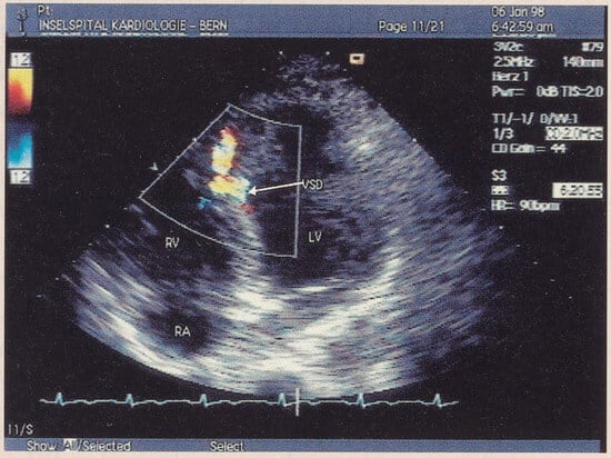
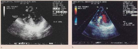
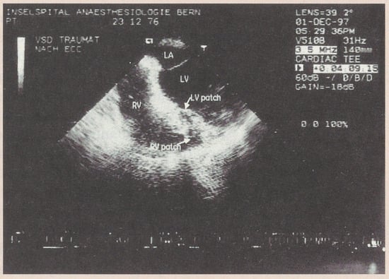
A 21-year-old white male was admitted to the hospital following a traffic accident. He had been run over by a car while taking a nap in the grass. He suffered a left lung contusion, subcapsular spleen haematoma and a left forearm fracture. Initial echocardiography demonstrated localised anterior hypokinesia in presence of preserved left ventricular function. The next day the patient developed a loud systolic murmur. A repeat echocardiographic examination revealed diffuse contusion injury of the left ventricle with mildly diminished global function. In the mid portion of the interventricular septum a haemodynamically relevant defect was detected (Qp:Qs 2.7) (Figure 1). The right ventricle was not dilated and the gradient across the tricuspid valve was compatible with moderate pulmonary hypertension. The patient remained clinically stable. Follow-up echocardiographies showed a normalisation of the systolic left ventricular function and the shunt remained unchanged. The patient was managed conservatively and subsequently discharged. An elective surgical repair of the ventricular septal defect was performed nine weeks later. The surgical approach was through the right atrium and tricuspid valve. After temporary removal of the posterior papillary muscle of the tricuspid valve, a gore-tex patch was placed at the right ventricular side of the septal defect. Intraoperative transoesophageal echocardiography showed a significant residual shunt with bulging of the patch into the right ventricle (Figure 2a and Figure 2b). The patient was taken back on cardiopulmonary bypass and through a left ventricular apical approach a second gore-tex patch was placed on the left side of the interventricular septum (Figure 3). At the end of the procedure, there was no relevant shunt as assessed by transoesophageal echocardiography. The clinical course was unremarkable. The patient was discharged two weeks later and has remained free of symptoms ever since.
Figure 1.
Interventricular septal defect with left-to-right shunt in a two-dimensional transthoracal four-chamber view with colour Doppler.
Figure 2.
Bulging of the patch placed on the right ventricular side of the septum (2a) and colour Doppler demonstration of significant residual left-to-right shunt (2b) in an intraoperative transoesophageal high-transgastric view.
Figure 3.
End result after second patch placement on the left ventricular side of the septum in an intraoperative transoesophageal hightransgastric view. LA = left atrium; RA = right atrium; RV = right ventricle; LV = left ventricle; VSD = ventricle septal defect.
© 1999 by the author. Attribution - Non-Commercial - NoDerivatives 4.0.