


Abstract
Carbohydrates and their constituent monosaccharides have historically been viewed as metabolic substrates that provide cellular energy. However, recent evidence demonstrates that monosaccharides can also function as bioactive signaling molecules that affect immune responses through cellular metabolic reprogramming, post-translational protein glycosylation, receptor complex modulation, and transcriptional regulation of immune cell differentiation. These discoveries have given rise to the emerging concept of glyconutraceuticals, bioactive carbohydrates with therapeutic potential. This review discusses current evidence for the immunomodulatory properties of glucose, D-mannose, sialic acid, L-fucose, galactose, N-acetylglucosamine, D-xylose, and L/D-arabinose, with particular emphasis on their mechanisms of action in T cell activation, subset differentiation, metabolic reprogramming, and anti-tumor cytotoxicity. The context-dependent and often opposing effects of individual monosaccharides across T cell subsets underscore the complexity of immunomodulation by glyconutrients.
1. Introduction
Dietary nutrients are increasingly recognized as key regulators of immune function, serving not only as metabolic substrates required for immune cell activation and proliferation, but also as structural precursors, signaling molecules, and epigenetic modifiers that shape immune cell differentiation, activation, and functional plasticity [1]. The relationship between nutritional status and immune competence is demonstrated by epidemiological and experimental evidence linking macronutrient imbalances, particularly diets high in refined carbohydrates, saturated fatty acids, and ultra-processed foods, to increased susceptibility to chronic inflammatory diseases, autoimmune conditions, and impaired anti-tumor immune surveillance. Through their effect on cellular metabolism, signal transduction, and gene expression, nutrients integrate environmental cues with immune cell fate and function, thereby influencing immune homeostasis and disease susceptibility [2,3].
Among dietary carbohydrates, monosaccharides have emerged as important immunomodulatory molecules that extend beyond their traditional role as metabolic fuels. Monosaccharides are the simplest carbohydrate units and serve not only as energy substrates but also as precursors for glycan biosynthesis and signaling intermediates [4]. Their relatively direct entry into metabolic and glycosylation pathways makes them uniquely positioned to regulate immune cell function compared with more complex carbohydrates, whose effects often depend on digestion, microbial fermentation, or indirect metabolic conversion [3,5]. Monosaccharides can serve as substrates for glycosylation reactions that modify proteins and lipids on the surface of immune cells, thereby regulating receptor clustering, signaling complex assembly, ligand-binding affinity, and checkpoint receptor surface retention. These glycosylated structures, also known as the glycocalyx, are directly determined by the availability of monosaccharides in the cellular milieu [6,7,8]. Specific monosaccharides can also fuel metabolic pathways, e.g., the hexosamine biosynthetic pathway (HBP), which generates UDP-N-acetylglucosamine (UDP-GlcNAc), the substrate for O-GlcNAcylation and N-glycosylation [9]. The flux of monosaccharides through this pathway acts as a nutrient-sensing mechanism that links the extracellular metabolic environment to intracellular immune programming [10]. Nutrient availability within the tissue microenvironment can also directly influence immune responses; the metabolic competition for glucose within the tumor microenvironment results in reduced anti-tumor T cell function by depriving effector T cells of the metabolic substrates required for cytokine production and cytotoxic activity [11,12].
T cells were selected as the focus of this review as they are metabolically dynamic and their activation, differentiation, and effector functions are tightly coupled to nutrient availability and metabolic reprogramming [4,13,14]. Although multiple classes of carbohydrates, including disaccharides, oligosaccharides, and polysaccharides, can influence immune responses, this review specifically focuses on monosaccharides that directly regulate T-cell biology [4]. We discuss evidence derived from experimental studies investigating direct effects on T-cell activation, proliferation, differentiation, cytokine production, and cytotoxic function, while broader systemic metabolic effects are addressed only when they provide mechanistic insight into T-cell regulation [11]. The review summarizes the role of eight representative monosaccharides—glucose, D-mannose, sialic acid, L-fucose, galactose, N-acetylglucosamine/glucosamine, D-xylose, and the arabinose isomers in regulating T-cell biology. We also discuss the emerging therapeutic implications of targeting monosaccharide metabolism or supplementation to modulate T-cell responses in cancer, autoimmune diseases, infection, and metabolic disorders, while highlighting current limitations and future research directions.
2. Overview of T Cell Function and Homeostasis
T cells are lymphocytes that are principal orchestrators of adaptive immunity. They play critical roles in defense against intracellular pathogens, immune surveillance of malignant cells, and the maintenance of immune tolerance through the suppression of autoreactive responses [15].
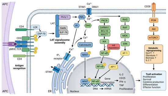
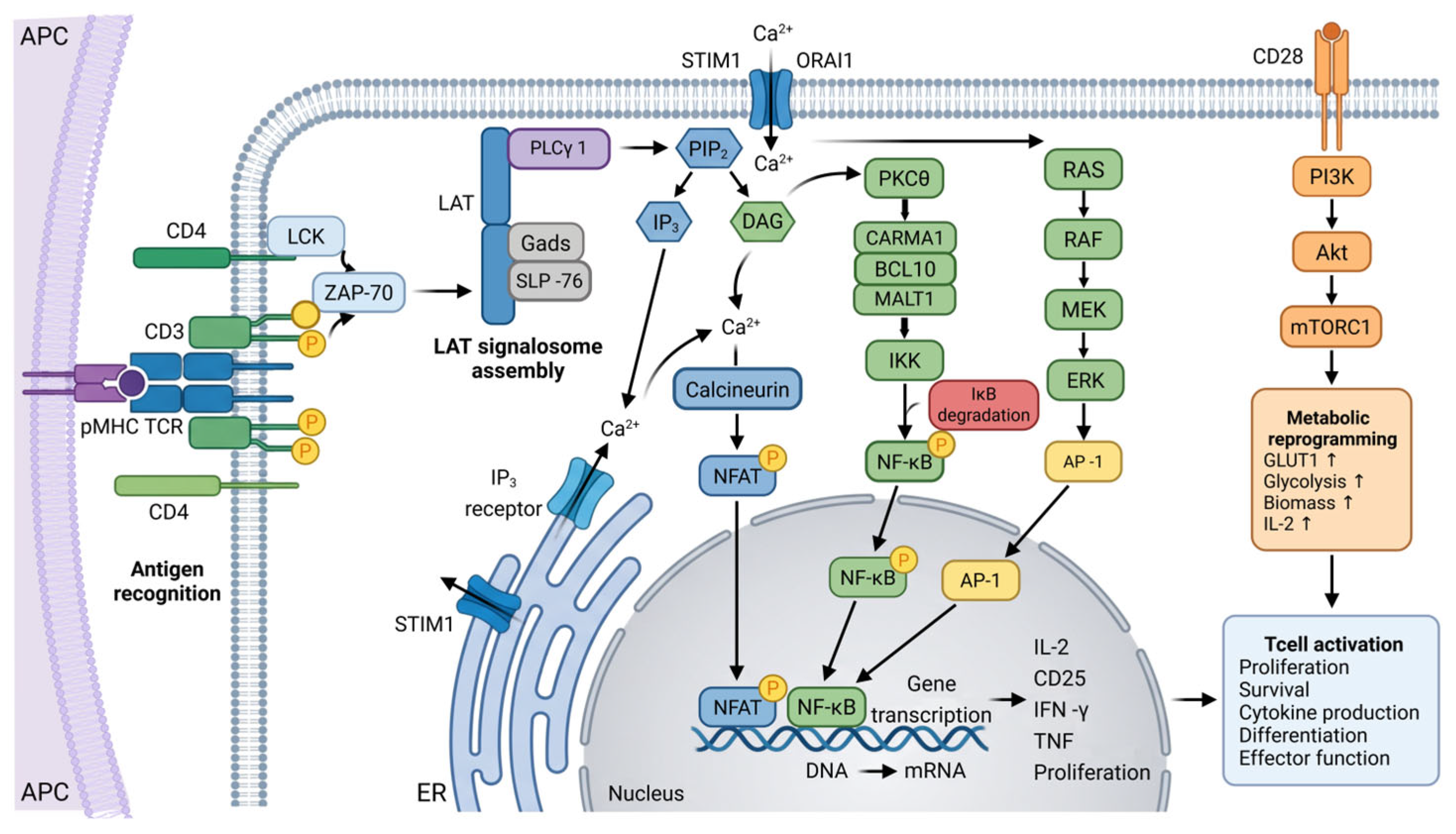
T cell activation (as demonstrated in Figure 1) begins with engagement of the multisubunit T cell receptor (TCR) by a cognate peptide–MHC complex (pMHC) on an antigen-presenting cell [16,17]. This recognition event triggers the phosphorylation of immunoreceptor tyrosine-based activation motifs (ITAMs) within the CD3 cytoplasmic tails (found within the TCR complex) by the Src family kinase LCK [15,18]. Phosphorylated ITAMs then recruit and activate ZAP70 leading to nucleation of the multimolecular signalosome. [15,16,17,19]. The signalosome activation results in the hydrolysis of membrane PIP2 into two critical second messengers: inositol (1,4,5)-trisphosphate (IP3) and diacylglycerol (DAG) [18,19]. IP3 triggers the release of Ca2+ stores from the endoplasmic reticulum, leading to the activation of CRAC channels (ORAI1/STIM1) and a sustained influx of extracellular Ca2+ [15,19]. Elevated intracellular Ca2+ activates the phosphatase calcineurin, which dephosphorylates NFAT, allowing its nuclear translocation [15,17]. Simultaneously, DAG activates PKCθ, which leads to the degradation of IκB and nuclear translocation of NF-κB [15,19]. DAG also drives the activation of AP-1 [15,17].
Figure 1.
Intracellular signaling pathways in T-cell activation. Figure created by BioRender.
T-cell activation is influenced by the glycosylation status of the cell surface. Mgat5-mediated N-glycan branching promotes the formation of a galectin-glycoprotein lattice, which restricts TCR mobility and clustering. This increases the threshold for T-cell activation [20]. However, disruption of this lattice, as seen in Mgat5-deficient T cells or when galectin binding is competed for by exogenous saccharides such as lactose, enhances TCR clustering and downstream signaling [20]. As monosaccharides serve as both metabolic substrates and glycosylation precursors, changes in their availability can affect N-glycan branching, membrane organization, TCR signaling and cellular metabolism. This can potentially impact the magnitude and duration of T-cell responses [20,21].
Various T cell subsets exist, and immune homeostasis depends on a regulated balance between pro- and anti-inflammatory effector cells. These include IFN-producing Th1, IL-4-producing Th2, and IL-17-producing Th17 cells, and immunosuppressive Foxp3+ regulatory T cells (Tregs) [22,23,24]. Dysregulation of the balance between populations underlies the pathogenesis of many autoimmune conditions as well as failure of anti-tumor immune surveillance in cancer [25].
Metabolic reprogramming, guided by environmental nutrient cues, underlies T cell proliferation and subset-specific differentiation [13,14,26]. Quiescent naive T cells rely on oxidative phosphorylation (OXPHOS) and fatty acid oxidation (FAO) for survival [21]. Upon activation, T cells undergo a rapid transition to aerobic glycolysis (the Warburg effect), a switch essential for meeting the anabolic demands of proliferation and biomass accumulation [21]. This metabolic shift is driven by TCR signals that upregulate nutrient transporters like Glut1 [15,21] and cause partitioning of glucose into biosynthetic pathways, including the pentose phosphate pathway (PPP) for nucleotide synthesis [21].
T-cell activation also increases PI3K/AKT/mTORC1 activity, linking antigen recognition to metabolic reprogramming. While quiescent T cells rely predominantly on oxidative phosphorylation (OXPHOS) to meet their relatively low energetic demands, activation induces a metabolic shift characterized by increased glucose uptake and glycolytic activity to support the biosynthetic requirements of cell growth, proliferation, and effector function [27,28,29]. mTORC1 contributes to this transition by promoting nutrient utilization, protein synthesis, lipid biosynthesis, and cellular growth. Conversely, increased metabolic demand or energetic stress activates AMP-activated protein kinase (AMPK), which promotes energy-conserving and catabolic pathways and restrains anabolic processes, including mTORC1 activity, thereby maintaining cellular energy homeostasis [28,29]. The level and timing of mTORC1 activity can also influence T-cell fate, with reduced mTORC1 signaling favoring the development and maintenance of memory CD8+ T-cell populations under specific conditions [27]. Thus, TCR signaling integrates transcriptional programs mediated by NFAT, NF-κB, and AP-1 with metabolic regulation through the PI3K/AKT/mTORC1 axis and energy-sensing pathways such as AMPK, allowing T cells to adapt their metabolic state to changing functional and energetic demands [14,27,28,29,30].
3. Monosaccharides
3.1. Glucose—The Central Metabolic Regulator of T Cell Activation
Glucose is the primary metabolic fuel required for T cell activation, clonal expansion, and functional differentiation [13,30]. Upon engagement of the T cell receptor (TCR) and CD28 co-stimulation, naive T cells undergo a rapid metabolic reprogramming from oxidative phosphorylation (OXPHOS) to aerobic glycolysis driven by upregulation of the glucose transporter GLUT1 and key glycolytic enzymes. This metabolic transition is indispensable for driving cells into an effector state, providing both the ATP and the biosynthetic precursors, including nucleotides, amino acids, and lipids necessary for rapid cellular growth, proliferation, and cytokine production [4,13]. Hexokinase 2 (HK2) catalyzes the first committed step of glycolysis by phosphorylating glucose to glucose-6-phosphate, thereby trapping glucose within the cell and directing it toward glycolysis and associated biosynthetic pathways [31,32,33]. Interestingly, HK2 is overexpressed in many tumors as part of their metabolic reprogramming, enabling rapid glucose utilization and also creating a competitive advantage over tumor-infiltrating lymphocytes (TILs) [33,34,35]. Reduced glucose flux through HK2 in T cells limits glycolytic capacity, cytokine production, proliferation, and cytotoxic activity, contributing to T-cell dysfunction within the tumor microenvironment [33,34]. This metabolic competition represents an additional mechanism of immune evasion and has prompted interest in therapeutic strategies aimed at selectively targeting tumor HK2 activity or restoring glycolytic fitness in T cells [31,33,34]. Pharmacological inhibition of tumor HK2 has therefore emerged as a promising strategy to reduce the metabolic advantage of cancer cells while increasing glucose availability for tumor-infiltrating T cells, potentially enhancing responses to immune checkpoint blockade and adoptive T-cell therapies [31,32,33].
Glucose utilization is also a critical determinant of T cell subset fate, with magnitude and duration of glycolytic flux influencing lineage commitment decisions. Proinflammatory effector cells including Th1, Th2, and Th17 cells have high glycolytic rates and increased GLUT1 expression, reflecting their need for glucose-fueled biosynthesis for rapid cytokine production and proliferation [13]. In contrast, Foxp3+ Tregs are generally less reliant on glucose, preferring fatty acid oxidation (FAO) for their energetic needs and long-term stability [13,36]. More nuanced evidence suggests that high-glucose environments can also promote Treg differentiation through an indirect feedback mechanism in which excessive lactate skews lineage commitment toward the regulatory fate [24].
Recent studies have expanded the role of glucose in T cell function beyond simple bioenergetics. In CD8+ effector T cells, a significant fraction of intracellular glucose is diverted into the biosynthesis of glycosphingolipids (GSLs) [4]. This pathway is essential for maintaining the integrity of plasma membrane lipid rafts, which are specialized cholesterol- and sphingolipid-enriched microdomains that serve as organizational platforms for T cell receptor (TCR) signaling complexes. Disruption of GSL synthesis impairs TCR signal transduction, granzyme B and perforin expression, and in vivo tumor control in experimental models [4].
Glucose also fuels the hexosamine biosynthetic pathway (HBP) to produce UDP-GlcNAc, the substrate for O-GlcNAc transferase (OGT), an enzyme that adds O-GlcNAc modifications to hundreds of intracellular proteins [14]. Loss of OGT-mediated O-GlcNAcylation in T cells blocks thymic progenitor renewal and prevents clonal expansion of mature peripheral T cells by destabilizing key transcriptional regulators [14,37]. This HBP-OGT-O-GlcNAc axis therefore functions as a molecular gauge that links nutrient availability to T cell proliferative capacity.
The immunological consequences of high-glucose environments are highly context- and subset-dependent. In primary human CD8+ cytotoxic T lymphocytes (CTLs), a high-glucose environment enhances killing efficiency through a calcium (Ca2+)-dependent mechanism, likely reflecting enhanced glycolytic support for the cytoskeletal remodeling and granule exocytosis required for target cell killing [38]. In contrast, in human Vγ9Vδ2 γδ T cells, high glucose induces a dysfunctional bioenergetic profile characterized by excessive lactate accumulation that suppresses AMPK activation and inhibits the secretion of lytic granules, impairing anti-tumor immunosurveillance [39]. This divergence underscores the importance of T cell subset identity in determining the immunological consequences of glucose availability.
While GLUT1 (SLC2A1) is the dominant glucose transporter in CD4+ T cells and is broadly required for effector cell differentiation, experimental data reveal that GLUT10 is selectively upregulated during CD8+ T cell activation and is specifically required for glucose uptake and anti-tumor responses in the competitive metabolic environment of the tumor microenvironment [11]. This transporter-level specialization suggests that therapeutic targeting of GLUT isoforms may provide opportunities to selectively modulate CD8+ cytotoxic function without broadly suppressing immune responses.
3.2. D-Mannose—Inducer of Tolerance and Regulator of CD8+ T Cell Fitness
D-mannose is a C-2 epimer of glucose found in plants and fruits and circulates in human blood at concentrations substantially lower than glucose, typically 50–100 µM, compared with the millimolar concentrations of glucose [40]. Despite its low physiological abundance, experimental evidence identifies D-mannose as a bioactive regulator of T-cell function, although many of its immunomodulatory effects have been demonstrated under supraphysiological concentrations achieved through oral supplementation or in vitro exposure [41,42,43]. Human studies indicate that oral D-mannose supplementation can safely elevate circulating mannose concentrations above physiological levels, but high doses may be associated with mild gastrointestinal adverse effects, including bloating, flatulence, abdominal discomfort, and osmotic diarrhea. Therefore, when evaluating the immunological effects of D-mannose, it is important to distinguish physiological regulation from pharmacological or therapeutic supplementation [40,43,44,45].
D-mannose has been reported to promote Foxp3+ Treg differentiation in primary T-cell cultures when added directly to the culture medium, potentially through activation of latent TGF-β, a key regulator of Foxp3 expression and Treg lineage commitment [41,43]. Similarly, oral supplementation with D-mannose resulted in altered Treg numbers in Peyer’s patches, mesenteric lymph nodes, lamina propria and spleen in response to ovalbumin [43]. Mannose effectively suppresses immunopathology in various models including experimental mouse models of autoimmune diabetes, airway inflammation, autoimmune hepatitis (AIH), and experimental autoimmune encephalomyelitis (EAE), suggesting a therapeutic applicability across many autoimmune conditions [41,43,46].
D-mannose has also emerged as a critical regulator of CD8+ T cell fitness in the tumor immune microenvironment. Experimental augmentation of mannose metabolism prevents the transition of CD8+ T cells to a state of terminal exhaustion and promotes T cell stemness, a property increasingly recognized as essential for anti-tumor immunity [42]. Mechanistically, D-mannose treatment increases OGT-mediated O-GlcNAcylation and stabilization of β-catenin, which maintains a stemness-associated epigenetic landscape and enhances the persistence and anti-tumor efficacy of adoptively transferred T cells in experimental models [42]. Furthermore, D-mannose has been reported to enhance T-cell cytotoxicity through a mechanism involving increased lysosomal degradation of PD-1, an inhibitory checkpoint receptor associated with T-cell dysfunction and exhaustion. This effect was associated with reduced PD-1 expression and enhanced cytotoxic T-cell activity following D-mannose treatment [47]. This process is mediated by the inactivation of GSK3β and the nuclear translocation of TFE3, a master regulator of lysosome biogenesis, leading to enhanced proteolytic clearance of PD-1 from the cell surface and synergistic improvement in the efficacy of anti-PD-1 and MEK inhibitor-based therapies in vivo [47]. Although preclinical findings support the therapeutic potential of D-mannose in autoimmunity and cancer [42,47], most mechanistic studies have been performed under supplementation conditions that exceed endogenous plasma concentrations [43,47,48]. Future studies should define the dose–response relationship between physiological and therapeutic mannose exposure, determine the concentrations required to modulate T-cell function in humans, and establish long-term safety and tolerability [41,46], particularly in relation to gastrointestinal side effects associated with high-dose supplementation [49,50]. These findings position D-mannose as a potentially valuable nutritional addition to immune checkpoint blockade in cancer immunotherapy.
Potential Pathogenic Roles: The immunological impact of mannose is highly context-dependent and can be pathogenic in specific autoimmune settings. While mannose metabolism supports Th1 cell differentiation and IFN-γ production under conditions of diminished glycolytic flux [48], host-derived mannose glycans can trigger deleterious autoreactive responses. In systemic lupus erythematosus (SLE), altered high-mannose glycosylation of kidney epithelial cells has been reported to contribute to pathogenic IL-17A-mediated inflammation through recognition by DC-SIGN-expressing γδ T cells. This pathway represents one potential mechanism linking altered glycosylation to renal inflammation in SLE, although additional innate and adaptive immune mechanisms may also contribute to IL-17A production and disease progression [22]. This autoreactive axis can be counteracted by GlcNAc supplementation, which promotes the biosynthesis of tolerogenic branched N-glycans that cap immunogenic mannose structures and restore homeostatic immune [22].
3.3. Sialic Acid—The Glycocalyx Checkpoint of T Cell Survival and Tolerance
Sialic acids (Sias) represent a family of nine-carbon carboxylated monosaccharides that can terminate glycan chains on the glycoproteins and glycolipids of the dense glycocalyx coating the surface of T cells [51,52]. The critical structural role of sialic acids in T cell survival has been demonstrated through T cell-specific deletion of the CMAS gene, which encodes the enzyme responsible for synthesizing the universal sialic acid precursor CMP-Sia [51]. Mice lacking sialic acids specifically on T cells exhibit T cell deficiency in peripheral lymphoid organs, driven by dramatically reduced naive T cell half-life through spontaneous caspase-3-dependent apoptosis and phosphatidylserine exposure. These findings suggest that sialic acid can act as a survival signal for circulating T cells by serving as a binding site for complement factor H, such that sialic acid loss renders T cells susceptible to autologous complement-mediated destruction [51].
From a nutritional perspective, it is important to distinguish between the predominant human sialic acid, N-acetylneuraminic acid (Neu5Ac), and the non-human sialic acid N-glycolylneuraminic acid (Neu5Gc), which is abundant in red meat and other mammalian-derived foods [53,54]. Humans cannot synthesize Neu5Gc because of an evolutionary inactivation of the CMAH gene [54,55]; however, Neu5Gc can be incorporated into human cell-surface glycoconjugates following dietary consumption [53,55]. As Neu5Gc is recognized as a foreign glycan, circulating anti-Neu5Gc antibodies can bind to incorporated Neu5Gc, generating a chronic inflammatory response termed xenosialitis [53,55]. This persistent low-grade inflammation has been implicated in the development of several chronic diseases, including cancer, atherosclerosis, and metabolic disorders [53,56]. Although direct effects of dietary Neu5Gc on T-cell biology remain incompletely understood, these findings highlight that dietary sialic acid composition may influence immune homeostasis and should be considered when evaluating nutraceutical strategies targeting glycocalyx remodeling and sialic acid-mediated immune regulation [54,56].
Beyond the endogenous and diet-derived effects of sialic acids, sialic acids also function as immunological checkpoints through their interactions with Siglec (sialic acid-binding immunoglobulin-type lectin) receptors expressed on immune cells [52,57]. Most Siglec family members contain immunoreceptor tyrosine-based inhibitory motifs (ITIMs) in their cytoplasmic domains that transduce inhibitory signals upon sialic acid engagement, dampening immune activation [57,58]. Studies using sialic acid-conjugated antigens (Sia-antigens) have demonstrated that these glycoconjugates instruct dendritic cells to promote de novo induction of Foxp3+ Tregs while actively suppressing Th1 and Th17 effector T cell differentiation [58]. Conversely, global removal of sialic acids from the surface of dendritic cells markedly enhances CD8+ T cell proliferation and cytotoxicity by facilitating high-avidity T cell–APC interactions and immunological synapse formation [57,59].
Hypersialylation, the aberrant overexpression of sialylated glycans on tumor cell surfaces, has been identified as a defining hallmark of tumor immune evasion [60,61]. Elevated tumor-derived sialic acids inhibit CD8+ T cell cytotoxicity by engaging inhibitory Siglec receptors on the T cell surface, establishing an immunosuppressive glycocalyx that shields tumor cells from cytotoxic killing [60]. Pharmacological disruption of tumor sialylation via intratumoral injections of sialic acid mimetics has been experimentally shown to reverse this immunosuppression, enhancing tumor infiltration by activated NK cells and CD8+ T cells while reducing the frequency of suppressive Tregs [60]. IDH1 is an extracellular protein with sialidase enzymatic activity capable of selectively desialylating the CD8 co-receptor on T cells, thereby increasing its avidity for peptide–MHC-I complexes and augmenting anti-tumor T cell recognition [62]. However, the therapeutic application of desialylation must be targeted, as complete systemic loss of sialic acids in certain cancer models can paradoxically drive tumor growth by augmenting antigen-independent T-cell apoptosis [61].
3.4. L-Fucose—Developmental Regulator from Thymus to Effector Function
Fucose is a deoxyhexose sugar utilized in both O-fucosylation and core N-fucosylation reactions. It plays critical roles in T cell biology spanning from initial thymic lineage commitment to peripheral effector function [63,64]. The developmental requirement for fucose is established at the earliest stages of T cell ontogeny, as O-fucose glycan attachment to the epidermal growth factor (EGF)-like repeats of Notch1 receptors is essential for Notch-ligand interactions required for T lineage specification in the thymus [64]. Experimental deletion of protein O-fucosyltransferase 1 (Pofut1) results in a complete absence of peripheral T cells as thymic progenitors fail to receive the Notch signals required for T lineage commitment. This developmental requirement for fucosylation cannot be bypassed by any other glycosylation pathway [64].
In mature peripheral T cells, core fucosylation, which encompasses the addition of α1,6-fucose to the innermost GlcNAc residue of N-glycans by the fucosyltransferase FUT8, serves as a critical structural prerequisite for TCR complex functionality and downstream signaling [63]. Genetic ablation of FUT8 severely impairs TCR-mediated signaling by destabilizing the TCR-CD3 complex and reducing the phosphorylation of proximal kinases including Lck, ZAP-70, and LAT, which effectively uncouples antigen recognition from the calcium-NFAT and MAP kinase effector pathways required for T cell activation [63,65]. Core fucosylation also regulates cell-surface stability of the immune checkpoint receptor PD-1 and blockade of core fucosylation reduces PD-1 surface levels and consequently enhances T cell activation and anti-tumor immune responses [66]. Therapeutic modulation of fucose metabolism using 2-deoxy-D-galactose (2-D-gal), a potent inhibitor of terminal fucosylation, has been shown to arrest the T cell cycle in G0/G1 phase, suppressing pro-inflammatory cytokines including IL-2, IFN-γ, and TNF-α, and inhibiting the Ca2+/calcineurin/NFAT axis [65].
Beyond cell-intrinsic roles, fucose serves as a key signal in the crosstalk between innate and adaptive immunity. Dietary L-fucose supplementation has been shown to stabilize MHC-II expression on tumor cells and polarize dendritic cells toward immunostimulatory subsets (cDC1 and moDC), which in turn enhances CD4+ and CD8+ T-cell proliferation and IFN-γ secretion [67,68]. Additionally, fucose residues on pathogens are recognized by the C-type lectin receptor DC-SIGN on dendritic cells, triggering a specific signaling pathway involving IKKϵ that directs the differentiation of T cells into TH2 and Follicular Helper T (TFH) cells [69,70].
3.5. Galactose—Metabolic Modulator of T Cell Fitness and Exhaustion Prevention
Galactose is a hexose monosaccharide primarily metabolized in the liver and exerts its effects on T cell functions through both direct metabolic modulation and indirect inter-organ signaling pathways [71,72]. Dietary galactose supplementation has been experimentally shown to reprogram hepatocyte metabolism, leading to inactivation of mTORC1 and subsequent upregulation of insulin-like growth factor binding protein-1 (IGFBP-1) production [71]. Elevated circulating IGFBP-1 neutralizes IGF-1 signaling in tumor-infiltrating CD8+ T cells, preventing their transition to a state of terminal exhaustion and increasing the frequency of progenitor-exhausted T cells, a functionally distinct subset that can respond to immune checkpoint blockade [71].
Direct metabolic modulation by galactose is also critical for T cell fitness at the cellular level. Culturing chimeric antigen receptor T (CAR-T) cells in galactose-based, glucose-free media forces a metabolic shift from aerobic glycolysis to mitochondrial OXPHOS, resulting in higher ATP production efficiency, enhanced mitochondrial membrane potential, and superior anti-tumor activity compared to standard glucose-expanded cells [72]. This metabolic reprogramming strategy represents a potentially clinically translatable approach to optimizing CAR-T cell manufacturing for adoptive cell therapy. In CD4+ T cells, galactose restricts glycolytic flux and promotes the diversion of fructose-6-phosphate into the hexosamine biosynthetic pathway, increasing N-glycan branching on the cell surface, a structural change that drives the differentiation of anti-inflammatory induced Tregs over pro-inflammatory Th17 cells [25]. Conversely, chronic D-galactose exposure serves as an established experimental model for immunosenescence; daily subcutaneous injections in rodents produce thymic and splenic structural degeneration and profound reduction in lymphocyte mitogenesis, recapitulating the immune decline observed during natural aging [73,74]. The biological effects of galactose are highly dependent on doses, duration of exposure, and route of administration [74]. Under physiological or controlled experimental conditions, galactose acts as an alternative carbon source that promotes oxidative phosphorylation, enhances mitochondrial fitness, and modulates glycosylation pathways involved in T-cell differentiation [74]. In contrast, chronic administration of supraphysiological doses of D-galactose, particularly through daily parenteral injections in rodent models, overwhelms normal galactose metabolism [74,75]. Excess galactose is diverted into oxidative pathways that increase reactive oxygen species (ROS) production and facilitate the formation of advanced glycation end products (AGEs), resulting in mitochondrial dysfunction, chronic inflammation, and cellular senescence [74,75,76]. These mechanisms underlie the widespread use of chronic D-galactose exposure as an experimental model of accelerated aging and immunosenescence rather than reflecting the physiological effects of dietary galactose intake [74,75,76].
3.6. N-Acetylglucosamine—Glycan Branching Regulators and Calcium Modulators
N-acetylglucosamine (GlcNAc) is a hexosamine derivative that serves as a structural component of cell-surface N-glycans and a direct immunomodulatory agent with therapeutic relevance in autoimmune disease [77,78,79,80]. The primary mechanism through which GlcNAc exerts immunomodulatory effects involves augmentation of N-glycan branching on T cell surface glycoproteins. Oral GlcNAc supplementation, in the form UDP-GlcNAc, provides a substrate for Golgi N-acetylglucosaminyltransferases, which catalyze the addition of branching GlcNAc residues to complex N-glycans [78]. Increased N-glycan branching has been proposed to strengthen the galectin-glycoprotein lattice formed by multivalent galectins and their branched N-glycan ligands on cell-surface receptors. According to this model, the galectin-glycoprotein lattice may influence the lateral organization and mobility of T-cell receptors, thereby increasing the threshold required for T-cell activation. Increased N-glycan branching has also been proposed to favor the cell-surface retention of inhibitory receptors such as CTLA-4 relative to activating receptors. These mechanisms were initially proposed by Demetriou et al. (2001) and subsequently discussed in relation to T-cell regulation by Grigorian et al. (2011) [20,78]. However, these mechanisms remain hypothetical and have not been conclusively established experimentally. The net functional consequence is effective inhibition of pro-inflammatory Th1 and Th17 differentiation and suppression of disease progression in experimental models of multiple sclerosis [20,79]. Additionally, the HBP-mediated N-glycan branching pathway functions as a molecular sensor of the overall nutrient environment. It integrates signals from glucose, glutamine, acetyl-CoA, and UTP availability and allows GlcNAc supplementation to partially rescue the reduction in branching when upstream nutrient supply is limited [79].
3.7. D-Xylose—Restoring Anti-Tumor Immunity Through Redox Regulation
D-xylose, the second most abundant sugar in nature, has recently been identified as a critical metabolic regulator within the tumor immune microenvironment [81]. Current evidence indicates that D-xylose influences anti-tumor immunity through both indirect effects on tumor cells and direct metabolic effects on CD8+ T cells [81,82]. The metabolic enzyme dihydrodiol dehydrogenase (DHDH) is upregulated in specific tumors, where it facilitates immune evasion by depleting local D-xylose concentrations [81,82]. In triple-negative breast cancer (TNBC) models, DHDH-mediated D-xylose depletion within tumor cells reduces processing and MHC-I-restricted presentation of tumor-derived antigens to T cells [81].
Experimental supplementation with D-xylose increases PSMB9 expression in tumor cells, resulting in enhanced antigen presentation, increased CD8+ T-cell infiltration, and elevated production of cytotoxic mediators, including IFN-γ and granzyme B [81]. D-xylose can also exert direct metabolic actions on CD8+ T cells. In hepatocellular carcinoma models, D-xylose serves as a substrate for DHDH-mediated NADPH generation, reducing oxidative stress, and preventing ROS-driven terminal exhaustion of tumor-infiltrating CD8+ T cells [82]. The immunomodulatory effects described in these studies have been attributed primarily to local metabolic regulation within the tumor microenvironment, involving both tumor cells and CD8+ T cells, rather than alterations in the gut microbiota [81,82]. Although D-xylose can influence gut microbial metabolism, current evidence does not support the gut microbiota as the primary mediator of its anti-tumor immune effects in these models [81].
3.8. Arabinose—Modulator of Mucosal Tolerance and Anti-Tumor Immunity
Arabinose isomers (L and D) have emerged as potent bioactive regulators of T-cell differentiation and effector function, particularly in the contexts of mucosal tolerance and oncological stress [83,84]. Experimental evidence demonstrates that L-arabinose, an aldopentose derived from plant polysaccharides, is a critical modulator of the Th1/Th2 paradigm [84]. In models of gliadin-induced food allergy, L-arabinose supplementation has been proven to restore immune balance by significantly suppressing the overactive Th2 response, evidenced by reduced expression of the transcription factor GATA-3 and its associated cytokines IL-4 and IL-5, while simultaneously promoting the Th1-associated factor T-bet and increasing IFN-γ production [84]. This systemic reprogramming of the T-cell profile leads to a marked reduction in total and allergen-specific IgE and histamine levels [84].
A central mechanism of L-arabinose-mediated immune competence is the de novo induction and expansion of CD4+ Foxp3+ Regulatory T cells (Tregs) [84]. Experimental data show that L-arabinose increases Treg populations in the spleen, mesenteric lymph nodes (MLNs), and Peyer’s patches. This tolerogenic effect is mechanistically driven by the upregulation of IL-2 and the enhanced phosphorylation of STAT5 (p-STAT5), a signaling hub that directly promotes the transcription of the master regulator Foxp3 [84]. Furthermore, L-arabinose treatment significantly increases the expression of the inhibitory checkpoint CTLA-4 and the suppressive cytokine TGF-β on these induced Tregs, which are essential for maintaining intestinal barrier homeostasis and preventing the infiltration of inflammatory cells into sensitive tissues [84].
The signaling mechanisms of arabinose isomers appear distinct from the bioenergetic requirements of other monosaccharides like glucose [84,85]. While glucose uptake is often a limiting factor for T-cell expansion and can enhance killing through calcium-dependent pathways, L-arabinose governs T-cell fate primarily through the STAT5 and MAPK/NF-κB signaling axes [84,85,86]. Supplementation with L-arabinose has been shown to inactivate the p65 NF-κB and p38 MAPK pathways, thereby reducing the pro-inflammatory environment that favors pathogenic T-cell activation and improving the intestinal microecological structure [84,86].
Finally, the rare isomer D-arabinose exhibits a specialized role in anti-tumor immunity by modulating the tumor environment [83]. In breast cancer models, D-arabinose induces G2/M cell cycle arrest and triggers autophagy via the activation of the p38 MAPK pathway. This metabolic and signaling arrest leads to increased inflammatory cell infiltration within tumor tissues, suggesting that D-arabinose may enhance the visibility of cold tumors to the adaptive immune system [83].
4. Summary
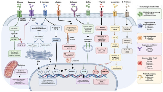
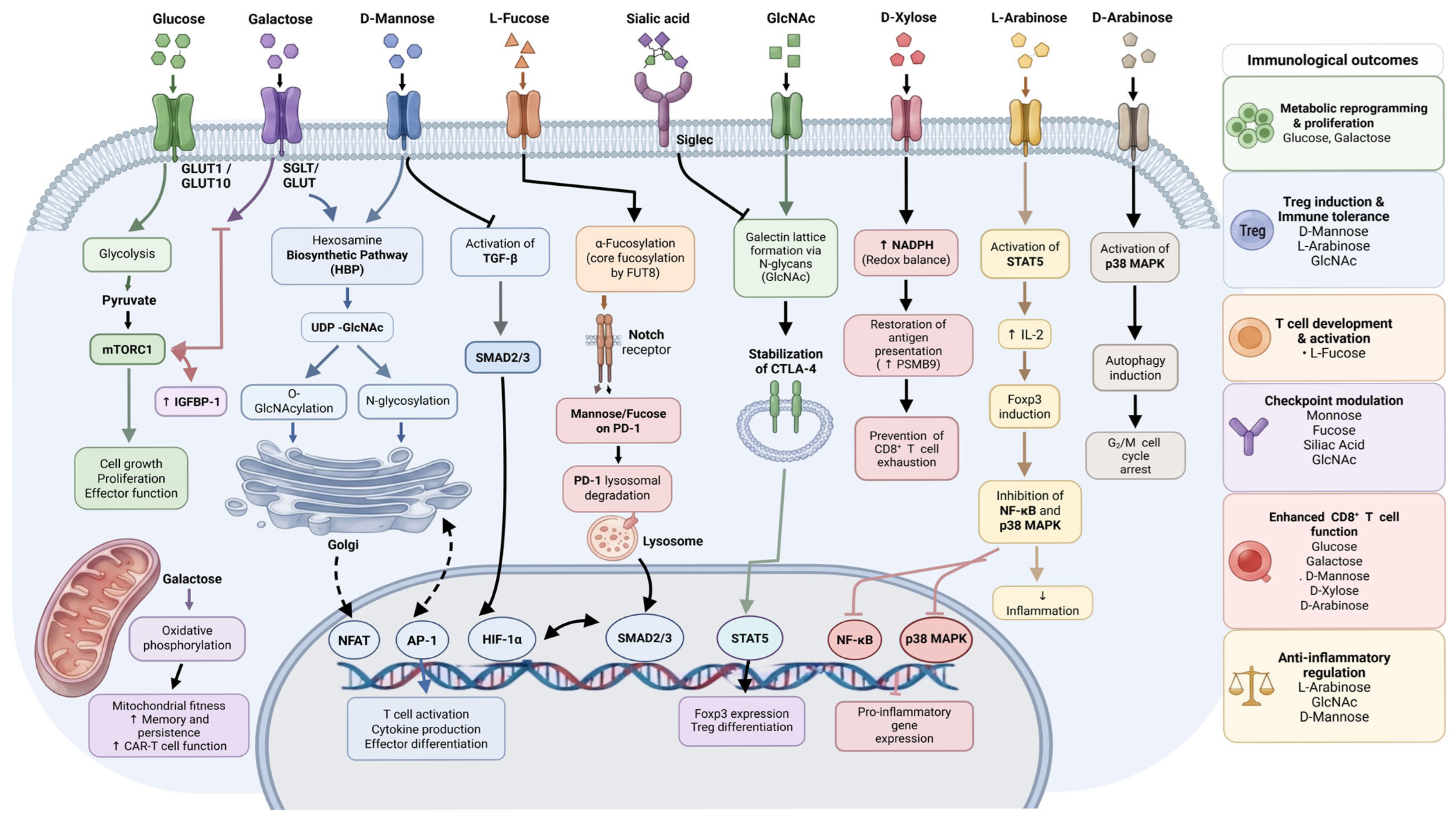
Figure 2 and Table 1 provide a summary of the effects of monosaccharides on T-cell function through distinct molecular mechanisms.
Figure 2.
Intracellular signaling pathways and immunological effects of bioactive monosaccharides in T-cell function. Figure created by BioRender.
Table 1.
Summary of the molecular mechanisms, T-cell responses, and therapeutic potential of immunomodulatory monosaccharides.
Collectively, the current evidence demonstrates that monosaccharides represent a previously underrecognized class of immunonutrients that regulate T-cell biology through multiple complementary mechanisms, including reprogramming of cellular metabolism, modifying glycosylation of immune receptors, regulating transcriptional and epigenetic pathways, thereby shaping T-cell activation, differentiation, immune tolerance, and effector function. Understanding these mechanisms opens the possibility of rationally designed glyconutritional strategies to achieve targeted immunological outcomes: inducing tolerance in autoimmunity, enhancing cytotoxicity in cancer, or restoring immune homeostasis in metabolic disease. However, the context-dependent effects of monosaccharides demand careful consideration of disease context, immune subset composition, and metabolic background before therapeutic application. As our understanding of the molecular interfaces between carbohydrate metabolism and immune regulation continues to deepen, glyconutritional strategies may emerge as important components of precision immunotherapy for autoimmune disease, cancer, and metabolic immune dysfunction.
5. Translational Perspectives, Challenges, and Future Directions of Monosaccharide-Based Immunomodulation
Nutritional regulation of immunity through monosaccharides represents an emerging field that integrates metabolism, glycobiology, and immunotherapy [13,21]. The studies discussed in this review demonstrate that monosaccharides are not merely energy substrates but function as bioactive molecules capable of regulating T-cell activation, differentiation, metabolic fitness, glycosylation patterns, and immune checkpoint pathways [14,20,42,43]. By influencing pathways such as PI3K/Akt/mTORC1, AMPK, the hexosamine biosynthetic pathway, O-GlcNAcylation, and glycocalyx remodeling, monosaccharides have the potential to modulate immune responses in diverse pathological contexts, including cancer, autoimmune disease, infection, and metabolic disorders [14,25,43,81,82]. However, despite promising preclinical findings, significant challenges remain before monosaccharide-based immunomodulatory strategies can be translated into clinical applications.
A major limitation is the incomplete understanding of the pharmacokinetics, bioavailability, and tissue distribution of individual monosaccharides in humans [74,82]. Although many monosaccharides can be orally administered and are generally considered safe nutritional compounds, their systemic concentrations are highly influenced by intestinal absorption, renal clearance, hepatic metabolism, and conversion into downstream metabolites [74]. Importantly, concentrations used in experimental models frequently exceed physiological circulating levels, raising questions regarding whether the observed immunological effects can be achieved through dietary supplementation alone. For example, D-mannose circulates at low micromolar concentrations under physiological conditions, whereas many immunomodulatory effects have been demonstrated using substantially higher concentrations in vitro or through supplementation protocols in animals [42,43]. Therefore, future clinical studies must establish dose–response relationships, pharmacodynamic thresholds, and the duration of exposure required to achieve beneficial immune effects without causing metabolic disturbances [74,82].
Another important consideration is the potential impact of monosaccharide supplementation on systemic metabolic homeostasis. Because carbohydrate metabolism is tightly connected to glucose regulation, insulin signaling, and energy balance, immunonutritional interventions must be carefully evaluated in metabolically heterogeneous populations [13]. Although alternative monosaccharides such as D-mannose or galactose may exert beneficial effects on immune function under specific conditions, excessive or inappropriate supplementation could potentially alter glycemic regulation, particularly in individuals with diabetes, insulin resistance, or impaired carbohydrate metabolism [13,74,87]. Similarly, chronic exposure to supraphysiological concentrations may produce biological effects distinct from physiological nutrient signaling, as demonstrated by high-dose D-galactose models of oxidative stress, advanced glycation end product (AGE) accumulation, and immunosenescence [73,74,75,76]. Thus, future translational studies should prioritize individualized approaches that consider disease status, metabolic phenotype, immune context, and baseline nutritional availability.
A further challenge is achieving immune-cell-specific and tissue-specific delivery. Systemic administration of monosaccharides may result in widespread metabolic effects that limit therapeutic precision [11,60]. In cancer, for example, enhancing CD8+ T-cell function requires overcoming the complex metabolic competition within the tumor microenvironment, where tumor cells frequently dominate nutrient utilization and create immunosuppressive conditions [13,88,89,90]. Similarly, immune tolerance-inducing strategies for autoimmune diseases may require selective modulation of pathogenic immune populations while preserving protective immunity [22,43,77]. Advances in targeted delivery technologies, including nanoparticle-based formulations, glycan-engineered carriers, tumor-targeting systems, and lymphoid tissue-directed delivery platforms, may provide opportunities to concentrate monosaccharide activity within specific immune niches while minimizing systemic exposure [60,82,91,92]. Such approaches could enhance therapeutic efficacy and improve safety profiles.
Future investigations should also address the complexity of host–microbiome–immune interactions in monosaccharide metabolism. Although several monosaccharides can influence intestinal microbial composition and metabolite production, the relative contribution of direct immune-cell effects versus microbiota-mediated mechanisms remains incompletely understood for many compounds [81,86,93,94]. Integrated approaches combining metabolomics, microbiome profiling, single-cell transcriptomics, and immune phenotyping will be essential to define the molecular pathways responsible for immunomodulatory effects in vivo [93].
Finally, clinical translation will require well-designed human studies evaluating safety, pharmacokinetics, immune biomarkers, and therapeutic efficacy [74,82]. Most current evidence supporting monosaccharide-mediated immune regulation originates from cellular systems and animal models, emphasizing the need for controlled clinical trials to determine whether these findings translate into meaningful improvements in human disease outcomes [42,72,78]. Future strategies may involve combining monosaccharides with existing immunotherapies, including immune checkpoint inhibitors, adoptive T-cell therapies, or anti-inflammatory treatments, to enhance therapeutic responses through metabolic and glycosylation-based immune reprogramming [11,42,47,60,72,82].
Collectively, monosaccharides represent a promising class of immunonutrients with the capacity to regulate T-cell biology through interconnected metabolic, glycosylation, and signaling mechanisms [4,14,21,42,43,79]. However, their clinical application requires careful consideration of bioavailability, dose optimization, metabolic safety, and targeted delivery [60,74,82]. A deeper understanding of the molecular interactions between monosaccharide availability and immune cell function will be essential for developing precision glyconutritional approaches capable of selectively enhancing immune protection while maintaining physiological immune balance.
Author Contributions
Conceptualization, M.v.d.M. and P.P.N.; investigation, T.A.R.; resources, M.v.d.M.; writing—original draft preparation, T.A.R. and P.P.N.; writing—review and editing, M.v.d.M. and P.P.N.; visualization, T.A.R.; supervision, M.v.d.M.; project administration, P.P.N. All authors have read and agreed to the published version of the manuscript.
Funding
This research received no external funding.
Institutional Review Board Statement
Not applicable.
Informed Consent Statement
Not applicable.
Data Availability Statement
No new data were created or analyzed in this study. All information presented in this review is derived from previously published literature cited in the article.
Acknowledgments
During the preparation of this manuscript, the authors used Claude, version Sonnet 5 for the purpose of reviewing the manuscript for general writing accuracy, and Scite, version 5.5.3 for the purpose of acquiring and confirming references. The authors have reviewed and edited the output and take full responsibility for the content of this publication.
Conflicts of Interest
The authors declare no conflicts of interest.
Abbreviations
The following abbreviations are used in this manuscript:
| 2-D-gal | 2-deoxy-D-galactose |
| AIH | Autoimmune hepatitis |
| AMPK | AMP-activated protein kinase |
| ATP | Adenosine triphosphate |
| Ca2+ | Calcium ion |
| CAR-T | Chimeric antigen receptor T cell |
| cDC1 | Conventional dendritic cell type 1 |
| CMAS | CMP-sialic acid synthetase |
| CMP-Sia | Cytidine monophosphate–sialic acid |
| CTL | Cytotoxic T lymphocyte |
| CTLA-4 | Cytotoxic T lymphocyte-associated protein 4 |
| DC-SIGN | Dendritic cell-specific intercellular adhesion molecule-3-grabbing non-integrin |
| DHDH | Dihydrodiol dehydrogenase |
| EAE | Experimental autoimmune encephalomyelitis |
| EGF | Epidermal growth factor |
| FAO | Fatty acid oxidation |
References
- Baker, S.A.; Rutter, J. Metabolites as Signalling Molecules. Nat. Rev. Mol. Cell Biol. 2023, 24, 355–374. [Google Scholar] [CrossRef] [Scilit] [PubMed]
- Christ, A.; Günther, P.; Lauterbach, M.A.R.; Duewell, P.; Biswas, D.; Pelka, K.; Scholz, C.J.; Oosting, M.; Haendler, K.; Baßler, K.; et al. Western Diet Triggers NLRP3-Dependent Innate Immune Reprogramming. Cell 2018, 172, 162–175.e14. [Google Scholar] [CrossRef] [Scilit] [PubMed]
- Wastyk, H.C.; Fragiadakis, G.K.; Perelman, D.; Dahan, D.; Merrill, B.D.; Yu, F.B.; Topf, M.; Gonzalez, C.G.; Van Treuren, W.; Han, S.; et al. Gut-Microbiota-Targeted Diets Modulate Human Immune Status. Cell 2021, 184, 4137–4153.e14. [Google Scholar] [CrossRef] [Scilit] [PubMed]
- Longo, J.; DeCamp, L.M.; Oswald, B.M.; Teis, R.; Reyes-Oliveras, A.; Dahabieh, M.S.; Ellis, A.E.; Vincent, M.P.; Damico, H.; Gallik, K.L.; et al. Glucose-Dependent Glycosphingolipid Biosynthesis Fuels CD8+ T Cell Function and Tumor Control. Cell Metab. 2025, 37, 1890–1906.e11. [Google Scholar] [CrossRef] [Scilit] [PubMed]
- Govers, C.; Tang, Y.; Stolte, E.H.; Wichers, H.J.; Mes, J.J. Wheat-Derived Arabinoxylans Reduced M2-Macrophage Functional Activity, but Enhanced Monocyte-Recruitment Capacity. Food Funct. 2020, 11, 7073–7083. [Google Scholar] [CrossRef] [Scilit] [PubMed]
- Pinho, S.S.; Reis, C.A. Glycosylation in Cancer: Mechanisms and Clinical Implications. Nat. Rev. Cancer 2015, 15, 540–555. [Google Scholar] [CrossRef] [Scilit] [PubMed]
- Schnaar, R.L. Glycobiology Simplified: Diverse Roles of Glycan Recognition in Inflammation. J. Leukoc. Biol. 2016, 99, 825–838. [Google Scholar] [CrossRef] [Scilit] [PubMed]
- Sun, Y.; Li, X.; Wang, T.; Li, W. Core Fucosylation Regulates the Function of Pre-BCR, BCR and IgG in Humoral Immunity. Front. Immunol. 2022, 13, 844427. [Google Scholar] [CrossRef] [Scilit] [PubMed]
- Wells, L.; Hart, G.W. O-GlcNAc Turns Twenty: Functional Implications for Post-Translational Modification of Nuclear and Cytosolic Proteins with a Sugar. FEBS Lett. 2003, 546, 154–158. [Google Scholar] [CrossRef] [Scilit] [PubMed]
- Hart, G.W.; Slawson, C.; Ramirez-Correa, G.; Lagerlof, O. Cross Talk between O-GlcNAcylation and Phosphorylation: Roles in Signaling, Transcription, and Chronic Disease. Annu. Rev. Biochem. 2011, 80, 825–858. [Google Scholar] [CrossRef] [Scilit] [PubMed]
- Liu, Y.; Wang, F.; Peng, D.; Zhang, D.; Liu, L.; Wei, J.; Yuan, J.; Zhao, L.; Jiang, H.; Zhang, T.; et al. Activation and Antitumor Immunity of CD8+ T Cells Are Supported by the Glucose Transporter GLUT10 and Disrupted by Lactic Acid. Sci. Transl. Med. 2024, 16, eadk7399. [Google Scholar] [CrossRef] [Scilit] [PubMed]
- Schuurmans, F.; Wagemans, K.E.; Adema, G.J.; Cornelissen, L.A.M. Tumor Glucose Metabolism and the T Cell Glycocalyx: Implication for T Cell Function. Front. Immunol. 2024, 15, 1409238. [Google Scholar] [CrossRef] [Scilit] [PubMed]
- Zhang, S.; Zhang, X.; Yang, H.; Liang, T.; Bai, X. Hurdle or Thruster: Glucose Metabolism of T Cells in Anti-Tumour Immunity. Biochim. Biophys. Acta Rev. Cancer 2024, 1879, 189022. [Google Scholar] [CrossRef] [Scilit] [PubMed]
- Swamy, M.; Pathak, S.; Grzes, K.M.; Damerow, S.; Sinclair, L.V.; Van Aalten, D.M.F.; Cantrell, D.A. Glucose and Glutamine Fuel Protein O-GlcNAcylation to Control T Cell Self-Renewal and Malignancy. Nat. Immunol. 2016, 17, 712–720. [Google Scholar] [CrossRef] [Scilit] [PubMed]
- Smith-Garvin, J.E.; Koretzky, G.A.; Jordan, M.S. T Cell Activation. Annu. Rev. Immunol. 2009, 27, 591–619. [Google Scholar] [CrossRef] [Scilit] [PubMed]
- Brownlie, R.J.; Zamoyska, R. T Cell Receptor Signalling Networks: Branched, Diversified and Bounded. Nat. Rev. Immunol. 2013, 13, 257–269. [Google Scholar] [CrossRef] [Scilit] [PubMed]
- Gaud, G.; Lesourne, R.; Love, P.E. Regulatory Mechanisms in T Cell Receptor Signalling. Nat. Rev. Immunol. 2018, 18, 485–497. [Google Scholar] [CrossRef] [Scilit] [PubMed]
- Courtney, A.H.; Lo, W.L.; Weiss, A. TCR Signaling: Mechanisms of Initiation and Propagation. Trends Biochem. Sci. 2018, 43, 108–123. [Google Scholar] [CrossRef] [Scilit] [PubMed]
- Huse, M. The T-Cell-Receptor Signalling Network. J. Cell Sci. 2009, 122, 1269–1273. [Google Scholar] [CrossRef] [Scilit] [PubMed]
- Demetriou, M.; Granovsky, M.; Quaggin, S.; Dennis, J.W. Negative Regulation of T-Cell Activation and Autoimmunity by Mgat5 N-Glycosylation. Nature 2001, 409, 733–739. [Google Scholar] [CrossRef] [Scilit] [PubMed]
- Pearce, E.L.; Pearce, E.J. Metabolic Pathways in Immune Cell Activation and Quiescence. Immunity 2013, 38, 633–643. [Google Scholar] [CrossRef] [Scilit] [PubMed]
- Alves, I.; Santos-Pereira, B.; De La Cruz, N.; Campar, A.; Pinto, V.; Rodrigues, P.M.; Araújo, M.; Santos, S.; Ramos-Soriano, J.; Vasconcelos, C.; et al. Host-Derived Mannose Glycans Trigger a Pathogenic Γδ T Cell/IL-17a Axis in Autoimmunity. Sci. Transl. Med. 2023, 15, eabo1930. [Google Scholar] [CrossRef] [Scilit] [PubMed]
- Angelin, A.; Gil-de-Gómez, L.; Dahiya, S.; Jiao, J.; Guo, L.; Levine, M.H.; Wang, Z.; Quinn, W.J.; Kopinski, P.K.; Wang, L.; et al. Foxp3 Reprograms T Cell Metabolism to Function in Low-Glucose, High-Lactate Environments. Cell Metab. 2017, 25, 1282–1293.e7. [Google Scholar] [CrossRef] [Scilit] [PubMed]
- Pitmon, E.; Meehan, E.V.; Ahmadi, E.; Adler, A.J.; Wang, K. High Glucose Promotes Regulatory T Cell Differentiation. PLoS ONE 2023, 18, e0280916. [Google Scholar] [CrossRef] [Scilit] [PubMed]
- Araujo, L.; Khim, P.; Mkhikian, H.; Mortales, C.-L.; Demetriou, M. Glycolysis and Glutaminolysis Cooperatively Control T Cell Function by Limiting Metabolite Supply to N-Glycosylation. eLife 2017, 6, e21330. [Google Scholar] [CrossRef] [Scilit] [PubMed]
- Bachoo, S.; Gudgeon, N.; Mann, R.; Stavrou, V.; Bishop, E.L.; Kelly, A.; Uribe, A.H.; Loeliger, J.; Frick, C.; Maddocks, O.D.K.; et al. IL-7 Promotes Integrated Glucose and Amino Acid Sensing during Homeostatic CD4+ T Cell Proliferation. Cell Rep. 2025, 44, 115199. [Google Scholar] [CrossRef] [Scilit] [PubMed]
- Almeida, L.; Lochner, M.; Berod, L.; Sparwasser, T. Metabolic Pathways in T Cell Activation and Lineage Differentiation. Semin. Immunol. 2016, 28, 514–524. [Google Scholar] [CrossRef] [Scilit] [PubMed]
- Ma, E.H.; Poffenberger, M.C.; Wong, A.H.T.; Jones, R.G. The Role of AMPK in T Cell Metabolism and Function. Curr. Opin. Immunol. 2017, 46, 45–52. [Google Scholar] [CrossRef] [Scilit] [PubMed]
- Chi, H. Regulation and Function of MTOR Signalling in T Cell Fate Decisions. Nat. Rev. Immunol. 2012, 12, 325–338. [Google Scholar] [CrossRef] [Scilit] [PubMed]
- Buck, M.D.; Sowell, R.T.; Kaech, S.M.; Pearce, E.L. Metabolic Instruction of Immunity. Cell 2017, 169, 570–586. [Google Scholar] [CrossRef] [Scilit] [PubMed]
- Guo, D.; Tong, Y.; Jiang, X.; Meng, Y.; Jiang, H.; Du, L.; Wu, Q.; Li, S.; Luo, S.; Li, M.; et al. Aerobic Glycolysis Promotes Tumor Immune Evasion by Hexokinase2-Mediated Phosphorylation of IκBα. Cell Metab. 2022, 34, 1312–1324.e6. [Google Scholar] [CrossRef] [Scilit] [PubMed]
- Wang, G. Hexokinase 2 Promotes Tumor Development and Progression. Am. J. Cancer Res. 2025, 15, 4499–4515. [Google Scholar] [CrossRef] [Scilit] [PubMed]
- Zur, R.T.; Zurinam, S.D.; Radman, M.; Funaro Balouka, E.; Borodianskiy-Shteinberg, T.; Saur, D.; Cohen, C.J. Hexokinase2-Engineered T Cells Display Increased Anti-Tumor Function. Front. Immunol. 2025, 16, 1477929. [Google Scholar] [CrossRef] [Scilit] [PubMed]
- Lin, J.; Fang, W.; Xiang, Z.; Wang, Q.; Cheng, H.; Chen, S.; Fang, J.; Liu, J.; Wang, Q.; Lu, Z.; et al. Glycolytic Enzyme HK2 Promotes PD-L1 Expression and Breast Cancer Cell Immune Evasion. Front. Immunol. 2023, 14, 1189953. [Google Scholar] [CrossRef] [Scilit] [PubMed]
- Li, R.; Mei, S.; Ding, Q.; Wang, Q.; Yu, L.; Zi, F. A Pan-Cancer Analysis of the Role of Hexokinase II (HK2) in Human Tumors. Sci. Rep. 2022, 12, 18807. [Google Scholar] [CrossRef] [Scilit] [PubMed]
- Villa, M.; O’Sullivan, D.; Pearce, E.L. Glucose Makes Treg Lose Their Temper. Cancer Cell 2021, 39, 460–462. [Google Scholar] [CrossRef] [Scilit] [PubMed]
- Lopez Aguilar, A.; Gao, Y.; Hou, X.; Lauvau, G.; Yates, J.R.; Wu, P. Profiling of Protein O-GlcNAcylation in Murine CD8+ Effector-and Memory-like T Cells. ACS Chem. Biol. 2017, 12, 3031–3038. [Google Scholar] [CrossRef] [Scilit] [PubMed]
- Zhu, J.; Yang, W.; Zhou, X.; Zöphel, D.; Soriano-Baguet, L.; Dolgener, D.; Carlein, C.; Hof, C.; Zhao, R.; Ye, S.; et al. High Glucose Enhances Cytotoxic T Lymphocyte-Mediated Cytotoxicity. Front. Immunol. 2021, 12, 689337. [Google Scholar] [CrossRef] [Scilit] [PubMed]
- Mu, X.; Xiang, Z.; Xu, Y.; He, J.; Lu, J.; Chen, Y.; Wang, X.; Tu, C.R.; Zhang, Y.; Zhang, W.; et al. Glucose Metabolism Controls Human Γδ T-Cell-Mediated Tumor Immunosurveillance in Diabetes. Cell. Mol. Immunol. 2022, 19, 944–956. [Google Scholar] [CrossRef] [Scilit] [PubMed]
- Zhang, S.; Zhang, K.; Zheng, C.X.; Gao, Y.F.; Deng, G.R.; Zhang, X.; Yuan, Y.; Jia, T.; Tang, S.Y.; He, G.X.; et al. D-Mannose Alleviates Type 2 Diabetes and Rescues Multi-Organ Deteriorations by Controlling Release of Pathological Extracellular Vesicles. Exploration 2025, 5, 20240133. [Google Scholar] [CrossRef] [Scilit] [PubMed]
- Ito, D.; Ito, H.; Ideta, T.; Kanbe, A.; Shimizu, M. D-Mannose Administration Improves Autoimmune Hepatitis by Upregulating Regulatory T Cells. Cell. Immunol. 2022, 375, 104517. [Google Scholar] [CrossRef] [Scilit] [PubMed]
- Qiu, Y.; Su, Y.; Xie, E.; Cheng, H.; Du, J.; Xu, Y.; Pan, X.; Wang, Z.; Chen, D.G.; Zhu, H.; et al. Mannose Metabolism Reshapes T Cell Differentiation to Enhance Anti-Tumor Immunity. Cancer Cell 2025, 43, 103–121.e8. [Google Scholar] [CrossRef] [Scilit] [PubMed]
- Zhang, D.; Chia, C.; Jiao, X.; Jin, W.; Kasagi, S.; Wu, R.; Konkel, J.E.; Nakatsukasa, H.; Zanvit, P.; Goldberg, N.; et al. D-Mannose Induces Regulatory T Cells and Suppresses Immunopathology. Nat. Med. 2017, 23, 1036–1045. [Google Scholar] [CrossRef] [Scilit] [PubMed]
- Sharma, V.; Ichikawa, M.; Freeze, H.H. Mannose Metabolism: More than Meets the Eye. Biochem. Biophys. Res. Commun. 2014, 453, 220–228. [Google Scholar] [CrossRef] [Scilit] [PubMed]
- Torretta, S.; Scagliola, A.; Ricci, L.; Mainini, F.; Di Marco, S.; Cuccovillo, I.; Kajaste-Rudnitski, A.; Sumpton, D.; Ryan, K.M.; Cardaci, S. D-Mannose Suppresses Macrophage IL-1β Production. Nat. Commun. 2020, 11, 6343. [Google Scholar] [CrossRef] [Scilit] [PubMed]
- Hwang, D.; Boehm, A.; Rostami, A.; Zhang, G.X.; Ciric, B. Oral D-Mannose Treatment Suppresses Experimental Autoimmune Encephalomyelitis via Induction of Regulatory T Cells. J. Neuroimmunol. 2022, 362, 577778. [Google Scholar] [CrossRef] [Scilit] [PubMed]
- Dong, W.; Lin, M.; Zhang, R.; Sun, X.; Li, H.; Liu, T.; Xu, Y.; Lv, L. D-Mannose Targets PD-1 to Lysosomal Degradation and Enhances T Cell-Mediated Anti-Tumor Immunity. Cancer Lett. 2024, 591, 216883. [Google Scholar] [CrossRef] [Scilit] [PubMed]
- Zygmunt, B.M.; Węgrzyn, A.; Gajska, W.; Yevsa, T.; Chodaczek, G.; Guzmán, C.A. Mannose Metabolism Is Essential for Th1 Cell Differentiation and IFN-γ Production. J. Immunol. 2018, 201, 1400–1411. [Google Scholar] [CrossRef] [Scilit] [PubMed]
- Davis, J.A.; Freeze, H.H. Studies of Mannose Metabolism and Effects of Long-Term Mannose Ingestion in the Mouse. Biochim. ET Biophys. Acta (BBA)-Gen. Subj. 2001, 1528, 116–126. [Google Scholar] [CrossRef] [Scilit] [PubMed]
- Mayatepek, E.; Schröder, M.; Kohlmüller, D.; Bieger, W.; Nützenadel, W. Continuous Mannose Infusion in Carbohydrate-deficient Glycoprotein Syndrome Type I. Acta Paediatr. 1997, 86, 1138–1140. [Google Scholar] [CrossRef] [Scilit] [PubMed]
- Schmidt, M.; Linder, A.T.; Korn, M.; Schellenberg, N.; Meyer, S.J.; Nimmerjahn, F.; Werner, A.; Abeln, M.; Gerardy-Schahn, R.; Münster-Kühnel, A.K.; et al. Sialic Acids on T Cells Are Crucial for Their Maintenance and Survival. Front. Immunol. 2024, 15, 1359494. [Google Scholar] [CrossRef] [Scilit] [PubMed]
- Varchetta, S.; Lusso, P.; Hudspeth, K.; Mikulak, J.; Mele, D.; Paolucci, S.; Cimbro, R.; Malnati, M.; Riva, A.; Maserati, R.; et al. Sialic Acid-Binding Ig-like Lectin-7 Interacts with HIV-1 Gp120 and Facilitates Infection of CD4 Pos T Cells and Macrophages. Retrovirology 2013, 10, 154. [Google Scholar] [CrossRef] [Scilit] [PubMed]
- Samraj, A.N.; Pearce, O.M.T.; Läubli, H.; Crittenden, A.N.; Bergfeld, A.K.; Band, K.; Gregg, C.J.; Bingman, A.E.; Secrest, P.; Diaz, S.L.; et al. A Red Meat-Derived Glycan Promotes Inflammation and Cancer Progression. Proc. Natl. Acad. Sci. USA 2015, 112, 542–547. [Google Scholar] [CrossRef] [Scilit] [PubMed]
- Varki, A. Uniquely Human Evolution of Sialic Acid Genetics and Biology. Proc. Natl. Acad. Sci. USA 2010, 107, 8939–8946. [Google Scholar] [CrossRef] [Scilit] [PubMed]
- Padler-Karavani, V.; Varki, A. Potential Impact of the Non-Human Sialic Acid N-Glycolylneuraminic Acid on Transplant Rejection Risk. Xenotransplantation 2011, 18, 1–5. [Google Scholar] [CrossRef] [Scilit] [PubMed]
- Varki, A. Sialic Acids in Human Health and Disease. Trends Mol. Med. 2008, 14, 351–360. [Google Scholar] [CrossRef] [Scilit] [PubMed]
- Balneger, N.; Cornelissen, L.A.M.; Wassink, M.; Moons, S.J.; Boltje, T.J.; Bar-Ephraim, Y.E.; Das, K.K.; Søndergaard, J.N.; Büll, C.; Adema, G.J. Sialic Acid Blockade in Dendritic Cells Enhances CD8+ T Cell Responses by Facilitating High-Avidity Interactions. Cell. Mol. Life Sci. 2022, 79, 98. [Google Scholar] [CrossRef] [Scilit] [PubMed]
- Perdicchio, M.; Ilarregui, J.M.; Verstege, M.I.; Cornelissen, L.A.M.; Schetters, S.T.T.; Engels, S.; Ambrosini, M.; Kalay, H.; Veninga, H.; Den Haan, J.M.M.; et al. Sialic Acid-Modified Antigens Impose Tolerance via Inhibition of T-Cell Proliferation and de Novo Induction of Regulatory T Cells. Proc. Natl. Acad. Sci. USA 2016, 113, 3329–3334. [Google Scholar] [CrossRef] [Scilit] [PubMed]
- Silva, M.; Silva, Z.; Marques, G.; Ferro, T.; Gonçalves, M.; Monteiro, M.; Van Vliet, S.J.; Mohr, E.; Lino, A.C.; Fernandes, A.R.; et al. Sialic Acid Removal from Dendritic Cells Improves Antigen Cross-Presentation and Boosts Anti-Tumor Immune Responses. Oncotarget 2016, 7, 41053–41066. [Google Scholar] [CrossRef] [Scilit] [PubMed]
- Büll, C.; Boltje, T.J.; Balneger, N.; Weischer, S.M.; Wassink, M.; Van Gemst, J.J.; Bloemendal, V.R.; Boon, L.; Van Der Vlag, J.; Heise, T.; et al. Sialic Acid Blockade Suppresses Tumor Growth by Enhancing T-Cell-Mediated Tumor Immunity. Cancer Res. 2018, 78, 3574–3588. [Google Scholar] [CrossRef] [Scilit] [PubMed]
- Cornelissen, L.A.M.; Blanas, A.; van der Horst, J.C.; Kruijssen, L.; Zaal, A.; O’Toole, T.; Wiercx, L.; van Kooyk, Y.; van Vliet, S.J. Disruption of Sialic Acid Metabolism Drives Tumor Growth by Augmenting CD8+ T Cell Apoptosis. Int. J. Cancer 2019, 144, 2290–2302. [Google Scholar] [CrossRef] [Scilit] [PubMed]
- Cordner, R.; Jhun, M.; Panwar, A.; Wang, H.Q.; Gull, N.; Murali, R.; McAbee, J.H.; Mardiros, A.; Sanchez-Takei, A.; Mazer, M.W.; et al. Glioma Immunotherapy Enhancement and CD8-Specific Sialic Acid Cleavage by Isocitrate Dehydrogenase (IDH)-1. Oncogene 2023, 42, 2088–2098. [Google Scholar] [CrossRef] [Scilit] [PubMed]
- Liang, W.; Mao, S.; Sun, S.; Li, M.; Li, Z.; Yu, R.; Ma, T.; Gu, J.; Zhang, J.; Taniguchi, N.; et al. Core Fucosylation of the T Cell Receptor Is Required for T Cell Activation. Front. Immunol. 2018, 9, 78. [Google Scholar] [CrossRef] [Scilit] [PubMed]
- Stanley, P.; Guidos, C.J. Regulation of Notch Signaling during T- and B-Cell Development by O-Fucose Glycans. Immunol. Rev. 2009, 230, 201–215. [Google Scholar] [CrossRef] [Scilit] [PubMed]
- Mao, K.; Luo, J.; Ye, J.; Li, L.; Lin, F.; Zhou, M.; Wang, D.; Yu, L.; Zhu, Z.; Zuo, D.; et al. 2-D-Gal Targets Terminal Fucosylation to Inhibit T-Cell Response in a Mouse Skin Transplant Model. Transplantation 2023, 107, 1291–1301. [Google Scholar] [CrossRef] [Scilit] [PubMed]
- Okada, M.; Chikuma, S.; Kondo, T.; Hibino, S.; Machiyama, H.; Yokosuka, T.; Nakano, M.; Yoshimura, A. Blockage of Core Fucosylation Reduces Cell-Surface Expression of PD-1 and Promotes Anti-Tumor Immune Responses of T Cells. Cell Rep. 2017, 20, 1017–1028. [Google Scholar] [CrossRef] [Scilit] [PubMed]
- Burton, C.; Bitaraf, A.; Snyder, K.; Zhang, C.; Yoder, S.J.; Avram, D.; Du, D.; Yu, X.; Lau, E.K. The Functional Role of L-Fucose on Dendritic Cell Function and Polarization. Front. Immunol. 2024, 15, 1353570. [Google Scholar] [CrossRef] [Scilit] [PubMed]
- Lester, D.K.; Burton, C.; Gardner, A.; Innamarato, P.; Kodumudi, K.; Liu, Q.; Adhikari, E.; Ming, Q.; Williamson, D.B.; Frederick, D.T.; et al. Fucosylation of HLA-DRB1 Regulates CD4+ T Cell-Mediated Anti-Melanoma Immunity and Enhances Immunotherapy Efficacy. Nat. Cancer 2023, 4, 222–239. [Google Scholar] [CrossRef] [Scilit] [PubMed]
- Gringhuis, S.I.; Kaptein, T.M.; Wevers, B.A.; Mesman, A.W.; Geijtenbeek, T.B.H. Fucose-Specific DC-SIGN Signalling Directs T Helper Cell Type-2 Responses via IKKε-and CYLD-Dependent Bcl3 Activation. Nat. Commun. 2014, 5, 3898. [Google Scholar] [CrossRef] [Scilit] [PubMed]
- Gringhuis, S.I.; Kaptein, T.M.; Wevers, B.A.; Van Der Vlist, M.; Klaver, E.J.; Van Die, I.; Vriend, L.E.M.; De Jong, M.A.W.P.; Geijtenbeek, T.B.H. Fucose-Based PAMPs Prime Dendritic Cells for Follicular T Helper Cell Polarization via DC-SIGN-Dependent IL-27 Production. Nat. Commun. 2014, 5, 5074. [Google Scholar] [CrossRef] [Scilit] [PubMed]
- Du, X.; Li, W.; Li, G.; Guo, C.; Tang, X.; Bao, R.; Li, R.; Huang, H.; Xu, S.; Yu, X.; et al. Diet-Derived Galactose Reprograms Hepatocytes to Prevent T Cell Exhaustion and Elicit Antitumour Immunity. Nat. Cell Biol. 2025, 27, 1357–1366. [Google Scholar] [CrossRef] [Scilit] [PubMed]
- Gross, G.; Alkadieri, S.; Meir, A.; Itzhaki, O.; Aharoni-Tevet, Y.; Ben Yosef, S.; Zenab, A.; Shbiro, L.; Toren, A.; Yardeni, T.; et al. Improved CAR-T Cell Activity Associated with Increased Mitochondrial Function Primed by Galactose. Leukemia 2024, 38, 1534–1540. [Google Scholar] [CrossRef] [Scilit] [PubMed]
- Uddin, M.N.; Nishio, N.; Ito, S.; Suzuki, H.; Isobe, K. Toxic Effects of D-Galactose on Thymus and Spleen that Resemble Aging. J. Immunotoxicol. 2010, 7, 165–173. [Google Scholar] [CrossRef] [Scilit] [PubMed]
- Azman, K.F.; Zakaria, R. D-Galactose-Induced Accelerated Aging Model: An Overview. Biogerontology 2019, 20, 763–782. [Google Scholar] [CrossRef] [Scilit] [PubMed]
- Song, X.; Bao, M.; Li, D.; Li, Y.M. Advanced Glycation in D-Galactose Induced Mouse Aging Model. Mech. Ageing Dev. 1999, 108, 239–251. [Google Scholar] [CrossRef] [Scilit] [PubMed]
- Cui, X.; Zuo, P.; Zhang, Q.; Li, X.; Hu, Y.; Long, J.; Packer, L.; Liu, J. Chronic Systemic D-Galactose Exposure Induces Memory Loss, Neurodegeneration, and Oxidative Damage in Mice: Protective Effects of R-α-Lipoic Acid. J. Neurosci. Res. 2006, 83, 1584–1590. [Google Scholar] [CrossRef] [Scilit] [PubMed]
- Grigorian, A.; Lee, S.U.; Tian, W.; Chen, I.J.; Gao, G.; Mendelsohn, R.; Dennis, J.W.; Demetriou, M. Control of T Cell-Mediated Autoimmunity by Metabolite Flux to N-Glycan Biosynthesis. J. Biol. Chem. 2007, 282, 20027–20035. [Google Scholar] [CrossRef] [Scilit] [PubMed]
- Grigorian, A.; Araujo, L.; Naidu, N.N.; Place, D.J.; Choudhury, B.; Demetriou, M. N-Acetylglucosamine Inhibits T-Helper 1 (Th1)/T-Helper 17 (Th17) Cell Responses and Treats Experimental Autoimmune Encephalomyelitis. J. Biol. Chem. 2011, 286, 40133–40141. [Google Scholar] [CrossRef] [Scilit] [PubMed]
- Lee, S.U.; Li, C.F.; Mortales, C.L.; Pawling, J.; Dennis, J.W.; Grigorian, A.; Demetriou, M. Increasing Cell Permeability of N-Acetylglucosamine via 6-Acetylation Enhances Capacity to Suppress T-Helper 1 (TH1)/TH17 Responses and Autoimmunity. PLoS ONE 2019, 14, e0214253. [Google Scholar] [CrossRef] [Scilit] [PubMed]
- Mkhikian, H.; Hayama, K.L.; Khachikyan, K.; Li, C.; Zhou, R.W.; Pawling, J.; Klaus, S.; Tran, P.Q.N.; Ly, K.M.; Gong, A.D.; et al. Age-Associated Impairment of T Cell Immunity Is Linked to Sex-Dimorphic Elevation of N-Glycan Branching. Nat. Aging 2022, 2, 231–242. [Google Scholar] [CrossRef] [Scilit] [PubMed]
- Wu, H.; Gong, Y.; Ling, Y.; Wu, S.; Ji, P.; Zhao, Q.; He, L.H.; Shao, Z.M.; Jiang, Y.Z.; Liu, G. DHDH-Mediated D-Xylose Metabolism Induces Immune Evasion in Triple-Negative Breast Cancer. Med 2025, 6, 100535. [Google Scholar] [CrossRef] [Scilit] [PubMed]
- Yu, H.; Wu, X.; Liu, Y.; Xin, C.; Guo, X.; Zhou, Y.; Ding, X. D-Xylose Suppresses Hepatocellular Carcinoma Progression by Regulating Dihydrodiol Dehydrogenase and Remodeling the Immune Microenvironment. Front. Immunol. 2026, 17, 1792196. [Google Scholar] [CrossRef] [Scilit] [PubMed]
- Tang, Z.; Song, H.; Qin, S.; Tian, Z.; Zhang, C.; Zhou, Y.; Cai, R.; Zhu, Y. D-Arabinose Induces Cell Cycle Arrest by Promoting Autophagy via P38 MAPK Signaling Pathway in Breast Cancer. Sci. Rep. 2024, 14, 11219. [Google Scholar] [CrossRef] [Scilit] [PubMed]
- Wang, Y.; Sun, J.; Xue, L.; Liu, J.; Nie, C.; Fan, M.; Qian, H.; Zhang, D.; Ying, H.; Li, Y.; et al. L -Arabinose Attenuates Gliadin-Induced Food Allergy via Regulation of Th1/Th2 Balance and Upregulation of Regulatory T Cells in Mice. J. Agric. Food Chem. 2021, 69, 3638–3646. [Google Scholar] [CrossRef] [Scilit] [PubMed]
- Jacobs, S.R.; Herman, C.E.; Maciver, N.J.; Wofford, J.A.; Wieman, H.L.; Hammen, J.J.; Rathmell, J.C. Glucose Uptake Is Limiting in T Cell Activation and Requires CD28-Mediated Akt-Dependent and Independent Pathways. J. Immunol. 2008, 180, 4476–4486. [Google Scholar] [CrossRef] [Scilit] [PubMed]
- Kang, L.; Pang, J.; Zhang, X.; Liu, Y.; Wu, Y.; Wang, J.; Han, D. L-Arabinose Attenuates LPS-Induced Intestinal Inflammation and Injury through Reduced M1 Macrophage Polarization. J. Nutr. 2023, 153, 3327–3340. [Google Scholar] [CrossRef] [Scilit] [PubMed]
- Morava, E. Galactose Supplementation in Phosphoglucomutase-1 Deficiency; Review and Outlook for a Novel Treatable CDG. Mol. Genet. Metab. 2014, 112, 275–279. [Google Scholar] [CrossRef] [Scilit] [PubMed]
- Chang, C.-H.; Qiu, J.; O’Sullivan, D.; Buck, M.D.; Noguchi, T.; Curtis, J.D.; Chen, Q.; Gindin, M.; Gubin, M.M.; van der Windt, G.J.W.; et al. Metabolic Competition in the Tumor Microenvironment Is a Driver of Cancer Progression. Cell 2015, 162, 1229–1241. [Google Scholar] [CrossRef] [Scilit] [PubMed]
- Ho, P.-C.; Bihuniak, J.D.; Macintyre, A.N.; Staron, M.; Liu, X.; Amezquita, R.; Tsui, Y.-C.; Cui, G.; Micevic, G.; Perales, J.C.; et al. Phosphoenolpyruvate Is a Metabolic Checkpoint of Anti-Tumor T Cell Responses. Cell 2015, 162, 1217–1228. [Google Scholar] [CrossRef] [Scilit] [PubMed]
- Ottensmeier, C.H.; Perry, K.L.; Harden, E.L.; Stasakova, J.; Jenei, V.; Fleming, J.; Wood, O.; Woo, J.; Woelk, C.H.; Thomas, G.J.; et al. Upregulated Glucose Metabolism Correlates Inversely with CD8+ T-Cell Infiltration and Survival in Squamous Cell Carcinoma. Cancer Res. 2016, 76, 4136–4148. [Google Scholar] [CrossRef] [Scilit] [PubMed]
- Bellato, F.; Feola, S.; Dalla Verde, G.; Bellio, G.; Pirazzini, M.; Salmaso, S.; Caliceti, P.; Cerullo, V.; Mastrotto, F. Mannosylated Polycations Target CD206+ Antigen-Presenting Cells and Mediate T-Cell-Specific Activation in Cancer Vaccination. Biomacromolecules 2022, 23, 5148–5163. [Google Scholar] [CrossRef] [Scilit] [PubMed]
- Du, Z.; Sui, D.; Xin, D.; Tang, X.; Li, M.; Liu, X.; Deng, Y.; Song, Y. Sialic Acid-Modified Doxorubicin Liposomes Target Tumor-Related Immune Cells to Relieve Multiple Inhibitions of CD8+ T Cells. J. Liposome Res. 2024, 34, 464–474. [Google Scholar] [CrossRef] [Scilit] [PubMed]
- Wu, Z.W.; Liu, X.C.; Quan, C.X.; Tao, X.Y.; Yi-Luo; Zhao, X.F.; Peng, X.R.; Qiu, M.H. Novel Galactose-Rich Polysaccharide from Ganoderma Lucidum: Structural Characterization and Immunomodulatory Activities. Carbohydr. Polym. 2025, 362, 123695. [Google Scholar] [CrossRef] [Scilit] [PubMed]
- Zhao, L.; Wang, Y.; Zhang, G.; Zhang, T.; Lou, J.; Liu, J. L-Arabinose Elicits Gut-Derived Hydrogen Production and Ameliorates Metabolic Syndrome in C57BL/6J Mice on High-Fat-Diet. Nutrients 2019, 11, 3054. [Google Scholar] [CrossRef] [Scilit] [PubMed]

Disclaimer/Publisher’s Note: The statements, opinions and data contained in all publications are solely those of the individual author(s) and contributor(s) and not of MDPI and/or the editor(s). MDPI and/or the editor(s) disclaim responsibility for any injury to people or property resulting from any ideas, methods, instructions or products referred to in the content. |
© 2026 by the authors. Licensee MDPI, Basel, Switzerland. This article is an open access article distributed under the terms and conditions of the Creative Commons Attribution (CC BY) license.