


Abstract
Probiotics are known to improve gut microbiota balance, enhance food digestion, and support overall health. Among these, Bacillus species are particularly promising due to their safety, spore-forming ability, environmental resilience, and diverse enzymatic activities. However, most Bacillus probiotics used in industry are of terrestrial origin, leaving marine-derived strains largely unexplored. Utilising the untapped potential of marine microbial biomass, this study presents a multi-stage methodology for identifying and evaluating marine-derived Bacillus strains with probiotic potential. A structured screening pipeline was applied to 67 microbial isolates from the Great Barrier Reef sponges. Initial selection focused on essential probiotic characteristics, including growth, stability, safety, and survival under gastrointestinal conditions. Strains meeting these criteria were then assessed for desirable properties, including digestive enzyme production and pathogen inhibition. Using this workflow, three marine-derived Bacillus strains were identified as potential probiotics, one of which demonstrated strong antimicrobial activity against Salmonella enterica at 5 and 10 mg/mL (p < 0.01). These findings demonstrate the capability of marine-associated Bacillus as novel bioproducts with functional antimicrobial properties.
1. Introduction
Probiotics are defined as “live microorganisms which when administered in adequate amounts confer a health benefit on the host” [1]. They are known to improve gut microbiota balance, support food digestion, and suppress pathogenic microorganisms [2,3]. Among various probiotic groups, Bacillus species (Bacillus spp.) are particularly promising due to their strong safety record [4,5] and their ability to form highly resilient endospores, enabling survival under harsh gastrointestinal (GI) and environmental conditions [6]. Additionally, Bacillus spp. produce a wide range of digestive enzymes and antimicrobial compounds, supporting nutrient utilisation and helping to suppress harmful pathogens [7,8].
Bacillus probiotics used in animals are mainly derived from B. subtilis strains isolated from host-associated sources, environmental habitats (soil, ponds, or seaweed), and fermented foods [9]. In contrast, marine environments harbour microbial communities that are compositionally distinct from those on land, even though their overall diversity is comparable [10]. Among marine habitats, sponges (phylum Porifera) are important sources of marine microorganisms. Their high seawater filtration rates and nutrient-rich tissues create stable microenvironments that support diverse and abundant microbial communities, with microorganisms comprising up to one-third of the sponge’s biomass [11,12,13]. These sponge-derived microorganisms produce novel enzymes and bioactive compounds, making them especially valuable for biotechnology and probiotic discovery [14,15,16].
Despite this biotechnological potential, the identification of sponge-derived probiotics for animal nutrition requires that candidate microorganisms meet both safety and regulatory standards. The Association of American Feed Control Officials (AAFCO) and the European Food Safety Authority (EFSA) provide strict criteria and approved strain lists for microbial feed additives, ensuring that probiotic strains used in animal feed are safe and compliant [4,5].
The NatureBank (www.griffith.edu.au/research/institute-biomedicine-glycomics/facilities/naturebank accessed on 19 March 2026) at the Institute for Biomedicine and Glycomics, Griffith University, comprises a microbial collection of over 2000 marine-derived microbial isolates. These microorganisms were predominantly isolated from sponges collected at the Great Barrier Reef [17]. This unique resource provides an opportunity to explore the probiotic potential of marine-derived Bacillus strains. The aim of this work was to develop an integrated screening framework to identify and evaluate marine-derived Bacillus strains with probiotic potential. The workflow first assessed essential probiotic characteristics, including growth performance, stability, safety, and gastrointestinal tolerance. It was followed by the evaluation of functional properties such as digestive enzyme production and antimicrobial activity. Using this pipeline to screen 67 sponge-derived isolates, we identified three safe Bacillus strains suitable for further development, including one with significant antimicrobial activity.
2. Results
2.1. Source of Marine-Derived Microorganisms and Screening Strategies
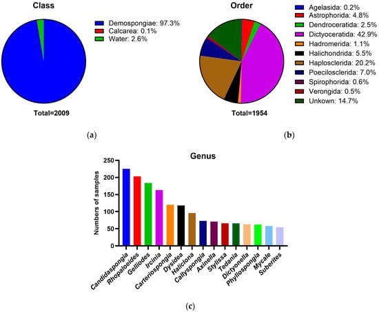
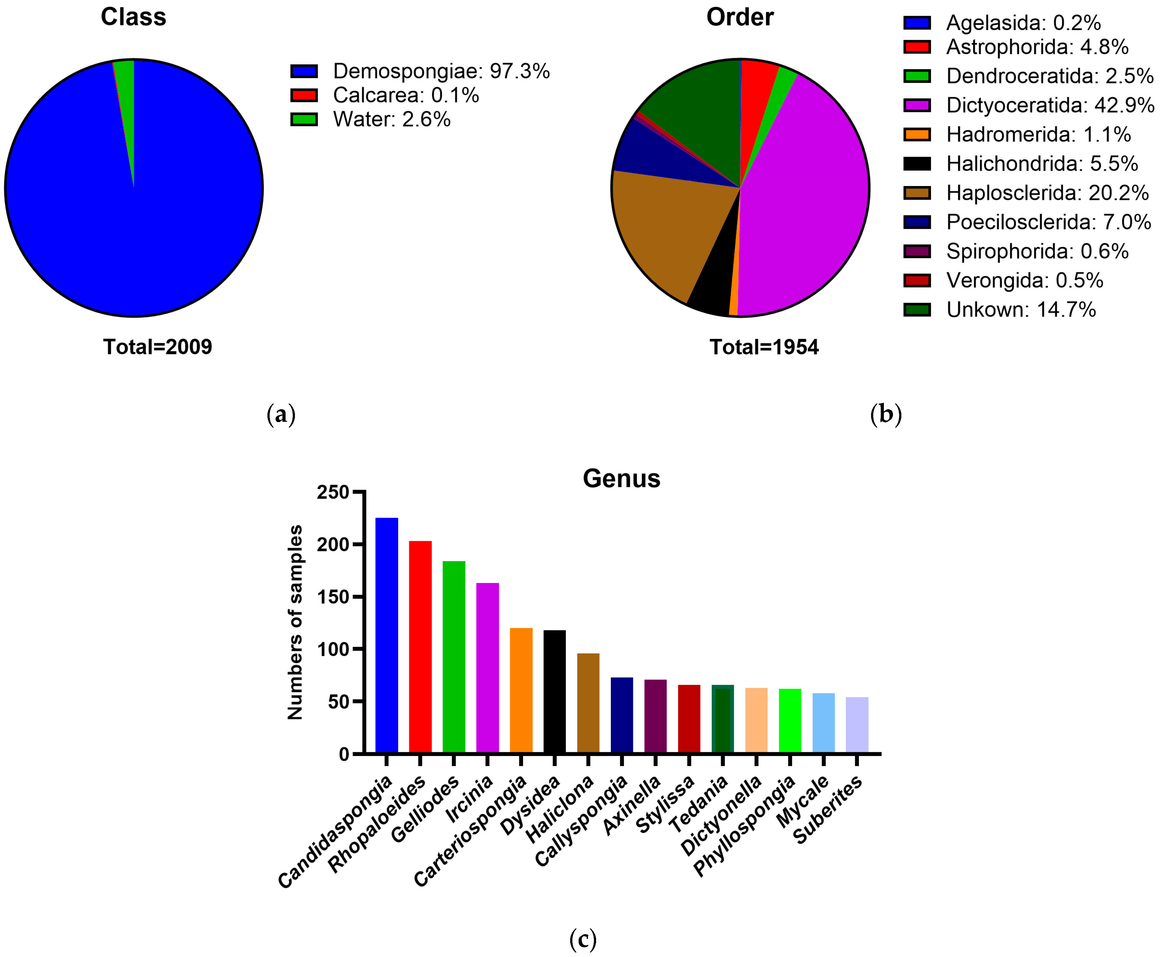
An extensive collection of over 2000 unknown marine-derived microorganisms was used in this study. These microbial isolates mainly originated from sponges collected from the Great Barrier Reef [17]. Host taxonomic information was recorded for the source sponges from which microbial isolates were derived. The collection comprised 2009 microorganisms, and their origins are dominated by the class Demospongiae (n = 1954), with a minor contribution from Calcarea (n = 3), alongside 52 seawater samples (Figure 1a). At the order level, Demospongiae host sponges were primarily represented by Dictyoceratida, Haplosclerida, Poecilosclerida, Halichondrida, and Astrophorida (Figure 1b). In total, 27 host families and 163 host genera were identified within Demospongiae. The 15 most abundant sponge host genera contributing to the microbial collection are shown in Figure 1c, with Candidaspongia, Rhopaloeides, Gelliodes, Ircinia, and Carteriospongia representing the most abundant host sponge genera. Species-level host identification was available for 875 sponge specimens, while the remaining lacked confident species assignments. The sponge samples were processed, and bacterial isolates were cultured on marine-based and selective agar media and then cryopreserved at −80 °C [18]. Colony morphology information and cryopreservation conditions were recorded for each isolate. No taxonomic information was available for these 2009 sponge-derived microbial isolates.
Figure 1.
Host taxonomic composition of sponge specimens associated with the microbial collection. (a) Class-level distribution of sample sources, showing sponge hosts belonging to the classes Demospongiae and Calcarea, alongside seawater samples; (b) Order-level taxonomic distribution of Demospongiae host sponges; (c) The 15 most abundant sponge host genera from which microorganisms were isolated.
2.2. Screening Strategies
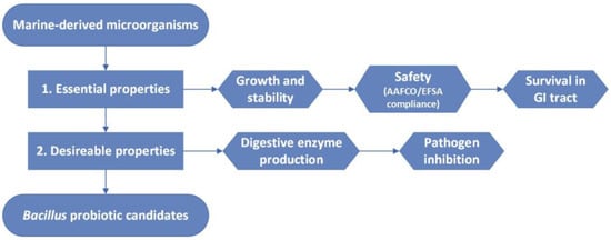
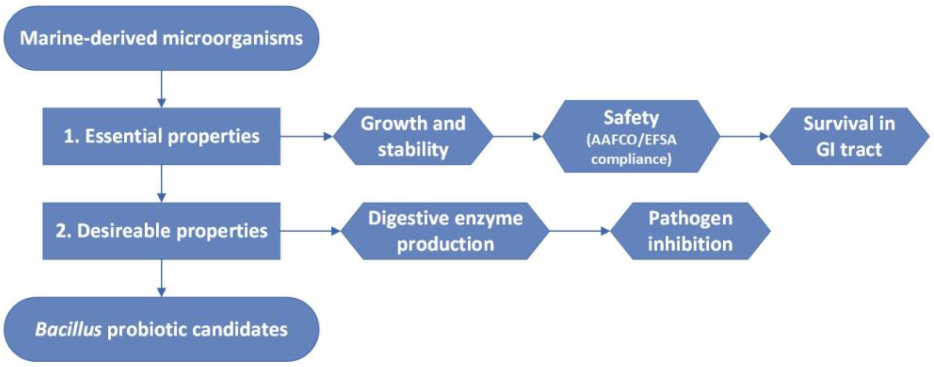
This study aimed to identify marine-derived Bacillus spp. that align with AAFCO or EFSA-approved strain lists for use as animal feed additives. Accordingly, microorganisms with circular or irregular colony morphology, characteristic of Bacillus, were randomly selected from the collection to screen for potential probiotics. A multi-step screening workflow was developed (Figure 2). Initial selection focused on essential probiotic characteristics, including growth performance, stability, safety, and gastrointestinal survival. Strains meeting these criteria were assessed for desirable properties, including digestive enzyme production and pathogen inhibition.
Figure 2.
Sequential screening workflow for marine-derived Bacillus probiotic candidates as animal feed additives. Marine-derived microorganisms were first screened for essential probiotic properties, including growth performance, stability, safety (AAFCO/EFSA compliance, including haemolysis, antimicrobial susceptibility, and taxonomic safety), and gastrointestinal survival. Next, isolates meeting these requirements were retained and evaluated for desirable properties, such as extracellular digestive enzyme production and pathogen inhibition. Lastly, isolates meeting all requirements were retained as Bacillus probiotic candidates.
The workflow also integrated AAFCO and EFSA guidelines, ensuring that the marine-derived isolates not only exhibit essential and desirable probiotic properties but also align with established regulatory requirements for animal feed additives, thereby enhancing their commercial relevance.
2.3. Screening for Essential Probiotic Properties
2.3.1. Growth and Stability
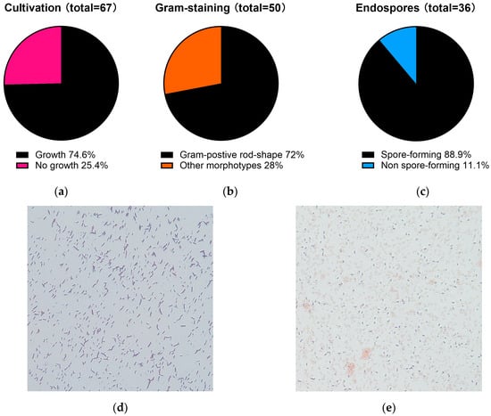
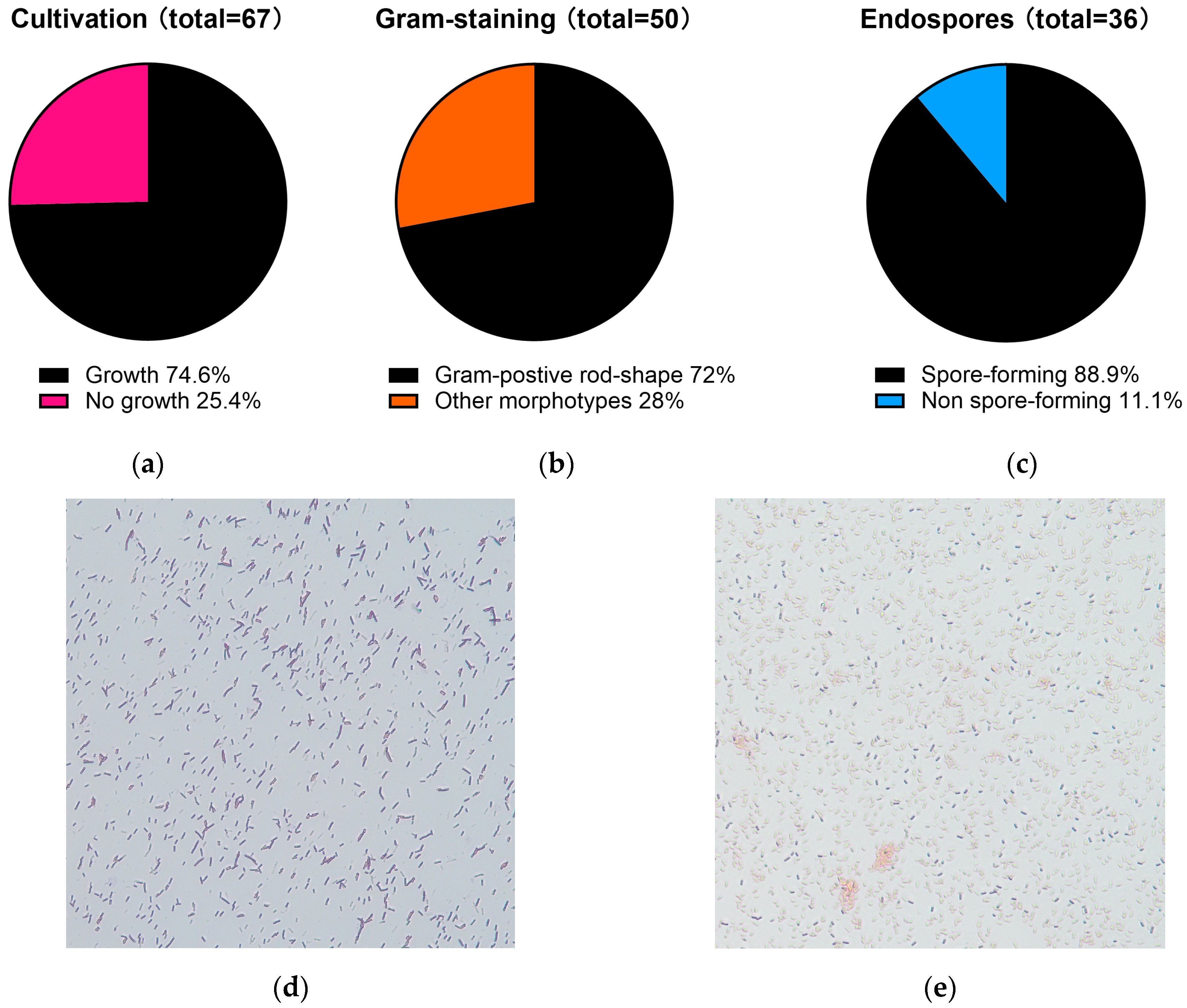
Sponges from the Great Barrier Reef typically inhabit waters with temperatures ranging from 22 to 28 °C, while experimental evidence shows that disruption of the sponge–microbial symbiosis occurs when seawater temperatures approach 33 °C [19]. Therefore, microorganisms derived from these sponges are expected to exhibit optimal growth within this temperature range in seawater. In contrast, typical Bacillus spp. generally exhibit optimal growth at higher temperatures (25–37 °C) [20]. Therefore, Bacillus favourable cultivation conditions were applied for the initial screening phase. A total of 67 sponge-derived bacterial isolates were cultivated under Bacillus favourable conditions (37 °C for 16 h), resulting in 50 isolates (74.6%, Figure 3a) that exhibit rapid growth. These isolates were subsequently subjected to Gram staining, and 36 out of 50 isolates (72%, Figure 3b) were identified as Gram-positive and rod-shaped bacteria (Figure 3d), which is consistent with typical Bacillus morphology. Endospore production was then confirmed using both Schaeffer–Fulton spore staining and heat-treatment (60 °C for 30 min). A total of 32 isolates (out of 36 Gram-positive rods, Figure 3c) demonstrated clear endospore formation (Figure 3e). They also remained stable at 60 °C, a temperature at which spores are mostly active without reducing germination efficiency [21]. Therefore, these isolates were retained for further characterisation.
Figure 3.
Distribution of marine-derived isolates meeting the growth and stability criteria. Marine-derived microbial isolates were sequentially screened under Bacillus-favourable cultivation conditions (37 °C for 16 h). (a) Under these conditions, 74.6% of isolates (50) exhibited rapid growth; (b) Growth-positive isolates were subjected to Gram staining and morphological analysis, showing that 72% (36) were Gram-positive rod-shaped bacteria; (c) Gram-positive rods were further evaluated for endospore-forming ability, with 32 isolates (88.9%) confirmed to form endospores; (d) Representative image of Gram-positive rod-shape marine-derived isolates under microscopy; (e) Representative image of a spore-forming marine-derived isolate under microscopy, with endospores in green colour. Images were cropped to enhance visualisation of spore morphology while preserving original staining characteristics.
2.3.2. Safety Tests
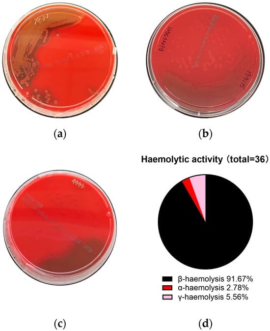
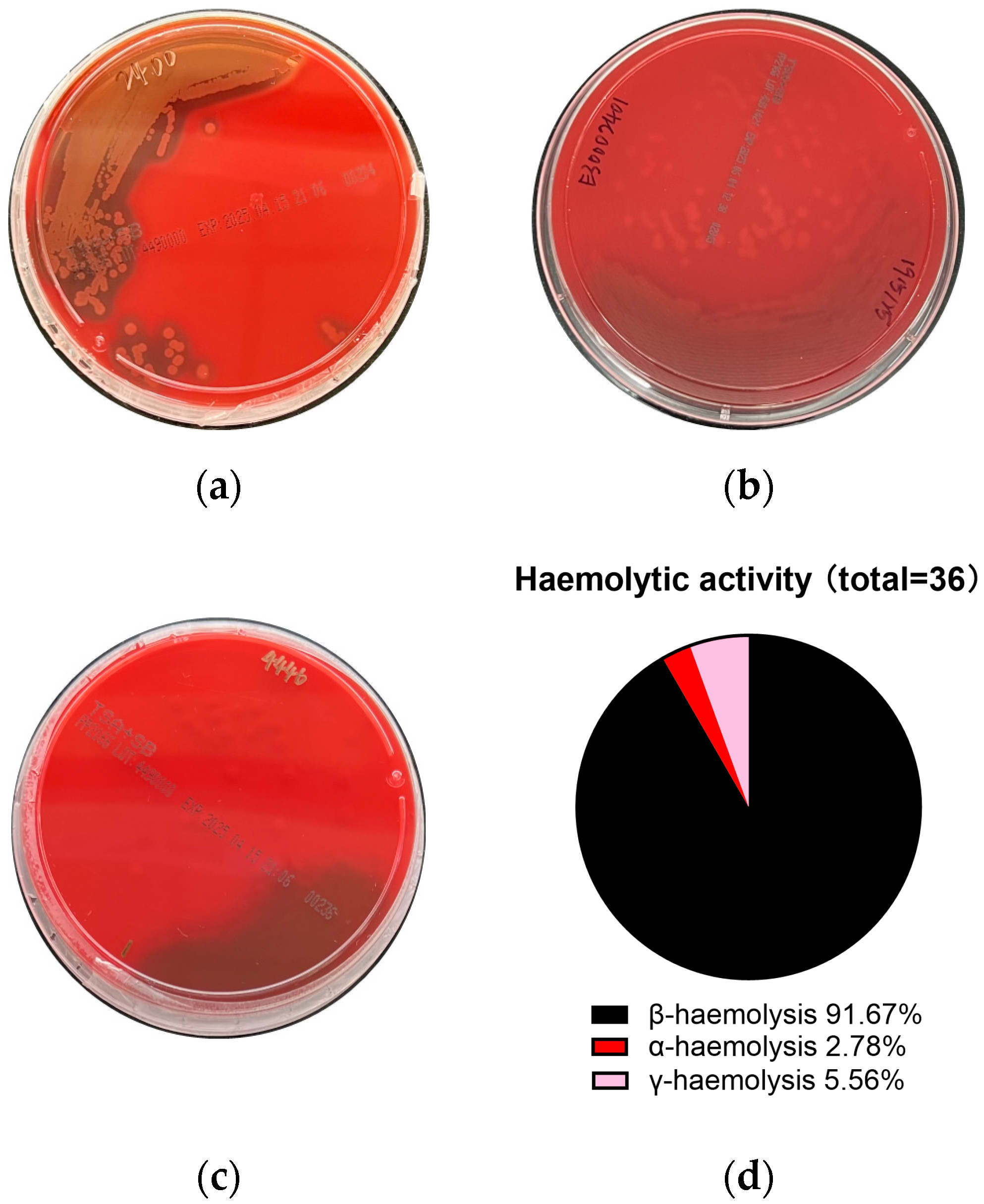
The marine-derived microorganisms were next evaluated for safety, beginning with a haemolysis test to determine their potential to lyse red blood cells. Haemolytic activity is commonly associated with toxicity and pathogenicity, such as streptolysin O and S produced by Group A Streptococcus (GAS), which contribute to β-haemolysis [22]. In contrast, non-haemolytic (γ-haemolysis) strains are preferred for human or animal use [23,24]. Haemolytic activity was assessed using sheep blood agar [25]. The presence of a clear zone surrounding bacterial colonies indicated β-haemolysis (Figure 4a), while partial haemolysis (greenish discolouration) and the absence of haemolysis corresponded to α- and γ-haemolysis (Figure 4b,c), respectively. Among the 36 isolates tested, 91.67% exhibited β-haemolysis, one isolate displayed α-haemolysis (2.78%), and two isolates showed γ-haemolysis (5.56%) (Figure 4d). Based on these results, only three isolates, which were non-haemolytic or exhibited weak haemolytic activity, were considered suitable for further assessments. They are abbreviated as candidates 1–3.
Figure 4.
Haemolytic activity of candidate isolates on sheep blood agar. Marine-derived isolates were inoculated onto sheep blood agar and incubated at 37 °C for 16–24 h. Examples of haemolytic activity were shown: (a) β-haemolysis; (b) α-haemolysis; (c) γ-haemolysis; (d) Distribution of marine-derived isolates showing different haemolytic activity.
The second safety assessment is antimicrobial susceptibility testing. According to EFSA, microbial feed additives should not add to the pool of antimicrobial resistance (AMR) genes already present in the gut bacterial population. To assess this, two types of data are required. Firstly, phenotypic testing is based on the determination of a minimum inhibitory concentration (MIC) for a selected group of antimicrobials. Secondly, genomic analysis for the presence of known AMR genes [26].
In this study, phenotypic MIC testing was performed for candidates 1–3 against a panel of antimicrobials, including vancomycin, gentamycin, kanamycin, streptomycin, erythromycin, tetracycline, and chloramphenicol. The MIC was determined by OD600 measurements and compared with the EFSA-recommended cut-off thresholds. As summarised in Table 1, the MIC values of candidates 1–3 for all tested antimicrobials were equal to or below the EFSA-recommended cut-off thresholds, indicating susceptibility and the absence of phenotypic resistance. Based on these results, candidates 1–3 were phenotypically safe, but confirmation of their genomic AMR status will require future whole-genome analysis.
Table 1.
MIC values of candidates 1 to 3 against EFSA-recommended antimicrobials.
2.3.3. Survival in Gastrointestinal (GI) Tract
Tolerance to acidic pH and bile salts is a key characteristic for probiotics to survive passage through the GI tract and exert functional effects in the host [27,28,29,30]. To evaluate GI stress tolerance, candidates 1–3 and five industrial benchmark Bacillus probiotic strains were evaluated under simulated gastrointestinal conditions.
Gastric pH varies widely among humans and animals, typically ranging from 1 to 3 in scavengers and carnivores, from 2.5 to 6.5 in herbivores, and around 2 to 4.5 in omnivores, depending on species and diet [31]. For this study, exposure to pH 4 was selected to mimic the acidic conditions encountered during gastric transit, providing a mild physiologically relevant stress capable of discriminating acid-tolerant strains. Bile salts are compounds synthesised from cholesterol in the liver, stored in the gallbladder, and released into the small intestine, where they are key determinants of the intestinal physicochemical environment [32]. To be effective, probiotics must survive this bile salt-rich environment. Therefore, this study used a bile salt concentration of 0.3% (w/v), which is widely adopted as a critical threshold for probiotic screening [33], to select bile salt-resistant probiotic strains.
Survival under acidic and bile conditions was assessed at 0, 2, and 4 h, corresponding to progressive exposure times during GI transit. For benchmark strains, survival at pH 4 ranged from 70 to 100% and 67 to 100% after 2 and 4 h of exposure, respectively (Table 2). All three candidates exhibited survival within this range. Under 0.3% bile salt stress, benchmark strains showed survival rates of 15–99% at 2 h and 12–98% at 4 h (Table 3). All three candidates displayed comparable bile tolerance to benchmark strains.
Table 2.
Percentage survival of benchmark Bacillus strains following exposure to acidic conditions (pH 4) for up to 4 h.
Table 3.
Percentage survival of benchmark Bacillus strains following exposure to 0.3% bile salts for up to 4 h.
To confirm the taxonomic identity of the three probiotic candidates, the 16S rRNA gene of candidates 1–3 was sequenced by Sanger sequencing. Comparison against reference databases showed that all three isolates clustered within the genus Bacillus (Supplementary Tables S1–S3). These verified marine-derived Bacillus isolates were then evaluated for additional desirable probiotic properties.
2.4. Screening for Desirable Probiotic Properties
2.4.1. Digestive Enzyme Production
Bacillus spp. are known to secrete extracellular digestive enzymes, such as proteases and amylases, which contribute to their ability to hydrolyse proteins and carbohydrates [8]. These enzymatic activities not only support nutrient acquisition but also strengthen their use as probiotics and industrial biocatalysts in food, feed, and biotechnological applications. Accordingly, screening for hydrolytic enzyme production was employed as a functional selection criterion to further prioritise isolates with potential applied functionality. All three candidates produced protease, while candidates 1 and 3 were additionally capable of amylase production (Supplementary Figure S1).
2.4.2. Antimicrobial Activity
Probiotics with antimicrobial activity not only enhance host health but also help reduce reliance on antibiotics. It is a critical functional trait, reflecting the ability of probiotic candidates to inhibit pathogenic microorganisms in the gastrointestinal environment. The antimicrobial potential of selected Bacillus candidates was evaluated against five pathogens, including Escherichia coli (E. coli), Salmonella enterica (S. enterica), Staphylococcus aureus (S. aureus), and Clostridium perfringens (C. perfringens), which are major zoonotic and foodborne pathogens [34], and Pseudomonas aeruginosa (P. aeruginosa), an opportunistic pathogen that can cause severe infections in various livestock and poultry species [35,36]. Together, these pathogens represent both Gram-negative and Gram-positive bacteria commonly encountered in gastrointestinal and environmental settings.
Agar diffusion assay showed that only candidates 2 and 3 exhibited antimicrobial activity, with candidate 2 inhibiting S. enterica and candidate 3 inhibiting both S. enterica and E. coli (Supplementary Figure S2). Because this method provides only qualitative evidence, quantitative assessment was performed using broth microdilution assays.
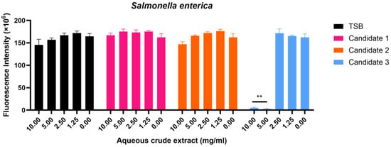
To evaluate both water-soluble and organic-soluble secondary metabolites, antimicrobial activity was tested using aqueous crude extracts and ethyl acetate (EtOAc) extracts from candidates 1–3. Among three crude aqueous extracts, only candidate 3 exhibited significant inhibition against S. enterica at 5 and 10 mg/mL (Figure 5, p < 0.01) and showed a MIC of 5 mg/mL, whereas TSB control, candidate 1, and candidate 2 showed no effects (Figure 5). None of the aqueous crude extracts from any candidate exhibited activity against the remaining four pathogens tested (Supplementary Figure S3). Similarly, none of the EtOAc crude extracts showed activity against any of the five pathogens (Supplementary Figure S4).
Figure 5.
Broth microdilution assay of aqueous crude extracts from Bacillus candidates 1–3 against pathogenic bacteria. S. enterica were cultured in MHB and incubated with aqueous crude extracts of TSB, and candidates 1–3 at 37 °C for 16 h. The growth of pathogenic bacteria was then assessed using Resazurin, and fluorescence intensity was measured. Mean ±SEM (n = 3) of three or four independent experiments, each performed in triplicate, is shown. One-way ANOVA followed by Dunnett’s test was employed to compare treated cells with untreated cells for each extract group. p ** < 0.01.
Overall, candidate 3 consistently demonstrated selective antimicrobial activity against S. enterica across all assays, confirming that this strain produces bioactive compounds capable of suppressing S. enterica growth.
3. Discussion
This study established an integrated workflow for identifying marine-derived Bacillus strains with probiotic potential. From 67 sponge-derived isolates, three safe Bacillus strains were identified, including one with significant, selective S. enterica inhibition. This represented a 4.5% hit rate for utilising marine microorganisms for probiotic discovery. The workflow also provided a cost-effective pipeline for the discovery of novel marine-derived probiotics for animal feed additives. Together, these findings expand the underexplored potential of marine microbial resources and demonstrate the capability of marine-associated Bacillus as novel bioproducts with functional antimicrobial properties.
Bacillus probiotics used in animals are mainly derived from B. subtilis strains originating from host-associated sources (intestine and faeces), environmental habitats (soil, ponds or seaweed), and fermented foods [9]. Despite this broad range of terrestrial and host-associated origins, marine-derived Bacillus strains remain largely unexplored. The results of this study, therefore, expand current knowledge by demonstrating that sponge-associated marine microorganisms also harbour Bacillus strains with probiotic potential. Marine sponges are known to contribute to diverse bioactive compounds [37], and microorganisms from marine sponges may possess unique enzymatic profiles, bioactive metabolites not commonly found in terrestrial and host-associated Bacillus, thereby contributing to distinct functional probiotic properties. Moreover, sponge-derived microorganisms are naturally adapted to a wide range of marine environments characterised by fluctuating salinity, temperature, nutrient availability, and microbial competition [38]. These adaptive traits are highly relevant to aquaculture systems, where probiotic candidates need to remain functional and stable under dynamic rearing conditions. Consequently, sponge-associated bacteria represent a promising and underexplored source of probiotic strains with potential applications in aquaculture, including improving animal health, enhancing feed utilisation, and supporting disease management strategies.
Among the three Bacillus probiotic candidates, only the aqueous crude extract of candidate 3, derived from the cell-free supernatant, exhibited antimicrobial activity. This extract selectively inhibited S. enterica, which is the only Gram-negative bacterium among the five tested pathogens (Figure 5, p < 0.01). This anti-salmonella activity is likely derived from the water-soluble metabolites produced by candidate 3. Bacillus spp. are known to produce bioactive metabolites that can contribute to antimicrobial activity [39,40]. Further isolation and structural characterisation of the bioactive metabolites will require chromatographic (HPLC) and spectroscopic techniques (LC–MS/MS and NMR).
For further commercial development and to elucidate the mode of action, additional investigations such as colonisation assays, large-scale fermentation, feed formulation, animal trials, and comprehensive genomic and metabolomic analyses will be required. Additionally, whole-genome analysis is needed to assess the presence of AMR genes, to provide species or strain-level taxonomic resolution, and to identify genomic features associated with functional properties.
4. Materials and Methods
4.1. Chemicals and Reagents
All chemicals and reagents used in this study were sourced from commercial suppliers. Tryptic soy broth (TSB), resazurin, Gram staining kits, Schaeffer and Fulton spore stain kits, bile salts, vancomycin hydrochloride, streptomycin sulphate, erythromycin, tetracycline, gentamicin solution, and kanamycin solution were obtained from Sigma-Aldrich/Merck (Melbourne, Australia). Mueller–Hinton broth (MHB), nutrient broth, bacteriological agar (Agar No. 1), tryptone, yeast extract, and dextrose were purchased from Oxoid (Thebarton, Australia). Tryptic Soy Agar (TSA) plates supplemented with 5% sheep blood were supplied by Thermo Fisher Scientific (Thebarton, Australia). Ethyl acetate, methanol, KCl, MgSO4·7H2O, KH2PO4, Ca(NO3)2, MnCl2, FeSO4, and chloramphenicol were obtained from ChemSupply (Gillman, Australia).
4.2. Microbial Strains
Sixty-seven marine-derived microorganisms were obtained from the NatureBank at the Institute for Biomedicine and Glycomics, Griffith University (Australia). Industrial benchmark strains: BPR-11 (Bacillus amyloliquefaciens, CBS#141692), BPR-14 (B. amyloliquefaciens, CBS#142360), BPR-16 (B. velezensis, CBS#148295), BPR-17 (B. amyloliquefaciens, CBS#148296), and BPR-20 (B. subtilis, CBS#144669) were obtained from Bioproton Pty Ltd. (Brisbane, Australia) [41]. The Centraalbureau voor Schimmelcultures (CBS) number was generated by Westerdijk Fungal Biodiversity Institute, Utrecht, The Netherlands. Pathogenic strains: C. perfringens (ATCC 13124), E. coli (ATCC 43887), P. aeruginosa (ATCC 10145), S. aureus (ATCC 6538), and S. enterica (ATCC 6960) were purchased from In Vitro Technologies (Melbourne, Australia).
4.3. Microbial Cultivation
Microorganisms were first streaked onto tryptic soy agar (TSA) plates and were incubated at 37 °C for 16–20 h in a static incubator. A single colony was then selected and inoculated into TSB, followed by incubation at 37 °C for 16–20 h at 200 rpm. Cultivation conditions followed standard microbiological procedures unless otherwise specified.
4.4. Gram Staining
Smears were prepared from a single bacterial colony or liquid culture on the microscopy slide. Gram staining was performed following the manufacturer’s instructions using crystal violet, iodine, decolourizer, and safranin. Slides were air-dried and examined under an Olympus BX63 microscope (Evident Scientific, Sydney, Australia) to assess cell morphology and Gram reaction.
4.5. Sporulation Stimulation and Verification
A single colony was inoculated into TSB and incubated overnight at 37 °C, 200 rpm until reaching the stationary growth phase. Then, 1–3% of the overnight culture was transferred into sporulation medium (Table S4), and the cultures were incubated at 37 °C for 48–72 h. Spore formation was then confirmed using the Schaeffer and Fulton spore stain kit (Merck, Melbourne, Australia), with spores appearing green and vegetative cells red or pink. The stability of spores was further tested by subjecting cultures to heat treatment at 60 °C for 30 min, followed by viability assessment on agar plates [21].
4.6. Haemolysis Determination
A 10 μL aliquot of bacterial suspension was spotted onto TSA supplemented with 5% defibrinated sheep blood (Thermo Fisher Scientific, Thebarton, Australia). Plates were incubated at 37 °C for 24 h. Haemolysis was evaluated based on the appearance of zones surrounding bacterial growth: complete clearance (β-haemolysis, indicating strong red blood cell lysis), partial/greenish discolouration (α-haemolysis, indicating partial lysis), or no visible change (γ-haemolysis, indicating absence of haemolytic activity) [25].
4.7. Antimicrobial Susceptibility Assay
Bacteria were cultured to the early log phase (OD600 ≈ 0.1 or equivalent to a 0.5 McFarland standard) in MHB by inoculating 1–3% of an overnight culture and incubating at 37 °C, 200 rpm. Once the cultures had reached their OD, they were further diluted 1:100 to achieve a final cell concentration of 1.5 × 10⁶ CFU/mL. Antimicrobials tested included vancomycin, gentamicin, kanamycin, streptomycin, erythromycin, tetracycline, and chloramphenicol [26], using two-fold serial dilutions from 16 μg/mL to 0 μg/mL. The diluted bacterial cultures and antimicrobials were combined in a final volume of 200 μL per well in 96-well microtiter plates. Plates were incubated statically at 37 °C for 16–20 h, and absorbance was measured at 600 nm using a SpectraMax iD5 plate reader (Bio-Strategy, Melbourne, Australia). The MIC was defined as the lowest antimicrobial concentration in wells with OD600 < 0.001.
4.8. Acid and Bile Salt Tolerance Assays
Spore suspensions were heat-treated at 60 °C for 30 min to eliminate vegetative cells and then cooled to room temperature. Subsequently, 2% of the heat-treated suspensions were inoculated into TSB, pH 4, or TSB supplemented with 0.3% bile salt. Cultures were incubated at 37 °C, 200 rpm, and samples were collected at 0, 2, and 4 h. At each time point, 100 μL was plated on TSA to assess viability. Colony confluency after 16–24 h of incubation was evaluated to determine spore survival under acidic and bile stress conditions. The method was adopted from AlGburi et al. with modifications [28].
4.9. Taxonomic Identification by Sanger Sequencing
Single colony of candidates 1–3 was resuspended in PrepMan Ultra buffer (Thermo Fisher Scientific, Thebarton, Australia) and submitted to Australian Genome Research Facility (AGRF) for 16S rRNA gene Sanger sequencing using the 16S microbial screen assay. Taxonomic identification was performed by AGRF, where the 16S rRNA gene was amplified and Sanger-sequenced. The resulting sequences were aligned against custom database called combined 16S database (version dated 20 Feb 2017) using Basic Local Alignment Search Tool (BLASTn, 2.17.0) to determine the closest taxonomic assignment.
4.10. Enzyme Activity Assays
Bacillus cultures (10 µL) were spotted onto skim milk agar (protease) or starch agar (amylase) (Table S4). Plates were incubated at 37 °C for 24 h. Protease activity was determined by clear zones on milk agar, indicating casein hydrolysis. Amylase activity was assessed by flooding starch agar with iodine; clear halos indicated starch degradation.
4.11. Agar Diffusion Assay
Pathogenic bacteria, including E. coli, P. aeruginosa, S. aureus, C. perfringens, and S. enterica, were cultured to the early log phase in MHB, diluted 1:20 in molten Mueller Hinton agar (MHA), and poured into petri dishes. After the agar solidified, 10 μL of the overnight Bacillus cultures were spotted onto the pathogenic MHA plates. The plates were incubated at 37 °C for 16–24 h. Clear zone around the spot indicated antimicrobial activities [41].
4.12. Preparation of Aqueous Crude Extracts
Bacillus candidates were cultured in TSB for 16 h at 37 °C, then they were centrifuged at 10,000× g for 20 min at 4 °C. Supernatant was collected and freeze-dried, then reconstituted in water and filter-sterilised to obtain the aqueous crude extracts.
4.13. Preparation of Ethyl Acetate (EtOAc) Extracts
Bacillus candidates were cultured in TSB for 16 h at 37 °C, then they were homogenised and centrifuged at 10,000× g for 20 min at 4 °C. The resulting supernatant was extracted with an equal volume of EtOAc three times using a separation funnel. The combined EtOAc phases were concentrated to dryness using a rotary evaporator, and the residue was dissolved in DMSO to obtain the EtOAc extracts.
4.14. Broth Microdilution Assay
Pathogenic bacteria, including E. coli, P. aeruginosa, S. aureus, C. perfringens, and S. enterica, were cultured to the early log phase in MHB, diluted 10 times, and stored on ice until further use. Aqueous crude extracts and EtOAc extracts were serially diluted in MHB in 96-well microtiter plates. An equal volume of pathogen suspension was added to each well to achieve the final working concentrations. Gentamicin (1 μg/mL) was used as the positive control, and MHB was used as the blank. Plates were incubated in a static incubator at 37 °C for 16 to 18 h. After incubation, resazurin (final concentration 0.05 mg/mL) was added to each well and incubated further for 4 h at 37 °C. Fluorescence was measured using a SpectraMax iD5 plate reader (Bio-Strategy, Melbourne, Australia) at excitation 530 nm and emission 590 nm to quantify metabolic activity [42].
4.15. Statistical Analysis
Broth microdilution assays included 3–4 biological replicates, each with three technical replicates. One-way ANOVA followed by Dunnett’s post-test was performed using GraphPad Prism 10. Data are reported as mean ± SEM.
Supplementary Materials
The following supporting information can be downloaded at: https://www.mdpi.com/article/10.3390/md24040137/s1, Table S1: 16S rRNA gene Sanger sequencing results of Bacillus candidates 1; Table S2: 16S rRNA gene Sanger sequencing results of Bacillus candidates 2; Table S3: 16S rRNA gene Sanger sequencing results of Bacillus candidates 3; Figure S1: Protease and amylase activities of Bacillus candidates 1–3 on skim milk and starch agar plates; Figure S2: Agar diffusion assay showing antimicrobial activity of Bacillus candidates against pathogenic bacteria; Figure S3: Broth microdilution assay of aqueous crude extracts from Bacillus candidates 1–3 against pathogenic bacteria; Figure S4: Broth microdilution assay of EtoAc extracts from Bacillus candidates 1–3 against pathogenic bacteria; Table S4: List of solutions and media.
Author Contributions
Conceptualisation, Y.L., X.C. and Y.F.; methodology, Y.L., X.C. and Y.F.; validation, Y.L.; formal analysis, Y.L.; investigation, Y.L.; data curation, Y.L.; writing—original draft preparation, Y.L.; writing—review and editing, Y.L., X.C. and Y.F.; supervision, X.C. and Y.F.; funding acquisition, X.C. and Y.F. All authors have read and agreed to the published version of the manuscript.
Funding
This research was funded by the Marine Bioproducts Cooperative Research Centre (CRC), grant number MBCRC319.
Institutional Review Board Statement
Not applicable.
Data Availability Statement
The data for the research results can be obtained from Supporting Information.
Acknowledgments
The authors acknowledge the financial support of the Marine Bioproducts Cooperative Research Centre (CRC), established and supported under the Australian Government’s CRC Program. The CRC Program supports industry-led collaborations between industry, researchers, and the community. The authors would also like to acknowledge the Benefit Sharing Agreement between the Queensland Government and Griffith University under the Biodiscovery Act 2004 (QLD), enabling equitable benefit sharing from biodiscovery using Queensland-sourced Native Biological Material. We thank NatureBank (www.griffith.edu.au/research/institute-biomedicine-glycomics/facilities/naturebank accessed on 19 March 2026) for supplying microorganisms and the Australian Institute for Marine Sciences (AIMS) for donating > 2000 marine invertebrate samples to NatureBank used during this study.
Conflicts of Interest
Author Xiaojing Chen is employed by Bioproton Pty Ltd. The remaining authors declare that the research was conducted in the absence of any commercial or financial relationships that could be construed as a potential conflict of interest. The funders had no role in the design of the study; in the collection, analyses, or interpretation of data; in the writing of the manuscript; or in the decision to publish the results.
Abbreviations
The following abbreviations are used in this manuscript:
| AAFCO | Association of American Feed Control Officials |
| AGRF | Australian Genome Research Facility |
| AMR | Antimicrobial Resistance |
| ANOVA | Analysis of Variance |
| ATCC | American Type Culture Collection |
| Bacillus spp. | Bacillus species |
| BLAST | Basic Local Alignment Search Tool |
| CBS | Centraalbureau voor Schimmelcultures |
| CFU | Colony Forming Unit |
| C. perfringens | Clostridium perfringens |
| DMSO | Dimethyl Sulfoxide |
| E. coli | Escherichia coli |
| EFSA | European Food Safety Authority |
| EtOAc | Ethyl Acetate |
| GI | Gastrointestinal |
| MHA | Mueller–Hinton agar |
| MHB | Mueller–Hinton broth |
| MIC | Minimum Inhibitory Concentration |
| OD600 | Optical Density at 600 nm |
| P. aeruginosa | Pseudomonas aeruginosa |
| QPS | Qualified Presumption of Safety |
| S. aureus | Staphylococcus aureus |
| S. enterica | Salmonella enterica |
| SEM | Standard Error of the Mean |
| TSB | Tryptic Soy Broth |
| TSA | Tryptic Soy Agar |
References
- FAO/WHO. Health and Nutrition Properties of Probiotics in Food Including Powder Milk with Live Lactic Acid Bacteria; FAO: Córdoba, Argentina; WHO: Córdoba, Argentina, 2001. [Google Scholar]
- Hemarajata, P.; Versalovic, J. Effects of probiotics on gut microbiota: Mechanisms of intestinal immunomodulation and neuromodulation. Ther. Adv. Gastroenterol. 2013, 6, 39–51. [Google Scholar] [CrossRef] [Scilit] [PubMed]
- Latif, A.; Shehzad, A.; Niazi, S.; Zahid, A.; Ashraf, W.; Iqbal, M.W.; Rehman, A.; Riaz, T.; Aadil, R.M.; Khan, I.M.; et al. Probiotics: Mechanism of action, health benefits and their application in food industries. Front. Microbiol. 2024, 15, 1378225. [Google Scholar] [CrossRef] [Scilit]
- FAO. Probiotics in Animal Nutrition—Production, Impact and Regulation; Bajagai, Y.S., Klieve, A.V., Dart, P.J., Bryden, W.L., Eds.; Food and Agriculture Organization of the United Nations: Rome, Italy, 2016. [Google Scholar]
- EFSA. Updated List of QPS-Recommended Microorganisms for Safety Risk Assessments Carried Out by EFSA; European Food Safety Authority (EFSA): Parma, Italy, 2026. [Google Scholar]
- Hobot, J.A. Chapter 2—Bacterial Ultrastructure. In Molecular Medical Microbiology, 2nd ed.; Tang, Y.-W., Sussman, M., Liu, D., Poxton, I., Schwartzman, J., Eds.; Academic Press: Boston, MA, USA, 2015; pp. 7–32. [Google Scholar]
- Abriouel, H.; Franz, C.M.; Ben Omar, N.; Galvez, A. Diversity and applications of Bacillus bacteriocins. FEMS Microbiol. Rev. 2011, 35, 201–232. [Google Scholar] [CrossRef] [Scilit]
- Danilova, I.; Sharipova, M. The Practical Potential of Bacilli and Their Enzymes for Industrial Production. Front. Microbiol. 2020, 11, 1782. [Google Scholar] [CrossRef] [Scilit]
- Mingmongkolchai, S.; Panbangred, W. Bacillus probiotics: An alternative to antibiotics for livestock production. J. Appl. Microbiol. 2018, 124, 1334–1346. [Google Scholar] [CrossRef] [Scilit]
- Ruff, S.E.; de Angelis, I.H.; Mullis, M.; Payet, J.P.; Magnabosco, C.; Lloyd, K.G.; Sheik, C.S.; Steen, A.D.; Shipunova, A.; Morozov, A.; et al. A global comparison of surface and subsurface microbiomes reveals large-scale biodiversity gradients, and a marine-terrestrial divide. Sci. Adv. 2024, 10, eadq0645. [Google Scholar] [CrossRef] [Scilit]
- Hentschel, U.; Usher, K.M.; Taylor, M.W. Marine sponges as microbial fermenters. FEMS Microbiol. Ecol. 2006, 55, 167–177. [Google Scholar] [CrossRef] [Scilit]
- Bell, J.J. The functional roles of marine sponges. Estuar. Coast. Shelf Sci. 2008, 79, 341–353. [Google Scholar] [CrossRef] [Scilit]
- Webster, N.S.; Thomas, T. The Sponge Hologenome. mBio 2016, 7, e00135-16. [Google Scholar] [CrossRef] [Scilit]
- Zhang, C.; Kim, S.K. Research and Application of Marine Microbial Enzymes: Status and Prospects. Mar. Drugs 2010, 8, 1920–1934. [Google Scholar] [CrossRef] [Scilit] [PubMed]
- Esposito, R.; Ruocco, N.; Viel, T.; Federico, S.; Zupo, V.; Costantini, M. Sponges and Their Symbionts as a Source of Valuable Compounds in Cosmeceutical Field. Mar. Drugs 2021, 19, 444. [Google Scholar] [CrossRef] [Scilit]
- Mehbub, M.F.; Yang, Q.; Cheng, Y.; Franco, C.M.M.; Zhang, W. Marine sponge-derived natural products: Trends and opportunities for the decade of 2011–2020. Front. Mar. Sci. 2024, 11, 1462825. [Google Scholar] [CrossRef] [Scilit]
- Webster, N.S.; Hill, R.T. The culturable microbial community of the Great Barrier Reef sponge Rhopaloeides odorabile is dominated by an α-Proteobacterium. Mar. Biol. 2001, 138, 843–851. [Google Scholar] [CrossRef] [Scilit]
- Brinkmann, C.M.; Kearns, P.S.; Evans-Illidge, E.; Kurtböke, D.I. Diversity and Bioactivity of Marine Bacteria Associated with the Sponges Candidaspongia flabellata and Rhopaloeides odorabile from the Great Barrier Reef in Australia. Diversity 2017, 9, 39. [Google Scholar] [CrossRef] [Scilit]
- Webster, N.S. Sea Sponges and Their Microbes Hit the Wall at 33 Degrees. Available online: https://www.aims.gov.au/information-centre/news-and-stories/sea-sponges-and-their-microbes-hit-wall-33-degrees (accessed on 16 March 2026).
- Fekete, T. Bacillus Species and Related Genera Other Than Bacillus anthracis. In Mandell, Douglas, and Bennett’s Principles and Practice of Infectious Diseases; Bennett, J.E., Dolin, R., Blaser, M.J., Eds.; Saunders: Philadelphia, PA, USA, 2015. [Google Scholar]
- Wen, J.; Smelt, J.P.P.M.; Vischer, N.O.E.; de Vos, A.L.; Setlow, P.; Brul, S. Heat Activation and Inactivation of Bacterial Spores: Is There an Overlap? Appl. Environ. Microbiol. 2022, 88, e0232421. [Google Scholar] [CrossRef] [Scilit]
- Sierig, G.; Cywes, C.; Wessels, M.R.; Ashbaugh, C.D. Cytotoxic effects of streptolysin O and streptolysin S enhance the virulence of poorly encapsulated group A streptococci. Infect. Immun. 2003, 71, 446–455. [Google Scholar] [CrossRef] [Scilit]
- Dabiré, Y.; Somda, N.S.; Somda, M.K.; Mogmenga, I.; Traoré, A.K.; Ezeogu, L.I.; Traoré, A.S.; Ugwuanyi, J.O.; Dicko, M.H. Molecular identification and safety assessment of Bacillus strains isolated from Burkinabe traditional condiment. Ann. Microbiol. 2022, 72, 10. [Google Scholar] [CrossRef] [Scilit]
- Golnari, M.; Bahrami, N.; Milanian, Z.; Khorasgani, M.R.; Asadollahi, M.A.; Shafiei, R.; Fatemi, S.S.A. Isolation and characterization of novel Bacillus strains with superior probiotic potential: Comparative analysis and safety evaluation. Sci. Rep. 2024, 14, 1457. [Google Scholar] [CrossRef] [Scilit]
- Yasmin, I.; Saeed, M.; Khan, W.A.; Khaliq, A.; Chughtai, M.F.J.; Iqbal, R.; Tehseen, S.; Naz, S.; Liaqat, A.; Mehmood, T.; et al. In Vitro Probiotic Potential and Safety Evaluation (Hemolytic, Cytotoxic Activity) of Bifidobacterium Strains Isolated from Raw Camel Milk. Microorganisms 2020, 8, 354. [Google Scholar] [CrossRef] [Scilit]
- EFSA Panel on Additives and Products or Substances used in Animal Feed (FEEDAP); Rychen, G.; Aquilina, G.; Azimonti, G.; Bampidis, V.; Bastos, M.d.L.; Bories, G.; Chesson, A.; Cocconcelli, P.S.; Flachowsky, G.; et al. Guidance on the characterisation of microorganisms used as feed additives or as production organisms. EFSA J. 2018, 16, e05206. [Google Scholar] [CrossRef] [Scilit]
- Kosin, B.; Rakshit, S.K. Microbial and Processing Criteria for Production of Probiotics: A Review. Food Technol. Biotechnol. 2006, 44, 371–379. [Google Scholar]
- AlGburi, A.; Volski, A.; Cugini, C.; Walsh, E.; Chistyakov, V.; Mazanko, M.; Bren, A.; Dicks, L.; Chikindas, M. Safety Properties and Probiotic Potential of Bacillus subtilis KATMIRA1933 and Bacillus amyloliquefaciens B-1895. Adv. Microbiol. 2016, 6, 432–452. [Google Scholar] [CrossRef]
- da Silva, M.N.; Tagliapietra, B.L.; Flores, V.D.; Richards, N.S.P.D. In vitro test to evaluate survival in the gastrointestinal tract of commercial probiotics. Curr. Res. Food Sci. 2021, 4, 320–325. [Google Scholar] [CrossRef] [Scilit]
- Khushboo; Karnwal, A.; Malik, T. Characterization and selection of probiotic lactic acid bacteria from different dietary sources for development of functional foods. Front. Microbiol. 2023, 14, 1170725. [Google Scholar] [CrossRef] [Scilit]
- Fujimori, S. Gastric acid level of humans must decrease in the future. World J. Gastroenterol. 2020, 26, 6706–6709. [Google Scholar] [CrossRef] [Scilit]
- Maillette de Buy Wenniger, L.; Pusl, T.; Beuers, U. Bile Salts. In Encyclopedia of Biological Chemistry; Lennarz, W.J., Lane, M.D., Eds.; Elsevier: Oxford, UK, 2013; pp. 167–171. [Google Scholar]
- Erkkilä, S.; Petäjä, E. Screening of commercial meat starter cultures at low pH and in the presence of bile salts for potential probiotic use. Meat Sci. 2000, 55, 297–300. [Google Scholar] [CrossRef] [Scilit] [PubMed]
- Ali, S.; Alsayeqh, A.F. Review of major meat-borne zoonotic bacterial pathogens. Front. Public Health 2022, 10, 1045599. [Google Scholar] [CrossRef] [Scilit] [PubMed]
- Abd El-Ghany, W.A. Pseudomonas aeruginosa infection of avian origin: Zoonosis and one health implications. Vet. World 2021, 14, 2155–2159. [Google Scholar] [CrossRef] [Scilit] [PubMed]
- Badawy, B.; Moustafa, S.; Shata, R.; Sayed-Ahmed, M.Z.; Alqahtani, S.S.; Ali, M.S.; Alam, N.; Ahmad, S.; Kasem, N.; Elbaz, E.; et al. Prevalence of Multidrug-Resistant Pseudomonas aeruginosa Isolated from Dairy Cattle, Milk, Environment, and Workers’ Hands. Microorganisms 2023, 11, 2775. [Google Scholar] [CrossRef] [Scilit]
- Varijakzhan, D.; Loh, J.Y.; Yap, W.S.; Yusoff, K.; Seboussi, R.; Lim, S.E.; Lai, K.S.; Chong, C.M. Bioactive Compounds from Marine Sponges: Fundamentals and Applications. Mar. Drugs 2021, 19, 246. [Google Scholar] [CrossRef] [Scilit]
- Amato, A.; Esposito, R.; Federico, S.; Pozzolini, M.; Giovine, M.; Bertolino, M.; Guida, M.; Manfra, L.; Libralato, G.; Zupo, V.; et al. Marine sponges as promising candidates for integrated aquaculture combining biomass increase and bioremediation: An updated review. Front. Mar. Sci. 2024, 10, 1234225. [Google Scholar] [CrossRef] [Scilit]
- Tran, C.; Cock, I.E.; Chen, X.; Feng, Y. Antimicrobial Bacillus: Metabolites and Their Mode of Action. Antibiotics 2022, 11, 88. [Google Scholar] [CrossRef] [Scilit]
- Nicolas, G.M. Secondary Metabolites from Bacillus spp. probiotics as potential treatments for multidrug-resistant pathogens: A comprehensive review. Curr. Res. Microb. Sci. 2025, 8, 100392. [Google Scholar] [CrossRef] [Scilit]
- Tran, C.; Horyanto, D.; Stanley, D.; Cock, I.E.; Chen, X.; Feng, Y. Antimicrobial Properties of Bacillus Probiotics as Animal Growth Promoters. Antibiotics 2023, 12, 407. [Google Scholar] [CrossRef] [Scilit] [PubMed]
- Balouiri, M.; Sadiki, M.; Ibnsouda, S.K. Methods for in vitro evaluating antimicrobial activity: A review. J. Pharm. Anal. 2016, 6, 71–79. [Google Scholar] [CrossRef] [Scilit] [PubMed]

Disclaimer/Publisher’s Note: The statements, opinions and data contained in all publications are solely those of the individual author(s) and contributor(s) and not of MDPI and/or the editor(s). MDPI and/or the editor(s) disclaim responsibility for any injury to people or property resulting from any ideas, methods, instructions or products referred to in the content. |
© 2026 by the authors. Licensee MDPI, Basel, Switzerland. This article is an open access article distributed under the terms and conditions of the Creative Commons Attribution (CC BY) license.