


Abstract
Tribonema species are filamentous microalgae rich in bioactive polysaccharides; however, their dense “H”-shaped cell wall limits extraction efficiency. This study established an ultrasound-assisted natural deep eutectic solvent (UA-NADES) strategy for polysaccharide extraction from Tribonema minus, focusing on process optimization and mechanistic understanding. Among ten NADES formulations, the choline chloride–malonic acid system was selected as the optimal NADES. Under optimized ultrasonic conditions (450 W, 80 °C, 60 min), the polysaccharide yield was 2.98-fold that of hot-water (HW) extraction. This enhancement arose from the synergistic effects of ultrasound-induced cavitation and NADES-mediated hydrogen-bond interactions, which effectively promoted cell-wall disruption. Density functional theory (DFT) calculations suggested multiple hydrogen-bonding sites and strong electrostatic interactions between NADES components and glucose units, facilitating polysaccharide solubilization. Although UA-NADES-extracted polysaccharides (UA-NADES-TPs) and HW-extracted polysaccharides (HW-TPs) were both glucose-dominant glucans with β-glycosidic linkages, UA-NADES-TPs exhibited a higher total sugar content (65.98% vs. 53.12%) and a higher predominant molecular weight (5.54 vs. 3.95 kDa), indicating better preservation of the major fraction. UA-NADES-TPs also exhibited a more balanced immunomodulatory response in RAW264.7 macrophages, enhancing phagocytosis and TNF-α mRNA expression while markedly upregulating IL-10 transcription. Overall, UA-NADES extraction offers a viable route for obtaining structurally preserved bioactive polysaccharides from T. minus.
1. Introduction
Tribonema are filamentous microalgae belonging to the class Xanthophyceae and are characterized by unbranched filaments and a distinctive cell-wall composed of interlocking “H”-shaped pieces [1]. This unique morphological feature facilitates resistance to rotifer predation and ease of harvest during large-scale cultivation [2]. Although previous studies have focused primarily on their lipid accumulation capacity [3], Tribonema species also synthesize chrysolaminarin, a water-soluble β-glucan that accounts for 7.34–17.20% of dry weight, depending on the strain and cultivation conditions [4,5]. Similar to paramylon in Euglena and laminarin in brown algae, chrysolaminarin functions as a major energy-storage polysaccharide in Tribonema and is generally characterized by a β-1,3-linked backbone with occasional β-1,6-branches [5,6]. Numerous studies have demonstrated that Tribonema-derived polysaccharides exhibit diverse biological activities, including antioxidant, anticancer, and immunomodulatory effects [7,8,9]. Moreover, dietary supplementation with Tribonema biomass has been reported to promote growth, improve hepatic health, and enhance flesh quality in aquaculture species, including fish and shrimp [9,10,11]. These findings highlight Tribonema as a promising source of bioactive polysaccharides with potential applications in functional foods, nutraceuticals, and functional feed ingredients.
However, the dense and robust cell wall of Tribonema constitutes a major physical barrier to polysaccharide release. In addition, harsh extraction conditions may promote glycosidic bond cleavage, resulting in structural changes and molecular weight (Mw) reduction, which may either enhance or impair the bioactivity of polysaccharides [12]. Therefore, developing an efficient extraction strategy that facilitates cell-wall disruption while minimizing structural degradation is essential for the high-value utilization of Tribonema polysaccharides.
Conventional hot-water (HW) extraction remains widely used for polysaccharide extraction because of its operational simplicity and low equipment requirements. Nevertheless, it generally requires prolonged extraction times and often provides relatively low yields [12]. Moreover, extended thermal exposure may promote glycosidic bond hydrolysis, leading to a reduction in Mw [13]. Physical field-assisted techniques, such as ultrasound and microwave treatment, can enhance cell-wall disruption and mass transfer [14]; however, water provides limited selectivity toward target polysaccharides, potentially resulting in crude extracts containing proteins and pigments that complicate subsequent purification.
Natural deep eutectic solvents (NADESs) have emerged as promising green alternatives, typically consisting of a hydrogen bond acceptor (HBA) and a hydrogen bond donor (HBD). Most of these components are primary metabolites of living organisms—such as sugars, organic acids, amino acids, and choline derivatives, which has good biocompatibility. This inherent biocompatibility has been verified in vivo: NADESs and NADES extracts were shown to be safe following both topical application and peroral administration in mice [15]. Their low volatility, tunable physicochemical properties, and generally favorable environmental characteristics make them attractive extraction media [12,16]. In recent years, the concept of “smart-NADES” has further evolved, in which the deep eutectic solvent serves not only as the extraction solvent during raw-material processing but also transitions directly into a functional excipient or active vehicle in the final formulation. Smart-NADES eliminates conventional downstream purification, toxic solvent evaporation, and complex reconstitution steps, thereby streamlining manufacturing and reducing energy consumption [17]. A representative example is a smart-NADES licorice extract hydrogel, which exhibited pronounced topical anti-inflammatory activity. For applications in the food and pharmaceutical fields, another issue of concern is whether undesirable constituents, particularly toxic trace elements, may be co-extracted from the raw biomass. However, a recent study on Fucus vesiculosus showed that all tested NADESs recovered As and Co at levels below the limit of quantification (LOQ) and most other elements at low levels, and the calculated daily intakes, hazard quotients, hazard indexes, and carcinogenic risks of all elements in the extracts were considerably below 1, indicating no health risk [18]. Mechanistically, NADESs may facilitate polysaccharide extraction by disrupting hydrogen-bond networks within the cell-wall matrix while establishing hydrogen-bond interactions with hydroxyl groups of polysaccharide chains, thereby enhancing polysaccharide solubilization [19]. By varying the HBA/HBD type, molar ratio, and water content, the hydrogen-bonding characteristics and viscosity of NADESs can be tailored to improve extraction efficiency and potentially reduce co-extraction of undesired components. This solvent capability is particularly relevant for compact biological matrices, in which polysaccharides are embedded within hierarchical cell wall networks and require a compatible solvent environment for desorption and diffusion after cell wall disruption [20]. By weakening matrix cohesion through hydrogen-bond competition and polarity adjustment, NADESs could act on the cell wall directly, complementing physical disruption techniques such as ultrasound, which promotes local interfacial renewal and helps maintain concentration gradients during extraction [20].
NADES-based extraction has shown potential advantages over HW extraction by improving polysaccharide yield and purity, promoting the recovery of target polysaccharides, and preserving their structural characteristics [21]. However, its application to Tribonema polysaccharides and the underlying extraction mechanisms remain largely unexplored. Furthermore, the relatively high viscosity of NADESs can restrict mass transfer. To address this limitation, ultrasound-assisted extraction can be integrated with NADESs to form an ultrasound-assisted NADES (UA-NADES) strategy [13]. Ultrasonic cavitation may disrupt the robust H-shaped cell-wall architecture, increase solvent–biomass contact, and enhance mass transfer, while NADESs may facilitate the selective solubilization of target polysaccharides. This synergistic combination of physical disruption and solvent-mediated interactions may overcome key limitations of conventional HW extraction, thereby enabling more efficient extraction of Tribonema polysaccharides.
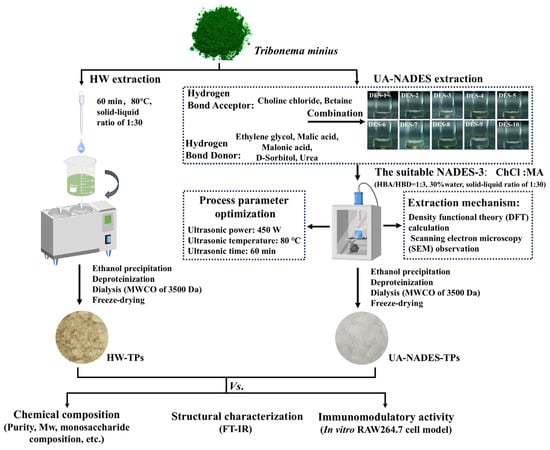
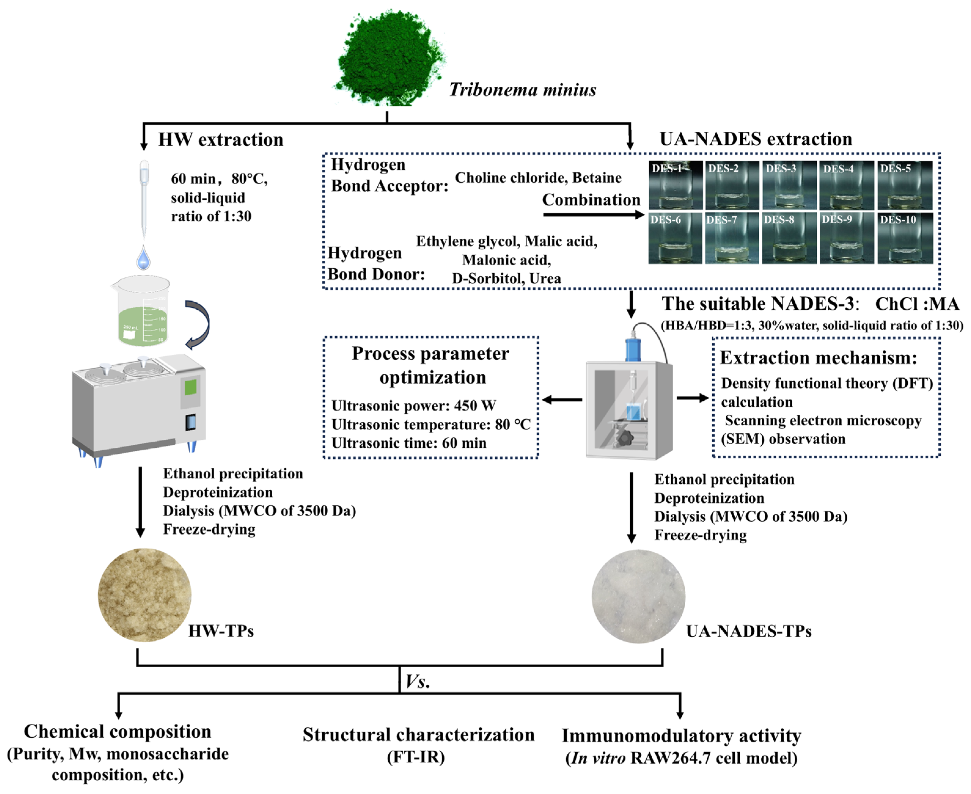
Accordingly, the present study aimed to (1) establish a UA-NADES extraction strategy for polysaccharides from Tribonema minus and elucidate the underlying extraction mechanism, with the overall workflow illustrated in Figure 1; (2) compare the chemical composition, physicochemical properties, and structural characteristics of polysaccharides obtained by different extraction methods; and (3) evaluate their immunomodulatory activities using the RAW264.7 macrophage model. This work provides mechanistic insights into UA-NADES-mediated polysaccharide extraction and advances the understanding of how extraction methods influence polysaccharide characteristics and immunomodulatory responses, supporting the sustainable valorization of Tribonema-derived polysaccharides.
Figure 1.
Schematic illustration of the extraction procedures for polysaccharides from Tribonema minus using hot-water (HW) and ultrasound-assisted natural deep eutectic solvent (UA-NADES) methods. ChCl, choline chloride; MA, malonic acid; Mw, molecular weight; MWCO, molecular weight cut off; HW-TPs, hot water-extracted Tribonema polysaccharides; UA-NADES-TPs, ultrasound-assisted natural deep eutectic solvent-extracted Tribonema polysaccharides.
2. Results and Discussion
2.1. Effects of Different NADESs on Polysaccharide Extraction Yield
To identify a suitable NADES for polysaccharide extraction from T. minus, ten NADESs were prepared using ChCl (choline chloride) or betaine as hydrogen bond acceptors (HBAs) and ethylene glycol, malic acid, MA (malonic acid), D-sorbitol, or urea as hydrogen bond donors (HBDs). As shown in Table 1, the ChCl-based NADESs generally produced significantly higher polysaccharide yields than HW extraction (77.32 mg/g), with yields ranging from 79.32 to 112.51 mg/g. In contrast, most betaine-based NADESs (NADES-6, NADES-7, and NADES-10) showed lower yields than HW extraction, whereas NADES-8 and NADES-9 resulted in only modest increases of 12.29% and 5.16%, respectively. These results indicate that NADES extraction performance was strongly dependent on the HBA–HBD combination, with ChCl-based systems generally outperforming betaine-based systems, consistent with previous reports [22].
Table 1.
Formulations, physicochemical properties, and polysaccharide extraction yields of the ten NADESs. NADES, natural deep eutectic solvent; HBA, hydrogen bond acceptor; HBD, hydrogen bond donor; ChCl, choline chloride; MA, malonic acid. pH values below 0.01 indicate that the acidity of the corresponding NADES systems exceeded the detection range of the pH meter. Different superscript letters within the same column indicate significant differences among treatment groups (p < 0.05).
The higher extraction efficiency of ChCl-based NADESs may be associated with their favorable hydrogen-bonding characteristics and lower viscosities. The chloride anion (Cl−) in ChCl exhibits strong HBA ability and can interact with hydroxyl groups within the cell-wall matrix, facilitating disruption of the cell-wall structure. In contrast, the zwitterionic structure of betaine, containing both quaternary ammonium cations and carboxylate groups (COO−), may result in different intermolecular interactions and be less effective in disrupting hydrogen-bond networks within cell-wall components [23]. In addition, the ChCl-based NADESs generally exhibited lower viscosities than the corresponding betaine-based systems, particularly when MA, malic acid, or D-sorbitol was used as the HBD (Table 1). Lower viscosity can favor solvent penetration and mass transfer, whereas excessive viscosity may restrict diffusion and consequently limit polysaccharide extraction [24].
Among the ChCl-based NADESs, the HBD type also significantly affected the extraction yield. The ChCl–MA system (NADES-3) gave the highest yield (112.51 mg/g), representing a 45.51% increase over HW extraction, followed by the D-sorbitol- (NADES-4) and malic acid-based (NADES-2) systems, with increases of 20.82% and 12.48%, respectively. In contrast, the ethylene glycol- and urea-based systems showed only slight improvements, with no significant difference between them (p > 0.05). The superior performance of NADES-3 may be attributed to the combined effects of strong hydrogen-bonding interactions, high acidity (pH < 0.01), and relatively low viscosity (96.00 mPa·s). As a dicarboxylic acid, MA can form extensive hydrogen-bonding interactions with ChCl and hydroxyl groups within the cell-wall matrix, thereby weakening the hydrogen-bond network and facilitating polysaccharide release. Furthermore, MA endowed the ChCl-based NADESs system with the lowest pH among the tested systems. The resulting acidic environment may facilitate disruption of the cell-wall structure, thereby increasing the accessibility of intracellular polysaccharides to the NADES and enhancing extraction efficiency [19]. This may explain why ChCl–D-sorbitol, despite its abundant hydrogen-bonding sites, showed a lower extraction yield, given its much weaker acidity (pH = 6.57). By contrast, the lower performance of ChCl–malic acid, which exhibited comparable acidity and hydrogen-bonding capability, may be primarily associated with its substantially higher viscosity (239.00 vs. 96.00 mPa·s), resulting in greater mass-transfer resistance.
Overall, NADES-3 exhibited the highest polysaccharide extraction yield among the tested NADES systems, likely due to the favorable balance of hydrogen-bonding interactions, acidity, and viscosity. Therefore, NADES-3 was selected for subsequent process optimization and mechanistic investigation.
2.2. Optimization of Ultrasonic-Assisted NADES Extraction by Single-Factor Experiments
Based on the screening results, the ChCl–MA system (NADES-3) was selected as the extraction solvent, and ultrasound assistance was subsequently introduced to further improve polysaccharide yield. Single-factor experiments were conducted to evaluate the effects of ultrasonic power, temperature, treatment time, HBA/HBD molar ratio, water content, and solid-to-liquid ratio on polysaccharide extraction yield.
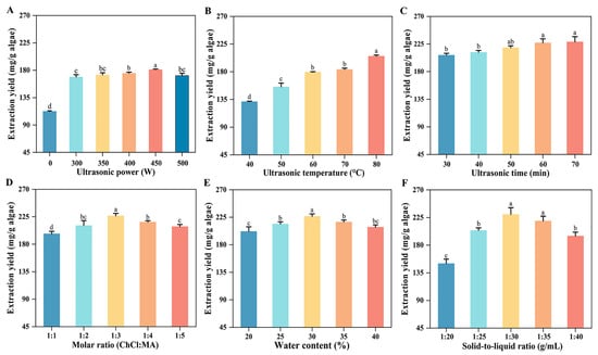
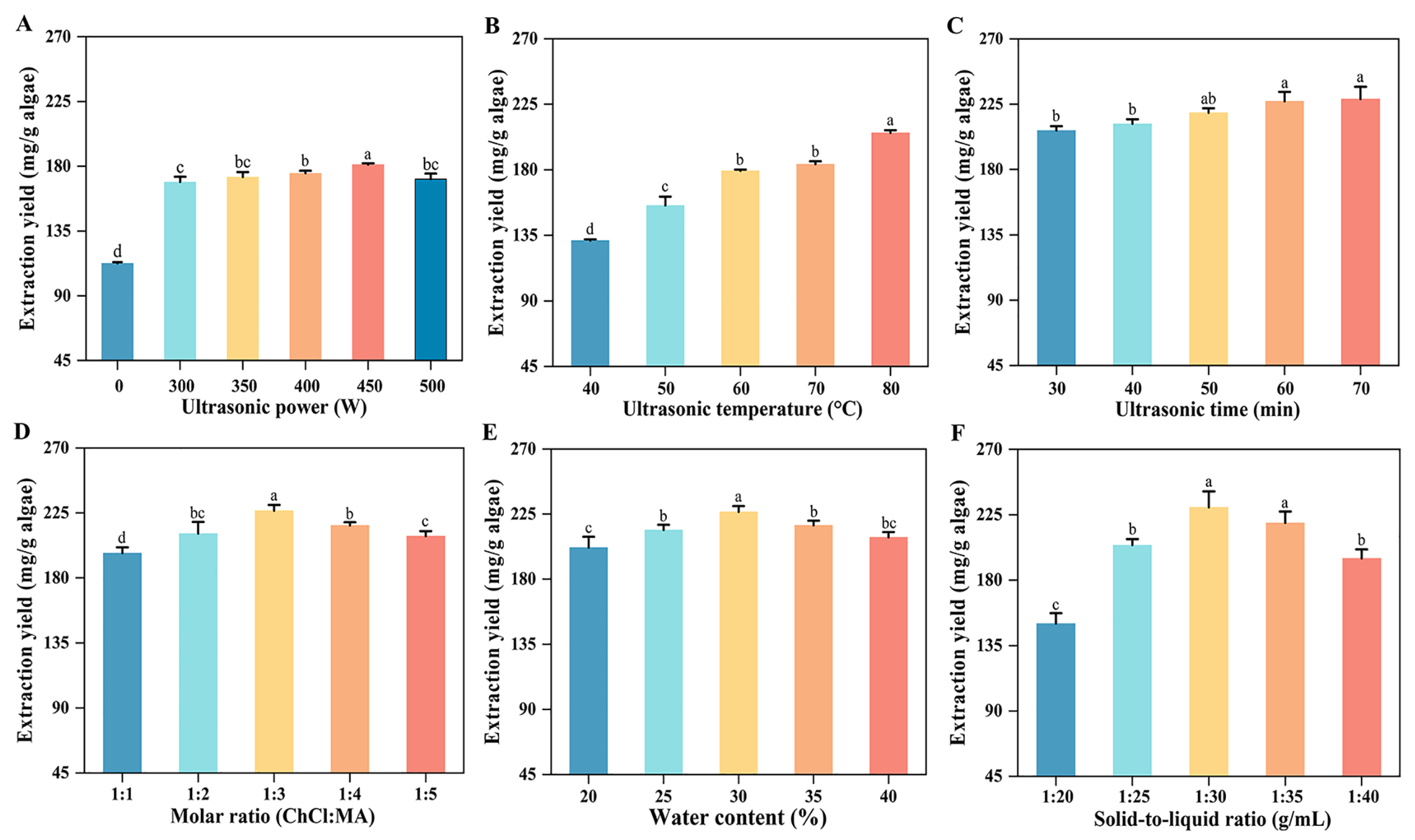
As shown in Figure 2A, ultrasound-assisted NADES-3 extraction produced significantly higher polysaccharide yields than NADES-3 extraction alone (p < 0.05), demonstrating the positive contribution of ultrasound to extraction efficiency. As the ultrasonic power increased from 300 to 450 W, the yield increased by 7.14% and reached a maximum of 181.05 mg/g at 450 W. This increase was mainly attributed to the enhanced cavitation effect, which promotes cell-wall disruption, solvent penetration, and mass transfer [25], consistent with the SEM observations of algal residues after extraction (as described below). However, a further increase to 500 W slightly reduced the yield by 5.51%. This trend is consistent with previous reports showing that polysaccharide yield exhibits a similar response pattern to ultrasonic power [13]. The decline at higher power may be due to excessive cavitation, which generates abundant bubbles and causes acoustic shielding, thereby reducing energy transfer efficiency and potentially promoting polysaccharide degradation [26]. Therefore, 450 W was selected as the optimal ultrasonic power for subsequent experiments.
Figure 2.
Effects of ultrasonic power (A), ultrasonic temperature (B), ultrasonic time (C), ChCl/MA (choline chloride/malonic acid) molar ratio (D), water content (E), and solid-to-liquid ratio (F) on polysaccharide yield from Tribonema minus. ChCl, choline chloride; MA, malonic acid. Different lowercase letters above the bars indicate significant differences among treatments (p < 0.05).
Increasing the ultrasonic temperature from 40 to 80 °C increased the polysaccharide yield by 56.32%, reaching 205.37 mg/g at 80 °C (Figure 2B). This trend can be explained by the fact that elevated temperature can reduce NADES viscosity and enhance molecular diffusion and mass transfer, thereby facilitating polysaccharide extraction [24,25]. Regarding ultrasonic time, the yield increased progressively from 30 to 60 min, and reached 227.00 mg/g at 60 min (Figure 2C), whereas extending the treatment time to 70 min produced no significant further increase (p > 0.05). Thus, the ultrasonic-assisted conditions were set at 450 W, 80 °C, and 60 min.
The composition and solvation environment of NADES-3 was subsequently optimized. The polysaccharide yield increased as the proportion of MA increased and reached a maximum of 227.20 mg/g at a ChCl/MA molar ratio of 1:3, followed by a decline at higher HBD proportions (Figure 2D). This response may reflect changes in the hydrogen-bonding environment, polarity, and viscosity of the NADES, which collectively influence polysaccharide solubilization and mass transfer [26]. Water content showed a similar response, with the highest yield (226.50 mg/g) obtained at 30% (Figure 2E), whereas further increase beyond 30% resulted in a significant decline (p > 0.05). An appropriate amount of water can partially weaken ionic and hydrogen-bonding interactions within the NADES, thereby reducing viscosity and facilitating diffusion. However, excessive water may disrupt the HBA–HBD interaction network and consequently weaken the solvation capacity of NADES for polysaccharides [27].
The solid-to-liquid ratio also significantly influenced extraction efficiency (Figure 2F). The yield increased as the ratio changed from 1:20 to 1:30 (g/mL), reaching 230.05 mg/g at 1:30, but decreased by 4.65% and 15.28% at 1:35 and 1:40, respectively. An adequate solvent volume favors contact between the biomass and extraction medium, whereas excessive solvent may reduce the effective ultrasonic energy density and provide little additional benefit to mass transfer [28]. Therefore, a solid-to-liquid ratio of 1:30 was selected.
Overall, the optimized UA-NADES conditions were 450 W, 80 °C, 60 min, a ChCl/MA molar ratio of 1:3, 30% water content, and a solid-to-liquid ratio of 1:30 (g/mL). Under these conditions, the resultant polysaccharide yield was 2.98-fold and 2.04-fold that obtained by HW extraction and NADES-3 extraction alone, respectively. These results demonstrate that the integration of ultrasound with NADES-3 significantly improved the polysaccharide extraction efficiency from T. minus.
2.3. Scale-Up and Reusability of the UA-NADES Extraction System
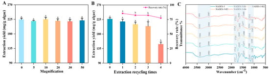
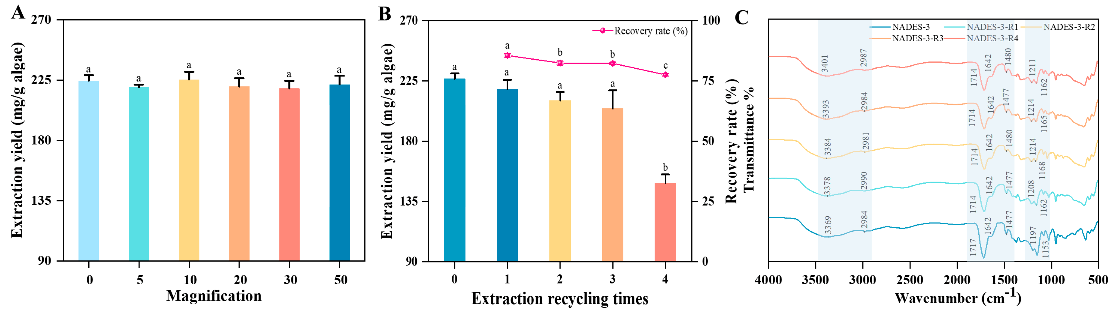
After establishing the UA-NADES extraction system, its scale-up stability and NADES reusability were evaluated to assess its practical applicability. Under a constant solid-to-liquid ratio of 1:30 g/mL, the extraction system was proportionally scaled up by 5, 10, 20, 30, and 50 times. As shown in Figure 3A, the polysaccharide yield remained relatively stable (216.31–225.47 mg/g) across all scale-up factors, with no significant differences among treatments (p > 0.05). These results indicate that the established UA-NADES system maintained consistent extraction performance during scale-up, demonstrating its potential for further process development. However, the UA-NADES extraction process established in this study was verified only at the laboratory scale and lacks the data support required for direct industrial scale-up, owing to the limited effective processing volume and rated power of the laboratory ultrasonic equipment, the polysaccharide yield decreased markedly upon further scale-up, and the applicability of this process at larger algal masses remains to be verified with engineered ultrasonic equipment in future work.
Figure 3.
Scale-up stability and reusability of the ultrasound-assisted natural deep eutectic solvent (UA-NADES) extraction system. (A) Effect of different scale-up factors on polysaccharide yield. (B) Polysaccharide extraction yields of NADES-3 over four consecutive reuse cycles. (C) FT–IR spectra of fresh and recycled NADES-3 after successive reuse cycles. Different lowercase letters above the bars indicate significant differences at p < 0.05.
The reusability of NADES-3 was subsequently investigated. The recovery rate remained above 82.33% during the first three recycling cycles but decreased to 77.57% in the fourth cycle, which is comparable to the recovery efficiencies that have been reported for other NADES systems used for polysaccharide extraction from Ganoderma lucidum [29]. As shown in Figure 3B, the polysaccharide yield gradually decreased during repeated cycles; however, no significant differences were observed among the first three cycles (p > 0.05), with yields maintained at 204.33–221.22 mg/g. FT–IR spectra further confirmed the stability of recycled NADES-3, as NADES-3-R1, NADES-3-R2, and NADES-3-R3 exhibited similar characteristic absorption bands to fresh NADES-3 (Figure 3C). The major absorption features associated with hydroxyl groups (3500–3200 cm−1), C–H stretching (3000–2800 cm−1), and the structural components of ChCl (1200–800 cm−1) and MA (1713 cm−1) [30] remained largely unchanged after three recycling cycles, indicating that the key functional groups of NADES-3 were preserved.
However, the extraction efficiency declined significantly after the fourth cycle, with the yield decreasing to 148.77 mg/g, corresponding to a 32.75% reduction compared with the first cycle. This decline may be attributed to changes in the NADES hydrogen-bonding network and the accumulation of non-target compounds during repeated use. Specifically, the O–H stretching band shifted from 3369 to 3401 cm−1 with increasing cycle number (Figure 3C), indicating alterations in the hydrogen-bonding environment or rearrangement of the NADES network [30]. In addition, the progressive darkening of recycled NADES-3 (Figure S1) suggested the accumulation of cell-derived impurities (e.g., pigments), which may interfere with the physicochemical properties of NADES and consequently reduce its extraction efficiency [23].
Overall, the UA-NADES extraction system exhibited good scale-up stability, and NADES-3 retained effective extraction performance for three consecutive cycles. These findings highlight the potential of UA-NADES as a sustainable strategy for polysaccharide extraction from T. minus.
2.4. Elucidation of the Extraction Mechanism
To elucidate the mechanism responsible for the enhanced polysaccharide extraction efficiency of the UA-NADES system, the microstructural changes in algal residues and intermolecular interactions were investigated using scanning electron microscopy (SEM) and density functional theory (DFT) calculations, respectively.
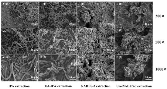
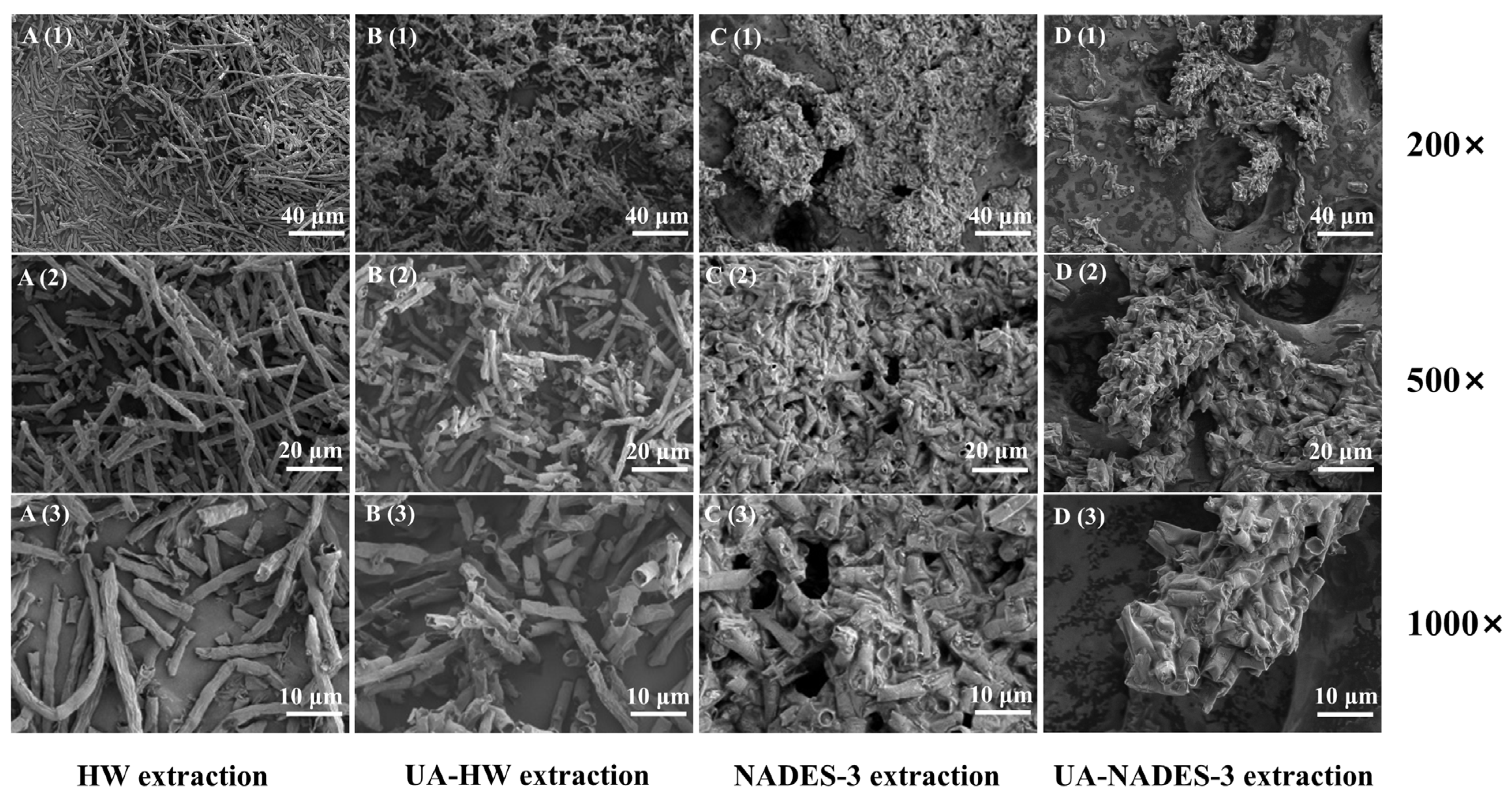
Figure 4 illustrates the morphological changes in algal residues after HW extraction, ultrasound-assisted HW extraction, NADES-3 extraction, and ultrasound-assisted NADES-3 extraction. As shown in Figure 4A(1–3), residues after HW extraction retained a relatively intact fibrous morphology with a smooth surface and limited structural disruption, indicating that HW extraction alone was insufficient to effectively overcome the robust cell-wall barrier. Compared with HW extraction, ultrasound-assisted HW extraction caused noticeable fiber breakage and surface roughening (Figure 4B(1–3)), suggesting that ultrasonic cavitation enhanced mechanical disruption of the algal matrix; however, the overall fibrous structure remained largely preserved.
Figure 4.
Scanning electron microscopy (SEM) images of algal residues after different treatments: hot water (HW) extraction (A(1)–A(3)), ultrasound-assisted hot water (UA-HW) extraction (B(1)–B(3)), NADES-3 (natural deep eutectic solvent, choline chloride–malonic acid) extraction (C(1)–C(3)), and ultrasound-assisted NADES-3 (UA-NADES-3) extraction (D(1)–D(3)). Images in rows 1, 2, and 3 were taken at magnifications of 200×, 500×, and 1000×, respectively.
NADES-3 extraction resulted in more pronounced morphological changes, including fiber aggregation, surface roughness, and the formation of irregular porous structures (Figure 4C(1–3)), indicating that NADES-mediated interactions promoted cell-wall disruption [31]. Notably, ultrasound-assisted NADES-3 extraction caused the most severe structural disruption, with the fibrous skeleton largely destroyed and a porous honeycomb-like morphology observed (Figure 4D(1–3)). These structural alterations likely increased solvent accessibility and contact area, thereby facilitating the release of intercellular polysaccharides. Similar synergistic disruption of plant cell-wall structures by ultrasound-assisted NADES extraction has also been reported during polysaccharide extraction from other biomass sources [19]. Collectively, the SEM results demonstrate that ultrasonic cavitation and NADES-mediated interactions exerted synergistic effects on disrupting the algal cell-wall structure, thereby contributing to the enhanced extraction efficiency of the UA-NADES system.
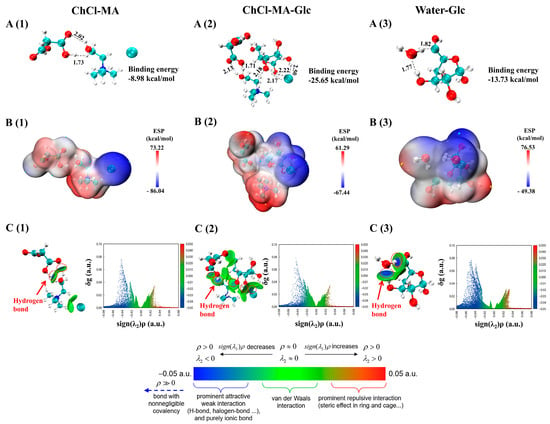
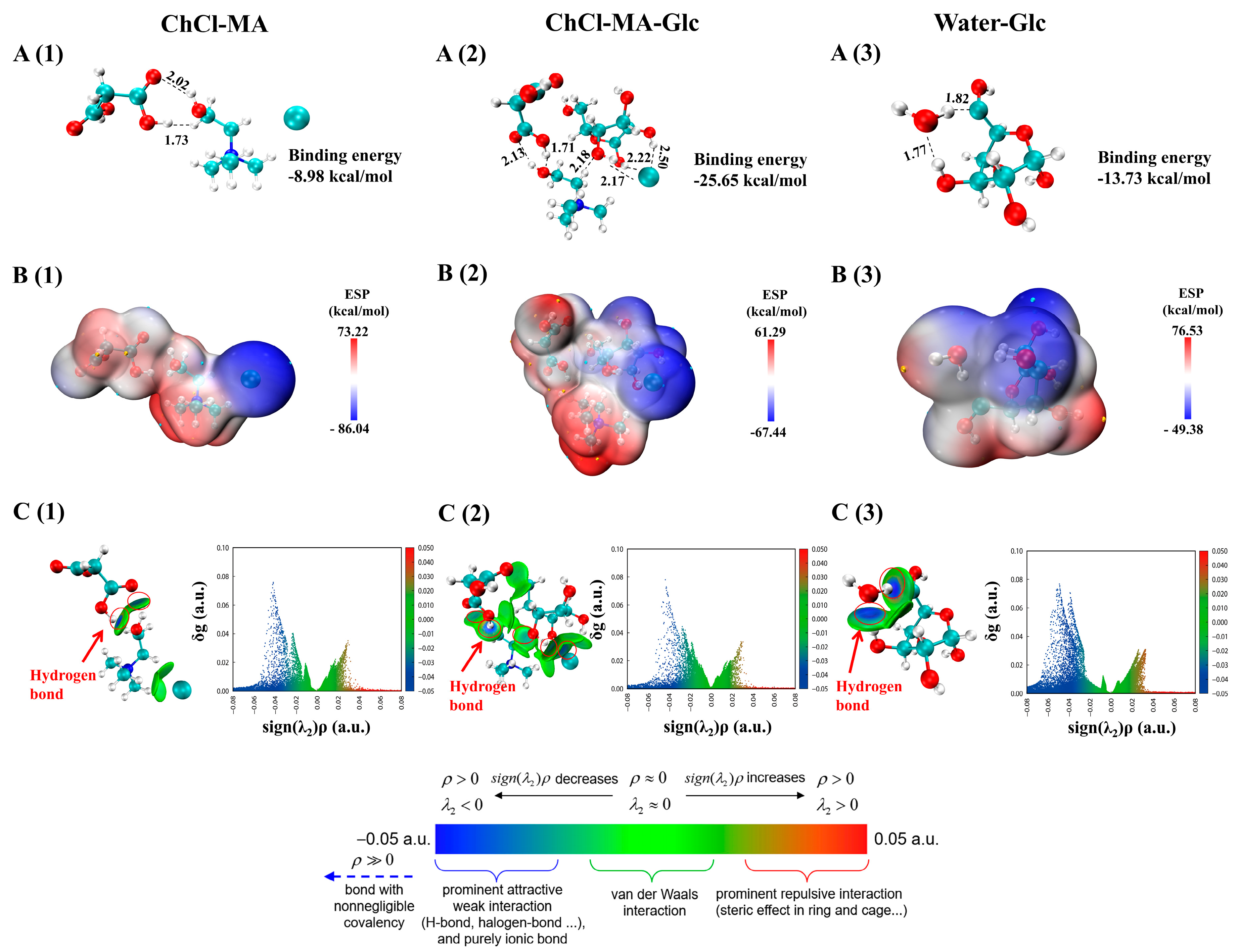
To further understand the superior extraction performance of NADES-3 at the molecular level, DFT calculations were performed to analyze the intermolecular interactions in the Water–Glc, ChCl–MA, and ChCl–MA–Glc systems (Figure 5). In the optimized ChCl–MA structure (Figure 5A(1)), two hydrogen bonds were identified between ChCl and MA, with bond lengths of 1.73 Å (O–H···O) and 2.02 Å (O···H–O), which fall within the typical range of hydrogen-bond interactions, indicating stable intermolecular association [31]. In the ChCl–MA–Glc complex (Figure 5A(2)), the original hydrogen bonds between ChCl and MA were maintained with slight changes in bond lengths (1.71 Å and 2.13 Å). Moreover, the introduction of Glc resulted in additional interactions between the Cl− anion of ChCl and hydroxyl groups of Glc, forming four hydrogen bonds with lengths of 2.17, 2.18, 2.22, and 2.50 Å. In contrast, only two intermolecular hydrogen bonds were observed in the Water–Glc system (Figure 5A(3)). The calculated interaction energies showed that the ChCl–MA–Glc complex exhibited a more negative binding energy (−25.65 kcal/mol) than ChCl–MA (−8.98 kcal/mol) and Water–Glc (−13.73 kcal/mol), suggesting stronger intermolecular interactions between NADES-3 and Glc than those formed in the water system. These interactions may contribute to enhanced polysaccharide solubilization and extraction efficiency.
Figure 5.
Non-covalent interaction analysis of ChCl–MA, ChCl–MA–Glc, and Water–Glc systems. (A) Optimized geometric structures and binding energies of ChCl–MA (A(1)), ChCl–MA–Glc (A(2)), and Water–Glc (A(3)). Hydrogen bonds are shown as dashed lines with bond lengths (Å). (B) Electrostatic potential (ESP) maps mapped on the total electron density surface (isovalue = 0.001 a.u.) for ChCl–MA (B(1)), ChCl–MA–Glc (B(2)), and Water–Glc (B(3)). (C) Gradient isosurfaces (inset) and corresponding sign(λ2)ρ scatter plots for ChCl–MA (C(1)), ChCl–MA–Glc (C(2)), and Water–Glc (C(3)). The color scale indicates the nature of interactions: blue for strong attractive interactions (e.g., hydrogen bonds), green for van der Waals interactions, and red for steric repulsion. ChCl, choline chloride; MA, malonic acid; Glc, glucose.
The ESP maps further revealed the charge distribution characteristics of these systems. The ChCl–MA system exhibited a broad ESP distribution range (−86.04 to 73.22 kcal/mol), reflecting heterogeneous charge distribution (Figure 5B(1)). After introducing Glc (Figure 5B(2)), the ESP distribution became more complementary, with positive regions mainly located around the quaternary ammonium group and hydroxyl hydrogens, while negative regions were concentrated around the hydroxyl oxygen atoms Glc and the Cl−. The altered ESP distribution suggests charge redistribution and enhanced electrostatic complementarity between NADES-3 and Glc. Notably, the ChCl–MA–Glc complex exhibited a narrower ESP range than the Water–Glc system (Figure 5B(3)) [32], further supporting stronger electrostatic interactions between NADES-3 and Glc.
The independent gradient model based on Hirshfeld partition (IGMH) analysis was further applied to visualize weak intermolecular interactions (Figure 5C). In the IGMH isosurfaces, blue regions represent strong attractive interactions (e.g., hydrogen bonds), green regions represent van der Waals (vdW) forces, and red regions correspond to steric repulsion [33]. The water–Glc system mainly exhibited hydrogen-bond interactions (Figure 5C(3)). In contrast, the ChCl–MA system showed abundant hydrogen-bonding regions accompanied by vdW interactions, whereas the ChCl–MA system showed abundant hydrogen-bonding regions accompanied by vdW interactions (Figure 5C(1)). After Glc incorporation, the ChCl–MA–Glc complex displayed more extensive hydrogen-bonding regions involving MA, ChCl, and Cl−, together with enhanced vdW interactions (Figure 5C(2)). Compared with ChCl–MA, the scatter plot of the ChCl–MA–Glc complex showed broader and more dispersed hydrogen-bonding regions with higher signal intensity and a larger vdW interaction area, indicating enhanced intermolecular interactions after Glc incorporation. These findings were consistent with the hydrogen-bond analysis and ESP results.
In summary, the enhanced extraction efficiency of the UA-NADES system resulted from the synergistic contribution of physical disruption and molecular interactions. Ultrasound-induced cavitation and NADES-3-mediated hydrogen-bond interactions worked in concert to disrupt the robust cell-wall matrix and improve solvent accessibility, while NADES-3 facilitated polysaccharide solubilization through multi-site hydrogen bonding, electrostatic interactions, and van der Waals forces with Glc units. These findings provide morphological and molecular-level insights into the synergistic mechanism underlying the enhanced extraction of T. minus polysaccharides by the UA-NADES system.
2.5. Comparison of Physicochemical Properties and Structural Features of Polysaccharides Extracted by Different Methods
In general, extraction methods can affect the chemical composition, physicochemical properties, and structural characteristics of polysaccharides, thereby influencing their biological functions [34]. Therefore, HW-TPs and UA-NADES-TPs were comparatively analyzed in terms of chemical composition, monosaccharide composition, Mw distribution, morphology, and spectroscopic characteristics.
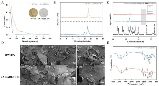
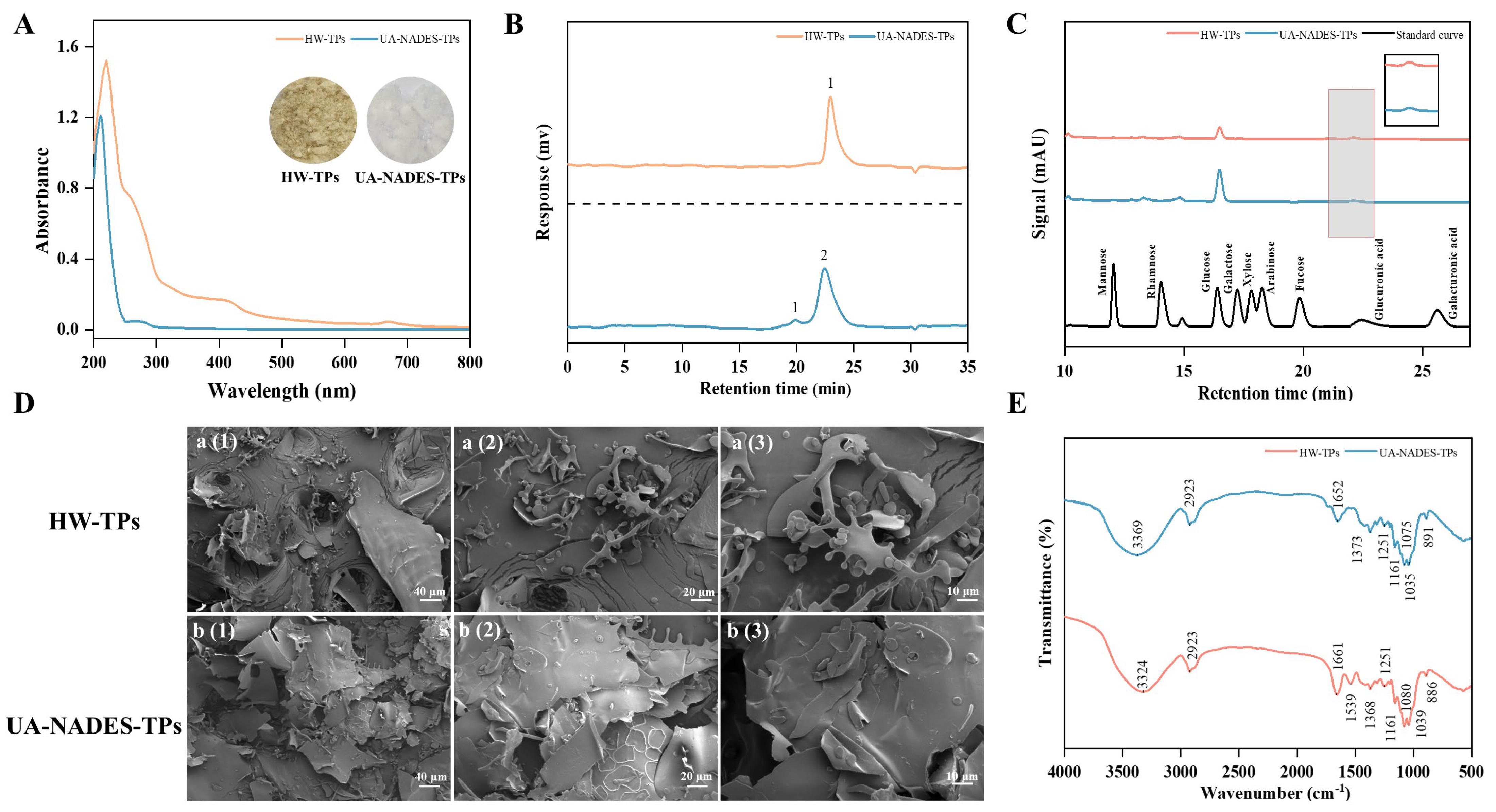
As shown in Table 2, UA-NADES-TPs exhibited a higher total sugar content (65.98%) and a lower protein content (7.36%) than HW-TPs, indicating enhanced polysaccharide enrichment and reduced protein co-extraction during UA-NADES extraction. This selective extraction capability may be associated with hydrogen-bonding interactions between NADES components and polysaccharide hydroxyl groups, which facilitate polysaccharide solubilization while limiting the co-extraction of non-target components [21]. Consistently, UA-NADES-TPs showed a lighter appearance and a weaker absorption peak at 280 nm compared with HW-TPs (Figure 6A), further confirming the lower residual pigment and protein content.
Table 2.
Chemical constituents, monosaccharide composition, and molecular weight characteristics of polysaccharides from Tribonema minus obtained by different extraction methods. HW-TPs and UA-NADES-TPs denote the polysaccharides obtained by hot water extraction and ultrasound-assisted NADES-3 extraction, respectively. HW-TPs, hot water-extracted Tribonema polysaccharides; UA-NADES-TPs, ultrasound-assisted natural deep eutectic solvent-extracted Tribonema polysaccharides; NADES-3, natural deep eutectic solvent-3 (choline chloride–malonic acid); Mn, number-average molecular weight; Mw, weight-average molecular weight.
Figure 6.
Physicochemical and structural characterization of polysaccharides from Tribonema minus extracted by hot water (HW-TPs) and ultrasound-assisted natural deep eutectic solvent (UA-NADES-TPs). (A) UV–visible absorption spectra of the polysaccharide samples (inset: visual appearance of lyophilized powders); (B) high-performance liquid chromatography (HPLC) chromatograms of monosaccharide composition; (C) molecular weight distribution profiles determined by gel permeation chromatography (GPC). (D) Scanning electron microscopy (SEM) images of HW-TPs (a(1)–a(3)) and UA-NADES-TPs (b(1)–b(3)) at magnifications of 200×, 500×, and 1000×, respectively. (E) Fourier-transform infrared spectroscopy (FT–IR) spectra of HW-TPs and UA-NADES-TPs. HW-TPs, hot water-extracted Tribonema polysaccharides; UA-NADES-TPs, ultrasound-assisted natural deep eutectic solvent-extracted Tribonema polysaccharides.
The monosaccharide composition analysis revealed that both polysaccharide samples were predominantly composed of Glc (Table 2 and Figure 6B). UA-NADES-TPs exhibited a higher Glc proportion (98.60%) than HW-TPs (94.92%), suggesting that both extracts were mainly glucan-type polysaccharides, consistent with previous reports on Tribonema-derived polysaccharides [35]. Meanwhile, small amounts of glucuronic acid (GlcA) were detected in both samples, with UA-NADES-TPs and HW-TPs containing 1.40% and 5.08% GlcA, respectively, in agreement with their corresponding uronic acid contents.
GPC analysis (Figure 6C) revealed distinct Mw distributions between HW-TPs and UA-NADES-TPs. HW-TPs exhibited a single symmetrical peak with an average Mw of 3.95 kDa, indicating a relatively homogeneous polysaccharide population. In contrast, UA-NADES-TPs displayed a bimodal distribution, consisting of a predominant fraction with an average Mw of 5.54 kDa (95.33%) and a minor high-Mw fraction with an average Mw of 33.31 kDa (4.67%).
Notably, the Mw of HW-TPs determined in this study was lower than that previously reported for HW-extracted Tribonema polysaccharides [35]. This discrepancy may partly arise from differences in GPC conditions, as the relative Mw determined by conventional GPC is dependent on the calibration standards and chromatographic conditions, future work should validate the absolute Mw using multi-angle light scattering (MALS). Nevertheless, under the same analytical conditions, the predominant fraction of UA-NADES-TPs exhibited a higher Mw than that of HW-TPs. This difference suggests that the NADES-3 extraction system provides a relatively mild extraction environment that better preserves the major polysaccharide fraction compared with conventional HW extraction. The prolonged thermal exposure during HW extraction may promote partial hydrolysis of glycosidic bonds, resulting in Mw reduction [36]. In contrast, NADES-mediated hydrogen-bonding interactions may offer a protective solvation environment that mitigates polysaccharide degradation during extraction. Similar protective effects of NADES systems under harsh extraction conditions have also been reported [19]. In addition, the minor high-Mw fraction detected in UA-NADES-TPs may be associated with enhanced disruption of the algal cell-wall matrix by ultrasound-assisted NADES extraction, which could facilitate the release of tightly associated polysaccharide components that are less accessible during HW extraction. A limitation of this study is that only the HW and UA-NADES extracts were fully characterized, while the ultrasound-assisted HW and NADES-only extracts served as intermediate controls and their molecular weights were not measured. Therefore, the contribution of cavitation alone could not be separated from that of the solvent, and the observed differences should be attributed to the combined effect.
The microstructural morphology of the polysaccharide samples was further examined by SEM. As shown in Figure 6D, HW-TPs exhibited a dense, compact block-like or sheet-like structure with a relatively smooth surface, whereas UA-NADES-TPs displayed a looser, porous, and fragmented structure with layered aggregation. These differences indicate that the extraction conditions influenced the aggregation behavior and surface morphology of the recovered polysaccharides. The more porous morphology of UA-NADES-TPs may contribute to improved interactions with aqueous environments and altered physicochemical properties.
The FT–IR spectra of both polysaccharides exhibited typical carbohydrate-related absorption bands. The broad band around 3369 cm−1 was attributed to O–H stretching vibrations, while the peak near 2923 cm−1 corresponded to C–H stretching vibrations. The absorption bands at approximately 1652 cm−1 were associated with O–H bending/carboxylate-related vibrations [32]. Both samples showed similar absorption patterns in the 800–1200 cm−1 region, with the characteristic band around 890 cm−1 indicating the presence of β-glycosidic linkages [35,37]. These results suggest that UA-NADES extraction did not cause detectable changes in the basic glycosidic configuration of the polysaccharides. Furthermore, the weaker absorption band around 1539 cm−1 in UA-NADES-TPs indicated lower residual protein content [38], consistent with UV–visible analysis and chemical composition determination.
Overall, both extraction methods preserved the basic glucan structure, while differences were observed in polysaccharide enrichment, Mw distribution, and surface morphology. These structural and compositional variations may account for the differential immunomodulatory responses observed for HW-TPs and UA-NADES-TPs. However, further purification and structural elucidation are required to clarify the specific structural features responsible for their differential biological activities.
2.6. Comparison of Immunomodulatory Activities of Polysaccharides Extracted by Different Methods
Previous studies have demonstrated that Tribonema-derived polysaccharides possess immunomodulatory potential [8,39]. In this study, RAW264.7 macrophages were used as an in vitro model to compare the immunomodulatory activities of UA-NADES-TPs and HW-TPs. As shown in Supplementary Figure S2, neither polysaccharide exhibited cytotoxic effects within the tested concentration range (1.25–500 μg/mL). Moreover, cell viability was slightly increased after treatment with 12.5–200 μg/mL polysaccharides. Therefore, concentrations of 50, 100, and 200 μg/mL were selected for subsequent experiments.
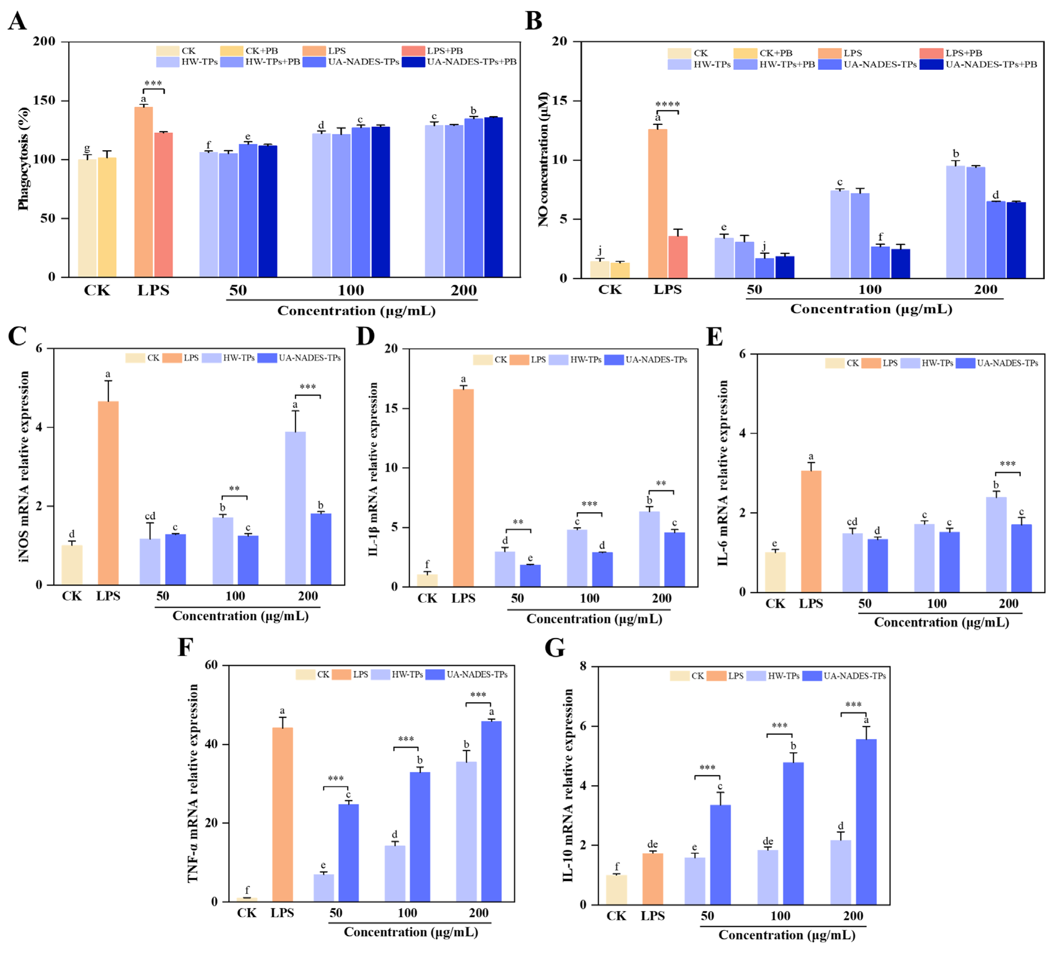
Macrophage phagocytosis is an important component of innate immune defense [40]. As shown in Figure 7A, both polysaccharide samples significantly enhanced phagocytic activity in a dose-dependent manner compared with the control group (p < 0.05). At 200 μg/mL, UA-NADES-TPs and HW-TPs increased phagocytic activity to 134.74% and 128.85%, respectively. Notably, UA-NADES-TPs induced significantly higher phagocytic activity than HW-TPs at the same concentration, suggesting a stronger capacity to enhance macrophage phagocytic function.
Figure 7.
Immunomodulatory effects of HW-TPs and UA-NADES-TPs on RAW264.7 macrophages. (A) Phagocytic activity determined by neutral red uptake assay. (B) NO production. (C–G) Relative mRNA expression levels of iNOS (C), IL-6 (D), IL-1β (E), TNF-α (F), and IL-10 (G) determined by qRT-PCR. PB, polymyxin B (30 μg/mL); LPS (1 μg/mL) served as the positive control. Different lowercase letters above the bars indicate significant differences among treatment groups (p < 0.05). Asterisks indicate significant differences between polysaccharide-treated groups with and without PB or between different polysaccharide treatments at the same concentration (** p < 0.01, *** p < 0.001, **** p < 0.0001). HW-TPs, hot water-extracted Tribonema polysaccharides; UA-NADES-TPs, ultrasound-assisted natural deep eutectic solvent-extracted Tribonema polysaccharides.
NO is a major effector molecule produced by macrophages and serves as an important marker of macrophage activation [41]. As shown in Figure 7B, treatment with the polysaccharide samples significantly increased NO production in a dose-dependent manner. At 200 μg/mL, NO concentrations reached 6.50 μM and 9.50 μM in the UA-NADES-TPs and HW-TPs groups, respectively. However, HW-TPs induced a stronger NO response than UA-NADES-TPs. Consistently, the mRNA expression level of iNOS showed a similar trend, with HW-TPs inducing a 114.36% higher iNOS transcriptional response than UA-NADES-TPs at 200 μg/mL.
To exclude possible interference from residual endotoxins, PB was included in the assay. PB markedly reduced the immune activation induced by LPS but did not significantly affect the phagocytic activity or NO production stimulated by the polysaccharide samples (p > 0.05, Figure 7A,B). These results suggest that the observed macrophage responses were mainly attributed to the polysaccharides themselves rather than endotoxin contamination.
The mRNA expression levels of immune-related cytokines, including TNF-α, IL-1β, IL-6, and IL-10, were further analyzed by qPCR (Figure 7C–F). Both polysaccharide samples significantly upregulated the expression of these immune-related cytokine genes compared with untreated cells, indicating their potential to activate macrophage immune responses [42]. However, distinct transcriptional response patterns were observed between UA-NADES-TPs and HW-TPs. Specifically, HW-TPs induced markedly higher mRNA expression levels of IL-1β and IL-6 than UA-NADES-TPs, consistent with their stronger NO-inducing activity. At 200 μg/mL, the expression levels of IL-1β and IL-6 induced by HW-TPs were 1.39-fold and 1.41-fold of those induced by UA-NADES-TPs, respectively, suggesting a stronger pro-inflammatory transcriptional response.
In contrast, UA-NADES-TPs induced higher TNF-α and IL-10 mRNA expression levels than HW-TPs at the same concentration. TNF-α is involved in macrophage activation and host defense, whereas IL-10 is an important immunoregulatory cytokine associated with the maintenance of inflammatory homeostasis [43,44]. The concurrent upregulation of TNF-α and IL-10 may therefore reflect a more balanced macrophage response, involving both immune activation and immunoregulatory signaling.
Collectively, the extraction method influenced the immunomodulatory responses of T. minus polysaccharides. HW-TPs exhibited a stronger pro-inflammatory activation pattern, characterized by elevated iNOS, IL-1β, and IL-6 transcription together with increased NO production. This response is consistent with a classical pro-inflammatory macrophage phenotype, in which activation of inflammatory signaling pathways, including TLR4/NF-κB, is commonly associated with pro-inflammatory cytokine expression and iNOS-mediated NO production [45]. In comparison, UA-NADES-TPs enhanced phagocytic activity while inducing higher TNF-α and IL-10 mRNA expression, indicating a distinct and relatively balanced immunomodulatory profile.
The different biological responses may be associated with differences in polysaccharide purity, Mw distribution, and structural characteristics resulting from different extraction processes. However, the samples evaluated in this study were crude polysaccharide preparations. Further purification combined with advanced structural elucidation techniques, such as NMR spectroscopy, will be required to clarify the specific structural features and molecular mechanisms underlying their differential immunomodulatory effects.
3. Materials and Methods
3.1. Materials and Reagents
The filamentous microalga T. minus used in this study was the same strain as that employed in our previous studies [4]. It was cultivated in a modified Blue Green (mBG-11) medium under continuous illumination at 150 ± 10 μmol photons/m2/s, with aeration using compressed air containing 1%CO2 (v/v). After 12 days of cultivation, the biomass was harvested. On a dry-weight basis, the biomass contained 33.33% lipids, 20.69% proteins, and 28.55% carbohydrates.
The mouse macrophage RAW264.7 cell line was purchased from Shanghai Jinyuan Biotechnology Co., Ltd. (Shanghai, China). Dulbecco’s Modified Eagle Medium (DMEM) was obtained from Cytiva Biotechnology Co., Ltd. (Hangzhou, Zhejiang, China), and fetal bovine serum (FBS) was obtained from CELL-BOX Biotechnology Co., Ltd. (Changsha, Hunan, China). Monosaccharide standards, 1-phenyl-3-methyl-5-pyrazolone (PMP), choline chloride (ChCl), betaine, malic acid, D-sorbitol, and trifluoroacetic acid (TFA) were purchased from Aladdin Industrial Co., Ltd. (Shanghai, China). Malonic acid (MA), ethylene glycol, and urea, were obtained from Sinopharm Chemical Reagent Co., Ltd. (Shanghai, China). The bicinchoninic acid (BCA) protein assay kit and nitric oxide (NO) assay kit were obtained from Beyotime Biotechnology Co., Ltd. (Shanghai, China). Unless otherwise specified, all other chemicals and reagents were of analytical grade.
3.2. Preparation of NADESs
The NADESs were prepared using a previously reported heating method with slight modifications [32]. ChCl and betaine were used as HBAs, while ethylene glycol, MA, D-sorbitol, malic acid and urea were used as HBDs. Briefly, each HBA was mixed with the corresponding HBD at a predetermined molar ratio and continuously stirred in a water bath at 80 °C for 1 h until a clear and homogeneous liquid phase was obtained. The water content was then adjusted to 30% (w/w), resulting in 10 NADESs formulations with different HBA/HBD combinations (Table 1). After cooling to room temperature, the NADESs were sealed and stored for further use. The pH and viscosity of the prepared NADESs were measured using a pH meter (pH700, APERA, Shanghai, China) and a viscometer (NPJ-9S, Shanghai, China), respectively, and the results are summarized in Table 1.
3.3. Screening of Suitable NADESs for Polysaccharide Extraction from T. minus
Freeze-dried algal powder (200.00 mg) was mixed with each NADES at a solid-to-liquid ratio of 1:30 (g/mL), with deionized water used as the control solvent. The mixtures were extracted in a water bath at 60 °C for 30 min under continuous stirring. After extraction, the suspensions were centrifuged at 8000 rpm for 10 min, and the resulting supernatants were collected and adjusted to a constant volume for subsequent analysis. The polysaccharide concentration (C, mg/mL) in the supernatants was determined using the phenol–sulfuric acid method with glucose (Glc) as the standard [18]. The calibration curve was established as Y = 6.4664x + 0.0594 (R2 = 0.9995). The polysaccharide extraction yield was calculated using the following equation:
where C is the polysaccharide concentration in the extract (mg/mL), V is the final volume of the extract (mL), D is the dilution factor, and M is the mass of algal powder used for extraction (g). Based on the extraction yields, the NADES showing the highest extraction efficiency was selected for subsequent experiments.
Extraction yield (mg/g algae) = (C × V × D)/M,
3.4. Establishment of Ultrasound-Assisted NADES Extraction System for Polysaccharides from T. minus
3.4.1. Single-Factor Experiments
Based on the NADES screening results, NADES-3 (ChCl–MA) was selected as the extraction solvent. Ultrasonic treatment was subsequently introduced to investigate whether the induced cavitation effect could further enhance polysaccharide extraction. Briefly, freeze-dried algal powder (200 mg) was suspended in 6.0 mL of NADES-3 in a centrifuge tube and treated using an ultrasonic processor (SCIENTZ-IID, Ningbo, China) at a fixed frequency of 22 kHz. The effects of ultrasonic powers (300–500 W), temperatures (40–80 °C), and treatment time (30–70 min) were first investigated, followed by the evaluation of NADES-3 HBA/HBD molar ratio (1:1–1:5), water content (20–40%, w/w), and solid-to-liquid ratio (1:20–1:40, g/mL) under the selected ultrasonic conditions. In each experiment, one parameter was varied while the others were kept constant. All experiments were performed in triplicate. After extraction, the supernatants were collected by centrifugation (8000 rpm, 10 min), and the polysaccharide concentration and extraction yield were determined as described in Section 3.3. The optimal conditions were selected based on the highest polysaccharide extraction yield.
3.4.2. Scale-Up and Recycling Experiments
To assess the scalability and reusability of the UA-NADES extraction process, scale-up and NADES recycling experiments were performed. For the scale-up experiments, the extraction system was proportionally increased by 5-, 10-, 20-, 30-, and 50-fold while maintaining a solid-to-liquid ratio of 1:30 (g/mL) and otherwise identical extraction conditions. The maximum scale-up factor was limited to 50-fold by the capacity of the ultrasonic equipment.
The reusability of NADES-3 was evaluated according to a previously reported method [30]. Briefly, under the optimized extraction conditions, polysaccharides were precipitated from the first-cycle extraction supernatant by adding ethanol. After polysaccharide precipitation, the residual ethanol and water were removed by rotary evaporation under reduced pressure. The recovered NADES-3 was then adjusted to a water content of 30% (w/w) and used for the next extraction cycle, designated as NADES-3-R1. The same procedure was repeated for three additional cycles, and the recovered NADES from the second, third, and fourth cycles were designated NADES-3-R2, NADES-3-R3, and NADES-3-R4, respectively. Each recycling experiment was performed in triplicate. The extraction performance of each recycled NADES was evaluated based on the polysaccharide concentration and extraction yield determined as described in Section 3.3.
The NADES recovery rate was calculated using the following formula:
where M0 is the initial mass of NADES-3 before extraction and M1 is the mass of recovered NADES-3 after extraction and rehydration.
Recovery rate (%) = (M0/M1) × 100%,
3.4.3. Density Functional Theory Analysis
Density functional theory (DFT) calculations were performed to investigate the molecular interactions underlying NADES-mediated polysaccharide extraction [30]. Because Glc was the predominant monosaccharide in the extracted polysaccharides from T. minus (as described below), it was selected as a representative structural unit for the quantum chemical calculations. The molecular structures of ChCl, MA, Glc, water, and their complexes, were fully optimized using Gaussian 16 at the B3LYP/6-31++G(d, p) level, incorporating Grimme’s D3 dispersion correction with Becke–Johnson damping (D3 (BJ)). Solvent effects were accounted for using the polarizable continuum model (PCM), with water as the solvent. Harmonic vibrational frequency calculations were performed at the same theoretical level to verify that the optimized structures corresponded to true local minima with no imaginary frequencies.
Molecular electrostatic potential (ESP) maps were generated to identify electron-rich and electron-deficient regions, as well as potential intermolecular recognition sites [46]. The independent gradient model based on Hirshfeld partition (IGMH) was applied to visualize and characterize the hydrogen-bonding, van der Waals, and steric interactions among the constituent components. Wavefunction analyses were performed using Multiwfn, and the resulting molecular surfaces were visualized using Visual Molecular Dynamics (VMD) [47,48].
The electronic interaction energy (ΔEint) was calculated from the energy difference between the complex and its constituent fragments according to the following equation:
where Ecomplex is the electronic energy of the complex and Ei is the electronic energy of each constituent fragment calculated at the same theoretical level. A negative ΔEint value indicates that association between the components is energetically favorable.
ΔEint = Ecomplex − Σi Ei,
3.5. Preparation and Characterization of Polysaccharides Extracted by Different Methods
Polysaccharides were extracted from T. minus using HW extraction and the optimized UA-NADES method, as illustrated in Figure 1. The resulting polysaccharide preparations were designated as HW-TPs and UA-NADES-TPs, respectively. After extraction, the mixtures from both methods were centrifuged at 8000 rpm for 10 min to obtain polysaccharide-containing supernatants, the algal residues were collected for subsequent scanning electron microscopy (SEM) analysis. The supernatants were diluted with a certain volume of deionized water and then subjected to ethanol precipitation, followed by deproteinization using the Sevag method and dialysis against deionized water for 4 days using a dialysis membrane with a molecular weight cutoff (MWCO) of 3500 Da. All procedures were performed according to previously reported methods [8,34]. The dialyzed samples were subsequently lyophilized, and the resulting polysaccharide preparations were used for comparative analyses of their chemical compositions, physicochemical properties, and structural characteristics.
3.5.1. Chemical Composition Determination
The total sugar, protein, and uronic acid contents of HW-TPs and UA-NADES-TPs were determined using the phenol–sulfuric acid method, a BCA protein assay kit, and the m-hydroxydiphenyl method, respectively, as described in previous reports [19]. The corresponding calibration curves were Y = 6.4664x + 0.0594 (R2 = 0.9995, linear range 0–0.15 mg/mL) for total sugars, Y = 0.9602x + 0.0655 (R2 = 0.9933, linear range 0–1.3 mg/mL) for proteins, and Y = 1.5769x + 0.0433 (R2 = 0.9917, linear range 0–0.3 mg/mL) for uronic acids. According to the ICH Q2 (R1) guidelines, the LOD and LOQ of the total sugar, protein, and uronic acid were determined as LOD = 3.3σ/S = 0.0016, 0.0167, 0.0061 µg/mL, and LOQ = 10σ/S = 0.0048, 0.0506, 0.0185 µg/mL, respectively. Before analysis, the polysaccharide samples were dissolved in deionized water at a concentration of 1.0 mg/mL.
3.5.2. Microstructure Observation by Scanning Electron Microscope
Algal residues and lyophilized polysaccharide samples obtained after extraction were mounted on sample stages using conductive adhesive and sputter-coated with a thin layer of gold. The surface morphology of samples was observed using a scanning electron microscope (SEM, SU8600, Hitachi, Ltd., Tokyo, Japan) at an accelerating voltage of 20.0 kV and magnifications of 200×, 500×, and 1000×.
3.5.3. Mw Distribution Analysis
The Mw distribution of HW-TPs and UA-NADES-TPs was determined by gel permeation chromatography (GPC) according to the method reported by Ma et al. [49]. Briefly, the polysaccharide samples (2 mg/mL) were filtered through a 0.45 µm membrane before injection. GPC analysis was performed using an Agilent 1260 system (Agilent Technologies, Santa Clara, CA, USA) equipped with a G1362A refractive index detector and a series of Waters Ultrahydrogel column (300 × 7.8 mm; 500, 250, and 120 Å). The mobile phase was an aqueous solution of 0.1 mol/L NaNO3 at a flow rate of 1.0 mL/min, and the column temperature was maintained at 40 °C. Polyethylene glycol (PEG) standards with molecular weights ranging from 102 to 105 Da were used for calibration, and the relative molecular weights of the polysaccharide samples were estimated based on the calibration curve.
3.5.4. Monosaccharide Composition Analysis
The monosaccharide composition of the polysaccharide samples was determined using the PMP derivatization method coupled with high-performance liquid chromatography (HPLC), as described in detail by Xu et al. [50]. Briefly, each polysaccharide sample (10 mg) was placed in a sealed hydrolysis tube and hydrolyzed with 2 mol/L TFA at 121 °C for 3 h. After hydrolysis, the TFA was removed by evaporation under a stream of nitrogen, and the residue was redissolved in deionized water. An aliquot of 200 μL of the polysaccharide hydrolysate or monosaccharide standard solution (10 mg/mL) was mixed with 200 μL of NaOH solution (0.6 mol/L), followed by the addition of 400 μL of PMP solution (0.5 mol/L). The mixture was derivatized at 70 °C for 60 min, and then neutralized with 400 μL of HCl solution (0.3 mol/L). To remove the residual PMP, the mixture was extracted repeatedly with chloroform until the lower phase became colorless. The resulting supernatant was filtered through a 0.22 µm membrane filter prior to HPLC analysis, and 50 μL of the filtrate was injected for analysis. Chromatographic separation was performed at a column temperature of 33 °C with a flow rate of 0.8 mL/min. A gradient elution program was employed using a mobile phase consisting of acetonitrile (A, 99%) and an aqueous solution of potassium dihydrogen phosphate and triethylamine (B) (Table S2). Monosaccharides were identified and quantified using external calibration curves established with the corresponding monosaccharide standards. In addition, the HPLC method used for monosaccharide composition analysis was adopted from previously established procedures but was not fully validated in terms of linearity, LOD/LOQ, and precision.
3.5.5. Spectroscopic Analysis
The ultraviolet (UV)–visible absorption spectrum of HW-TPs and UA-NADES-TPs were recorded over the wavelength range of 200–800 nm using a UV–visible spectrophotometer (A580, AOELAB, Ningbo, China).
For Fourier-transform infrared (FT–IR) spectroscopy, 2 mg of freeze-dried polysaccharide sample was thoroughly ground with 200 mg of KBr, ground into a 2 mm-thick pellet. The FT–IR spectra were recorded over the range of 4000–500 cm−1 using an FT–IR spectrometer (PerkinElmer, Buckinghamshire, UK).
3.6. Evaluation of Immunomodulatory Activity
3.6.1. Cell Viability
RAW264.7 cells were cultured in DMEM supplemented with 10%FBS, 1% penicillin–streptomycin at 37 °C in a humidified atmosphere containing 5%CO2. Cell viability was assessed using the MTT assay. Briefly, cells were seeded into 96-well plates at 1 ×10⁴ cells/well and cultured for 24 h, followed by treatment with HW-TPs or UA-NADES-TPs (1.25–500 μg/mL) for 24 h. Subsequently, 100 μL of MTT solution (0.5 mg/mL) was added and incubated for an additional 2 h. After removing the supernatant, 100 μL of dimethyl sulfoxide (DMSO) was added to dissolve the formazan crystals. The plates were incubated in the dark for 20 min, and the absorbance was measured at 570 nm using a microplate reader (SuPerMax 3100, Shanpu, Shanghai, China).
3.6.2. Phagocytosis Assay
The phagocytic activity of RAW264.7 cells was evaluated using the neutral red uptake method [51]. Cells were seeded into 96-well plates at 1 × 10⁴ cells/well and treated with HW-TPs or UA-NADES-TPs (50, 100, and 200 μg/mL) for 24 h. LPS (1 μg/mL) was used as a positive control, and polymyxin B (PB, 30 μg/mL) was included to assess potential interference from residual endotoxins. Subsequently, 100 μL of neutral red solution (0.1%, w/v) was added and incubated for 3 h. The cells were then lysed with 200 μL of glacial acetic acid/anhydrous ethanol (1:1, v/v), and the absorbance was measured at 540 nm.
3.6.3. Determination of NO Production
RAW264.7 cells were seeded into 6-well plates at 1 × 10⁶ cells/well and treated with HW-TPs or UA-NADES-TPs (50, 100, and 200 μg/mL) for 24 h. LPS (1 μg/mL) and untreated cells were used as positive and negative controls, respectively. NO production in the culture supernatants was determined using a commercial NO assay kit (Beyotime Biotechnology, Shanghai, China) according to the manufacturer’s instructions. Each treatment was performed with three biological replicates.
3.6.4. Quantification of Cytokine Gene Expression by Real-Time Quantitative PCR
RAW264.7 cells were treated as described in Section 3.6.3. After 24 h of incubation, total RNA was extracted and reverse-transcribed into cDNA. The mRNA expression levels of TNF-α, IL-1β, iNOS, IL-6, and IL-10 were quantified by real-time quantitative PCR (qPCR) using the primers listed in Supplementary Table S1. Relative gene expression was calculated using the 2−ΔΔCt method, with β-actin serving as the internal reference gene for normalization.
3.7. Statistical Analysis
All experiments were performed at least in triplicate, and the data are expressed as mean ± standard deviation (SD). Statistical analyses were carried out using SPSS (version 16.0), and graphs were plotted using OriginPro 2021. Differences among groups were evaluated by one-way analysis of variance (ANOVA) followed by Tukey’s multiple comparison test. A p-value < 0.05 was considered statistically significant.
4. Conclusions
In this study, a UA-NADES strategy was successfully established for polysaccharide extraction from T. minus. Through systematic optimization of NADES composition and extraction parameters, the optimized UA-NADES process achieved a polysaccharide yield 2.98-fold that obtained by HW extraction, while demonstrating satisfactory scale-up stability and NADES reusability. Mechanistic investigations revealed that ultrasound-induced cavitation and NADES-mediated hydrogen-bonding interactions acted synergistically to disrupt the algal cell-wall matrix, while molecular interactions between NADES components and polysaccharides facilitated solute solubilization, as supported by SEM observations and DFT calculations. Compared with HW extraction, UA-NADES extraction not only improved polysaccharide purity and preserved the integrity of the major polysaccharide fraction but also led to distinct immunomodulatory profiles. Notably, UA-NADES-TPs elicited a more balanced immunomodulatory response in RAW264.7 macrophages, whereas HW-TPs triggered a stronger pro-inflammatory response. However, the immunomodulatory mechanisms were evaluated only at the cytokine expression level in RAW264.7 macrophages using crude polysaccharides, without receptor-blocking or pathway-inhibition experiments; in addition, the extraction process was verified only at the laboratory scale using a probe-type ultrasonic system. Future work will focus on polysaccharide purification, mechanistic validation at the receptor level, and process scale-up verification at the industrial scale. Overall, the UA-NADES strategy developed in this work offers an efficient and sustainable approach for the high-value utilization of T. minus polysaccharides, and provides new insights into how extraction methodologies shape polysaccharide characteristics and biological functions.
Supplementary Materials
The following supporting information can be downloaded at https://www.mdpi.com/article/10.3390/md24110405/s1, Table S1. Primer sequences used for quantitative real-time PCR analysis. Table S2. HPLC conditions for monosaccharide composition analysis. Figure S1. Visual appearance of NADES-3 after different reuse cycles, showing progressive color darkening with increasing cycles. Figure S2. Effects of HW‑TPs (A) and UA‑DES‑TPs (B) on the viability of RAW264.7 macrophages. PB, polymyxin B (30 μg/mL); LPS (1 μg/mL) served as the positive control. Different lowercase letters above the bars indicate significant differences among treatment groups (p < 0.05). Asterisks indicate significant differences between polysaccharide-treated groups with and without PB or between different polysaccharide treatments at the same concentration (* p < 0.05, ** p < 0.01, *** p < 0.001).
Author Contributions
Y.H.: conceptualization, data curation, writing—original draft, project administration, investigation. Y.M.: conceptualization, data curation, writing—original draft, review and editing, formal analysis, visualization. Z.Z. (Zichun Zhang): data curation, validation. Y.Z.: project administration, validation. R.Y.: conceptualization. Y.S.: methodology. F.W.: writing—review and editing, visualization, supervision, resources, project administration, funding acquisition, conceptualization. Z.Z. (Zhenzhou Zhu): supervision, funding acquisition. All authors have read and agreed to the published version of the manuscript.
Funding
This research was funded by the Emerging Interdisciplinary Research Funding of Wuhan Polytechnic University (WHPU) (No. WHPU-EIRF-2608).
Institutional Review Board Statement
Not applicable.
Data Availability Statement
The raw data supporting the conclusions of this article will be made available by the authors on request.
Conflicts of Interest
The authors declare no conflicts of interest.
References
- Machová, K.; Elster, J.; Hydrobiologia, L.A.J. Xanthophyceaen assemblages during winter–spring flood: Autecology and ecophysiology of Tribonema fonticolum and T. monochloron. Hydrobiologia 2008, 600, 155–168. [Google Scholar] [CrossRef] [Scilit]
- Liu, T.; Wang, H.; Zhang, W.; Gao, L.; Chen, L.; Dong, Q.; Zhou, W. Special biochemical responses to nitrogen deprivation of filamentous oleaginous microalgae Tribonema sp. Bioresour. Technol. 2014, 158, 19–24. [Google Scholar] [CrossRef] [Scilit]
- Hong, J.; Liu, Y.; Yu, B.; Zhang, C.; Gao, B. Combined transcriptomic and proteomic analysis reveals the influence of sulfur availability on nitrogen and carbon metabolism and lipid accumulation in Tribonema minus (Xanthophyceae). Biotechnol. Biofuels Bioprod. 2026, 19, 55. [Google Scholar] [CrossRef] [Scilit]
- Wang, F.; Li, Y.; Yang, R.; Zhang, N.; Li, S.; Zhu, Z. Effects of sodium selenite on the growth, biochemical composition and selenium biotransformation of the filamentous microalga Tribonema minus. Bioresour. Technol. 2023, 384, 129313. [Google Scholar] [CrossRef] [Scilit]
- Wang, F.; Gao, B.; Dai, C.; Su, M.; Zhang, C. Comprehensive utilization of the filamentous oleaginous microalga Tribonema utriculosum for the production of lipids and chrysolaminarin in a biorefinery concept. Algal Res. 2020, 50, 101973. [Google Scholar] [CrossRef] [Scilit]
- Yi, Z.; Su, Y.; Brynjolfsson, S.; Olafsdóttir, K.; Fu, W. Bioactive polysaccharides and their derivatives from microalgae: Biosynthesis, applications, and challenges. Stud. Nat. Prod. Chem. 2021, 71, 67–85. [Google Scholar] [CrossRef] [Scilit]
- Huo, S.; Wang, H.; Chen, J.; Hu, X.; Zan, X.; Zhang, C.; Elshobary, M. A preliminary study on polysaccharide extraction, purification, and antioxidant properties of sugar-rich filamentous microalgae Tribonema minus. J. Appl. Phycol. 2022, 34, 2755–2767. [Google Scholar] [CrossRef] [Scilit]
- Wang, F.; Yang, R.; Guo, Y.; Zhang, C. Isolation, characterization and Immunomodulatory activity evaluation of chrysolaminarin from the filamentous microalga Tribonema aequale. Mar. Drugs 2022, 21, 13. [Google Scholar] [CrossRef] [Scilit]
- Chen, W.; Luo, L.; Han, D.; Long, F.; Chi, Q.; Hu, Q. Effect of dietary supplementation with filamentous microalga Tribonema ultriculosum on growth performance, fillet quality and immunity of rainbow trout Oncorhynchus mykiss. Aquacult. Nutr. 2021, 27, 1232–1243. [Google Scholar] [CrossRef] [Scilit]
- Deng, H.; Sun, Y.; Chen, X.; Jiang, Y.; Gu, W.; Huang, L.; Shen, P. Dual application of Tribonema sp. GXU-A10 for purifying molasses vinasse and promoting the growth and health of Oreochromis niloticus. Algal Res. 2023, 76, 103298. [Google Scholar] [CrossRef] [Scilit]
- Zhao, W.; Fang, H.; Gao, B.; Dai, C.; Liu, Z.; Zhang, C.; Niu, J. Dietary Tribonema sp. supplementation increased growth performance, antioxidant capacity, immunity and improved hepatic health in golden pompano (Trachinotus ovatus). Aquaculture 2020, 529, 735667. [Google Scholar] [CrossRef] [Scilit]
- Yusoff, M.H.M.; Shafie, M.H. Pioneering polysaccharide extraction with deep eutectic solvents: A review on impacts to extraction yield, physicochemical properties and bioactivities. Int. J. Biol. Macromol. 2025, 306, 141469. [Google Scholar] [CrossRef] [Scilit]
- Shu, P.; Wang, N.; Meng, Y.; Fan, S.; Liu, S.; Fan, X.; Wang, X. Ultrasound-assisted deep eutectic solvent extraction of polysaccharides from Cercis chinensis bark: Optimization, kinetics and antioxidant activities. Ultrason. Sonochem. 2025, 121, 107535. [Google Scholar] [CrossRef] [Scilit]
- Garcia-Vaquero, M.; Ummat, V.; Tiwari, B.; Rajauria, G. Exploring ultrasound, microwave and ultrasound–microwave assisted extraction technologies to increase the extraction of bioactive compounds and antioxidants from brown macroalgae. Mar. Drugs 2020, 18, 172. [Google Scholar] [CrossRef] [Scilit]
- Shikov, A.N.; Obluchinskaya, E.D.; Flisyuk, E.V.; Terninko, I.I.; Generalova, Y.E.; Pozharitskaya, O.N. The Impact of Natural Deep Eutectic Solvents and Extraction Method on the Co-Extraction of Trace Metals from Fucus vesiculosus. Mar. Drugs 2022, 20, 5. [Google Scholar] [CrossRef] [Scilit]
- Hadidi, M.; Tarahi, M.; Mcclements, D.J.; Aghababaei, F. Unlocking the potential of deep eutectic solvents for sustainable pectin extraction. Carbohydr. Polym. 2025, 362, 123683. [Google Scholar] [CrossRef] [Scilit]
- Shikova, V.A.; Pozharitskaya, O.N.; Flisyuk, E.V.; Ivkin, D.Y.; Shikov, A.N. Smart-NADES Licorice Extract as a Pharmacologically Active Phytocomplex: From Antioxidant Synergism to Anti-Inflammatory Hydrogel Efficacy. Future Pharmacol. 2026, 6, 42. [Google Scholar] [CrossRef] [Scilit]
- Shikova, V.A.; Pozharitskaya, O.N.; Flisyuk, E.V.; Ivkin, D.Y.; Borovikov, D.N.; Balabanova, O.L.; Shikov, A.N. Safety of NADES Extract of Glycyrrhiza Roots After Topical Application and Peroral Administration to Mice. Molecules 2025, 30, 24. [Google Scholar] [CrossRef] [Scilit]
- Imran, M.; Weng, C.; Wolde, G.S.; Chen, Y.; Wu, Y.; Huang, S.; Lin, Y. Ultrasound-assisted deep eutectic solvent-based extraction of polysaccharides from Okra: Optimization by response surface methodology and artificial neural network modeling. Ultrason. Sonochem. 2026, 124, 107715. [Google Scholar] [CrossRef] [Scilit]
- Cheong, K.L.; Xu, S.; Yao, W.; Hafeez, F.A.; Sabir, A.; Aziz, A.; Cao, Z.; Veerabagu, U. Ultrasound-Assisted Deep Eutectic Solvent Extraction of Polysaccharides: Mechanistic Foundations, Structural Consequences, and Process Optimization. Polymers 2026, 18, 18. [Google Scholar] [CrossRef] [Scilit]
- Zhang, H.; Lin, Y.; Bao, Y.; Li, W.; Hong, B.; Zhao, M. A high selective separation method for high-purity polysaccharides from dandelions by density-oriented deep eutectic solvent ultrasonic-assisted system. J. Sustain. Agric. 2024, 42, 101844. [Google Scholar] [CrossRef] [Scilit]
- Feng, S.; Zhang, J.; Luo, X.; Xu, Z.; Liu, K.; Chen, T.; Ding, C. Green extraction of polysaccharides from Camellia oleifera fruit shell using tailor-made deep eutectic solvents. Int. J. Biol. Macromol. 2023, 253, 127286. [Google Scholar] [CrossRef] [Scilit]
- Meng, Y.; Zou, W.; Guo, T.; Zhang, L.; Zhang, P.; Shu, P. Optimization of ultrasound-assisted deep eutectic solvent extraction, characterization, and bioactivities of flavonoids from Cercis glabra leaves. Ultrason. Sonochem. 2025, 120, 107434. [Google Scholar] [CrossRef] [Scilit]
- Ling, J.K.U.; Hadinoto, K. Deep eutectic solvent as green solvent in extraction of biological macromolecules: A review. Int. J. Mol. Sci. 2022, 23, 3381. [Google Scholar] [CrossRef] [Scilit]
- Wen, Y.; Yan, X.; Chen, H. Optimization of ultrasonic-assisted deep eutectic solvent extraction, characterization, and bioactivities of polysaccharides from Eucommia ulmoides. Prep. Biochem. Biotechnol. 2020, 55, 577–589. [Google Scholar] [CrossRef] [Scilit]
- Gu, H.; Liang, L.; Wei, Y.; Wang, J.; Ma, Y.; Shi, J.; Li, B. Green extraction of tea polysaccharides using ultrasonic-assisted deep eutectic solvents and an analysis of their physicochemical and antioxidant properties. Foods 2025, 14, 2601. [Google Scholar] [CrossRef] [Scilit]
- Liu, J.; Duan, H.; Wang, H.; Gao, Q.; Liu, L.; Liang, Y.; Guo, X. Deep eutectic solvent extraction of chlorogenic acid from dandelion with ultrasonic-assisted: Process optimization, purification, and bioactivity. Ultrason. Sonochem. 2025, 122, 107579. [Google Scholar] [CrossRef] [Scilit]
- Ojulari, O.V.; Park, J.H.; Lee, J.H.; Lee, W.; Nam, J.O.; Olawuyi, I.F.; Hahn, D. Optimization of bioactive polysaccharide extractionfrom protaetia brevitarsis seulensis larvae using deep eutectic solvents. ACS Omega 2026, 11, 8457–8469. [Google Scholar] [CrossRef] [Scilit]
- Cai, C.; Wang, Y.; Yu, W.; Wang, C.; Li, F.; Tan, Z. Temperature-responsive deep eutectic solvents as green and recyclable media for the efficient extraction of polysaccharides from Ganoderma lucidum. J. Clean. Prod. 2020, 274, 123074. [Google Scholar] [CrossRef] [Scilit]
- Yan, K.; Liu, X.; Li, L.; Zhu, S.; Zheng, L.; He, S.; Yang, F. Ultrasound-assisted deep eutectic solvents extraction of polysaccharides from Loquat leaf: Process optimization and bioactivity study. Int. J. Biol. Macromol. 2024, 274, 133308. [Google Scholar] [CrossRef] [Scilit]
- Gu, J.; Lin, L.; Zhao, M. Demonstration of feasibility and effectiveness of deep eutectic solvent-water system extraction of RG-I type pectin from wolfberry based on target polysaccharide, solvent and their interactions. Food Hydrocoll. 2023, 144, 103298. [Google Scholar] [CrossRef] [Scilit]
- Zhou, D.; Hu, Y.; Zhou, T.; Peng, S.; Li, R.; He, J.; Tang, Q. Deep eutectic solvent extraction of polysaccharides from Gastrodiae elata with ultrasonic and enzymes assistance: Process optimization, structure, and antioxidant activity. Ultrason. Sonochem. 2025, 119, 107383. [Google Scholar] [CrossRef] [Scilit]
- Xi, M.; Zhu, J.; Dong, J.; Jiang, L.; Huang, Y.; Jing, F.; Jiang, M. Polysaccharide extraction from Rhodiola species using solvents of varying pH and polarity: A comparative study and mechanistic analysis. Biotechnology 2025, 56, 1133–1144. [Google Scholar] [CrossRef] [Scilit]
- Ke, Y.; Lin, L.; Zhao, M. Lotus leaf polysaccharides prepared by alkaline water, deep eutectic solvent and high pressure homogenization-assisted dual enzyme extraction: A comparative study of structural features, prebiotic activities and functionalities. Food Hydrocoll. 2023, 143, 108870. [Google Scholar] [CrossRef] [Scilit]
- Yang, R.; Guo, Y.; Mao, Y.; Zhang, W.; Wang, F.; Zhu, Z. Isolation of a novel polysaccharide from Tribonema minus and preparation of polysaccharide–selenium nanoparticles with enhanced antioxidant activity. Algal Res. 2025, 88, 104020. [Google Scholar] [CrossRef] [Scilit]
- Benito-Román, O.; Alonso, E.; Cocero, M.J. Pressurized hot water extraction of β-glucans from waxy barley. J. Supercrit. Fluids 2013, 73, 120–125. [Google Scholar] [CrossRef] [Scilit]
- Chen, L.; Li, J.; Jiang, J.; Feng, S. Effects of ultrasound-assisted deep eutectic solvents method on the physicochemical characteristics and biological activity of polysaccharide from highland barley distiller’s grains. Innov. Food Sci. Emerg. Technol. 2026, 112, 104641. [Google Scholar] [CrossRef] [Scilit]
- Wang, X.; Chang, P.; Li, Z.; Wang, H.; Liang, H.; Cao, X.; Chen, Y. Chitosan-coated cellulose/soy protein membranes with improved physical properties and hemocompatibility. Bioresources 2011, 6, 1392–1413. [Google Scholar] [CrossRef] [Scilit]
- Chen, X.; Song, L.; Wang, H.; Liu, S.; Yu, H.; Wang, X.; Li, P. Partial Characterization, the immune modulation and anticancer activities of sulfated polysaccharides from filamentous microalgae Tribonema sp. Molecules 2019, 24, 322. [Google Scholar] [CrossRef] [Scilit]
- Hirayama, D.; Iida, T.; Nakase, H. The Phagocytic Function of Macrophage-enforcing innate immunity and tissue homeostasis. Int. J. Mol. Sci. 2018, 19, 92. [Google Scholar] [CrossRef] [Scilit]
- Coleman, J.W. Nitric oxide in immunity and inflammation. Int. Immunopharmacol. 2001, 1, 1397–1406. [Google Scholar] [CrossRef] [Scilit]
- Li, R.; Qin, X.; Liu, S.; Zhang, X.; Zeng, X.; Guo, H.; Wang, T.; Zhang, Y.; Zhang, J.; Zhang, J.; et al. [HNMP] HSO4 catalyzed synthesis of selenized polysaccharide and its immunomodulatory effect on RAW264.7 cells via MAPKs pathway. Int. J. Biol. Macromol. 2020, 160, 1066–1077. [Google Scholar] [CrossRef] [Scilit]
- Ke, J.; Song, J.; Li, L.; He, Z.; Huang, Z.; Liu, Z.; Chen, X. Advancements of macrophages involvement in pathological progression of colitis-associated colorectal cancer and associated pharmacological interventions. Chin. J. Integr. Med. 2024, 30, 565–576. [Google Scholar] [CrossRef] [Scilit]
- Wang, Y.; Zhang, Y.; Shao, J.; Ren, X.; Jia, J.; Li, B. Study on the immunomodulatory activity of a novel polysaccharide from the lichen Umbilicaria Esculenta. Int. J. Biol. Macromol. 2019, 121, 846–851. [Google Scholar] [CrossRef] [Scilit]
- Liu, X.; Chen, X.; Xie, L.; Xie, J.; Shen, M. Sulfated Chinese yam polysaccharide enhances the immunomodulatory activity of RAW 264.7 cells via the TLR4-MAPK/NF-κB signaling pathway. Food Funct. 2022, 13, 1316–1326. [Google Scholar] [CrossRef] [Scilit]
- Zhang, J.; Lu, T. Efficient evaluation of electrostatic potential with computerized optimized code. Phys. Chem. Chem. Phys. 2021, 23, 20323–20328. [Google Scholar] [CrossRef] [Scilit]
- Humphrey, W.; Dalke, A.; Schulten, K. VMD: Visual molecular dynamics. J. Mol. Graph. 1996, 14, 33–39. [Google Scholar] [CrossRef] [Scilit]
- Lu, T.; Chen, F. Multiwfn: A multifunctional wavefunction analyzer. J. Comput. Chem. 2012, 33, 580–592. [Google Scholar] [CrossRef] [Scilit]
- Ma, Y.; Jiang, S.; Zeng, M. In vitro simulated digestion and fermentation characteristics of polysaccharide from oyster (Crassostrea gigas), and its effects on the gut microbiota. Food Res. Int. 2021, 149, 110646. [Google Scholar] [CrossRef] [Scilit]
- Xu, C.; Qin, N.; Yan, C.; Wang, S. Isolation, purification, characterization and bioactivities of a glucan from the root of Pueraria lobata. Food Funct. 2018, 9, 2644–2652. [Google Scholar] [CrossRef] [Scilit]
- Qian, Z.; Li, S.; Liu, X.; Liu, J.; Zhou, J.; Huang, Y.; Huang, W. Structure characterization and in vitro immunomodulatory effects of a novel galactoglucomannan in cultivated Chinese cordyceps (Cordyceps sinensis). Carbohydr. Polym. 2026, 371, 124520. [Google Scholar] [CrossRef] [Scilit]

Disclaimer/Publisher’s Note: The statements, opinions and data contained in all publications are solely those of the individual author(s) and contributor(s) and not of MDPI and/or the editor(s). MDPI and/or the editor(s) disclaim responsibility for any injury to people or property resulting from any ideas, methods, instructions or products referred to in the content. |
© 2026 by the authors. Licensee MDPI, Basel, Switzerland. This article is an open access article distributed under the terms and conditions of the Creative Commons Attribution (CC BY) license.