


Abstract
Initial carious lesions represent a reversible stage of the caries process in which non-operative strategies can prevent lesion progression and preserve dental hard tissues. This comprehensive review provides an overview of peptide-based approaches for the management of initial carious lesions, with emphasis on self-assembling peptides. The literature was identified through PubMed electronic searches complemented by manual screening of reference lists. Only randomized clinical trials and controlled clinical studies published in English were included. The PICOS framework guided the structure of the review, focusing on patients of any age with initial carious lesions, peptide-based interventions aimed at enamel remineralization, comparisons with placebo, alternative treatments, or standard preventive care (e.g., fluoride products), and outcomes related to de-/remineralisation. Overall, the available evidence suggests that peptide-based strategies can mimic natural biomineralization and promote subsurface hydroxyapatite formation. Among the investigated approaches, the self-assembling peptide P11-4 is the most extensively studied. Evidence supports its safety and its potential to enhance initial carious lesion remineralisation, with possible advantages over fluoride alone in selected cases. In conclusion, peptide-based potentially regenerative approaches, particularly P11-4, represent a promising adjunct in minimally invasive caries management, although further long-term and comparative clinical studies are needed to define their role in routine dental practice.
1. Introduction
Dental caries remains a major global health challenge and continues to affect a substantial proportion of children, adolescents, and adults worldwide [1,2]. Despite overall declines in prevalence in several industrialised countries, early carious lesions are still frequently detected in clinical practice and represent a critical stage in disease development [3,4,5]. Conventional operative approaches, commonly referred to as “drill and fill,” address the consequences of caries rather than the disease process itself and often initiate a cycle of repeated restorative interventions and progressive loss of sound tooth structure.
Contemporary caries management, therefore, emphasises prevention, early detection, and minimally invasive strategies aimed at preserving dental hard tissues [6,7,8]. Nevertheless, many preventive measures depend heavily on patients’ motivation to implement preventive strategies, which is particularly relevant in children and adolescents. While fluoride-based products and sealants remain the cornerstone of preventive care, their ability to regenerate enamel is limited, particularly in subsurface lesions [9,10,11]. This has led to growing interest in biomimetic approaches that seek not only to arrest lesion progression but also to promote potential enamel regeneration. Biomimetic materials are generally defined as materials designed to mimic the structure, function, or biological processes of natural tissues or systems in order to potentially support repair or regeneration [12]. In dentistry, peptide-based technologies have emerged as a promising strategy to restore enamel-like mineral structures in early carious lesions.
Rationale for Peptide-Based Regeneration
Dental enamel is a highly mineralised tissue that is mainly composed of hydroxyapatite crystals organised in a prismatic structure. This structure gives enamel its hardness and resistance to mechanical forces. However, as enamel lacks cellular components, it has no intrinsic capacity for biological repair once mineral loss has occurred. Initial carious lesions develop when the balance between demineralisation and remineralisation is disturbed. The metabolism of fermentable carbohydrates by plaque bacteria produces organic acids that lower the pH at the tooth surface, leading to the dissolution of minerals from enamel. In the early stages, mineral loss occurs beneath an apparently intact enamel surface layer, resulting in a subsurface porous lesion. This occurs because the superficial enamel layer may remain relatively preserved due to partial remineralization from saliva, while ongoing acid diffusion causes continued mineral loss in the underlying enamel. At this stage, the process is potentially reversible if remineralisation exceeds ongoing demineralisation [13,14]. Remineralisation depends on the availability of essential minerals, such as calcium and phosphate ions, as well as an adequate pH environment and buffer factors, such as saliva. Saliva plays a key role in this process by buffering acids, supplying minerals and facilitating the re-precipitation of minerals into partially demineralised enamel [15]. Fluoride enhances this process by promoting the formation of more acid-resistant mineral phases and reducing further mineral loss. However, conventional fluoride-based approaches primarily act at or near the lesion surface, which may limit deeper mineral penetration into the subsurface lesion [13].
True regeneration of initial carious lesions requires remineralisation to occur not only at the enamel surface, but also within the lesion itself. This requires remineralising agents to penetrate the lesion and promote the formation of minerals that closely resemble the composition and structural organisation of natural enamel. While conventional preventive agents primarily enhance surface remineralisation and increase resistance to acid challenges, they often fail to restore the enamel’s original structural organisation throughout the lesion.
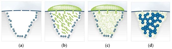
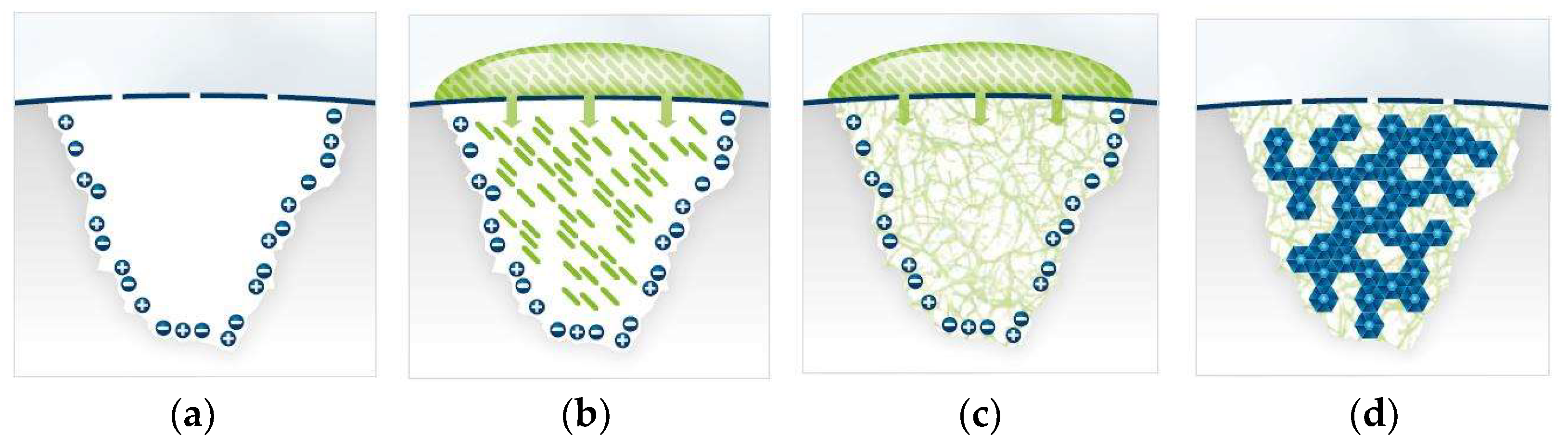
Consequently, regenerative strategies aim to support the guided, localized deposition of minerals within the lesion, enabling completer and more stable repair of early enamel lesions. In this context, different classes of peptides have been investigated (e.g., self-assembling peptides, amelogenin-derived peptides, and synthetic and engineered peptides) for their ability to promote enamel regeneration by mimicking natural biomineralisation processes. Despite their differences in origin, molecular structure and mechanisms of action, these peptides share the common goal of facilitating hydroxyapatite formation and subsurface lesion remineralization at the molecular level [16,17,18,19]. Among these, the self-assembling peptide P11-4 has been extensively investigated as a biomimetic approach for the management of initial carious lesions. P11-4 is designed to diffuse into the porous subsurface lesion, where it self-assembles in situ into a three-dimensional biomimetic scaffold that facilitates hydroxyapatite nucleation and promotes subsurface remineralisation (Figure 1). By harnessing natural salivary mineralisation processes, this approach aims to support lesion repair in a minimally invasive and biologically driven manner. Numerous in vitro and in vivo studies have analysed the potential of P11-4 for biomimetic remineralization and enamel regeneration, supporting its role as an innovative treatment option for the management of early-stage carious lesions [18,19,20,21].
Figure 1.
(a–d) Schematic illustration of the proposed mechanism of biomimetic enamel remineralisation by self-assembling peptide P11-4 in initial carious lesions. Presence of an initial carious lesion (a); A drop of Curodont™ Repair (green) is applied to the tooth surface after etching with phosphoric acid and diffuses through the pores of the surface layer into the carious lesion (b); within the lesion, Curodont™ Repair forms a three-dimensional network (c). Over a period of 1–6 months, new hydroxyapatite crystals form around the Curodont™ Repair network, resulting in potential “regeneration” or remineralisation of the carious lesion (d). (Graphic formerly provided by Credentis AG, reproduced with permission).
2. Literature Search and Methodological Approach
- -
- This work provides a comprehensive review with a narrative character, informed by a systematic literature search in PubMed, aiming to deliver a structured and evidence-based overview of the topic. The PICOS framework was used to guide study eligibility, search strategy, and content organisation: Population (P): patients of any age presenting with initial carious lesions;
- -
- Intervention (I): peptide-based therapies aimed at enamel regeneration;
- -
- Comparison (C): placebo, no treatment, another treatment (e.g., resin infiltration), or standard care (e.g., fluoride-based products such as varnishes or gels);
- -
- Outcomes (O): outcomes related to enamel demineralisation and/or remineralisation, independent of the assessment method used (e.g., visual scoring systems, laser fluorescence, etc.);
- -
- Study design (S): randomised controlled trials (RCTs) and prospective controlled clinical trials (CCTs).
The literature search was conducted primarily in PubMed/MEDLINE, to identify relevant English-language studies published between 2000 and 3 January 2026. Search terms were developed based on the PICOS framework and adapted to the objectives and narrative character of this review.
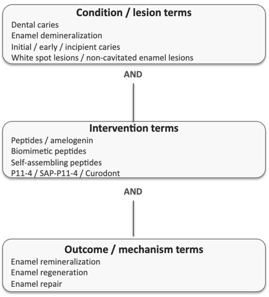
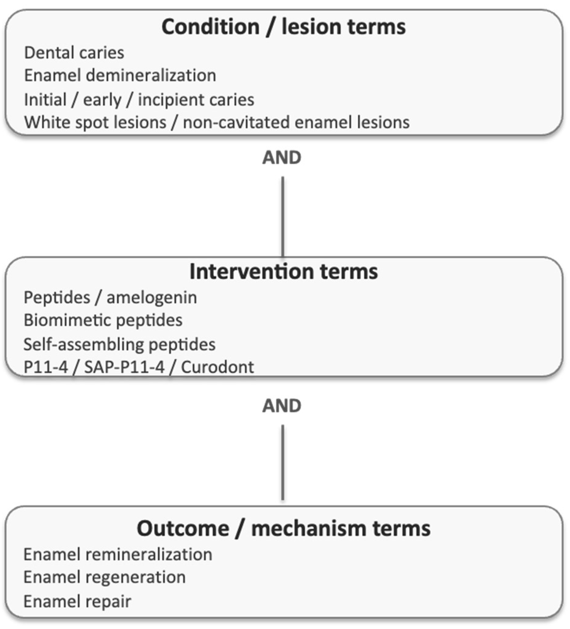
Titles and abstracts were screened for relevance, followed by full-text assessment of potentially eligible studies (Figure 2). The literature search on 3 January 2026 was guided by representative keywords and MeSH terms, combining terms related to dental caries and enamel demineralization (“Dental Caries” [MeSH] OR “Enamel Demineralization” [MeSH] OR initial caries OR early caries OR incipient caries OR white spot lesions OR enamel lesions), peptide-based interventions (“Peptides” [MeSH] OR “Amelogenin” [MeSH] OR peptide OR biomimetic peptide OR self-assembling peptide* OR P11-4 OR enamel regeneration peptide*), enamel remineralization/regeneration (“Enamel Remineralization” [MeSH] OR “Tooth Remineralization” [MeSH] OR remineralization OR remineralisation OR demineralization OR demineralisation OR enamel regeneration OR enamel repair*), and conventional preventive care comparators (placebo* OR fluoride* OR “standard care” OR “preventive care”). Only clinical studies meeting the predefined PICOS criteria were included. On the other hand, laboratory studies, animal studies, observational cohort studies, case reports, and retrospective analyses were excluded. Given the heterogeneity of study designs, outcome measures, and follow-up periods, a qualitative synthesis was performed. A formal risk-of-bias assessment was not undertaken, as the primary aim of this comprehensive narrative review was to provide a structured overview of existing clinical evidence and identify current knowledge gaps, rather than to perform a formal systematic evidence synthesis or generate pooled effect estimates through meta-analysis.
Figure 2.
Search strategy diagram and key search terms used for the comprehensive narrative review.
3. Results
All eligible studies were independently reviewed in full by an experienced investigator for data extraction. The accuracy of the extracted data was subsequently verified by a second senior researcher. The main findings of the included studies are summarized below.
3.1. Types of Peptides Used for Enamel Regeneration
Different classes of peptides have been investigated for their potential to promote enamel regeneration by mimicking natural biomineralisation processes all aim to support hydroxyapatite formation and lesion remineralisation at the molecular level (Table 1).
Table 1.
Comparison of peptide-based approaches for enamel regeneration.
3.1.1. Self-Assembling Peptides
Self-assembling peptides are the most extensively studied peptide group for enamel regeneration, with evidence from in vitro, in situ, and clinical studies supporting its remineralisation potential. Its application aims to promote natural enamel regeneration by mimicking endogenous remineralisation processes [17,19,20,21,27].
3.1.2. Amelogenin-Derived Peptides
Amelogenin-derived peptides are based on fragments of natural enamel matrix proteins involved in physiological enamel formation during tooth development. These peptides aim to replicate specific functional domains of amelogenin that regulate crystal growth and orientation. Experimental studies have demonstrated their ability to guide enamel-like crystal formation; however, most investigations remain at the in vitro or early preclinical stage, and clinical evidence is currently limited [28].
3.1.3. Synthetic and Engineered Peptides
Synthetic and engineered peptides are designed to optimise stability, binding affinity, and remineralisation capacity. These peptides are often modified to enhance interaction with enamel surfaces or mineral ions. While promising results have been reported in laboratory models, their translation into clinical application is still under development, and long-term clinical data are lacking.
3.2. Self-Assembling Peptides: Peptide P11-4
3.2.1. Mechanisms of Action
Peptide P11-4 is a self-assembling peptide composed of 11 amino acids (Ace-Gln-Gln-Arg-Phe-Glu-Trp-Glu-Phe-Glu-Gln-Gln-NH2). P11-4 can infiltrate subsurface enamel lesions and spontaneously assemble into three-dimensional scaffolds that act as nucleation templates for hydroxyapatite formation. Their ability to form organised matrices within demineralised enamel makes them particularly suitable for the treatment of initial, non-cavitated carious lesions. In this type of lesions P11-4 penetrates the subsurface enamel, where the peptides self-assemble into elongated structures resembling ladder rungs. This peptide scaffold acts as a matrix that attracts and incorporates calcium, phosphate, and hydroxyl ions, thereby promoting hydroxyapatite formation. The peptide’s high affinity for hydroxyapatite enhances this process, and the addition of fluoride further supports enamel remineralisation [12,20].
3.2.2. Safety and Biocompatibility
Experimental studies have shown that the self-assembling peptide P11-4 does not exhibit cytotoxic properties or trigger immunological responses [18,20]. These findings are supported by in vivo investigations and retrospective clinical studies, which consistently report a favourable safety profile for P11-4. No treatment-related adverse events, medical complications or allergic reactions were observed across these studies, either immediately after application or during follow-up periods of up to three and six months [27]. The clinical safety of P11-4 has been further substantiated by a recent systematic review including meta-analyses, which confirm its suitability for use in patients and reinforce its biocompatibility and clinical applicability [20]. Table 2 provides an overview of the randomized controlled trials (RCTs) included in this review and summarizes their main characteristics and principal findings.
Table 2.
In vivo/clinical studies on the self-assembling peptide P11-4.
3.2.3. New Evidence
Over the past decade, a growing body of research has examined the clinical effectiveness of the self-assembling peptide P11-4 in managing initial carious lesions. This evidence comprises well-designed randomized controlled clinical trials, clinical studies (Table 2), and in vitro investigations. The information displayed in Table 2 is ordered by year of publication and does not reflect study quality or reported outcomes.
Around 2019, a modified formulation of the self-assembling peptide P11-4, which incorporates 500 ppm sodium fluoride, was introduced to clinical use. A recent clinical investigation compared the remineralization efficacy of this peptide–fluoride formulation with that of fluoride varnish alone (NaF, Bifluorid 10) in 58 early carious lesions among 28 patients [36]. Lesion changes were monitored using laser fluorescence measurements (DIAGNOdent) at baseline as well as after 1, 3, and 6 months.
While both treatments resulted in improvements over time, the peptide–fluoride group exhibited significantly greater remineralization at the 3- and 6-month evaluations (p < 0.05; Table 2). These results indicate that the adjunctive use of P11-4 with fluoride may enhance the management of early carious lesions beyond what can be achieved with high-concentration fluoride varnish alone. At present, direct comparative studies between P11-4 alone and Curodont™ Repair Fluoride Plus are lacking. Such investigations would be valuable in clarifying the specific contribution of fluoride within peptide-based remineralisation approaches and in refining the assessment of their clinical effectiveness. In general, all studies consistently support the remineralization potential and clinical applicability of P11-4.
4. Discussion
Although a growing number of studies have investigated the self-assembling peptide P11-4, the overall volume of evidence remains limited when compared with other established approaches in minimal and non-invasive caries management for early lesions. Several systematic reviews and meta-analyses have pointed out methodological limitations within the current literature, including small study populations, heterogeneity in outcome measures, and relatively short observation periods [12,20]. Importantly, however, the restricted quantity of available studies should not be interpreted as evidence of ineffectiveness. On the contrary, published investigations to date consistently report favourable safety profiles and positive effects of P11-4 on enamel remineralization across different study designs [12,20,21,22], with associated risks considered minimal due to its application to acellular enamel and its degradation into natural amino acids or excretion if ingested [17]. Although experimental and in situ studies have shown potential of other peptide-based approaches than P11-4, these remain in the research and developmental stage and are not yet widely available for routine clinical use.
Results from clinical and experimental studies suggest that P11-4 may achieve higher levels of caries arrest and remineralization than conventional preventive measures such as sodium fluoride varnish. Nonetheless, head-to-head comparisons with other minimally invasive treatment options are rare. Only a single study has reported superior outcomes for resin infiltration compared with P11-4 [29]. As a result, it remains uncertain whether P11-4 offers should be favored regarding remineralization over alternative non-invasive or minimally invasive strategies, including resin infiltration or silver diamine fluoride (SDF), and further comparative research is warranted.
Based on the currently available evidence, P11-4 can be considered a complementary option for the management of initial carious lesions on various tooth surfaces, including occlusal, approximal, and buccal sites. Its use may be particularly appropriate in clinical situations where fluoride-based measures alone do not achieve sufficient lesion control, while operative intervention is not yet justified [12,22]. In such cases, factors such as patient cooperation, treatment acceptance, and personal economic considerations and structure of the dental healthcare coverage may strongly influence clinical decision-making.
Furthermore, as a relatively recent innovation, the clinical technique and underlying concept of P11-4 remain unfamiliar to many practitioners. Limited exposure and training may reduce confidence in its application and lead to its omission during patient counselling. Similarly to resin infiltration, peptide application does not result in an immediately visible change for the patient, which may complicate communication and acceptance of the treatment. In addition, evaluating treatment success in daily practice remains challenging. Objective assessment tools such as laser fluorescence devices are mainly confined to research settings and are not routinely available in general dental practices. Conventional radiographic imaging also fails to visualise changes associated with peptide-induced remineralisation, making longitudinal monitoring difficult for both clinicians and patients.
This review brings together the currently available evidence supporting the remineralization potential of self-assembling peptides (P11-4), drawing on findings from in vitro, in situ, and clinical studies. However, several limitations inherent to the narrative design of this review should be acknowledged. Although informed by a structured literature search in PubMed, the search was not intended to be fully exhaustive across multiple databases, as would be expected in a formal systematic review. Consequently, some relevant studies may not have been identified. Moreso, no formal risk-of-bias assessment was conducted, due to the nature of this review, which presents another methodological limitation. Nevertheless, the approach in this work provides a clinically focused and structured overview of the currently available evidence while highlighting important areas requiring further research.
Beyond caries management, emerging evidence indicates that P11-4 may have broader applications. Recent studies suggest potential protective effects against enamel erosion, indicating a possible role in preventive strategies beyond caries control [38]. These findings open perspectives for the use of P11-4 in conditions such as erosive tooth wear and dentin hypersensitivity. However, robust clinical studies are still needed to confirm its effectiveness in these indications and to compare it with established treatment approaches.
5. Conclusions
In conclusion, P11-4 represents an innovative, biologically driven, and minimally invasive addition to modern caries management. Unlike traditional restorative interventions based on tissue removal, this peptide-based approach aims to promote natural enamel repair and preserve tooth structure. From a practical clinical perspective, P11-4 appears to be a user-friendly and feasible option for the remineralization of initial carious lesions, offering clinicians an additional non-invasive treatment approach that may be readily integrated into preventive and minimally invasive care strategies. However, further well-designed long-term clinical studies are essential to clarify its comparative effectiveness and to possibly support broader integration into routine dental care.
Author Contributions
Conceptualization, R.M.S., M.A., C.H.S. and J.S.; search of the literature, R.M.S. and J.S.; data extraction, R.M.S. and J.S.; writing the original draft preparation, R.M.S. writing, review and editing, R.M.S., M.A., C.H.S. and J.S.; supervision, R.M.S. and J.S.; All authors have read and agreed to the published version of the manuscript.
Funding
This study received internal departmental support from the Department of Pediatric Dentistry at the University Medicine Greifswald in Germany, but no specific grant was obtained. No external funding was obtained.
Data Availability Statement
The original contributions presented in this study are included in the article. Further inquiries can be directed to the corresponding author.
Conflicts of Interest
The Department of Pediatric Dentistry received financial research support from Credentis AG in 2013 and 2014 for the conduct of a randomised controlled trial using the product Curodont Repair. Currently, the company vVardis distributes the product Curodont Repair™. The authors declare that there are currently no conflicts of interest.
References
- Bernabe, E.; Marcenes, W.; Hernandez, C.R.; Bailey, J.; Abreu, L.G.; Alipour, V.; Kassebaum, N.J. Global, Regional, and National Levels and Trends in Burden of Oral Conditions from 1990 to 2017: A Systematic Analysis for the Global Burden of Disease 2017 Study. J. Dent. Res. 2020, 99, 362–373. [Google Scholar] [CrossRef] [Scilit] [PubMed]
- Kassebaum, N.J.; Bernabé, E.; Dahiya, M.; Bhandari, B.; Murray, C.J.L.; Marcenes, W. Global burden of untreated caries: A systematic review and metaregression. J. Dent. Res. 2015, 94, 650–658. [Google Scholar] [CrossRef] [Scilit] [PubMed]
- Drancourt, N.; Roger-Leroi, V.; Martignon, S.; Jablonski-Momeni, A.; Pitts, N.; Domejean, S. Carious lesion activity assessment in clinical practice: A systematic review. Clin. Oral. Investig. 2019, 23, 1513–1524. [Google Scholar] [CrossRef] [Scilit]
- Pitts, N.B.; Zero, D.T.; Marsh, P.D.; Ekstrand, K.; Weintraub, J.A.; Ramos-Gomez, F.; Tagami, J.; Twetman, S.; Tsakos, G.; Ismail, A. Dental caries. Nat. Rev. Dis. Prim. 2017, 3, 17030. [Google Scholar] [CrossRef] [Scilit]
- Zafar, M.; Levy, S.M.; Warren, J.J.; Xie, X.J.; Kolker, J.; Pendleton, C. Prevalence of non-cavitated lesions and progression, regression, and no change from age 9 to 23 years. J. Public Health Dent. 2022, 82, 313–320. [Google Scholar] [CrossRef] [Scilit]
- BaniHani, A.; Santamaría, R.M.; Hu, S.; Maden, M.; Albadri, S. Minimal intervention dentistry for managing carious lesions into dentine in primary teeth: An umbrella review. Eur. Arch. Paediatr. Dent. 2022, 23, 667–693. [Google Scholar] [CrossRef] [Scilit]
- Banerjee, A.; Frencken, J.E.; Schwendicke, F.; Innes, N.P.T. Contemporary operative caries management: Consensus recommendations on minimally invasive caries removal. Br. Dent. J. 2017, 223, 215–222. [Google Scholar] [CrossRef] [Scilit]
- Leal, S.C.; Dame-Teixeira, N.; De Brito Barbosa, C.; Kominami, P.A.A.; Raposo, F.; Nakagawa, E.M.T.; Banerjee, A. Minimum intervention oral care: Defining the future of caries management. Braz. Oral. Res. 2022, 36, e135. [Google Scholar] [CrossRef] [Scilit]
- Marinho, V.C.C.; Worthington, H.V.; Walsh, T.; Clarkson, J.E. Fluoride varnishes for preventing dental caries in children and adolescents. Cochrane Database Syst. Rev. 2013, 7, CD002279. [Google Scholar] [CrossRef] [Scilit]
- Walsh, T.; Worthington, H.V.; Glenny, A.M.; Appelbe, P.; Marinho, V.C.; Shi, X. Fluoride toothpastes of different concentrations for preventing dental caries in children and adolescents. Cochrane Database Syst. Rev. 2010, 1, CD007868. [Google Scholar] [CrossRef] [Scilit] [PubMed]
- Cvikl, B.; Moritz, A.; Bekes, K. Pit and Fissure Sealants-A Comprehensive Review. Dent. J. 2018, 6, 18. [Google Scholar] [CrossRef] [Scilit]
- Wierichs, R.J.; Carvalho, T.S.; Wolf, T.G. Efficacy of a self-assembling peptide to remineralize initial caries lesions—A systematic review and meta-analysis. J. Dent. 2021, 109, 103652. [Google Scholar] [CrossRef] [Scilit]
- Philip, N. State of the Art Enamel Remineralization Systems: The Next Frontier in Caries Management. Caries Res. 2019, 53, 284–295. [Google Scholar] [CrossRef] [Scilit]
- Selwitz, R.H.; Ismail, A.I.; Pitts, N.B. Dental caries. Lancet 2007, 369, 51–59. [Google Scholar] [CrossRef] [Scilit]
- Farooq, I.; Bugshan, A. The role of salivary contents and modern technologies in the remineralization of dental enamel: A narrative review. F1000Research 2020, 9, 171. [Google Scholar] [CrossRef] [Scilit]
- Schlee, M.; Schad, T.; Koch, J.H.; Cattin, P.C.; Rathe, F. Clinical performance of self-assembling peptide P11-4 in the treatment of initial proximal carious lesions: A practice-based case series. J. Investig. Clin. Dent. 2018, 9, e12286. [Google Scholar] [CrossRef] [Scilit]
- Brunton, P.A.; Davies, R.P.W.; Burke, J.L.; Smith, A.; Aggeli, A.; Brookes, S.J.; Kirkham, J. Treatment of early caries lesions using biomimetic self-assembling peptides--a clinical safety trial. Br. Dent. J. 2013, 215, E6. [Google Scholar] [CrossRef] [Scilit] [PubMed]
- Kind, L.; Stevanovic, S.; Wuttig, S.; Wimberger, S.; Hofer, J.; Müller, B.; Pieles, U. Biomimetic Remineralization of Carious Lesions by Self-Assembling Peptide. J. Dent. Res. 2017, 96, 790–797. [Google Scholar] [CrossRef] [Scilit] [PubMed]
- Kirkham, J.; Firth, A.; Vernals, D.; Boden, N.; Robinson, C.; Shore, R.C.; Aggeli, A. Self-assembling peptide scaffolds promote enamel remineralization. J. Dent. Res. 2007, 86, 426–430. [Google Scholar] [CrossRef] [Scilit] [PubMed]
- Keeper, J.H.; Kibbe, L.J.; Thakkar-Samtani, M.; Heaton, L.J.; Desrosiers, C.; Vela, K.; Amaechi, B.T.; Jablonski-Momeni, A.; Young, D.A.; MacLean, J.; et al. Systematic review and meta-analysis on the effect of self-assembling peptide P11-4 on arrest, cavitation, and progression of initial caries lesions. J. Am. Dent. Assoc. 2023, 154, 580–591.e11. [Google Scholar] [CrossRef] [Scilit]
- Alkilzy, M.; Qadri, G.; Splieth, C.H.; Santamaría, R.M. Biomimetic Enamel Regeneration Using Self-Assembling Peptide P11-4. Biomimetics 2023, 8, 290. [Google Scholar] [CrossRef] [Scilit]
- Alkilzy, M.; Santamaria, R.M.; Schmoeckel, J.; Splieth, C.H. Treatment of Carious Lesions Using Self-Assembling Peptides. Adv. Dent. Res. 2018, 29, 42–47. [Google Scholar] [CrossRef] [Scilit]
- Ruan, Q.; Moradian-Oldak, J. Development of amelogenin-chitosan hydrogel for in vitro enamel regrowth with a dense interface. J. Vis. Exp. 2014, 89, 51606. [Google Scholar]
- Fan, Y.; Nelson, J.R.; Alvarez, J.R.; Hagan, J.; Berrier, A.; Xu, X. Amelogenin-assisted ex vivo remineralization of human enamel: Effects of supersaturation degree and fluoride concentration. Acta Biomater. 2011, 7, 2293–2302. [Google Scholar] [CrossRef] [Scilit] [PubMed]
- Ding, L.; Han, S.; Wang, K.; Zheng, S.; Zheng, W.; Peng, X.; Niu, Y.; Li, W.; Zhang, L. Remineralization of enamel caries by an amelogenin-derived peptide and fluoride in vitro. Regen. Biomater. 2020, 7, 283–292. [Google Scholar] [CrossRef] [Scilit] [PubMed]
- Xu, J.; Shi, H.; Luo, J.; Yao, H.; Wang, P.; Li, Z.; Wei, J. Advanced materials for enamel remineralization. Front Bioeng. Biotechnol. 2022, 10, 985881. [Google Scholar] [CrossRef] [Scilit]
- Alkilzy, M.; Tarabaih, A.; Santamaria, R.M.; Splieth, C.H. Self-assembling Peptide P11-4 and Fluoride for Regenerating Enamel. J. Dent. Res. 2018, 97, 148–154. [Google Scholar] [CrossRef] [Scilit]
- Hu, D.; Ren, Q.; Li, Z.; Han, S.; Ding, L.; Lu, Z.; Zhang, L. Unveiling the mechanism of an amelogenin-derived peptide in promoting enamel biomimetic remineralization. Int. J. Biol. Macromol. 2023, 253, 127322. [Google Scholar] [CrossRef] [Scilit]
- Gözetici, B.; Öztürk-Bozkurt, F.; Toz-Akalın, T. Comparative Evaluation of Resin Infiltration and Remineralisation of Noncavitated Smooth Surface Caries Lesions: 6-month Results. Oral. Health Prev. Dent. 2019, 17, 99–106. [Google Scholar]
- Bröseler, F.; Tietmann, C.; Bommer, C.; Drechsel, T.; Heinzel-Gutenbrunner, M.; Jepsen, S. Randomised clinical trial investigating self-assembling peptide P11-4 in the treatment of early caries. Clin. Oral. Investig. 2020, 24, 123–132. [Google Scholar] [CrossRef] [Scilit]
- Doberdoli, D.; Bommer, C.; Begzati, A.; Haliti, F.; Heinzel-Gutenbrunner, M.; Juric, H. Randomized Clinical Trial investigating Self-Assembling Peptide P11-4 for Treatment of Early Occlusal Caries. Sci. Rep. 2020, 10, 4195. [Google Scholar] [CrossRef] [Scilit]
- Welk, A.; Ratzmann, A.; Reich, M.; Krey, K.F.; Schwahn, C. Effect of self-assembling peptide P11-4 on orthodontic treatment-induced carious lesions. Sci. Rep. 2020, 10, 6819. [Google Scholar] [CrossRef] [Scilit]
- Atteya, S.M.; Amer, H.A.; Saleh, S.M.; Safwat, Y. Self-assembling peptide and nano-silver fluoride in remineralizing early enamel carious lesions: Randomized controlled clinical trial. BMC Oral. Health 2023, 23, 577. [Google Scholar] [CrossRef] [Scilit]
- Natchiyar, N.; Asokan, S.; Priya, G.P.; Viswanath, S. Comparison of Remineralizing Agents in the Management of White Spot Lesions In Three- to Five-year-old Children: A Clinical Trial. Pediatr. Dent. 2023, 45, 99–106. [Google Scholar] [PubMed]
- Gohar, R.A.A.E.G.; Ibrahim, S.H.; Safwat, O.M. Evaluation of the remineralizing effect of biomimetic self-assembling peptides in post-orthodontic white spot lesions compared to fluoride-based delivery systems: Randomized controlled trial. Clin. Oral. Investig. 2023, 27, 613–624. [Google Scholar] [CrossRef] [Scilit] [PubMed]
- Shaalan, O.; Fawzy El-Sayed, K.; Abouauf, E. Evaluation of the remineralization potential of self-assembling peptide P11-4 with fluoride compared to fluoride varnish in the management of incipient carious lesions: A randomized controlled clinical trial. Clin. Oral. Investig. 2024, 28, 438. [Google Scholar] [CrossRef] [Scilit]
- Elaziz, B.S.A.; Hamdy, D.; Galal, M.; Khattab, N.M.A. The remineralizing ability of self-assembling peptide P11-4, 2% arginine enriched sodium fluoride and functionalized tri calcium phosphate fluoride varnishes in treatment of white spot lesions—A randomized controlled trial. BDJ Open 2025, 11, 70. [Google Scholar] [CrossRef] [Scilit]
- Yilmaz Sen, B.; Akcay, M. Comparative analysis of the effect of self-assembling peptide P11-4 on enamel erosion: A confocal laser scanning microscopy study. Clin. Oral. Investig. 2024, 29, 29. [Google Scholar] [CrossRef] [Scilit] [PubMed]

Disclaimer/Publisher’s Note: The statements, opinions and data contained in all publications are solely those of the individual author(s) and contributor(s) and not of MDPI and/or the editor(s). MDPI and/or the editor(s) disclaim responsibility for any injury to people or property resulting from any ideas, methods, instructions or products referred to in the content. |
© 2026 by the authors. Published by MDPI on behalf of the Lithuanian University of Health Sciences. Licensee MDPI, Basel, Switzerland. This article is an open access article distributed under the terms and conditions of the Creative Commons Attribution (CC BY) license.