


Abstract
Background and Objectives: Computed tomography (CT) characterizes intra-articular and transitional pediatric ankle fractures, but its additional effect on clinical decision-making and patient outcomes requires clarification. Materials and Methods: PubMed/MEDLINE, Scopus, and CENTRAL were searched from inception to 4 September 2026. Eligible studies compared radiographs and CT within patients. Quality was assessed using eight core Methodological Index for Non-Randomized Studies (MINORS) items. Narrative synthesis separated classification, fixation planning, and treatment recommendations. Results: Ten full-text studies described 574 patients, including 551 CT recipients; outcome-specific denominators differed. In one study of 25 triplane fractures, classification changed in 115/250 observer ratings. Substantial screw-plan changes occurred in 61/150 surgeon ratings; 4/15 initially nonoperative ratings changed to an operative recommendation. Another study reported operative recommendation changes in 24/64 patients. CT clarified displacement and fracture geometry, while observer agreement did not consistently improve. Evidence for additional benefit in confidently characterized extra-articular injuries was limited. No included study reported cohort-specific numerical radiation dose estimates. Conclusions: CT may support decisions when radiographs leave unresolved articular or fixation-planning questions. Changes in recommendations do not establish improved function or fewer complications. Heterogeneous observational evidence supports cautious selective use; the proposed framework requires prospective validation.
1. Introduction
Pediatric ankle fractures account for approximately 5% of childhood fractures and 15–20% of physeal injuries. The distal tibial physis contributes about 40% of tibial growth and 17% of lower-extremity growth [1,2,3]. Physeal injury can cause premature closure, limb-length discrepancy, and angular deformity. In one retrospective distal tibial physeal cohort, premature closure occurred in 12/107 patients assessable for late complications and angular deformity in 11/107; these selected follow-up denominators should not be interpreted as population incidence [4]. Accurate assessment of fracture morphology, displacement, and articular congruity is therefore clinically relevant.
During asymmetric distal tibial physeal closure, adolescents may sustain Tillaux or triplane fractures. Tillaux injuries involve the anterolateral epiphysis, whereas triplane injuries extend through several anatomical planes [2,3]. Radiography remains the initial examination, but overlapping structures and complex fracture geometry can obscure articular extension or displacement. Computed tomography (CT) provides multiplanar characterization of these features and may influence fracture classification and fixation planning [5,6,7,8].
An imaging-related change in diagnosis or treatment recommendation is distinct from improvement in patient outcomes. Observer studies may identify additional fracture detail without producing more consistent decisions, and changes in operative indication do not establish that surgery is necessary or beneficial for every reclassified injury [8]. The relevant question is which diagnostic or planning uncertainty CT resolves and whether that resolution justifies the examination.
In children, CT should be justified for a specific clinical question and performed using an optimized protocol [9,10]. Routine acquisition is difficult to justify when radiographs adequately characterize an injury and further detail is unlikely to affect a decision. Conversely, uncertain articular involvement or fixation geometry may provide a reason to obtain cross-sectional imaging.
Radiographic markers may help identify such uncertainty. Metaphyseal displacement on the lateral view and epiphyseal displacement or tibiofibular clear-space widening have been associated with greater CT-measured displacement [11,12]. These associations are candidates for patient selection rather than validated rules for omitting CT or proceeding to surgery.
The literature combines patient-level cohorts, repeated observer assessments, displacement measurements, and planning exercises. This heterogeneity complicates estimates of how often CT changes decisions and requires explicit separation of outcomes and their denominators.
This systematic review evaluates the additional diagnostic and decision-making information provided by CT beyond radiography in pediatric ankle fractures. We examine fracture reclassification, articular displacement, operative recommendations, fixation planning, and observer agreement, and assess the evidence supporting selective CT use.
2. Materials and Methods
2.1. Protocol
This systematic review is reported with reference to the Preferred Reporting Items for Systematic Reviews and Meta-Analyses (PRISMA) 2020 statement [13]. The completed PRISMA 2020 checklist is provided in Supplementary Table S5. The review was not registered, and no review protocol was prepared.
2.2. Eligibility Criteria
Eligibility was defined a priori using the population, index test, comparator, and outcome framework summarized in Table 1. Studies were eligible when they: (1) investigated pediatric distal tibial and/or fibular fractures; (2) included at least ten patients; (3) reported radiography and CT in the same patients, with an extractable paired diagnostic, displacement, decision, or planning endpoint; and (4) presented original observational or diagnostic data. In mixed-imaging cohorts, only the extractable CT-imaged subgroup contributed to paired outcomes. The original population definition allowed studies explicitly described as pediatric; accordingly, reports with an upper age boundary of 18 years were retained and identified in Table 2 [6,12].
Table 1.
PICO framework used to define eligibility.
Studies were excluded if they contained an exclusively adult or nonextractable mixed-age population, fractures outside the ankle, absence of an extractable direct radiograph–CT comparison, fewer than ten patients, nonoriginal data, a biomechanical or cadaveric design, or duplicate cohorts. Comparing CT recipients with non-recipients without a paired imaging endpoint did not satisfy the comparator criterion. Full-text reports were required for eligibility assessment.
2.3. Information Sources and Search Strategy
PubMed/MEDLINE, Scopus, and Cochrane CENTRAL were searched from inception to 4 September 2026. The queries combined pediatric, ankle/physeal fracture, and CT concepts. PubMed used MeSH and free-text terms with human, English-language, and preprint-exclusion filters; Scopus was limited to English. The complete original queries, filters, and database-specific yields are provided separately in Supplementary Table S1. No new database search was performed for this revision.
Reference lists of the originally included reports were screened for additional eligible studies; none were identified through this route.
2.4. Study Selection Process
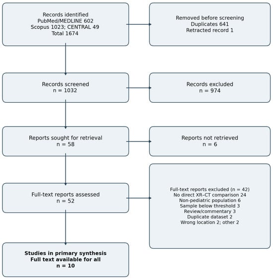
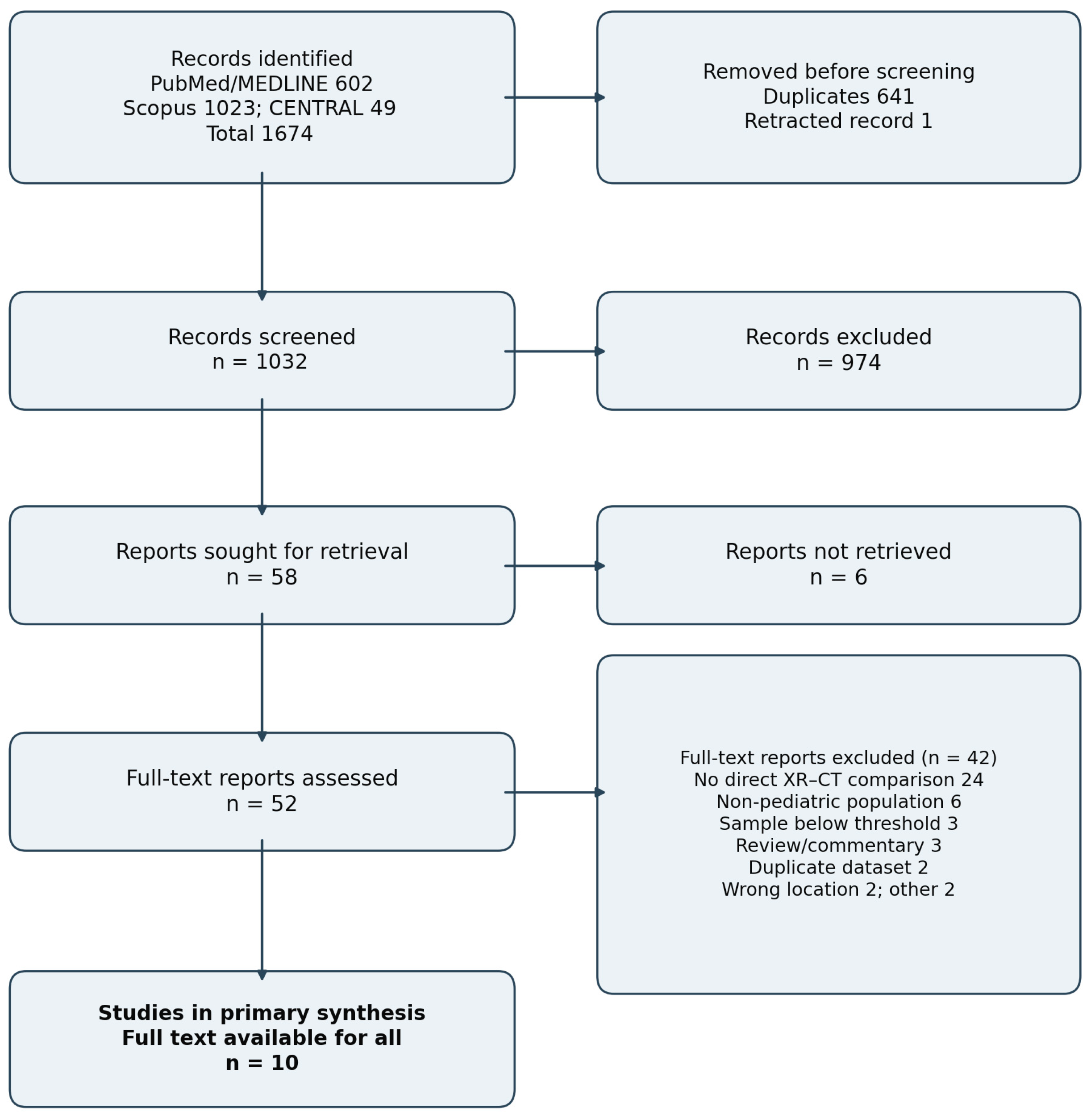
Database records were imported into Zotero (version 10.0.5, Corporation for Digital Scholarship, Falls Church, VA, USA) and deduplicated. Two reviewers independently screened titles and abstracts and then assessed full texts against the eligibility criteria. Disagreements were resolved by discussion or by a third reviewer. Full-text decisions and exclusion reasons were recorded in a standardized spreadsheet. The revised selection flow is presented in Section 3.1 and Figure 1.
Figure 1.
PRISMA 2020 flow diagram. Of 58 reports sought, 6 were not retrieved, and 52 full texts were assessed. Forty-two reports were excluded, and ten studies were included. Database and title/abstract screening counts are unchanged [13].
2.5. Data Extraction
One reviewer extracted data using a standardized Excel (version 16.115 for Mac, build 26110837, Microsoft Corporation, Redmond, WA, USA) form, and a second reviewer verified the extraction; disagreements were resolved by discussion. Variables included the study design and period, patient numbers, CT-imaged subgroups, demographics, fracture patterns, imaging protocols, assessors and blinding, reference standards, reclassification, displacement, treatment recommendations, fixation planning, and observer agreement. The revision audit distinguished patient counts from observer ratings and selected reasons, recorded outcome-specific numerators and denominators, and checked source pages and tables. Radiation reporting was separated into cohort-specific dose measurements, acquisition/optimization descriptions, and externally cited dose estimates.
2.6. Methodological Quality Assessment
Methodological quality was reassessed using the eight core Methodological Index for Non-Randomized Studies (MINORS) items, scored as 0 (not reported), 1 (reported inadequately), or 2 (reported adequately), with a maximum of 16 [14]. The four additional between-group items were not applied to the paired-imaging evidence because the comparison used the same patients rather than independent clinical groups. For immediate imaging endpoints, item 6 addressed the interval between images or repeat readings, and item 7 addressed completeness of paired imaging or ratings; these review-specific interpretations are stated in Supplementary Table S4. Item 3 required evidence of a prospective clinical accrual protocol, rather than a planned retrospective rereading exercise. A single reassessment was completed with a source-based rationale for every item; independent duplicate reassessment was not performed. All included studies were assessed from their full reports. Totals are descriptive and were not converted into verbal quality categories or used to weight results.
2.7. Data Synthesis and Analysis
A narrative synthesis was undertaken because fracture populations, displacement definitions, observer designs, and decision endpoints differed substantially. Findings were organized by fracture pattern and by the mechanism of decision impact. Fracture reclassification, change in operative recommendation, and modification of an existing fixation plan were treated as separate outcomes. Each percentage is accompanied by its original numerator, denominator, and unit where available. Patient-level results were not pooled with repeated ratings, and overlapping samples within a study were not added. Imaging availability across whole cohorts was summarized separately from outcome-specific analytic samples. No pooled frequency of CT-attributable change was calculated. No sensitivity analyses, formal assessment of reporting bias, or formal outcome-level certainty assessment were performed.
3. Results
3.1. Study Selection
The search identified 1674 records; 1032 remained after the removal of 641 duplicates and a single retracted record. Of fifty-eight reports sought, six were unavailable, and fifty-two full texts were assessed. Forty-two were excluded, leaving ten full-text studies in the primary synthesis (Figure 1). Three previously listed reports, El-Karef, D’Angelo, and Seel, were excluded from that synthesis because no extractable paired radiograph–CT endpoint was identified [4,15,16].
3.2. Study Characteristics
The ten retained reports described cohorts of 24–130 patients, totaling 574 patients, with 551 documented CT recipients (Table 2). These figures summarize imaging availability, not a common paired-outcome sample. Aletto reported CT in 124/130 patients but paired recorded emergency-department classification in only 22; a separate 61-case observer survey had unspecified overlap with that subgroup [17]. Bund reported paired measurements for 21/38 patients [18]. The other eight reports described CT for all patients in their stated samples. Lemburg included thirty-three patients with suspected growth-plate injury, but CT excluded fracture in two [6]. Most studies used retrospective case identification; none compared randomized imaging strategies with patient-centered outcomes. Fracture categories use the Salter–Harris (SH) classification.
Table 2.
Characteristics and analytic samples of the ten studies retained in the primary paired-imaging synthesis. Cohort and CT counts are distinct from outcome-specific denominators. CT, computed tomography; ED, emergency department.
3.3. Methodological Quality and Bias
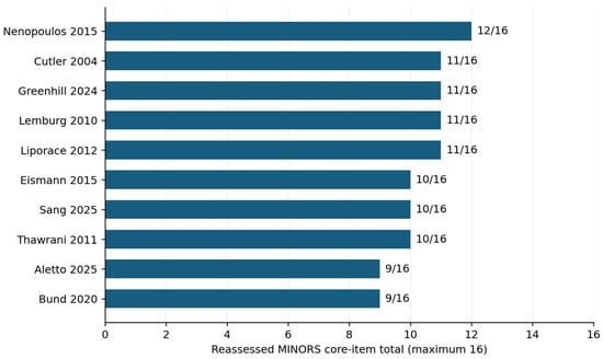
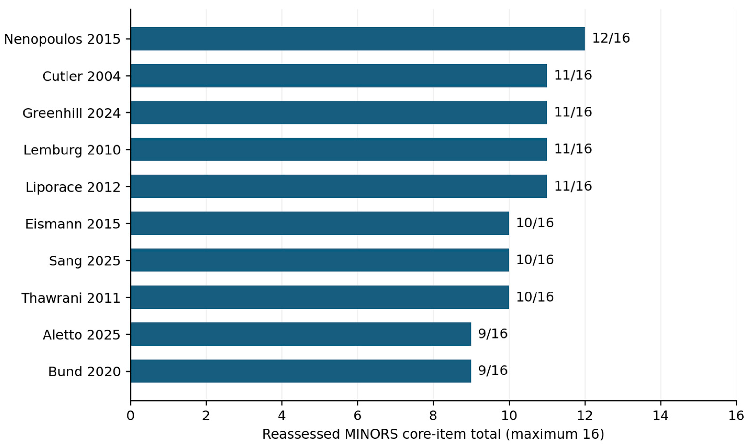
The reassessed eight-item MINORS totals ranged from 9/16 to 12/16 across the ten included studies (Figure 2; Supplementary Table S4). No report established a prospective clinical accrual protocol or prospective sample-size calculation in the available material. Incomplete documentation of consecutive inclusion and assessor masking, together with missing paired data in some cohorts, contributed to lower item scores. No verbal quality categories were assigned.
Figure 2.
Reassessed MINORS totals using the eight core items, with a maximum of 16. Item-level scores, operational definitions, and source-based explanations are provided in Supplementary Table S4. Totals are descriptive and do not represent validated overall risk-of-bias categories [14]. The studies shown are reported in references [5,6,7,8,11,12,17,18,19,20].
The source reports indicate recurrent concerns about retrospective selection of CT recipients, small samples, variable blinding, and repeated assessments of the same cases. Planning and classification endpoints commonly used CT itself as the reference, and imaging changes were rarely connected to long-term patient outcomes. These design limitations inform the interpretation alongside the item-level appraisal.
3.4. Findings by Fracture Pattern
The studies reported different diagnostic and decision endpoints across transitional, mixed intra-articular, and extra-articular patterns. Study-specific changes and their units are presented in Table 3.
Table 3.
Study-specific diagnostic and decision changes after CT. Rows represent different outcomes and overlapping samples; they must not be summed or interpreted as comparable patient-level event rates.
3.4.1. Transitional Fractures (Tillaux and Triplane)
In 25 intra-articular triplane cases, Eismann reported fracture-pattern changes in 115/250 paired observer ratings (46.0%). A substantial screw-plan change occurred in 61/150 paired surgeon ratings (40.7%). Of fifteen ratings initially recommending nonoperative treatment, four changed to an operative recommendation after CT (26.7%); this is a conditional rating-level proportion [5]. In the transitional subgroup of Nenopoulos, operative recommendations changed in 16/32 cases assessed by one blinded observer [7]. Liporace reported Tillaux-to-triplane reclassification in 7/144 paired observer ratings (4.9%). Recommendations changed from nonoperative to operative in 19/144 ratings (13.2%; p = 0.033): six toward percutaneous fixation and thirteen toward open reduction and internal fixation. This percentage uses all 144 ratings, rather than only initially nonoperative ratings, and does not describe treatment actually delivered [20].
3.4.2. Mixed Physeal and Intra-Articular Cohorts
In the sixty-four-case intra-articular series of Nenopoulos, operative recommendations increased from eighteen to forty-two after CT, corresponding to 24/64 case-level changes; eight changes occurred among thirty-two non-transitional SH III/IV cases [7]. Lemburg reported different SH classifications in 10/33 patients (30.3%), including six radiograph-based SH III injuries reclassified as SH IV; triplane morphology was missed or misclassified in five out of seven cases [6]. Thawrani reported reclassification in 54/250 observer ratings (21.6%) and a surgical/non-surgical recommendation change in 43/250 (17.2%): 26 toward surgery and 17 away from surgery [8]. Aletto reported eight classification changes among the twenty-two patients with both a recorded initial SH classification and CT (36.4%). In its separate survey, all six responders matched the CT-based classification in 21/61 cases; this unanimous-case proportion is distinct from accuracy across individual ratings [17]. Mixed-cohort findings were not presented as estimates confined to non-transitional SH III/IV fractures.
3.4.3. Extra-Articular Salter–Harris I and II Fractures
Lemburg found no radiographic misclassification among six SH I/II cases [6]. However, Cutler’s planning exercise included 29 SH II fractures and found improved drawing accuracy with CT across fracture types [19]. Therefore, limited classification change should not be equated with an absence of potential planning information. No robust patient-level estimate of treatment change was available for confidently established uncomplicated extra-articular injuries.
3.5. Displacement Assessment and Fixation Planning
Paired imaging studies also reported displacement measurements, planning accuracy, and candidate radiographic predictors. These endpoints were considered separately from changes in operative recommendation.
3.5.1. Articular Displacement and Study-Specific Thresholds
Eismann found that 53/136 ratings initially measuring ≤2 mm of articular displacement on radiographs exceeded 2 mm after CT (39.0%) [5]. Greenhill reported mean sagittal articular gaps of 1.0 mm on radiographs and 3.3 mm on CT; mean axial CT diastasis was 4.2 mm [11]. In Bund’s 21-patient paired subgroup, radiographic gap measurements were associated with CT measurements and were smaller; subgroup means are reported in the original Table III [18]. These measurement findings do not by themselves quantify a treatment benefit. Thresholds differed: Greenhill assessed CT gaps ≥2.5 mm, while several other reports used approximately 2 mm.
3.5.2. Precision of Spatial Surgical Planning
In Cutler’s 62-case drawing exercise, four observers planned screw placement using radiographs and then CT. Mean deviation from a CT-derived ideal entry point decreased from 12 mm to 2 mm, and angular deviation decreased from 31° to 6.5° (both p < 0.0001). These are planning-surrogate endpoints, not measurements of actual postoperative implant accuracy or functional improvement [19]. In the original study by Thawrani et al., Table 5 recorded screw planning as 131 of 232 selected reasons explaining CT usefulness; multiple reasons were permitted, so this 56.5% does not represent a proportion of patients or of all 250 observer ratings [8].
3.5.3. Radiographic Predictors of CT-Measured Displacement
In Greenhill, 56 of 60 patients with >1 mm metaphyseal displacement on the lateral radiograph had an articular gap ≥2.5 mm on CT. The intraclass correlation coefficient (ICC) for the lateral measurement was 0.84; the reported negative predictive value was only 39% [11]. In Sang’s 61-patient series, lateral epiphyseal displacement correlated with CT diastasis (ρ = 0.55, p < 0.001), as did mortise tibiofibular clear space more weakly (ρ = 0.27, p = 0.041) [12]. These were association studies, not prospective validation of a CT-selection rule. Observer agreement also varied: Thawrani reported interobserver κ values of 0.64 with radiographs and 0.57 after CT for classification, and 0.48 versus 0.36 for treatment recommendation [8].
3.5.4. Radiation Dose Reporting
No cohort-specific numerical volume CT dose index (CTDIvol), dose-length product, or effective-dose estimates were identified in any of the ten included full-text reports. Aletto described acquisition and reconstruction optimization without numerical dose outputs [17]. Lemburg cited external estimates, which were not counted as measurements from its cohort [6]. Supplementary Table S2 records dose reporting for all ten studies.
3.6. Conceptual Framework for Selective CT
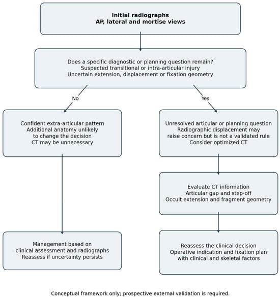
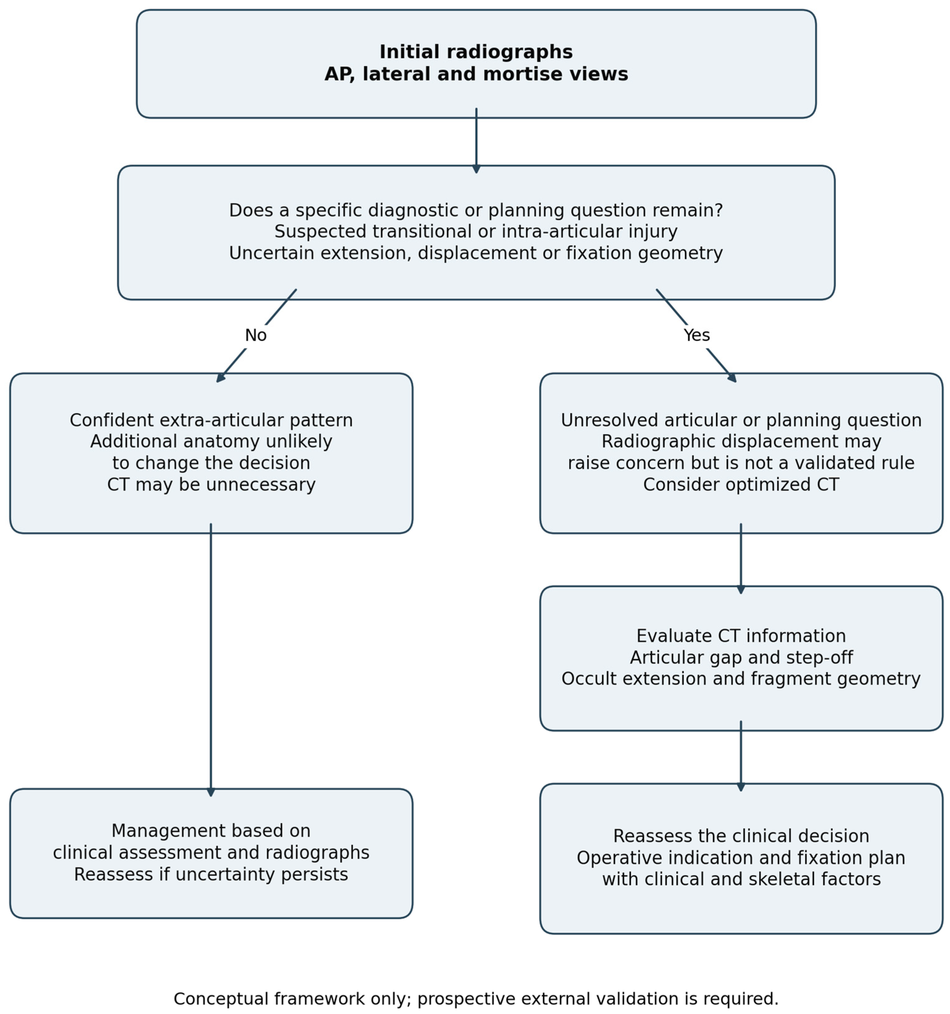
Figure 3 integrates the recurring diagnostic and planning questions identified in the retained studies. It separates a radiograph-based management branch from a branch in which CT may clarify an unresolved articular or fixation-planning question. The framework is a conceptual synthesis and has not been prospectively validated.
Figure 3.
Conceptual framework for selective CT after initial radiography. The radiograph-only branch ends separately and does not feed into CT findings. Radiographic associations may prompt further assessment but are not validated thresholds for excluding CT or mandating surgery. Changes in decisions do not establish improved patient outcomes. CT, computed tomography; AP, anteroposterior.
4. Discussion
CT most often added decision-relevant information when radiographs did not fully characterize a transitional or intra-articular injury. The contribution included clarifying fracture geometry, identifying a larger articular gap, and refining fixation plans [5,6,7,8,11,12,17,18,19,20]. The evidence is less informative about the value of CT in unselected children with ankle trauma because the cohorts were commonly enriched for patients already selected for advanced imaging.
The revised synthesis avoids a single range for “management change”. A classification revision, a change in operative recommendation, and an alteration in screw trajectory answer different questions. Repeated ratings of the same fracture also cannot be interpreted as independent patients. Reporting study-specific counts makes these distinctions visible without implying a pooled probability of benefit.
4.1. Decision Changes and Patient Outcomes
A change from a nonoperative to an operative recommendation does not demonstrate that the eventual clinical outcome will be better. Observer exercises measure the influence of information on a proposed decision; they do not determine whether that decision improves pain, function, growth, complications, or quality of life. Changes in the opposite direction, such as those reported by Thawrani, also show that additional imaging does not invariably increase recommendations for surgery [8].
Improved planning accuracy is a plausible intermediate benefit when fixation is already indicated, but the CT-derived reference used in planning exercises can favor CT by design [19]. None of the retained studies established the causal effect of obtaining CT on long-term patient outcomes. The findings therefore support a possible diagnostic and planning contribution, with uncertainty about its downstream clinical benefit.
Clinical cohorts excluded from the paired-imaging synthesis provide context rather than direct estimates of incremental decision impact. D’Angelo and Seel compared selected CT recipients with non-recipients, while El-Karef described a triplane treatment series [4,15,16]. Their outcomes are affected by injury severity, treatment selection, and follow-up. Associations with better reduction cannot be attributed to CT alone.
For a confidently characterized extra-articular injury, the limited evidence suggests less additional classification information from CT. However, the absence of misclassification among six SH I/II cases in Lemburg is a small selected observation, not proof that CT has no role in every apparent extra-articular fracture [6]. Uncertain fracture extension or a specific fixation question may still justify additional imaging.
4.2. Selective CT and Radiographic Predictors
CT may be considered when the expected anatomical information could resolve a defined clinical uncertainty, including occult articular extension, incompletely measured diastasis, or fragment geometry needed for fixation. This approach requires documenting the question before imaging and reconsidering the decision after the result, rather than treating any additional detail as an indication for intervention.
Metaphyseal displacement and lateral or mortise-view parameters are candidate selection markers [11,12]. Greenhill’s low negative predictive value cautions against using an apparently reassuring measurement to rule out relevant articular displacement. Sang’s correlations were modest to moderate and did not establish safe cutoffs for omitting CT. Their diagnostic performance requires external validation in a broader spectrum of injuries.
Displacement thresholds should be interpreted within the study and clinical setting. The ≥2.5 mm criterion examined by Greenhill differs from the approximately 2 mm thresholds used elsewhere; these values cannot be treated as interchangeable biological boundaries. Fracture pattern, skeletal maturity, stability, reducibility, and soft-tissue status also influence treatment [1,2,3,5,11].
The conceptual pathway in Figure 3 may help organize these questions, but it cannot currently be presented as a guideline or validated decision rule. A prospective study must establish how often it avoids unnecessary CT while preserving detection of findings that meaningfully affect treatment.
4.3. Radiation Exposure and Dose Optimization
Pediatric CT requires justification and optimization [9,10]. Cohort-specific numerical dose measurements were absent from all ten included full reports. Aletto described protocol optimization without numerical dose outputs [17]. Lemburg’s quoted dose estimates were taken from the other literature rather than measured in its cohort [6]. These data do not support a quantitative dose–benefit comparison.
External technical studies provide relevant context and were not added to the included-study count. In 90 pediatric lower-extremity CT examinations, Shah et al. compared protocols with and without adaptive statistical iterative reconstruction. Dose indices decreased with iterative reconstruction, but subjective sharpness and diagnostic acceptability also decreased [21]. Protocol choice must therefore preserve the image quality needed for the specific fracture question.
In pediatric ankle phantoms, Tschauner et al. reported mean surface doses of 3.8 ± 2.1 mGy with dedicated extremity cone-beam CT and 4.5 ± 1.3 mGy with optimized multidetector CT, with comparable subjective general image quality [22]. These are phantom surface-dose measurements, not effective doses or direct pediatric fracture-outcome data. They illustrate protocol-dependent dose reduction rather than a universal dose applicable to the studies reviewed here.
4.4. Limitations
Selection of more complex or uncertain injuries for CT limits generalizability. Heterogeneous fracture patterns, imaging protocols, measurement definitions, and observer experience further restrict comparison. Repeated ratings are clustered within patients and observers, and outcome-specific subsets can overlap. The available aggregate data do not permit reconstruction of independent patient-level change rates.
The paired-imaging criterion excluded three previously listed reports from the primary synthesis, reducing the direct evidence base. Requiring an extractable paired endpoint also favors studies with complete radiograph and CT series and may limit applicability to patients not selected for CT. All ten included studies were assessed in full text.
MINORS was developed for nonrandomized surgical studies and has not been validated specifically for these paired-imaging tasks [14]. We therefore reported item-level explanations and made the interpretation of the observation interval and paired-assessment completeness explicit. The reassessment was not independently duplicated, which permits assessor subjectivity. Scores were not used to assert low risk of bias, weight effects, or establish comparative clinical effectiveness. Reporting bias and certainty of evidence were not formally assessed.
4.5. Future Research
Prospective multicenter studies should record the diagnosis, displacement estimate, treatment recommendation, and fixation plan before and after CT, using predefined definitions of a meaningful change. Patient-level data and observer-level ratings should be reported separately, with analyses accounting for repeated observations.
Validation of selection markers should include both complex and minimally displaced injuries, prespecified thresholds, and explicit assessment of missed clinically relevant findings. Outcomes should extend to function, pain, complications, growth disturbance, patient-reported measures, resource use, and radiation exposure. CTDIvol, dose-length product, scan length, and any effective-dose estimation method should be reported with the protocol and patient characteristics.
5. Conclusions
CT may add diagnostic and fixation-planning information in selected pediatric transitional or intra-articular ankle fractures when radiographs leave uncertainty about extension, displacement, or geometry. It appears less useful for routine characterization of confidently diagnosed uncomplicated extra-articular injuries. These heterogeneous observational findings may support selective use, but changes in recommendations do not establish improved patient outcomes, and the proposed selection framework requires prospective validation.
Supplementary Materials
The following supporting information can be downloaded at: https://www.mdpi.com/article/10.3390/medicina62112244/s1, Table S1, Complete original database-specific search strategies, filters, and search yields for PubMed/MEDLINE, Scopus, and CENTRAL; Table S2, Audit of radiation-dose reporting in all ten included studies; Table S3, Outcome-specific numerators, denominators, and units of analysis; Table S4, Reassessed MINORS items, operational scoring definitions, and study-specific explanations. Table S5, Completed PRISMA 2020 reporting checklist. Sources for Table S2 are cited in references [4,5,6,7,8,11,12,15,16,17,18,19,20], those for Table S3 in [5,6,7,8,11,17,20], and those for Table S4 in [5,6,7,8,11,12,14,17,18,19,20]. Table S5 is based on the PRISMA 2020 statement [13].
Author Contributions
Conceptualization, K.S.; methodology, K.S. and J.M.; software, K.S. and J.M.; validation, K.S., J.M., and R.T.; formal analysis, K.S., J.M., and R.T.; investigation, K.S.; data curation, K.S. and J.M.; writing—original draft preparation, K.S. and J.M.; writing—review and editing, K.S. and R.T.; visualization, J.M.; supervision, R.T.; project administration, K.S. All authors have read and agreed to the published version of the manuscript.
Funding
This research received no external funding.
Institutional Review Board Statement
Not applicable.
Informed Consent Statement
Not applicable.
Data Availability Statement
Extracted aggregate data and source locations are provided in the Supplementary Materials. Original data remain available from the respective publications. No individual patient dataset was generated.
Conflicts of Interest
The authors declare no conflicts of interest.
References
- Venkatadass, K.; Sangeet, G.; Durga Prasad, V.; Rajasekaran, S. Paediatric ankle fractures: Guidelines to management. Indian J. Orthop. 2021, 55, 35–46. [Google Scholar] [CrossRef] [Scilit] [PubMed]
- Su, A.W.; Larson, A.N. Pediatric ankle fractures: Concepts and treatment principles. Foot Ankle Clin. 2015, 20, 705–719. [Google Scholar] [CrossRef] [Scilit] [PubMed]
- Blackburn, E.W.; Aronsson, D.D.; Rubright, J.H.; Lisle, J.W. Ankle fractures in children. J. Bone Jt. Surg. Am. 2012, 94, 1234–1244. [Google Scholar] [CrossRef] [Scilit] [PubMed]
- Seel, E.H.; Noble, S.; Clarke, N.M.P.; Uglow, M.G. Outcome of distal tibial physeal injuries. J. Pediatr. Orthop. B 2011, 20, 242–248. [Google Scholar] [CrossRef] [Scilit] [PubMed]
- Eismann, E.A.; Stephan, Z.A.; Mehlman, C.T.; Denning, J.; Mehlman, T.; Parikh, S.N.; Tamai, J.; Zbojniewicz, A. Pediatric triplane ankle fractures: Impact of radiographs and computed tomography on fracture classification and treatment planning. J. Bone Jt. Surg. Am. 2015, 97, 995–1002. [Google Scholar] [CrossRef] [Scilit] [PubMed]
- Lemburg, S.P.; Lilienthal, E.; Heyer, C.M. Growth plate fractures of the distal tibia: Is CT imaging necessary? Arch. Orthop. Trauma Surg. 2010, 130, 1411–1417. [Google Scholar] [CrossRef] [Scilit] [PubMed]
- Nenopoulos, A.; Beslikas, T.; Gigis, I.; Sayegh, F.; Christoforidis, I.; Hatzokos, I. The role of CT in diagnosis and treatment of distal tibial fractures with intra-articular involvement in children. Injury 2015, 46, 2177–2180. [Google Scholar] [CrossRef] [Scilit] [PubMed]
- Thawrani, D.; Kuester, V.; Gabos, P.G.; Kruse, R.W.; Littleton, A.G.; Rogers, K.J.; Holmes, L.; Thacker, M.M. Reliability and necessity of computerized tomography in distal tibial physeal injuries. J. Pediatr. Orthop. 2011, 31, 745–750. [Google Scholar] [CrossRef] [Scilit] [PubMed]
- Brody, A.S.; Frush, D.P.; Huda, W.; Brent, R.L.; American Academy of Pediatrics Section on Radiology. Radiation risk to children from computed tomography. Pediatrics 2007, 120, 677–682. [Google Scholar] [CrossRef] [Scilit] [PubMed]
- Miglioretti, D.L.; Johnson, E.; Williams, A.; Greenlee, R.T.; Weinmann, S.; Solberg, L.I.; Feigelson, H.S.; Roblin, D.; Flynn, M.J.; Vanneman, N.; et al. Pediatric computed tomography and associated radiation exposure and estimated cancer risk. JAMA Pediatr. 2013, 167, 700–707. [Google Scholar] [CrossRef] [Scilit] [PubMed]
- Greenhill, D.A.; Mundluru, S.N.; Gomez, R.W.; Romero, J.; Riccio, A.I. Metaphyseal fracture displacement is predictive of intra-articular diastasis in adolescent triplane ankle fractures. J. Pediatr. Orthop. 2024, 44, 94–98. [Google Scholar] [CrossRef] [Scilit] [PubMed]
- Sang, L.; Garcia, S.M.; Youn, A.; Bach, K.; Swarup, I. Three radiographic parameters correlate with computed tomography displacement in transitional ankle fractures. J. Child. Orthop. 2025, 19, 235–241. [Google Scholar] [CrossRef] [Scilit] [PubMed]
- Page, M.J.; McKenzie, J.E.; Bossuyt, P.M.; Boutron, I.; Hoffmann, T.C.; Mulrow, C.D.; Shamseer, L.; Tetzlaff, J.M.; Akl, E.A.; Brennan, S.E.; et al. The PRISMA 2020 statement: An updated guideline for reporting systematic reviews. BMJ 2021, 372, n71. [Google Scholar] [CrossRef] [Scilit] [PubMed]
- Slim, K.; Nini, E.; Forestier, D.; Kwiatkowski, F.; Panis, Y.; Chipponi, J. Methodological index for non-randomized studies (minors): Development and validation of a new instrument. ANZ J. Surg. 2003, 73, 712–716. [Google Scholar] [CrossRef] [Scilit] [PubMed]
- El-Karef, E.; Sadek, H.I.; Nairn, D.S.; Aldam, C.H.; Allen, P.W. Triplane fracture of the distal tibia. Injury 2000, 31, 729–736. [Google Scholar] [CrossRef] [Scilit] [PubMed]
- D’Angelo, F.; Solarino, G.; Tanas, D.; Zani, A.; Cherubino, P.; Moretti, B. Outcome of distal tibia physeal fractures: A review of cases as related to risk factors. Injury 2017, 48, S7–S11. [Google Scholar] [CrossRef] [Scilit] [PubMed]
- Aletto, C.; Marsiolo, M.; Florio, M.; Aulisa, A.G.; Toniolo, R.M.; Falciglia, F.; Maffulli, N. Utility of computed tomography in children’s ankle fractures from classification to surgical planning. J. Orthop. Traumatol. 2025, 26, 45. [Google Scholar] [CrossRef] [Scilit] [PubMed]
- Bund, L.; Tricard, T.; Alexis, W.; Schneider, L.; Karger, C.; Gicquel, P. Functional outcome following closed reduction and percutaneous osteosynthesis of distal tibia articular fractures in children. Acta Orthop. Belg. 2020, 86, 102–108. [Google Scholar] [PubMed]
- Cutler, L.; Molloy, A.; Dhukuram, V.; Bass, A. Do CT scans aid assessment of distal tibial physeal fractures? J. Bone Jt. Surg. Br. 2004, 86, 239–243. [Google Scholar] [CrossRef] [Scilit] [PubMed]
- Liporace, F.A.; Yoon, R.S.; Kubiak, E.N.; Parisi, D.M.; Koval, K.J.; Feldman, D.S.; Egol, K.A. Does adding computed tomography change the diagnosis and treatment of Tillaux and triplane pediatric ankle fractures? Orthopedics 2012, 35, e208–e212. [Google Scholar] [CrossRef] [Scilit] [PubMed]
- Shah, A.; Rees, M.; Kar, E.; Bolton, K.; Lee, V.; Panigrahy, A. Adaptive statistical iterative reconstruction use for radiation dose reduction in pediatric lower-extremity CT: Impact on diagnostic image quality. Skelet. Radiol. 2018, 47, 785–793. [Google Scholar] [CrossRef] [Scilit] [PubMed]
- Tschauner, S.; Marterer, R.; Nagy, E.; Apfaltrer, G.; Riccabona, M.; Singer, G.; Stücklschweiger, G.; Guss, H.; Sorantin, E. Surface radiation dose comparison of a dedicated extremity cone beam computed tomography (CBCT) device and a multidetector computed tomography (MDCT) machine in pediatric ankle and wrist phantoms. PLoS ONE 2017, 12, e0178747. [Google Scholar] [CrossRef] [Scilit] [PubMed]

Disclaimer/Publisher’s Note: The statements, opinions and data contained in all publications are solely those of the individual author(s) and contributor(s) and not of MDPI and/or the editor(s). MDPI and/or the editor(s) disclaim responsibility for any injury to people or property resulting from any ideas, methods, instructions or products referred to in the content. |
© 2026 by the authors. Published by MDPI on behalf of the Lithuanian University of Health Sciences. Licensee MDPI, Basel, Switzerland. This article is an open access article distributed under the terms and conditions of the Creative Commons Attribution (CC BY) license.