


Abstract
Severely calcified coronary lesions remain among the most technically demanding substrates for percutaneous coronary intervention (PCI), and their prevalence is rising with population ageing and the growing burden of diabetes mellitus and chronic kidney disease. Since 2023, the field has evolved rapidly: the ROLLER COASTR-EPIC22 trial provided the first randomized comparison of rotational atherectomy, intravascular lithotripsy and excimer laser coronary angioplasty; the ECLIPSE trial randomized more than 2000 patients to orbital atherectomy or balloon-based preparation; and the DUAL-PREP registry supported the safety of intravascular lithotripsy after atherectomy, contributing to the subsequent revision of the Japanese imaging-guided device-selection consensus. This narrative review summarizes the pathophysiology and morphology of coronary calcification, the roles of non-invasive and intravascular imaging in lesion assessment, the mechanisms and evidence for each modification technique, combination strategies, the drug-coated balloon concept after lesion preparation, and long-term outcomes. An imaging-driven, stepwise algorithm integrating these options is proposed. Because no modification technique has shown superiority for hard clinical endpoints, matching the technique to calcium morphology and imaging-guided optimization of the final result appear to be important modifiable determinants of outcome.
1. Introduction
Coronary artery calcification (CAC) is one of the most frequent and clinically relevant findings encountered in contemporary interventional cardiology. Calcified coronary artery disease is angiographically visible in 25–30% of patients undergoing percutaneous coronary intervention (PCI) [1], although this likely substantially underestimates its true prevalence given the limited sensitivity of angiography for calcium detection compared with intravascular imaging. The burden of calcified lesions is increasing alongside the ageing of the population referred for revascularization and the growing prevalence of its main risk factors, including diabetes mellitus, chronic kidney disease, and hypertension [2]. Moderate-to-severe CAC reduces vessel compliance, impairs device delivery, and limits adequate stent expansion, thereby increasing the risk of procedural failure, in-stent restenosis, and major adverse cardiac events. Importantly, this adverse impact extends beyond the procedural setting. In a SYNTAXES substudy of 1800 patients with three-vessel and/or left main disease, Kawashima et al. showed that the 532 patients with at least one heavily calcified lesion had higher 10-year all-cause mortality (36.4% vs. 22.3%), and heavy calcification remained an independent predictor of death, with a similar prognosis after PCI or CABG, suggesting that CAC represents not only a technical challenge, but also a marker of more aggressive coronary disease [3].
The management of calcified coronary lesions has changed substantially over the past decade. Rotational atherectomy, introduced into clinical practice in the late 1980s, remained for many years the main dedicated calcium-modification strategy. The introduction and rapid adoption of intravascular lithotripsy (IVL) in the late 2010s (U.S. approval in 2021) significantly expanded the available treatment options, providing a wire-based, balloon-delivered technique capable of fracturing both superficial and deep calcium without tissue removal and with a considerably shorter learning curve than atherectomy [4]. In parallel, the increasing use of intravascular imaging, particularly the development of OCT-based calcium scoring systems allowing for the quantification of calcium arc, length, and thickness, provided a more objective framework for lesion characterization and device selection [5].
Several recent developments have further changed the management of calcified coronary disease, although they have not settled the choice of modification technique. The ROLLER COASTR-EPIC22 trial provided the first randomized head-to-head comparison of RA, IVL, and excimer laser coronary angioplasty (ELCA) in severely calcified lesions, providing randomized evidence for strategies that had previously been compared mainly through observational data; its primary endpoint was stent expansion on OCT, an imaging-derived measure [6]. The ECLIPSE trial randomized more than 2000 patients to orbital atherectomy or conventional balloon preparation, providing the largest randomized dataset in calcified lesion PCI to date, and showed no difference in either of its primary endpoints, minimum stent area and 1-year target vessel failure [7]. The DUAL-PREP registry supported the short-term safety of sequential RA followed by IVL, a combination that had been contraindicated in the 2023 Japanese imaging-guided device-selection consensus, and contributed to its subsequent revision [8]. More recently, the concept of drug-coated balloon (DCB) treatment following adequate lesion preparation has emerged as a potential stentless strategy in selected patients with diffuse calcified disease or high bleeding risk, introducing the possibility of avoiding permanent metallic implantation altogether [9].
The aim of this narrative state-of-the-art review is to provide a comprehensive and updated synthesis of the contemporary management of severely calcified coronary lesions, covering the pathophysiology and morphological characteristics of coronary calcification, the role of non-invasive and intravascular imaging in lesion characterization and procedural planning, and the evidence and practical application of currently available calcium-modification strategies. Beyond an updated summary of devices and trials, its main intended contribution is a practical decision-making framework that integrates lesion crossability, calcium morphology on intravascular imaging and escalation strategies, as summarized in the stepwise algorithms presented in Figure 1 and Figure 2. Its central message is that technique selection depends primarily on calcium morphology and on whether devices can cross the lesion, whereas comparative evidence for hard clinical endpoints remains limited. Throughout the review, we therefore distinguish procedural results, imaging-derived endpoints such as stent expansion, and long-term clinical outcomes, because most comparative trials used procedural or imaging primary endpoints.
2. Methods
This is a narrative state-of-the-art review rather than a systematic review; PRISMA methodology was therefore not applied. Publications were identified mainly through PubMed, Google and Google Scholar, Scopus, ClinicalTrials.gov, the reference lists of the reviewed articles and the authors’ knowledge of key publications in the field; a structured PubMed/MEDLINE search, run retrospectively on 20 October 2026 (Appendix A), retrieved 67 of the 70 cited publications indexed in PubMed. Studies were selected by two authors (T.P.K. and F.-L.L.) on the basis of clinical relevance, with priority given to randomized controlled trials and their meta-analyses, then large prospective or multicentre registries, smaller series and expert consensus documents, and to studies that developed or validated imaging-based calcium scores. The selection was not exhaustive, and no formal quality assessment of individual studies or grading of the evidence was performed.
3. Pathophysiology of Coronary Calcification
Coronary artery calcification (CAC) is not a passive degenerative process, but rather an active, highly regulated biological mechanism sharing several features with osteogenesis. Under the influence of chronic inflammatory stimuli, oxidative stress, and apoptotic signaling, vascular smooth muscle cells undergo phenotypic transformation into osteoblast-like cells, acquiring the capacity to deposit hydroxyapatite crystals within the arterial wall [10,11]. This process is further amplified by the release of matrix vesicles from apoptotic cells and the local accumulation of lipids, which serve as nucleation sites for mineral deposition [12].
From an interventional standpoint, the spatial distribution and morphological characteristics of calcium deposits carry greater practical significance than their mere presence. Intimal calcification, located at the plaque–lumen interface, directly impairs balloon expansion and stent deployment by creating a rigid scaffolding resistant to radial forces. Medial calcification, in contrast, affects the deeper arterial wall and is more frequently encountered in patients with diabetes mellitus and chronic kidney disease, where it contributes to diffuse vessel stiffness rather than focal obstruction [10]. The distinction between these two patterns is relevant when selecting modification strategies, as superficial calcium is the primary target of rotational atherectomy, while intravascular lithotripsy can effectively address both superficial and deep calcium deposits through its acoustic pressure wave mechanism [4].
Calcium morphology further modulates procedural risk and strategy selection. Calcium arcs with a circumference of >270° of the vessel wall on intravascular imaging severely restrict vessel compliance and render balloon-based modification largely ineffective without prior dedicated treatment. Eccentric deposits, while potentially less obstructive to balloon expansion, may create asymmetric stent deployment and predispose to edge dissection. A distinct and particularly challenging morphological subtype is the calcified nodule, characterized by nodular protrusions of calcium into the lumen, with or without fibrous cap disruption. Eruptive calcified nodules with surface disruption and associated thrombus represent an independent substrate for acute coronary syndromes and carry specific procedural implications such as a higher risk of burr entrapment and distal embolization during rotational atherectomy [2,13].
4. Assessment of Calcified Coronary Lesions: From Non-Invasive Imaging to Intravascular Characterization
4.1. Non-Invasive Imaging
Coronary artery calcium (CAC) scoring by non-contrast cardiac computed tomography (CT) represents the most widely used non-invasive tool for the detection and quantification of coronary calcification. The Agatston score is derived from the product of calcium lesion area and a density-weighted coefficient, and it has been extensively validated as an independent predictor of coronary heart disease events, with a stepwise increase in risk corresponding to higher calcium burden [14]. It also directly influences decisions regarding preventive pharmacotherapy [15].
From an interventional standpoint, however, the utility of non-contrast CT is limited. While it reliably identifies and quantifies calcium burden at the vessel level, it provides no information on luminal stenosis severity, calcium distribution within the plaque, or the spatial relationship between calcium deposits and the vessel wall—all of which are critical determinants of procedural strategy. Coronary CT angiography (CCTA) partially addresses these limitations by enabling simultaneous visualization of the coronary lumen and vessel wall, and the recent clinical introduction of photon-counting CT technology has substantially improved spatial resolution compared to conventional energy-integrating detector systems, with early data suggesting improved delineation of coronary plaque morphology and a reduction in calcium blooming artefacts even in heavily calcified segments [16]. Nonetheless, heavy calcification remains a significant limitation of CCTA, consistently overestimating luminal stenosis severity in the most calcified segments, where accurate characterization matters most [17]. Photon-counting detectors may improve the reproducibility of calcium quantification [18], whereas conventional CCTA overestimates calcium volume by approximately 60% compared with OCT because of blooming [19].
4.2. Coronary Angiography
Conventional coronary angiography detects calcification as radiopaque opacities visible without contrast injection, most reliably appreciated as the classical “tram-track” appearance when calcium is present on opposing sides of the arterial wall. While this finding is specific for significant calcification, angiography is notoriously insensitive for its detection. Wang X et al. demonstrated in a cohort of 440 lesions that calcium was detected by angiography in only 40.2% of cases compared to 82.7% by IVUS and 76.8% by OCT [20], a detection rate consistent with earlier seminal data from Mintz GS et al., who in 1155 lesions reported angiographic calcium detection in only 38% overall [21]. Furthermore, even when detected angiographically, no quantitative information is available regarding calcium arc, thickness, or depth, and the technique is unable to distinguish superficial intimal calcium from deep medial deposits. As such, angiographic assessment alone is insufficient to guide modification strategy selection and should be regarded as a screening instrument rather than a planning tool in the context of calcified lesion PCI [22].
4.3. Intravascular Ultrasound
Intravascular ultrasound (IVUS) offers a substantially more detailed assessment of coronary calcium than angiography, providing information on calcium arc, longitudinal extent, and distribution relative to the vessel wall. On IVUS, calcium presents as a bright hyperechoic signal with posterior acoustic shadowing, which in fact represents the technique’s principal limitation: the acoustic shadow prevents the visualization of structures deep to the calcium deposit, precluding reliable measurement of calcium thickness [22]. This is a meaningful constraint, as calcium thickness is one of the strongest determinants of stent underexpansion and a key variable in the decision to escalate modification strategy.
Notwithstanding this limitation, IVUS-derived calcium characterization retains important clinical value. Zhang M et al. developed an IVUS-based calcium score for the prediction of stent underexpansion, based on four features of superficial calcium in lesions with a maximum superficial calcium angle above 270°—an arc greater than 270° over more than 5 mm, 360° of superficial calcium, a calcified nodule, and a vessel diameter below 3.5 mm—with a score of 2 or more identifying lesions in which calcium modification is recommended [23]. IVUS also provides reliable information on reference vessel diameter and lesion length and retains a particular advantage in large-caliber vessels such as the left main, where its superior tissue penetration outperforms OCT. Importantly, in a single-centre comparison of OCT-guided versus IVUS-guided rotational atherectomy, Teng W et al. reported significantly greater stent expansion in the OCT-guided group (82 ± 8% vs. 75 ± 9%; p = 0.001), albeit with a larger contrast volume and more frequent use of scoring balloons after atherectomy, suggesting an added value of OCT in this specific context [24].
4.4. Optical Coherence Tomography—The Current Standard for Calcium Characterization
Optical coherence tomography (OCT) has emerged as the modality of choice for detailed calcium characterization prior to and following plaque modification. Operating on near-infrared light with an axial resolution approximately tenfold higher than that of IVUS, OCT uniquely enables the simultaneous measurement of calcium arc, longitudinal length, and calcium thickness, the latter appearing as a low-signal region with well-delineated borders [23]. This combination of parameters forms the basis of the OCT-derived calcium scoring system developed by Fujino A et al. [5], assigning two points for a maximum calcium arc greater than 180° and one point each for a maximum thickness exceeding 0.5 mm and a lesion length greater than 5 mm, for a maximum score of four. A score of four was independently associated with a significantly higher rate of stent underexpansion, establishing a practical, imaging-based threshold to guide the decision for upfront plaque modification. This scoring system was subsequently revised and refined by Sato T et al. in lesions with a maximum superficial calcium angle greater than 270°, incorporating minimum rather than maximum calcium thickness, with improved discriminatory ability for stent underexpansion prediction [25]. This primacy is not absolute: in large vessels, the left main, and when contrast use must be limited, IVUS remains equivalent or preferable (Section 4.3).
OCT plays an equally important role in the assessment of modification efficacy. The identification of calcium cracks serves as the primary indicator of successful plaque modification [26]. Of note, after IVL, stent expansion in the DISRUPT CAD III OCT substudy was similar whether or not fractures were visible [4]. Post-stent OCT assessment further enables the detection of underexpansion, malapposition, and edge dissection [22].
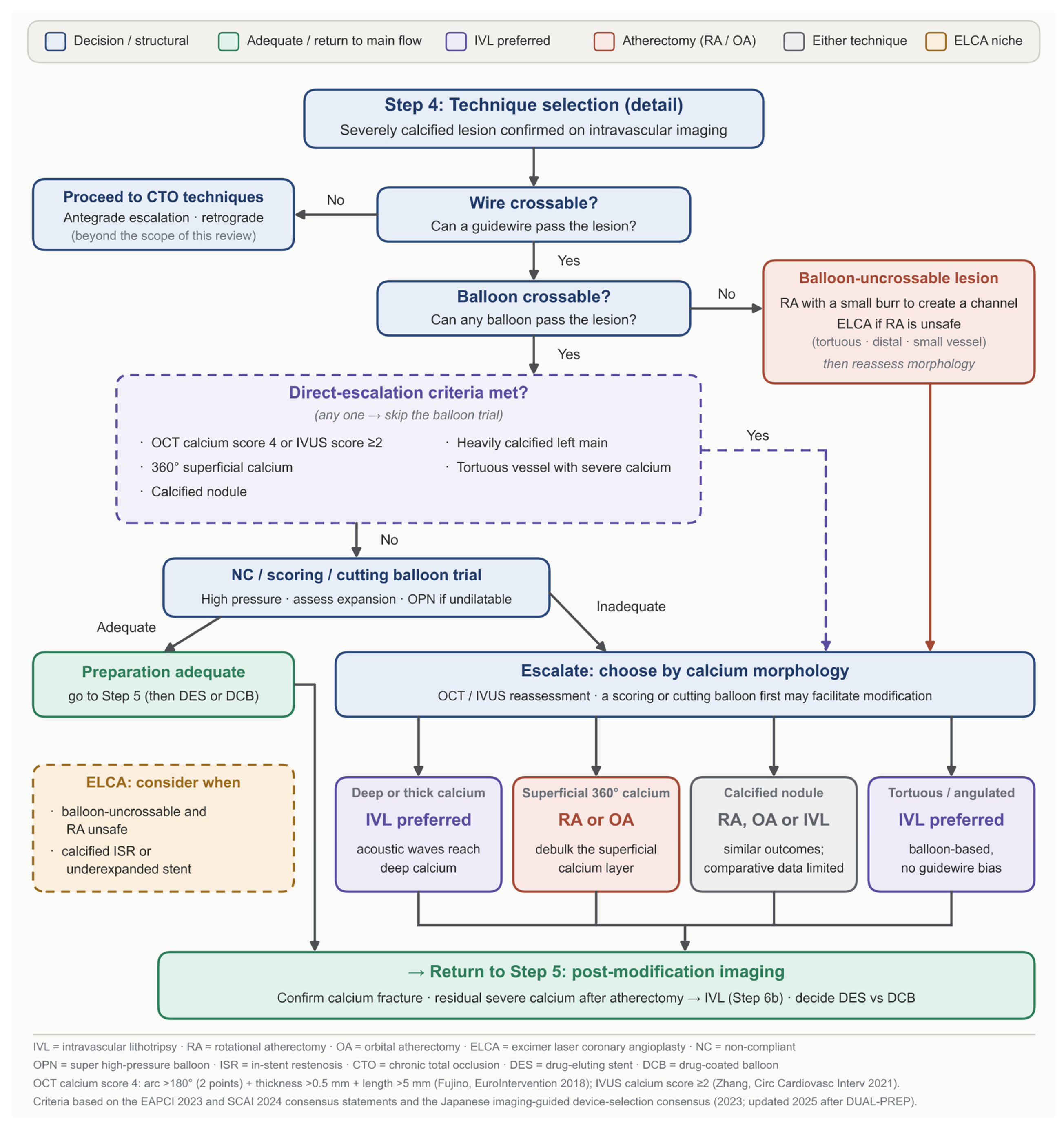
The practical application of the imaging-guided stepwise algorithm for calcium modification technique choice is illustrated in Figure 1 and Figure 2 of the present review.
Figure 1.
Imaging-guided algorithm for the interventional management of severely calcified coronary lesions. Pre-procedural planning and intravascular imaging inform calcium risk stratification [5,23,25], which determines whether balloon pre-dilation is attempted or plaque modification is undertaken directly. Post-modification imaging governs the choice between stent implantation, further escalation, including intravascular lithotripsy after rotational atherectomy, and the drug-coated balloon stentless pathway, the latter conditional on an imaging-confirmed quality check with bail-out stenting available at all times. Both algorithms (Figure 1 and Figure 2) combine recommendations of the EAPCI and SCAI consensus documents [2,22] and of the updated Japanese device-selection consensus (Section 6.2), with the evidence summarized in Table 1; where evidence is lacking, they reflect the authors’ practical proposal. Abbreviations: CCTA, coronary computed tomography angiography; CT, computed tomography; DAPT, dual antiplatelet therapy; DCB, drug-coated balloon; DES, drug-eluting stent; ELCA, excimer laser coronary angioplasty; HBR, high bleeding risk; IVL, intravascular lithotripsy; IVUS, intravascular ultrasound; MSA, minimum stent area; NC, non-compliant; OA, orbital atherectomy; OCT, optical coherence tomography; RA, rotational atherectomy; TIMI, thrombolysis in myocardial infarction.
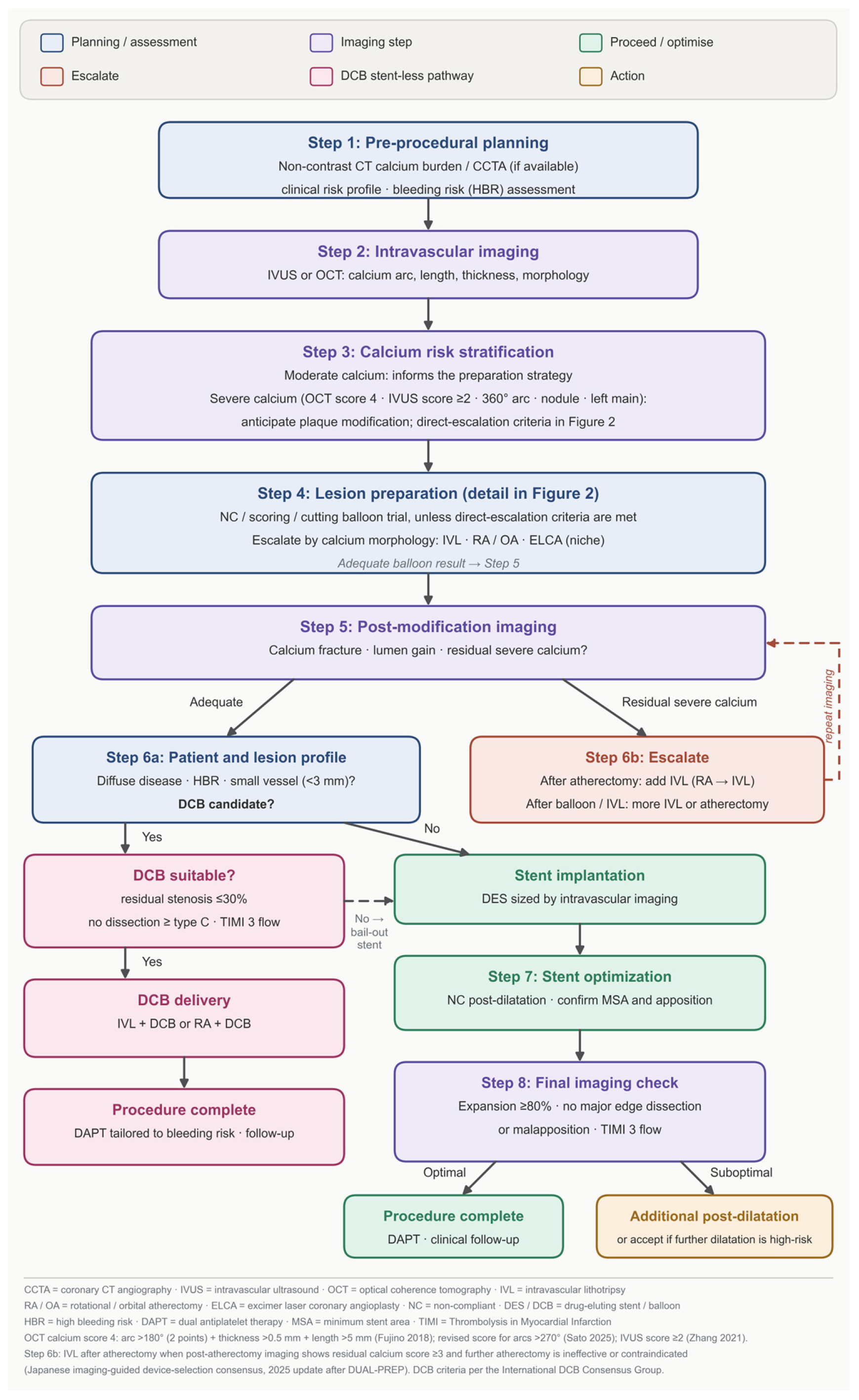
Figure 2.
Technique selection in severely calcified lesions (detail of Step 4 in Figure 1). Selection proceeds through wire crossability, then balloon crossability, and finally calcium morphology on intravascular imaging. Lesions meeting the direct escalation criteria bypass the balloon trial. Calcium morphology determines the preferred modification technique, with excimer laser coronary angioplasty reserved for the indications shown. All pathways return to post-modification imaging (Step 5). Abbreviations: CTO, chronic total occlusion; DCB, drug-coated balloon; DES, drug-eluting stent; ELCA, excimer laser coronary angioplasty; ISR, in-stent restenosis; IVL, intravascular lithotripsy; IVUS, intravascular ultrasound; NC, non-compliant; OA, orbital atherectomy; OCT, optical coherence tomography; OPN, super high-pressure balloon; RA, rotational atherectomy.
5. Plaque Modification Strategies: Mechanisms, Evidence, and Selection
Three situations should be distinguished, as they lead to different strategies (Figure 2). In the wire-uncrossable lesion, no guidewire can be advanced through the lesion; this requires chronic total occlusion techniques and is outside the scope of this review. In the balloon-uncrossable lesion, the guidewire has crossed but the smallest balloon cannot; atherectomy is the usual option, but because RA requires the dedicated RotaWire with microcatheter, ELCA may be needed when the exchange microcatheter cannot cross [2,22]. In the balloon-undilatable lesion, the balloon crosses but does not expand fully despite high pressure; IVL, atherectomy and ultra-high-pressure balloons are the main options (Section 5.1, Section 5.2, Section 5.3 and Section 5.4).
5.1. Balloon-Based Strategies
Balloon-based preparation remains the first-line approach in the majority of calcified lesions where wire and balloon passage is achievable, and represents the foundation upon which all escalation decisions are built. Non-compliant (NC) balloons, inflated at high pressure (typically 16+ atm), are the initial tool of choice, providing radial force capable of fracturing mild-to-moderate calcium deposits and achieving adequate lesion preparation in a substantial proportion of cases. Their principal advantages are simplicity, widespread availability, and low cost; however, their effectiveness is limited in the presence of severe circumferential or thick calcium, where the rigid plaque architecture resists adequate dilation and balloon waisting signals the need for escalation.
When NC balloon dilatation proves insufficient, scoring and cutting balloons represent a logical next step before proceeding to atherectomy or lithotripsy. Scoring balloons, such as the AngioSculpt device, incorporate a helical nitinol element that concentrates radial force along defined scoring lines, creating controlled longitudinal calcium fractures and reducing the risk of uncontrolled dissection compared to standard high-pressure inflation. Cutting balloons, equipped with three or four atherotomes mounted longitudinally on the balloon surface, operate on a similar principle of focused plaque incision. In PREPARE-CALC, a strategy based on modified balloons (scoring or cutting) was compared to upfront rotational atherectomy in severely calcified lesions; while RA achieved superior strategy success (98% vs. 81%), the modified balloon strategy succeeded in most cases, failures being driven mainly by crossover to RA for uncrossable or undilatable lesions, supporting their role as an intermediate escalation step before committing to atherectomy [27]. ECLIPSE used a different comparator, a conventional balloon angioplasty-based strategy rather than a protocol-defined modified-balloon strategy, and enrolled only lesions judged suitable for either technique; in its optical coherence tomography substudy, minimum stent area did not differ significantly from that obtained after orbital atherectomy [7].
It is worth noting that scoring and cutting balloons also retain a useful role as adjuncts after atherectomy, providing additional crack propagation through calcium that has been debulked but not fully disrupted, and facilitating IVL balloon passage in lesions where atherectomy has created a channel but residual calcification persists.
A further balloon-based option is the ultra-high-pressure non-compliant balloon, of which the OPN NC is the most widely used. Its twin-layer construction permits inflation to pressures well above those tolerated by conventional non-compliant balloons—35 atm and beyond—delivering very high radial force without the uncontrolled expansion that would otherwise accompany such pressures [28]. In the randomised ISAR-CALC trial, 74 patients whose severely calcified lesions had failed to respond to conventional non-compliant balloon preparation were assigned to an ultra-high-pressure balloon or a scoring balloon before drug-eluting stent implantation. The primary endpoint, stent expansion index on OCT, did not differ between arms (0.72 ± 0.12 vs. 0.68 ± 0.13; p = 0.22), although strategy success (91.9% vs. 83.8%; p = 0.48) and procedural success (100% vs. 89.2%; p = 0.12) were numerically higher with the ultra-high-pressure balloon, and angiographic core-laboratory analysis showed a larger final minimum lumen diameter and lower residual stenosis [29]. As such, the ultra-high-pressure balloon is best positioned as an escalation option in the balloon-crossable but undilatable lesion, rather than as a first-line preparation strategy. The comparative mechanisms, sizing and inflation strategies of scoring, cutting and ultra-high-pressure balloons have recently been reviewed in detail [30].
5.2. Intravascular Lithotripsy
Intravascular lithotripsy (IVL) represents the most significant addition to the calcified lesion armamentarium in the past decade. The Shockwave IVL system delivers localized, pulsatile acoustic pressure waves from emitters mounted within a dedicated balloon catheter, generating sonic pressure waves that selectively fracture both superficial and deep calcium deposits while sparing soft tissue. This feature represents a significant advantage over rotational atherectomy, which acts exclusively on superficial plaque [4]. The absence of guidewire bias, the balloon-based delivery system compatible with standard 0.014-inch coronary wires, and the relatively straightforward learning curve have contributed to its rapid adoption in centers worldwide.
The pivotal evidence base for IVL is derived from the DISRUPT CAD program, a series of prospective single-arm studies that collectively enrolled over 600 patients with severely calcified de novo lesions. In a patient-level pooled analysis of the DISRUPT CAD studies [31], procedural success, defined as stent delivery with residual stenosis ≤ 30% without in-hospital MACE, was achieved in 92.4% of cases; serious angiographic complications occurred in 2.1% of patients after IVL and 0.3% at final angiography, with no IVL-associated perforations. In DISRUPT CAD III (431 patients enrolled, 384 in the intention-to-treat population), 92.2% of patients were free from major adverse cardiovascular events at 30 days [4]. At one year, major adverse cardiovascular events had occurred in 13.8% of patients and target lesion failure in 11.9% (Kaplan–Meier estimates); most of these events were non-Q-wave myocardial infarctions (9.2%), the majority occurring in hospital, whereas cardiac death (1.1%) and ischaemia-driven target lesion revascularisation (4.3%) were infrequent, and definite or probable stent thrombosis was observed in 1.1%, only one event of which occurred beyond 30 days [32]. These results, obtained in a population with angiographically severe calcium and a high prevalence of complex anatomical features, established IVL as a safe and effective calcium-modification strategy.
A particularly relevant morphological subtype is the calcified nodule. In a pooled analysis of the DISRUPT CAD OCT substudies (155 lesions, 29 with nodules), IVL achieved comparable minimum stent area and stent expansion in nodular and non-nodular lesions, with numerically more calcium fractures in nodules and no significant difference in 2-year target lesion failure (13.9% vs. 8.0%) [13]. IVL has also been evaluated in the anatomically challenging setting of heavily calcified left main disease. In an analysis of the prospective BENELUX registry, technical success in left main lesions treated with IVL was 93.2% (55 out of 59 lesions), not significantly different from the 89.8% achieved in non-left main lesions (p = 0.41), with comparable MACE rates at discharge, 30 days, and 1 year, reinforcing its applicability beyond conventional lesion subsets [33].
From a practical standpoint, IVL is particularly well-suited to tortuous anatomy where rotational atherectomy carries increased procedural risk, and to lesions where deep calcium, inaccessible to atherectomy, is identified on pre-procedural OCT imaging. Its principal limitation is the requirement for balloon crossability: the IVL catheter, while trackable in most lesions, cannot cross a balloon-uncrossable lesion, and requires pre-treatment with a small balloon or atherectomy to establish a channel when the lumen is critically narrowed. The integration of IVL into combination strategies is addressed separately in Section 6.
5.3. Rotational Atherectomy
Rotational atherectomy (RA) has been the cornerstone of calcified lesion preparation for over three decades and remains the most widely adopted dedicated modification strategy in contemporary practice. The Rotablator system operates through a diamond-coated burr rotating at 135,000–180,000 rpm, advancing over a dedicated 0.009-inch guidewire and selectively ablating inelastic, calcified plaque through the principle of differential cutting, preferentially removing hard, calcified tissue while deflecting away from elastic, healthy vessel wall components [22]. This mechanism results in the pulverization of superficial calcium into microparticles small enough to pass through the coronary microcirculation, creating a smoother, more compliant luminal surface amenable to subsequent balloon dilatation and stent delivery.
The randomized evidence base for RA in severely calcified lesions is anchored primarily by two trials. ROTAXUS randomized 240 patients with complex calcified lesions to RA followed by paclitaxel-eluting stent implantation versus stenting alone, demonstrating a significantly larger acute lumen gain and higher procedural success with RA; however, the primary endpoint was not met: in-stent late lumen loss at nine months was in fact significantly higher in the rotational atherectomy arm (0.44 ± 0.58 vs. 0.31 ± 0.52 mm; p = 0.04), while binary restenosis, target lesion revascularisation and MACE did not differ between groups, a pattern generally attributed to the limitations of paclitaxel-eluting technology rather than to atherectomy itself [34]. More relevant to contemporary practice, PREPARE-CALC randomized 200 patients with severely calcified lesions to RA versus a modified balloon strategy (scoring or cutting balloon), demonstrating significantly higher strategy success with RA (98% vs. 81%; p = 0.0001), lower crossover rates, and superior acute procedural results, with comparable 9-month outcomes between the two strategies [27]. These data established RA as the preferred upfront strategy when modified balloons are anticipated to be insufficient.
Longer-term follow-up of PREPARE-CALC, however, suggests that this early equivalence does not persist indefinitely. At two years, no significant difference in target vessel failure or MACE was observed; however, rotational atherectomy appeared more favourable in longer lesions, and target lesion revascularisation tended to be lower with RA (2% vs. 8%; p = 0.053) [35]. At five years, target vessel failure remained comparable between strategies, but target lesion revascularisation was significantly less frequent after rotational atherectomy than after modified balloon preparation (3% vs. 12%; HR 0.28, 95% CI 0.08–0.98; p = 0.048), with rotational atherectomy independently protective against TLR on multivariable analysis [36]. This late divergence is relevant to the interpretation of trials reporting only mid-term outcomes, and suggests that the choice of preparation strategy may influence durability even where acute results appear equivalent.
Real-world data from the British Cardiovascular Intervention Society national database, encompassing 7740 rotational atherectomy procedures performed between 2013 and 2016, showed that lower annual operator RA volume was associated with a higher risk of major adverse cardiac and cerebrovascular events, highlighting the importance of institutional volume and operator expertise [37]. The Euro4C registry, which enrolled 966 patients undergoing RA across 19 centres in eight European countries, documented favourable results in contemporary practice, reporting clinical success in 91.9% of procedures, an in-hospital MACE rate of 4.7%, and a one-year MACE rate of 13.2% despite the high complexity of the population studied [38].
The principal limitation of RA is that it modifies only superficial calcium and cannot address deep calcium deposits that remain intact beneath the ablated layer. This is the mechanistic rationale for combination with IVL in selected cases, as discussed in Section 6. In bifurcation lesions, side-branch guidewires should be removed during RA because of the risk of wire fracture or burr entrapment, leaving the side branch unprotected during ablation [22]; this limitation is relevant to the treatment of calcified bifurcations (Section 7.1). Additional technical considerations include the risk of burr entrapment in severely angulated or tortuous segments, the mandatory use of a dedicated 0.009-inch RotaWire with its own handling characteristics, and the potential for slow-flow or no-reflow from microparticle embolization. These risks can be mitigated by appropriate burr sizing (burr-to-artery ratio ≤0.6), adequate rotational speed, and judicious use of vasodilators and anticoagulation.
5.4. Orbital Atherectomy
Orbital atherectomy (OA) relies on a distinct mechanism from rotational atherectomy, despite sharing the general principle of mechanical plaque modification. The Diamondback 360 system uses an eccentrically mounted, diamond-coated crown that when activated, orbits around the vessel centreline at variable speeds rather than rotating along a fixed axis. This orbital motion generates centrifugal force, allowing the ablation diameter to adapt to vessel size and enabling a single device to treat different vessel diameters without crown exchange. At the same time, the system modifies superficial calcium and the surrounding plaque, potentially creating a larger and more symmetrical lumen than a conventional rotational pass [2].
For many years, evidence supporting OA in calcified coronary lesions was largely limited to single-arm registries and observational studies, with ORBIT II providing the main prospective data and demonstrating high procedural success rate and a 30-day MACE-free rate of 89.6% with a 30-day TVR of only 1.4% [39]. More recently, the ECLIPSE trial provided the first adequately powered randomized comparison of OA and conventional balloon angioplasty as lesion preparation strategies before drug-eluting stent implantation in severely calcified coronary lesions [7]. The trial randomized 2005 patients across 104 centres in the United States and failed to demonstrate the superiority of routine OA on either of its two primary endpoints: target vessel failure at one year occurred in 11.5% of the OA group versus 10.0% of the balloon group (p = 0.28), while in the imaging cohort, the mean minimum stent area at the site of maximal calcification was 7.67 mm2 versus 7.42 mm2 (mean difference 0.26 mm2; p = 0.078). Crossover in the balloon arm occurred in only 4.9% of cases, substantially less frequently than in previous atherectomy-versus-balloon studies such as PREPARE-CALC. A safety signal deserving mention is the excess of early mortality in the OA arm, with cardiac death at 30 days occurring in 0.8% of OA-treated patients versus none in the balloon group (p = 0.005), four of these deaths being adjudicated as related or possibly related to the device, in the absence of any corresponding excess of dissection or perforation. Two important caveats qualify the interpretation of these results: enrolment was restricted to lesions in which the operator considered both strategies to be reasonable, thereby excluding the most severely calcified lesions in which atherectomy would have been deemed mandatory, and the trial was conducted almost entirely before IVL became widely available, with lithotripsy used in less than 1% of cases in either arm. These findings nonetheless suggest that in a broadly selected population with severely calcified lesions, the incremental benefit of routine OA over optimized balloon preparation may be more limited than previously anticipated and further emphasize the importance of appropriate lesion selection, preferably guided by intravascular imaging.
The role of OA in contemporary practice is therefore mainly related to several practical and anatomical advantages. Its ability to treat different vessel diameters with a single device may simplify procedures in situations where RA burr exchange is less desirable, while bidirectional crown movement may offer advantages in selected anatomical configurations. However, similar to RA, OA acts predominantly on superficial calcium and has limited ability to modify deep or circumferential calcium that remains beneath the treated surface [19].
5.5. Excimer Laser Coronary Angioplasty
Excimer laser coronary angioplasty (ELCA) is the oldest dedicated plaque-modification technology still in clinical use, having been introduced in the late 1980s, but continues to retain a relevant, although relatively niche, role in the contemporary treatment of calcified coronary lesions. The CVX-300 system delivers ultraviolet energy at a wavelength of 308 nm through a multifibre catheter, producing tissue ablation through three simultaneous mechanisms: photochemical disruption of molecular bonds, photothermal vaporization of tissue water, and photomechanical shock waves generated by rapid bubble formation and collapse [22]. Unlike purely mechanical techniques, ELCA can modify not only calcified tissue, but also fibrotic plaque, thrombus, and neointimal tissue within stents, extending its applicability across a broader spectrum of lesion substrates than atherectomy alone.
In calcified coronary lesions, ELCA is conventionally described as addressing “balloon failure”, an umbrella term covering both balloon-uncrossable and balloon-undilatable lesions. The two indications are, however, mechanistically distinct and are supported by different techniques and different bodies of evidence. In the uncrossable lesion, the limitation is device delivery, and the relevant property of the laser catheter is its profile: a 0.9 mm catheter advanced over a conventional 0.014-inch guidewire ablates tissue as it is advanced, creating a channel through which balloons, atherectomy devices or stents may subsequently pass. Bilodeau et al. evaluated this approach prospectively in 100 calcified and/or balloon-resistant lesions treated with the 0.9 mm catheter under saline flush, with increased energy parameters used in 29 resistant lesions, and reported laser technical success in 92%, procedural success in 93%, and clinical success in 86% [40].
In the undilatable lesion, the balloon has already crossed and catheter profile is irrelevant; what is required is disruption of the resistant substrate itself. This is achieved by lasing at high energy during contrast injection rather than under saline flush, a deliberate reversal of the standard technique that amplifies the photoacoustic and photomechanical component of laser–tissue interaction. Because lasing during contrast injection also increases the risk of no-reflow and perforation [2], it should be regarded as a specialised, higher-risk manoeuvre reserved for experienced operators rather than as a simple alternative to saline flush. The ELLEMENT registry applied this technique in 28 patients with stent underexpansion refractory to high-pressure non-compliant balloon inflation and achieved successful laser-assisted dilatation in 27 cases (96.4%), with minimum stent diameter increasing from 1.6 ± 0.6 mm to 2.6 ± 0.6 mm and minimum stent area from 3.5 ± 1.1 mm2 to 7.1 ± 1.9 mm2; periprocedural myocardial infarction occurred in 7.1%, transient slow flow in 3.6%, and target lesion revascularisation in 6.7% during follow-up [41]. It should be emphasised that this indication has been characterised predominantly in underexpanded and restenotic stents rather than in de novo calcified disease, and the extrapolation to the undilatable de novo calcified lesion is therefore indirect. Historical data are also relevant here: in the PELCA multicentre registry, Ahmed et al. concluded that although laser-facilitated angioplasty was effective for lesions not dilatable by balloon alone, success rates were lower specifically in calcified lesions, and that such lesions were better approached with alternative therapy [42]. As such, in the de novo severely calcified undilatable lesion, rotational atherectomy or intravascular lithotripsy remain the more established options, and ELCA is most defensible where a balloon cannot be delivered at all, where the substrate includes thrombus or neointima, or where atherectomy is anatomically hazardous.
The main practical limitations of ELCA include the need for saline flushing during energy delivery to avoid excessive bubble formation in the presence of contrast, a more limited depth of calcium modification compared with IVL, and a learning curve related to energy titration and catheter manipulation. Coronary perforation remains an uncommon but recognized complication and requires careful energy delivery, particularly in tortuous or angulated segments. Nevertheless, in centres with adequate experience, ELCA represents an important complementary option to RA and IVL, especially in lesions where the risk of burr entrapment can make RA more challenging [19].
5.6. How Do They Compare?—What the Data Actually Show
Despite the mechanistic rationale for technique-specific advantages and the growing body of procedural data supporting each individual modification strategy, the question of whether any single approach translates into superior hard clinical outcomes compared to others remains largely unanswered; the key studies discussed in this review are summarized in Table 1. A systematic review and meta-analysis by Kristensen et al., incorporating 31 randomized controlled trials and 8453 patients, of which 24 trials and 4392 patients addressed lesion preparation techniques specifically, found no significant difference between techniques for all-cause mortality or serious adverse events at any follow-up timepoint [43]. The certainty of evidence was rated low to very low across most comparisons, reflecting the persistent limitations of the existing trial landscape: small sample sizes, heterogeneous patient populations, variable imaging guidance, inconsistent definitions of calcification severity, and a general underpowering for mortality endpoints. These findings, while perhaps counterintuitive given the procedural differences between strategies, are consistent with the broader literature on complex PCI, where differences in acute procedural metrics do not reliably translate into differences in long-term clinical outcomes when confounding by lesion complexity and patient selection is adequately addressed.
ROLLER COASTR-EPIC22 provides the most direct comparative evidence, randomising 171 patients with moderate-to-severe calcified coronary lesions at eight Spanish centres to RA, IVL or ELCA, with stent expansion on OCT as the primary endpoint [6]. IVL was non-inferior to RA (85.6 ± 13.3% vs. 86.4 ± 14.1%; p = 0.77), whereas ELCA did not reach non-inferiority in the intention-to-treat analysis (80.3 ± 13.3%). Minimum stent area, procedural success and complication rates did not differ between the three strategies, and at one year, no significant differences were observed in major adverse cardiovascular events or their components—cardiac death, target vessel myocardial infarction, target lesion and target vessel revascularisation, and stent thrombosis [44].
The BALI trial addressed a different question, namely whether IVL adds to conventional lesion preparation [45]. In 200 patients with severely calcified lesions, adding IVL reduced the primary composite of procedural failure or 1-year target vessel failure (35% vs. 52%; relative risk 0.69, 95% CI 0.48–0.97; p = 0.02), mainly through fewer cases of residual area stenosis ≥ 20% on OCT (32% vs. 45%), with no difference in safety endpoints.
The ROLLING STONE registry (2026) is, to our knowledge, the most recent comparison between these techniques. It enrolled 1005 consecutive patients with complex calcified lesions across 23 Italian centres, of whom 544 treated with IVL and 380 treated with rotational or orbital atherectomy were compared, after the exclusion of 81 patients who received both [46]. Procedural success was comparable between techniques (85.4% with IVL vs. 86.3% with atherectomy; relative risk 1.01, 95% CI 0.88–1.17), whereas 30-day MACE was significantly lower in the IVL group (5.7% vs. 8.6%; p = 0.045), a difference driven predominantly by a reduction in cardiovascular death (1.7% vs. 3.9%; p = 0.03) and which remained significant at 12 months after propensity matching (6.8% vs. 14.3%; HR 0.43; p = 0.022). These findings should nonetheless be interpreted with caution, as device selection was left to operator discretion and may have been influenced by both lesion complexity and individual operator experience, such that part of the observed safety difference may reflect patient and lesion selection rather than the technique itself.
This equipoise should not, however, be read as complete interchangeability. It is most secure for mortality and myocardial infarction, endpoints for which the existing trials are uniformly underpowered, and least secure for repeat revascularisation, where the five-year PREPARE-CALC data show a significant advantage for rotational atherectomy over modified balloon preparation that was not apparent at nine months [36]. Taken together, these data suggest that—at least within the range of lesion complexity captured by existing trials, and over the follow-up durations they report—the choice of modification technique may matter less for hard safety endpoints than the quality of its execution and the adequacy of subsequent stent optimization, while differences in durability may yet emerge with longer follow-up.
These comparisons share a structural limitation. The techniques do not act on the same target: atherectomy ablates superficial calcium and can create a channel in the balloon-uncrossable lesion, whereas IVL requires balloon delivery and fractures superficial and deep calcium in the crossable but undilatable lesion. Comparative trials such as ECLIPSE selected lesions by overall, mainly angiographic, calcium severity [7] rather than by calcium type on intravascular imaging, and thus compared devices with different mechanisms across mixed substrates. A neutral result in such a population does not show that the techniques are interchangeable; they are complementary and frequently combined (Section 6.1). Future comparisons would be more informative if restricted to lesions with predefined, comparable calcium characteristics on intravascular imaging.
In contrast to the clinical equipoise observed between different modification techniques, the benefit of intravascular imaging guidance in calcified lesion PCI has been demonstrated with considerably greater consistency across the literature. The OCTOBER trial, while not restricted to calcified lesions, demonstrated that OCT-guided PCI reduced major adverse cardiac events at 2 years compared with angiography-guided PCI in complex bifurcation lesions (10.1% vs. 14.1%; HR 0.70) [47]. Registry data have consistently linked IVUS guidance to reduced target lesion revascularization and improved survival in complex PCI, including heavily calcified subsets, with the benefit appearing most pronounced precisely in those anatomical situations where angiography is most likely to underestimate lesion severity and stent underexpansion [2,22]. In the randomized RENOVATE-COMPLEX-PCI trial, in which severe calcification was one of the qualifying complex-lesion features, imaging-guided PCI reduced the composite of cardiac death, target-vessel myocardial infarction or target-vessel revascularization compared with angiography guidance (7.7% vs. 12.3%; HR 0.64) [48].
The mechanistic explanation for this consistent imaging benefit is straightforward: in calcified lesions, the consequences of stent underexpansion are directly detectable only by intravascular imaging, and their correction requires imaging-guided post-dilatation that cannot be reliably performed on the basis of angiographic appearance alone. Although OCTOBER and RENOVATE-COMPLEX-PCI were not designed to compare calcium-modification strategies, these trials, together with the class I guideline recommendation for imaging-guided PCI of anatomically complex lesions [49], support imaging-guided stent optimization as an important determinant of long-term outcomes, possibly more so than the choice of modification technique.
The influence of stent platform on outcomes in calcified lesions has also been examined. Rheude T et al. reported a pooled analysis of the ISAR-TEST 4 and 5 trials at 10-year follow-up, demonstrating an incremental increase in adverse events with the degree of calcification, with 10-year mortality rising from 25.3% in patients without calcification to 46.5% in those with severe calcification, while in moderate-to-severe, the calcification outcomes did not differ according to stent polymer strategy, suggesting that polymer choice does not alter the prognostic penalty of calcified disease in the long-term [50]. These data collectively reinforce the concept that calcification should be viewed as a systemic disease marker requiring aggressive medical management in parallel with optimized revascularization technique.
The main complications of each modification technique, together with preventive measures and bail-out strategies, are summarized in Table 2 [2,19,22].
Table 1.
Overview of key studies discussed in the review.
Table 2.
Main complications of calcium-modification techniques, with preventive measures and bail-out strategies.
6. The Evolving Paradigm: Combination Strategies and the Post-DUAL-PREP Era
6.1. Rationale for Combination Approaches
The single-technique limitations of each individual modification strategy provide a strong mechanistic rationale for sequential or combined use. Rotational atherectomy, as discussed, acts exclusively on superficial calcium and is unable to address deep calcium that remains intact beneath the ablated layer; IVL, while capable of fracturing both superficial and deep calcium, requires balloon crossability and cannot be used in a balloon-uncrossable lesion without prior preparation; ELCA provides ablation but with limited calcium penetration depth compared to IVL. These complementary mechanisms suggest that in the most severely calcified lesions where a single strategy proves insufficient, sequential use of two techniques targeting different calcium compartments may achieve superior modification than either alone.
The combination of RA followed by IVL is mechanistically the most logical of these approaches: RA debulks and smooths superficial calcium, reducing plaque bulk and improving luminal geometry, while subsequent IVL fractures the residual deep calcium that atherectomy cannot reach, maximizing vessel compliance before stent delivery. This sequence (“RotaTripsy”) had been described in European case series and a multicentre registry [19,56], but the 2023 Japanese imaging-guided device-selection consensus listed it as contraindicated until dedicated safety data became available (Section 6.2).
6.2. RA + IVL: From Contraindication to Accepted Strategy
The shift from contraindication to accepted practice was driven mainly by two datasets. The multicentre Rota-Shock registry (160 patients, 23 centres) showed that IVL used electively or as bail-out after RA achieved a procedural success rate of 96.9%, although perforation occurred in 2.5% of patients [56]. Subsequently, the DUAL-PREP registry provided prospective, imaging-guided safety data, prospectively enrolling 118 patients with 120 severely calcified lesions across 20 Japanese centres in whom post-atherectomy intravascular imaging confirmed residual heavy calcium deemed insufficient for direct stenting. The OCT-derived calcium score in the enrolled population was 4 for all lesions and atherectomy was performed with a rotational burr in 83.9% and an orbital crown in 16.1% of cases. IVL was successfully delivered in every case, with a procedural success rate of 98.3% and 30-day freedom from MACE of 98.3% [8]. No perforation, acute occlusion or severe dissection occurred during IVL or stent implantation, and at 1 year, MACE had occurred in 7.6% of patients [57]. As a prospective single-arm registry without a control group, DUAL-PREP supports the feasibility and short-term safety of the combination rather than proving its efficacy [8,57].
These data contributed to the revision of the 2023 imaging-guided device selection consensus document [51]. In the updated algorithm, the fundamental principle of imaging-guided planning is retained; however, the critical modification is the explicit acknowledgment that IVL may now be considered after atherectomy when post-atherectomy intravascular imaging confirms persistent severe calcium (residual calcium score ≥3) and further atherectomy is judged either unlikely to be effective based on lesion characteristics or contraindicated for clinical reasons, such as reduced left ventricular function or impaired renal function [52]. This represents the most clinically significant algorithmic evolution in calcified lesion PCI since the introduction of IVL itself, which is directly reflected in the stepwise algorithm presented in Figure 1 of this review.
6.3. Drug-Coated Balloons After Lesion Preparation—A Stentless Strategy in Diffuse Calcified Disease
The concept of a stentless approach to calcified coronary disease—delivering antiproliferative drug therapy to the vessel wall without permanent metallic implantation—has gained increasing traction in recent years, driven by the recognition that total stent length is an independent predictor of target vessel failure and that diffuse calcified lesions, by their very nature, often require long stenting segments with attendant risks of stent malapposition, underexpansion, and restenosis [58,59]. The appeal is particularly compelling in specific patient subsets: those at high bleeding risk (HBR) in whom prolonged dual antiplatelet therapy carries prohibitive haemorrhagic risk, elderly patients with diffuse small-vessel disease, and those with chronic kidney disease where the consequences of stent thrombosis or restenosis are disproportionately severe [59,60].
The fundamental prerequisite for a DCB strategy in calcified disease is the absolute requirement for adequate lesion preparation prior to balloon delivery. Drug transfer from a paclitaxel or sirolimus coated balloon to the vessel wall is critically dependent on direct, homogeneous balloon-wall contact; residual calcium, which prevents full balloon apposition and creates zones of non-contact, will result in unpredictable drug delivery [59]. In de novo lesions more broadly, propensity-matched data show long-term outcomes after DCB comparable to those after DES [61]. As such, the DCB in calcified disease is not a standalone strategy but rather the terminal step of a carefully planned preparation sequence, with the quality of lesion preparation serving as the decisive determinant of whether DCB delivery is feasible, safe, and likely to be effective.
From a mechanistic standpoint, IVL is particularly well-suited as a preparatory strategy before DCB delivery. Unlike atherectomy, which removes tissue and may create a roughened, irregular surface, IVL modifies calcium through fracture while preserving vessel wall continuity and achieving a relatively smooth luminal surface, theoretically optimizing conditions for uniform drug transfer. Räsänen et al. reported a retrospective single-centre series of 34 consecutive patients with severely calcified de novo lesions treated with IVL followed by paclitaxel DCB alone, after the exclusion of five who required bail-out stenting, with the majority (76%) classified as high bleeding risk; there were no acute vessel closures or periprocedural myocardial infarctions, and at 12 months, MACE occurred in 15% (n = 5), driven predominantly by cardiovascular death (9%, n = 3), representing, to our knowledge, the most direct available evidence for IVL+DCB as a stentless strategy in this population [9].
The evidence base for RA followed by DCB is more mature, albeit still predominantly observational. Nagai T et al. reported outcomes in 190 lesions treated with RA+DCB at a single centre; among the 87 lesions with adequate follow-up angiography at approximately 6.5 months, the angiographic restenosis rate was 17.8%, late lumen loss 0.23 mm, and late lumen enlargement was observed in 39.1% of lesions, a phenomenon reflecting positive vessel remodeling in the absence of a metallic scaffold and analogous to that observed in DCB-treated CTOs [55,59]. Dong H et al. provided the most direct comparative data in a retrospective analysis of 318 consecutive patients with severe coronary calcification treated with either RA+DCB (n = 57) or RA+DES (n = 261), demonstrating no significant difference in MACCE or target lesion revascularization between strategies at mid-term follow-up, a finding that, while hypothesis-generating given the observational design and sample size, supports the biological plausibility of the RA+DCB approach [54]. Ueno et al. compared DCB with new-generation DES after imaging-guided RA in 123 patients and found similar 3-year target lesion revascularization (15.6% vs. 16.3%), with lower late lumen loss after DCB (0.09 vs. 0.52 mm) [62].
However, no randomized controlled trial has specifically addressed the DCB-after-preparation strategy in calcified coronary lesions, all existing data are observational and largely from single centers, and long-term data beyond 1 year are absent for the IVL+DCB combination. The ongoing TRANSFORM II trial, evaluating DCB versus DES in de novo coronary lesions including complex calcified subsets, will provide the first prospective randomized data to inform this strategy, though its primary results are awaited [63].
6.4. Practical Algorithm for Technique Selection in 2026
The contemporary management of severely calcified coronary lesions is best approached as a stepwise, imaging-guided escalation strategy rather than as a choice between competing technologies. As illustrated in Figure 1 and Figure 2, the algorithm begins with pre-procedural risk assessment and calcium quantification by CT or intravascular imaging, followed by the evaluation of wire and balloon crossability and OCT- or IVUS-based characterization of calcium severity to guide the need for and intensity of plaque modification.
In lesions with a moderate calcium burden that remain balloon-crossable, initial preparation with an NC, scoring, or cutting balloon is generally appropriate. An adequate response, defined by complete balloon expansion without significant waist and satisfactory lumen gain on imaging, allows progression to stent implantation. When balloon-based preparation is insufficient, or when intravascular imaging identifies a calcium pattern requiring upfront modification, device selection should be adapted to lesion morphology. IVL is particularly useful in lesions with a dominant deep calcium component or tortuous anatomy; RA is better suited for extensive superficial circumferential calcium and balloon-uncrossable lesions where atherectomy can facilitate subsequent device delivery; while ELCA remains an important option in selected balloon- or microcatheter-uncrossable lesions and calcified ISR. Following plaque modification, repeat imaging is essential to assess calcium fracture or full balloon expansion [2] and lumen gain and to determine whether stent implantation can proceed directly, whether further escalation or a combination strategy is required, or whether DCB treatment may be considered in selected patients with diffuse disease or HBR. When stenting is performed, high-pressure NC post-dilatation and imaging-confirmed optimization complete the procedural sequence.
Importantly, no single calcium-modification technique has demonstrated consistent superiority for hard clinical endpoints in adequately powered randomized trials, as discussed in Section 5.6. This further supports an individualized, morphology-based approach in which intravascular imaging guides both initial device selection and subsequent optimization, with procedural outcome depending not only on the technique used for lesion preparation, but also on the quality of the final result. Within this sequence, balloon-based preparation, atherectomy, IVL and imaging-guided stent optimization reflect established practice, whereas IVL after atherectomy and DCB after lesion preparation remain emerging strategies supported mainly by observational data (Section 7.4).
7. Special Considerations and Future Directions
7.1. Specific Lesion Subsets
Certain anatomical configurations within the broader category of calcified coronary disease warrant specific consideration, as the general principles outlined in the preceding sections require meaningful modification in their application.
Calcified left main disease represents the highest-stakes anatomical substrate in this context, where procedural complications carry the most severe haemodynamic consequences and where the penalty for suboptimal stent expansion is disproportionately high given the large myocardial territory subtended. IVL has emerged as a particularly attractive modification strategy in this setting, combining effective calcium modification with a safety profile that avoids the rotational risks associated with RA in large-caliber, bifurcating vessels with a technical success of 93.2% in left main lesions in the BENELUX registry, comparable to non-left main lesions and with comparable 1-year MACE [33], and of 99.4% in a multicentre series of 184 patients [64]; this evidence base is growing but remains observational. RA retains a role in left main disease, particularly for balloon-uncrossable lesions or those with dominant ostial calcification, but requires particular attention to burr sizing as the large vessel diameter of the left main mandates careful burr-to-artery ratio selection to avoid the risk of slow-flow from excessive ablation [22].
Calcified bifurcations present the combined challenge of calcium modification and bifurcation management, with the additional complexity that aggressive preparation of the main vessel may compromise side-branch access and that post-dilatation after bifurcation stenting must account for the calcium-related resistance to adequate kissing balloon inflation. IVL is mechanistically advantageous in this setting due to its ability to modify calcium at the polygon of confluence without the guidewire bias that may deflect an atherectomy burr away from the carina, though dedicated evidence in calcified bifurcations specifically remains limited [65] and represents an important area for future investigation, with case-level experience suggesting that IVL-facilitated DCB delivery is feasible in this setting, with angiographic evidence of late luminal enlargement at follow-up [19,60].
Calcified nodules constitute a distinct and particularly challenging morphological subtype, as discussed in Section 3. The eruptive variant—characterized by surface disruption, fibrous cap rupture, and associated thrombus—is a recognized substrate for acute coronary syndromes and carries specific procedural implications. RA requires special caution in this setting due to the risk of burr entrapment against the protruding nodule and distal embolization of nodular material; IVL has demonstrated feasibility and acceptable outcomes in dedicated substudies of the DISRUPT CAD program [13]. Yasumura K et al. compared RA, OA and IVL for calcified nodules (54 lesions) under OCT guidance and found broadly similar minimum stent area and 1-year target vessel failure with the three strategies, providing, to our knowledge, the most direct comparative data currently available for this morphological subtype [66].
7.2. High-Risk Patient Subgroups
The management of calcified coronary lesions must be further individualized according to patient-level characteristics that modify both procedural risk and the optimal strategy selection.
Elderly patients (above 75 years old) represent a growing proportion of the calcified lesion population, given the strong age-related increase in calcium burden and the high prevalence of concomitant frailty, chronic kidney disease, and polypharmacy. In this subgroup, the risk–benefit calculation for aggressive modification strategies shifts meaningfully: while adequate lesion preparation remains essential for durable outcomes, the procedural risks of atherectomy, such as slow-flow, no-reflow, and contrast-associated acute kidney injury, are amplified, and the DCB-after-preparation strategy, by eliminating the need for prolonged dual antiplatelet therapy, carries particular appeal.
Patients with chronic kidney disease (CKD) and end-stage renal disease represent another high-risk subgroup in whom calcified lesions are both more prevalent and more consequential, given the dramatically elevated rates of stent restenosis and target lesion revascularization in this population. Contrast volume minimization, combined with judicious use of intravascular imaging to reduce the number of contrast injections required for procedural guidance, should be a priority in CKD patients undergoing calcified lesion PCI [67].
Patients with diabetes mellitus warrant a special mention given the distinct calcium morphology, which is often diffuse and involves small vessels and the medial, thus responding differently to modification strategies primarily designed for focal intimal calcium. The combination of small vessel reference diameter, diffuse calcium distribution, and HBR status in many diabetic patients makes the DCB-after-preparation strategy conceptually attractive, though dedicated evidence in this subgroup remains limited.
Finally, haemodynamic support with mechanical circulatory assistance devices such as Impella may be considered in selected patients with severely reduced left ventricular function undergoing complex calcified lesion PCI, where the combination of prolonged procedural duration, repeated contrast injections, and the haemodynamic consequences of potential no-reflow may precipitate acute decompensation; the decision should be individualized rather than based on ventricular dysfunction alone [22].
7.3. Emerging Technologies and Unanswered Questions
Several technological developments on the horizon have the potential to further reshape the management of calcified coronary lesions over the coming years.
High-definition IVUS (HD-IVUS) systems, operating at substantially higher frequencies than conventional IVUS, offer improved axial resolution that approaches the calcium characterization capability of OCT while retaining IVUS’s superior tissue penetration in large-caliber vessels and its advantage in the absence of blood-clearing requirements [68]. The clinical impact of HD-IVUS on procedural decision-making and outcomes in calcified lesions specifically remains to be established in dedicated prospective studies.
Photon-counting CT, as discussed in Section 4.1, represents the most promising non-invasive advance for pre-procedural calcium characterization, with its substantially reduced blooming artefacts and improved spatial resolution offering the potential to reliably measure calcium arc and thickness from a non-invasive scan. If this technology is validated against intravascular imaging standards, it could meaningfully inform pre-procedural strategy planning and reduce the number of patients requiring intravascular imaging solely for calcium assessment [16].
Artificial intelligence-assisted calcium quantification from OCT and IVUS pullbacks represents a further frontier, with several groups reporting automated calcium scoring algorithms with similar results to expert reader performance with substantially reduced analysis time [69]. The integration of real-time AI-assisted imaging interpretation into the catheterization laboratory workflow could democratize the benefits of intravascular imaging-guided calcium assessment to centres without dedicated imaging expertise.
From a therapeutic standpoint, the most critical unmet need remains the absence of adequately powered randomized trials with mortality endpoints comparing modification strategies against each other and against optimized balloon preparation. As Kristensen et al. demonstrated, the existing evidence base, despite including more than 4000 patients randomized to lesion-preparation strategies, remains insufficient to detect clinically meaningful differences in hard endpoints between techniques [43]. Ongoing studies include randomized comparisons of balloon-based preparation with IVL, such as ISAR-CALC 2 [70], the TRANSFORM II trial of DCB versus DES in de novo lesions [63], and single-arm device studies such as FORWARD CAD, which is evaluating a forward-firing IVL catheter in difficult-to-cross lesions, similar to the Shockwave Javelin Peripheral IVL already used in PAD treatment [71]. Until their results are available, clinical decision-making in calcified lesion PCI will continue to rest on a foundation of mechanistic rationale, procedural endpoints, and expert consensus rather than definitive outcome data, a limitation that should be acknowledged both in clinical practice and in the conclusions of reviews such as this.
7.4. Strength of the Evidence
The recommendations of this review rest on evidence of unequal strength. First, the need for adequate lesion preparation and for intravascular imaging guidance in complex calcified lesions is supported by relatively strong evidence: imaging-based calcium scores predict stent underexpansion [5,23,25], and randomized trials of imaging-guided PCI in complex lesions showed fewer adverse events, consistent with current guideline recommendations [47,48,49]. Second, the choice between RA, IVL, OA and ELCA depends largely on anatomy, calcium morphology and procedural considerations; randomized comparisons have used mainly procedural or imaging endpoints [6,7,27,29,45], and a meta-analysis found no difference in hard clinical endpoints, with low to very low certainty of evidence [43]. Third, RA followed by IVL [8,56,57] and DCB after lesion preparation [9,54,62] are promising but supported only by registries and observational series, and require randomized validation [63].
7.5. Limitations
This review has limitations. The literature search was not systematic, so relevant studies may have been missed, and the evidence was not formally graded. The studies discussed differ in patient and lesion selection, definitions of calcium severity and endpoints; most randomized comparisons were underpowered for clinical endpoints [43], and the evidence for emerging strategies, such as RA followed by IVL and DCB after lesion preparation, is largely observational [8,9,54,56,57,62]. The algorithms in Figure 1 and Figure 2 therefore combine evidence of unequal strength with expert consensus and the authors’ practical experience.
8. Conclusions
The management of severely calcified coronary lesions has become an imaging-driven discipline. Angiography identifies fewer than half of the lesions that intravascular imaging shows to be calcified and provides little of the quantitative parameters that predict stent underexpansion, so OCT- and IVUS-derived calcium assessment now constitutes the practical basis for deciding whether, and how aggressively, to modify a lesion before stenting.
Against that background, no single modification technique has demonstrated superiority for hard clinical endpoints, but this equipoise is not interchangeability. Rotational atherectomy, intravascular lithotripsy, excimer laser angioplasty and balloon-based preparation act on different calcium compartments and carry different anatomical constraints, and the relevant question is therefore which technique suits a given lesion rather than which is superior in general. Moreover, combinations of such techniques can prove useful in the direst of situations, as is the example of RA+IVL.
After adequate lesion preparation, DCB represents a promising stentless option in carefully selected calcified lesions, although randomized evidence is still lacking. Adequately powered trials with clinical endpoints, incorporating systematic imaging guidance and contemporary modification strategies, however, remain the principal unmet need in this field.
Author Contributions
Conceptualization, T.P.K., F.-L.L., C.H., M.B. and D.-M.O.; investigation and literature search, T.P.K. and F.-L.L.; writing—original draft preparation, T.P.K.; writing—review and editing, F.-L.L., C.H., M.O. (Mihai Ober), D.T., M.S., M.O. (Maria Olinic), M.B. and D.-M.O.; visualization, T.P.K.; supervision, C.H. and D.-M.O.; project administration, D.-M.O. All authors have read and agreed to the published version of the manuscript.
Funding
This research received no external funding.
Institutional Review Board Statement
Not applicable.
Informed Consent Statement
Not applicable.
Data Availability Statement
No new data were created or analyzed in this study. Data sharing is not applicable to this article.
Acknowledgments
During the preparation of this manuscript, the authors used Claude Opus 5.5 (Anthropic, PBC) for literature synthesis, drafting and language editing of the manuscript text, generation of the figures, and compilation of the summary table. All content generated with this tool was subsequently verified against the primary literature, reviewed and edited by the authors, who take full responsibility for the accuracy, integrity and originality of the published work.
Conflicts of Interest
The authors declare no conflicts of interest.
Appendix A
Table A1.
PubMed/MEDLINE searches, run on 20 October 2026 with the limits english[la] AND (“1990/01/01”[dp]: “2026/09/30”[dp]). The combined search retrieved 67 of the 70 cited publications indexed in PubMed (not retrieved: [15,17,49]); no publication was added on the basis of this search.
References
- McInerney, A.; Hynes, S.O.; Gonzalo, N. Calcified Coronary Artery Disease: Pathology, Prevalence, Predictors and Impact on Outcomes. Interv. Cardiol. 2025, 20, e02. [Google Scholar] [CrossRef] [Scilit]
- Riley, R.F.; Patel, M.P.; Abbott, J.D.; Bangalore, S.; Brilakis, E.S.; Croce, K.J.; Doshi, D.; Kaul, P.; Kearney, K.E.; Kerrigan, J.L.; et al. SCAI Expert Consensus Statement on the Management of Calcified Coronary Lesions. J. Soc. Cardiovasc. Angiogr. Interv. 2024, 3, 101259. [Google Scholar] [CrossRef] [Scilit]
- Kawashima, H.; Serruys, P.W.; Hara, H.; Ono, M.; Gao, C.; Wang, R.; Garg, S.; Sharif, F.; de Winter, R.J.; Mack, M.J.; et al. 10-Year All-Cause Mortality Following Percutaneous or Surgical Revascularization in Patients With Heavy Calcification. JACC Cardiovasc. Interv. 2022, 15, 193–204. [Google Scholar] [CrossRef] [Scilit]
- Hill, J.M.; Kereiakes, D.J.; Shlofmitz, R.A.; Klein, A.J.; Riley, R.F.; Price, M.J.; Herrmann, H.C.; Bachinsky, W.; Waksman, R.; Stone, G.W.; et al. Intravascular Lithotripsy for Treatment of Severely Calcified Coronary Artery Disease. J. Am. Coll. Cardiol. 2020, 76, 2635–2646. [Google Scholar] [CrossRef] [Scilit]
- Fujino, A.; Mintz, G.S.; Matsumura, M.; Lee, T.; Kim, S.Y.; Hoshino, M.; Usui, E.; Yonetsu, T.; Haag, E.S.; Shlofmitz, R.A.; et al. A new optical coherence tomography-based calcium scoring system to predict stent underexpansion. EuroIntervention 2018, 13, e2182–e2189. [Google Scholar] [CrossRef] [Scilit]
- Jurado-Román, A.; Gómez-Menchero, A.; Rivero-Santana, B.; Amat-Santos, I.J.; Jiménez-Valero, S.; Caballero-Borrego, J.; Ojeda, S.; Miñana, G.; Gonzálvez-García, A.; Tébar-Márquez, D.; et al. Rotational Atherectomy, Lithotripsy, or Laser for Calcified Coronary Stenosis: The ROLLER COASTR-EPIC22 Trial. JACC Cardiovasc. Interv. 2025, 18, 606–618. [Google Scholar] [CrossRef] [Scilit]
- Kirtane, A.J.; Généreux, P.; Lewis, B.; Shlofmitz, R.A.; Dohad, S.; Choudary, J.; Dahle, T.; Pineda, A.M.; Shunk, K.; Maehara, A.; et al. Orbital atherectomy versus balloon angioplasty before drug-eluting stent implantation in severely calcified lesions eligible for both treatment strategies (ECLIPSE): A multicentre, open-label, randomised trial. Lancet 2025, 405, 1240–1251. [Google Scholar] [CrossRef] [Scilit]
- Nakamura, M.; Kuriyama, N.; Tanaka, Y.; Yamazaki, S.; Kawasaki, T.; Muramatsu, T.; Kadota, K.; Ashikaga, T.; Takahashi, A.; Otsuji, S.; et al. Dual-Prep registry: Atherectomy devices and intravascUlAr lithotripsy for the PREParation of heavily calcified coronary lesions registry. Cardiovasc. Interv. Ther. 2025, 40, 553–564. [Google Scholar] [CrossRef] [Scilit]
- Räsänen, A.; Eranti, A.; Rissanen, T.T. Safety and feasibility of the treatment of calcified de novo coronary artery lesions with drug-coated balloon angioplasty after intravascular lithotripsy. Front. Cardiovasc. Med. 2026, 13, 1753826. [Google Scholar] [CrossRef] [Scilit]
- Madhavan, M.V.; Tarigopula, M.; Mintz, G.S.; Maehara, A.; Stone, G.W.; Généreux, P. Coronary artery calcification: Pathogenesis and prognostic implications. J. Am. Coll. Cardiol. 2014, 63, 1703–1714. [Google Scholar] [CrossRef] [Scilit]
- Leopold, J.A. Vascular calcification: Mechanisms of vascular smooth muscle cell calcification. Trends Cardiovasc. Med. 2015, 25, 267–274. [Google Scholar] [CrossRef] [Scilit]
- Demer, L.L.; Tintut, Y. Vascular calcification: Pathobiology of a multifaceted disease. Circulation 2008, 117, 2938–2948. [Google Scholar] [CrossRef] [Scilit]
- Ali, Z.A.; Shin, D.; Singh, M.; Malik, S.; Sakai, K.; Honton, B.; Kereiakes, D.J.; Hill, J.M.; Saito, S.; Mario, C.D.; et al. Outcomes of coronary intravascular lithotripsy for the treatment of calcified nodules: A pooled analysis of the Disrupt CAD studies. EuroIntervention 2024, 20, e1454–e1464. [Google Scholar] [CrossRef] [Scilit]
- Detrano, R.; Guerci, A.D.; Carr, J.J.; Bild, D.E.; Burke, G.; Folsom, A.R.; Liu, K.; Shea, S.; Szklo, M.; Bluemke, D.A.; et al. Coronary calcium as a predictor of coronary events in four racial or ethnic groups. N. Engl. J. Med. 2008, 358, 1336–1345. [Google Scholar] [CrossRef] [Scilit]
- Grundy, S.M.; Stone, N.J.; Bailey, A.L.; Beam, C.; Birtcher, K.K.; Blumenthal, R.S.; Braun, L.T.; de Ferranti, S.; Faiella-Tommasino, J.; Forman, D.E.; et al. 2018 AHA/ACC/AACVPR/AAPA/ABC/ACPM/ADA/AGS/APhA/ASPC/NLA/PCNA Guideline on the Management of Blood Cholesterol: A Report of the American College of Cardiology/American Heart Association Task Force on Clinical Practice Guidelines. Circulation 2019, 139, e1082–e1143. [Google Scholar] [CrossRef] [Scilit]
- Flohr, T.; Schmidt, B.; Ulzheimer, S.; Alkadhi, H. Cardiac imaging with photon counting CT. Br. J. Radiol. 2023, 96, 20230407. [Google Scholar] [CrossRef] [Scilit]
- Ran, Q.; Chen, D.; Zhang, H.; Zhang, L. When can coronary computed tomography angiography in patients with calcified plaque be accurate? Front. Cardiovasc. Med. 2025, 12, 1570517. [Google Scholar] [CrossRef] [Scilit]
- van der Werf, N.R.; Booij, R.; Greuter, M.J.W.; Bos, D.; van der Lugt, A.; Budde, R.P.J.; van Straten, M. Reproducibility of coronary artery calcium quantification on dual-source CT and dual-source photon-counting CT: A dynamic phantom study. Int. J. Cardiovasc. Imaging 2022, 38, 1613–1619. [Google Scholar] [CrossRef] [Scilit]
- Pesarini, G.; Hellig, F.; Seth, A.; Shlofmitz, R.A.; Ribichini, F.L. Percutaneous coronary intervention for calcified and resistant lesions. EuroIntervention 2025, 21, e339–e355. [Google Scholar] [CrossRef] [Scilit]
- Wang, X.; Matsumura, M.; Mintz, G.S.; Lee, T.; Zhang, W.; Cao, Y.; Fujino, A.; Lin, Y.; Usui, E.; Kanaji, Y.; et al. In Vivo Calcium Detection by Comparing Optical Coherence Tomography, Intravascular Ultrasound, and Angiography. JACC Cardiovasc. Imaging 2017, 10, 869–879. [Google Scholar] [CrossRef] [Scilit]
- Mintz, G.S.; Popma, J.J.; Pichard, A.D.; Kent, K.M.; Satler, L.F.; Chuang, Y.C.; Ditrano, C.J.; Leon, M.B. Patterns of calcification in coronary artery disease: A statistical analysis of intravascular ultrasound and coronary angiography in 1155 lesions. Circulation 1995, 91, 1959–1965. [Google Scholar] [CrossRef] [Scilit]
- Barbato, E.; Gallinoro, E.; Abdel-Wahab, M.; Andreini, D.; Carrié, D.; Di Mario, C.; Dudek, D.; Escaned, J.; Fajadet, J.; Guagliumi, G.; et al. Management strategies for heavily calcified coronary stenoses: An EAPCI clinical consensus statement in collaboration with the EURO4C-PCR group. Eur. Heart J. 2023, 44, 4340–4356. [Google Scholar] [CrossRef] [Scilit]
- Zhang, M.; Matsumura, M.; Usui, E.; Noguchi, M.; Fujimura, T.; Fall, K.N.; Zhang, Z.; Nazif, T.M.; Parikh, S.A.; Rabbani, L.E.; et al. Intravascular Ultrasound-Derived Calcium Score to Predict Stent Expansion in Severely Calcified Lesions. Circ. Cardiovasc. Interv. 2021, 14, e010296. [Google Scholar] [CrossRef] [Scilit]
- Teng, W.; Li, Q.; Ma, Y.; Cao, C.; Liu, J.; Zhao, H.; Lu, M.; Hou, C.; Wang, W. Comparison of optical coherence tomography-guided and intravascular ultrasound-guided rotational atherectomy for calcified coronary lesions. BMC Cardiovasc. Disord. 2021, 21, 290. [Google Scholar] [CrossRef] [Scilit]
- Sato, T.; Matsumura, M.; Yamamoto, K.; Sugizaki, Y.; Shlofmitz, E.; Moses, J.W.; Khalique, O.K.; Thomas, S.V.; Malik, S.; Dakroub, A.; et al. A Revised Optical Coherence Tomography-Derived Calcium Score to Predict Stent Underexpansion in Severely Calcified Lesions. JACC Cardiovasc. Interv. 2025, 18, 622–633. [Google Scholar] [CrossRef] [Scilit]
- Maejima, N.; Hibi, K.; Saka, K.; Akiyama, E.; Konishi, M.; Endo, M.; Iwahashi, N.; Tsukahara, K.; Kosuge, M.; Ebina, T.; et al. Relationship Between Thickness of Calcium on Optical Coherence Tomography and Crack Formation After Balloon Dilatation in Calcified Plaque Requiring Rotational Atherectomy. Circ. J. 2016, 80, 1413–1419. [Google Scholar] [CrossRef] [Scilit]
- Abdel-Wahab, M.; Toelg, R.; Byrne, R.A.; Geist, V.; El-Mawardy, M.; Allali, A.; Rheude, T.; Robinson, D.R.; Abdelghani, M.; Sulimov, D.S.; et al. High-Speed Rotational Atherectomy Versus Modified Balloons Prior to Drug-Eluting Stent Implantation in Severely Calcified Coronary Lesions. Circ. Cardiovasc. Interv. 2018, 11, e007415. [Google Scholar] [CrossRef] [Scilit]
- Secco, G.G.; Ghione, M.; Mattesini, A.; Dall’Ara, G.; Ghilencea, L.; Kilickesmez, K.; De Luca, G.; Fattori, R.; Parisi, R.; Marino, P.N.; et al. Very high-pressure dilatation for undilatable coronary lesions: Indications and results with a new dedicated balloon. EuroIntervention 2016, 12, 359–365. [Google Scholar] [CrossRef] [Scilit]
- Rheude, T.; Rai, H.; Richardt, G.; Allali, A.; Abdel-Wahab, M.; Sulimov, D.S.; Mashayekhi, K.; Ayoub, M.; Cuculi, F.; Bossard, M.; et al. Super high-pressure balloon versus scoring balloon to prepare severely calcified coronary lesions: The ISAR-CALC randomised trial. EuroIntervention 2021, 17, 481–488. [Google Scholar] [CrossRef] [Scilit]
- Colletti, G.; Ruzsa, Z.; Gach, O.; Jossart, A.; Peter, L.; Spanò, A.; Cocco, N.; Boukhris, M.; Ungureanu, C. Calcium modifying dedicated balloons: A contemporary review. Front. Cardiovasc. Med. 2026, 13, 1700877. [Google Scholar] [CrossRef] [Scilit]
- Kereiakes, D.J.; Di Mario, C.; Riley, R.F.; Fajadet, J.; Shlofmitz, R.A.; Saito, S.; Ali, Z.A.; Klein, A.J.; Price, M.J.; Hill, J.M.; et al. Intravascular Lithotripsy for Treatment of Calcified Coronary Lesions: Patient-Level Pooled Analysis of the Disrupt CAD Studies. JACC Cardiovasc. Interv. 2021, 14, 1337–1348. [Google Scholar] [CrossRef] [Scilit]
- Kereiakes, D.J.; Hill, J.M.; Shlofmitz, R.A.; Klein, A.J.; Riley, R.F.; Price, M.J.; Herrmann, H.C.; Bachinsky, W.; Waksman, R.; Stone, G.W.; et al. Intravascular Lithotripsy for Treatment of Severely Calcified Coronary Lesions: 1-Year Results From the Disrupt CAD III Study. J. Soc. Cardiovasc. Angiogr. Interv. 2022, 1, 100001. [Google Scholar] [CrossRef] [Scilit]
- Oliveri, F.; van Oort, M.J.H.; Phagu, A.A.S.; Al Amri, I.; Bingen, B.O.; Claessen, B.E.P.M.; Dimitriu-Leen, A.C.; Kefer, J.; Girgis, H.; Vossenberg, T.; et al. Intravascular lithotripsy in calcified left main coronary artery: Procedural success and 1-year clinical outcomes. Int. J. Cardiol. 2025, 423, 132996. [Google Scholar] [CrossRef] [Scilit]
- Abdel-Wahab, M.; Richardt, G.; Joachim Büttner, H.; Toelg, R.; Geist, V.; Meinertz, T.; Schofer, J.; King, L.; Neumann, F.J.; Khattab, A.A. High-speed rotational atherectomy before paclitaxel-eluting stent implantation in complex calcified coronary lesions: The randomized ROTAXUS (Rotational Atherectomy Prior to Taxus Stent Treatment for Complex Native Coronary Artery Disease) trial. JACC Cardiovasc. Interv. 2013, 6, 10–19. [Google Scholar] [CrossRef] [Scilit]
- Allali, A.; Richardt, G.; Toelg, R.; Elbasha, K.; Sulimov, D.S.; Kastrati, A.; Geist, V.; El-Mawardy, M.; Rheude, T.; Abdel-Wahab, M. High-speed rotational atherectomy versus modified balloons for plaque preparation of severely calcified coronary lesions: Two-year outcomes of the randomised PREPARE-CALC trial. EuroIntervention 2023, 18, e1365–e1367. [Google Scholar] [CrossRef] [Scilit]
- Mankerious, N.; Richardt, G.; Allali, A.; Geist, V.; Kastrati, A.; El-Mawardy, M.; Rheude, T.; Sulimov, D.; Toelg, R.; Abdel-Wahab, M. Lower revascularization rates after high-speed rotational atherectomy compared to modified balloons in calcified coronary lesions: 5-year outcomes of the randomized PREPARE-CALC trial. Clin. Res. Cardiol. 2024, 113, 1051–1059. [Google Scholar] [CrossRef] [Scilit]
- Kinnaird, T.; Gallagher, S.; Sharp, A.; Protty, M.; Salim, T.; Ludman, P.; Copt, S.; Curzen, N.; Mamas, M.A. Operator Volumes and In-Hospital Outcomes: An Analysis of 7,740 Rotational Atherectomy Procedures From the BCIS National Database. JACC Cardiovasc. Interv. 2021, 14, 1423–1430. [Google Scholar] [CrossRef] [Scilit]
- Bouisset, F.; Barbato, E.; Reczuch, K.; Dobrzycki, S.; Meyer-Gessner, M.; Bressollette, E.; Cayla, G.; Lhermusier, T.; Zajdel, W.; Palazuelos Molinero, J.; et al. Clinical outcomes of PCI with rotational atherectomy: The European multicentre Euro4C registry. EuroIntervention 2020, 16, e305–e312. [Google Scholar] [CrossRef] [Scilit]
- Chambers, J.W.; Feldman, R.L.; Himmelstein, S.I.; Bhatheja, R.; Villa, A.E.; Strickman, N.E.; Shlofmitz, R.A.; Dulas, D.D.; Arab, D.; Khanna, P.K.; et al. Pivotal trial to evaluate the safety and efficacy of the orbital atherectomy system in treating de novo, severely calcified coronary lesions (ORBIT II). JACC Cardiovasc. Interv. 2014, 7, 510–518. [Google Scholar] [CrossRef] [Scilit]
- Bilodeau, L.; Fretz, E.B.; Taeymans, Y.; Koolen, J.; Taylor, K.; Hilton, D.J. Novel use of a high-energy excimer laser catheter for calcified and complex coronary artery lesions. Catheter. Cardiovasc. Interv. 2004, 62, 155–161. [Google Scholar] [CrossRef] [Scilit]
- Latib, A.; Takagi, K.; Chizzola, G.; Tobis, J.; Ambrosini, V.; Niccoli, G.; Sardella, G.; DiSalvo, M.E.; Armigliato, P.; Valgimigli, M.; et al. Excimer Laser LEsion modification to expand non-dilatable stents: The ELLEMENT registry. Cardiovasc. Revasc. Med. 2014, 15, 8–12. [Google Scholar] [CrossRef] [Scilit]
- Ahmed, W.H.; al-Anazi, M.M.; Bittl, J.A. Excimer laser-facilitated angioplasty for undilatable coronary narrowings. Am. J. Cardiol. 1996, 78, 1045–1046. [Google Scholar] [CrossRef] [Scilit]
- Kristensen, A.T.; Faltermeier, P.; Kamp, C.B.; Jakobsen, J.C.; Olsen, N.T. Percutaneous Coronary Interventions for Calcified Lesions: A Systematic Review with Meta-analyses of Randomised Trials. Interv. Cardiol. 2025, 20, e31. [Google Scholar] [CrossRef] [Scilit]
- Basile, M.; Gómez-Menchero, A.; Rivero-Santana, B.; Amat-Santos, I.J.; Jiménez-Valero, S.; Caballero-Borrego, J.; Ojeda, S.; Miñana, G.; Gonzálvez-García, A.; Tébar-Márquez, D.; et al. Rotational Atherectomy, Lithotripsy, or Laser for Calcified Coronary Stenosis: One-Year Outcomes From the ROLLER COASTER-EPIC22 Trial. Catheter. Cardiovasc. Interv. 2025, 106, 702–710. [Google Scholar] [CrossRef] [Scilit]
- Kristensen, A.T.; Christiansen, E.H.; Holm, N.R.; Kelbæk, H.; Engstrøm, T.; Veien, K.; Raungaard, B.; Kleveland, O.; Bennett, J.; Laanmets, P.; et al. Balloon Lithotripsy Added to Conventional Preparation Before Stent Implantation in Severely Calcified Coronary Lesions. JACC Cardiovasc. Interv. 2026, 19, 331–341. [Google Scholar] [CrossRef] [Scilit]
- Cerrato, E.; Pavani, M.; Zecchino, S.; Leoncini, M.; Zanda, G.; Abdirashid, M.; Cordone, S.; Nicolino, A.; Franzè, A.; Bernardi, A.; et al. Intravascular Lithotripsy or Mechanical Debulking in Complex Calcified Coronary Arteries: Multicenter, Prospective ROLLING STONE Study. JACC Cardiovasc. Interv. 2026, 19, 571–582. [Google Scholar] [CrossRef] [Scilit]
- Holm, N.R.; Andreasen, L.D.; Neghabat, O.; Laanmets, P.; Kumsars, I.; Bennett, J.; Olsen, N.T.; Odenstedt, J.; Hoffmann, P.; Dens, J.; et al. OCT or Angiography Guidance for PCI in Complex Bifurcation Lesions. N. Engl. J. Med. 2023, 389, 1477–1487. [Google Scholar] [CrossRef] [Scilit]
- Lee, J.M.; Choi, K.H.; Song, Y.B.; Lee, J.Y.; Lee, S.J.; Lee, S.Y.; Kim, S.M.; Yun, K.H.; Cho, J.Y.; Kim, C.J.; et al. Intravascular Imaging-Guided or Angiography-Guided Complex PCI. N. Engl. J. Med. 2023, 388, 1668–1679. [Google Scholar] [CrossRef] [Scilit]
- Vrints, C.; Andreotti, F.; Koskinas, K.C.; Rossello, X.; Adamo, M.; Ainslie, J.; Banning, A.P.; Budaj, A.; Buechel, R.R.; Chiariello, G.A.; et al. 2024 ESC Guidelines for the Management of Chronic Coronary Syndromes. Eur. Heart J. 2024, 45, 3415–3537. [Google Scholar] [CrossRef] [Scilit]
- Rheude, T.; Koch, T.; Joner, M.; Lenz, T.; Xhepa, E.; Wiebe, J.; Coughlan, J.J.; Aytekin, A.; Cassese, S.; Laugwitz, K.L.; et al. Ten-year clinical outcomes of drug-eluting stents with different polymer coating strategies by degree of coronary calcification: A pooled analysis of the ISAR-TEST 4 and 5 randomised trials. EuroIntervention 2023, 18, 1188–1196. [Google Scholar] [CrossRef] [Scilit]
- Ikari, Y.; Saito, S.; Nakamura, S.; Shibata, Y.; Yamazaki, S.; Tanaka, Y.; Ako, J.; Yokoi, H.; Kobayashi, Y.; Kozuma, K. Device indication for calcified coronary lesions based on coronary imaging findings. Cardiovasc. Interv. Ther. 2023, 38, 163–165. [Google Scholar] [CrossRef] [Scilit]
- Ikari, Y.; Sugano, T.; Ogata, N.; Sonoda, S.; Nakazato, K.; Ako, J.; Shinke, T.; Kobayashi, Y.; Kozuma, K. Device indication for calcified coronary lesions based on coronary imaging findings. Cardiovasc. Interv. Ther. 2025, 40, 733–735. [Google Scholar] [CrossRef] [Scilit]
- Blachutzik, F.; Meier, S.; Weissner, M.; Schlattner, S.; Gori, T.; Ullrich, H.; Gaede, L.; Achenbach, S.; Möllmann, H.; Chitic, B.; et al. Coronary intravascular lithotripsy and rotational atherectomy for severely calcified stenosis: Results from the ROTA.shock trial. Catheter. Cardiovasc. Interv. 2023, 102, 823–833. [Google Scholar] [CrossRef] [Scilit]
- Dong, H.; Shan, Y.; Gong, S.; Li, R.; Li, Y.; Lu, X.; Sun, G. Clinical research of drug-coated balloon after rotational atherectomy for severe coronary artery calcification. BMC Cardiovasc. Disord. 2023, 23, 40. [Google Scholar] [CrossRef] [Scilit]
- Nagai, T.; Mizobuchi, M.; Funatsu, A.; Kobayashi, T.; Nakamura, S. Acute and mid-term outcomes of drug-coated balloon following rotational atherectomy. Cardiovasc. Interv. Ther. 2020, 35, 242–249. [Google Scholar] [CrossRef] [Scilit]
- Sardella, G.; Stefanini, G.; Leone, P.P.; Boccuzzi, G.; Fovero, N.T.; Van Mieghem, N.; Giacchi, G.; Escaned, J.; Fineschi, M.; Testa, L.; et al. Coronary Lithotripsy as Elective or Bail-Out Strategy After Rotational Atherectomy in the Rota-Shock Registry. Am. J. Cardiol. 2023, 198, 1–8. [Google Scholar] [CrossRef] [Scilit]
- Nakamura, M.; Kuriyama, N.; Tanaka, Y.; Yamazaki, S.; Kawasaki, T.; Muramatsu, T.; Kadota, K.; Ashikaga, T.; Takahashi, A.; Otsuji, S.; et al. Dual-Prep registry: Atherectomy devices and intravascUlAr lithotripsy for the PREParation of heavily calcified coronary lesions registry, 1-year results. Cardiovasc. Interv. Ther. 2026, 41, 530–539. [Google Scholar] [CrossRef] [Scilit]
- Cortese, B.; Kalkat, H.; Bathia, G.; Basavarajaiah, S. The evolution and revolution of drug coated balloons in coronary angioplasty: An up-to-date review of literature data. Catheter. Cardiovasc. Interv. 2023, 102, 1069–1077. [Google Scholar] [CrossRef] [Scilit]
- Lazar, F.L.; Onea, H.L.; Homorodean, C.; Bitea, I.C.; Lazar, D.R.; Ober, M.C.; Tataru, D.; Olinic, M.; Spinu, M.; Kacso, T.P.; et al. Drug-Coated Balloons in All-Comer Population-Are We There Yet? J. Clin. Med. 2025, 14, 3608. [Google Scholar] [CrossRef] [Scilit]
- Lazar, F.L.; Khasha, S.; Anderco, P.; Kacso, T.P.; Onea, H.L. Drug-Coated Balloon Performance in a Calcified Coronary Bifurcation Complicated by Cardiogenic Shock. JACC Case Rep. 2026, 31, 109445. [Google Scholar] [CrossRef] [Scilit]
- Baumer, U.; Steinacher, E.; Hammer, A.; Kazem, N.; Hofer, F.; Frey, B.; Lang, I.; Hengstenberg, C.; Hemetsberger, R.; Sulzgruber, P.; et al. Drug-coated balloon vs drug-eluting stent in de novo coronary lesions: A propensity score matched cohort study. Clin. Res. Cardiol. 2026, 115, 60–68. [Google Scholar] [CrossRef] [Scilit]
- Ueno, K.; Morita, N.; Kojima, Y.; Takahashi, H.; Kawasaki, M.; Ito, R.; Kondo, H.; Minatoguchi, S.; Yoshida, T.; Hashimoto, Y.; et al. Safety and Long-Term Efficacy of Drug-Coated Balloon Angioplasty following Rotational Atherectomy for Severely Calcified Coronary Lesions Compared with New Generation Drug-Eluting Stents. J. Interv. Cardiol. 2019, 2019, 9094178. [Google Scholar] [CrossRef] [Scilit]
- Greco, A.; Sciahbasi, A.; Abizaid, A.; Mehran, R.; Rigattieri, S.; de la Torre Hernandez, J.M.; Alfonso, F.; Cortese, B. Sirolimus-coated balloon versus everolimus-eluting stent in de novo coronary artery disease: Rationale and design of the TRANSFORM II randomized clinical trial. Catheter. Cardiovasc. Interv. 2022, 100, 544–552. [Google Scholar] [CrossRef] [Scilit]
- Riley, R.F.; Miller, L.E.; Davies, R.; Alaswad, K.; Al-Jebaje, Z.; Doshi, D.; Jaffer, F.A.; Adusumalli, S.; Frizzell, J.D.; Kumar, K.; et al. Retrospective Multicenter Analysis of Intravascular Lithotripsy Use During Calcified Left Main Coronary Artery Percutaneous Coronary Interventions. J. Soc. Cardiovasc. Angiogr. Interv. 2024, 3, 101213. [Google Scholar] [CrossRef] [Scilit]
- Oliveri, F.; D’Acunto, G.; van Oort, M.J.H.; Phagu, A.; Al Amri, I.; Bingen, B.O.; Paradies, V.; Mincione, G.; Claessen, B.E.; Dimitriu-Leen, A.C.; et al. Intravascular Lithotripsy for Calcific Coronary Bifurcations: Procedural Success and 1-Year Clinical Outcomes. Catheter. Cardiovasc. Interv. 2025, 106, 1489–1498. [Google Scholar] [CrossRef] [Scilit]
- Yasumura, K.; Koshy, A.N.; Vinayak, M.; Vengrenyuk, Y.; Minatoguchi, S.; Krishnamoorthy, P.; Hooda, A.; Sharma, R.; Kapur, V.; Sweeny, J.; et al. Rotational, orbital atherectomy and intravascular lithotripsy for coronary calcified nodules: Insights from optical coherence tomography. Catheter. Cardiovasc. Interv. 2024, 104, 1373–1386. [Google Scholar] [CrossRef] [Scilit]
- Mariani, J.; Guedes, C.; Soares, P.; Zalc, S.; Campos, C.M.; Lopes, A.C.; Spadaro, A.G.; Perin, M.A.; Filho, A.E.; Takimura, C.K.; et al. Intravascular ultrasound guidance to minimize the use of iodine contrast in percutaneous coronary intervention: The MOZART (Minimizing cOntrast utiliZation With IVUS Guidance in coRonary angioplasTy) randomized controlled trial. JACC Cardiovasc. Interv. 2014, 7, 1287–1293. [Google Scholar] [CrossRef] [Scilit]
- Wu, W.; Zhao, S.; Banga, A.; Trivedi, Y.V.; Dasari, V.S.; Munjal, P.; Bhat, R.R.; Tapia-Orihuela, R.K.A.; Oguz, U.M.; Zafar, H.; et al. High-Definition Intravascular Ultrasound Versus Optical Coherence Tomography: Lumen Size and Plaque Morphology. J. Soc. Cardiovasc. Angiogr. Interv. 2025, 4, 102520. [Google Scholar] [CrossRef] [Scilit]
- McCarthy, K.J.; Larnard, E.A.; Anderson, C.K.; Chowdhury, M.; Shalev, R.; Hakim, D.A.; Croce, K.J.; Osborn, E.A. Automated Artificial Intelligence Mapping of Coronary Plaque Calcification: A Comparison with Manual Intravascular Image Analysis. J. Clin. Med. 2025, 14, 8166. [Google Scholar] [CrossRef] [Scilit]
- Scalamogna, M.; Abdel-Wahab, M.; Mashayekhi, K.; Fusaro, M.; Leistner, D.M.; Ayoub, M.; Xhepa, E.; Joner, M.; Kastrati, A.; Cassese, S.; et al. Randomized ComparIson of Strategies to PrepAre SeveRely CALCified Coronary Lesions 2: Design and Rationale of the ISAR-CALC 2 Trial. Cardiovasc. Revasc. Med. 2023, 49, 22–27. [Google Scholar] [CrossRef] [Scilit]
- FORWARD CAD IDE Study. ClinicalTrials.gov Identifier: NCT06662500. Available online: https://clinicaltrials.gov/study/NCT06662500 (accessed on 22 September 2026).
Disclaimer/Publisher’s Note: The statements, opinions and data contained in all publications are solely those of the individual author(s) and contributor(s) and not of MDPI and/or the editor(s). MDPI and/or the editor(s) disclaim responsibility for any injury to people or property resulting from any ideas, methods, instructions or products referred to in the content. |
© 2026 by the authors. Published by MDPI on behalf of the Lithuanian University of Health Sciences. Licensee MDPI, Basel, Switzerland. This article is an open access article distributed under the terms and conditions of the Creative Commons Attribution (CC BY) license.