


Abstract
Background and Objectives: Presbyopia is an age-related condition that affects daily activities requiring sustained near vision. This study evaluated real-world functional near-vision outcomes, distance-vision preservation, tolerability, and ocular safety after pilocarpine-diclofenac eye drops in presbyopic adults. Materials and Methods: This retrospective single-center cohort included 741 presbyopic patients treated with pilocarpine 1%, 2%, or 3% combined with diclofenac 0.05%. Binocular uncorrected near visual acuity was assessed using a Jaeger near-vision chart. Because Jaeger categories are ordinal and chart-dependent, outcomes were reported as categorical clinical endpoints, including Jaeger 1 and functional near vision, defined as Jaeger ≤ 2. Distance visual acuity, intraocular pressure, adverse events, and retinal complications were assessed at the baseline, 1 h after instillation, and follow-up visits. Results: At 1 h, 462/741 patients achieved Jaeger 1 and 593/741 achieved Jaeger ≤ 2. By concentration, Jaeger ≤ 2 was achieved by 143/143 patients in the 1% group, 222/245 in the 2% group, and 228/353 in the 3% group. Pilocarpine concentration was individualized according to baseline clinical characteristics, with higher concentrations preferentially used in patients with more severe near-vision impairment. Therefore, concentration-specific outcomes and adjusted concentration estimates are susceptible to confounding by indication and should not be interpreted as evidence of an inverse pharmacological doseresponse relationship. Functional near vision was maintained in 551/689 patients (80.0%) at the first follow-up visit and in 445/552 patients (80.6%) at the second follow-up visit. Mild transient adverse events were reported by 308/741 patients (41.6%), most commonly temporary dim vision or decreased light perception in 245/741 (33.1%). No clinically significant intraocular pressure elevation, retinal tear, or rhegmatogenous retinal detachment was documented in the available clinical records during follow-up. Conclusions: Pilocarpine-diclofenac eye drops were associated with rapid and sustained functional near-vision improvement and the preservation of distance vision. No retinal tear or rhegmatogenous retinal detachment was documented in the available records; however, the retrospective design and absence of a comparative monotherapy group preclude conclusions regarding comparative retinal safety.
1. Introduction
Presbyopia is a highly prevalent age-related visual condition, estimated to affect approximately 1.8 billion people worldwide in 2015 [1], characterized by the progressive loss of accommodation and increasing difficulty performing near-vision tasks. Although presbyopia is traditionally considered a physiological consequence of aging, its functional impact has become increasingly relevant in modern visual environments, where daily activities frequently require sustained near and intermediate vision. Reading, writing, occupational tasks, mobile device use, and prolonged screen exposure may amplify the perceived burden of presbyopia and increase the demand for effective, acceptable, and flexible treatment strategies. Uncorrected or insufficiently corrected presbyopia can impair quality of life, productivity, and independence in daily activities [2,3].
Conventional presbyopia management relies mainly on optical correction, including reading spectacles, bifocal or multifocal spectacles, and contact lenses. These options are effective for many patients but may be perceived as inconvenient, limiting, or incompatible with specific occupational and lifestyle demands. Surgical alternatives, including corneal refractive procedures and intraocular lens-based strategies, may reduce spectacle dependence but are invasive and may be associated with visual symptoms, reduced contrast sensitivity, halos, glare, refractive unpredictability, or surgical complications. In this context, pharmacological treatment has emerged as a non-surgical approach aimed at improving functional near vision while preserving distance vision and minimizing disruption to daily visual activities [4,5].
Topical pilocarpine is one of the most widely studied pharmacological options for presbyopia. Its miotic effect increases depth of focus, and ciliary muscle stimulation may contribute to improved near-vision performance. Pilocarpine 1.25% was the first topical pharmacological treatment approved by the United States Food and Drug Administration for presbyopia; however, pilocarpine-based approaches had been explored earlier, including the pioneering work of Benozzi and colleagues [6,7]. Despite its demonstrated ability to improve near visual function, pilocarpine use may be associated with adverse effects such as headache, ocular discomfort, dim vision, and in selected patients, potential retinal safety concerns, including rhegmatogenous retinal detachment [6,8,9]. These concerns highlight the need for careful patient selection, safety monitoring, and real-world evidence on long-term tolerability.
The combination of pilocarpine with diclofenac has been proposed as a strategy to improve near vision while potentially reducing pilocarpine-related discomfort. Diclofenac, a non-steroidal anti-inflammatory drug widely used in ophthalmology, may attenuate inflammatory and ciliary muscle-related symptoms associated with chronic miotic therapy. Previous retrospective studies have suggested that pilocarpine-diclofenac eye drops may provide sustained near-vision improvement without clinically relevant deterioration in distance vision [10,11]. Additional mechanistic hypotheses, including effects on prostaglandin pathways, nitric oxide production, ciliary muscle response, and aquaporin-related mechanisms, have been proposed, although their clinical relevance in presbyopia treatment remains to be fully established [12,13].
A further challenge in pharmacological presbyopia research is the lack of standardization in near-vision assessment. Studies frequently report improvement in near visual acuity using different charts, distances, notations, and definitions of clinically meaningful gain. Jaeger notation remains widely used in routine clinical practice, but it is ordinal and chart-dependent, limiting direct comparison with standardized logarithmic near-vision scales. Therefore, real-world studies using Jaeger charts should report near-vision outcomes in clinically interpretable categories, such as the proportion of patients achieving Jaeger 1 or functional near vision, rather than relying exclusively on mean line changes. Recent work with logarithmic near-vision charts has further emphasized the importance of standardized near-vision assessment and the limitations of direct equivalence between Jaeger categories and logMAR values [14,15].
Given these considerations, this study aimed to evaluate real-world functional near-vision outcomes, preservation of distance vision, tolerability, and documented ocular safety events after individualized pilocarpine-diclofenac treatment in presbyopic adults managed under routine clinical practice conditions.
2. Methods
2.1. Study Design
This was a retrospective, single-center, real-world cohort study conducted at the Centro de Investigación Avanzada de la Presbicia, Buenos Aires, Argentina. The study reviewed clinical records of presbyopic adults treated with pilocarpine-diclofenac eye drops between January 2018 and October 2023. The study was conducted in accordance with the tenets of the Declaration of Helsinki. Owing to its retrospective design, use of previously collected clinical data, and anonymized data analysis, the project was reviewed by the institutional review board of the center and was considered exempt from full ethics committee review. During routine clinical care, all patients were informed about the characteristics of the pharmacological treatment and its potential adverse effects. Written informed consent was obtained from all patients for treatment and for the use of their anonymized clinical data for research purposes. Patient identity and confidentiality were protected throughout data collection, analysis, and reporting. All patients had been informed in routine clinical care about the characteristics of the pharmacological treatment and its potential adverse effects, including itching, burning, foreign body sensation, conjunctival hyperemia, dim vision, and headache.
2.2. Participants
The final analytic cohort included adult patients aged 42 to 77 years with ophthalmologist-confirmed presbyopia who received topical treatment with pilocarpine 1%, 2%, or 3%, each combined with diclofenac 0.05%, as part of routine clinical practice. Presbyopia was clinically defined as an age-related reduction in the eye’s focusing range resulting in insufficient near visual clarity for the individual’s visual needs, consistent with the BCLA CLEAR definition of presbyopia [16]. For study eligibility, patients were additionally required to have binocular uncorrected near visual acuity of Jaeger 3 or worse at a fixed testing distance of 40 cm. Eligible patients also had a mean spherical equivalent between −3.00 D and +2.50 D, with an astigmatic cylinder magnitude of no more than 2.00 D.
Patients were excluded if they had active ocular infection or inflammation at baseline, intraocular surgery within the previous 6 months, pseudophakia, glaucoma, macular disease, retinal disease, previous retinal detachment, previous vitreoretinal surgery, previous retinal laser photocoagulation, keratoconus, corneal scarring, prior corneal refractive surgery, clinically significant cataract defined as Lens Opacities Classification System III (LOCS III) NO3-NC3 or higher, baseline intraocular pressure greater than 21 mmHg, amblyopia, relevant anisometropia, or any condition that could affect stable binocular near-vision assessment. Patients receiving medications known to interfere with accommodation or ocular surface status were also excluded.
2.3. Treatment and Real-World Concentration Selection
Patients were treated with one of three pilocarpine-diclofenac formulations: pilocarpine 1% plus diclofenac 0.05%, pilocarpine 2% plus diclofenac 0.05%, or pilocarpine 3% plus diclofenac 0.05%. The concentration used in each patient reflected individualized clinical decision-making in routine practice, considering baseline near-vision impairment, refractive status, symptoms, expected functional needs, and tolerability. Therefore, the treatment groups should be interpreted as real-world clinical subgroups rather than randomized or prospectively assigned dose groups. This observational study did not constitute a clinical trial, and clinical trial registration was not applicable.
Patients were instructed to instill one drop in each eye twice daily, once upon waking and again approximately 6 h later. A third dose was allowed as needed in cases of symptom recurrence or insufficient duration of near-vision benefit. When clinically indicated, treatment concentration could be modified according to patient-reported tolerability and the treating physician’s judgment, particularly in cases of discomfort, headache, or excessive dim vision.
The ophthalmic formulations contained pilocarpine 1%, 2%, or 3%, each combined with diclofenac 0.05%. They were sterile compounded ophthalmic preparations dispensed in 15-mL plastic dropper bottles rather than commercially manufactured branded finished products; therefore, no finished-product trade name applies. The preparations were compounded by a licensed pharmacist in Buenos Aires, Argentina.
2.4. Clinical Endpoints
Baseline ophthalmic evaluation included demographic and clinical data, refractive status, binocular uncorrected near visual acuity, uncorrected distance visual acuity, corrected distance visual acuity when available, slit-lamp examination, intraocular pressure measurement by Goldmann applanation tonometry, and fundoscopic examination.
Follow-up data were extracted from routine clinical visits. Short-term response was assessed 1 h after instillation. The early post-instillation assessment formed part of the routine clinical workflow used to evaluate initial treatment response and tolerability. A near-vision assessment recorded at approximately 1 h after instillation was available for all 741 patients included in the analytic cohort.
The first follow-up visit was defined as the first available near-vision assessment between 7 and 120 days after treatment initiation. The second follow-up visit was defined as the available long-term near-vision assessment occurring more than 120 days after treatment initiation. No upper limit for long-term follow-up was imposed, provided that the assessment corresponded to valid clinical follow-up during treatment. These follow-up intervals reflected available routine-care visits rather than research-mandated assessment times. Accordingly, the second follow-up represents an available long-term assessment after day 120 and should not be interpreted as a uniform fixed-time endpoint.
At follow-up, available assessments included binocular uncorrected near visual acuity, uncorrected distance visual acuity, corrected distance visual acuity when available, slit-lamp examination, intraocular pressure, adverse events, and fundoscopic evaluation for retinal complications.
2.5. Near-Vision Measurement and Jaeger Reporting
Binocular uncorrected near visual acuity was assessed using the same Jaeger near-vision reading card routinely employed at the study center, at a fixed reading distance of 40 cm. Lower Jaeger numbers correspond to smaller readable print and therefore better near-vision performance. Examinations were performed under routine photopic illumination in the ophthalmic examination room; however, luminance was not instrumentally measured or recorded at individual encounters. Because Jaeger notation is ordinal and chart-dependent, no direct Jaeger-to-logMAR conversion was used for inferential analysis.
Near-vision outcomes were therefore reported primarily as clinically meaningful categorical endpoints. Functional near vision was defined as Jaeger ≤ 2. Additional near-vision outcomes included the proportion of patients achieving Jaeger 1 and the distribution of Jaeger categories during follow-up. An estimated nominal equivalence table for the Jaeger Argentina/Pförtner chart is available from the corresponding author upon reasonable request to facilitate clinical interpretation and comparison with previous studies; however, this equivalence should be considered descriptive and chart-specific, not a universal Jaeger-to-logMAR conversion.
2.6. Outcomes
The primary outcome was the proportion of patients achieving functional binocular uncorrected near vision, defined as Jaeger ≤ 2, 1 h after treatment.
Secondary outcomes included the proportion of patients achieving Jaeger 1 at 1 h, maintenance of functional near vision during follow-up, uncorrected distance visual acuity, corrected distance visual acuity when available, intraocular pressure, early tolerability, and long-term ocular safety. Early tolerability was evaluated using symptoms reported after treatment initiation, particularly at the 1-h post-instillation assessment. Long-term ocular safety was assessed by clinically relevant events documented during follow-up, including rhegmatogenous retinal detachment, retinal tear, clinically significant intraocular pressure elevation, severe ocular inflammation, and permanent treatment discontinuation due to adverse events.
2.7. Data Collection and Management
Data were extracted retrospectively from the institution’s clinical records using patient-level anonymized records. Clinical information was obtained from a structured routine-care record used at the study center. The record included predefined fields for refractive and ophthalmic characteristics, near- and distance-visual acuity, treatment concentration, treatment-initiation date and daily dosing, ophthalmic examination findings, and post-treatment symptoms (Supplementary Figure S1). This structured record was part of routine clinical care rather than a prospective research protocol, and the presence of a predefined field did not imply that every variable was available at every clinical encounter.
The analysis included available real-world observations from patients who completed each corresponding follow-up visit. No imputation was performed for missing data. The number of patients available at each follow-up time point was reported to allow for the interpretation of follow-up retention and potential attrition.
Follow-up analyses were based on available routine-care assessments, with no imputation of missing observations. No research-specific telephone follow-up, direct patient contact, or protocol-mandated return visits were performed. The denominator at each follow-up was therefore the number of patients with an available near-vision assessment within the corresponding follow-up window. When a patient did not return for a later routine-care visit, the reason for non-return was recorded only if documented in the clinical record; absence of follow-up was not assumed to indicate treatment failure, treatment discontinuation, adverse events, or lack of benefit.
2.8. Bias Safeguards and Independent Data Analysis
To reduce the potential for bias related to intellectual property interests in the pilocarpine-diclofenac formulation, data extraction, data verification, database cleaning, outcome coding, and statistical analyses were performed by AFZ and MAM-G, who had no patent or ownership interest in the formulation. GB, the patent holder and study funder, did not perform or supervise these activities, and reviewed aggregate results and manuscript drafts after completion of the primary analyses.
Data accuracy checks were performed before analysis by reviewing variable ranges, eligibility criteria, internal consistency of follow-up dates, near-vision outcomes, distance visual acuity values, and adverse-event fields. Discrepancies or implausible values were resolved before database lock. The statistical analyses were conducted using reproducible scripts on the anonymized analytic dataset.
GB had access to clinical information within the context of routine clinical care and clinical supervision; however, GB did not modify the anonymized analytic dataset, did not participate in database cleaning or statistical modeling, and reviewed only aggregate results and manuscript drafts after the primary analyses had been completed. Interpretation of the statistical results was led by AFZ and MAM.
2.9. Statistical Analysis
Study size was determined by the number of eligible clinical records available during the predefined retrospective study period; no prospective recruitment target or a priori sample-size calculation was used. Statistical precision was assessed using 95% confidence intervals. Post hoc observed-power calculations were not performed.
Continuous variables were summarized as mean ± standard deviation or mean ± standard error, as appropriate. Categorical variables were summarized as frequencies and percentages. Because Jaeger categories are ordinal and chart-dependent, near-vision outcomes were analyzed and reported primarily as categorical clinical endpoints, including the proportion of patients achieving Jaeger 1 and Jaeger ≤ 2. Mean Jaeger-line changes were not used as the main inferential outcome. No direct Jaeger-to-logMAR conversion was applied for statistical testing.
The unit of analysis was the patient. Treatment concentration was individually selected during routine clinical care and was not randomized; consequently, unequal subgroup sizes reflect the observed prescribing distribution. All eligible patients were retained in the analysis rather than subsampled solely to equalize group sizes. Comparisons among pilocarpine concentration groups were therefore interpreted cautiously, because between-group differences may reflect baseline presbyopic severity, refractive profile, and individualized prescribing patterns rather than a direct concentration-response effect.
To explore patient-level predictors of functional near-vision response at 1 h, an adjusted binary logistic regression model was prespecified using Jaeger ≤2 as the dependent variable. Candidate predictors included age, sex, baseline Jaeger score, baseline uncorrected distance visual acuity, mean spherical equivalent, prior near-spectacle use, and pilocarpine concentration. Because complete separation was observed for the primary endpoint in one treatment subgroup, conventional maximum-likelihood logistic regression was considered inappropriate for the primary adjusted model. Therefore, Firth penalized logistic regression was used to estimate adjusted odds ratios and 95% confidence intervals. Model discrimination was evaluated using the area under the receiver operating characteristic curve. All statistical analyses were performed using R software (version 4.6.1; R Foundation for Statistical Computing, Vienna, Austria). Firth penalized logistic regression was performed using the logistf package (version 1.26.1), and ROC curve analysis and AUC estimation were conducted using the pROC package (version 1.19.1).
Safety outcomes, including adverse events, clinically significant intraocular pressure elevation, retinal tear, rhegmatogenous retinal detachment, and permanent treatment discontinuation due to adverse events, were summarized descriptively. No formal time-to-event analysis was performed for rhegmatogenous retinal detachment because no events were documented in the available clinical records.
3. Results
3.1. Study Population
A total of 741 presbyopic patients were included in this retrospective real-world cohort. The mean age was 56.2 ± 5.38 years, with a range of 42 to 77 years. The cohort included 378 men and 363 women. The mean spherical equivalent was 0.93 ± 0.02 D, and baseline uncorrected distance visual acuity was 0.51 ± 0.14 in decimal notation. A history of near-spectacle use was reported by 85.3% of patients. Baseline demographic, refractive, and visual characteristics are summarized in Table 1.
Table 1.
Baseline characteristics of the study cohort.
The distribution according to pilocarpine concentration was 143 patients in the 1% group, 245 patients in the 2% group, and 353 patients in the 3% group. Baseline characteristics according to pilocarpine concentration are shown in Table 2. Patients treated with higher pilocarpine concentrations had lower baseline uncorrected distance visual acuity, higher spherical equivalent, and greater near-spectacle dependence. These differences suggest that treatment concentration reflected individualized real-world clinical decision-making rather than randomized allocation.
Table 2.
Baseline characteristics according to pilocarpine concentration.
3.2. Short-Term Functional Near-Vision Outcomes
Short-term functional near-vision outcomes are summarized in Table 3. At 1 h after instillation, 462 out of 741 patients achieved Jaeger 1, corresponding to 62.3% of the total cohort. Functional near vision, defined as Jaeger ≤ 2, was achieved by 593 out of 741 patients, corresponding to 80.0% of the cohort. Overall, 131 patients achieved Jaeger 2 (17.7%), and 148 patients remained at Jaeger ≥ 3 (20.0%).
Table 3.
Short-term functional and distance-vision outcomes 1 hour after instillation.
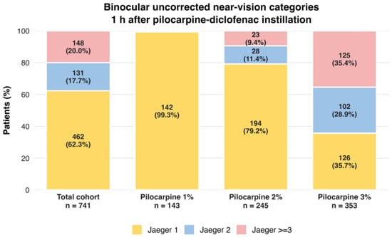
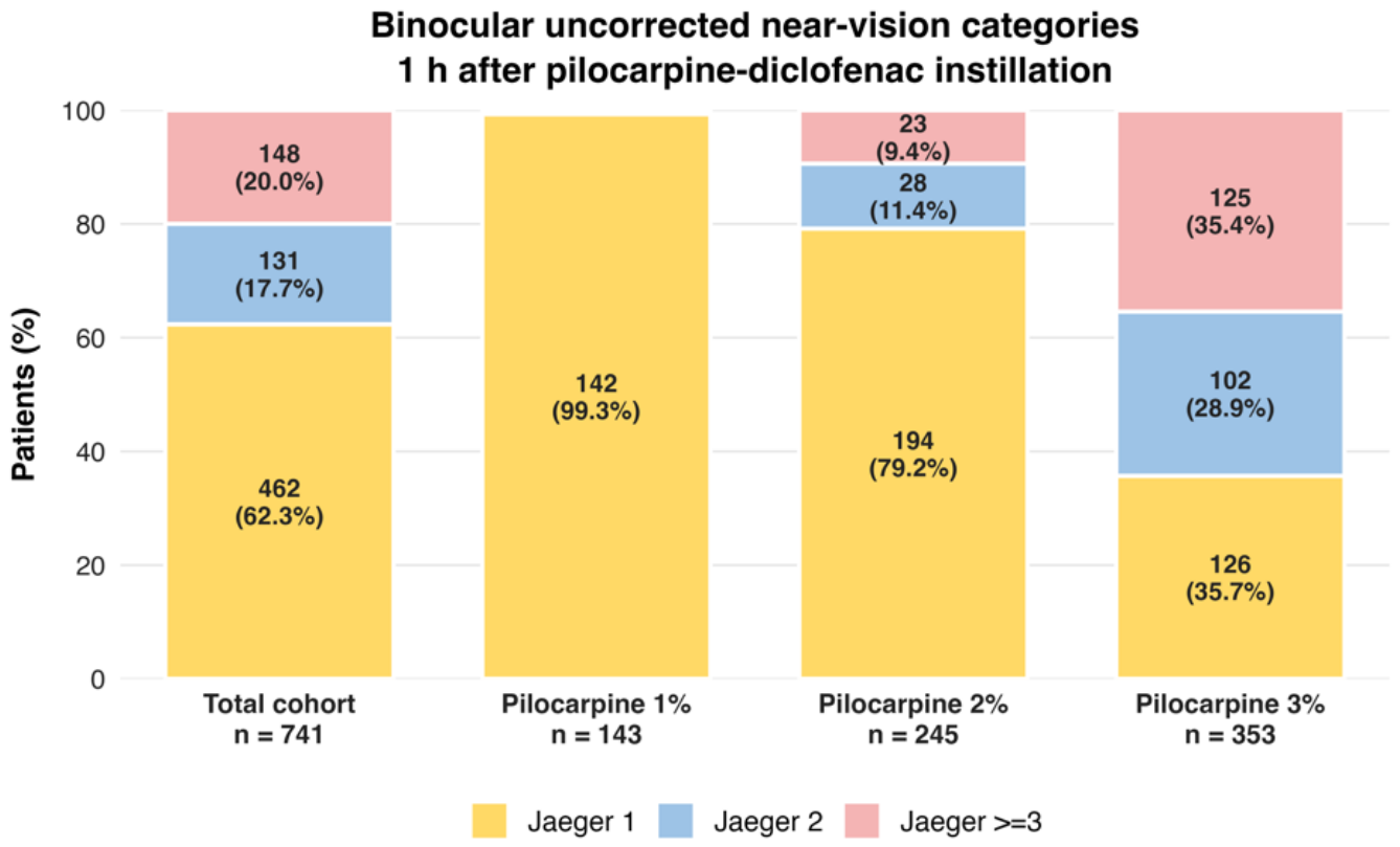
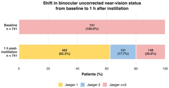
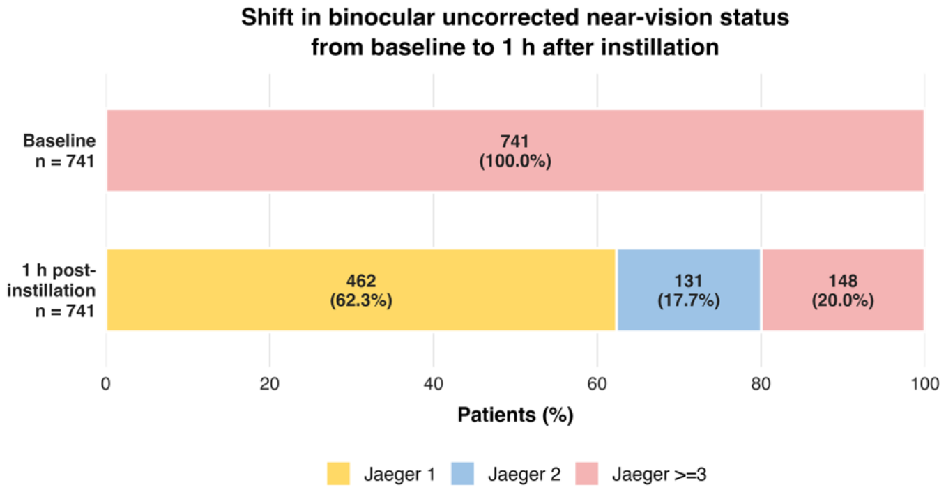
The distribution of binocular near-vision categories 1 h after instillation according to pilocarpine concentration is shown in Figure 1. Jaeger 1 was achieved by 142 out of 143 patients (99.3%) in the 1% group, 194 out of 245 patients (79.2%) in the 2% group, and 126 out of 353 patients (35.7%) in the 3% group. Functional near vision, defined as Jaeger ≤2, was achieved by 143 out of 143 patients in the 1% group, 222 out of 245 patients (90.6%) in the 2% group, and 228 out of 353 patients (64.6%) in the 3% group. Figure 2 illustrates the aggregate functional shift in binocular uncorrected near-vision status from the baseline to 1 h after instillation. Baseline status is shown according to the study-entry criterion of Jaeger ≥3, whereas the 1-h distribution is reported as clinically meaningful Jaeger categories. This figure is intended to summarize the aggregate functional change in the cohort, not individual category transitions.
Figure 1.
Distribution of binocular uncorrected near-vision categories 1 h after pilocarpine-diclofenac instillation. Functional near vision was defined as Jaeger ≤ 2. The figure shows the proportion of patients achieving Jaeger 1, Jaeger 2, or remaining at Jaeger ≥ 3 in the total cohort and according to pilocarpine concentration. Jaeger 2 was calculated as Jaeger ≤ 2 minus Jaeger 1, and Jaeger ≥ 3 as the remaining proportion. Treatment concentration reflected routine clinical decision-making and was not randomized.
Figure 2.
Shift in binocular uncorrected near-vision status from the baseline to 1 h after instillation. At study entry, patients met the clinical inclusion criterion of Jaeger ≥ 3. One hour after pilocarpine-diclofenac instillation, 62.3% achieved Jaeger 1, 17.7% achieved Jaeger 2, and 20.0% remained at Jaeger ≥ 3. This figure represents aggregate functional status, not individual category transitions.
Because concentration selection was individualized and not randomized, these subgroup findings should not be interpreted as a direct dose-response comparison. The lower proportion of patients reaching Jaeger 1 in the 3% group should be interpreted in the context of real-world clinical prescribing patterns and baseline clinical differences among groups.
Uncorrected distance visual acuity did not show clinically relevant deterioration 1 h after treatment. According to patient-level data, mean UDVA was 0.51 ± 0.14 at the baseline and 0.89 ± 0.16 at 1 h after instillation.
3.3. Long-Term Functional Near-Vision Outcomes
Follow-up availability and long-term functional outcomes are summarized in Table 4. A first follow-up visit with near-vision assessment was available for 689 out of 741 patients, corresponding to 93.0% of the original cohort. A second long-term follow-up visit with near-vision assessment was available for 552 patients, corresponding to 74.5% of the original cohort. The mean time elapsed was 40.3 days for the first follow-up visit, with a range of 7 to 120 days, and 518.3 days for the second follow-up visit, with a range of 122 to 1328 days.
Table 4.
Follow-up availability and long-term functional outcomes.
Functional near vision without near spectacles, defined as Jaeger ≤ 2, was observed in 551/689 patients (80.0%) at the first follow-up visit and 445/552 patients (80.6%) at the second follow-up visit. UDVA remained stable during follow-up, with mean values of 0.88 ± 0.16 and 0.86 ± 0.15 at the first and second follow-up visits, respectively. By initial pilocarpine concentration, functional near vision (Jaeger ≤ 2) at the first follow-up assessment was documented in 127/127 patients (100.0%) in the 1% group, 204/224 (91.1%) in the 2% group, and 220/338 (65.1%) in the 3% group. At the second follow-up assessment, the corresponding proportions were 89/96 (92.7%), 150/179 (83.8%), and 206/277 (74.4%), respectively. These subgroups are defined according to the initial pilocarpine concentration. Because treatment concentration could be modified during routine clinical care, these findings should not be interpreted as fixed-dose longitudinal efficacy comparisons. A second follow-up assessment was unavailable for 189 out of 741 patients (25.5%). In most cases, the available records indicated only that the patient did not return for a subsequent routine-care assessment; specific reasons for non-return were not systematically documented.
3.4. Predictors of Functional Near-Vision Response
Adjusted predictors of functional near-vision response at 1 h are shown in Table 5. Complete separation was observed for functional near vision at 1 h because all patients in the 1% pilocarpine-diclofenac group achieved Jaeger ≤ 2. Accordingly, adjusted associations were estimated using Firth penalized logistic regression.
Table 5.
Adjusted predictors of functional near-vision response at 1 h.
In the adjusted model, older age and higher mean spherical equivalent were associated with a lower likelihood of achieving functional near vision at 1 h. Higher baseline Jaeger score showed an inverse association, although the confidence interval crossed unity. The model showed good discrimination, with an area under the receiver operating characteristic curve of 0.878. The lower adjusted odds observed for the 2% and 3% concentration groups relative to the 1% group should be interpreted in the context of individualized prescribing and confounding by indication, rather than as evidence of a direct concentration–response relationship.
3.5. Tolerability and Safety
Treatment was generally well-tolerated. Mild transient adverse events were reported by 308/741 patients (41.6%). The most frequently reported symptom was transient dim vision or decreased light perception, reported by 245/741 patients (33.1%), followed by headache in 29/741 (3.9%) and ocular burning in 33/741 (4.5%).
By initial pilocarpine concentration, at least one early adverse event was documented in 48/143 patients (33.6%) in the 1% group, 95/245 (38.8%) in the 2% group, and 165/353 (46.7%) in the 3% group. Transient dim vision or decreased light perception was documented in 30, 78, and 137 patients, respectively; headache in 5, 9, and 15 patients; and ocular burning in 12, 8, and 13 patients. These concentration-specific frequencies are descriptive and should not be interpreted as causal concentration-toxicity estimates because treatment concentration was individually selected and was not randomized.
No permanent treatment discontinuation due to adverse events was reported in the available clinical records. No clinically significant intraocular pressure elevation, retinal tear, or rhegmatogenous retinal detachment was documented during the available follow-up period.
4. Discussion
The present study evaluated real-world functional near-vision outcomes after pilocarpine-diclofenac eye drops in a large cohort of presbyopic adults treated under routine clinical practice conditions. The main finding was that 80.0% of patients achieved functional near vision, defined as Jaeger ≤ 2, 1 h after instillation, while 62.3% reached Jaeger 1. Functional near vision was maintained in 551/689 patients (80.0%) at the first follow-up visit and in 445/552 patients (80.6%) at the second follow-up visit, with no clinically relevant deterioration in uncorrected distance visual acuity. These findings are clinically relevant because presbyopia is not only an optical consequence of aging, but also a functional condition that affects reading, work-related near tasks, mobile device use, and daily independence. Berdahl et al. emphasized the patient and economic burden associated with presbyopia, while Frick et al. highlighted the global productivity loss attributable to uncorrected presbyopia [2,3]. In this context, real-world outcomes focused on functional near vision may provide useful information for clinical decision-making.
Pharmacological treatment for presbyopia has gained increasing attention as a non-surgical strategy intended to improve near vision while preserving distance vision and avoiding the limitations of optical or surgical approaches. Haghpanah and Alany described the expanding field of topical pharmacological treatments for presbyopia, including miotics and combination therapies, while Renna et al. reviewed modern pharmacological perspectives and emphasized the potential role of miotic agents in increasing depth of focus [4,5]. More recently, Grzybowski et al. provided an updated systematic review of pharmacological treatments for presbyopia, and Singh et al. summarized the evidence for topical pilocarpine 1.25% in a systematic review and meta-analysis [17,18]. In parallel, Holland et al. reported pooled phase 3 data for CSF-1, a low-dose pilocarpine 0.4% formulation, supporting the broader concept that miotic pharmacotherapy can improve near vision without clinically meaningful loss of distance vision [19]. Korenfeld et al. also evaluated UNR844, a lipoic acid choline ester formulation, illustrating that non-miotic approaches aimed at lens biomechanics have also been explored [20]. These studies highlight the rapidly evolving landscape of pharmacological presbyopia treatment.
Comparisons with previous pharmacological studies should be made cautiously. Waring et al. evaluated pilocarpine 1.25% using standardized distance-corrected near visual acuity endpoints in the GEMINI 1 trial, whereas Holland et al. evaluated CSF-1 using randomized phase 3 data, and Lievens et al. explored pilocarpine 1.25% in patients with or without prior laser vision correction [6,19,21]. Farid et al. recently reported randomized phase 2b data evaluating low-dose pilocarpine/diclofenac sodium and pilocarpine alone, providing additional contemporary evidence for combination strategies in pharmacological presbyopia treatment [22]. Mousavi et al. also reported that pilocarpine-based drops improved near vision while maintaining stable distance vision in a comparative clinical study [23]. These studies differ from the present cohort in design, population, outcome definitions, testing conditions, and visual acuity notation. Therefore, the present findings should not be interpreted as evidence of superiority over other pharmacological treatments, but rather as complementary real-world evidence for a pilocarpine-diclofenac strategy in routine clinical practice.
The present study differs from randomized clinical trials because it reflects routine clinical practice, individualized concentration selection, and long-term real-world follow-up. Earlier work by Benozzi et al. introduced a pilocarpine-based pharmacological approach for presbyopia and subsequently reported long-term retrospective outcomes with pilocarpine-diclofenac eye drops [7,10]. In a multicentric study, Benozzi et al. also reported sustained improvement in near vision with pharmacological treatment for presbyopia [11]. The current cohort expands this clinical experience by reporting outcomes across three pilocarpine concentrations combined with diclofenac 0.05% while emphasizing categorical functional endpoints rather than mean Jaeger-line changes. Because treatment concentration was individualized rather than randomly assigned, this study was not designed to establish superiority among pilocarpine concentrations or to validate an optimal concentration-selection algorithm. Concentration-specific findings should therefore be interpreted as observational outcomes in clinically selected real-world subgroups rather than as evidence of a direct concentration-response effect.
The distribution of near-vision categories according to concentration should be interpreted in the context of individualized prescribing. The 1% and 2% groups showed higher proportions of patients achieving Jaeger 1 or Jaeger ≤ 2, whereas the 3% group had a larger residual proportion of patients remaining at Jaeger ≥ 3. This should not be interpreted as lower activity of the 3% formulation. Instead, it likely reflects the clinical selection pattern in which higher concentrations were generally used in patients with a more unfavorable baseline profile, including higher hyperopic spherical equivalent, lower baseline uncorrected distance visual acuity, and greater near-spectacle dependence. This real-world prescribing pattern is clinically plausible but introduces confounding by indication. Consequently, the findings support individualized concentration selection as a pragmatic clinical strategy, while prospective controlled studies are needed to define optimal concentration criteria more rigorously. Accordingly, the lower adjusted odds associated with the 2% and 3% groups in Table 5 should not be interpreted as causal concentration effects or as evidence that lower pilocarpine concentrations are pharmacologically more effective than higher concentrations.
Distance-vision preservation is a critical outcome in pharmacological presbyopia treatment. Miotic agents may improve near visual function by increasing depth of focus, but excessive miosis can potentially reduce retinal illumination and induce dim vision, especially under mesopic or low-light conditions. Abdelkader reported improved presbyopic vision with miotics, and Vargas et al. also reported near-vision improvement with topical pharmacological treatment [24,25]. In the present study, uncorrected distance visual acuity did not show clinically relevant deterioration at 1 h or during follow-up. This finding is clinically relevant because an acceptable presbyopia treatment should provide useful near-vision improvement without compromising distance function in daily activities.
The combination of pilocarpine with diclofenac may have theoretical advantages over miotic monotherapy, although the mechanisms remain incompletely established. Pilocarpine-induced miosis increases depth of focus and may stimulate the ciliary muscle, while diclofenac may contribute to tolerability by modulating inflammatory pathways and reducing discomfort associated with chronic miotic use. Benozzi et al. recently explored pilocarpine-induced nitric oxide and prostaglandin production in porcine ciliary muscle, supporting the biological plausibility of ciliary muscle-mediated pharmacological effects [12]. Gerometta and Candia also proposed that aquaporin-related changes may contribute to presbyopia pathophysiology, although this remains a mechanistic hypothesis rather than a clinically validated therapeutic target [13]. Therefore, the clinical outcomes observed in this cohort should be interpreted primarily as real-world evidence of functional response and tolerability, not as proof of a specific mechanistic pathway.
Safety is particularly relevant in presbyopia pharmacotherapy because treatment is usually elective and directed at improving daily visual function in otherwise healthy eyes. In the present cohort, early adverse events were generally mild and transient, with dim vision or decreased light perception being the most frequently reported symptom. No clinically significant intraocular pressure elevation, retinal tear, or rhegmatogenous retinal detachment was documented in the available clinical records during follow-up. However, retrospective ascertainment from routine-care records may incompletely capture rare events, particularly those occurring between visits or managed outside the study center. Accordingly, the absence of documented retinal events should not be interpreted as evidence of established retinal safety. The study was neither prospectively powered nor specifically designed to estimate the incidence of rare retinal complications or to compare retinal safety with pilocarpine monotherapy. Eton et al. reported cases of rhegmatogenous retinal detachment after the initiation of pilocarpine hydrochloride 1.25% for presbyopia, and Elhusseiny et al. used real-world data to evaluate the association between topical pilocarpine use and retinal detachment [8,9]. Regulatory information for newer miotic therapies also supports the need for caution. The official prescribing information for aceclidine ophthalmic solution includes warnings regarding dim or dark vision and rare retinal tear or detachment in susceptible individuals, reinforcing that retinal safety monitoring is a broader issue for miotic pharmacotherapy in presbyopia [26]. Together, these sources support appropriate patient selection, retinal history assessment, and clinical monitoring, particularly in patients with high myopia, peripheral retinal degeneration, previous retinal pathology, or other predisposing risk factors.
A major methodological issue in this field is the heterogeneity of near-vision measurement. Many pharmacological presbyopia studies report improvement using different near charts, testing distances, and acuity notations, making direct comparisons difficult. Jaeger notation remains familiar and clinically practical, but it is ordinal and chart-dependent. Jaeger notation categorizes near-vision performance according to the smallest near-print size that can be read at the specified testing distance. Lower Jaeger numbers indicate smaller readable print and better near-vision performance; therefore, Jaeger 1 represents better near vision than Jaeger 5. However, Jaeger categories are ordinal and chart-dependent because print sizes and category assignments may vary among different reading cards. They should consequently not be considered equally spaced visual-acuity units or directly converted to logMAR values. Torres emphasized the limitations of Jaeger testing and the value of logarithmic near-vision charts for standardized assessment [14]. More recently, Valvecchia et al. reported near visual acuity using logarithmic measurements at 32 and 40 cm and showed that Jaeger categories do not provide a simple universal equivalence to logMAR values [15]. Radner argued that homologated and standardized reading charts are necessary for presbyopia research because historical near charts do not provide a unique standard for analyzing reading performance [27]. Chen et al. further demonstrated that commonly used near-vision charts are not interchangeable, even when conversion tables are proposed [28]. Together, these findings support the present decision to report Jaeger outcomes as clinically meaningful categories, including Jaeger 1 and Jaeger ≤2, rather than as continuous visual acuity values.
The lack of systematic patient-reported outcomes in the present cohort is therefore an important limitation [29], particularly because modern presbyopia management should address reading ability, occupational near tasks, visual comfort, satisfaction, and spectacle independence.
The main strengths of this study include its large real-world cohort, long observation period, inclusion of three clinically used pilocarpine concentrations combined with diclofenac, and focus on functional near-vision categories, distance-vision preservation, tolerability, and retinal safety. The Latin American setting also adds practical value because real-world presbyopia management may vary across regions according to patient expectations, access to optical correction, working conditions, and clinical practice patterns.
This study also has important limitations. First, it was retrospective, single-center, and non-randomized, which introduces selection bias and confounding by indication. Second, treatment concentration was selected according to routine clinical judgment, so the three concentration groups cannot be interpreted as randomized dose groups. Third, near vision was assessed using a Jaeger chart, which is clinically useful but not equivalent to a standardized logarithmic near-vision scale. Fourth, follow-up occurred as part of routine clinical care rather than through protocol-mandated research visits. No research-specific telephone follow-up or direct patient contact was used to ensure return visits, and a second follow-up assessment was unavailable for 189/741 patients (25.5%). Specific reasons for non-return were not systematically documented. Patients who returned for subsequent assessments may therefore differ from those who did not, introducing potential attrition bias. Accordingly, long-term response estimates apply to patients with available follow-up assessments and may not represent outcomes in the entire original cohort. Adverse events occurring after the last recorded visit or managed outside the study center may likewise have been incompletely captured. Fifth, subjective patient-reported outcomes, pupil size, contrast sensitivity, and detailed adherence data were not systematically available. Although near-vision testing was performed at a fixed distance of 40 cm under routine photopic examination-room conditions, luminance was not instrumentally measured or recorded at each encounter; therefore, uniform illumination across all assessments cannot be confirmed. Finally, although no retinal tear or rhegmatogenous retinal detachment was documented in the available clinical records during follow-up, the study was not designed or powered to establish comparative retinal safety versus pilocarpine monotherapy or other pilocarpine formulations.
Future prospective, multicenter studies should use standardized logarithmic near-vision charts, predefined lighting and testing distances, patient-reported outcome measures, pupil-size assessment, and prespecified safety monitoring. Randomized or well-controlled comparative designs will be needed to determine the optimal concentration of pilocarpine-diclofenac, identify patient profiles most likely to benefit, and clarify long-term safety. Until such evidence is available, the present findings support pilocarpine-diclofenac eye drops as a potentially useful non-surgical option for selected presbyopic adults in routine clinical practice, while emphasizing the need for cautious interpretation and individualized clinical judgment.
5. Conclusions
In this large real-world Latin American cohort, pilocarpine-diclofenac eye drops were associated with rapid and sustained improvement in functional near vision among selected presbyopic adults, with preservation of distance vision. No retinal tear or rhegmatogenous retinal detachment was documented in the available clinical records during follow-up. However, because adverse-event ascertainment was retrospective and the study was neither powered nor designed to compare retinal safety with pilocarpine monotherapy, no conclusions regarding comparative retinal safety can be drawn. Similarly, the findings do not establish superiority over other presbyopia treatments, an optimal pilocarpine concentration, or the independent contribution of diclofenac. Future prospective studies using standardized logarithmic near-vision charts are warranted to evaluate comparative efficacy and safety and to define concentration-selection criteria more rigorously.
Supplementary Materials
The following supporting information can be downloaded at: https://www.mdpi.com/article/10.3390/medicina62112113/s1, Figure S1: Structured routine-care clinical record used at the Centro de Investigación Avanzada de la Presbicia, Buenos Aires, Argentina.
Author Contributions
Conceptualization, G.B., A.F.Z. and M.A.M.-G.; methodology, G.B., J.L., A.F.Z. and M.A.M.-G.; validation, A.F.Z. and M.A.M.-G.; formal analysis, A.F.Z. and M.A.M.-G.; investigation, J.L. and G.B.; data curation, A.F.Z. and M.A.M.-G.; writing—original draft preparation, A.F.Z.; writing—review and editing, M.A.M.-G., J.L. and G.B.; resources and supervision, G.B. All authors have read and agreed to the published version of the manuscript.
Funding
This research was funded by G.B., A.F.Z. and M.A.M.-G. received support from the Ministry of Science, Technology and Innovation of Colombia—MinCiencias (Grant 111589786169). MinCiencias had no role in the design of the study; the collection, analysis or interpretation of data; the writing of the manuscript; or the decision to publish the results.
Institutional Review Board Statement
The study was conducted in accordance with the Declaration of Helsinki and the ethical principles established in Resolution No. 1480/2011 of the Argentine Ministry of Health, which approved the Guide for Research in Human Health. The Institutional Ethics Review Committee of the Centro de Investigación Avanzada de la Presbicia, Buenos Aires, Argentina, reviewed the project and determined that full committee review was not required because of its retrospective design, use of previously collected clinical records, minimal risk, and anonymized data analysis. This determination was recorded in Minute No. 02/Reference No. 001, dated 20 February 2025.
Informed Consent Statement
During routine clinical care, all patients were informed about the characteristics of the pharmacological treatment and its potential adverse effects. Written informed consent was obtained from all patients for treatment and for the use of their anonymized clinical data for research purposes.
Data Availability Statement
The data presented in this study are available on request from the corresponding author. The data are not publicly available due to patient privacy and institutional restrictions.
Acknowledgments
The authors thank Janeth Mora of LinkPharma for her advisory contribution and input on the topic.
Conflicts of Interest
G.B. holds patents related to the pilocarpine-diclofenac formulation. A.F.Z. reports receiving honoraria for lectures from AbbVie, Bayer, Novartis, Sanofi, Novo Nordisk, Takeda, Adium, Merck, Pfizer and Amgen. No pharmaceutical company had any role in the study design, data analysis, interpretation of results, manuscript preparation or the decision to submit this manuscript for publication. Data extraction, data verification, database cleaning, outcome coding and statistical analyses were performed by A.F.Z. and M.A.M.-G., who had no patent or ownership interest in the formulation. G.B. did not conduct or supervise the statistical analyses. The remaining authors declare no conflicts of interest.
References
- Fricke, T.R.; Tahhan, N.; Resnikoff, S.; Papas, E.; Burnett, A.; Ho, S.M.; Naduvilath, T.; Naidoo, K.S. Global prevalence of presbyopia and vision impairment from uncorrected presbyopia: Systematic review, meta-analysis, and modelling. Ophthalmology 2018, 125, 1492–1499. [Google Scholar] [CrossRef] [Scilit]
- Berdahl, J.; Bala, C.; Dhariwal, M.; Lemp-Hull, J.; Thakker, D.; Jawla, S. Patient and Economic Burden of Presbyopia: A Systematic Literature Review. Clin. Ophthalmol. 2020, 14, 3439–3450. [Google Scholar] [CrossRef] [Scilit]
- Frick, K.D.; Joy, S.M.; Wilson, D.A.; Naidoo, K.S.; Holden, B.A. The Global Burden of Potential Productivity Loss from Uncorrected Presbyopia. Ophthalmology 2015, 122, 1706–1710. [Google Scholar] [CrossRef] [Scilit]
- Haghpanah, N.; Alany, R. Pharmacological treatment of presbyopia: A systematic review. Eur. J. Transl. Myol. 2022, 32, 10781. [Google Scholar] [CrossRef] [Scilit]
- Renna, A.; Alió, J.L.; Vejarano, L.F. Pharmacological treatments of presbyopia: A review of modern perspectives. Eye Vis. 2017, 4, 3. [Google Scholar] [CrossRef] [Scilit]
- Waring GO4th Price FWJr Wirta, D.; McCabe, C.; Moshirfar, M.; Guo, Q.; Gore, A.; Liu, H.; Safyan, E.; Robinson, M.R. Safety and Efficacy of AGN-190584 in Individuals With Presbyopia: The GEMINI 1 Phase 3 Randomized Clinical Trial. JAMA Ophthalmol. 2022, 140, 363–371. [Google Scholar] [CrossRef] [Scilit]
- Benozzi, J.; Benozzi, G.; Orman, B. Presbyopia: A new potential pharmacological treatment. Med. Hypothesis Discov. Innov. Ophthalmol. 2012, 1, 3–5. [Google Scholar]
- Eton, E.A.; Zhao, P.Y.; Johnson, M.W.; Rao, R.C.; Huvard, M.J. Rhegmatogenous Retinal Detachment after Initiation of Pilocarpine Hydrochloride Ophthalmic Solution 1.25% for Treatment of Presbyopia. Retin. Cases Brief. Rep. 2024, 18, 98–100. [Google Scholar] [CrossRef] [Scilit]
- Elhusseiny, A.M.; Chauhan, M.Z.; Jabbehdari, S.; Alshammari, N.; Jong, S.; Phillips, P.H.; Sallam, A.B. Using Real-World Data to Assess the Association of Retinal Detachment With Topical Pilocarpine Use. Am. J. Ophthalmol. 2025, 271, 1–6. [Google Scholar] [CrossRef] [Scilit]
- Benozzi, G.; Perez, C.; Leiro, J.; Facal, S.; Orman, B. Presbyopia Treatment With Eye Drops: An Eight Year Retrospective Study. Transl. Vis. Sci. Technol. 2020, 9, 25. [Google Scholar] [CrossRef]
- Benozzi, G.; Cortina, M.E.; Gimeno, E.; Vantesone, D.L.; Solas, A.E.; Lorda, G.M.; Facal, S.; Leiro, J.; Orman, B. A multicentric study of pharmacological treatment for presbyopia. Graefes Arch. Clin. Exp. Ophthalmol. 2021, 259, 2441–2450. [Google Scholar] [CrossRef] [Scilit]
- Benozzi, G.; Quinteros Villarruel, E.; Zuluaga, A.F.; Orman, B. Mechanisms of Pilocarpine-Induced Nitric Oxide and Prostaglandin Production in Porcine Ciliary Muscle. FASEB Bioadv 2025, 7, e70064. [Google Scholar] [CrossRef] [Scilit]
- Gerometta, R.; Candia, O.A. A decrease in the permeability of aquaporin zero as a possible cause for presbyopia. Med. Hypotheses 2016, 86, 132–134. [Google Scholar] [CrossRef] [Scilit]
- Torres, R.M.; Rivero, J.S.; Daponte, P. Cartillas de lectura en escala logarítmica: ¿por qué el test de Jaeger es obsoleto en el presente? Oftalmol. Clin. Exp. 2023, 16, e255. [Google Scholar] [CrossRef] [Scilit]
- Valvecchia, G.; Castro, T.M.; Calero-Vera, D.E.; Masseroni, M.; Nasif, N.; Icaza, E.; Aguirre, L.; Vargas, N.; Gómez-Caride, G.; Perrone, L. Early Real-World Outcomes with a Novel Hybrid Full Range of Vision Intraocular Lens. Medicina 2026, 62, 576. [Google Scholar] [CrossRef] [Scilit]
- Wolffsohn, J.S.; Naroo, S.A.; Bullimore, M.A.; Craig, J.P.; Davies, L.N.; Markoulli, M.; Schnider, C.; Morgan, P.B. BCLA CLEAR Presbyopia: Definitions. Cont. Lens Anterior Eye 2024, 47, 102155. [Google Scholar] [CrossRef] [Scilit]
- Grzybowski, A.; Kapitanovaite, L.; Zemaitiene, R. An updated systematic review of pharmacological treatments for presbyopia. Adv. Ophthalmol. Pract. Res. 2024, 4, 220–225. [Google Scholar] [CrossRef] [Scilit]
- Singh, M.; Sinha, B.P.; Dutta, S.; Deokar, K.K.; Mishra, D.; Goswami, K. A Systematic Review and Meta-Analysis on the Efficacy and Safety of Topical Pilocarpine 1.25% in Presbyopia Treatment. J. Curr. Ophthalmol. 2024, 36, 111–121. [Google Scholar] [CrossRef] [Scilit]
- Holland, E.; Karpecki, P.; Fingeret, M.; Schaeffer, J.; Gupta, P.; Fram, N.; Smits, G.; Ignacio, T.; Lindstrom, R. Efficacy and Safety of CSF-1 (0.4% Pilocarpine Hydrochloride) in Presbyopia: Pooled Results of the NEAR Phase 3 Randomized Clinical Trials. Clin. Ther. 2024, 46, 104–113. [Google Scholar] [CrossRef] [Scilit]
- Korenfeld, M.S.; Robertson, S.M.; Stein, J.M.; Evans, D.G.; Rauchman, S.H.; Sall, K.N.; Venkataraman, S.; Chen, B.L.; Wuttke, M.; Burns, W. Topical lipoic acid choline ester eye drop for improvement of near visual acuity in subjects with presbyopia: A safety and preliminary efficacy trial. Eye 2021, 35, 3292–3301. [Google Scholar] [CrossRef] [Scilit]
- Lievens, C.W.; Hom, M.M.; McLaurin, E.B.; Yuan, J.; Safyan, E.; Liu, H. Pilocarpine HCl 1.25% for treatment of presbyopia after laser vision correction: Pooled analysis of two phase 3 randomized trials (GEMINI 1 and 2). J. Cataract Refract Surg. 2024, 50, 57–63. [Google Scholar] [CrossRef] [Scilit]
- Farid, M.; Rowen, S.L.; Moshirfar, M.; Cunningham, D.; Gaddie, I.B.; Smits, G.; Ignacio, T.; Gupta, P.K. Combination Low-Dose Pilocarpine/Diclofenac Sodium and Pilocarpine Alone for Presbyopia: Results of a Randomized Phase 2b Clinical Trial. Clin. Ophthalmol. 2024, 18, 3425–3439. [Google Scholar] [CrossRef] [Scilit]
- Mousavi, S.A.; Rajavi, Z.; Sabbaghi, H.; Abdi, S.; Montazerin, N.; Kheiri, B.; Haseli-Monfared, A.; Sheibani, K.; Baghban Jaldian, H. Pharmacological Treatment of Presbyopia Using Pilocarpine 1.25% Eye Drops. J. Ophthalmic Vis. Res. 2024, 19, 468–475. [Google Scholar] [CrossRef] [Scilit]
- Abdelkader, A. Improved Presbyopic Vision With Miotics. Eye Contact Lens 2015, 41, 323–327. [Google Scholar] [CrossRef] [Scilit]
- Vargas, V.; Vejarano, F.; Alió, J.L. Near Vision Improvement with the Use of a New Topical Compound for Presbyopia Correction: A Prospective, Consecutive Interventional Non-Comparative Clinical Study. Ophthalmol. Ther. 2019, 8, 31–39. [Google Scholar] [CrossRef] [Scilit]
- DailyMed. VIZZ (Aceclidine Ophthalmic Solution) Prescribing Information; National Library of Medicine: Bethesda, MD, USA, 2026. [Google Scholar]
- Radner, W. Near vision examination in presbyopia patients: Do we need good homologated near vision charts? Eye Vis. 2016, 3, 29. [Google Scholar] [CrossRef] [Scilit]
- Chen, D.Z.; Tham, Y.C.; Shen, L.; Chee, S.P. A Comparative Validation Study of Near Visual Acuity Assessment Using Different Handheld Acuity Charts. Ophthalmol. Sci. 2025, 5, 100790. [Google Scholar] [CrossRef] [Scilit]
- Bentley, S.; Findley, A.; Chiva-Razavi, S.; Naujoks, C.; Patalano, F.; Johnson, C.; Arbuckle, R.; Wolffsohn, J.S. Evaluation of the content validity of patient-reported outcome instruments developed for use with individuals with phakic presbyopia, including the Near Activity Visual Questionnaire-Presbyopia (NAVQ-P) and the Near Vision Correction Independence (NVCI) instrument. J. Patient Rep. Outcomes 2021, 5, 109. [Google Scholar] [CrossRef] [Scilit]

Disclaimer/Publisher’s Note: The statements, opinions and data contained in all publications are solely those of the individual author(s) and contributor(s) and not of MDPI and/or the editor(s). MDPI and/or the editor(s) disclaim responsibility for any injury to people or property resulting from any ideas, methods, instructions or products referred to in the content. |
© 2026 by the authors. Published by MDPI on behalf of the Lithuanian University of Health Sciences. Licensee MDPI, Basel, Switzerland. This article is an open access article distributed under the terms and conditions of the Creative Commons Attribution (CC BY) license.