


Abstract
Background and Objectives: To evaluate whether contrast extravasation on contrast-enhanced computed tomography (CE-CT) is associated with hemorrhage severity, transfusion requirements, and treatment strategies in patients with postpartum vulvovaginal hematoma. Materials and Methods: This single-center retrospective cohort study included 21 patients with vulvovaginal hematoma who underwent CE-CT between 2018 and 2024. Extravasation-positive cases were defined as those demonstrating arterial-phase contrast leakage that persisted on delayed-phase imaging. Clinical variables, including blood loss, laboratory findings, transfusion requirements, and management strategies (conservative treatment, surgery, and transcatheter arterial embolization) were compared between extravasation-positive and -negative groups. Results: Maternal and obstetric characteristics were similar between groups, except for maternal age (32.5 vs. 30 years, p = 0.013). The extravasation-positive group received significantly more red blood cell (RBC) transfusions (median: 13 vs. 6 units, p = 0.02) and fresh frozen plasma (14 vs. 4 units, p = 0.035) and had a higher frequency of massive transfusions (>10 RBC units). Estimated blood loss was higher in the extravasation-positive group (median: 4772 vs. 1945 g), although this difference was not statistically significant. Hematoma volume and location were not associated with extravasation. Clinical success was achieved in all 13 transcatheter arterial embolization (TAE) procedures (100%). Conclusions: CT-detected extravasation was associated with greater transfusion requirements and may provide clinically useful information as an adjunct to overall clinical assessment when considering hemostatic management in patients with vulvovaginal hematoma. Early CE-CT evaluation and prompt consideration of TAE when extravasation is identified may potentially contribute to improved hemostatic management.
1. Introduction
Vulvovaginal hematoma is a potentially life-threatening complication of vaginal delivery, occurring in approximately 1 in 300–1500 deliveries [1,2,3,4]. Pelvic vessel injury may directly result from episiotomy, vaginal laceration, or operative vaginal delivery, or indirectly from excessive stretching of the birth canal during prolonged labor or in cases of fetal macrosomia [5,6,7]. Reported risk factors include primigravidity, obesity, excessive gestational weight gain, smoking, extreme maternal age, hypertensive disorders of pregnancy, and coagulopathies [4,8]. Rapid expansion of a vulvovaginal hematoma can result in severe pain and hemodynamic instability, necessitating massive transfusion and urgent hemostatic intervention, including surgical evacuation or transcatheter arterial embolization (TAE).
Early recognition and appropriate management are important because the clinical course of vulvovaginal hematoma can vary considerably, ranging from stable hematomas that can be observed to rapidly progressive bleeding requiring urgent intervention. When a puerperal genital hematoma is suspected, evaluation includes physical examination, laboratory testing, and imaging modalities, such as ultrasonography and contrast-enhanced computed tomography (CE-CT) [3,4,6]. CE-CT can delineate the location and extent of the hematoma and may identify active contrast extravasation and the potential bleeding source [3,4,6]. These findings may provide clinically relevant information when selecting between observation, surgical management, and endovascular treatment. However, evidence regarding the clinical significance of CT-detected extravasation in vulvovaginal hematoma remains limited. Previous reports have primarily focused on individual cases, small case series, or the technical and clinical outcomes of surgical or endovascular treatment. Relatively few studies have directly compared patients with and without CT-detected extravasation to determine whether this imaging finding is associated with transfusion requirements or patterns of subsequent management. Furthermore, the relationship between extravasation and conventional anatomical characteristics of the hematoma, such as its size and location, has not been well characterized. Clarifying these associations may help define the role of CE-CT as an adjunct to clinical assessment in this uncommon but potentially serious postpartum complication.
Therefore, in this exploratory single-center study, we aimed to examine the association between CT-detected extravasation and transfusion requirements and to describe treatment strategies according to CE-CT findings in patients with vulvovaginal hematoma. We also explored whether hematoma size and location differed according to the presence or absence of extravasation.
2. Materials and Methods
2.1. Study Population
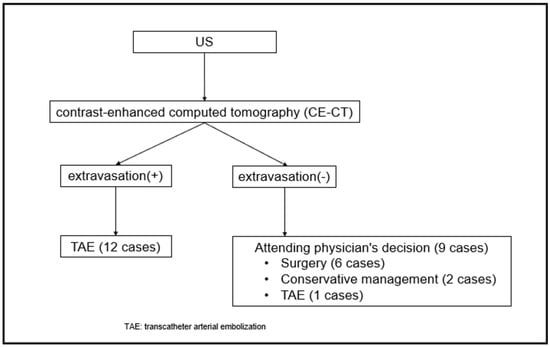
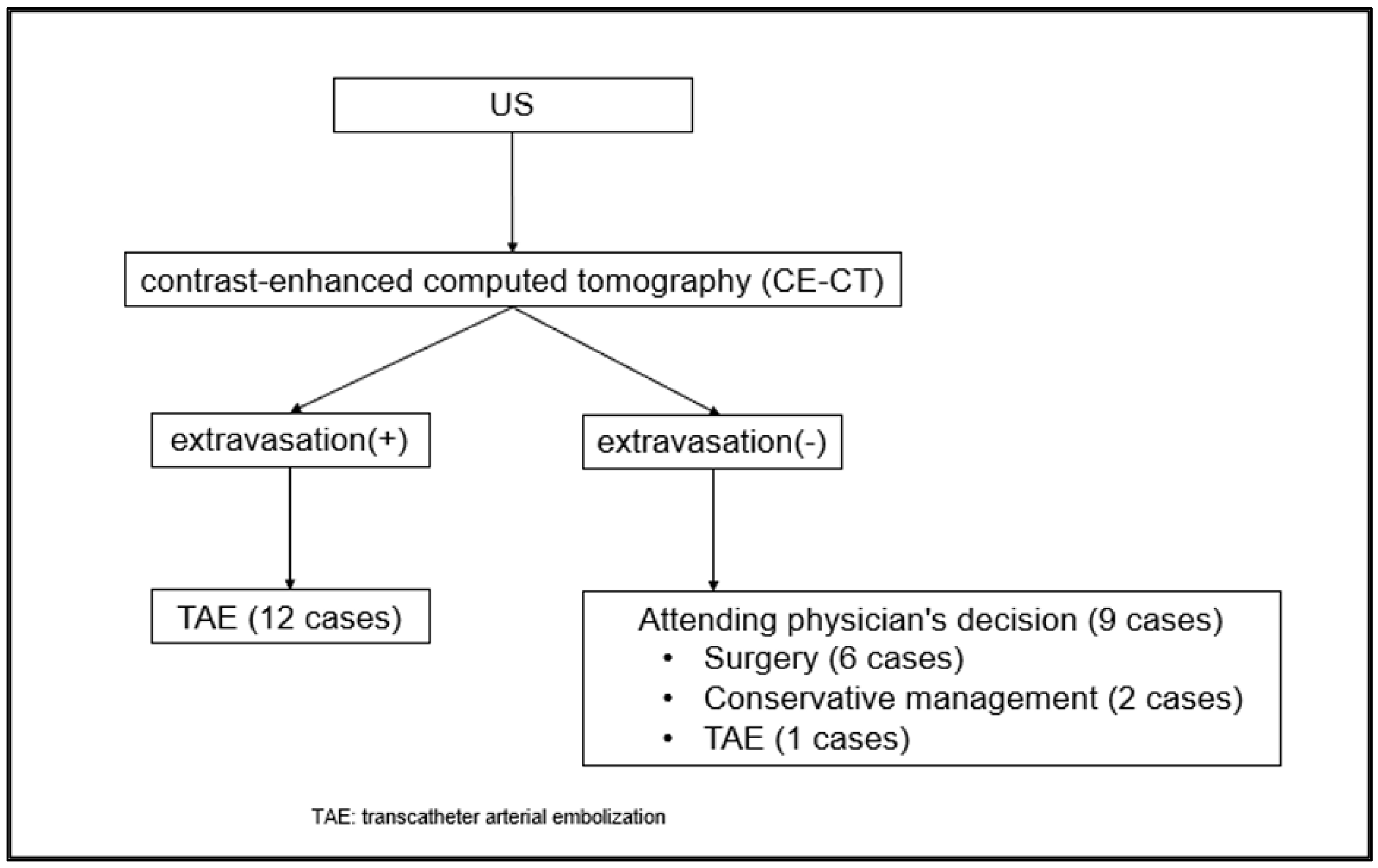
This study was approved by our Institutional Review Board (IRB) and the requirement for written informed consent was waived. This retrospective study included pregnancies complicated by vulvovaginal hematoma treated at our institution between 1 January 2018 and 31 December 2024. During the study period, 21 patients were diagnosed with vulvovaginal hematoma using CE-CT (Figure 1).
Figure 1.
Management strategy for vulvovaginal hematoma.
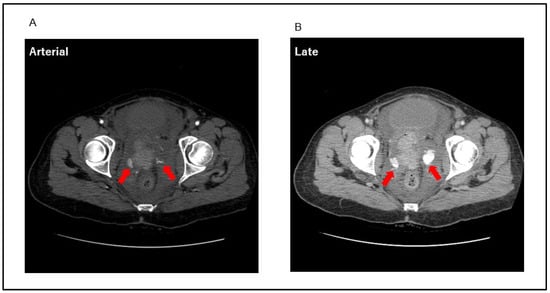
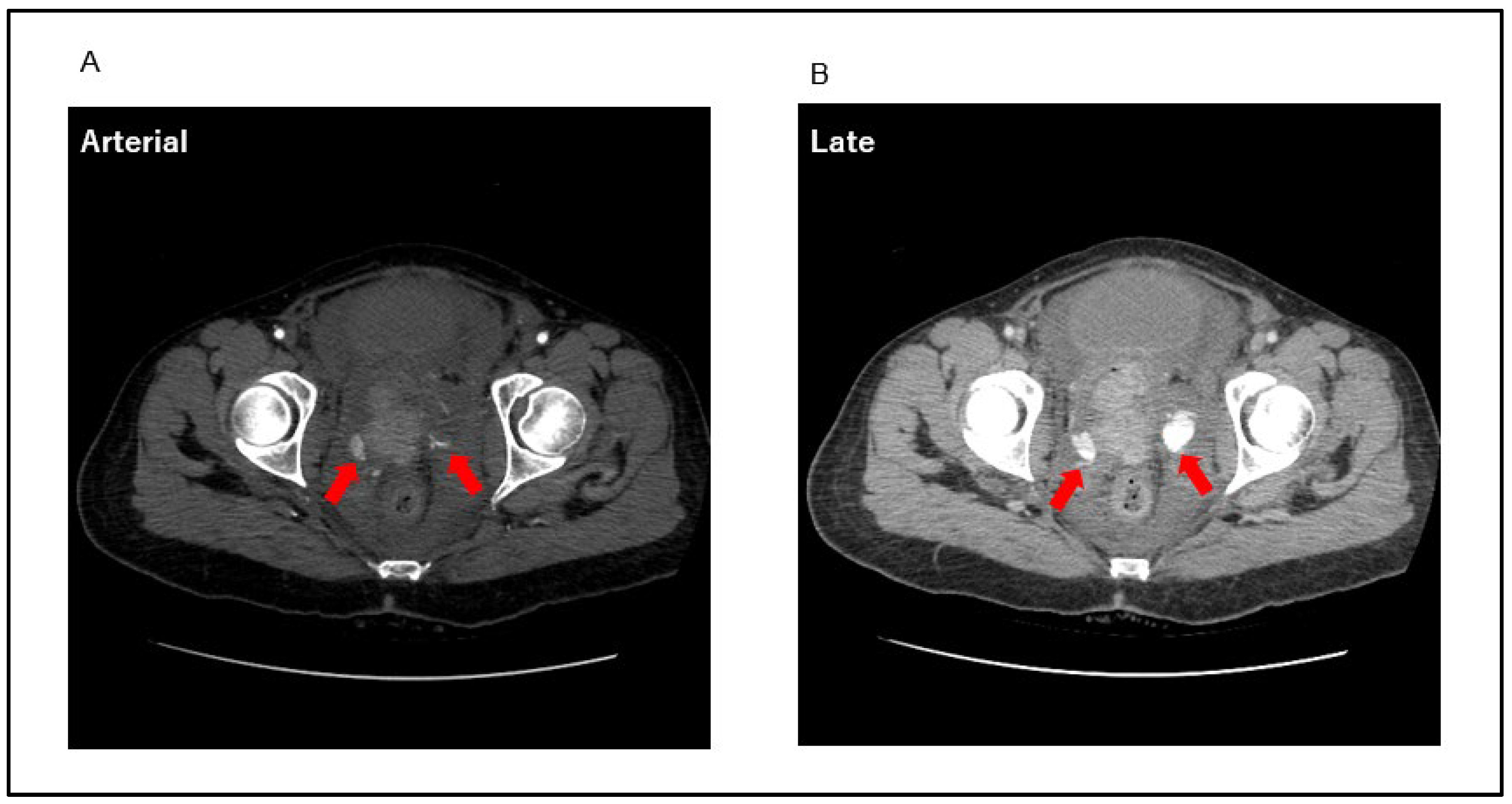
Patients were eligible for inclusion if they developed a vulvovaginal hematoma following delivery and underwent CE-CT during the study period. The decision to perform CE-CT was made at the discretion of the attending physician based on the clinical condition. CE-CT was generally not performed in patients with mild hematomas that could be managed conservatively or in those in whom adequate hemostasis was considered to have been achieved by immediate surgical intervention. Accordingly, the study population comprised 21 patients with vulvovaginal hematomas who underwent CE-CT. The CT protocol included pre-contrast, early-phase, and delayed-phase imaging, with a scan range extending from the upper abdomen to the vulvar region. Extravasation-positive cases were defined as those demonstrating active contrast extravasation on arterial-phase CT that persisted on delayed-phase imaging (Figure 2). In this study, the term “extravasation” refers specifically to contrast extravasation detected on CE-CT and is distinguished from active bleeding identified on angiography. According to our institutional policy, TAE is performed as the primary treatment for patients with active extravasation on CE-CT, whereas the treatment strategy for extravasation-negative cases was determined at the discretion of the attending physician (Figure 1).
Figure 2.
Representative extravasation-positive case. Extravasation-positive cases were defined as active contrast extravasation identified on arterial-phase computed tomography that persisted on delayed-phase imaging. (A) Arterial-phase image demonstrating active contrast extravasation within the hematoma (arrow). (B) Delayed-phase image depicting persistent contrast pooling consistent with ongoing arterial bleeding (arrow).
A total of 21 patients were diagnosed with vulvovaginal hematoma using contrast-enhanced computed tomography (CE-CT). Transcatheter arterial embolization (TAE) was selected as the first-line treatment for extravasation-positive cases. In contrast, treatment strategies for extravasation-negative cases were determined at the discretion of the attending physician.
Extravasation-positive cases were defined as active contrast extravasation identified on arterial-phase computed tomography that persisted on delayed-phase imaging. (A) Arterial-phase image demonstrating active contrast extravasation within the hematoma (arrow). (B) Delayed-phase image depicting persistent contrast pooling consistent with ongoing arterial bleeding (arrow).
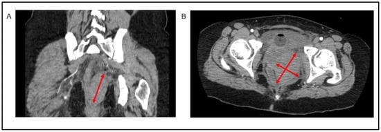
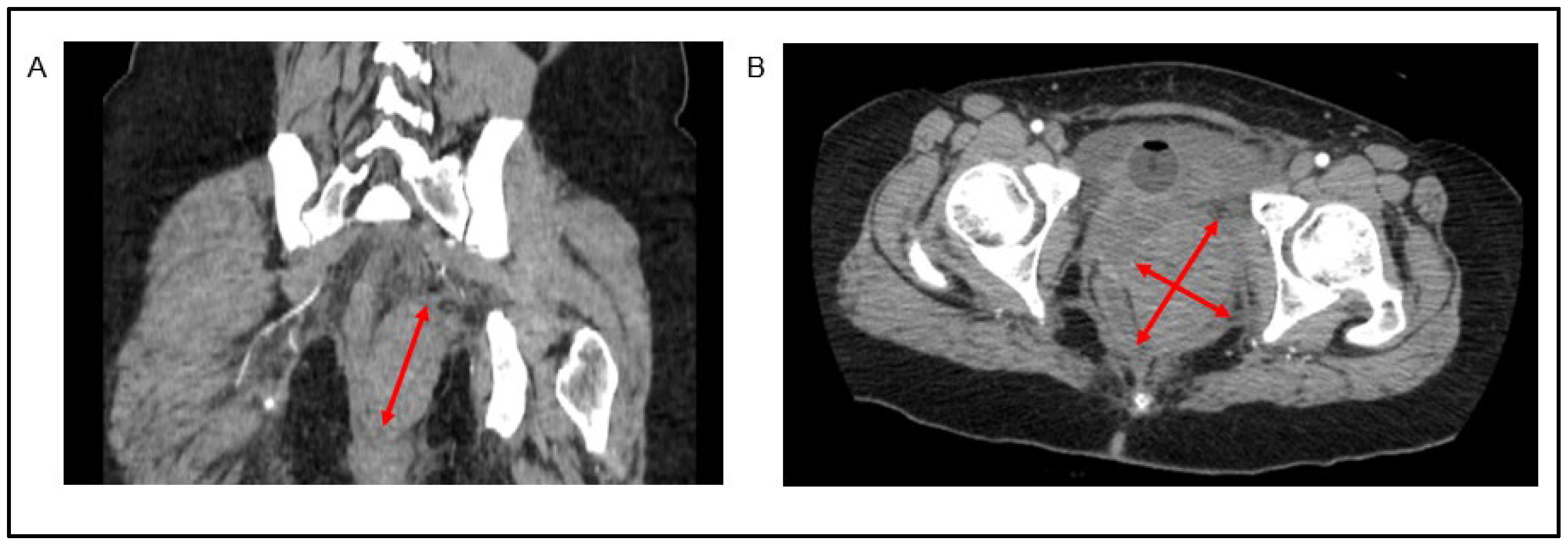
Hematoma volume was calculated by multiplying the maximal diameters measured in three orthogonal directions on CT images (coronal and axial planes) and using the formula V = 0.52 × (length × width × height) (cm3), assuming an ellipsoidal shape (Figure 3). Patients were categorized into the extravasation-positive and -negative groups based on CE-CT findings, and their clinical characteristics were compared. The patient selection flowchart is illustrated in Figure 1.
Figure 3.
Measurement of hematoma volume. Hematoma volume was calculated from the maximal diameters measured in three orthogonal directions on computed tomography (CT) images using the formula V = 0.52 × (length × width × height), assuming an ellipsoidal shape. (A) Coronal CT image demonstrating measurement of the maximal longitudinal diameter. (B) Axial CT image demonstrating the measurement of the maximal transverse diameter.
Hematoma volume was calculated from the maximal diameters measured in three orthogonal directions on computed tomography (CT) images using the formula V = 0.52 × (length × width × height), assuming an ellipsoidal shape. (A) Coronal CT image demonstrating measurement of the maximal longitudinal diameter. (B) Axial CT image demonstrating the measurement of the maximal transverse diameter.
2.2. Embolization Procedure
Informed consent for the procedure was obtained from all patients or their families. Under local anesthesia, a 5-French (F) sheath was inserted via the left or right common femoral artery, and angiography of the internal iliac artery was performed using a 5-F Cobra catheter to identify the bleeding source. If the bleeding point could not be identified, the target vessel was selected using a 5-F catheter or a microcatheter (1.7–1.9 F) based on CE-CT findings or hematoma location. Subsequently, a microcatheter was advanced into the target vessel, with the tip placed as close to the bleeding point as possible. When feasible, the catheter tip was positioned directly at the site of arterial rupture. Embolic materials included a mixture of n-butyl-2-cyanoacrylate (NBCA; Histoacryl, B. Braun, Melsungen, Germany), iodized oil (Lipiodol; Guerbet, Aulnay-sous-Bois, France), and gelatin sponge particles (Serescue, Nippon Kayaku, Tokyo, Japan), which were prepared using the pumping or cutting method, or metallic coils. The embolic agent was selected at the discretion of the operator according to catheter position and the degree of extravasation.
Digital subtraction angiography and obstetric pelvic examination were performed immediately after TAE to confirm hemostasis. If bleeding persisted, the bleeding source was reassessed using pelvic angiography or aortography, and additional TAE was performed. The procedure was completed after confirmation of no active bleeding on angiography and speculum examination. Clinical success was defined as the cessation of ongoing bleeding without the need for additional hemostatic intervention.
2.3. Data Collection
Clinical data were retrospectively collected from the institutional perinatal database and electronic medical records. Collected variables included maternal age, gestational age at delivery, body mass index (BMI) at delivery, delivery history, use of assisted reproductive technology, birth weight, oxytocin use, epidural anesthesia, instrumental delivery, uterine fundal pressure, cardiotocography findings, shock index, blood loss, hemoglobin level, platelet count, fibrinogen level, blood transfusion, hematoma volume, and hematoma location. Cumulative estimated blood loss was defined as the total estimated volume of blood loss during the first 48 h postpartum. The amount of blood products transfused was determined on a case-by-case basis according to the patient’s vital signs, trends in laboratory parameters (hemoglobin, fibrinogen, and platelet count), and the presence or absence of ongoing bleeding. The decision to initiate transfusion, including its timing and the number of units administered, was made at the discretion of the attending physician. Massive transfusion was defined as the administration of over 10 units of RBCs during the clinical course from delivery to definitive hemostasis.
2.4. Statistical Analysis
Statistical analyses were performed using the Mann–Whitney U test (χ2 approximation) for continuous variables and Fisher’s exact test for categorical variables. Maternal background characteristics, obstetric outcomes, and short-term neonatal outcomes were compared between the extravasation-positive and extravasation-negative groups. A p-value < 0.05 was considered statistically significant. All statistical analyses were performed using JMP Student’s Edition, version 18 (SAS Institute Inc., Cary, NC, USA).
3. Results
Maternal characteristics and maternal and obstetric outcomes did not differ significantly between the two groups, except for maternal age (33 vs. 30 years, p = 0.013; Table 1). Patients in the extravasation-positive group exhibited significantly more severe conditions than those in the extravasation-negative group. Specifically, the extravasation-positive group required significantly greater numbers of RBC transfusions (median: 13 vs. 6 units, p = 0.02), fresh frozen plasma transfusions (median: 14 vs. 4 units, p = 0.035), and massive transfusions (>10 RBC units; Table 2) than the extravasation-negative group. Although median blood loss was higher in the extravasation-positive group than in the extravasation-negative group (median: 4772 vs. 1945 g), the difference did not reach statistical significance (Table 2).
Table 1.
Maternal characteristics and obstetric outcomes.
Table 2.
Clinical outcomes, laboratory findings, and requirement for blood transfusion.
A significant difference in maternal age was observed between the extravasation-positive and extravasation-negative groups; however, no significant differences were identified in other maternal characteristics or obstetric outcomes.
Hematoma characteristics, including volume and location according to the presence of contrast extravasation, are summarized in Table 3. No significant differences were observed between the extravasation-positive and extravasation-negative groups in terms of time from delivery to CT scan (median [range]: 363 [132–956] vs. 385 [131–2746] min, p = 0.97) or hematoma volume (138.3 [0–354.9] vs. 183.7 [0–468.2] cm3, p = 0.97). Regarding hematoma location, vaginal hematomas were the most common in both groups (33.3 vs. 38.1%), followed by labial hematomas (19.0 vs. 4.76%). Retroperitoneal hematoma was observed only in the extravasation-positive group (4.76%); however, the difference was not statistically significant (p = 0.17).
Table 3.
Hematoma volume and location.
Treatment strategies according to transfusion requirements are presented in Table 4. Patients requiring over 10 units of RBC transfusion tended to undergo TAE more frequently than those requiring ≤10 units (81.8 vs. 40.0%), whereas surgical management was more common in the ≤10-unit group (50.0 vs. 9.1%). Conservative management was infrequently selected in both groups (9.1 vs. 10.0%). However, although a trend was observed, the difference did not reach statistical significance (p = 0.075).
Table 4.
Treatment strategies according to transfusion requirements.
Details of the TAE procedures are summarized in Table 5. Among the 13 patients who underwent TAE, contrast extravasation on CE-CT was observed in 12 patients (92.3%). The vaginal artery was the most frequently embolized vessel (8/13, 61.5%), followed by the uterine artery (4/13, 30.8%), internal iliac artery (4/13, 30.8%; including the bilateral anterior division in one case), and internal pudendal artery (3/13, 23.1%). Embolization of the superior rectal artery arising from the inferior mesenteric artery was performed in two cases (15.4%). In terms of embolic materials, NBCA was used in eight cases (61.5%) and gelatin sponge in seven cases (53.8%), with the combined use of NBCA and gelatin sponge in three cases (23.1%), and coil embolization in one case (7.7%). A retroperitoneal hematoma was identified in one patient, and a pseudoaneurysm was observed in another. Notably, clinical success was achieved in all 13 TAE procedures (100%).
Table 5.
Summary of transcatheter arterial embolization (TAE) procedures.
4. Discussion
In the present study, our results revealed that CT-detected contrast extravasation was associated with increased transfusion requirements in patients with vulvovaginal hematoma. In contrast, no statistically significant associations were detected between contrast extravasation and hematoma volume or location, nor were statistically significant differences observed in estimated blood loss, shock index, nadir hemoglobin, platelet count, or fibrinogen levels between the groups.
An imaging-guided escalation strategy may help standardize management of vulvovaginal hematoma, for which no consensus approach currently exists. To date, an optimal treatment strategy for vulvovaginal hematoma has not been established. Some reports describe TAE as a rescue therapy after failure of conservative management or surgical evacuation [9], whereas others advocate primary TAE in selected cases [10]. At our institution, CE-CT is used to determine treatment strategies, and TAE is the first-line therapy for extravasation-positive cases. In the present study, CT-detected extravasation was found to be potentially associated with increased blood transfusion requirements. Notably, hematoma volume itself was not correlated with hemorrhagic severity, consistent with findings from trauma studies in which contrast extravasation was a stronger predictor of hemodynamic compromise than hematoma volume alone [11,12]. Additionally, in our cohort, patients requiring massive transfusions frequently underwent TAE, suggesting that extravasation may serve as a practical imaging marker to guide early escalation. Although a causal relationship cannot be established owing to the retrospective design, the association between extravasation and transfusion burden highlights its potential clinical value.
The clinical significance of CT extravasation is biologically plausible and supported by evidence from trauma care studies. Previous reports on postpartum hemorrhage have primarily focused on uterine bleeding, such as uterine atony or placental abnormalities [13,14,15], whereas limited data are available regarding clinical predictors of severity in vulvovaginal hematoma [2]. However, in studies on trauma, active contrast extravasation, which is often described as “contrast blush,” is strongly associated with ongoing arterial bleeding and the need for TAE [11,16]. For example, White et al. demonstrated that contrast blush on CT significantly predicted angiographic arterial injury and the need for embolization in patients with pelvic fractures [16]. Blackmore et al. also reported that CT findings, including contrast extravasation, were independent predictors of major hemorrhage in pelvic trauma [11]. Although these studies were conducted in non-obstetric settings, the fundamental concept that contrast extravasation reflects active arterial bleeding is likely applicable to obstetric pelvic hematomas.
CE-CT may be particularly valuable when the bleeding source arises outside the typical internal iliac system and may be missed on routine pelvic angiography. In the present study, two cases (Cases 5 and 6) involved bleeding from the superior rectal artery, a branch of the inferior mesenteric artery. In such cases, the bleeding source may not be identified on routine pelvic angiography alone, particularly when the causative vessel originates from non-internal iliac branches or collateral pathways [6]. These observations further support our CE-CT-driven strategy. Overall, our findings suggest that a vulvovaginal hematoma is suspected, CE-CT should be performed early, and prompt TAE may be considered if extravasation is detected to achieve hemostasis.
TAE has been reported to be effective for puerperal hematomas, with a low complication rate [17]. The clinical success rate of TAE as first-line therapy in extravasation-positive cases reportedly ranges from 88–100% [17], consistent with our findings (100%). However, the optimal first-line embolic material for TAE in vulvovaginal hematoma remains controversial [18]. In our cohort, NBCA was used in 8 of 13 TAE cases (62%). Unlike gelatin sponges, NBCA may remain effective even in the presence of coagulopathy, including disseminated intravascular coagulation associated with obstetric hemorrhage [19,20,21]. Nevertheless, NBCA requires meticulous technique, as proximal embolization may permit persistent bleeding via collateral circulation. Therefore, embolization should be performed as distally and selectively as possible. Accordingly, in TAE for vaginal wall hematoma, the choice between NBCA and gelatin sponge should be individualized based on factors such as the volume and rate of extravasation, vascular injury characteristics (including suspected vessel size and bleeding rate), angiographic accessibility for selective catheterization, and the coagulation status of the patient.
The optimal management of extravasation-negative vulvovaginal hematomas remains unclear. Although the absence of CT-detected extravasation may suggest the absence of active arterial bleeding at the time of imaging, it does not necessarily indicate that conservative management is appropriate. In the present study, treatment decisions regarding observation, surgical evacuation, and TAE were made according to the overall clinical condition and varied among the attending physicians. Consequently, among the nine patients without extravasation, six underwent surgery, two were treated conservatively, and one underwent TAE. Nevertheless, all patients achieved favorable outcomes without the need for additional treatment (Figure 1). These findings indicate that CE-CT findings alone are insufficient to determine the treatment strategy for extravasation-negative patients. Takahashi et al. reported that conservative management may be feasible in patients without extravasation on CE-CT regardless of hematoma volume; however, 2 of 19 patients (11%) initially managed conservatively subsequently required therapeutic intervention [2]. Thus, conservative management may be considered in selected clinically stable patients without extravasation, provided that close observation and prompt intervention are available if deterioration occurs. Treatment decisions should incorporate the overall clinical context, including vital signs, hemoglobin trends, hematoma volume and location, evidence of hematoma expansion, and the potential risk of rebleeding. Conversely, the presence of extravasation may support closer monitoring and consideration of early embolization, although CE-CT findings should not be used as the sole determinant of treatment.
Certain limitations should be considered when interpreting the findings of the present study and translating them into clinical practice. First, the retrospective, single-center design may limit the generalizability of the findings, as hemostatic management may vary across institutions. Second, the small sample size limited the statistical power of this study, and the possibility of type II error cannot be excluded. Therefore, further studies with larger sample sizes are warranted to improve statistical power and reduce the risk of type II error. In addition, maternal age differed significantly between the groups and may have acted as a confounding factor. Because of the small sample size and limited number of events, reliable multivariable adjustment was not feasible; therefore, the observed associations should not be interpreted as independent effects of CT-detected extravasation. Third, CE-CT was not uniformly performed in all vulvovaginal hematoma cases, which may have introduced selection bias toward more severe presentations. Fourth, TAE was the preferred treatment for patients with contrast extravasation detected on CE-CT, which may have introduced treatment selection bias and influenced the observed associations between extravasation and clinical outcomes. Therefore, prospective multicenter studies are warranted to validate CT-detected extravasation as a decision-making marker in obstetric pelvic hematomas.
5. Conclusions
In this exploratory single-center study, CT-detected extravasation was associated with greater transfusion requirements and may provide clinically useful information as an adjunct to overall clinical assessment when considering hemostatic management in patients with vulvovaginal hematoma. In our cohort, extravasation was associated with increased transfusion requirements, whereas hematoma volume and location were not. Therefore, an imaging-guided strategy involving early CE-CT followed by prompt consideration of TAE when extravasation is identified may help optimize hemostatic management. Conversely, in extravasation-negative cases, treatment decisions should be based on the overall clinical condition rather than CE-CT findings alone, with close monitoring for possible deterioration.
Author Contributions
Conceptualization, H.G. and D.O.; Methodology, H.G. and D.O.; Formal analysis, H.G.; Investigation, H.G. and R.W.; Data curation, H.G.; Writing—original draft, H.G.; Writing—review & editing, H.G., R.W., H.I., Y.Y. (Yu Yamazaki), Y.Y. (Yoshihiro Yoshimura), K.H., T.S., S.H., K.F., Y.K., T.M. and D.O.; Visualization, H.G.; Supervision, D.O. All authors have read and agreed to the published version of the manuscript.
Funding
This research received no external funding.
Institutional Review Board Statement
All procedures performed in studies involving human participants were in accordance with the ethical standards of the institutional and/or national research committee and with the 1964 Helsinki Declaration and its later amendments or comparable ethical standards. The study was approved by the Ethics Committee of Kitasato University School of Medicine and Hospital (approval number: B23-109, 3 October 2025).
Informed Consent Statement
This retrospective observational study was conducted using the opt-out method, as per the guidance of the ethics committee. The requirement for informed consent was waived by the ethics committee because of the retrospective design. An opt-out approach was employed in accordance with institutional policy. For this type of study, consent for publication is not required.
Data Availability Statement
The data supporting the findings of this study are provided in the article. Additional data are not publicly available due to patient privacy and ethical restrictions but may be available from the corresponding author on reasonable request, subject to approval by the relevant institutional review board.
Conflicts of Interest
The authors declare that they have no conflicts of interest.
References
- Egan, E.; Dundee, P.; Lawrentschuk, N. Vulvar hematoma secondary to spontaneous rupture of the internal iliac artery: Clinical review. Am. J. Obstet. Gynecol. 2009, 200, e17–e18. [Google Scholar] [CrossRef] [Scilit]
- Takahashi, T.; Tomita, H.; Hamada, H.; Tadakawa, M.; Iwama, N.; Saito, M. Transcatheter arterial embolization outperforms surgery in reducing blood transfusions for postpartum vulvovaginal hematoma. Am. J. Obstet. Gynecol. 2024, 231, 653.e1–653.e8. [Google Scholar] [CrossRef] [Scilit]
- Distefano, M.; Casarella, L.; Amoroso, S.; Di Stasi, C.; Scambia, G.; Tropeano, G. Selective arterial embolization as a first-line treatment for postpartum hematomas. Obstet. Gynecol. 2013, 121, 443–447. [Google Scholar] [CrossRef] [Scilit]
- Tsumagari, A.; Ohara, R.; Mayumi, M.; Yagi, H.; Nagai, Y.; Obata-Yasuoka, M.; Hamada, H.; Satoh, T. Clinical characteristics, treatment indications and treatment algorithm for post-partum hematomas. J. Obstet. Gynaecol. Res. 2019, 45, 1127–1133. [Google Scholar] [CrossRef] [Scilit]
- Onyeodi, I.; Azubuike, I.; Ukpeteru, F. Post-partum vulva haematoma management in a low resource setting: A case report. Trop. J. Obstet. Gynaecol. 2025, 43, 236–239. [Google Scholar]
- Woodhams, R.; Fujii, K.; Kurihara, Y.; Ochiai, D. Interventional radiology for primary postpartum hemorrhage: Optimal strategy based on the pathogenesis of bleeding. Interv. Radiol. 2025, 10, e20250014. [Google Scholar] [CrossRef] [Scilit]
- Dixon, S.; Tapping, C.R.; Chuah, P.S.; Bratby, M.; Uberoi, R.; Anthony, S. Successful fibroid embolization of pelvic and inferior mesenteric artery collaterals after previous uterine artery embolization. Acta Radiol. 2012, 53, 292–295. [Google Scholar] [CrossRef] [Scilit]
- Rani, S.; Verma, M.; Pandher, D.K.; Takkar, N.; Huria, A. Risk factors and incidence of puerperal genital haematomas. J. Clin. Diagn. Res. 2017, 11, QC01–QC03. [Google Scholar] [CrossRef] [Scilit]
- Dahdouh, E.M.; Balayla, J.; Dubé, J. Angiographic embolization of a postpartum vulvovaginal hematoma in a patient with situs inversus totalis: An effective second-line treatment. Case Rep. Obstet. Gynecol. 2013, 2013, 323781. [Google Scholar] [CrossRef] [Scilit]
- Takagi, K.; Akashi, K.; Horiuchi, I.; Nakamura, E.; Samejima, K.; Ushijima, J.; Okochi, T.; Hamamoto, K.; Tanno, K. Managing vulvovaginal hematoma by arterial embolization as first-line hemostatic therapy. Taiwan J. Obstet. Gynecol. 2017, 56, 224–226. [Google Scholar] [CrossRef] [Scilit]
- Blackmore, C.C.; Cummings, P.; Jurkovich, G.J.; Linnau, K.F.; Hoffer, E.K.; Rivara, F.P. Predicting major hemorrhage in patients with pelvic fracture. J. Trauma 2006, 61, 346–352. [Google Scholar] [CrossRef] [Scilit]
- Rich, H.; Shah, N.; Rahman, S.; Baghdanian, A.; Baghdanian, A.; Sax, A.; Anderson, S.; LeBedis, C. Utility of pelvic CT angiography in blunt pelvic trauma. Curr. Probl. Diagn. Radiol. 2025, 54, 355–360. [Google Scholar] [CrossRef] [Scilit]
- Evensen, A.; Anderson, J.M.; Fontaine, P. Postpartum hemorrhage: Prevention and treatment. Am. Fam. Physician 2017, 95, 442–449. [Google Scholar]
- Sheldon, W.R.; Blum, J.; Vogel, J.P.; Souza, J.P.; Gulmezoglu, A.M.; Winikoff, B. WHO Multicountry Survey on Maternal; Newborn Health Research Network. Postpartum haemorrhage management, risks, and maternal outcomes: Findings from the World Health Organization Multicountry Survey on Maternal and newborn Health. BJOG 2014, 121, 5–13. [Google Scholar] [CrossRef] [Scilit]
- Sentilhes, L.; Kayem, G.; Chandraharan, E.; Palacios-Jaraquemada, J.; Jauniaux, E.; FIGO Placenta Accreta Diagnosis and Management Expert Consensus Panel. FIGO consensus guidelines on placenta accreta spectrum disorders: Conservative management. Int. J. Gynaecol. Obstet. 2018, 140, 291–298. [Google Scholar] [CrossRef] [Scilit]
- White, C.E.; Hsu, J.R.; Holcomb, J.B. Haemodynamically unstable pelvic fractures. Injury 2009, 40, 1023–1030. [Google Scholar] [CrossRef] [Scilit]
- Vedantham, S.; Goodwin, S.C.; McLucas, B.; Mohr, G. Uterine artery embolization: An underused method of controlling pelvic hemorrhage. Am. J. Obstet. Gynecol. 1997, 176, 938–948. [Google Scholar] [CrossRef] [Scilit]
- Misugi, T.; Hamuro, A.; Kitada, K.; Kurihara, Y.; Tahara, M.; Terayama, E.; Kageyama, K.; Yamamoto, A.; Tachibana, D. Transcatheter arterial embolization (TAE) using N-butyl-2-cyanoacrylate (NBCA) as the first choice for postpartum vulvovaginal hematoma; case series and reviews of the literature. Diagnostics 2022, 12, 1429. [Google Scholar] [CrossRef] [Scilit]
- Kanematsu, M.; Watanabe, H.; Kondo, H.; Goshima, S.; Kato, H.; Furui, T.; Toyoki, H.; Morishige, K. Postpartum hemorrhage in coagulopathic patients: Preliminary experience with uterine arterial embolization with N-butyl cyanoacrylate. J. Vasc. Interv. Radiol. 2011, 22, 1773–1776. [Google Scholar] [CrossRef] [Scilit]
- Yonemitsu, T.; Kawai, N.; Sato, M.; Sonomura, T.; Takasaka, I.; Nakai, M.; Minamiguchi, H.; Sahara, S.; Iwasaki, Y.; Naka, T.; et al. Comparison of hemostatic durability between N-butyl cyanoacrylate and gelatin sponge particles in transcatheter arterial embolization for acute arterial hemorrhage in a coagulopathic condition in a swine model. Cardiovasc. Intervent. Radiol. 2010, 33, 1192–1197. [Google Scholar] [CrossRef] [Scilit]
- Yonemitsu, T.; Kawai, N.; Sato, M.; Tanihata, H.; Takasaka, I.; Nakai, M.; Minamiguchi, H.; Sahara, S.; Iwasaki, Y.; Shima, Y.; et al. Evaluation of transcatheter arterial embolization with gelatin sponge particles, microcoils, and n-butyl cyanoacrylate for acute arterial bleeding in a coagulopathic condition. J. Vasc. Interv. Radiol. 2009, 20, 1176–1187. [Google Scholar] [CrossRef] [Scilit]

Disclaimer/Publisher’s Note: The statements, opinions and data contained in all publications are solely those of the individual author(s) and contributor(s) and not of MDPI and/or the editor(s). MDPI and/or the editor(s) disclaim responsibility for any injury to people or property resulting from any ideas, methods, instructions or products referred to in the content. |
© 2026 by the authors. Published by MDPI on behalf of the Lithuanian University of Health Sciences. Licensee MDPI, Basel, Switzerland. This article is an open access article distributed under the terms and conditions of the Creative Commons Attribution (CC BY) license.