


Abstract
Metabolic syndrome (MetS) is a clinical condition defined by the coexistence of interconnected cardiometabolic risk factors, including central obesity, dyslipidemia, elevated blood pressure, and impaired glucose regulation, which collectively increase the risk of type 2 diabetes mellitus and cardiovascular disease. Beyond these clinical diagnostic features, MetS is characterized by complex pathophysiological alterations involving systemic dysregulation of metabolic signaling across adipose tissue, skeletal muscle, liver, vascular endothelium, and the immune system. Key molecular alterations include impaired insulin receptor substrate (IRS)–Akt signaling, chronic nuclear factor kappa B (NF-κB) activation, mitochondrial dysfunction, and oxidative stress. Physical exercise is recognized as a pleiotropic biomedical intervention capable of restoring metabolic homeostasis through coordinated modulation of intracellular signaling pathways and inter-organ communication. Exercise activates AMP-activated protein kinase (AMPK), enhances peroxisome proliferator-activated receptor gamma coactivator-1 alpha (PGC-1α)-mediated mitochondrial biogenesis, and stimulates nuclear factor erythroid 2-related factor 2 (Nrf2)-dependent antioxidant responses. These adaptations improve glucose uptake, enhance fatty acid oxidation, and reduce ectopic lipid accumulation across metabolically active tissues. At the systemic level, skeletal muscle functions as an endocrine organ by releasing myokines such as irisin, interleukin-6 (IL-6), and fibroblast growth factor 21 (FGF21), which contribute to metabolic regulation across the liver, adipose tissue, and vasculature. These exercise-induced signals promote immune modulation, reduce pro-inflammatory cytokine production, and improve endothelial function. Different exercise modalities including aerobic, resistance, and high-intensity interval training (HIIT) activate both common and modality-specific molecular pathways, supporting individualized exercise strategies. Collectively, exercise targets the multi-organ pathophysiology of MetS and provides a mechanistic foundation for precision exercise medicine in cardiometabolic disease management.
1. Introduction
Metabolic syndrome (MetS) is a clinical condition defined by the coexistence of interconnected cardiometabolic risk factors, including central obesity, elevated blood pressure, dyslipidemia, and impaired glucose regulation, which collectively increase the risk of cardiovascular disease and type 2 diabetes mellitus worldwide [1,2,3,4]. Beyond its clinical definition, MetS is characterized by a complex pathophysiological network involving dysregulated signaling across multiple organ systems, including adipose tissue, skeletal muscle, liver, vascular endothelium, and immune cells [5,6,7,8]. Although insulin resistance and chronic low-grade inflammation are considered central mechanisms underlying MetS, they are not diagnostic criteria and may not be present in all affected individuals. From a biomedical perspective, MetS is therefore not merely a clinical aggregation of risk factors but a complex network disease involving dysregulated signaling across multiple organ systems, including adipose tissue, skeletal muscle, liver, vascular endothelium, and immune cells [5,6,7,8].
At the cellular level, MetS is driven by persistent metabolic overload and impaired nutrient sensing, leading to disruption of insulin receptor substrate (IRS)–Akt signaling, chronic activation of stress pathways such as c-Jun N-terminal kinase (JNK) and nuclear factor kappa B (NF-κB), and progressive mitochondrial dysfunction [9,10,11,12]. These alterations result in reduced glucose uptake, enhanced hepatic gluconeogenesis, increased lipolysis in adipose tissue, and ectopic lipid deposition in liver and muscle, thereby amplifying systemic metabolic dysfunction [7,13].
A key biomedical hallmark of MetS is chronic low-grade inflammation, characterized by increased circulating levels of tumor necrosis factor-alpha (TNF-α), interleukin-6 (IL-6), and C-reactive protein (CRP), together with altered adipokine profiles including decreased adiponectin and increased leptin [14,15]. This inflammatory milieu contributes to endothelial dysfunction, impaired nitric oxide bioavailability, and accelerated atherogenesis, linking metabolic dysfunction to cardiovascular disease [16,17].
Mitochondrial dysfunction and oxidative stress further exacerbate disease progression by impairing oxidative phosphorylation, increasing reactive oxygen species (ROS) production, and activating redox-sensitive signaling pathways [18,19]. Collectively, these processes create a self-perpetuating cycle of metabolic inflexibility, inflammation, and cellular injury that defines the pathophysiology of MetS.
In this context, physical exercise has emerged as a powerful biomedical intervention capable of modulating multiple disease-driving mechanisms simultaneously [20,21,22,23,24]. Exercise activates AMP-activated protein kinase (AMPK), enhances peroxisome proliferator-activated receptor gamma coactivator-1 alpha (PGC-1α)-mediated mitochondrial biogenesis, improves insulin signaling through IRS–Akt pathway restoration, and activates nuclear factor erythroid 2-related factor 2 (Nrf2)-dependent antioxidant defense systems [25,26,27,28]. Importantly, skeletal muscle acts as an endocrine organ during contraction, releasing myokines such as irisin, IL-6, and fibroblast growth factor 21 (FGF21), which are associated with inter-organ communication and systemic metabolic adaptation [29,30].
Despite substantial advances in understanding the physiological effects of exercise, the integration of its molecular mechanisms into a unified biomedical framework for MetS remains incomplete. A systems-level synthesis linking intracellular signaling, organ crosstalk, and clinical outcomes is required to support its implementation as a precision therapeutic strategy. Therefore, this narrative review aims to critically analyze the molecular and cellular mechanisms through which exercise modulates MetS and to discuss their implications for translational and clinical medicine.
2. Literature Search Strategy
Although this manuscript is presented as a narrative review, a structured literature search was performed to ensure a comprehensive and balanced synthesis of current evidence regarding the molecular mechanisms through which exercise modulates MetS. Electronic searches were conducted in PubMed/MEDLINE, Scopus, and Web of Science for articles published from January 2000 to June 2026. The search strategy combined Medical Subject Headings (MeSH) and free-text terms related to exercise and metabolic syndrome, including “metabolic syndrome,” “exercise,” “physical activity,” “aerobic exercise,” “resistance training,” “high-intensity interval training,” “myokines,” “exerkines,” “AMPK,” “PGC-1α,” “Nrf2,” “mitochondrial biogenesis,” “oxidative stress,” “inflammation,” “insulin resistance,” “organ crosstalk,” and “precision exercise”. Priority was given to systematic reviews, meta-analyses, randomized controlled trials, prospective cohort studies, and high-quality experimental studies published in peer-reviewed journals. Seminal studies that established key molecular mechanisms were also included when considered essential for understanding current concepts. Additional relevant articles were identified through manual screening of the reference lists of selected publications. Research was selected based on its scientific quality, methodological rigor, biological relevance, and direct contribution to understanding the molecular pathways, inter-organ communication, biomarkers, and clinical implications of exercise in MetS.
3. Pathophysiological Basis of Metabolic Syndrome: An Integrated Cellular and Molecular Perspective
MetS is increasingly recognized as a systemic disorder of inter-organ metabolic dysregulation rather than a collection of isolated clinical risk factors [1]. At the cellular level, its pathophysiology arises from the convergence of adipose tissue dysfunction, immune–metabolic reprogramming, endothelial impairment, and mitochondrial failure [1,12]. These interconnected processes establish a self-sustaining network of metabolic inflexibility, chronic inflammation, and oxidative stress that drives disease progression toward type 2 diabetes mellitus and cardiovascular disease [1,12,13,14] (Figure 1).
Figure 1.
Pathophysiological Background of MetS. Source: own elaboration. Akt: protein kinase B, ETC: electron transport chain, eNOS: endothelial nitric oxide synthase, HIF-1α: hypoxia-inducible factor-1 alpha, ICAM-1: intercellular adhesion molecule-1, IL-1β: interleukin-1 beta, IL-6: interleukin-6, IRS: insulin receptor substrate, JNK: c-Jun N-terminal kinase, MCP-1: monocyte chemoattractant protein-1, MetS: metabolic syndrome, mTOR: mechanistic target of rapamycin, mtDNA: mitochondrial DNA, NF-κB: nuclear factor kappa B, NLRP3: NOD-like receptor family pyrin domain-containing 3, NO: nitric oxide, PGC-1α: peroxisome proliferator-activated receptor gamma coactivator-1 alpha, PI3K: phosphoinositide 3-kinase, ROS: reactive oxygen species, Ser-IRS: serine phosphorylation of insulin receptor substrate, Treg: regulatory T cell, Th1: T helper 1 cell, Th17: T helper 17 cell, TNF-α: tumor necrosis factor-alpha, VCAM-1: vascular cell adhesion molecule-1.
3.1. Adipose Tissue Inflammation and Dysfunction
Adipose tissue is no longer considered a passive energy storage depot but rather a highly active endocrine and immunometabolic organ that plays a central role in whole-body energy homeostasis and inflammatory regulation [31]. In MetS, chronic positive energy balance drives sustained lipid accumulation within adipocytes, leading initially to hypertrophy and, subsequently, hyperplasia as a compensatory mechanism [32,33]. However, this expansion often exceeds the capacity of the local vascular network, resulting in impaired angiogenesis, reduced oxygen diffusion, and the development of a hypoxic microenvironment within adipose depots [34]. Hypoxia-inducible factor-1α (HIF-1α) is consequently stabilized, triggering transcriptional programs associated with fibrosis, cellular stress, and inflammatory signaling [35,36].
This hypoxic and metabolically stressed environment promotes adipocyte dysfunction, endoplasmic reticulum stress, and eventual apoptosis or necrosis of enlarged adipocytes [37,38]. These cellular events act as chemotactic signals for immune cell infiltration, particularly monocytes that differentiate into classically activated M1-polarized macrophages [39]. These macrophages characteristically aggregate around dead or dying adipocytes, forming crown-like structures (CLS), which are a histological hallmark of adipose tissue inflammation in obesity and MetS [40,41]. The progressive accumulation of CLS reflects a shift from a homeostatic tissue-resident macrophage phenotype toward a sustained pro-inflammatory immune state [42].
Activated macrophages, together with hypertrophic and dysfunctional adipocytes, establish a dysregulated secretory profile characterized by elevated production of pro-inflammatory cytokines and chemokines, including TNF-α, IL-6, and monocyte chemoattractant protein-1 (MCP-1), among others [43,44]. In parallel, there is a marked reduction in the secretion of adiponectin, an anti-inflammatory and insulin-sensitizing adipokine that plays a protective role in glucose and lipid metabolism [45]. This imbalance in adipokine secretion contributes to systemic metabolic dysregulation, promoting insulin resistance in peripheral tissues and favoring ectopic lipid accumulation in organs such as the liver and skeletal muscle, thereby exacerbating lipotoxicity and metabolic impairment [46,47].
At the intracellular signaling level, adipose tissue inflammation is primarily mediated through the activation of canonical inflammatory pathways, including NF-κB and JNK [48]. Activation of these pathways is closely linked to stress responses induced by free fatty acids, oxidative stress, and cytokine signaling [49]. NF-κB and JNK signaling converge to impair insulin receptor substrate (IRS) function through serine phosphorylation, which inhibits its ability to effectively transduce insulin signals [50,51]. This disruption leads to downstream attenuation of the phosphoinositide 3-kinase (PI3K)–Akt signaling cascade, ultimately reducing glucose transporter type 4 (GLUT4) translocation and impairing insulin-stimulated glucose uptake [52].
Collectively, these molecular and cellular alterations establish adipose tissue as a central driver of systemic insulin resistance and chronic low-grade inflammation [53,54]. Beyond its metabolic role, inflamed adipose tissue acts as an active signaling hub that perpetuates crosstalk with the liver, skeletal muscle, and vascular endothelium, thereby contributing to the multisystem pathophysiology characteristic of MetS [55].
3.2. Immunometabolic Dysregulation
MetS is fundamentally characterized by immunometabolic reprogramming, where immune cells undergo phenotypic and metabolic shifts that reinforce systemic inflammation [56]. Macrophages in insulin-resistant adipose tissue switch from oxidative phosphorylation to glycolysis-dominant metabolism, a process regulated by hypoxia-inducible factor 1-alpha (HIF-1α) and mechanistic target of rapamycin (mTOR) signaling [57].
This metabolic shift enhances pro-inflammatory cytokine production and sustains chronic inflammation. In parallel, T-cell polarization is altered, with increased Th1 and Th17 responses and reduced regulatory T-cell (Treg) activity, further amplifying inflammatory signaling [58,59]. These immune alterations extend beyond adipose tissue, affecting hepatic Kupffer cells and vascular immune populations [60].
At systemic level, circulating cytokines such as TNF-α and IL-6 induce insulin resistance in liver and skeletal muscle by serine phosphorylation of IRS proteins and suppression of insulin-mediated glucose uptake [61]. This establishes a bidirectional loop between metabolic dysfunction and immune activation, defining MetS as a true immunometabolic disease [62].
Mitochondrial dysfunction is a unifying mechanism underlying metabolic inflexibility in MetS [12]. Excess nutrient supply leads to mitochondrial overload, impaired electron transport chain efficiency, and incomplete fatty acid oxidation [63]. This results in excessive ROS generation, which damages mitochondrial DNA, lipids, and proteins, further impairing oxidative phosphorylation [64,65].
A key defect involves reduced activity of PGC-1α, a master regulator of mitochondrial biogenesis [66]. Downregulation of PGC-1α limits mitochondrial turnover and reduces oxidative capacity in skeletal muscle, liver, and adipose tissue [53,54]. Additionally, impaired mitochondrial dynamics—imbalanced fusion and fission processes mediated by mitofusins (Mfn1/2) and dynamin-related protein 1 (Drp1)—contributes to accumulation of dysfunctional mitochondria [67]. Defective mitophagy further exacerbates cellular stress, leading to activation of inflammasomes such as NLRP3, linking mitochondrial damage to immune activation [68,69].
This energetic failure reinforces insulin resistance, lipid accumulation, and inflammatory signaling, positioning mitochondrial dysfunction as a central hub in MetS pathophysiology [70,71].
3.3. Endothelial Dysfunction and Vascular Inflammation
Endothelial dysfunction is a central pathological feature linking metabolic abnormalities to cardiovascular complications in MetS, acting as an early and critical event in the progression toward atherosclerosis and vascular disease [72,73]. Under physiological conditions, insulin exerts vasoprotective effects on the endothelium by activating the phosphoinositide 3-kinase (PI3K)–Akt signaling pathway, which leads to phosphorylation and activation of endothelial nitric oxide synthase (eNOS) [74,75]. This process promotes the production of nitric oxide (NO), a key vasodilatory molecule that maintains vascular tone, inhibits platelet aggregation, and suppresses leukocyte adhesion to the endothelium [76,77]. In addition, NO plays an essential role in preserving endothelial integrity and preventing vascular smooth muscle proliferation [78].
In MetS, insulin resistance disrupts this signaling axis, selectively impairing the PI3K–Akt–eNOS pathway while leaving mitogen-activated protein kinase (MAPK)-dependent vasoconstrictive pathways relatively unopposed [79,80]. As a result, eNOS activity is reduced, leading to decrease NO bioavailability and a shift toward increased vasoconstriction, vascular reactivity, and impaired endothelial-dependent relaxation [74,75,76,77]. This imbalance contributes to early functional alterations in the vasculature, even before structural changes become evident.
Concurrently, metabolic disturbances associated with MetS, including hyperglycemia, elevated free fatty acids, and dyslipidemia, promote excessive production of ROS through mitochondrial dysfunction and activation of NADPH oxidase [18,81]. These ROS rapidly inactivate NO by forming peroxynitrite, a highly reactive nitrogen species that not only reduces NO bioavailability but also induces oxidative damage to lipids, proteins, and DNA within endothelial cells [82]. This oxidative stress amplifies endothelial injury and further perpetuates vascular dysfunction in a self-sustaining loop [83].
At the inflammatory signaling level, activation of NF-κB in endothelial cells plays a pivotal role in propagating vascular inflammation [84]. NF-κB upregulates the expression of adhesion molecules, including vascular cell adhesion molecule-1 (VCAM-1) and intercellular adhesion molecule-1 (ICAM-1), which facilitate the adhesion, rolling, and transmigration of circulating leukocytes into the subendothelial space [85,86]. This process marks a key step in the initiation and progression of atherosclerotic plaque formation, promoting chronic vascular wall inflammation and immune cell infiltration [87].
In parallel, endothelial cells undergo a phenotypic shift toward a pro-thrombotic and pro-constrictive state [1]. This is characterized by increased expression of endothelin-1, a potent vasoconstrictor peptide, and plasminogen activator inhibitor-1 (PAI-1), which inhibits fibrinolysis and promotes thrombus stability [88,89]. These changes contribute to increased vascular stiffness, elevated peripheral resistance, hypertension, and accelerated atherogenesis [89]. Over time, the combined effects of impaired vasodilation, oxidative stress, inflammation, and pro-thrombotic signaling establish endothelial dysfunction as a key vascular manifestation of systemic metabolic inflammation in MetS, bridging metabolic dysregulation with overt cardiovascular disease [88].
3.4. Mitochondrial Dysfunction and Cellular Energetic Failure
Mitochondrial dysfunction is widely recognized as a unifying mechanistic hallmark underlying metabolic inflexibility in MetS, integrating disturbances in energy metabolism, redox balance, and inflammatory signaling [12]. Under conditions of chronic nutrient excess, particularly elevated glucose and free fatty acid availability, mitochondria become metabolically overloaded, leading to saturation of oxidative phosphorylation capacity [63,90]. This overload disrupts the normal coupling efficiency of the electron transport chain (ETC), resulting in electron leakage at complexes I and III and impaired proton gradient maintenance [90,91,92]. As a consequence, incomplete β-oxidation of fatty acids occurs, favoring the accumulation of lipid intermediates such as diacylglycerols and ceramides, which further interfere with insulin signaling pathways [93].
A major downstream consequence of ETC inefficiency is the excessive generation of ROS, which overwhelms endogenous antioxidant defenses such as superoxide dismutase and glutathione peroxidase [94,95]. Elevated ROS levels induce oxidative damage to mitochondrial DNA (mtDNA), which is particularly vulnerable due to its proximity to the ETC and limited repair capacity [96,97]. In addition, ROS-mediated lipid peroxidation compromises mitochondrial membrane integrity, while protein oxidation alters the function of key respiratory chain complexes, further exacerbating mitochondrial inefficiency and establishing a self-perpetuating cycle of energetic dysfunction [96,97].
A critical molecular defect in MetS involves the downregulation and functional impairment of PGC-1α, a master transcriptional coactivator regulating mitochondrial biogenesis and oxidative metabolism [66,98,99]. Reduced PGC-1α activity limits the transcription of nuclear and mitochondrial genes required for ETC assembly, fatty acid oxidation, and mitochondrial replication [100]. This impairment leads to diminished mitochondrial density and reduced oxidative capacity in metabolically active tissues, including skeletal muscle, liver, and adipose tissue, thereby contributing to systemic metabolic inflexibility and reduced substrate switching between lipids and carbohydrates [53,54].
In addition to defects in mitochondrial biogenesis, MetS is characterized by profound alterations in mitochondrial dynamics, particularly the balance between fusion and fission processes [101,102]. Mitochondrial fusion, mediated by mitofusins (Mfn1 and Mfn2) and optic atrophy 1 (OPA1), is essential for maintaining mitochondrial integrity and functional complementation, whereas fission, regulated primarily by dynamin-related protein 1 (Drp1), facilitates mitochondrial quality control and removal of damaged organelles [67,103,104,105]. In MetS, this balance is disrupted in favor of excessive fission and/or impaired fusion, leading to fragmented and energetically inefficient mitochondria [106]. Such structural abnormalities compromise ATP production and increase susceptibility to oxidative stress [107].
Furthermore, defective mitophagy, the selective autophagic removal of damaged mitochondria, prevents the efficient clearance of dysfunctional organelles, allowing their accumulation within cells [108]. This failure of mitochondrial quality control amplifies cellular stress signaling and contributes to the activation of innate immune pathways, particularly the NOD-like receptor family pyrin domain-containing 3 (NLRP3) inflammasome [109,110]. Activation of the NLRP3 inflammasome links mitochondrial dysfunction directly to chronic low-grade inflammation through the maturation and release of pro-inflammatory cytokines such as interleukin-1β (IL-1β) and interleukin-18 (IL-18) [109,110,111].
Collectively, these interrelated mechanisms establish mitochondrial dysfunction as a central hub in the pathophysiology of MetS, bridging impaired energy metabolism with oxidative stress, insulin resistance, lipid accumulation, and chronic inflammation [12]. This energetic failure not only compromises cellular homeostasis but also reinforces systemic metabolic dysregulation, positioning mitochondria as a key integrative node in disease progression [12].
3.5. Inter-Organ Communication and Exercise-Induced Crosstalk in MetS
MetS is increasingly recognized as a disorder of impaired inter-organ communication rather than a condition confined to individual tissues [1,55,112]. Under physiological conditions, skeletal muscle, adipose tissue, liver, vascular endothelium, and immune cells continuously exchange endocrine, paracrine, and autocrine signals that coordinate energy metabolism and maintain metabolic homeostasis [1,55,112]. In MetS, this communication network becomes profoundly disrupted, leading to maladaptive signaling loops that perpetuate insulin resistance, chronic inflammation, and metabolic inflexibility [1,55,112].
Adipose tissue plays a central role in this dysregulated crosstalk through the excessive secretion of pro-inflammatory adipokines and cytokines, including TNF-α, IL-6, and MCP-1, while simultaneously reducing the release of protective mediators such as adiponectin [113,114]. These alterations promote insulin resistance in skeletal muscle and liver, impair endothelial function, and facilitate immune cell activation [115]. In parallel, hepatic lipid accumulation contributes to the release of hepatokines such as fetuin-A, which further suppress insulin signaling and amplify systemic inflammation [116].
Skeletal muscle represents a major regulatory hub within this communication network [117]. Beyond its role in glucose disposal, contracting skeletal muscle functions as an endocrine organ through the release of myokines, including irisin, IL-6, FGF21, and brain-derived neurotrophic factor (BDNF), which have been implicated in exercise-induced metabolic adaptation [118,119]. These molecules exert systemic effects on adipose tissue metabolism, hepatic lipid handling, endothelial function, and immune regulation [120]. Exercise-induced myokine secretion promotes browning of white adipose tissue, enhances fatty acid oxidation, improves insulin sensitivity, and attenuates chronic inflammatory signaling, thereby counteracting many of the pathophysiological mechanisms underlying MetS [21,121,122].
The immune system acts as both a mediator and amplifier of inter-organ communication [123]. Pro-inflammatory macrophages and activated T lymphocytes produce cytokines that influence insulin signaling in multiple tissues, while metabolic stress-derived signals from adipocytes and hepatocytes further modulate immune cell phenotype and function [124,125]. This bidirectional communication establishes a self-reinforcing cycle between metabolic dysfunction and chronic inflammation that contributes to disease progression [126].
Exercise represents one of the most effective interventions for restoring physiological organ crosstalk [127,128]. Through coordinated modulation of myokines, adipokines, hepatokines, endothelial mediators, and immune signaling pathways, exercise re-establishes communication between metabolic organs and promotes systemic homeostasis [127,128]. Consequently, the therapeutic effects of exercise in MetS should not be viewed solely as tissue-specific adaptations but rather as the restoration of an integrated network of inter-organ signaling that underlies cardiometabolic health [21,122].
4. Exercise as a Molecular Modulator of MetS
Regular exercise activates key signaling pathways, including AMPK, PGC-1α, PI3K/Akt, and Nrf2, resulting in improved insulin sensitivity, enhanced mitochondrial function, increased fatty acid oxidation, improved glucose uptake, and attenuation of chronic inflammation and oxidative stress. Furthermore, skeletal muscle functions as an endocrine organ by releasing myokines such as irisin, IL-6, and FGF21, which have been implicated in inter-organ communication with adipose tissue, the liver, and other metabolic organs, thereby contributing to systemic metabolic homeostasis (Figure 2).
Figure 2.
Exercise as a Molecular Modulator of Metabolic Syndrome. Source: own elaboration. Akt: protein kinase B, AMPK: AMP: activated protein kinase, BDNF: brain-derived neurotrophic factor, CaMKII: calcium/calmodulin-dependent protein kinase II, CAT: catalase, ETC: electron transport chain, FGF21: fibroblast growth factor 21, GLUT4: glucose transporter type 4, HDL: high-density lipoprotein cholesterol, HO-1: heme oxygenase-1, IL-6: interleukin-6, IL-15: interleukin-15, LDL: low-density lipoprotein cholesterol, MAPK: mitogen-activated protein kinase, MetS: metabolic syndrome, mtDNA: mitochondrial DNA, NF-κB: nuclear factor kappa B, Nrf2: nuclear factor erythroid 2-related factor 2, PGC-1α: peroxisome proliferator-activated receptor gamma coactivator 1-alpha, PI3K: phosphoinositide 3-kinase, ROS: reactive oxygen species, SOD: superoxide dismutase, TG: triglycerides.
4.1. Regulation of Glucose and Lipid Metabolism
Physical exercise is a cornerstone non-pharmacological intervention for the prevention and management of MetS, targeting multiple interconnected mechanisms underlying insulin resistance, dyslipidemia, visceral adiposity, and impaired metabolic flexibility [21,23,24,121,129].
From a translational perspective, exercise should no longer be regarded merely as a lifestyle recommendation but rather as a dose-dependent intervention that induces systemic molecular adaptations with multi-target effects [130,131]. Similar to pharmacological interventions, its therapeutic efficacy depends on exercise modality, intensity, frequency, and duration, each of which activates distinct but interconnected signaling pathways [132]. Unlike conventional drugs that generally target a single molecular pathway, exercise simultaneously influences skeletal muscle, adipose tissue, liver, vasculature, and immune function, thereby addressing the multisystem nature of MetS [21,23,24,121,129].
One of the primary molecular effects of exercise in MetS is the regulation of glucose and lipid metabolism [133]. Contractile activity increases cellular energy demand, leading to activation of AMP-activated protein kinase (AMPK), a central energy sensor that promotes catabolic pathways while suppressing anabolic processes [25]. AMPK activation enhances fatty acid oxidation through inhibition of acetyl-CoA carboxylase, decreases malonyl-CoA concentrations, and facilitates mitochondrial fatty acid transport and utilization [25,134]. Simultaneously, exercise suppresses lipogenesis and improves lipid utilization, thereby reducing ectopic fat accumulation and circulating triglyceride levels [135].
A major downstream effector of AMPK is PGC-1α, which promotes oxidative metabolism and metabolic flexibility [136]. Through activation of the AMPK–PGC-1α axis, exercise enhances mitochondrial oxidative capacity, improves substrate utilization, and facilitates the transition from lipid storage toward lipid oxidation [137]. These adaptations are particularly relevant in MetS, where impaired metabolic flexibility contributes to chronic energy surplus and insulin resistance [122].
Exercise also enhances insulin signaling through activation of the phosphoinositide 3-kinase/protein kinase B (PI3K/Akt) pathway [27,122]. This signaling cascade promotes glucose transporter type 4 (GLUT4) translocation to the plasma membrane, increasing glucose uptake in skeletal muscle and adipose tissue [138]. Restoration of PI3K/Akt signaling improves insulin-mediated glucose disposal and contributes to reductions in insulin resistance and hyperglycemia [27].
Different exercise modalities induce complementary metabolic adaptations [139]. Aerobic exercise predominantly enhances oxidative metabolism through sustained AMPK activation and increased PGC-1α signaling, while also promoting angiogenesis through vascular endothelial growth factor (VEGF), thereby improving oxygen delivery and substrate utilization [140,141]. Resistance training induces mechanical tension and muscle fiber recruitment, activating mTOR pathways that promote protein synthesis, muscle remodeling, and increases in lean body mass [142]. Given that skeletal muscle is the primary site of postprandial glucose disposal, these adaptations play a critical role in improving glycemic control and insulin sensitivity [139]. High-Intensity Interval Training (HIIT) integrates metabolic and mechanical stressors, producing robust activation of AMPK, calcium/calmodulin-dependent protein kinase (CaMK), and p38 mitogen-activated protein kinase (p38 MAPK), thereby enhancing mitochondrial biogenesis, glucose transport capacity, and cardiorespiratory fitness despite relatively low training volumes [143,144,145].
Furthermore, exercise induces favorable remodeling of adipose tissue biology. Reductions in visceral adiposity, improvements in adipocyte function, increased adiponectin secretion, and attenuation of leptin resistance contribute to enhanced insulin sensitivity and systemic metabolic regulation [137]. Collectively, these integrated adaptations directly target the central metabolic disturbances that characterize MetS [23].
4.2. Mitochondrial Adaptations Induced by Exercise
Exercise-induced improvements in mitochondrial function represent a fundamental mechanism through which physical activity counteracts MetS [23]. Mitochondrial dysfunction is increasingly recognized as a central contributor to insulin resistance, impaired substrate oxidation, reduced metabolic flexibility, and cardiometabolic disease progression [146,147].
Regular exercise stimulates the expression and activation of PGC-1α, a master regulator of mitochondrial biogenesis and oxidative metabolism [99]. Activation of PGC-1α occurs through multiple upstream pathways, including AMPK, CaMK, and p38 MAPK, which respond to energetic and mechanical stress generated during exercise [66,99]. Upregulation of PGC-1α promotes coordinated transcription of genes involved in mitochondrial respiration, oxidative phosphorylation, ATP synthesis, and fatty acid oxidation, resulting in increased mitochondrial density and improved bioenergetic efficiency [136].
Enhanced mitochondrial function increases the capacity for substrate oxidation and reduces the accumulation of toxic lipid intermediates associated with insulin resistance [148,149]. Consequently, exercise restores metabolic flexibility, enabling more efficient switching between carbohydrate and lipid utilization according to metabolic demand [150].
Beyond increasing mitochondrial quantity, exercise also improves mitochondrial quality through modulation of mitochondrial dynamics, including fusion, fission, and mitophagy [103,106,107]. Enhanced turnover of dysfunctional mitochondria preserves cellular bioenergetics and maintains metabolic competence [67]. These adaptations reduce reactive oxygen species production and improve mitochondrial respiratory efficiency [139].
Importantly, the magnitude of mitochondrial adaptation appears to be exercise-modality dependent [132]. Endurance-oriented exercise primarily promotes mitochondrial expansion through sustained activation of AMPK–PGC-1α signaling, whereas HIIT induces comparable increases in mitochondrial biogenesis through repeated energetic perturbations despite lower overall exercise volume [22,140,141,143]. Emerging evidence also suggests considerable inter-individual variability in mitochondrial responsiveness, influenced by baseline fitness, insulin sensitivity, metabolic phenotype, and genetic background [137]. Such findings support the growing concept of personalized exercise interventions aimed at maximizing mitochondrial adaptations in MetS [122].
Through improvements in both mitochondrial quantity and quality, exercise directly addresses one of the central pathophysiological defects underlying MetS, namely impaired oxidative metabolism and reduced metabolic flexibility [122].
4.3. Anti-Inflammatory and Antioxidant Effects
Chronic low-grade inflammation and oxidative stress are key drivers of MetS and contribute directly to insulin resistance, endothelial dysfunction, and cardiovascular risk [151,152]. Regular exercise exerts potent anti-inflammatory and antioxidant effects that attenuate these pathological processes and promote metabolic resilience [153].
Physical activity reduces activation of inflammatory signaling pathways such as nuclear factor-kappa B (NF-κB) and c-Jun N-terminal kinase (JNK), resulting in decreased production of pro-inflammatory cytokines including TNF-α, interleukin-1β (IL-1β), and monocyte chemoattractant protein-1 (MCP-1) [154,155,156]. These effects alleviate chronic metabolic inflammation and improve insulin signaling across multiple tissues [137].
Exercise-induced reductions in inflammatory burden are reflected by decreases in circulating biomarkers such as high-sensitivity C-reactive protein (hs-CRP) and TNF-α, both of which are strongly associated with cardiometabolic risk and endothelial dysfunction [157,158]. Improvements in adipose tissue function further contribute to anti-inflammatory effects through increased adiponectin concentrations and improvements in the adiponectin-to-leptin ratio, an emerging marker of adipose tissue health and systemic metabolic regulation [159].
In parallel, exercise activates nuclear factor erythroid 2-related factor 2 (Nrf2), a master regulator of cellular antioxidant defense mechanisms [28]. Nrf2 activation stimulates transcription of antioxidant enzymes including superoxide dismutase (SOD), catalase, and glutathione peroxidase, enhancing endogenous protection against oxidative stress [160]. Consequently, exercise reduces lipid peroxidation, decreases circulating malondialdehyde (MDA) levels, and limits oxidative damage to proteins, lipids, and nucleic acids [161].
The coordinated suppression of inflammatory pathways and enhancement of antioxidant defenses interrupts the self-perpetuating cycle linking oxidative stress, adipose tissue dysfunction, insulin resistance, and cardiometabolic deterioration [21,162,163]. These molecular adaptations not only contribute to improved metabolic health but also provide measurable biomarkers that may be useful for monitoring exercise responsiveness in individuals with MetS [164].
4.4. Endocrine Communication and Myokine Signaling
An increasingly recognized mechanism underlying the metabolic benefits of exercise is the endocrine communication between skeletal muscle and other metabolically active tissues [137]. During contraction, skeletal muscle functions as an endocrine organ by releasing a diverse repertoire of myokines, extracellular vesicles, and signaling molecules that coordinate systemic metabolic adaptations [137,165].
Among the growing repertoire of exercise-induced myokines, irisin, IL-6, FGF21, myonectin (CTRP15), meteorin-like (METRNL), brain-derived neurotrophic factor (BDNF), and decorin have been implicated in the regulation of metabolic processes [137,166,167,168]. Irisin, generated by proteolytic cleavage of fibronectin type III domain-containing protein 5 (FNDC5) following exercise-induced activation of PGC-1α, promotes the browning of white adipose tissue through induction of uncoupling protein-1 (UCP1), thereby enhancing mitochondrial thermogenesis, energy expenditure, and systemic insulin sensitivity [137,139]. Exercise-induced IL-6, when released acutely from contracting skeletal muscle, acts as a metabolic regulator rather than a pro-inflammatory cytokine, promoting glucose uptake, lipid mobilization, and anti-inflammatory signaling [169]. Mechanistically, muscle-derived IL-6 activates AMPK in skeletal muscle and liver, stimulating GLUT4 translocation, glucose uptake, and fatty acid oxidation. Concurrently, IL-6 activates the Janus kinase/signal transducer and activator of transcription 3 (JAK/STAT3) pathway in immune cells and hepatocytes, promoting the production of anti-inflammatory mediators such as IL-10 while suppressing TNF-α. Importantly, these transient exercise-induced responses differ fundamentally from the chronic elevation of IL-6 observed in obesity and metabolic syndrome, where persistent signaling contributes to low-grade inflammation and insulin resistance. Similarly, FGF21 contributes to improvements in fatty acid oxidation, mitochondrial function, energy expenditure, and metabolic flexibility [29]. Myonectin promotes fatty acid uptake and lipid homeostasis, METRNL contributes to adipose tissue remodeling and anti-inflammatory responses, BDNF regulates energy balance and glucose metabolism, whereas decorin has been associated with skeletal muscle remodeling and improved insulin sensitivity [166,167,168]. In addition, myostatin acts as a negative regulator of skeletal muscle mass through activation of the activin receptor type IIB (ActRIIB) and the transforming growth factor-beta (TGF-β)/SMAD2/3 signaling pathway. Chronic elevation of myostatin suppresses protein synthesis, impairs mitochondrial function, and contributes to insulin resistance and metabolic dysfunction, whereas regular exercise downregulates myostatin expression, thereby facilitating muscle hypertrophy, metabolic flexibility, and improved glucose homeostasis.
The circulating concentrations of irisin, IL-6, and FGF21 have therefore emerged as potential biomarkers of exercise responsiveness, reflecting the endocrine activity of skeletal muscle and the extent of systemic metabolic remodeling induced by training [170]. In addition to classical myokines, exercise influences adipokines, hepatokines, and immune-derived mediators that collectively participate in inter-organ communication [137].
Beyond protein-based mediators, exercise modulates the release of extracellular vesicles and exercise-responsive microRNAs (miRNAs), including miR-126, miR-133, and miR-206 [171,172,173]. These small non-coding RNAs regulate pathways involved in angiogenesis, myogenesis, mitochondrial biogenesis, inflammation, and metabolic reprogramming while simultaneously acting as mediators of intercellular communication [174]. Specifically, miR-126 promotes endothelial repair and angiogenesis by regulating vascular endothelial growth factor (VEGF)-dependent signaling and the PI3K/Akt/endothelial nitric oxide synthase (eNOS) pathway, thereby improving endothelial function and vascular homeostasis. In contrast, miR-133a and miR-206 regulate skeletal muscle regeneration by modulating satellite cell activation and myogenic differentiation through the regulation of serum response factor (SRF), paired box protein Pax7 (PAX7), and other genes involved in muscle remodeling. Emerging evidence further indicates that exercise-responsive miRNAs regulate key metabolic signaling pathways, including phosphatidylinositol-3 kinase/protein kinase B (PI3K/Akt), mammalian target of rapamycin (mTOR), AMP-activated protein kinase (AMPK), and Forkhead box O (FOXO), thereby influencing protein synthesis, autophagy, mitochondrial adaptation, and insulin sensitivity [137]. Rather than acting independently, these exercise-responsive miRNAs orchestrate coordinated regulatory networks linking specific target genes with intracellular signaling pathways involved in angiogenesis, mitochondrial remodeling, muscle regeneration, inflammation, and metabolic adaptation, providing an additional layer of molecular regulation underlying the systemic benefits of exercise. Due to their stability in circulation and tissue-specific expression patterns, these molecules have emerged as promising biomarkers for monitoring long-term exercise adaptations [137].
Beyond miRNAs, exercise also induces the transient release of circulating cell-free DNA (cfDNA) derived from skeletal muscle, immune cells, and other metabolically active tissues [171,172,173,174]. Although traditionally considered a biomarker of cellular stress, accumulating evidence suggests that cfDNA may participate in intercellular communication by modulating inflammatory responses, immune activation, and metabolic signaling across multiple organs, representing an additional component of the exercise-induced endocrine response. However, its physiological role remains under active investigation [171,172,173,174].
Recent advances in transcriptomics, proteomics, metabolomics, and microbiome research have further expanded the understanding of exercise-induced endocrine signaling networks [128]. Multi-omics approaches reveal coordinated adaptations across skeletal muscle, adipose tissue, liver, vasculature, immune cells, and the gut microbiota, providing a systems-level perspective of exercise biology [137,165]. These technologies have identified molecular signatures associated with improvements in insulin sensitivity, mitochondrial function, inflammatory regulation, and substrate utilization, offering new opportunities for individualized exercise prescription [175].
Through this integrated endocrine network, exercise coordinates metabolic responses across multiple organs, reinforcing systemic insulin sensitivity, metabolic flexibility, and whole-body metabolic homeostasis [137]. This inter-organ crosstalk highlights exercise as a powerful biological regulator capable of addressing the multifactorial nature of MetS and supports the emerging paradigm of precision exercise medicine [123].
5. Influence of Exercise Modalities on Molecular Pathways in MetS
The mechanistic pathways underlying MetS provide a strong rationale for exercise as a first-line, multi-target therapeutic strategy [122]. Exercise simultaneously modulates metabolic, inflammatory, vascular, and mitochondrial pathways, positioning it as a systems-level intervention that targets multiple pathophysiological mechanisms underlying MetS [21,122]. Current evidence supporting these molecular adaptations has been obtained predominantly from skeletal muscle, although important contributions from adipose tissue, vascular endothelium, liver, and immune cells have also been reported (Figure 3).
Figure 3.
Influence of Exercise Modalities on MetS. Source: own elaboration. AMPK: AMP-activated protein kinase, ATP: adenosine triphosphate, BDNF: brain-derived neurotrophic factor, CAT: catalase, CRP: C-reactive protein, FGF21: fibroblast growth factor 21, GLUT4: glucose transporter type 4, HDL: high-density lipoprotein cholesterol, HIIT: high-intensity interval training, HO-1: heme oxygenase-1, IDF: International Diabetes Federation, IL-1β: interleukin-1 beta, IL-6: interleukin-6, IL-15: interleukin-15, LDL: low-density lipoprotein cholesterol, MetS: metabolic syndrome, mTOR: mechanistic target of rapamycin, NF-κB: nuclear factor kappa B, NO: nitric oxide, Nrf2: nuclear factor erythroid 2-related factor 2, PGC-1α: peroxisome proliferator-activated receptor gamma coactivator-1 alpha, ROS: reactive oxygen species, SOD: superoxide dismutase, T2DM: type 2 diabetes mellitus, TG: triglycerides, TNF-α: tumor necrosis factor-alpha, VO2max: maximal oxygen uptake.
5.1. Aerobic Exercise and Oxidative Metabolic Remodeling
Growing evidence indicates that distinct exercise modalities induce partially overlapping yet mechanistically specific molecular adaptations relevant to MetS [21,122]. Aerobic training predominantly enhances oxidative metabolism through sustained activation of AMPK and upregulation of PGC-1α, leading to increased mitochondrial biogenesis, enhanced oxidative phosphorylation, and greater fatty acid oxidation capacity [22,140,141,143].
Prolonged rhythmic muscle contractions increase energy demand and promote systemic lipid mobilization, facilitating the utilization of fatty acids as metabolic substrates [6,117,139]. These adaptations reduce circulating triglyceride levels, decrease visceral adiposity, and improve metabolic flexibility. In parallel, aerobic exercise suppresses lipogenic pathways while enhancing mitochondrial lipid oxidation, thereby improving overall lipid homeostasis [6,117,139].
Aerobic training also promotes angiogenesis through increased VEGF signaling, improving capillary density, oxygen delivery, and nutrient transport in metabolically active tissues [176,177]. Enhanced vascular function contributes to improved insulin sensitivity and mitochondrial efficiency, further supporting cardiometabolic health [176].
Collectively, these adaptations result in improved glucose regulation, reduced adiposity, enhanced insulin sensitivity, and greater oxidative capacity, making aerobic exercise a cornerstone intervention for individuals with MetS [21,122].
5.2. Resistance Training and Peripheral Metabolic Control
Although traditionally associated with musculoskeletal adaptations, resistance training has emerged as a potent metabolic intervention capable of improving several components of MetS independently of substantial weight loss [178,179].
By increasing skeletal muscle mass and strength, resistance exercise enhances whole-body glucose disposal and elevates resting metabolic rate [180]. Given that skeletal muscle represents the largest site of insulin-stimulated glucose uptake, increases in lean mass substantially improve glycemic control and insulin sensitivity [181].
At the molecular level, resistance training stimulates PI3K/Akt signaling and promotes GLUT4 translocation to the sarcolemmal membrane, facilitating both insulin-dependent and insulin-independent glucose uptake [182,183]. Concurrent activation of mTOR signaling promotes protein synthesis, muscle remodeling, and hypertrophic adaptations that contribute to long-term metabolic improvements [142].
Beyond glucose regulation, resistance exercise favorably influences adipokine profiles and stimulates the secretion of myokines involved in inflammation control, mitochondrial function, and lipid metabolism [184]. These endocrine adaptations contribute to reductions in insulin resistance and improvements in metabolic flexibility [185].
Consequently, resistance training represents a particularly valuable strategy for individuals with sarcopenic obesity, impaired glucose regulation, or reduced functional capacity, all of which frequently coexist within the MetS phenotype [186].
5.3. High-Intensity Interval Training and Rapid Metabolic Adaptations
HIIT combines brief periods of vigorous exercise with recovery intervals, generating substantial metabolic stress within a relatively short time frame [187]. This modality produces rapid perturbations in cellular energy balance that strongly activate AMPK, CaMK, and p38 mitogen-activated protein kinase (p38 MAPK) [143,145].
The convergence of these signaling pathways results in robust stimulation of PGC-1α, promoting mitochondrial biogenesis, oxidative enzyme activity, and improvements in metabolic flexibility [188]. HIIT has consistently demonstrated the ability to enhance insulin sensitivity, increase glucose transport capacity, and improve cardiorespiratory fitness despite lower total exercise volume compared with traditional endurance training [189,190]. The high-intensity nature of HIIT also increases catecholamine-mediated lipolysis, facilitating mobilization of fatty acids from adipose tissue and contributing to reductions in visceral fat mass [191]. Furthermore, HIIT has been associated with significant reductions in inflammatory biomarkers and improvements in endothelial function, making it an attractive time-efficient strategy for individuals with MetS [22].
Given its ability to simultaneously target mitochondrial dysfunction, insulin resistance, adiposity, and cardiovascular fitness, HIIT represents one of the most effective exercise modalities for inducing rapid cardiometabolic adaptations [192].
5.4. Combined Training Approaches and Integrated Molecular Benefits
Although aerobic, resistance, and HIIT modalities activate distinct primary pathways, all forms of exercise ultimately converge on common molecular targets relevant to MetS, including activation of AMPK and PGC-1α signaling, enhancement of insulin responsiveness through PI3K/Akt pathways, suppression of chronic inflammatory signaling, stimulation of antioxidant defenses through Nrf2 activation, and improvements in mitochondrial quality and function [188,193,194].
Importantly, many exercise-induced metabolic benefits occur independently of substantial weight loss, emphasizing that exercise exerts direct molecular and cellular effects beyond simple reductions in body mass [195]. Improvements in insulin sensitivity, inflammatory status, mitochondrial function, and metabolic flexibility can therefore occur even when changes in body weight are modest [196].
Emerging evidence suggests that combined training approaches integrating aerobic and resistance exercise may provide additive or synergistic benefits by simultaneously targeting oxidative capacity, muscle mass, glucose regulation, and cardiometabolic fitness [157]. Such multimodal programs appear particularly effective for reducing visceral adiposity, improving glycemic control, attenuating systemic inflammation, and enhancing physical function [195].
From a precision exercise medicine perspective, exercise prescription should be individualized according to metabolic phenotype, functional capacity, comorbidities, and patient preferences [197]. Individuals with predominant visceral obesity and insulin resistance may benefit from greater emphasis on aerobic or HIIT interventions, whereas those with sarcopenia or reduced functional capacity may require a stronger resistance training component [198,199]. Consequently, combined and periodized exercise programs may represent the most comprehensive strategy for optimizing molecular, physiological, and clinical outcomes in MetS [200].
6. Clinical and Translational Implications of Exercise in MetS
The expanding understanding of exercise-induced molecular adaptations provides a compelling framework for translating mechanistic insights into clinical strategies for metabolic syndrome management [137]. By simultaneously targeting insulin resistance, adipose tissue dysfunction, mitochondrial impairment, chronic low-grade inflammation, and inter-organ communication, exercise addresses the multifactorial nature of metabolic syndrome more comprehensively than most pharmacological approaches [201]. These pleiotropic effects support the integration of structured physical activity as a cornerstone intervention across the entire cardiometabolic disease spectrum, from early metabolic dysfunction to advanced cardiovascular risk states [202,203].
Importantly, exercise-mediated improvements in insulin sensitivity, glycemic control, lipid metabolism, blood pressure regulation, and body composition frequently occur independently of substantial weight loss, emphasizing that metabolic remodeling rather than body mass reduction per se drives many of the health benefits associated with physical activity [204]. This observation has considerable clinical relevance, as it broadens therapeutic opportunities for individuals who struggle to achieve or maintain meaningful weight loss [205]. Moreover, improvements in cardiorespiratory fitness and muscle strength provide additional protection against cardiovascular morbidity and mortality [137,206].
From a translational perspective, emerging biomarkers reflecting exercise responsiveness may facilitate individualized intervention strategies [164]. Circulating levels of FGF21, irisin, inflammatory cytokines, adipokines, oxidative stress markers, and exercise-regulated microRNAs may provide valuable insights into metabolic adaptations to exercise. Nevertheless, their clinical utility for personalized exercise prescription has not yet been established and remains an area of ongoing investigation [29,169,170,171,172,173,174,175]. Advanced imaging modalities assessing body composition and visceral adiposity, along with non-invasive metabolic assessments such as glucose homeostasis indices, lipid profiles, and vascular function measures, further enable monitoring of physiological responses to training [21,23,24,121,129,137,191,195]. Integration of these biomarkers into clinical practice may allow stratification of patients according to molecular phenotype and predicted responsiveness to specific exercise modalities [164,170].
The heterogeneity of MetS underscores the need for personalized exercise prescriptions [20,21,22,23,24]. Patients differ markedly in metabolic status, degree of insulin resistance, adiposity distribution, physical capacity, and comorbidity burden, necessitating tailored programs that balance efficacy, adherence, and safety [20,21,22,23,24]. Aerobic exercise may be prioritized in individuals with obesity, dyslipidemia, and impaired cardiorespiratory fitness, whereas resistance training may provide particular benefits in sarcopenic, insulin-resistant, or physically deconditioned phenotypes [21,122,176,177]. HIIT offers a time-efficient alternative for selected individuals, while combined aerobic and resistance training may maximize cardiometabolic adaptations and overall health outcomes [143,145,190,191,192]. In clinical practice, current evidence supports prescribing at least 150–300 min/week of moderate-intensity aerobic exercise or 75–150 min/week of vigorous-intensity exercise, combined with resistance training performed 2–3 days/week involving the major muscle groups [207]. Exercise intensity should be individualized according to functional capacity, comorbidities, and cardiovascular risk, with gradual progression to optimize adherence and minimize adverse events [130]. Such precision-based frameworks align with contemporary concepts of personalized medicine and may enhance long-term treatment success [187]. A practical summary of exercise prescription, including exercise modality, typical dose, target population, expected clinical outcomes, safety considerations, and level of evidence, is presented in Table 1.
Table 1.
Exercise prescription recommendations for MetS.
Exercise also exhibits substantial synergistic potential when combined with dietary interventions and emerging pharmacotherapies [207,208,209]. Nutritional strategies that improve energy balance and reduce metabolic stress may amplify exercise-induced adaptations, while pharmacological agents targeting obesity, glucose dysregulation, dyslipidemia, or cardiovascular risk factors may complement the systemic benefits of physical activity [208,209].
Finally, Figure 4 summarizes the clinical and translational implications of exercise in metabolic syndrome.
Figure 4.
Translational and clinical perspectives from molecular mechanisms to therapeutic applications. Source: own elaboration.
7. Limitations and Future Directions
Despite considerable advances in understanding the molecular mechanisms through which exercise improves MetS, several methodological and conceptual limitations remain. Significant heterogeneity in study populations, diagnostic criteria, exercise protocols, and outcome measures complicates comparisons across studies and limits the generalizability of findings. Individuals with MetS present diverse clinical phenotypes characterized by varying degrees of obesity, insulin resistance, dyslipidemia, hypertension, and physical deconditioning, which may influence both molecular and physiological responses to exercise. Furthermore, many mechanistic insights originate from animal models or cellular studies that only partially reproduce the complex metabolic, endocrine, immune, and behavioral factors influencing exercise adaptations in humans.
Another important limitation is the lack of standardized exercise prescriptions and molecular assessment protocols. Aerobic, resistance, high-intensity interval training (HIIT), and combined exercise modalities activate overlapping yet distinct signaling pathways, including AMPK, PGC-1α, PI3K/Akt, Nrf2, and CaMK. However, differences in exercise intensity, volume, frequency, and duration often result in heterogeneous outcomes. In addition, acute and chronic exercise induce distinct molecular responses, and many studies lack comprehensive temporal profiling of exercise-induced adaptations. This limitation is particularly relevant for the assessment of circulating biomarkers such as myokines, adipokines, inflammatory mediators, oxidative stress markers, and exercise-responsive microRNAs, whose concentrations are influenced by multiple physiological and environmental factors.
Future progress will require the integration of multi-omics approaches, including transcriptomics, proteomics, metabolomics, epigenomics, and microbiome analyses, within well-controlled human exercise studies. These technologies may provide a more comprehensive understanding of the molecular networks underlying exercise responsiveness and help identify biological signatures associated with favorable cardiometabolic adaptations. Nevertheless, the implementation of multi-omics strategies in clinical and exercise settings remains expensive and methodologically demanding, highlighting the need for standardized analytical pipelines and reproducible research frameworks.
Future research should also prioritize longitudinal and translational studies capable of linking molecular adaptations with clinically meaningful outcomes. The identification of biomarkers predictive of exercise responsiveness could facilitate the development of precision exercise medicine for individuals with MetS. In this context, personalized exercise prescriptions may represent a promising future strategy, allowing exercise modality, intensity, volume, and progression to be tailored according to individual metabolic phenotypes. Molecular characteristics such as insulin signaling efficiency, AMPK activation capacity, mitochondrial adaptability, inflammatory status, oxidative stress burden, and exercise-responsive microRNA profiles may help predict therapeutic responsiveness and optimize cardiometabolic outcomes. Such individualized approaches could maximize improvements in insulin sensitivity, glucose homeostasis, lipid metabolism, body composition, and cardiovascular risk reduction.
Ultimately, advancing exercise-based therapies for MetS will require interdisciplinary collaboration among exercise physiologists, molecular biologists, endocrinologists, cardiologists, nutrition scientists, and public health specialists. The development of standardized, reproducible, and clinically applicable frameworks integrating molecular biomarkers with exercise prescription may facilitate the translation of mechanistic discoveries into personalized interventions capable of addressing the heterogeneity and complexity of metabolic syndrome.
8. Conclusions
MetS is a multifactorial cardiometabolic disorder driven by interconnected alterations in insulin signaling, mitochondrial function, inflammatory pathways, endothelial homeostasis, and inter-organ communication. Growing evidence indicates that these mechanisms are not isolated events but components of an integrated pathophysiological network that promotes metabolic dysfunction and cardiovascular risk.
Exercise represents a unique systems-level therapeutic intervention capable of simultaneously modulating many of these disease-driving pathways. Through the activation of molecular regulators such as AMPK, PGC-1α, Akt, and Nrf2, exercise improves metabolic flexibility, enhances mitochondrial function, attenuates chronic inflammation, and restores physiological communication among metabolic organs. These adaptations provide a mechanistic basis for the well-established clinical benefits of aerobic, resistance, and high-intensity interval training in individuals with metabolic syndrome.
Current advances in biomarker discovery, multi-omics technologies, and digital health tools support the emergence of precision exercise medicine as a promising strategy for individualized cardiometabolic care. Future research should focus on identifying molecular predictors of exercise responsiveness and refining personalized exercise prescriptions to maximize therapeutic efficacy and long-term health outcomes in metabolic syndrome.
Author Contributions
Conceptualization, H.F.-B.; methodology, H.F.-B. and R.A.-E.; formal analysis, H.F.-B., R.A.-E., M.A.-R. and C.F.-F.; investigation, H.F.-B., R.A.-E., M.A.-R. and C.F.-F.; resources, H.F.-B., R.A.-E., M.A.-R. and C.F.-F.; data curation, H.F.-B., R.A.-E., M.A.-R. and C.F.-F.; writing—original draft preparation, H.F.-B., R.A.-E., M.A.-R. and C.F.-F.; writing—review and editing, H.F.-B., R.A.-E., M.A.-R. and C.F.-F.; visualization, H.F.-B., R.A.-E., M.A.-R. and C.F.-F.; supervision, H.F.-B. All authors have read and agreed to the published version of the manuscript.
Funding
This research received no external funding.
Institutional Review Board Statement
Not applicable.
Informed Consent Statement
Not applicable.
Data Availability Statement
No new data were created or analyzed in this study. Data sharing is not applicable to this article.
Acknowledgments
The authors would like to thank Lisse Angarita Davila for their valuable technical assistance and support with data collection during the initial stages of this study. Additionally, during the preparation of this manuscript, the authors used Chat GPT 5 to improve grammatical style. The authors have reviewed and edited the output and take full responsibility for the content of this publication.
Conflicts of Interest
The authors declare no conflicts of interest.
References
- Engin, A. The Definition and Prevalence of Obesity and Metabolic Syndrome: Correlative Clinical Evaluation Based on Phenotypes. In Obesity and Lipotoxicity; Advances in Experimental Medicine and Biology; Springer: Cham, Switzerland, 2024; Volume 1460, pp. 1–25. [Google Scholar]
- Koufakis, T.; Vlahakos, D.; Vlachopoulos, C.; Kallistratos, E.; Kotsa, K.; Liberopoulos, E.N.; Stefanidis, I.; Hatziagelaki, E. Definition, Classification, Diagnosis, and Management of an Emerging Threat: Cardio-Renal-Metabolic Syndrome. Am. J. Cardiovasc. Drugs 2026, 26, 11–19. [Google Scholar] [CrossRef] [Scilit] [PubMed]
- Cases, A.; Broseta, J.J.; Marqués, M.; Cigarrán, S.; Julián, J.C.; Alcázar, R.; Ortiz, A. Cardiovascular-kidney-metabolic syndrome definition and its role in the prevention, risk staging, and treatment. An opportunity for the Nephrology. Nefrol. Engl. Ed. 2024, 44, 771–783. [Google Scholar] [CrossRef] [Scilit] [PubMed]
- Yue, Y.; Li, P.; Sun, Z.; Murayama, R.; Li, Z.; Hashimoto, K.; Zhang, Y. Association of novel triglyceride-glucose-related indices with incident stroke in early-stage cardiovascular-kidney-metabolic syndrome. Cardiovasc. Diabetol. 2025, 24, 301. [Google Scholar] [CrossRef] [Scilit] [PubMed]
- Zhu, K.; Jin, Y.; Liu, W.; Wen, C.; Zheng, X.; Li, Z.; Chen, Y.; Niu, Y.; Pan, W.; Jiang, Y.; et al. Clinical Investigations and Therapeutic Perspectives on Metabolic Syndrome following Kidney Transplantation. Kidney Blood Press. Res. 2025, 50, 232–239. [Google Scholar] [CrossRef] [Scilit] [PubMed]
- Richter-Stretton, G.L.; Fenning, A.S.; Vella, R.K. Skeletal muscle—A bystander or influencer of metabolic syndrome? Diabetes Metab. Syndr. 2020, 14, 867–875. [Google Scholar] [CrossRef] [Scilit] [PubMed]
- Carobbio, S.; Pellegrinelli, V.; Vidal-Puig, A. Adipose Tissue Dysfunction Determines Lipotoxicity and Triggers the Metabolic Syndrome: Current Challenges and Clinical Perspectives. In Obesity and Lipotoxicity; Advances in Experimental Medicine and Biology; Springer: Cham, Switzerland, 2024; Volume 1460, pp. 231–272. [Google Scholar]
- Zhu, Y.; Shrestha, A. Metabolic syndrome and its effect on immune cells in apical periodontitis- a narrative review. Clin. Oral Investig. 2025, 29, 67. [Google Scholar] [CrossRef] [Scilit] [PubMed]
- Acierno, C.; Nevola, R.; Barletta, F.; Rinaldi, L.; Sasso, F.C.; Adinolfi, L.E.; Caturano, A. Multidrug-Resistant Infections and Metabolic Syndrome: An Overlooked Bidirectional Relationship. Biomedicines 2025, 13, 1343. [Google Scholar] [CrossRef] [Scilit] [PubMed]
- Garg, R.; Kumariya, S.; Katekar, R.; Verma, S.; Goand, U.K.; Gayen, J.R. JNK signaling pathway in metabolic disorders: An emerging therapeutic target. Eur. J. Pharmacol. 2021, 901, 174079. [Google Scholar] [CrossRef] [Scilit] [PubMed]
- Catrysse, L.; van Loo, G. Inflammation and the Metabolic Syndrome: The Tissue-Specific Functions of NF-κB. Trends Cell Biol. 2017, 27, 417–429. [Google Scholar] [CrossRef] [Scilit] [PubMed]
- Todosenko, N.; Khaziakhmatova, O.; Malashchenko, V.; Yurova, K.; Bograya, M.; Beletskaya, M.; Vulf, M.; Gazatova, N.; Litvinova, L. Mitochondrial Dysfunction Associated with mtDNA in Metabolic Syndrome and Obesity. Int. J. Mol. Sci. 2023, 24, 12012. [Google Scholar] [CrossRef] [Scilit] [PubMed]
- Özyüncü, N.; Erol, Ç. From Metabolic Syndrome to Cardiovascular-Kidney-Metabolic Syndrome and Systemic Metabolic Disorder: A Call to Recognize the Progressive Multisystemic Dysfunction. Anatol. J. Cardiol. 2025, 29, 384–393. [Google Scholar] [CrossRef] [Scilit] [PubMed]
- Martínez-Pérez, L.A.; González-Sánchez, G.D.; Martínez-Esquivias, F.; Becerra-Ruiz, J.S.; Guzmán-Flores, J.M. The Inflammatory Response in Metabolic Syndrome. Endocr. Metab. Immune Disord. Drug Targets 2026, 26, E18715303385742. [Google Scholar]
- Bashandy, S.A.E.; El-Seidy, A.M.A.; Ibrahim, F.A.A.; Abdelrahman, S.S.; Abdelmottaleb Moussa, S.A.; ElBaset, M.A. Zinc nanoparticles ameliorated obesity-induced cardiovascular disease: Role of metabolic syndrome and iron overload. Sci. Rep. 2023, 13, 16010, Erratum in Sci Rep. 2024, 14, 3746. https://doi.org/10.1038/s41598-024-53947-8. [Google Scholar] [CrossRef] [Scilit] [PubMed]
- Marchesi, C.; Ebrahimian, T.; Angulo, O.; Paradis, P.; Schiffrin, E.L. Endothelial nitric oxide synthase uncoupling and perivascular adipose oxidative stress and inflammation contribute to vascular dysfunction in a rodent model of metabolic syndrome. Hypertension 2009, 54, 1384–1392. [Google Scholar] [CrossRef] [Scilit] [PubMed]
- Liao, G.Z.; He, C.H.; Zhang, Y.H.; Zhang, J. Addressing the “Nitric Oxide Crisis” in Cardiovascular-Kidney-Metabolic Syndrome: Therapeutic Potential of the Inorganic Nitrate-Nitrite-NO Pathway. Obes. Rev. 2026, 27, e70021. [Google Scholar] [CrossRef] [Scilit] [PubMed]
- Lee, H.; Jose, P.A. Coordinated Contribution of NADPH Oxidase- and Mitochondria-Derived Reactive Oxygen Species in Metabolic Syndrome and Its Implication in Renal Dysfunction. Front. Pharmacol. 2021, 12, 670076. [Google Scholar] [CrossRef] [Scilit] [PubMed]
- Tain, Y.L.; Hsu, C.N. Metabolic Syndrome Programming and Reprogramming: Mechanistic Aspects of Oxidative Stress. Antioxidants 2022, 11, 2108. [Google Scholar] [CrossRef] [Scilit] [PubMed]
- Miranda-Tueros, M.; Ramirez-Peña, J.; Cabanillas-Lazo, M.; Paz-Ibarra, J.L.; Pinedo-Torres, I. Effects of aerobic exercise on components of the metabolic syndrome in older adults with type 2 diabetes mellitus: Systematic review and meta-analysis. Rev. Peru. Med. Exp. Salud Publica 2024, 41, 146–155. [Google Scholar] [CrossRef] [Scilit] [PubMed]
- Liang, M.; Pan, Y.; Zhong, T.; Zeng, Y.; Cheng, A.S.K. Effects of aerobic, resistance, and combined exercise on metabolic syndrome parameters and cardiovascular risk factors: A systematic review and network meta-analysis. Rev. Cardiovasc. Med. 2021, 22, 1523–1533. [Google Scholar] [CrossRef] [Scilit] [PubMed]
- Poon, E.T.; Wongpipit, W.; Li, H.Y.; Wong, S.H.; Siu, P.M.; Kong, A.P.; Johnson, N.A. High-intensity interval training for cardiometabolic health in adults with metabolic syndrome: A systematic review and meta-analysis of randomised controlled trials. Br. J. Sports Med. 2024, 58, 1267–1284. [Google Scholar] [CrossRef] [Scilit] [PubMed]
- Ancín-Osés, A.; Izquierdo, M.; Cuesta, M.J.; Sáez de Asteasu, M.L. Effects of physical exercise on metabolic syndrome in psychotic disorders: A systematic review with meta-analysis of randomized controlled trials. Eur. Psychiatry 2025, 68, e101. [Google Scholar] [CrossRef] [Scilit] [PubMed]
- Tan, A.; Thomas, R.L.; Campbell, M.D.; Prior, S.L.; Bracken, R.M.; Churm, R. Effects of exercise training on metabolic syndrome risk factors in post-menopausal women—A systematic review and meta-analysis of randomised controlled trials. Clin. Nutr. 2023, 42, 337–351. [Google Scholar] [CrossRef] [Scilit] [PubMed]
- Spaulding, H.R.; Yan, Z. AMPK and the Adaptation to Exercise. Annu. Rev. Physiol. 2022, 84, 209–227. [Google Scholar] [CrossRef] [Scilit] [PubMed]
- Neto, I.V.S.; Pinto, A.P.; Muñoz, V.R.; de Cássia Marqueti, R.; Pauli, J.R.; Ropelle, E.R.; Silva, A.S.R.D. Pleiotropic and multi-systemic actions of physical exercise on PGC-1α signaling during the aging process. Ageing Res. Rev. 2023, 87, 101935. [Google Scholar] [CrossRef] [Scilit] [PubMed]
- Wang, H.; Wang, J.; Zhu, Y.; Yan, H.; Lu, Y. Effects of Different Intensity Exercise on Glucose Metabolism and Hepatic IRS/PI3K/AKT Pathway in SD Rats Exposed with TCDD. Int. J. Environ. Res. Public Health 2021, 18, 13141. [Google Scholar] [CrossRef] [Scilit] [PubMed]
- Powers, S.K.; Lategan-Potgieter, R.; Goldstein, E. Exercise-induced Nrf2 activation increases antioxidant defenses in skeletal muscles. Free Radic. Biol. Med. 2024, 224, 470–478. [Google Scholar] [CrossRef] [Scilit] [PubMed]
- Pérez-López, A.; Gonzalo-Encabo, P.; Pérez-Köhler, B.; García-Honduvilla, N.; Valadés, D. Circulating myokines IL-6, IL-15 and FGF21 response to training is altered by exercise type but not by menopause in women with obesity. Eur. J. Sport Sci. 2022, 22, 1426–1435. [Google Scholar] [PubMed]
- Zhang, Y.; Qiu, Y. Exercise-Induced Myokines in Obesity-Related Metabolic Disorders and Cardiovascular Protection: A Narrative Review. Sports 2026, 14, 212. [Google Scholar] [CrossRef] [Scilit] [PubMed]
- Chavakis, T.; Alexaki, V.I.; Ferrante, A.W., Jr. Macrophage function in adipose tissue homeostasis and metabolic inflammation. Nat. Immunol. 2023, 24, 757–766, Correction in Nat. Immunol. 2024, 25, 576. [Google Scholar] [CrossRef] [Scilit] [PubMed]
- Naous, E.; Achkar, A.; Mitri, J. Intermittent Fasting and Its Effects on Weight, Glycemia, Lipids, and Blood Pressure: A Narrative Review. Nutrients 2023, 15, 3661. [Google Scholar] [CrossRef] [Scilit] [PubMed]
- Fu, X.; Wang, Y.; Lu, Y.; Liu, J.; Li, H. Association between metabolic syndrome and benign prostatic hyperplasia: The underlying molecular connection. Life Sci. 2024, 358, 123192. [Google Scholar] [CrossRef] [Scilit] [PubMed]
- Scholz, G.H.; Hanefeld, M. Metabolic Vascular Syndrome: New Insights into a Multidimensional Network of Risk Factors and Diseases. Visc. Med. 2016, 32, 319–326. [Google Scholar] [CrossRef] [Scilit] [PubMed]
- Warbrick, I.; Rabkin, S.W. Hypoxia-inducible factor 1-alpha (HIF-1α) as a factor mediating the relationship between obesity and heart failure with preserved ejection fraction. Obes. Rev. 2019, 20, 701–712. [Google Scholar] [CrossRef] [Scilit] [PubMed]
- Hu, Y.; Li, J.; Chen, H.; Shi, Y.; Ma, X.; Wang, Y.; Li, X.; Zhong, Q.; Wang, Y.; Jiang, D.; et al. Autophagy Related 5 Promotes Mitochondrial Fission and Inflammation via HSP90-HIF-1α-Mediated Glycolysis in Kidney Fibrosis. Adv. Sci. 2025, 12, e2414673. [Google Scholar] [CrossRef] [Scilit] [PubMed]
- Kierans, S.J.; Taylor, C.T. Regulation of glycolysis by the hypoxia-inducible factor (HIF): Implications for cellular physiology. J. Physiol. 2021, 599, 23–37. [Google Scholar] [CrossRef] [Scilit] [PubMed]
- Bae, T.; Hallis, S.P.; Kwak, M.K. Hypoxia, oxidative stress, and the interplay of HIFs and NRF2 signaling in cancer. Exp. Mol. Med. 2024, 56, 501–514. [Google Scholar] [CrossRef] [Scilit] [PubMed]
- Ross, E.A.; Devitt, A.; Johnson, J.R. Macrophages: The Good, the Bad, and the Gluttony. Front. Immunol. 2021, 12, 708186. [Google Scholar] [CrossRef] [Scilit] [PubMed]
- Pierzynová, A.; Šrámek, J.; Cinkajzlová, A.; Kratochvílová, H.; Lindner, J.; Haluzík, M.; Kučera, T. The number and phenotype of myocardial and adipose tissue CD68+ cells is associated with cardiovascular and metabolic disease in heart surgery patients. Nutr. Metab. Cardiovasc. Dis. 2019, 29, 946–955. [Google Scholar] [CrossRef] [Scilit] [PubMed]
- Tanaka, M. Molecular mechanism of obesity-induced adipose tissue inflammation; the role of Mincle in adipose tissue fibrosis and ectopic lipid accumulation. Endocr. J. 2020, 67, 107–111. [Google Scholar] [CrossRef] [Scilit] [PubMed]
- Gálvez, I.; Hinchado, M.D.; Martín-Cordero, L.; Morán-Plata, F.J.; Graham, G.; Francisco-Morcillo, J.; Ortega, E. The anti-inflammatory and bioregulatory effects of habitual exercise in high-fat diet-induced obesity involve crown-like structures and MCP-1 in white adipose tissue. Exerc. Immunol. Rev. 2023, 29, 111–120. [Google Scholar] [PubMed]
- Zhao, Y.; Xu, P.; Guo, L.; Wang, H.; Min, Y.; Feng, Q.; Hou, Y.; Sun, T.; Li, G.; Ji, X.; et al. Tumor Necrosis Factor-α Blockade Corrects Monocyte/Macrophage Imbalance in Primary Immune Thrombocytopenia. Thromb. Haemost. 2021, 121, 767–781. [Google Scholar] [CrossRef] [Scilit] [PubMed]
- Cseri, K.; Szentesi, P.; Csernoch, L. IL-6 production of C2C12 cells is enhanced in the presence of macrophages and pravastatin. Gen. Physiol. Biophys. 2021, 40, 307–315. [Google Scholar] [CrossRef] [Scilit] [PubMed]
- Wada, T.; Yamamoto, Y.; Takasugi, Y.; Ishii, H.; Uchiyama, T.; Saitoh, K.; Suzuki, M.; Uchiyama, M.; Yoshitane, H.; Fukada, Y.; et al. Adiponectin regulates the circadian rhythm of glucose and lipid metabolism. J. Endocrinol. 2022, 254, 121–133. [Google Scholar] [CrossRef] [Scilit] [PubMed]
- Surina, S.; Scisciola, L.; Basilicata, M.G.; Pesapane, A.; Fontanella, R.A.; Balzano, N.; Palazzo, A.M.M.; Zia, A.; Tortorella, G.; Ulfat, Z.; et al. Age-related adiponectin resistance in human skeletal muscle dysfunction: In vivo and in vitro evidence. J. Transl. Med. 2026, 24, 504. [Google Scholar] [CrossRef] [Scilit] [PubMed]
- Saadati, S.; Godini, R.; Reddy, A.; Teede, H.; Mousa, A. Metabolic crossroads in insulin resistance: Exploring lipid dysregulation and inflammation. Front. Immunol. 2025, 16, 1692742. [Google Scholar] [CrossRef] [Scilit] [PubMed]
- Wu, J.G.; Yang, W.K.; Huang, H.Z.; Tang, K.; Zheng, G.D. MiRNA-6870-3p Regulates Lipopolysaccharide Induced Epicardial Adipose Tissue Inflammatory Genes via Targeting Tollip-Mediated JNK and NF-κB Signaling in Coronary Artery Disease. Int. Heart J. 2022, 63, 915–927. [Google Scholar] [CrossRef] [Scilit] [PubMed]
- Jiang, L.; Yan, J. The relationship between free fatty acids and mitochondrial oxidative stress damage to trophoblast cell in preeclampsia. BMC Pregnancy Childbirth 2022, 22, 273. [Google Scholar] [CrossRef] [Scilit] [PubMed]
- Heo, Y.J.; Choi, S.E.; Jeon, J.Y.; Han, S.J.; Kim, D.J.; Kang, Y.; Lee, K.W.; Kim, H.J. Visfatin Induces Inflammation and Insulin Resistance via the NF-κB and STAT3 Signaling Pathways in Hepatocytes. J. Diabetes Res. 2019, 2019, 4021623. [Google Scholar] [CrossRef] [Scilit] [PubMed]
- Ozcan, U.; Cao, Q.; Yilmaz, E.; Lee, A.H.; Iwakoshi, N.N.; Ozdelen, E.; Tuncman, G.; Görgün, C.; Glimcher, L.H.; Hotamisligil, G.S. Endoplasmic reticulum stress links obesity, insulin action, and type 2 diabetes. Science 2004, 306, 457–461. [Google Scholar] [CrossRef] [Scilit] [PubMed]
- Suaifan, G.A.R.Y.; Alkhawaja, B.; Shehadeh, M.B.; Sharmaa, M.; Hor Kuan, C.; Okechukwu, P.N. Glucosamine substituted sulfonylureas: IRS-PI3K-PKC-AKT-GLUT4 insulin signalling pathway intriguing agent. RSC Med. Chem. 2024, 15, 695–703. [Google Scholar] [CrossRef] [Scilit] [PubMed]
- Fuentes-Barría, H.; Aguilera-Eguía, R.; Flores-Fernández, C.; Angarita-Davila, L.; Alarcón-Rivera, M. Exercise as a Molecular Therapeutic Tool in MASLD: From Signaling Pathways to Clinical Translation—A Narrative Review. Biomedicines 2026, 14, 577. [Google Scholar] [CrossRef] [Scilit] [PubMed]
- Fuentes-Barría, H.; Aguilera-Eguía, R.; Flores-Fernández, C.; Angarita-Davila, L.; Alarcón-Rivera, M. Type 2 Diabetes Mellitus as a Multisystem Disease: From Insulin Resistance to Organ Crosstalk—A Narrative Review. Biomedicines 2026, 14, 752. [Google Scholar] [CrossRef] [Scilit] [PubMed]
- Juguilon, C.; Khosravi, R.; Radisic, M.; Wu, J.C. In Vitro Modeling of Interorgan Crosstalk: Multi-Organ-on-a-Chip for Studying Cardiovascular-Kidney-Metabolic Syndrome. Circ. Res. 2025, 136, 1476–1493. [Google Scholar] [CrossRef] [Scilit] [PubMed]
- Omoto, A.C.M.; do Carmo, J.M.; da Silva, A.A.; Hall, J.E.; Mouton, A.J. Immunometabolism, extracellular vesicles and cardiac injury. Front. Endocrinol. 2024, 14, 1331284. [Google Scholar] [CrossRef] [Scilit] [PubMed]
- Cheng, S.C.; Quintin, J.; Cramer, R.A.; Shepardson, K.M.; Saeed, S.; Kumar, V.; Giamarellos-Bourboulis, E.J.; Martens, J.H.; Rao, N.A.; Aghajanirefah, A.; et al. mTOR- and HIF-1α-mediated aerobic glycolysis as metabolic basis for trained immunity. Science 2014, 345, 1250684, Erratum in Science 2014, 346, aaa1503. https://doi.org/10.1126/science.aaa1503. [Google Scholar] [CrossRef] [Scilit] [PubMed]
- Zhang, J.; Chen, X.; Liu, W.; Zhang, C.; Xiang, Y.; Liu, S.; Zhou, Z. Metabolic surgery improves the unbalanced proportion of peripheral blood myeloid dendritic cells and T lymphocytes in obese patients. Eur. J. Endocrinol. 2021, 185, 819–829. [Google Scholar] [CrossRef] [Scilit] [PubMed]
- More, M.P.; Saha, P.; Roy, S.; Trivedi, A.; Chatterjee, S. Underlying metabolic syndrome exacerbates Vibrio vulnificus-induced acute kidney injury via systemic Th17/Treg dysregulation. Am. J. Physiol. Ren. Physiol. 2025, 329, F627–F644. [Google Scholar] [CrossRef] [Scilit] [PubMed]
- Ezhilarasan, D.; Najimi, M. Intercellular communication among liver cells in the perisinusoidal space of the injured liver: Pathophysiology and therapeutic directions. J. Cell Physiol. 2023, 238, 70–81. [Google Scholar] [CrossRef] [Scilit] [PubMed]
- Liu, K.; Zhao, W.; Gao, X.; Huang, F.; Kou, J.; Liu, B. Diosgenin ameliorates palmitate-induced endothelial dysfunction and insulin resistance via blocking IKKβ and IRS-1 pathways. Atherosclerosis 2012, 223, 350–358. [Google Scholar] [CrossRef] [Scilit] [PubMed]
- Apaza, C.J.; Cerezo, J.F.; García-Tejedor, A.; Giménez-Bastida, J.A.; Laparra-Llopis, J.M. Revisiting the Immunometabolic Basis for the Metabolic Syndrome from an Immunonutritional View. Biomedicines 2024, 12, 1825. [Google Scholar] [CrossRef] [Scilit] [PubMed]
- Casuso, R.A. Mitochondrial puzzle in muscle: Linking the electron transport system to overweight. Obes. Rev. 2024, 25, e13794. [Google Scholar] [CrossRef] [Scilit] [PubMed]
- Xu, X.; Pang, Y.; Fan, X. Mitochondria in oxidative stress, inflammation and aging: From mechanisms to therapeutic advances. Signal Transduct. Target. Ther. 2025, 10, 190. [Google Scholar] [CrossRef] [Scilit] [PubMed]
- Ray, P.D.; Huang, B.W.; Tsuji, Y. Reactive oxygen species (ROS) homeostasis and redox regulation in cellular signaling. Cell. Signal. 2012, 24, 981–990. [Google Scholar] [CrossRef] [Scilit] [PubMed]
- Halling, J.F.; Pilegaard, H. PGC-1α-mediated regulation of mitochondrial function and physiological implications. Appl. Physiol. Nutr. Metab. 2020, 45, 927–936. [Google Scholar] [CrossRef] [Scilit] [PubMed]
- Zhang, T.; Li, Z.; Xu, Y.; Xu, C.; Wang, H.; Rui, T. Regulation of mitochondrial dynamics in cardiomyocytes: Implications for cardiac health and disease. Front. Cell Dev. Biol. 2025, 13, 1652683. [Google Scholar] [CrossRef] [Scilit] [PubMed]
- Yadav, S.; Kumar, G.; Kumar, S.; Aran, K.R.; Kosey, S. Mitochondrial dysfunction-driven inflammation and β-Cell apoptosis in type 2 diabetes mellitus: Mechanistic insights and therapeutic implications. Mol. Biol. Rep. 2026, 53, 662. [Google Scholar] [CrossRef] [Scilit] [PubMed]
- Ko, M.S.; Yun, J.Y.; Baek, I.J.; Jang, J.E.; Hwang, J.J.; Lee, S.E.; Heo, S.H.; Bader, D.A.; Lee, C.H.; Han, J.; et al. Mitophagy deficiency increases NLRP3 to induce brown fat dysfunction in mice. Autophagy 2021, 17, 1205–1221. [Google Scholar] [CrossRef] [Scilit] [PubMed]
- Bovolini, A.; Garcia, J.; Andrade, M.A.; Duarte, J.A. Metabolic Syndrome Pathophysiology and Predisposing Factors. Int. J. Sports Med. 2021, 42, 199–214. [Google Scholar] [CrossRef] [Scilit] [PubMed]
- Zhang, B.; Shi, H.; Cai, W.; Yang, B.; Xiu, W. Metabolic syndrome in children and adolescents: Definitions, epidemiology, pathophysiology, interventions, and challenges. Front. Endocrinol. 2025, 16, 1512642. [Google Scholar] [CrossRef] [Scilit] [PubMed]
- Wang, H.; Guo, J.; Chung, E. Metabolic Syndrome-Associated Erectile Dysfunction: Multiple Vascular Endothelial Dysfunction Mechanisms and Potential Therapeutic Targets. Int. J. Biol. Sci. 2025, 21, 5842–5858. [Google Scholar] [CrossRef] [Scilit] [PubMed]
- Xu, S.; Ilyas, I.; Little, P.J.; Li, H.; Kamato, D.; Zheng, X.; Luo, S.; Li, Z.; Liu, P.; Han, J.; et al. Endothelial Dysfunction in Atherosclerotic Cardiovascular Diseases and Beyond: From Mechanism to Pharmacotherapies. Pharmacol. Rev. 2021, 73, 924–967. [Google Scholar] [CrossRef] [Scilit] [PubMed]
- Wang, B.Q.; Duan, Y.Y.; Chen, M.; Ma, Y.F.; Chen, R.; Huang, C.; Gao, F.; Xu, R.; Duan, C.M. Endothelial Cell Integrin α6 Regulates Vascular Remodeling Through the PI3K/Akt-eNOS-VEGFA Axis After Stroke. Neurosci. Bull. 2025, 41, 1522–1536. [Google Scholar] [CrossRef] [Scilit] [PubMed]
- Mu, Q.; Lv, K.; Yu, J.; Chu, S.; Zhang, L.; Kong, L.; Zhang, L.; Tian, Y.; Jia, X.; Liu, B.; et al. Hydrogen Repairs LPS-Induced Endothelial Progenitor Cells Injury via PI3K/AKT/eNOS Pathway. Front. Pharmacol. 2022, 13, 894812. [Google Scholar] [CrossRef] [Scilit] [PubMed]
- Cyr, A.R.; Huckaby, L.V.; Shiva, S.S.; Zuckerbraun, B.S. Nitric Oxide and Endothelial Dysfunction. Crit. Care Clin. 2020, 36, 307–321. [Google Scholar] [CrossRef] [Scilit] [PubMed]
- Aguilar, G.; Córdova, F.; Koning, T.; Sarmiento, J.; Boric, M.P.; Birukov, K.; Cancino, J.; Varas-Godoy, M.; Soza, A.; Alves, N.G.; et al. TNF-α-activated eNOS signaling increases leukocyte adhesion through the S-nitrosylation pathway. Am. J. Physiol. Heart Circ. Physiol. 2021, 321, H1083–H1095. [Google Scholar] [CrossRef] [Scilit] [PubMed]
- Lazzarato, L.; Bianchi, L.; Andolfo, A.; Granata, A.; Lombardi, M.; Sinelli, M.; Rolando, B.; Carini, M.; Corsini, A.; Fruttero, R.; et al. Proteomics Studies Suggest That Nitric Oxide Donor Furoxans Inhibit In Vitro Vascular Smooth Muscle Cell Proliferation by Nitric Oxide-Independent Mechanisms. Molecules 2023, 28, 5724. [Google Scholar] [CrossRef] [Scilit] [PubMed]
- Li, R.; Cui, K.; Liu, K.; Li, H.; Zhang, Y.; Liu, X.; Chen, R.; Li, M.; Wang, T.; Wang, S.; et al. Metabolic syndrome in rats is associated with erectile dysfunction by impairing PI3K/Akt/eNOS activity. Sci. Rep. 2017, 7, 13464. [Google Scholar] [CrossRef] [Scilit] [PubMed]
- Ye, L.; Hu, P.; Feng, L.P.; Huang, L.L.; Wang, Y.; Yan, X.; Xiong, J.; Xia, H.L. Protective Effects of Ferulic Acid on Metabolic Syndrome: A Comprehensive Review. Molecules 2022, 28, 281. [Google Scholar] [CrossRef] [Scilit] [PubMed]
- Lin, L.; Tan, W.; Pan, X.; Tian, E.; Wu, Z.; Yang, J. Metabolic Syndrome-Related Kidney Injury: A Review and Update. Front. Endocrinol. 2022, 13, 904001. [Google Scholar] [CrossRef] [Scilit] [PubMed]
- Matsubara, K.; Higaki, T.; Matsubara, Y.; Nawa, A. Nitric oxide and reactive oxygen species in the pathogenesis of preeclampsia. Int. J. Mol. Sci. 2015, 16, 4600–4614. [Google Scholar] [CrossRef] [Scilit] [PubMed]
- Gupta, V.; Garg, A.; Tomar, R.; Arora, M.K. Oxidative Stress: Meeting Multiple Targets in Pathogenesis of Vascular Endothelial Dysfunction. Curr. Drug Targets 2022, 23, 902–912. [Google Scholar] [CrossRef] [Scilit] [PubMed]
- Lopacinska, N.; Wesoly, J.; Bluyssen, H.A.R. Interplay Between KLF4, STAT, IRF, and NF-κB in VSMC and Macrophage Plasticity During Vascular Inflammation and Atherosclerosis. Int. J. Mol. Sci. 2025, 26, 10205. [Google Scholar] [CrossRef] [Scilit] [PubMed]
- Yang, J.; Chen, X.; He, X.; Fang, X.; Liu, S.; Zou, L.; Cao, H.; Liu, J.; Zuo, J.; Yu, L.; et al. Tanreqing injection demonstrates anti-dengue activity through the regulation of the NF-κB-ICAM-1/VCAM-1 axis. Phytomedicine 2024, 130, 155764. [Google Scholar] [CrossRef] [Scilit] [PubMed]
- Zhang, Y.; Liu, H.; Tang, W.; Qiu, Q.; Peng, J. Resveratrol prevents TNF-α-induced VCAM-1 and ICAM-1 upregulation in endothelial progenitor cells via reduction of NF-κB activation. J. Int. Med. Res. 2020, 48, 300060520945131. [Google Scholar] [CrossRef] [Scilit] [PubMed]
- Li, L.; Zhuang, S.; Jiang, S. Muscone inhibits the progression of atherosclerotic plaques in mice aorta by inhibiting the NF-κB/p65 pathway. Biochem. Biophys. Res. Commun. 2024, 702, 149628. [Google Scholar] [CrossRef] [Scilit] [PubMed]
- Vecchiola, A.; García, K.; González-Gómez, L.M.; Tapia-Castillo, A.; Artigas, R.; Baudrand, R.; Kalergis, A.M.; Carvajal, C.A.; Fardella, C.E. Plasminogen Activator Inhibitor-1 and Adiponectin Are Associated with Metabolic Syndrome Components. Am. J. Hypertens. 2022, 35, 311–318. [Google Scholar] [CrossRef] [Scilit] [PubMed]
- Kudinova, A.Y.; Kulak, M.J.; Daniels, T.E.; Angeles, W.L.L.; de la Monte, S.; Mathis, K.J.; Beck, Q.M.; Laumann, L.E.; Tyrka, A.R. Increased plasminogen activator inhibitor-1 (PAI-1) and its associations with metabolic risk in healthy young adults with early life stress. Psychoneuroendocrinology 2024, 166, 107071. [Google Scholar] [CrossRef] [Scilit] [PubMed]
- Nisr, R.B.; Shah, D.S.; Ganley, I.G.; Hundal, H.S. Proinflammatory NFkB signalling promotes mitochondrial dysfunction in skeletal muscle in response to cellular fuel overloading. Cell. Mol. Life Sci. 2019, 76, 4887–4904. [Google Scholar] [CrossRef] [Scilit] [PubMed]
- Spinelli, J.B.; Rosen, P.C.; Sprenger, H.G.; Puszynska, A.M.; Mann, J.L.; Roessler, J.M.; Cangelosi, A.L.; Henne, A.; Condon, K.J.; Zhang, T.; et al. Fumarate is a terminal electron acceptor in the mammalian electron transport chain. Science 2021, 374, 1227–1237. [Google Scholar] [CrossRef] [Scilit] [PubMed]
- Weinberg, S.E.; Singer, B.D.; Steinert, E.M.; Martinez, C.A.; Mehta, M.M.; Martínez-Reyes, I.; Gao, P.; Helmin, K.A.; Abdala-Valencia, H.; Sena, L.A.; et al. Mitochondrial complex III is essential for suppressive function of regulatory T cells. Nature 2019, 565, 495–499. [Google Scholar] [CrossRef] [Scilit] [PubMed]
- Guo, W.; Zhang, C.; Zhou, Q.; Chen, T.; Xu, X.; Zhang, J.; Yu, X.; Wu, H.; Zhang, X.; Ma, L.; et al. Mitochondrial CCN1 drives ferroptosis via fatty acid β-oxidation. Dev. Cell 2025, 60, 2294–2312.e11. [Google Scholar] [CrossRef] [Scilit] [PubMed]
- Chenna, S.; Koopman, W.J.H.; Prehn, J.H.M.; Connolly, N.M.C. Mechanisms and mathematical modeling of ROS production by the mitochondrial electron transport chain. Am. J. Physiol. Cell Physiol. 2022, 323, C69–C83. [Google Scholar] [CrossRef] [Scilit] [PubMed]
- Nolfi-Donegan, D.; Braganza, A.; Shiva, S. Mitochondrial electron transport chain: Oxidative phosphorylation, oxidant production, and methods of measurement. Redox Biol. 2020, 37, 101674. [Google Scholar] [CrossRef] [Scilit] [PubMed]
- Indo, H.P.; Davidson, M.; Yen, H.C.; Suenaga, S.; Tomita, K.; Nishii, T.; Higuchi, M.; Koga, Y.; Ozawa, T.; Majima, H.J. Evidence of ROS generation by mitochondria in cells with impaired electron transport chain and mitochondrial DNA damage. Mitochondrion 2007, 7, 106–118. [Google Scholar] [CrossRef] [Scilit] [PubMed]
- Mohamed, H.R.H.; Essam, R.; Mohamed, B.A.; Hakeem, G.M.; Elnawasani, S.H.; Nagy, M.; Safwat, G.; Diab, A. Potent cytotoxicity and induction of ROS-mediated genomic instability, mitochondrial dysfunction, and apoptosis by Y2O3 NPs in Hep-G2 hepatic cancer cells. Naunyn Schmiedebergs Arch. Pharmacol. 2025, 398, 12673–12683. [Google Scholar] [CrossRef] [Scilit] [PubMed]
- Chuluun-Erdene, A.; Sengeragchaa, O.; Altangerel, T.A.; Sanjmyatav, P.; Dagdan, B.; Battulga, S.; Enkhbat, L.; Byambasuren, N.; Malchinkhuu, M.; Janlav, M. Association of Candidate Gene Polymorphism with Metabolic Syndrome among Mongolian Subjects: A Case-Control Study. Med. Sci. 2020, 8, 38. [Google Scholar] [CrossRef] [Scilit] [PubMed]
- Fontecha-Barriuso, M.; Martin-Sanchez, D.; Martinez-Moreno, J.M.; Monsalve, M.; Ramos, A.M.; Sanchez-Niño, M.D.; Ruiz-Ortega, M.; Ortiz, A.; Sanz, A.B. The Role of PGC-1α and Mitochondrial Biogenesis in Kidney Diseases. Biomolecules 2020, 10, 347. [Google Scholar] [CrossRef] [Scilit] [PubMed]
- Guo, M.; McDermott, M.M.; Dayanidhi, S.; Leeuwenburgh, C.; Wohlgemuth, S.; Ferrucci, L.; Peterson, C.A.; Kosmac, K.; Tian, L.; Zhao, L.; et al. Cigarette smoking and mitochondrial dysfunction in peripheral artery disease. Vasc. Med. 2023, 2, 28–35. [Google Scholar] [CrossRef] [Scilit] [PubMed]
- Sahariah, P.; Saikia, L.; Bharali, A.; Law, D.; Sen, S.; Dutta, P.P. Modulation of mitochondrial biogenesis by flavonoids via SIRT1 signalling in metabolic syndrome: A systematic review. Mol. Biol. Rep. 2026, 53, 422. [Google Scholar] [CrossRef] [Scilit] [PubMed]
- Xu, J.; Wei, H.; Sun, Z.; Li, W.; Long, J.; Liu, J.; Feng, Z.; Cao, K. Hydroxytyrosol as a Mitochondrial Homeostasis Regulator: Implications in Metabolic Syndrome and Related Diseases. Antioxidants 2025, 14, 398. [Google Scholar] [CrossRef] [Scilit] [PubMed]
- Song, Y.; Ren, S.; Chen, X.; Li, X.; Chen, L.; Zhao, S.; Zhang, Y.; Shen, X.; Chen, Y. Inhibition of MFN1 restores tamoxifen-induced apoptosis in resistant cells by disrupting aberrant mitochondrial fusion dynamics. Cancer Lett. 2024, 590, 216847. [Google Scholar] [CrossRef] [Scilit] [PubMed]
- Noone, J.; O’Gorman, D.J.; Kenny, H.C. OPA1 regulation of mitochondrial dynamics in skeletal and cardiac muscle. Trends Endocrinol. Metab. 2022, 33, 710–721. [Google Scholar] [CrossRef] [Scilit] [PubMed]
- Tábara, L.C.; Segawa, M.; Prudent, J. Molecular mechanisms of mitochondrial dynamics. Nat. Rev. Mol. Cell Biol. 2025, 26, 123–146. [Google Scholar] [CrossRef] [Scilit] [PubMed]
- Del Campo, A.; Bustos, C.; Mascayano, C.; Acuña-Castillo, C.; Troncoso, R.; Rojo, L.E. Metabolic Syndrome and Antipsychotics: The Role of Mitochondrial Fission/Fusion Imbalance. Front. Endocrinol. 2018, 9, 144. [Google Scholar] [CrossRef] [Scilit] [PubMed]
- Liu, X.; Wang, X.; Liu, F. Mitochondrial dynamics and transfer in mesenchymal stromal cells: Redox regulation, therapeutic mechanisms, and engineering strategies. Free Radic. Biol. Med. 2026, 253, 272–288. [Google Scholar] [CrossRef] [Scilit] [PubMed]
- Wang, H.; Ye, J.; Peng, Y.; Ma, W.; Chen, H.; Sun, H.; Feng, Z.; He, W.; Li, G.; Chu, S.; et al. CKLF induces microglial activation via triggering defective mitophagy and mitochondrial dysfunction. Autophagy 2024, 20, 590–613. [Google Scholar] [CrossRef] [Scilit] [PubMed]
- Liu, D.; Zhou, C.; Wang, X.; Luo, Z.; Xu, R.; Huo, S.; Guo, L.; Luo, X.; Yang, S.; Click, A.; et al. Nucleotide metabolic rewiring enables NLRP3 inflammasome hyperactivation in obesity. Science 2026, 391, eadq9006. [Google Scholar] [CrossRef] [Scilit] [PubMed]
- Yuan, Y.; Shen, Q.; Jin, J. Interaction between NOD-like receptor protein 3 inflammatory corpuscles and mitochondrial dysfunction in the pathogenesis of septic cardiomyopathy. Zhonghua Wei Zhong Bing Ji Jiu Yi Xue 2024, 36, 313–319. [Google Scholar] [PubMed]
- Swanson, K.V.; Deng, M.; Ting, J.P. The NLRP3 inflammasome: Molecular activation and regulation to therapeutics. Nat. Rev. Immunol. 2019, 19, 477–489. [Google Scholar] [CrossRef] [Scilit] [PubMed]
- Kita, S.; Maeda, N.; Shimomura, I. Interorgan communication by exosomes, adipose tissue, and adiponectin in metabolic syndrome. J. Clin. Investig. 2019, 129, 4041–4049. [Google Scholar] [CrossRef] [Scilit] [PubMed]
- Scheele, C.; Wolfrum, C. Brown Adipose Crosstalk in Tissue Plasticity and Human Metabolism. Endocr. Rev. 2020, 41, 53–65. [Google Scholar] [CrossRef] [Scilit] [PubMed]
- Engin, A. Reappraisal of Adipose Tissue Inflammation in Obesity. In Obesity and Lipotoxicity; Advances in Experimental Medicine and Biology; Springer: Cham, Switzerland, 2024; Volume 1460, pp. 297–327. [Google Scholar]
- Michailidou, Z.; Gomez-Salazar, M.; Alexaki, V.I. Innate Immune Cells in the Adipose Tissue in Health and Metabolic Disease. J. Innate Immun. 2022, 14, 4–30. [Google Scholar] [CrossRef] [Scilit] [PubMed]
- Miao, X.; Alidadipour, A.; Saed, V.; Sayyadi, F.; Jadidi, Y.; Davoudi, M.; Amraee, F.; Jadidi, N.; Afrisham, R. Hepatokines: Unveiling the molecular and cellular mechanisms connecting hepatic tissue to insulin resistance and inflammation. Acta Diabetol. 2024, 61, 1339–1361. [Google Scholar] [CrossRef] [Scilit] [PubMed]
- Brooks, S.V.; Guzman, S.D.; Ruiz, L.P. Skeletal muscle structure, physiology, and function. Handb. Clin. Neurol. 2023, 195, 3–16. [Google Scholar] [CrossRef] [Scilit] [PubMed]
- Mănescu, D.C.; Plastoi, C.D.; Pîrvan, A.; Dîrnu, R.; Floroiu, E.A.; Popescu, A. The Myokine Adaptome in Health and Disease: Exercise-Induced Cellular Signaling, Muscle-Organ Crosstalk, and Therapeutic Plasticity. Cells 2026, 15, 1236. [Google Scholar] [CrossRef] [Scilit] [PubMed]
- Aslam, M.A.; Lee, J.; Scott Bowen, T.; Huh, J.Y. Exercise-induced myokines in metabolic regulation: Mechanisms, mimetics, and translational potential. Arch. Pharm. Res. 2026, 49, 781–819. [Google Scholar] [CrossRef] [Scilit] [PubMed]
- Chen, Z.T.; Weng, Z.X.; Lin, J.D.; Meng, Z.X. Myokines: Metabolic regulation in obesity and type 2 diabetes. Life Metab. 2024, 3, loae006. [Google Scholar] [CrossRef] [Scilit] [PubMed]
- Bettariga, F.; Taaffe, D.R.; Galvão, D.A.; Lopez, P.; Bishop, C.; Markarian, A.M.; Natalucci, V.; Kim, J.S.; Newton, R.U. Exercise training mode effects on myokine expression in healthy adults: A systematic review with meta-analysis. J. Sport Health Sci. 2024, 13, 764–779. [Google Scholar] [CrossRef] [Scilit] [PubMed]
- Roberts, C.K.; Hevener, A.L.; Barnard, R.J. Metabolic syndrome and insulin resistance: Underlying causes and modification by exercise training. Compr. Physiol. 2013, 3, 1–58. [Google Scholar] [CrossRef] [Scilit]
- Nakai, A.; Leach, S.; Suzuki, K. Control of immune cell trafficking through inter-organ communication. Int. Immunol. 2021, 33, 327–335. [Google Scholar] [CrossRef] [Scilit] [PubMed]
- Ross, S.H.; Cantrell, D.A. Signaling and Function of Interleukin-2 in T Lymphocytes. Annu. Rev. Immunol. 2018, 36, 411–433. [Google Scholar] [CrossRef] [Scilit] [PubMed]
- Politiek, F.A.; Turkenburg, M.; Ofman, R.; Waterham, H.R. Mevalonate kinase-deficient THP-1 cells show a disease-characteristic pro-inflammatory phenotype. Front. Immunol. 2024, 15, 1379220. [Google Scholar] [CrossRef] [Scilit] [PubMed]
- Grippo, A.J.; Scotti, M.A. Stress and neuroinflammation. Mod. Trends Pharmacopsychiatry 2013, 28, 20–32. [Google Scholar] [CrossRef] [Scilit] [PubMed]
- Sabaratnam, R.; Wojtaszewski, J.F.P.; Højlund, K. Factors mediating exercise-induced organ crosstalk. Acta Physiol. 2022, 234, e13766. [Google Scholar] [CrossRef] [Scilit] [PubMed]
- Ahn, C.; Hevener, A.L.; Goodyear, L.J.; Bodine, S.C.; Esser, K.A.; Seldin, M.M.; Sparks, L.M. Exercise training remodels inter-organ endocrine networks. Mol. Metab. 2025, 99, 102219. [Google Scholar] [CrossRef] [Scilit] [PubMed]
- Tibana, R.A.; Franco, O.L.; Pereira, R.W.; Navalta, J.; Prestes, J. Exercise as an Effective Transgenerational Strategy to Overcome Metabolic Syndrome in the Future Generation: Are We There? Exp. Clin. Endocrinol. Diabetes 2017, 125, 347–352. [Google Scholar] [CrossRef] [Scilit] [PubMed]
- Wewege, M.A.; Thom, J.M.; Rye, K.A.; Parmenter, B.J. Aerobic, resistance or combined training: A systematic review and meta-analysis of exercise to reduce cardiovascular risk in adults with metabolic syndrome. Atherosclerosis 2018, 274, 162–171. [Google Scholar] [CrossRef] [Scilit] [PubMed]
- Fenwick, P.H.; Jeejeebhoy, K.; Dhaliwal, R.; Royall, D.; Brauer, P.; Tremblay, A.; Klein, D.; Mutch, D.M. Lifestyle genomics and the metabolic syndrome: A review of genetic variants that influence response to diet and exercise interventions. Crit. Rev. Food Sci. Nutr. 2019, 59, 2028–2039. [Google Scholar] [CrossRef] [Scilit] [PubMed]
- Bishop, D.J.; Beck, B.; Biddle, S.J.H.; Denay, K.L.; Ferri, A.; Gibala, M.J.; Headley, S.; Jones, A.M.; Jung, M.; Lee, M.J.; et al. Physical Activity and Exercise Intensity Terminology: A Joint American College of Sports Medicine (ACSM) Expert Statement and Exercise and Sport Science Australia (ESSA) Consensus Statement. Med. Sci. Sports Exerc. 2025, 57, 2599–2613. [Google Scholar] [CrossRef] [Scilit] [PubMed]
- Chomiuk, T.; Niezgoda, N.; Mamcarz, A.; Śliż, D. Physical activity in metabolic syndrome. Front. Physiol. 2024, 15, 1365761. [Google Scholar] [CrossRef] [Scilit] [PubMed]
- Desjardins, E.M.; Day, E.A.; Scott, J.W.; Steinberg, G.R. Sensing of Long-Chain Fatty Acyl-CoA Esters by AMPK. In Nutrient Sensing in Eukaryotes; Methods in Molecular Biology; Humana: New York, NY, USA, 2025; Volume 2882, pp. 121–137. [Google Scholar]
- Paoli, A. The Influence of Physical Exercise, Ketogenic Diet, and Time-Restricted Eating on De Novo Lipogenesis: A Narrative Review. Nutrients 2025, 1, 663. [Google Scholar]
- Abu Shelbayeh, O.; Arroum, T.; Morris, S.; Busch, K.B. PGC-1α Is a Master Regulator of Mitochondrial Lifecycle and ROS Stress Response. Antioxidants 2023, 12, 1075. [Google Scholar] [CrossRef] [Scilit] [PubMed]
- Fuentes-Barría, H.; Aguilera-Eguía, R.; Alarcón-Rivera, M.; López-Soto, O.; Aristizabal-Hoyos, J.A.; Roco-Videla, Á.; Caviedes-Olmos, M.; Rojas-Gómez, D. Exercise-Induced Molecular Adaptations in Chronic Non-Communicable Diseases-Narrative Review. Int. J. Mol. Sci. 2025, 26, 12096. [Google Scholar] [CrossRef] [Scilit] [PubMed]
- Flores-Opazo, M.; McGee, S.L.; Hargreaves, M. Exercise and GLUT4. Exerc. Sport Sci. Rev. 2020, 48, 110–118. [Google Scholar] [CrossRef] [Scilit] [PubMed]
- Thomas, A.C.Q.; Stead, C.A.; Burniston, J.G.; Phillips, S.M. Exercise-specific adaptations in human skeletal muscle: Molecular mechanisms of making muscles fit and mighty. Free Radic. Biol. Med. 2024, 223, 341–356. [Google Scholar] [CrossRef] [Scilit] [PubMed]
- Shen, Q.; Wu, X.; Huang, C.; Ding, X.; Wan, C. Aerobic Exercise Activates AMPK/PGC-1α Pathway, Inhibits Cardiomyocyte Apoptosis Improves Mitochondrial and Infarcted Heart Function. Dokl. Biochem. Biophys. 2024, 518, 420–428. [Google Scholar] [CrossRef] [Scilit] [PubMed]
- Karakilic, A.; Yuksel, O.; Kizildag, S.; Hosgorler, F.; Topcugil, B.; Ilgin, R.; Gumus, H.; Guvendi, G.; Koc, B.; Kandis, S.; et al. Regular aerobic exercise increased VEGF levels in both soleus and gastrocnemius muscles correlated with hippocampal learning and VEGF levels. Acta Neurobiol. Exp. 2021, 81, 1–9. [Google Scholar] [CrossRef] [Scilit] [PubMed]
- Zhao, Y.C.; Huang, T. Research progress on molecular mechanism of resistance training-induced skeletal muscle hypertrophy: The crucial role of mTOR signaling. Sheng Li Xue Bao 2025, 77, 573–586. [Google Scholar] [CrossRef] [Scilit] [PubMed]
- Wang, Y.; Yuan, J.; Liu, H.; Chen, J.; Zou, J.; Zeng, X.; Du, L.; Sun, X.; Xia, Z.; Geng, Q.; et al. Elevated meteorin-like protein from high-intensity interval training improves heart function via AMPK/HDAC4 pathway. Genes Dis. 2023, 1, 101100. [Google Scholar] [CrossRef] [Scilit] [PubMed]
- Gurd, B.J.; Menezes, E.S.; Arhen, B.B.; Islam, H. Impacts of altered exercise volume, intensity, and duration on the activation of AMPK and CaMKII and increases in PGC-1α mRNA. Semin. Cell Dev. Biol. 2023, 143, 17–27. [Google Scholar] [CrossRef] [Scilit] [PubMed]
- Wang, C.; Hu, Y.; Chen, J.; Wu, Y. High-intensity intermittent training promotes adipose tissue browning via the IL-27/p38 MAPK-PGC-1α signaling pathway in diet-induced obese rats. Front. Physiol. 2026, 17, 1745363. [Google Scholar] [CrossRef] [Scilit] [PubMed]
- Feng, Z.; Tan, Z.; Lu, D. Mitochondrial bioenergetics dysfunction in T2DM: Linking oxidative stress to insulin resistance. Front. Endocrinol. 2025, 16, 1674477. [Google Scholar] [CrossRef] [Scilit] [PubMed]
- de Las Heras, N.; Lahera, V. Relevance of mitochondrial dysfunction in heart disease associated with insulin resistance conditions. Pflug. Arch. 2022, 474, 21–31. [Google Scholar] [CrossRef] [Scilit] [PubMed]
- Smolková, K.; Gotvaldová, K. Fatty Acid Trafficking Between Lipid Droplets and Mitochondria: An Emerging Perspective. Int. J. Biol. Sci. 2025, 21, 1863–1873. [Google Scholar] [CrossRef] [Scilit] [PubMed]
- Ioannou, M.S.; Jackson, J.; Sheu, S.H.; Chang, C.L.; Weigel, A.V.; Liu, H.; Pasolli, H.A.; Xu, C.S.; Pang, S.; Matthies, D.; et al. Neuron-Astrocyte Metabolic Coupling Protects against Activity-Induced Fatty Acid Toxicity. Cell 2019, 177, 1522–1535.e14. [Google Scholar] [CrossRef] [Scilit] [PubMed]
- Liu, J.; Fan, J.J.; Zhou, B. Exercise intervention for obesity via modulating metabolic flexibility: A molecular perspective (review). Physiol. Int. 2026, 113, 12–33. [Google Scholar] [CrossRef] [Scilit] [PubMed]
- Cen, M.; Song, L.; Fu, X.; Gao, X.; Zuo, Q.; Wu, J. Associations between metabolic syndrome and anxiety, and the mediating role of inflammation: Findings from the UK Biobank. Brain Behav. Immun. 2024, 116, 1–9. [Google Scholar] [CrossRef] [Scilit] [PubMed]
- Chen, Y.; Ju, H.; Xie, K.; Zhao, X. Association of inflammatory score with all-cause and cardiovascular mortality in patients with metabolic syndrome: NHANES longitudinal cohort study. Front. Immunol. 2024, 15, 1410871. [Google Scholar] [CrossRef] [Scilit] [PubMed]
- Galle, F.A.; Martella, D.; Bresciani, G. Antioxidant and anti-inflammatory modulation of exercise during aging. Rev. Esp. Geriatr. Gerontol. 2018, 53, 279–284. [Google Scholar] [PubMed]
- Cui, N.; Li, H.; Dun, Y.; Ripley-Gonzalez, J.W.; You, B.; Li, D.; Liu, Y.; Qiu, L.; Li, C.; Liu, S. Exercise inhibits JNK pathway activation and lipotoxicity via macrophage migration inhibitory factor in nonalcoholic fatty liver disease. Front. Endocrinol. 2022, 13, 961231. [Google Scholar] [CrossRef] [Scilit] [PubMed]
- Liu, J.; Jia, S.; Yang, Y.; Piao, L.; Wang, Z.; Jin, Z.; Bai, L. Exercise induced meteorin-like protects chondrocytes against inflammation and pyroptosis in osteoarthritis by inhibiting PI3K/Akt/NF-κB and NLRP3/caspase-1/GSDMD signaling. Biomed. Pharmacother. 2023, 158, 114118. [Google Scholar] [CrossRef] [Scilit] [PubMed]
- Song, L.; Huang, Y.; Liu, L.; Chang, X.; Hu, L.; Wang, G.; Xu, L.; Zhang, T.; Wang, Y.; Xiao, Y.; et al. Meteorin-like alleviates hepatic steatosis by regulating hepatic triglyceride secretion and fatty acid oxidation. Cell Rep. 2025, 44, 115246. [Google Scholar] [CrossRef] [Scilit] [PubMed]
- Al-Mhanna, S.B.; Batrakoulis, A.; Wan Ghazali, W.S.; Mohamed, M.; Aldayel, A.; Alhussain, M.H.; Afolabi, H.A.; Wada, Y.; Gülü, M.; Elkholi, S.; et al. Effects of combined aerobic and resistance training on glycemic control, blood pressure, inflammation, cardiorespiratory fitness and quality of life in patients with type 2 diabetes and overweight/obesity: A systematic review and meta-analysis. PeerJ 2024, 12, e17525. [Google Scholar] [CrossRef] [Scilit] [PubMed]
- Su, X.; He, J.; Cui, J.; Li, H.; Men, J. The effects of aerobic exercise combined with resistance training on inflammatory factors and heart rate variability in middle-aged and elderly women with type 2 diabetes mellitus. Ann. Noninvasive Electrocardiol. 2022, 27, e12996. [Google Scholar] [CrossRef] [Scilit] [PubMed]
- An, S.M.; Cho, S.H.; Yoon, J.C. Adipose Tissue and Metabolic Health. Diabetes Metab. J. 2023, 47, 595–611. [Google Scholar] [CrossRef] [Scilit] [PubMed]
- Mao, Z.; Wang, C.; Liu, J.; Li, X.; Duan, H.; Ye, Y.; Liu, H.; Lv, L.; Xue, G.; He, Z.; et al. Superoxide dismutase 1-modified dental pulp stem cells alleviate high-altitude pulmonary edema by inhibiting oxidative stress through the Nrf2/HO-1 pathway. Gene Ther. 2024, 31, 422–433. [Google Scholar] [CrossRef] [Scilit] [PubMed]
- Kamiya, T.; Yamaguchi, Y.; Oka, M.; Hara, H. Combined action of FOXO1 and superoxide dismutase 3 promotes MDA-MB-231 cell migration. Free Radic. Res. 2022, 56, 106–114. [Google Scholar] [CrossRef] [Scilit] [PubMed]
- AlZaim, I.; Eid, A.H.; Abd-Elrahman, K.S.; El-Yazbi, A.F. Adipose tissue mitochondrial dysfunction and cardiometabolic diseases: On the search for novel molecular targets. Biochem. Pharmacol. 2022, 206, 115337. [Google Scholar] [CrossRef] [Scilit] [PubMed]
- Konstantinidou, S.K.; Argyrakopoulou, G.; Tentolouris, N.; Karalis, V.; Kokkinos, A. Interplay between baroreflex sensitivity, obesity and related cardiometabolic risk factors (Review). Exp. Ther. Med. 2022, 23, 67. [Google Scholar] [CrossRef] [Scilit] [PubMed]
- Dieli-Conwright, C.M.; Courneya, K.S.; Demark-Wahnefried, W.; Sami, N.; Lee, K.; Buchanan, T.A.; Spicer, D.V.; Tripathy, D.; Bernstein, L.; Mortimer, J.E. Effects of Aerobic and Resistance Exercise on Metabolic Syndrome, Sarcopenic Obesity, and Circulating Biomarkers in Overweight or Obese Survivors of Breast Cancer: A Randomized Controlled Trial. J. Clin. Oncol. 2018, 36, 875–883, Erratum in J. Clin. Oncol. 2020, 38, 1370–1370. https://doi.org/10.1200/JCO.20.00521; Erratum in J. Clin. Oncol. 2020, 38, 2115–2115. https://doi.org/10.1200/JCO.20.01277. [Google Scholar] [CrossRef] [Scilit] [PubMed]
- Egan, B.; Sharples, A.P. Molecular responses to acute exercise and their relevance for adaptations in skeletal muscle to exercise training. Physiol. Rev. 2023, 103, 2057–2170. [Google Scholar] [CrossRef] [Scilit] [PubMed]
- Alizadeh, H. Meteorin-like protein (Metrnl): A metabolic syndrome biomarker and an exercise mediator. Cytokine 2022, 157, 155952. [Google Scholar] [CrossRef] [Scilit] [PubMed]
- Marmondi, F.; Ferrando, V.; Filipas, L.; Codella, R.; Ruggeri, P.; La Torre, A.; Faelli, E.L.; Bonato, M. Exercise-Induced Biomarker Modulation in Sarcopenia: From Inflamm-Aging to Muscle Regeneration. Sports 2025, 13, 444. [Google Scholar] [CrossRef] [Scilit] [PubMed]
- Seldin, M.M.; Peterson, J.M.; Byerly, M.S.; Wei, Z.; Wong, G.W. Myonectin (CTRP15), a novel myokine that links skeletal muscle to systemic lipid homeostasis. J. Biol. Chem. 2012, 287, 11968–11980. [Google Scholar] [CrossRef] [Scilit] [PubMed]
- Jouffroy, R.; Anglicheau, D.; Mansencal, N.; Toussaint, J.F.; Antero, J. Relationship between Exercise Intensity and IL-6 Increase during an 80 km Long-Distance Running Race. Int. J. Environ. Res. Public Health 2022, 19, 6368. [Google Scholar] [CrossRef] [Scilit] [PubMed]
- da Silveira Campos, R.M.; Dâmaso, A.R.; Masquio, D.C.L.; Duarte, F.O.; Sene-Fiorese, M.; Aquino, A.E., Jr.; Savioli, F.A.; Quintiliano, P.C.L.; Kravchychyn, A.C.P.; Guimarães, L.I.; et al. The effects of exercise training associated with low-level laser therapy on biomarkers of adipose tissue transdifferentiation in obese women. Lasers Med. Sci. 2018, 33, 1245–1254. [Google Scholar] [CrossRef] [Scilit] [PubMed]
- Ma, Y.; Liu, H.; Wang, Y.; Xuan, J.; Gao, X.; Ding, H.; Ma, C.; Chen, Y.; Yang, Y. Roles of physical exercise-induced MiR-126 in cardiovascular health of type 2 diabetes. Diabetol. Metab. Syndr. 2022, 14, 169. [Google Scholar] [CrossRef] [Scilit] [PubMed]
- Bahrami, F.; Fathi, M.; Ahmadvand, H.; Pajohi, N. Endurance training changes the expression of miR-1 and miR-133 and predicted genes in slow and fast twitch muscles. Arch. Gerontol. Geriatr. 2023, 108, 104929. [Google Scholar] [CrossRef] [Scilit] [PubMed]
- Chai, L.; Cao, Q.; Liu, K.; Zhu, R.; Li, H.; Yu, Y.; Wang, J.; Niu, R.; Zhang, D.; Yang, B.; et al. Exercise Alleviates Fluoride-Induced Learning and Memory Impairment in Mice: Role of miR-206-3p and PREG. Biol. Trace Elem. Res. 2024, 202, 5126–5144. [Google Scholar] [CrossRef] [Scilit] [PubMed]
- Kotewitsch, M.; Heimer, M.; Schmitz, B.; Mooren, F.C. Non-coding RNAs in exercise immunology: A systematic review. J. Sport Health Sci. 2024, 13, 311–338. [Google Scholar] [CrossRef] [Scilit] [PubMed]
- Wood, L.; Ash, G. Exercise prescription and patient outcomes. BMC Prim. Care 2025, 26, 233. [Google Scholar] [CrossRef] [Scilit] [PubMed]
- Jiang, L.; Ding, H.; Lee, D.; Chun, B. The Optimal Type and Dose of Exercise Interventions on VEGF Levels in Healthy Individuals, as Well as Obesity and Chronic Disease Populations: A Network Meta-Analysis. Biomedicines 2025, 13, 2548. [Google Scholar] [CrossRef] [Scilit] [PubMed]
- Shaw, P.; Dwivedi, S.K.D.; Bhattacharya, R.; Mukherjee, P.; Rao, G. VEGF signaling: Role in angiogenesis and beyond. Biochim. Biophys. Acta Rev. Cancer 2024, 1879, 189079. [Google Scholar] [CrossRef] [Scilit] [PubMed]
- Moreno-Cabañas, A.; Ortega, J.F.; Morales-Palomo, F.; Ramirez-Jimenez, M.; Alvarez-Jimenez, L.; Mora-Rodriguez, R. Concurrent endurance and resistance training enhances muscular adaptations in individuals with metabolic syndrome. Scand. J. Med. Sci. Sports 2021, 31, 1440–1449. [Google Scholar] [CrossRef] [Scilit] [PubMed]
- Currier, B.S.; D’Souza, A.C.; Singh, M.A.F.; Lowisz, C.V.; Rawson, E.S.; Schoenfeld, B.J.; Smith-Ryan, A.E.; Steen, J.P.; Thomas, G.A.; Triplett, N.T.; et al. American College of Sports Medicine Position Stand. Resistance Training Prescription for Muscle Function, Hypertrophy, and Physical Performance in Healthy Adults: An Overview of Reviews. Med. Sci. Sports Exerc. 2026, 58, 851–872. [Google Scholar] [CrossRef] [Scilit] [PubMed]
- D’Onofrio, G.; Kirschner, J.; Prather, H.; Goldman, D.; Rozanski, A. Musculoskeletal exercise: Its role in promoting health and longevity. Prog. Cardiovasc. Dis. 2023, 77, 25–36. [Google Scholar] [CrossRef] [Scilit] [PubMed]
- Burns, R.D.; Fu, Y.; Zhang, P. Resistance Training and Insulin Sensitivity in Youth: A Meta-analysis. Am. J. Health Behav. 2019, 43, 228–242. [Google Scholar] [CrossRef] [Scilit] [PubMed]
- Zhou, H.; Yang, J.; Li, N.; Li, J.; Ran, J.; Zheng, Y.; Long, Y.; Cheng, F.; Liao, Y. Impacts of resistance training combined with vibration training on the IGF-1/PI3K/AKT/FOXO3 axis and clinical outcomes in patients with sarcopenia: A protocol for a randomized controlled trial. PLoS ONE 2025, 20, e0333343. [Google Scholar] [CrossRef] [Scilit] [PubMed]
- de Melo Yamamoto, A.P.; Chiba, F.Y.; Astolphi, R.D.; de Oliveira da Mota, M.S.; Louzada, M.J.Q.; de Lima Coutinho Mattera, M.S.; Garbin, C.A.S.; Ervolino, E.; Tsosura, T.V.S.; Belardi, B.E.; et al. Effect of resistance training on osteopenic rat bones in neonatal streptozotocin-induced diabetes: Analysis of GLUT4 content and biochemical, biomechanical, densitometric, and microstructural evaluation. Life Sci. 2021, 287, 120143. [Google Scholar] [CrossRef] [Scilit] [PubMed]
- Stokes, T.; Hector, A.J.; Morton, R.W.; McGlory, C.; Phillips, S.M. Recent Perspectives Regarding the Role of Dietary Protein for the Promotion of Muscle Hypertrophy with Resistance Exercise Training. Nutrients 2018, 10, 180. [Google Scholar] [CrossRef] [Scilit] [PubMed]
- Griffen, C.; Renshaw, D.; Duncan, M.; Dallaway, A.; Randeva, H.; Weickert, M.O.; Hattersley, J. Metabolic flexibility following resistance exercise and a high protein diet in older men: Results from a 12-week randomized controlled trial. Exp. Gerontol. 2026, 217, 113101, Erratum in Exp Gerontol. 2026, 219, 113140. https://doi.org/10.1016/j.exger.2026.113140. [Google Scholar] [CrossRef] [Scilit] [PubMed]
- von Korn, P.; Keating, S.; Mueller, S.; Haller, B.; Kraenkel, N.; Dinges, S.; Duvinage, A.; Scherr, J.; Wisløff, U.; Tjønna, A.E.; et al. The Effect of Exercise Intensity and Volume on Metabolic Phenotype in Patients with Metabolic Syndrome: A Randomized Controlled Trial. Metab. Syndr. Relat. Disord. 2021, 19, 107–114. [Google Scholar] [CrossRef] [Scilit] [PubMed]
- Aguilera Eguía, R.; Vergara Miranda, C.; Quezada Donoso, R.; Sepúlveda Silva, M.; Coccio, N.; Cortés, P.; Delarze, C.; Flores, C. High-Intensity Interval exercise therapy to reduce cardiovascular risk factors in people with the metabolic syndrome; systematic review with meta-analysis. Nutr. Hosp. 2015, 32, 2460–2471. [Google Scholar] [PubMed]
- Wang, Y.; Chen, X.; Baker, J.S.; Davison, G.W.; Xu, S.; Zhou, Y.; Bao, X. Astaxanthin promotes mitochondrial biogenesis and antioxidant capacity in chronic high-intensity interval training. Eur. J. Nutr. 2023, 62, 1453–1466. [Google Scholar] [CrossRef] [Scilit] [PubMed]
- Tønnessen, E.; Sandbakk, Ø.; Sandbakk, S.B.; Seiler, S.; Haugen, T. Training Session Models in Endurance Sports: A Norwegian Perspective on Best Practice Recommendations. Sports Med. 2024, 54, 2935–2953. [Google Scholar] [CrossRef] [Scilit] [PubMed]
- Winding, K.M.; Munch, G.W.; Iepsen, U.W.; Van Hall, G.; Pedersen, B.K.; Mortensen, S.P. The effect on glycaemic control of low-volume high-intensity interval training versus endurance training in individuals with type 2 diabetes. Diabetes Obes. Metab. 2018, 20, 1131–1139, Erratum in Diabetes Obes Metab. 2019, 21, 202. https://doi.org/10.1111/dom.13558. [Google Scholar] [CrossRef] [Scilit] [PubMed]
- Maillard, F.; Pereira, B.; Boisseau, N. Effect of High-Intensity Interval Training on Total, Abdominal and Visceral Fat Mass: A Meta-Analysis. Sports Med. 2018, 48, 269–288. [Google Scholar] [CrossRef] [Scilit] [PubMed]
- Carrasco-Poyatos, M.; López-Osca, R.; Martínez-González-Moro, I.; Granero-Gallegos, A. HRV-guided training vs traditional HIIT training in cardiac rehabilitation: A randomized controlled trial. Geroscience 2024, 46, 2093–2106. [Google Scholar] [CrossRef] [Scilit] [PubMed]
- Da Silva, M.A.R.; Baptista, L.C.; Neves, R.S.; De França, E.; Loureiro, H.; Lira, F.S.; Caperuto, E.C.; Veríssimo, M.T.; Martins, R.A. The Effects of Concurrent Training Combining Both Resistance Exercise and High-Intensity Interval Training or Moderate-Intensity Continuous Training on Metabolic Syndrome. Front. Physiol. 2020, 11, 572. [Google Scholar] [CrossRef] [Scilit] [PubMed]
- Reljic, D.; Dieterich, W.; Herrmann, H.J.; Neurath, M.F.; Zopf, Y. “HIIT the Inflammation”: Comparative Effects of Low-Volume Interval Training and Resistance Exercises on Inflammatory Indices in Obese Metabolic Syndrome Patients Undergoing Caloric Restriction. Nutrients 2022, 14, 1996. [Google Scholar] [CrossRef] [Scilit] [PubMed]
- Dupuit, M.; Rance, M.; Morel, C.; Bouillon, P.; Pereira, B.; Bonnet, A.; Maillard, F.; Duclos, M.; Boisseau, N. Moderate-Intensity Continuous Training or High-Intensity Interval Training with or without Resistance Training for Altering Body Composition in Postmenopausal Women. Med. Sci. Sports Exerc. 2020, 52, 736–745. [Google Scholar] [CrossRef] [Scilit] [PubMed]
- Zhang, J.Z.; Liu, X.M.; Lu, S.Q.; Cao, Z.B.; Zhu, Z. Compared to moderate-intensity continuous training, short-term high-intensity interval training demonstrates enhanced effects on metabolic flexibility in adult males with obesity. J. Exerc. Sci. Fit. 2025, 23, 370–378. [Google Scholar] [CrossRef] [Scilit] [PubMed]
- Davenport, T.E.; Griech, S.F.; Deamer, K.E.; Gale, L.R. Beyond “Exercise as Medicine” in Physical Therapy: Toward the Promotion of Exercise as a Public Good. Phys. Ther. 2022, 102, pzac087. [Google Scholar] [CrossRef] [Scilit] [PubMed]
- Dun, Y.; Zhang, W.; Du, Y.; Xie, K.; Liu, Y.; Li, C.; Qiu, L.; Fu, S.; Olson, T.P.; Long, Y.; et al. High-Intensity Interval Training Mitigates Sarcopenia and Suppresses the Myoblast Senescence Regulator EEF1E1. J. Cachexia Sarcopenia Muscle 2024, 15, 2574–2585. [Google Scholar] [CrossRef] [Scilit] [PubMed]
- Liu, M.; Li, J.; Xu, J.; Chen, Y.; Chien, C.; Zhang, H.; Zhang, Q.; Wang, L. Graded Progressive Home-Based Resistance Combined with Aerobic Exercise in Community-Dwelling Older Adults with Sarcopenia: A Randomized Controlled Trial. Clin. Interv. Aging 2024, 19, 1581–1595. [Google Scholar] [CrossRef] [Scilit] [PubMed]
- Peiris, C.L.; van Namen, M.; O’Donoghue, G. Education-based, lifestyle intervention programs with unsupervised exercise improve outcomes in adults with metabolic syndrome. A systematic review and meta-analysis. Rev. Endocr. Metab. Disord. 2021, 22, 877–890. [Google Scholar] [CrossRef] [Scilit] [PubMed]
- Ambrose, K.R.; Golightly, Y.M. Physical exercise as non-pharmacological treatment of chronic pain: Why and when. Best Pract. Res. Clin. Rheumatol. 2015, 29, 120–130. [Google Scholar] [CrossRef] [Scilit] [PubMed]
- Tucker, W.J.; Fegers-Wustrow, I.; Halle, M.; Haykowsky, M.J.; Chung, E.H.; Kovacic, J.C. Exercise for Primary and Secondary Prevention of Cardiovascular Disease: JACC Focus Seminar 1/4. J. Am. Coll. Cardiol. 2022, 80, 1091–1106. [Google Scholar] [PubMed]
- Lavie, C.J.; Arena, R.; Swift, D.L.; Johannsen, N.M.; Sui, X.; Lee, D.C.; Earnest, C.P.; Church, T.S.; O’Keefe, J.H.; Milani, R.V.; et al. Exercise and the cardiovascular system: Clinical science and cardiovascular outcomes. Circ. Res. 2015, 117, 207–219. [Google Scholar] [PubMed]
- Ansari, M.; Hardcastle, S.; Myers, S.; Williams, A.D. The Health and Functional Benefits of Eccentric versus Concentric Exercise Training: A Systematic Review and Meta-Analysis. J. Sports Sci. Med. 2023, 22, 288–309. [Google Scholar] [CrossRef] [Scilit] [PubMed]
- Bellini, A.; Scotto di Palumbo, A.; Nicolò, A.; Bazzucchi, I.; Sacchetti, M. Exercise Prescription for Postprandial Glycemic Management. Nutrients 2024, 16, 1170. [Google Scholar] [CrossRef] [Scilit] [PubMed]
- Skou, S.T.; Nyberg, M.; Dideriksen, M.; Rasmussen, H.; Overgaard, J.A.; Bodilsen, C.; Soja, A.M.B.; Attarzadeh, A.P.; Bieder, M.J.; Dridi, N.P.; et al. Exercise therapy and self-management support for individuals with multimorbidity: A randomized and controlled trial. Nat. Med. 2025, 31, 3176–3182. [Google Scholar] [CrossRef] [Scilit] [PubMed]
- Bull, F.C.; Al-Ansari, S.S.; Biddle, S.; Borodulin, K.; Buman, M.P.; Cardon, G.; Carty, C.; Chaput, J.-P.; Chastin, S.; Chou, R.; et al. World Health Organization 2020 guidelines on physical activity and sedentary behaviour. Br. J. Sports Med. 2020, 54, 1451–1462. [Google Scholar] [CrossRef] [Scilit] [PubMed]
- Hashimoto, Y.; Takahashi, F.; Okamura, T.; Hamaguchi, M.; Fukui, M. Diet, exercise, and pharmacotherapy for sarcopenia in people with diabetes. Metabolism 2023, 144, 155585. [Google Scholar] [CrossRef] [Scilit] [PubMed]
- Locatelli, J.C.; Costa, J.G.; Haynes, A.; Naylor, L.H.; Fegan, P.G.; Yeap, B.B.; Green, D.J. Incretin-Based Weight Loss Pharmacotherapy: Can Resistance Exercise Optimize Changes in Body Composition? Diabetes Care 2024, 47, 1718–1730. [Google Scholar] [CrossRef] [Scilit] [PubMed]
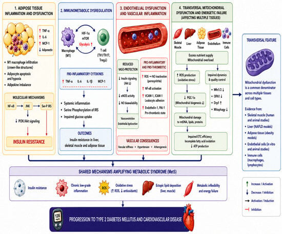
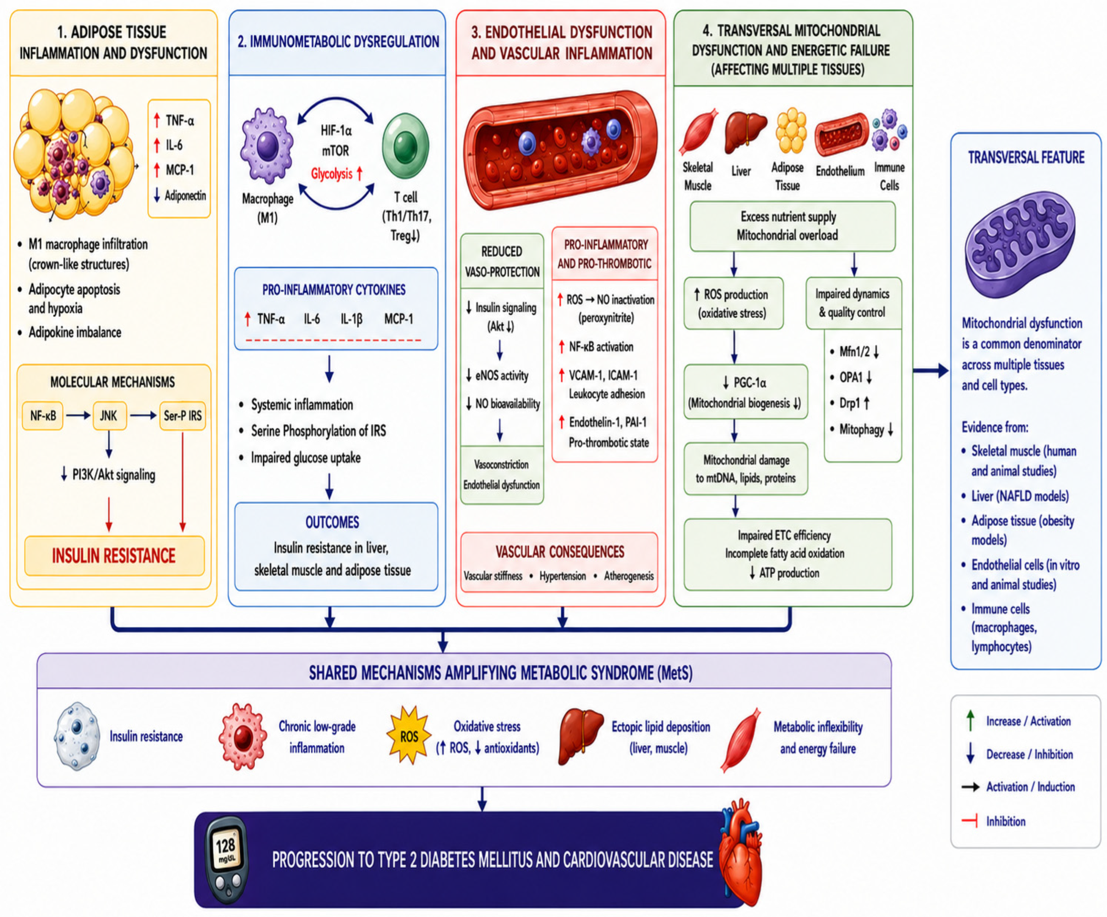
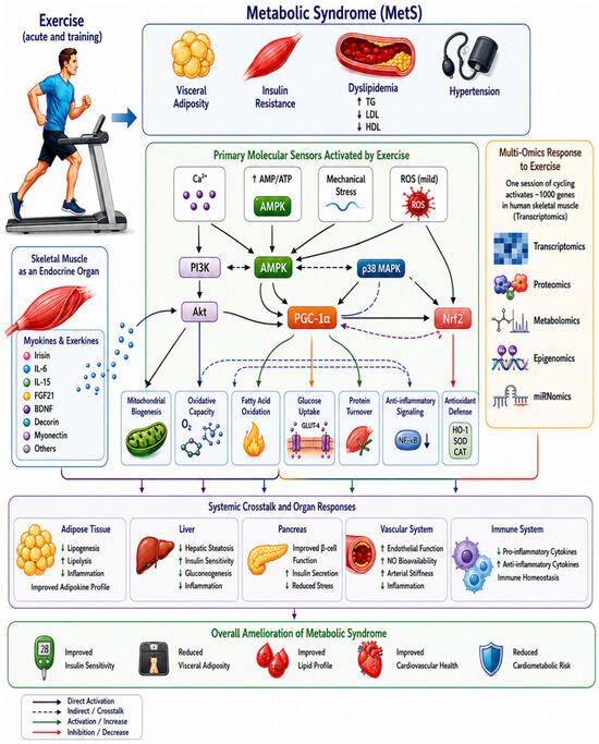
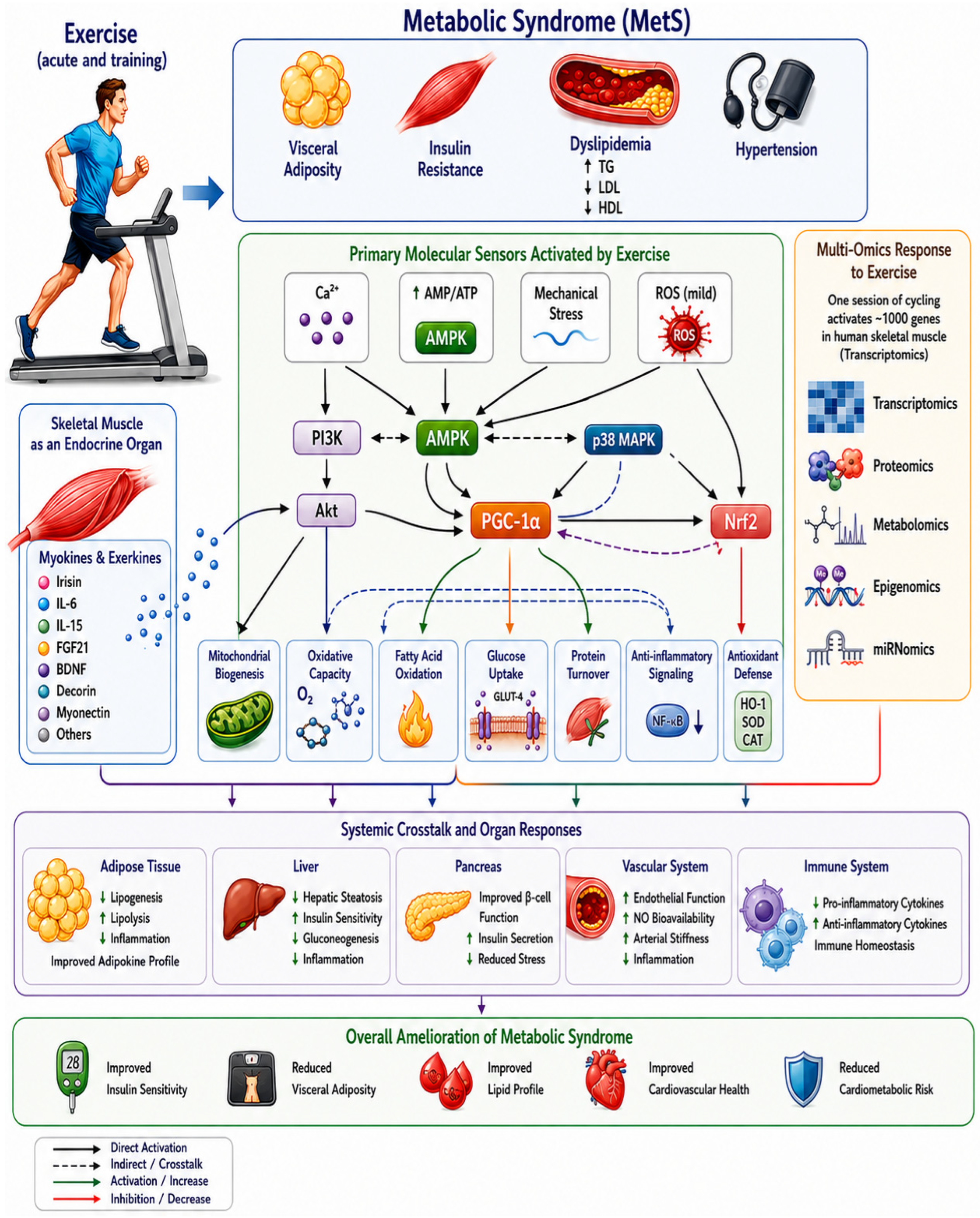
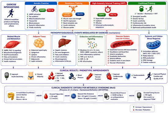
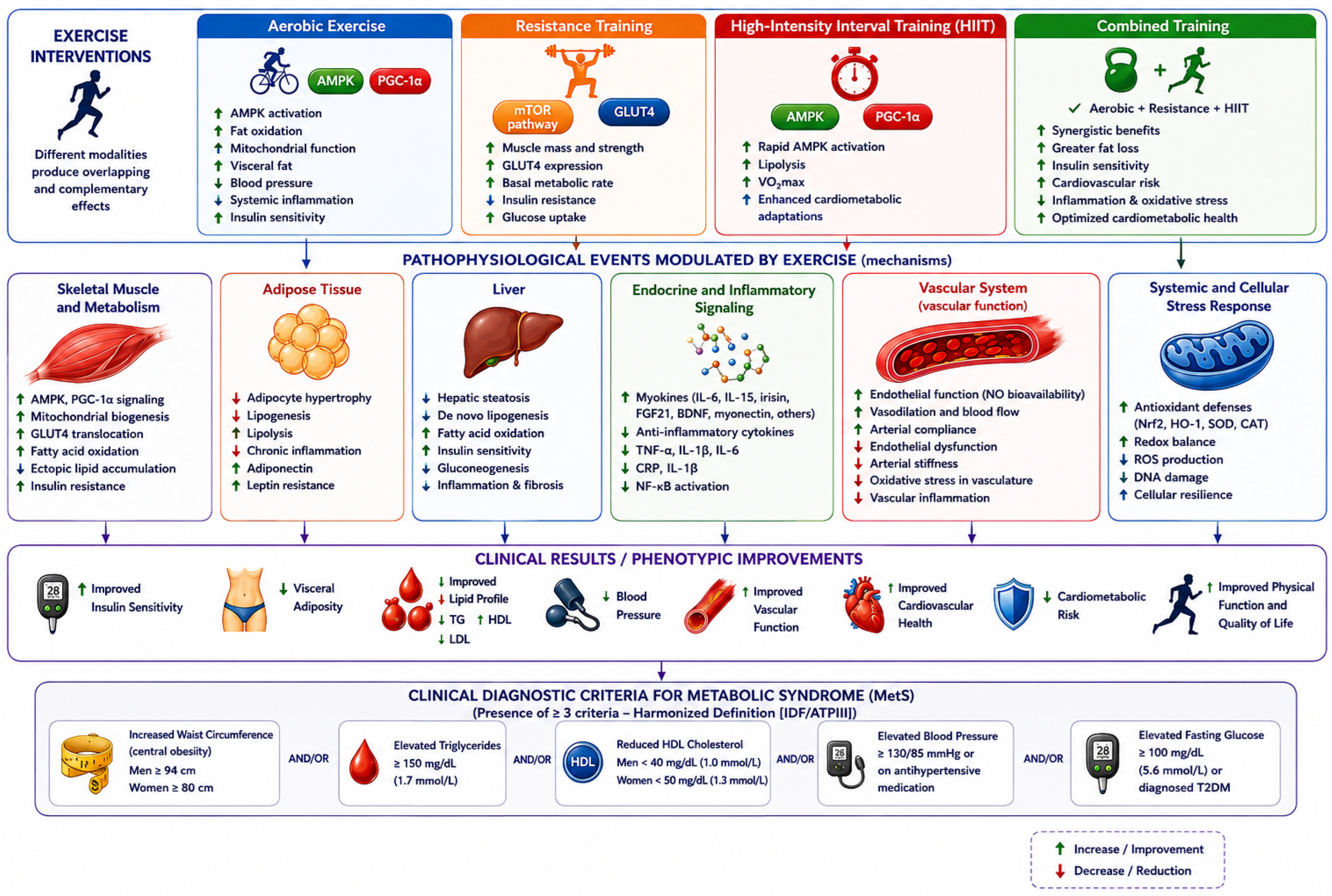
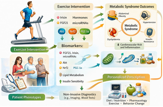
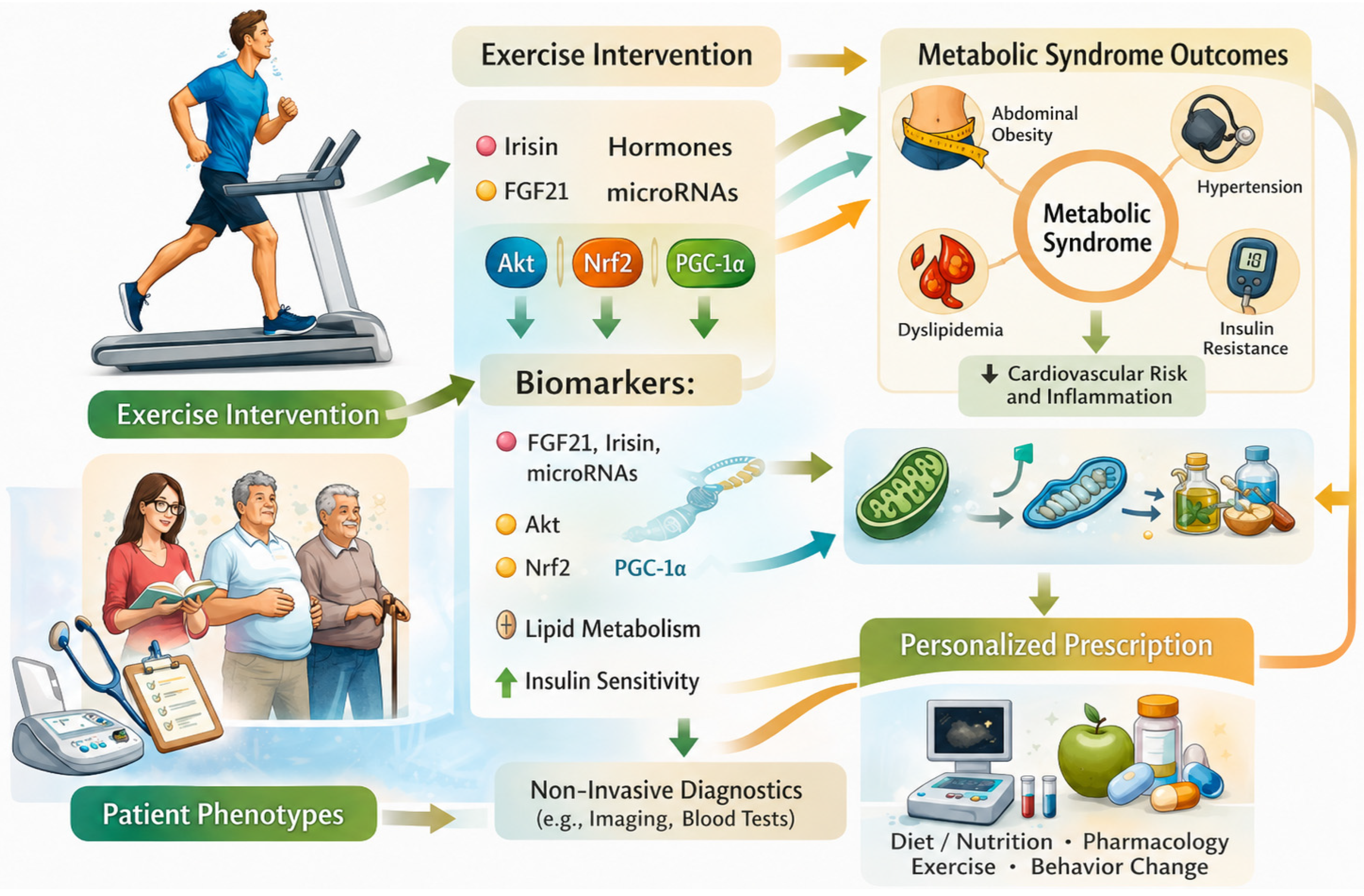

Disclaimer/Publisher’s Note: The statements, opinions and data contained in all publications are solely those of the individual author(s) and contributor(s) and not of MDPI and/or the editor(s). MDPI and/or the editor(s) disclaim responsibility for any injury to people or property resulting from any ideas, methods, instructions or products referred to in the content. |
© 2026 by the authors. Licensee MDPI, Basel, Switzerland. This article is an open access article distributed under the terms and conditions of the Creative Commons Attribution (CC BY) license.