


Abstract
Balanced control of intracellular deoxyribonucleoside-triphosphate (dNTP) pools is crucial for accurate DNA replication and genome stability. Deoxycytidine triphosphate pyrophosphatase 1 (DCTPP1) is a cytosolic nucleotide-sanitizing enzyme that selectively hydrolyzes abnormal cytosine-derived triphosphates to maintain pyrimidine pool homeostasis. Dysregulated DCTPP1 expression disrupts this balance, leading to replication stress and genomic instability. Recent studies have revealed that DCTPP1 is frequently upregulated across various human cancers and contributes to tumor progression, stress tolerance, and chemoresistance. Particularly in hepatocellular carcinoma, an oncogenic DCTPP1/MYC feedback loop has been reported to rewire pyrimidine metabolism. This review summarizes the current understanding of DCTPP1 biology, highlights its emerging role in hepatocellular carcinoma, and discusses its potential therapeutic implications.
1. Introduction
Deoxyribonucleoside triphosphate (dNTP) pools are essential for accurate DNA replication and the preservation of genome integrity. Both excesses and shortages of individual dNTPs lead to elevated mutation rates, replication stress, and activation of DNA damage responses, and perturbations of dNTP balance are increasingly recognized as contributors to tumorigenesis and therapy responses [1,2].
To protect the genome from incorporation of damaged or non-canonical nucleotides, cells deploy a set of “house-cleaning” enzymes that surveil and sanitize the nucleotide pool. These enzymes include members of the Nudix hydrolase family such as MTH1 [3,4] and a separate superfamily of all-α nucleoside triphosphate (NTP) pyrophosphatases (MazG-like proteins and their eukaryotic homologs) [4,5]. They selectively hydrolyze aberrant NTP/dNTP species and thereby are thought to limit mutagenic incorporation into DNA. The concept of nucleotide “sanitization” as a distinct and evolutionarily conserved layer of genome maintenance has been articulated in structural and functional studies of these enzyme families [6]. Within the all-α NTP pyrophosphatase group, deoxycytidine triphosphate pyrophosphatase 1 (DCTPP1), also annotated as XTP3-transactivated protein A (XTP3TPA), has emerged as a cytosolic nucleotide-sanitizing enzyme with strong biochemical preference for cytosine-derived triphosphates, including modified forms such as 5-methyl-dCTP [7]. Recent structural studies and comprehensive reviews of nucleotide sanitization enzymes have placed DCTPP1 within the Nucleoside triphosphate pyrophosphohydrolase (MazG)/dCTPase-like fold and have emphasized its role in maintaining pyrimidine pool homeostasis in mammalian cells [8,9]. In addition, broader reviews highlight DCTPP1 as a component of this genome-protective enzymatic framework [4].
The trajectory of DCTPP1 research spans computational/structural annotation and subsequent cellular and biochemical characterization. Early structure-guided analyses identified RS21-C6/XTP3TPA as a MazG-like candidate with putative nucleotide-sanitizing activity; more recent biochemical and cellular studies have confirmed high selectivity of the enzyme for cytosine nucleotides and have connected DCTPP1 perturbation to altered dNTP ratios, uracil accumulation in genomic DNA, and activation of DNA damage responses in cultured cells. Collectively, these observations support the view that DCTPP1 may act as a conserved key regulator of pyrimidine pool quality whose dysregulation could influence cancer phenotypes and chemosensitivity. However, important mechanistic and tissue-specific questions remain [6].
In this review we summarize current knowledge about DCTPP1 from structural features and substrate specificity to physiological roles in maintaining replication fidelity and emerging evidence linking DCTPP1 to human malignancies. Importantly, while functional data in liver biology have been scarce [1,10], recent studies have identified an oncogenic feedback loop in liver cancer, repositioning DCTPP1 as a central player in metabolic rewiring [11]. Accordingly, this review should be interpreted as a synthesis of current evidence and a framework for hypothesis generation, rather than as definitive proof of clinical utility.
2. Structure and Biochemical Properties of DCTPP1
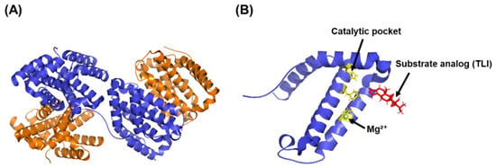
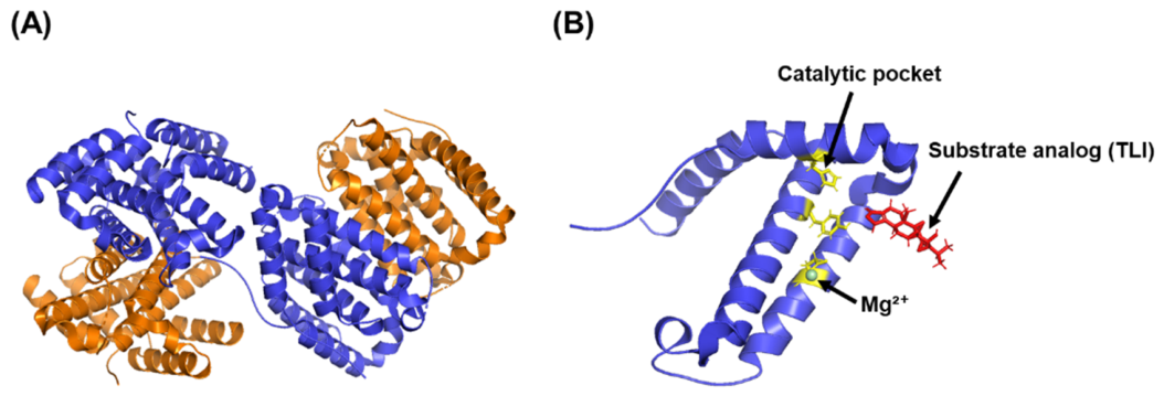
DCTPP1 is a member of the all-α NTP pyrophosphatase superfamily. Structural analyses indicate that DCTPP1 assembles as a homotetramer with a predominantly α-helical architecture, with each subunit contains a conserved MazG-like catalytic domain required for pyrophosphatase activity (Figure 1) [12]. The active site is proposed to coordinate divalent metal ions, primarily Mg2+, which appear to be required for efficient hydrolysis of its substrates. Structural studies of the murine homolog RS21-C6 indicate a four-helix bundle fold that forms a compact binding pocket likely involved in substrate recognition and catalysis [12].
Figure 1.
Structural organization of human DCTPP1. (A) Ribbon representation of the human DCTPP1 homotetramer showing its tetrameric assembly. Individual protomers are displayed in alternating colors. (B) Enlarged view of a representative catalytic pocket showing the bound substrate analog (TLI, red), catalytic Mg2+ ion (green sphere), and representative substrate-binding residues (His38, Glu63, and Tyr102; yellow). Structures were generated using PyMOL version 3.1.8 (Schrödinger, LLC, New York, NY, USA) based on the crystal structure of human DCTPP1 (PDB ID: 7MU5).
Biochemically, DCTPP1 has been reported to exhibit a strong substrate preference for cytosine-derived triphosphates, including canonical dCTP such as 5-methyl-, 5-bromo-, and 5-iodo-dCTP [7]. Hydrolysis of these nucleotides to deoxycytidine monophosphate (dCMP) and pyrophosphate is thought to reduce their misincorporation into DNA and thereby preserves genome integrity [8]. Enzymatic assays have shown optimal activity at alkaline pH (pH > 7; at mildly alkaline pH (approximately 8.5–8.7)) in the presence of Mg2+ and markedly lower activity toward other canonical dNTPs, supporting a relatively high specificity for cytosine derivatives [7]. Structural modeling identifies His38, Glu63, and Tyr102 as key residues potentially involved in substrate recognition via hydrogen bonding and hydrophobic contacts, while the 5-methyl group of modified substrates may stabilize binding through interactions with Trp47 and Trp73 [12].
Subcellular localization studies have indicated that DCTPP1 is predominantly cytosolic but can also be detected in the nucleus and mitochondria under specific cellular conditions, including highly proliferative conditions and the S phase of the cell cycle [8,13]. This compartmental flexibility suggests a possible role for DCTPP1 in safeguarding nucleotide integrity across multiple replication-associated compartments. Crucially, recent functional evidence has expanded this paradigm to non-proliferative states, reporting that DCTPP1 plays an indispensable role in quiescent cells. DCTPP1 orchestrates dCTP pool dynamics specifically within the mitochondrial compartment, thereby safeguarding mitochondrial DNA (mtDNA) stability and preventing mutational deletion phenotypes under metabolic conditions independent of active nuclear replication [14].
At the physiological level, DCTPP1 is proposed to contribute to nucleotide-pool homeostasis by degrading abnormal cytosine nucleotides, including 5-methyl-dCTP and uracil-containing dNTPs, thereby limiting mutagenic DNA incorporation [8]. DCTPP1 loss disrupts dCTP/deoxythymidine triphosphate (dTTP)/deoxyuridine triphosphate (dUTP) balance, causing uracil accumulation, strand breaks, and replication stress that can be rescued by thymidine supplementation or dUTPase overexpression [9]. Collectively, these structural and biochemical observations establish the molecular basis for understanding the emerging role of DCTPP1 in tumor biology, which is discussed in the following sections.
3. DCTPP1 in Human Cancers
Accumulating evidence indicates that DCTPP1 is aberrantly expressed in a variety of human malignancies and may be associated with tumor progression, therapeutic resistance, and patient outcomes [15]. Large-scale transcriptomic analyses from The Cancer Genome Atlas (TCGA), Genotype-Tissue Expression (GTEx), and Gene Expression Omnibus (GEO) indicate that DCTPP1 is upregulated in many solid tumors compared with normal tissues. However, reduced expression has also been reported in several cancer types, highlighting the context-dependent nature of DCTPP1 dysregulation.
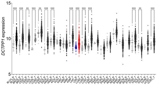
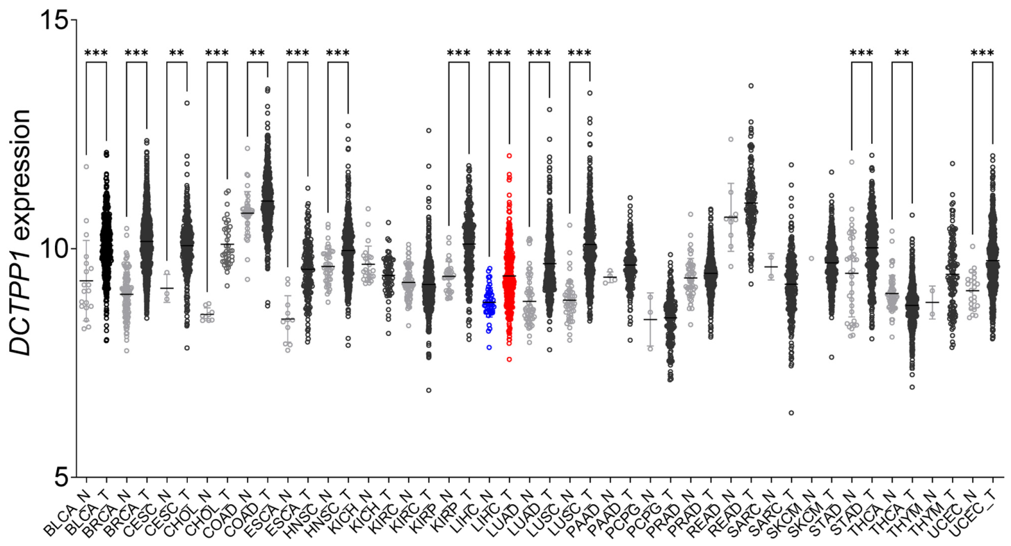
To complement the published literature, we performed a pan-cancer bioinformatic analysis using publicly available datasets from TCGA via the UCSC Xena Browser. The analysis workflow and statistical methods are described in the legend of Figure 2. Notably, elevated DCTPP1 expression was observed in multiple tumor types. In contrast, a limited number of cancer types exhibited reduced DCTPP1 expression in tumor tissues relative to normal controls, indicating that the direction and magnitude of DCTPP1 dysregulation may vary in a cancer-type-dependent manner. Moreover, high DCTPP1 expression has been consistently associated with adverse survival outcomes in several cancer types, although the underlying mechanisms appear to be context-dependent [16]. These findings are illustrated in Figure 2, and representative cancer types showing DCTPP1 dysregulation are summarized in Table 1 [15].
Figure 2.
Pan-cancer expression profile of DCTPP1. Scatter plots illustrating DCTPP1 expression levels (log2(TPM + 1)) in tumor (T, colored) and normal (N, grey) tissues across 33 cancer types. Pan-cancer gene expression data were obtained from the TCGA Pan-Cancer Atlas Hub via the UCSC Xena Browser (Illumina HiSeq RNASeqV2 dataset; n = 11,060). Tumor and normal samples were classified according to TCGA sample ID nomenclature (codes 01–09, tumor; 10–19, normal). Differential expression was evaluated using the Wilcoxon rank-sum test with multiple-testing correction. Data processing and statistical analyses were performed in R, and figures were generated using the ggplot2 package. LIHC is highlighted (N in blue, T in red). Statistical significance is indicated as follows: ns, not significant; ** p < 0.01; *** p < 0.001. Abbreviations for cancer types are as follows: bladder urothelial carcinoma (BLCA), breast invasive carcinoma (BRCA), cervical squamous cell carcinoma and endocervical adenocarcinoma (CESC), cholangiocarcinoma (CHOL), colon adenocarcinoma (COAD), esophageal carcinoma (ESCA), head and neck squamous cell carcinoma (HNSC), kidney chromophobe (KICH), kidney renal clear cell carcinoma (KIRC), kidney renal papillary cell carcinoma (KIRP), liver hepatocellular carcinoma (LIHC), lung adenocarcinoma (LUAD), lung squamous cell carcinoma (LUSC), pancreatic adenocarcinoma (PAAD), pheochromocytoma and paraganglioma (PCPG), prostate adenocarcinoma (PRAD), rectum adenocarcinoma (READ), sarcoma (SARC), skin cutaneous melanoma (SKCM), stomach adenocarcinoma (STAD), thyroid carcinoma (THCA), thymoma (THYM), and uterine corpus endometrial carcinoma (UCEC).
In breast cancer, TCGA and GEO meta-analyses revealed that DCTPP1 is significantly upregulated, and high expression correlates with reduced overall and recurrence-free survival [7,13,17]. In triple-negative breast cancer, FOXA1-mediated transcriptional activation of DCTPP1 suppresses ferroptosis and promotes cisplatin resistance [18]. High DCTPP1 expression has also been linked to an immunosuppressive tumor microenvironment by promoting M2 macrophage polarization [19]. In luminal A tumors, elevated DCTPP1 expression has been associated with reduced chemotherapy responsiveness [20].
Similarly, in gastric cancer, DCTPP1 overexpression has been linked to unfavorable prognosis and chemoresistance via upregulation of the multidrug resistance 1 gene (MDR1), which reduces 5-fluorouracil sensitivity [21]. Colorectal cancer data also identify DCTPP1 as a poor-prognosis-associated factor, with natural small-molecule inhibitors reported to modulate DCTPP1-dependent metabolic reprogramming [22].
In ovarian cancer, DCTPP1 overexpression has been associated with cisplatin resistance by reducing oxidative stress and promoting cell survival during chemotherapy [23,24]. These findings further support a role for DCTPP1 in redox homeostasis and therapy resistance across multiple cancer types. In lung adenocarcinoma, DCTPP1 overexpression has been associated with increased tumorigenicity, chemotherapy resistance, and potential diagnostic and prognostic value [25]. Furthermore, a pan-cancer analysis underscores the context-dependent prognostic role of DCTPP1, with high expression associated with both favorable immunotherapeutic responses and poor outcomes in different tumor types [15]. Together, these findings indicate that DCTPP1 is a nucleotide-metabolizing enzyme with broad oncogenic relevance and suggest that nucleotide imbalance, oxidative stress tolerance, and therapy resistance contribute to malignant progression across multiple human cancers.
Recent evidence further suggests that DCTPP1 contributes to post-transcriptional regulation and stemness-related phenotypes in multiple cancer types [26]. Functional studies have shown that DCTPP1 depletion suppresses cell proliferation, stem cell-like properties, and chemoresistance in breast and gastric cancer models [7,21]. These findings support a role for DCTPP1 in maintaining nucleotide balance and redox homeostasis while promoting tumor progression through diverse molecular mechanisms. Although these pan-cancer studies provide important biological context, the remainder of this review focuses primarily on the emerging role of DCTPP1 in hepatocellular carcinoma, where recent functional studies have begun to elucidate its biological significance.
Table 1.
Differential Expression of DCTPP1 in Various Cancers. Only representative cancer types with reported DCTPP1 upregulation are included.
4. DCTPP1 in Liver Biology and Disease
Although DCTPP1 has not been extensively studied in liver biology or hepatocarcinogenesis, several observations suggest its potential oncogenic relevance. In MYC-driven mouse models of liver tumorigenesis, DCTPP1 expression has been reported to be elevated in MYC-overexpressing liver stem/progenitor cells, suggesting an association between DCTPP1 expression and dedifferentiated or highly proliferative cellular states [9,36]. Consistent with these findings, DCTPP1 has been observed to accumulate in the nucleus of liver carcinoma cells, which may indicate potential nuclear roles in DNA replication or repair processes [16]. Furthermore, pan-cancer transcriptomic analyses, including hepatocellular carcinoma (HCC) datasets, indicate that DCTPP1 expression is higher in liver tumors relative to normal hepatic tissues [15].
To further assess the clinical relevance of these observations, we performed a pan-cancer survival analysis using the publicly available Tumor Immune Estimation Resource version 3 (TIMER3) platform, and the results are summarized in Table 2. Among the analyzed cancer types, high DCTPP1 expression was significantly associated with poor clinical outcomes in liver hepatocellular carcinoma (LIHC), ranking among the strongest prognostic associations across cancer types after multiple-testing adjustment. As these findings are derived from in silico analyses, they should be interpreted as correlative rather than indicative of causality.
Table 2.
Association between DCTPP1 expression and overall survival across cancer types. Pan-cancer survival association of DCTPP1 expression based on univariate Cox proportional hazards analysis using the TIMER3 platform. Data were obtained from the TCGA Pan-Cancer Atlas via TIMER3 (accessed on 15 July 2026). Z-score represents the standardized Cox regression coefficient (positive values indicate that higher DCTPP1 expression is associated with increased mortality risk, whereas negative values indicate an association with improved survival). p-value indicates the univariate Cox regression p-value, and adjusted p-value represents the Benjamini–Hochberg false discovery rate (FDR)-adjusted p-value.
Consistent with a biomarker-oriented interpretation, immunohistochemical data from the Human Protein Atlas indicate that DCTPP1 is variably expressed in human liver cancer tissues, with predominantly cytoplasmic localization and heterogeneous staining intensity across patients. This inter-patient variability highlights the potential utility of DCTPP1 as a prognostic or patient-stratification biomarker in HCC. Nevertheless, heterogeneous protein expression alone cannot establish functional dependency or therapeutic tractability, emphasizing the need for mechanistic studies to define the biological role of DCTPP1 in liver tumorigenesis.
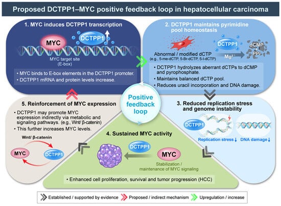
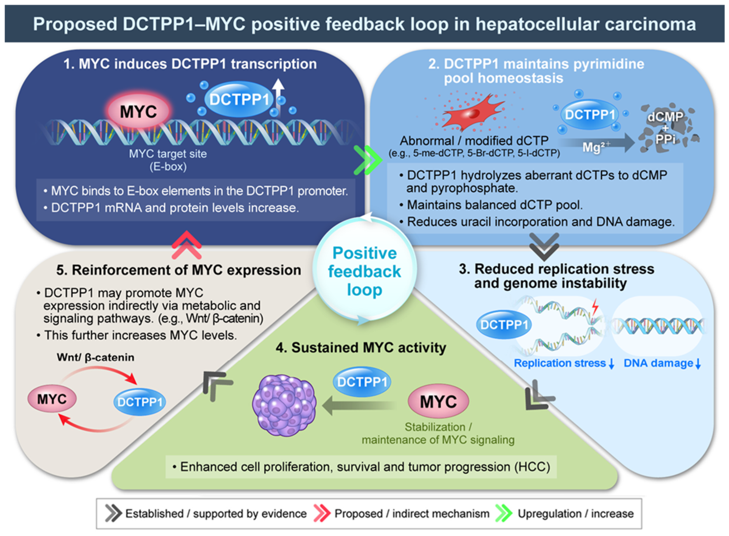
Despite these correlations, functional studies directly examining DCTPP1 in hepatocyte physiology or hepatocarcinogenesis remain limited. A recent study has begun to address this gap by reporting a novel oncogenic DCTPP1/MYC positive-feedback loop that may contribute to HCC progression (Figure 3).
Figure 3.
Proposed mechanistic model of the DCTPP1–MYC regulatory axis in hepatocellular carcinoma (HCC). MYC transcriptionally activates DCTPP1 through E-box elements within the DCTPP1 promoter, resulting in increased DCTPP1 expression in HCC cells (Step 1). Elevated DCTPP1 maintains pyrimidine nucleotide homeostasis by hydrolyzing aberrant or modified cytosine-derived triphosphates into deoxycytidine monophosphate (dCMP) and inorganic pyrophosphate (PPi), thereby preserving deoxycytidine triphosphate (dCTP)pool balance and limiting uracil incorporation into DNA (Step 2). Maintenance of nucleotide-pool homeostasis is proposed to reduce replication stress and genome instability (Step 3), thereby supporting sustained MYC activity and promoting cell proliferation, survival, and tumor progression (Step 4). Based on recent findings, DCTPP1 may also indirectly reinforce MYC expression or signaling through metabolic remodeling and pathways such as Wnt/β-catenin, forming a proposed positive-feedback loop (Step 5). Black arrows indicate experimentally supported relationships, red arrows denote proposed or indirect mechanisms that require further experimental validation, and green arrows represent upregulation or increased activity. This schematic integrates the established biochemical functions of DCTPP1 with the current working model in HCC while highlighting hypotheses that remain to be experimentally validated. In the diagram, white upward arrows indicate transcriptional upregulation, green arrows indicate upregulation or increased activity, red curved arrows represent regulatory feedback circuits, blue downward arrows denote reduction, and grey straight arrows indicate mechanistic connections.
Specifically, the study proposes a model in which MYC transcriptionally induces DCTPP1 expression, whereas elevated DCTPP1 may maintain pyrimidine nucleotide homeostasis, thereby alleviate replication stress and support sustained MYC-driven tumor growth. Although several components remain hypothetical, this model provides a useful conceptual framework for understanding the emerging role of DCTPP1 in hepatocellular carcinoma.
At the molecular level, this regulatory axis is initiated by MYC-mediated transcriptional activation of DCTPP1 [11]. Elevated DCTPP1 may subsequently reinforce MYC-driven metabolic adaptation by maintaining pyrimidine nucleotide homeostasis [11,37]. These findings suggest that DCTPP1 may play a broader role than nucleotide sanitization alone by integrating nucleotide quality control with oncogenic metabolic reprogramming. Beyond identifying DCTPP1 as a downstream target of MYC, this newly identified regulatory axis provides important insights into the metabolic adaptation of hepatocellular carcinoma.
The biological significance of this regulatory axis becomes particularly evident in MYC-driven tumors, which exhibit increased nucleotide biosynthesis, accelerated DNA replication, and elevated replication stress [38,39]. These hallmarks of highly proliferative cancers create a continuous demand for balanced intracellular nucleotide pools [37,40]. Under these conditions, the accumulation of non-canonical or damaged pyrimidine nucleotides is expected to increase, thereby creating a greater dependence on nucleotide-sanitizing enzymes such as DCTPP1 to preserve replication fidelity. Within this context, DCTPP1 may function as a metabolic safeguard that preserves pyrimidine nucleotide homeostasis by preventing the incorporation of non-canonical pyrimidine nucleotides into DNA. Maintenance of nucleotide quality is proposed to alleviate MYC-driven replication stress, thereby supporting sustained tumor cell proliferation and metabolic adaptation [39,41]. Rather than acting solely as a housekeeping enzyme involved in nucleotide sanitization, DCTPP1 may therefore serve as a metabolic regulator linking nucleotide quality control with MYC-driven metabolic reprogramming in HCC [11,42]. Although this model is currently supported primarily by a single functional study, it provides a biologically plausible rationale for understanding the metabolic dependency of rapidly proliferating liver cancer cells. Collectively, these results provide functional evidence that DCTPP1 may represent a promising therapeutic candidate in hepatocellular carcinoma. Importantly, these findings extend beyond the correlative clinical observations described above but remain insufficient to establish DCTPP1 as a clinically validated therapeutic target. Additional mechanistic, pharmacological, and preclinical studies are therefore required before the therapeutic potential of DCTPP1 in hepatocellular carcinoma can be fully established.
Despite the proposed mechanistic model summarized in Figure 3, several key mechanistic questions remain unresolved [11]. Accordingly, this model should be regarded as a working hypothesis rather than a fully established signaling pathway. It remains unclear whether DCTPP1 directly regulates MYC stability or transcriptional activity, or whether the observed positive-feedback loop is mediated indirectly through metabolic remodeling. Although MYC is currently the best-characterized upstream regulator of DCTPP1, additional oncogenic signaling pathways may also contribute to DCTPP1 regulation.
Moreover, MYC is a well-established downstream effector of the Wnt/β-catenin signaling pathway, one of the most frequently dysregulated oncogenic pathways in hepatocellular carcinoma [43,44,45]. Whether DCTPP1 functionally interacts with Wnt/β-catenin signaling or whether DCTPP1 dependency is preferentially associated with MYC-activated HCC remains to be determined. Addressing these questions will be essential for determining whether DCTPP1 represents a true therapeutic vulnerability rather than simply a prognostic biomarker [15]. These observations provide a basis for future studies aimed at validating the proposed DCTPP1/MYC regulatory model and determining whether DCTPP1 constitutes a genuine metabolic dependency in hepatocellular carcinoma. Considering its established biochemical role in dNTP homeostasis and replication fidelity [8], liver-focused research using conditional knockout or overexpression models is warranted. Experimental strategies may include liver- or progenitor-specific DCTPP1 perturbation in MYC-driven HCC models, human liver organoids, or stem-cell-derived hepatocytes combined with dNTP metabolomics, uracil-incorporation assays, and replication-stress markers. Such approaches could elucidate whether DCTPP1 contributes causally to liver tumor initiation or progression and evaluate its suitability as a therapeutic target in HCC.
Importantly, although transcriptomic, immunohistochemical, and survival analyses consistently support the clinical relevance of DCTPP1 in HCC, these observations remain largely correlative. Therefore, current evidence is sufficient to support DCTPP1 as a promising prognostic biomarker and an emerging therapeutic candidate, but not yet as a fully validated therapeutic target. Future mechanistic and preclinical studies will ultimately determine whether DCTPP1 represents a genuine therapeutic vulnerability in hepatocellular carcinoma.
5. Targeting DCTPP1: Experimental and Therapeutic Strategies
Functional interrogation and therapeutic targeting of DCTPP1 can be approached using both genetic and pharmacological strategies. Transient small interfering RNA (siRNA) or short hairpin RNA (shRNA) mediated knockdown allows short-term phenotypic assessment and serves as an initial validation step, ideally employing multiple independent siRNAs with appropriate controls [46]. For definitive gene disruption, Clustered regularly interspaced short palindromic repeats/CRISPR-associated protein 9 (CRISPR/Cas9)-mediated knockout or allele-specific editing provides robust loss-of-function models that may be suitable for long-term and in vivo analyses [47].
Complementary to genetic approaches, small-molecule inhibitors offer the possibility of rapid and reversible modulation of DCTPP1 activity, thereby providing experimental tools to explore its potential therapeutic relevance in liver diseases and HCC. Several chemical scaffolds have been reported, including triptolide [48], Pyrcoumin [49], and synthetic series such as piperazin-1-ylpyridazines [50], and 3,6-disubstituted triazolothiadiazoles [51]. Among these, triptolide is the most well-characterized compound known to suppress DCTPP1 activity by directly binding and inhibiting its enzymatic function [48]. Importantly, triptolide’s water-soluble pro-drug Minnelide is under clinical investigation for various solid tumors including liver cancers [52,53]. Beyond these generic loss-of-function and pharmacological approaches, DCTPP1 may be of particular interest in the context of MYC-driven tumorigenesis. MYC is a master regulator of nucleotide biosynthesis, ribosome biogenesis, and DNA replication, and MYC overexpression is known to be associated with severe replication stress by imposing extreme demands on dNTP pool balance and replication fidelity [54,55]. In MYC-driven liver tumor models, elevated DCTPP1 expression in liver stem/progenitor cell populations has been observed, raising the possibility that DCTPP1 may function as an adaptive safeguard that buffers MYC-induced metabolic and replicative stress by sanitizing damaged or excess cytosine nucleotides [36]. Under MYC-hyperactivated conditions, functional disruption of DCTPP1 would be predicted to exacerbate nucleotide imbalance, increase uracil misincorporation into genomic DNA, and amplify DNA damage signaling, thereby limiting tumor cell viability. This model positions DCTPP1 not merely as a passive metabolic marker but as a context-dependent vulnerability in MYC-driven hepatocarcinogenesis. This therapeutic rationale has now been validated empirically by the discovery of the reciprocal regulation between DCTPP1 and MYC in HCC models, where targeting the DCTPP1/MYC axis successfully disrupted pyrimidine metabolic homeostasis and suppressed tumor progression [11]. Such a hypothesis can be directly tested using combinatorial experimental systems that integrate MYC overexpression with DCTPP1 genetic perturbation in hepatocyte or liver progenitor models, allowing discrimination between correlation and functional dependency. To ensure experimental specificity, rigorous on-target validation is essential. Standard strategies include phenocopy assays, where chemically distinct inhibitors reproduce the cellular phenotypes seen with genetic knockdown or knockout, and non-additivity or epistasis testing, where combining an inhibitor with CRISPR-mediated DCTPP1 loss fails to produce further effects [56,57]. Rescue experiments are also critical, with re-expression of RNAi-resistant or wild-type DCTPP1 restoring the phenotype, whereas catalytically inactive mutants do not. Complementary target engagement assays such as enzyme kinetics, thermal shift assays, Cellular Thermal Shift Assay (CETSA), and chemoproteomic pulldown provide biochemical confirmation of compound binding [58,59]. Together, these orthogonal strategies—integrating genetic depletion, pharmacological inhibition, and robust validation—represent recommended experimental standards for interrogating DCTPP1 function and for assessing its potential therapeutic relevance, rather than constituting definitive proof of clinical utility. A concise summary of representative experimental strategies, chemical scaffolds, phenotypic outcomes, and validation methods is provided in Table 3.
Table 3.
Experimental and therapeutic strategies for targeting DCTPP1.
6. Future Perspectives and Challenges
Biochemical and cellular studies have established DCTPP1 as an essential regulator of dNTP pool homeostasis and genome stability [8,9]. Nevertheless, critical mechanistic questions remain unanswered, particularly how DCTPP1 activity is integrated into oncogenic signaling networks, tissue-specific nucleotide metabolism, and in vivo tumor progression. In the liver, this question gains particular significance because hepatocytes display uniquely high nucleotide turnover and replication potential, driven by continuous metabolic flux and regenerative capacity. Under such regenerative conditions, tight control of nucleotide pool quality becomes especially critical, suggesting that DCTPP1 activity may be particularly important in safeguarding replication fidelity during liver regeneration and repair. Chronic hepatic injury, including viral hepatitis, steatohepatitis, and cirrhosis, creates an environment of oxidative stress, inflammation, and repeated cell proliferation that profoundly perturbs nucleotide balance and genome stability, processes closely linked to hepatocarcinogenesis [10,60,61]. In such settings, increased oxidative nucleotide damage and sustained replicative demand are expected to increase cellular reliance on nucleotide pool-sanitizing enzymes such as DCTPP1, potentially increasing the biological relevance of DCTPP1 in chronic liver disease. Although multiple cell-active and chemically distinct DCTPP1 inhibitors have been developed, including scaffolds that can enhance the cytotoxicity of nucleoside analogs, their pharmacological selectivity and translational relevance remain incompletely characterized [49,62].
Pan-cancer transcriptomic analyses and targeted knockdown studies suggest that DCTPP1 suppression reduces proliferation and influences tumor immune and drug-response signatures across several tumor models. These observations support the hypothesis that DCTPP1 may represent a context-dependent therapeutic vulnerability rather than a universal oncogenic driver. However, given that nucleotide metabolism is profoundly reprogrammed during liver disease progression—from chronic inflammation and fibrosis to malignant transformation—it will be important to evaluate whether DCTPP1 expression or activity correlates with hepatic disease stage or HCC subtype, particularly in the context of liver regeneration and metabolic reprogramming that accompany chronic liver injury and tumor progression. Recent multi-omic analyses in HCC cohorts have revealed that dysregulation of pyrimidine metabolism enzymes contributes to tumor proliferation and poor prognosis, suggesting that DCTPP1 could play a similar, but context-dependent, role in hepatic oncogenesis [63,64].
Importantly, the therapeutic exploitation of DCTPP1 must carefully balance antitumor efficacy with tissue-specific toxicity. Notably, systematic toxicity profiles of selective DCTPP1 inhibition in normal tissues, including the liver, remain largely undefined, underscoring the need for careful preclinical evaluation of therapeutic specificity and safety. Experience from other nucleotide metabolism targets such as thymidylate synthase and ribonucleotide reductase demonstrate that on-target toxicities, metabolic compensation, and acquired resistance frequently limit therapeutic windows [2,65,66]. These risks are particularly pronounced in the liver, where extensive metabolic redundancy, detoxification capacity, and regenerative potential may either buffer or exacerbate the consequences of DCTPP1 inhibition. Moreover, the high proliferative and regenerative capacity of hepatocytes raises the possibility that sustained DCTPP1 inhibition could impair normal hepatic repair processes or provoke adaptive metabolic rewiring, highlighting the importance of defining liver-specific toxicity profiles and safe dosing strategies early in preclinical development [67,68].
Future work should therefore prioritize the development of high-quality, selective chemical probes with validated cellular target engagement (for example, CETSA and chemoproteomic assays) and integration with orthogonal genetic models, including conditional loss- and gain-of-function systems. Rigorous functional analyses combining quantitative dNTP and uracil-incorporation assays, replication-stress markers, and DNA damage readouts will be essential to define the biological consequences of DCTPP1 modulation in physiological and oncogenic contexts. To accurately assess therapeutic specificity and toxicity, such studies should be conducted in liver-relevant experimental systems, including primary hepatocytes, hepatic organoids, and inflammation- or fibrosis-driven HCC models, which better capture tissue-specific metabolic and microenvironmental influences on DCTPP1 function.
In parallel, biomarker-guided combination strategies—such as pairing DCTPP1 inhibition with nucleoside analogs or DNA-damaging agents—may offer a rational approach to enhance therapeutic efficacy while minimizing systemic toxicity. Defining such rational therapeutic windows, together with predictive biomarkers of response and toxicity, will be critical for de-risking clinical translation and enabling the safe advancement of DCTPP1-targeted interventions into cancer therapy [2,58].
7. Conclusions
DCTPP1 is a conserved nucleotide-pool-sanitizing enzyme that contributes to the maintenance of replication fidelity by hydrolyzing aberrant cytosine nucleotides. Convergent structural, biochemical, and cellular studies support its role in genome maintenance and suggest that dysregulation of DCTPP1 may be associated with tumor biology, including context-dependent effects on proliferation, stress tolerance, and drug response. However, the majority of current evidence is derived from in vitro systems, bioinformatic analyses, and non-hepatic cancer models, and therefore should be interpreted with appropriate caution. Selective chemical probes combined with orthogonal genetic approaches will be essential for establishing on-target mechanisms and validating DCTPP1 biology. At present, direct functional evidence linking DCTPP1 activity to liver physiology or hepatocarcinogenesis remains limited, particularly in the context of the biological heterogeneity and lineage plasticity of primary liver cancers [69]. Therefore, its proposed role in HCC should be regarded as a testable hypothesis rather than a fully demonstrated causal mechanism. Accordingly, DCTPP1 should not yet be considered a validated therapeutic target in liver cancer, but rather a candidate molecule whose biological and translational relevance requires further empirical validation. Future liver-focused studies using physiologically relevant experimental models and clinical specimens will be essential to determine whether DCTPP1 plays a functional role in hepatic tumor initiation, progression, or therapeutic response.
A disciplined target-assessment framework—emphasizing specificity, in-cell target engagement, therapeutic window definition, and systematic toxicity evaluation—will be critical before DCTPP1 can be credibly advanced toward clinical translation. Taken together, the available data position DCTPP1 as a promising but preliminary node within the complex metabolic landscape of cancer, underscoring the need for rigorous experimental validation to clarify its role in HCC.
Author Contributions
J.W.E.: Conceptualization, Investigation, Writing—Review & Editing, Visualization, Funding Acquisition, Supervision. H.I.K.: Methodology, Investigation, Writing—Review & Editing, Funding Acquisition. S.H.J.: Methodology, Investigation, Writing—Original Draft, Visualization. H.S.J.: Methodology, Validation. H.S.K.: Methodology, Validation, Review & Editing, Funding. All authors have read and agreed to the published version of the manuscript.
Funding
This work was supported by the National Research Foundation of Korea (NRF), funded by the Ministry of Science and ICT (MSIT), Republic of Korea (grant numbers: RS-2022-NR070489, RS-2023-00210847 and RS-2024-00344635), the Commercializations Promotion Agency for R&D Outcomes (COMPA) funded by Ministry of Science and ICT (RS-2024-00422549), and the Korea Health Technology R&D Project of the Korea Health Industry Development Institute (KHIDI), funded by the Ministry of Health & Welfare, Republic of Korea (HR21C1003).
Institutional Review Board Statement
Not applicable.
Informed Consent Statement
Not applicable.
Data Availability Statement
No new experimental datasets were generated during this study. The structural model shown in Figure 1 was generated using PyMOL based on the publicly available crystal structure of human DCTPP1 (Protein Data Bank, PDB ID: 7MU5). Publicly available transcriptomic and survival datasets used for Figure 2 and Table 2 were obtained from the TCGA Pan-Cancer Atlas via the UCSC Xena Browser and the Tumor Immune Estimation Resource version 3 (TIMER3), respectively.
Conflicts of Interest
The authors declare no conflicts of interest.
References
- Pai, C.-C.; Kearsey, S.E. A critical balance: dNTPs and the maintenance of genome stability. Genes 2017, 8, 57. [Google Scholar] [CrossRef] [Scilit] [PubMed]
- Mullen, N.J.; Singh, P.K. Nucleotide metabolism: A pan-cancer metabolic dependency. Nat. Rev. Cancer 2023, 23, 275–294. [Google Scholar] [CrossRef] [Scilit] [PubMed]
- Nakabeppu, Y.; Ohta, E.; Abolhassani, N. MTH1 as a nucleotide pool sanitizing enzyme: Friend or foe? Free Radic. Biol. Med. 2017, 107, 151–158. [Google Scholar] [CrossRef] [Scilit]
- Broderick, K.; Moutaoufik, M.T.; Aly, K.A.; Babu, M. Sanitation enzymes: Exquisite surveillance of the noncanonical nucleotide pool to safeguard the genetic blueprint. In Seminars in Cancer Biology; Academic Press: Cambridge, MA, USA, 2023; pp. 11–20. [Google Scholar]
- Lyu, L.-D.; Tang, B.-K.; Fan, X.-Y.; Ma, H.; Zhao, G.-P. Mycobacterial MazG safeguards genetic stability via housecleaning of 5-OH-dCTP. PLoS Pathog. 2013, 9, e1003814. [Google Scholar] [CrossRef] [Scilit] [PubMed]
- Moroz, O.V.; Murzin, A.G.; Makarova, K.S.; Koonin, E.V.; Wilson, K.S.; Galperin, M.Y. Dimeric dUTPases, HisE, and MazG belong to a new superfamily of all-α NTP pyrophosphohydrolases with potential “house-cleaning” functions. J. Mol. Biol. 2005, 347, 243–255. [Google Scholar] [CrossRef] [Scilit] [PubMed]
- Song, F.; Xia, L.; Ji, P.; Tang, Y.; Huang, Z.; Zhu, L.; Zhang, J.; Wang, J.; Zhao, G.; Ge, H.; et al. Human dCTP pyrophosphatase 1 promotes breast cancer cell growth and stemness through the modulation on 5-methyl-dCTP metabolism and global hypomethylation. Oncogenesis 2015, 4, e159. [Google Scholar] [CrossRef] [Scilit] [PubMed]
- Requena, C.E.; Pérez-Moreno, G.; Ruiz-Pérez, L.M.; Vidal, A.E.; González-Pacanowska, D. The NTP pyrophosphatase DCTPP1 contributes to the homoeostasis and cleansing of the dNTP pool in human cells. Biochem. J. 2014, 459, 171–180. [Google Scholar] [CrossRef] [Scilit] [PubMed]
- Martínez-Arribas, B.; Requena, C.E.; Pérez-Moreno, G.; Ruíz-Pérez, L.M.; Vidal, A.E.; González-Pacanowska, D. DCTPP1 prevents a mutator phenotype through the modulation of dCTP, dTTP and dUTP pools. Cell. Mol. Life Sci. 2020, 77, 1645–1660. [Google Scholar] [PubMed]
- Yang, F.; Hilakivi-Clarke, L.; Shaha, A.; Wang, Y.; Wang, X.; Deng, Y.; Lai, J.; Kang, N. Metabolic reprogramming and its clinical implication for liver cancer. Hepatology 2023, 78, 1602–1624. [Google Scholar] [CrossRef] [Scilit] [PubMed]
- Li, J.; Liang, J.; Zhang, M.; Ma, R.; Zhou, R.; Xi, J.; Yu, H.; Li, Y.; Wang, C.; Bai, Z. Oncogenic DCTPP1/MYC feedback loop rewires pyrimidine metabolism to drive hepatocellular carcinoma. Funct. Integr. Genom. 2026, 26, 88. [Google Scholar] [CrossRef] [Scilit] [PubMed]
- Scaletti, E.; Claesson, M.; Helleday, T.; Jemth, A.-S.; Stenmark, P. The first structure of an active mammalian dCTPase and its complexes with substrate analogs and products. J. Mol. Biol. 2020, 432, 1126–1142. [Google Scholar] [CrossRef] [Scilit] [PubMed]
- Muñoz, J.P.; Soto-Jiménez, D.; Calaf, G.M. DCTPP1 expression as a predictor of chemotherapy response in luminal a breast cancer patients. Biomedicines 2024, 12, 1732. [Google Scholar] [CrossRef] [Scilit] [PubMed]
- Fernandez, B.; Perez-Moreno, G.; Martinez-Arribas, B.; Vidal, A.E.; Ruiz-Perez, L.M.; Gonzalez-Pacanowska, D. DCTPP1 orchestrates dCTP pool dynamics and mtDNA stability in quiescent cells. Cell Death Dis. 2026, 17, 404. [Google Scholar] [CrossRef] [Scilit] [PubMed]
- Li, H.; Xu, Y.; Shan, J.; Lun, Y. Pan-cancer landscape of DCTPP1 and preliminary exploration of DCTPP1 in renal clear cell carcinoma. Sci. Rep. 2024, 14, 27710. [Google Scholar] [CrossRef] [Scilit] [PubMed]
- Zhang, Y.; Ye, W.; Wang, J.; Wang, S.; Ji, P.; Zhou, G.; Zhao, G.; Ge, H.; Wang, Y. dCTP pyrophosphohydrase exhibits nucleic accumulation in multiple carcinomas. Eur. J. Histochem. EJH 2013, 57, e29. [Google Scholar] [CrossRef] [Scilit] [PubMed]
- Niu, M.; Shan, M.; Liu, Y.; Song, Y.; Han, J.-g.; Sun, S.; Liang, X.-s.; Zhang, G.-q. DCTPP1, an oncogene regulated by miR-378a-3p, promotes proliferation of breast cancer via DNA repair signaling pathway. Front. Oncol. 2021, 11, 641931. [Google Scholar] [CrossRef] [Scilit] [PubMed]
- He, Y.; Li, G.; Shi, X.; Bie, J.; Du, G.; Feng, X.; Yu, F.; He, Y. DCTPP1 is Transcriptionally Activated by FOXA1 to Affect Cisplatin Sensitivity in Triple-Negative Breast Cancer via Suppression of Ferroptosis. Cell Biochem. Biophys. 2025, 83, 4755–4768. [Google Scholar] [CrossRef] [Scilit] [PubMed]
- Chi, J.; Liu, W.; Zhai, Z.; Wang, L.; Wang, X.; Ma, Z. DCTPP1 drives immunosuppression and poor prognosis in breast cancer by promoting M2 macrophage polarization. Sci. Rep. 2026, 16, 8767. [Google Scholar] [CrossRef] [Scilit] [PubMed]
- Zhang, Y.; Jia, S.; Song, H.; Zhao, S.; Huang, L.; Zhang, Q.; Cheng, S.; Huang, Y. Silencing DSCAM-AS1 suppresses the growth and invasion of ER-positive breast cancer cells by downregulating both DCTPP1 and QPRT. Aging 2020, 12, 14754–14774. [Google Scholar] [CrossRef] [Scilit]
- Xia, L.-l.; Tang, Y.-b.; Song, F.-f.; Xu, L.; Ji, P.; Wang, S.-j.; Zhu, J.-m.; Zhang, Y.; Zhao, G.-p.; Wang, Y.; et al. DCTPP1 attenuates the sensitivity of human gastric cancer cells to 5-fluorouracil by up-regulating MDR1 expression epigenetically. Oncotarget 2016, 7, 68623–68637. [Google Scholar] [CrossRef] [Scilit] [PubMed]
- Feng, L.; Wang, X.; Guo, X.; Shi, L.; Su, S.; Li, X.; Wang, J.; Tan, N.; Ma, Y.; Wang, Z. Identification of Novel Target DCTPP1 for Colorectal Cancer Therapy with the Natural Small-Molecule Inhibitors Regulating Metabolic Reprogramming. Angew. Chem. 2024, 136, e202402543. [Google Scholar] [CrossRef] [Scilit]
- Wang, Y.; Chen, X.; Chen, Q.; Liu, T.; Wu, Y.; Huang, L.; Chen, Y. Expression of human dCTP pyrophosphatase 1 (DCTPP1) and its association with cisplatin resistance characteristics in ovarian cancer. J. Cell. Mol. Med. 2024, 28, e18371. [Google Scholar] [CrossRef] [Scilit] [PubMed]
- Wang, Y.; Chen, P.; Chen, X.; Gong, D.; Wu, Y.; Huang, L.; Chen, Y. ROS-induced DCTPP1 upregulation contributes to cisplatin resistance in ovarian cancer. Front. Mol. Biosci. 2022, 9, 838006. [Google Scholar] [CrossRef] [Scilit] [PubMed]
- Luo, R.; Li, X.; Gao, R.; Yang, M.; Cai, J.; Dai, L.; Lou, N.; Fan, G.; Zhu, H.; Wang, S.; et al. A Novel IgG–IgM Autoantibody Panel Enhances Detection of Early-stage Lung Adenocarcinoma from Benign Nodules. Genom. Proteom. Bioinform. 2024, 22, qzae085. [Google Scholar] [CrossRef] [Scilit]
- Zhai, D.; Li, T.; Ye, R.; Bi, J.; Kuang, X.; Shi, Y.; Shao, N.; Lin, Y. LncRNA LGALS8-AS1 Promotes Breast Cancer Metastasis Through miR-125b-5p/SOX12 Feedback Regulatory Network. Front. Oncol. 2021, 11, 711684. [Google Scholar] [CrossRef] [Scilit] [PubMed]
- Lu, J.; Dong, W.; He, H.; Han, Z.; Zhuo, Y.; Mo, R.; Liang, Y.; Zhu, J.; Li, R.; Qu, H.; et al. Autophagy induced by overexpression of DCTPP1 promotes tumor progression and predicts poor clinical outcome in prostate cancer. Int. J. Biol. Macromol. 2018, 118, 599–609. [Google Scholar] [CrossRef] [Scilit] [PubMed]
- Wei, J.; Qin, Y.; Zhang, L.; Gong, X. Combined protein and transcriptomics identifies DCTPP1 as a putative biomarkers for predicting immunotherapy responsiveness in gastric cancer patients. Anti-Cancer Drugs 2025, 36, 415–426. [Google Scholar] [CrossRef] [Scilit] [PubMed]
- Xu, L.; Xia, L.; Song, F.; Ji, P.; Wang, S.; Zhang, Y.; Shen, L.; Wang, Y. Expression and distribution of dCTP pyrophosphatase 1 in gastric cancer tissues and its clinical relevance. J. Shanghai Jiaotong Univ. (Med. Sci.) 2016, 36, 172. [Google Scholar] [CrossRef] [Scilit]
- Morisaki, T.; Yashiro, M.; Kakehashi, A.; Inagaki, A.; Kinoshita, H.; Fukuoka, T.; Kasashima, H.; Masuda, G.; Sakurai, K.; Kubo, N.; et al. Comparative proteomics analysis of gastric cancer stem cells. PLoS ONE 2014, 9, e110736. [Google Scholar] [CrossRef] [Scilit] [PubMed]
- Fang, Y.; Zhang, Q.; Guo, C.; Zheng, R.; Liu, B.; Zhang, Y.; Wu, J. Mitochondrial-related genes as prognostic and metastatic markers in breast cancer: Insights from comprehensive analysis and clinical models. Front. Immunol. 2024, 15, 1461489. [Google Scholar] [CrossRef] [Scilit] [PubMed]
- Zhang, B.; Zhang, Y.; Chang, K.; Hou, N.; Fan, P.; Ji, C.; Liu, L.; Wang, Z.; Li, R.; Wang, Y.; et al. Risk assessment model based on nucleotide metabolism-related genes highlights SLC27A2 as a potential therapeutic target in breast cancer. J. Cancer Res. Clin. Oncol. 2024, 150, 258. [Google Scholar] [CrossRef] [Scilit] [PubMed]
- Zhao, X.; Lin, J. Construction and Validation of a Prognostic Model Based on mRNAsi-Related Genes in Breast Cancer. Comput. Math. Methods Med. 2022, 2022, 6532591. [Google Scholar] [CrossRef] [Scilit] [PubMed]
- Jordaens, S.; Cooksey, L.; Bonney, S.; Orchard, L.; Coutinho, M.; Van Tendeloo, V.; Mills, K.I.; Orchard, K.; Guinn, B. Serum profiling identifies ibrutinib as a treatment option for young adults with B-cell acute lymphoblastic leukaemia. Br. J. Haematol. 2020, 189, 500–512. [Google Scholar] [CrossRef] [Scilit] [PubMed]
- Requena, C.E.; Pérez-Moreno, G.; Horváth, A.; Vértessy, B.G.; Ruiz-Pérez, L.M.; González-Pacanowska, D.; Vidal, A.E. The nucleotidohydrolases DCTPP1 and dUTPase are involved in the cellular response to decitabine. Biochem. J. 2016, 473, 2635–2643. [Google Scholar] [CrossRef] [Scilit] [PubMed]
- Aravalli, R.N.; Talbot, N.C.; Steer, C.J. Gene expression profiling of MYC-driven tumor signatures in porcine liver stem cells by transcriptome sequencing. World J. Gastroenterol. WJG 2015, 21, 2011–2029. [Google Scholar] [CrossRef] [Scilit] [PubMed]
- Liu, S.; Feng, L.; Wang, Z. DCTPP1: A promising target in cancer therapy and prognosis through nucleotide metabolism. Drug Discov. Today 2025, 30, 104348. [Google Scholar] [CrossRef] [Scilit] [PubMed]
- Liu, Y.C.; Li, F.; Handler, J.; Huang, C.R.; Xiang, Y.; Neretti, N.; Sedivy, J.M.; Zeller, K.I.; Dang, C.V. Global regulation of nucleotide biosynthetic genes by c-Myc. PLoS ONE 2008, 3, e2722. [Google Scholar] [CrossRef] [Scilit] [PubMed]
- Peripolli, S.; Meneguello, L.; Perrod, C.; Singh, T.; Patel, H.; Rahman, S.T.; Kiso, K.; Thorpe, P.; Calvanese, V.; Bertoli, C.; et al. Oncogenic c-Myc induces replication stress by increasing cohesins chromatin occupancy in a CTCF-dependent manner. Nat. Commun. 2024, 15, 1579. [Google Scholar] [CrossRef] [Scilit] [PubMed]
- Chen, J.; Yang, S.; Li, Y.; Ziwen, X.; Zhang, P.; Song, Q.; Yao, Y.; Pei, H. De novo nucleotide biosynthetic pathway and cancer. Genes Dis. 2023, 10, 2331–2338. [Google Scholar] [CrossRef] [Scilit] [PubMed]
- Rohban, S.; Campaner, S. Myc induced replicative stress response: How to cope with it and exploit it. Biochim. Biophys Acta (BBA) Gene Regul. Mech. 2015, 1849, 517–524. [Google Scholar] [CrossRef] [Scilit] [PubMed]
- Sequera, C.; Grattarola, M.; Holczbauer, A.; Dono, R.; Pizzimenti, S.; Barrera, G.; Wangensteen, K.J.; Maina, F. MYC and MET cooperatively drive hepatocellular carcinoma with distinct molecular traits and vulnerabilities. Cell Death Dis. 2022, 13, 994. [Google Scholar] [CrossRef] [Scilit] [PubMed]
- Xu, C.; Xu, Z.; Zhang, Y.; Evert, M.; Calvisi, D.F.; Chen, X. β-Catenin signaling in hepatocellular carcinoma. J. Clin. Investig. 2022, 132, e154515. [Google Scholar] [CrossRef] [Scilit] [PubMed]
- Gajos-Michniewicz, A.; Czyz, M. WNT/β-catenin signaling in hepatocellular carcinoma: The aberrant activation, pathogenic roles, and therapeutic opportunities. Genes Dis. 2024, 11, 727–746. [Google Scholar] [CrossRef] [Scilit] [PubMed]
- Khalaf, A.M.; Fuentes, D.; Morshid, A.I.; Burke, M.R.; Kaseb, A.O.; Hassan, M.; Hazle, J.D.; Elsayes, K.M. Role of Wnt/β-catenin signaling in hepatocellular carcinoma, pathogenesis, and clinical significance. J. Hepatocell. Carcinoma 2018, 5, 61–73. [Google Scholar] [CrossRef] [Scilit] [PubMed]
- Tuzmen, S.; Kiefer, J.; Mousses, S. Validation of short interfering RNA knockdowns by quantitative real-time PCR. In Protocols for Nucleic Acid Analysis by Nonradioactive Probes; Springer: Berlin/Heidelberg, Germany, 2007; pp. 177–203. [Google Scholar]
- Sentmanat, M.F.; Peters, S.T.; Florian, C.P.; Connelly, J.P.; Pruett-Miller, S.M. A survey of validation strategies for CRISPR-Cas9 editing. Sci. Rep. 2018, 8, 888. [Google Scholar] [CrossRef] [Scilit] [PubMed]
- Corson, T.W.; Cavga, H.; Aberle, N.; Crews, C.M. Triptolide directly inhibits dCTP pyrophosphatase. Chembiochem 2011, 12, 1767–1773. [Google Scholar] [CrossRef] [Scilit] [PubMed]
- Friese, A.; Kapoor, S.; Schneidewind, T.; Vidadala, S.R.; Sardana, J.; Brause, A.; Förster, T.; Bischoff, M.; Wagner, J.; Janning, P.; et al. Chemical genetics reveals a role of dCTP pyrophosphatase 1 in Wnt signaling. Angew. Chem. 2019, 131, 13143–13147. [Google Scholar] [CrossRef] [Scilit]
- Llona-Minguez, S.; Hoglund, A.; Ghassemian, A.; Desroses, M.; Calderon-Montano, J.M.; Burgos Moron, E.a.; Valerie, N.C.; Wiita, E.; Almlof, I.; Koolmeister, T.; et al. Piperazin-1-ylpyridazine derivatives are a novel class of human dCTP pyrophosphatase 1 inhibitors. J. Med. Chem. 2017, 60, 4279–4292, Correction in J. Med. Chem. 2017, 60, 7614. [Google Scholar] [CrossRef] [Scilit] [PubMed]
- Llona-Minguez, S.; Hoglund, A.; Wiita, E.; Almlof, I.; Mateus, A.; Calderon-Montano, J.M.; Cazares-Korner, C.; Homan, E.; Loseva, O.; Baranczewski, P.; et al. Identification of triazolothiadiazoles as potent inhibitors of the dCTP pyrophosphatase 1. J. Med. Chem. 2017, 60, 2148–2154. [Google Scholar] [CrossRef] [Scilit] [PubMed]
- Banerjee, S.; Saluja, A. Minnelide, a novel drug for pancreatic and liver cancer. Pancreatology 2015, 15, S39–S43. [Google Scholar] [CrossRef] [Scilit] [PubMed]
- Borazanci, E.; Saluja, A.; Gockerman, J.; Velagapudi, M.; Korn, R.; Von Hoff, D.; Greeno, E. First-in-human phase I study of minnelide in patients with advanced gastrointestinal cancers: Safety, pharmacokinetics, pharmacodynamics, and antitumor activity. Oncologist 2024, 29, 132–141. [Google Scholar] [CrossRef] [Scilit] [PubMed]
- Curti, L.; Campaner, S. MYC-induced replicative stress: A double-edged sword for cancer development and treatment. Int. J. Mol. Sci. 2021, 22, 6168. [Google Scholar] [CrossRef] [Scilit] [PubMed]
- Dong, Y.; Tu, R.; Liu, H.; Qing, G. Regulation of cancer cell metabolism: Oncogenic MYC in the driver’s seat. Signal Transduct. Target. Ther. 2020, 5, 124. [Google Scholar] [CrossRef] [Scilit] [PubMed]
- Kasap, C.; Elemento, O.; Kapoor, T.M. DrugTargetSeqR: A genomics-and CRISPR-Cas9–based method to analyze drug targets. Nat. Chem. Biol. 2014, 10, 626–628. [Google Scholar] [CrossRef] [Scilit] [PubMed]
- Smurnyy, Y.; Cai, M.; Wu, H.; McWhinnie, E.; Tallarico, J.A.; Yang, Y.; Feng, Y. DNA sequencing and CRISPR-Cas9 gene editing for target validation in mammalian cells. Nat. Chem. Biol. 2014, 10, 623–625. [Google Scholar] [CrossRef] [Scilit] [PubMed]
- Molina, D.M.; Jafari, R.; Ignatushchenko, M.; Seki, T.; Larsson, E.A.; Dan, C.; Sreekumar, L.; Cao, Y.; Nordlund, P. Monitoring drug target engagement in cells and tissues using the cellular thermal shift assay. Science 2013, 341, 84–87. [Google Scholar] [CrossRef] [Scilit]
- Emmerich, C.H.; Gamboa, L.M.; Hofmann, M.C.; Bonin-Andresen, M.; Arbach, O.; Schendel, P.; Gerlach, B.; Hempel, K.; Bespalov, A.; Dirnagl, U.; et al. Improving target assessment in biomedical research: The GOT-IT recommendations. Nat. Rev. Drug Discov. 2021, 20, 64–81. [Google Scholar] [CrossRef] [Scilit] [PubMed]
- Friedman, S.L.; Pinzani, M. Hepatic fibrosis 2022: Unmet needs and a blueprint for the future. Hepatology 2022, 75, 473–488. [Google Scholar] [CrossRef] [Scilit] [PubMed]
- Kisseleva, T.; Brenner, D. Molecular and cellular mechanisms of liver fibrosis and its regression. Nat. Rev. Gastroenterol. Hepatol. 2021, 18, 151–166. [Google Scholar] [PubMed]
- Llona-Minguez, S.; Hoglund, A.; Jacques, S.A.; Johansson, L.; Calderon-Montano, J.M.; Claesson, M.; Loseva, O.; Valerie, N.C.; Lundback, T.; Piedrafita, J.; et al. Discovery of the first potent and selective inhibitors of human dCTP pyrophosphatase 1. J. Med. Chem. 2016, 59, 1140–1148. [Google Scholar] [CrossRef] [Scilit] [PubMed]
- Lu, J.; Shi, L.; Zhang, C.; Zhang, F.; Cai, M. Prognostic significance of pyrimidine metabolism-related genes as risk biomarkers in hepatocellular carcinoma. J. Chemother. 2025, 37, 448–464. [Google Scholar] [PubMed]
- Tenen, D.G.; Chai, L.; Tan, J.L. Metabolic alterations and vulnerabilities in hepatocellular carcinoma. Gastroenterol. Rep. 2021, 9, 1–13. [Google Scholar]
- Stine, Z.E.; Schug, Z.T.; Salvino, J.M.; Dang, C.V. Targeting cancer metabolism in the era of precision oncology. Nat. Rev. Drug Discov. 2022, 21, 141–162. [Google Scholar] [PubMed]
- Lemberg, K.M.; Gori, S.S.; Tsukamoto, T.; Rais, R.; Slusher, B.S. Clinical development of metabolic inhibitors for oncology. J. Clin. Investig. 2022, 132, e148550. [Google Scholar] [CrossRef] [Scilit] [PubMed]
- Liu, Q.; Wang, S.; Fu, J.; Chen, Y.; Xu, J.; Wei, W.; Song, H.; Zhao, X.; Wang, H. Liver regeneration after injury: Mechanisms, cellular interactions and therapeutic innovations. Clin. Transl. Med. 2024, 14, e1812. [Google Scholar] [CrossRef] [Scilit] [PubMed]
- Clemens, M.M.; McGill, M.R.; Apte, U. Mechanisms and biomarkers of liver regeneration after drug-induced liver injury. Adv. Pharmacol. 2019, 85, 241–262. [Google Scholar] [CrossRef] [Scilit] [PubMed]
- Yoon, J. Systemic treatment strategies for combined hepatocellular-cholangiocarcinoma: Current evidence and future direction. J. Liver Cancer 2026, 26, 45–54. [Google Scholar] [CrossRef] [Scilit] [PubMed]

Disclaimer/Publisher’s Note: The statements, opinions and data contained in all publications are solely those of the individual author(s) and contributor(s) and not of MDPI and/or the editor(s). MDPI and/or the editor(s) disclaim responsibility for any injury to people or property resulting from any ideas, methods, instructions or products referred to in the content. |
© 2026 by the authors. Licensee MDPI, Basel, Switzerland. This article is an open access article distributed under the terms and conditions of the Creative Commons Attribution (CC BY) license.