


Abstract
Breast cancer remains a leading cause of cancer-related mortality worldwide, with epigenetic mechanisms like N6 methyladenosine (m6A) modification playing a crucial role in tumorigenesis. The interaction between microRNAs and m6A regulators, such as the methyltransferase METTL14, is increasingly recognized as a key pathway in oncogenesis. This study investigated whether miR-30c-2-3p regulates METTL14 expression to influence global m6A levels and cell migration in breast epithelial (MCF12A) and breast cancer (MCF7) cell lines. Following transfection with miR-30c-2-3p mimics, successful overexpression was confirmed in both cell lines. Subsequent RT-qPCR and Western blotting analyses demonstrated that METTL14 mRNA and protein levels were significantly reduced at 24 and 48 h post-transfection (p < 0.05). Concurrently, global m6A RNA methylation levels decreased, with a more pronounced reduction observed in MCF12A cells (p < 0.001). Functionally, wound healing assays revealed that miR-30c-2-3p significantly inhibited migration, reducing wound closure by 30–44% in MCF7 cells and by 66–72% in MCF12A cells. These findings reveal a novel regulatory axis involving miR-30c-2-3p, METTL14, and m6A, suggesting that miR-30c-2-3p functions as a tumor suppressor and represents a promising biomarker and therapeutic target in breast cancer.
1. Introduction
Breast cancer (BC) remains one of the leading causes of cancer-related mortality among women worldwide, with its incidence continuing to rise globally [1]. It represents a highly heterogeneous disease at the molecular level and challenges such as recurrence, metastasis, and drug resistance are frequently encountered [2,3]. Therefore, elucidating the molecular mechanisms underlying the development and progression of breast cancer is crucial for identifying novel biomarkers that can aid in early diagnosis, therapeutic targeting, and evaluation of prognosis [4]. MicroRNAs (miRNAs) are endogenous, single-stranded, non-coding RNAs of 18–24 nucleotides that post-transcriptionally regulate gene expression by binding to the 3′ untranslated region (3′UTR) of target mRNAs, leading to translational inhibition or mRNA degradation [5]. Numerous studies have demonstrated that miRNAs play pivotal roles in various cancer types, functioning as either oncogenes or tumor suppressors [6,7]. Through these mechanisms, miRNAs modulate key cellular processes including proliferation, apoptosis, invasion, and metastasis. Among miRNA families, the miR-30 family—comprising miR 30a, -30b, -30c-1, -30c-2, -30d, and -30e—has been shown to act as a tumor suppressor in multiple cancers [8]. miR-30c, a member of this family, regulates essential cellular processes such as proliferation, differentiation, metabolism, and metastasis, and its reduced expression of this microRNA has been associated with poor prognosis [9,10]. In particular, increased miR-30c expression has been linked to improved response to tamoxifen therapy in estrogen receptor (ER)–positive breast cancer patients [7]. Conversely, miR-30c-2-3p expression has been reported to be downregulated in ER-negative breast cancers, where its overexpression suppresses cell migration and invasion by modulating the NF-κB signaling pathway and cell-cycle progression [11].
Epigenetic mechanisms, including histone modifications, DNA methylation, and RNA methylation, play crucial roles in regulating gene expression. Among RNA modifications, N6-methyladenosine (m6A) is the most prevalent internal modification in eukaryotic mRNAs [12,13]. m6A marks are dynamically regulated by methyltransferases (“writers,” such as METTL3, METTL14, and WTAP), demethylases (“erasers,” such as FTO and ALKBH5), and reader proteins that recognize and interpret these modifications [14]. Aberrant m6A modification has been implicated in tumor initiation and progression through its effects on mRNA stability, splicing, translation, and degradation [15]. Recent studies have demonstrated a bidirectional regulatory relationship between m6A modifications and non-coding RNAs, including miRNAs. While m6A influences miRNA biogenesis and maturation, miRNAs can also target m6A-related enzymes, thereby modulating m6A dynamics across the transcriptome [16,17]. This reciprocal regulation plays an important role in maintaining the balance of gene expression and has been implicated in cancer development.
In breast cancer, however, the role of METTL14—a core component of the m6A methyltransferase complex—remains controversial. Some studies have shown that METTL14 expression is downregulated in breast tumors, and that its overexpression suppresses cell proliferation, colony formation, and migration [18]. In contrast, other studies have reported elevated METTL14 levels that enhance cancer cell invasion and metastasis through m6A-dependent mechanisms [19]. Considering these conflicting findings, the potential role of the miR-30c-2-3p/METTL14 axis in breast cancer biology warrants further investigation.
MicroRNA-30c-2-3p (miR-30c-2-3p) is recognized as a regulatory miRNA involved in breast cancer biology, particularly through pathways associated with cell-cycle control and NF-κB signaling [11]. To explore its potential interaction with METTL14, we analyzed publicly available datasets, including GEPIA2 and the Human Protein Atlas, and observed alterations in METTL14 expression between breast cancer and normal tissues. Furthermore, bioinformatic prediction tools such as TargetScanHuman 8.0 and miRTarBase 9.0 identified a putative miR-30c-2-3p binding site within the METTL14 transcript, suggesting a possible regulatory relationship. Collectively, these findings provide preliminary evidence for a previously uncharacterized link between miR-30c-2-3p and m6A regulation via METTL14. Given that both miR-30c-2-3p and m6A modifications have been independently implicated in breast cancer, our study aimed to determine whether miR-30c-2-3p modulates m6A RNA methylation by targeting METTL14 and to characterize the downstream effects on cell migration in breast cancer and normal mammary epithelial cells.
2. Materials and Methods
2.1. Cell Culture
MCF7 (HTB-22) human breast adenocarcinoma and MCF12A (CRL-3598) non-tumorigenic mammary epithelial cell lines were obtained from the American Type Culture Collection (ATCC, Manassas, VA, USA). MCF7 cells were cultured in Dulbecco’s Modified Eagle Medium (DMEM) supplemented with 10% fetal bovine serum (FBS) and 1% penicillin–streptomycin (Pen/Strep). MCF12A cells were maintained in DMEM Ham’s F12 medium containing 10% FBS, 1% Pen/Strep, 20 ng/mL epidermal growth factor (EGF), and 0.5 µg/mL hydrocortisone. All cells were incubated at 37 °C in a humidified atmosphere containing 5% CO2.
2.2. miRNA Transfection
Cells were seeded into 6-well plates and allowed to adhere overnight before transfection. Transfections were performed using Lipofectamine RNAiMAX reagent (Thermo Fisher Scientific, Waltham, MA, USA) according to the manufacturer’s protocol, which is optimized for miRNA delivery. Cells were transfected with miR-30c-2-3p mimics (mirVana® miRNA mimic (hsa-miR-30c-2-3p, Thermo Fisher Scientific (Carlsbad, CA, USA) to induce overexpression or with a negative control miRNA as a reference group. Following transfection, the cells were incubated for 24 h or 48 h, after which they were harvested for RNA isolation, protein extraction, or migration assays.
2.3. RNA Isolation and RT-qPCR
Total RNA was isolated from transfected and control cells using TRIzol reagent (Invitrogen, Carlsbad, CA, USA) based on the method originally described by Chomczynski and Sacchi (1987) [20]. The isolated RNA was dissolved in RNase-free water and stored at −80 °C until use. RNA concentration and purity were determined using a NanoDrop 2000 spectrophotometer (Thermo Scientific, Wilmington, DE, USA), and RNA integrity was verified by agarose gel electrophoresis.
For miRNA expression analysis, cDNA was synthesized using miRNA-specific stem-loop primers, whereas random hexamer primers were used for mRNA analysis. Quantitative real-time PCR (RT-qPCR) was carried out using SYBR Green Master Mix (SensiFAST, Bioline, London, UK). The RT-qPCR temperature cycling conditions used in this study are summarized in Table A1. U6 small nuclear RNA was used as the internal control for miRNA expression, and TATA-binding protein (TBP) was used for normalization of mRNA levels (Table A2). TBP and U6 expression levels showed no substantial variation across the experimental conditions evaluated in this study. Relative expression values were calculated using the ΔΔCt method, and all experiments were performed in biological triplicates.
2.4. Global m6A RNA Methylation Assay
Global m6A RNA methylation levels were determined using a colorimetric m6A RNA Methylation Quantification Kit (Epigentek, P-9005, Farmingdale, NY, USA) according to the manufacturer’s protocol. Total RNA samples were bound to assay wells, incubated with capture and detection antibodies, and treated with a colorimetric substrate. Absorbance was measured at 450 nm using a microplate reader. Relative m6A methylation levels were normalized to those of the control group.
2.5. Western Blot Analysis
Total proteins were extracted using RIPA buffer supplemented with protease and phosphatase inhibitors. Cell lysates were sonicated and centrifuged to remove debris, and the supernatants were collected. Protein concentrations were determined using the BCA Protein Assay Kit (Thermo Fisher Scientific, Waltham, MA, USA). Equal amounts of protein (30 µg) were separated by SDS–PAGE on 4–12% Bis-Tris gels (Invitrogen, Carlsbad, CA, USA) and transferred onto PVDF membranes using the iBlot Transfer Stack system (Invitrogen, Carlsbad, CA, USA). Membranes were blocked with 5% non-fat dry milk in TBS-T for 1 h at room temperature, followed by overnight incubation at 4 °C with primary antibodies against METTL14 (1:1000, Cell Signaling Technology, Danvers, MA, USA) and β-actin (1:5000, Sigma-Aldrich, Louis, MO, USA). After washing, membranes were incubated with HRP-conjugated secondary antibodies for 1 h at room temperature. Signals were visualized using enhanced chemiluminescence (WesternBright Sirius, Advansta, San Jose, CA, USA) and quantified with ImageJ software (version 1.54f; NIH, Bethesda, MD, USA).
2.6. Scratch Wound Healing Assay
To evaluate cell migration, a scratch wound healing assay was conducted. Cells were seeded in 6-well plates and cultured to 90–100% confluence. A sterile 200 µL pipette tip was used to create a linear scratch in the monolayer. Wells were gently washed with PBS to remove detached cells and then incubated in medium containing 1% FBS to minimize proliferation. Images were captured at 0, 24, and 48 h post-scratch using an inverted phase-contrast microscope. Wound areas were measured using ImageJ software, and migration rates were calculated as the percentage of wound closure relative to the initial wound area.
2.7. Statistical Analysis
All experiments were performed in biological triplicates. Data are expressed as mean ± standard error of the mean (SEM). Normality of data distribution was tested prior to analysis. Comparisons between two groups were performed using either an unpaired two-tailed Student’s t-test (for normally distributed data) or a Mann–Whitney U test (for non-parametric data). Statistical analyses were conducted using GraphPad Prism 8, and differences were considered statistically significant at p < 0.05.
3. Results
3.1. miR-30c-2-3p Expression in MCF7 and MCF12A Cells
To assess the efficiency of miR-30c-2-3p overexpression, MCF7 and MCF12A cell lines were transfected with miR-30c-2-3p mimics and incubated for 24 and 48 h. Total RNA was subsequently isolated and analyzed by real-time quantitative PCR (RT-qPCR). miR-30c-2-3p expression levels were markedly elevated compared to the corresponding controls. Specifically, in MCF7 cells, expression increased approximately 5145-fold at 24 h and 6706-fold at 48 h post-transfection. In MCF12A cells, miR-30c-2-3p levels increased by approximately 1362-fold at 24 h and 15514-fold at 48 h.
3.2. The Effect of miR-30c-2-3p on METTL14
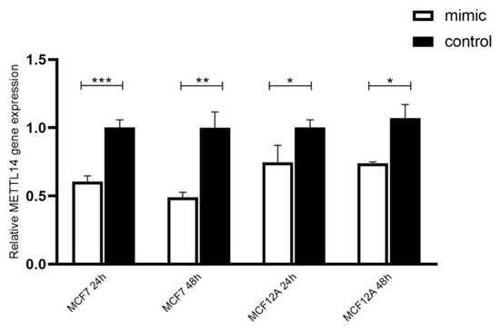
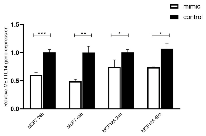
Following miR-30c-2-3p mimic transfection, the upregulation of miRNA expression was confirmed by RT-qPCR, and METTL14 mRNA levels were subsequently evaluated in MCF7 and MCF12A cell lines. In both cell types, a significant reduction in METTL14 expression was observed following mimic transfection. In MCF7 cells, METTL14 mRNA levels were significantly decreased at 24 h (p = 0.0006) and 48 h (p = 0.0019) compared to the control group. Similarly, in MCF12A cells, the reduction in METTL14 expression was also statistically significant at both 24 h (p = 0.0331) and 48 h (p = 0.0053) (Figure 1). Additional raw RT-qPCR data are provided in the Supplementary Data.
Figure 1.
Relative expression of METTL14 mRNA levels in MCF7 and MCF12A cells. Relative expression of METTL14 mRNA levels in MCF7 and MCF12A cells transfected with miR-30c-2-3p mimic or negative control. Bars represent mean ± SEM (n = 3 per group). Control groups are shown as solid black bars, while miR-30c-2-3p mimic groups are shown as solid white bars. Expression levels were normalized to the control group within each condition. Statistical analysis was performed using unpaired two-tailed Student’s t-test. Significance levels are indicated as follows: * p < 0.05, ** p < 0.01, *** p < 0.001.
3.3. Expression of miR-30c-2-3p Reduces Global m6A RNA Methylation Levels
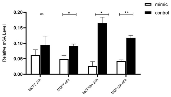
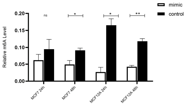
To investigate the regulation of global m6A RNA methylation by miR-30c-2-3p, MCF12A and MCF7 cell lines were transfected with miR-30c-2-3p mimics. Total RNA was isolated 24 and 48 h after transfection, after which global m6A levels were measured using a colorimetric assay kit. In MCF12A cells, expression of miR-30c-2-3p was associated with a significant decrease in global m6A levels compared to the control group after 24 and 48 h of incubation (p = 0.0009 and p = 0.0008, respectively). A decrease in m6A levels was also observed in MCF7 cells after mimic transfection. However, a statistically significant difference was only found at the 48 h time point (p = 0.0107), while the difference at the 24 h time point was not statistically significant (p = 0.1805) (Figure 2). Additional global m6A methylation data are provided in the Supplementary Data.
Figure 2.
miR-30c-2-3p overexpression reduces global m6A RNA methylation levels. miR-30c-2-3p overexpression reduces global m6A RNA methylation levels in breast epithelial and cancer cell lines. Each condition was then compared to its respective negative control. Data are shown as the mean ± standard error of the mean (SEM) of three biological replicates. Asterisks indicate statistical significance between the control and mimic groups (* p < 0.05, ** p < 0.01; ns: not significant).
3.4. miR-30c-2-3p Reduces METTL14 Protein Expression
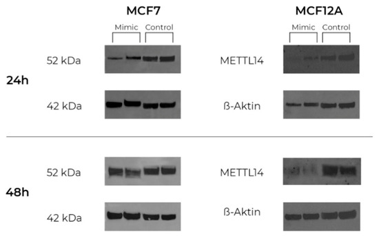
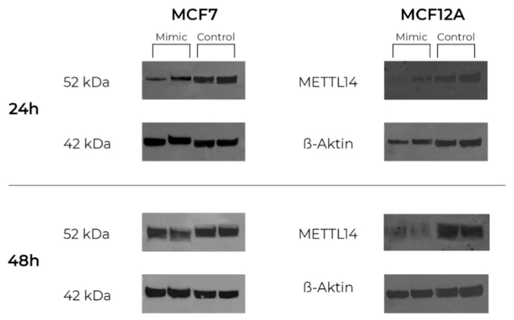
To evaluate the effect of miR-30c-2-3p on METTL14 protein levels, Western blot analysis was performed in MCF7 and MCF12A cell lines following transfection with miR-30c-2-3p mimics. METTL14 and the loading control β-actin were analyzed at 24 and 48 h post-transfection. A reduction in METTL14 protein levels was observed in both cell lines upon miR-30c-2-3p overexpression compared to their respective controls. In MCF7 cells, this reduction was statistically significant at both 24 h (p = 0.032) and 48 h (p = 0.008). Similarly, in MCF12A cells, a statistically significant decrease was detected at 24 h (p = 0.045), which became more pronounced at 48 h (p = 0.011), (Figure 3).
Figure 3.
miR-30c-2-3p overexpression decreases METTL14 protein levels. miR-30c-2-3p overexpression decreases METTL14 protein levels in MCF7 and MCF12A cells. Western blot analysis showing METTL14 and β-actin (loading control) in MCF7 and MCF12A cell lines 24 and 48 h after transfection with miR-30c-2-3p mimics or negative control. In both cell lines, METTL14 protein levels were markedly reduced following miR-30c-2-3p overexpression.
3.5. miR-30c-2-3p Reduces Cell Migration
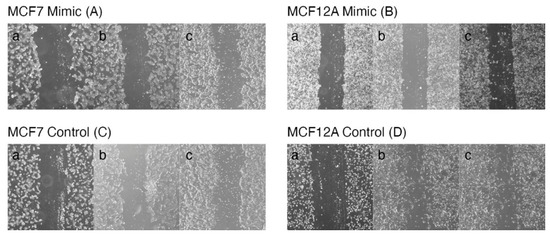
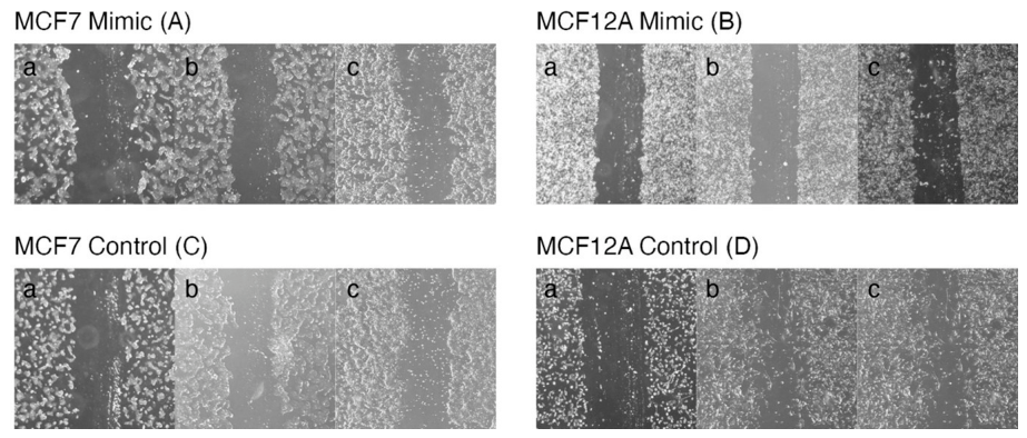
To evaluate the effect of miR-30c-2-3p on cell migration, a scratch wound healing assay was performed using MCF7 and MCF12A cell lines. Images were captured at 0, 24, and 48 h post-transfection, and wound closure was quantified using ImageJ software. In the control groups, progressive wound closure was observed over time in both cell lines, indicating active migration. In contrast, miR-30c-2-3p overexpression was associated with a reduction in wound closure, suggesting an inhibitory effect on cellular migration.
In MCF7 cells, miR-30c-2-3p mimic transfection resulted in a 30% reduction in migration at 24 h and a 44% reduction at 48 h compared to control cells. The inhibitory effect was even more pronounced in MCF12A cells, with migration reduced by 66% at 24 h and 72% at 48 h. These results suggest that miR-30c-2-3p overexpression may impair the migratory capacity of both breast epithelial and breast cancer cell lines, with a stronger effect observed in the non-tumorigenic MCF12A cells (Figure 4). Additional raw migration assay data are provided in the Supplementary Data.
Figure 4.
Effect of miR-30c-2-3p overexpression on cell migration in MCF7 and MCF12A cells. Scratch wound healing assay demonstrating the effect of miR-30c-2-3p overexpression on cell migration in MCF7 mimic (A), MCF12A mimic (B), MCF7 control (C), and MCF12A control (D) cells. Cells were photographed at 0 h (a), 24 h (b), and 48 h (c) post-transfection. In the control groups, progressive wound closure was observed over time, indicating active cell migration. In contrast, miR-30c-2-3p mimic transfection resulted in visibly reduced wound closure in both cell lines, showing its inhibitory effect on cellular migration.
4. Discussion
Our study demonstrates that miR-30c-2-3p overexpression significantly downregulates METTL14 expression at both mRNA and protein levels, leading to a pronounced reduction in global m6A RNA methylation and impaired cell migration in both MCF7 and MCF12A cells. Notably, we observed a temporal difference in the reduction in global m6A levels between the two cell lines; while MCF12A cells exhibited a significant decrease as early as 24 h, MCF7 cells showed a significant reduction only at 48 h. This discrepancy may reflect differences in metabolic rates or epigenetic plasticity between non-tumorigenic and cancerous cells. Cancer cells often possess more robust compensatory mechanisms or altered RNA turnover rates, which might delay the manifestation of global methylation changes compared to normal epithelial cells. Collectively, these findings suggest that miR-30c-2-3p functions as a tumor suppressor in breast tissue, potentially by regulating METTL14-mediated m6A RNA methylation.
Members of the miR-30 family are known to regulate tumor progression by targeting genes involved in proliferation, differentiation, apoptosis, and invasion [6]. In particular, miR-30c-2-3p has been reported to inhibit NF-κB signaling and cell-cycle progression by downregulating TRADD and CCNE1 in estrogen receptor–negative breast cancer cells [11], consistent with our observation that its overexpression reduces migratory ability. Furthermore, decreased miR-30c-2-3p expression has been correlated with poor prognosis or inferior therapeutic response in breast cancer, supporting its role as a tumor suppressor [7,9].
Epigenetic RNA modifications such as N6-methyladenosine (m6A) are critical regulators of post-transcriptional gene expression [12,14]. METTL14, a core component of the m6A methyltransferase complex, functions with METTL3 and WTAP to catalyze adenosine methylation on mRNA. Dysregulation of m6A modification has been implicated in tumor initiation and progression through altered mRNA stability, translation, and degradation [14]. However, the biological role of METTL14 appears to be context-dependent across different cancer types. In hepatocellular carcinoma (HCC), for example, reduced METTL14 expression suppresses m6A-dependent microRNA maturation and promotes metastasis [21]. Likewise, colorectal cancer (CRC) models have shown that METTL14 downregulation facilitates epithelial-to-mesenchymal transition (EMT) and tumor progression [22,23]. Conversely, elevated METTL14 expression enhances proliferation and is associated with poor prognosis in pancreatic cancer and acute myeloid leukemia (AML) [24,25]. In breast cancer, findings remain contradictory. Dong et al. reported that METTL14 expression is significantly reduced in tumor tissues compared with normal controls and that its overexpression suppresses cell proliferation, colony formation, and migration [18]. In contrast, Yi et al. found that METTL14 expression was elevated in breast tumors and promoted invasion through m6A-dependent regulation of miR-146a-5p [19]. Our results are more consistent with the former model, as METTL14 levels decreased and migration was reduced following miR-30c-2-3p overexpression, suggesting that METTL14 suppression may be a downstream effect of miR-30c-2-3p activity.
Recent studies have highlighted a reciprocal regulatory relationship between miRNAs and the m6A machinery, where m6A influences miRNA biogenesis and miRNAs can, in this relationship, m6A influences miRNA biogenesis, and miRNAs can regulate the expression of m6A-related enzymes, including METTL3, METTL14, and FTO. [15,16]. Our findings are consistent with this bidirectional interaction and suggest that miR-30c-2-3p may act as an upstream regulator of METTL14 and m6A methylation dynamics in breast cancer.
In summary, we propose a miR-30c-2-3p/METTL14/m6A regulatory axis that could modulate both RNA methylation and cell migration in breast cancer. Considering the contrasting roles of METTL14 in different tumor types, this axis might represent a context-dependent epigenetic pathway in breast tissue. The ability of miR-30c-2-3p to influence both gene expression and RNA modification suggests that it could be further investigated as a potential diagnostic or prognostic biomarker and as a target for modulating m6A-related mechanisms in cancer.
5. Limitations of Study
This study was conducted using in vitro breast cell line models, which may not fully recapitulate the complexity of tumor behavior in vivo. Although we demonstrated that miR-30c-2-3p regulates METTL14 expression and affects cell migration, additional experiments using animal models or patient-derived samples would be necessary to confirm these findings in a physiological context. We also noted a substantial overexpression of miR-30c-2-3p, particularly in MCF12A cells (>13,000-fold); while this confirms successful transfection, we acknowledge that supraphysiological levels could potentially lead to non-specific off-target effects, although the consistent phenotypic results in both cell lines suggest a specific mechanism. Future studies using lower and more physiological levels of miR-30c-2-3p overexpression may further clarify whether the observed phenotypic effects persist under conditions that more closely reflect the in vivo cellular environment. Furthermore, to definitively confirm that the observed inhibition of migration is mediated exclusively through METTL14, future studies should include “rescue” experiments involving the overexpression of a METTL14 construct lacking the 3′UTR in miR-30c-2-3p-transfected cells. Finally, only limited downstream targets of METTL14 were examined; therefore, further studies are warranted to elucidate the broader impact of the miR-30c-2-3p–METTL14 interaction on m6A-dependent gene regulation.
Supplementary Materials
The following supporting information can be downloaded at: https://www.mdpi.com/article/10.3390/cimb48060545/s1.
Author Contributions
Z.E. conceived and designed the study. Z.E., S.P., V.Z. and S.U. performed the cell culture, miRNA transfection, RNA isolation, RT-qPCR, m6A quantification, Western blot, and scratch wound healing experiments, and collected and processed the data. Z.E. and S.P. analyzed and interpreted the experimental results and performed the statistical analyses. Z.E. prepared the draft manuscript and created the figures. Z.E., S.P., V.Z., S.S.E. and N.A. critically revised and edited the manuscript. Z.E., N.A. and S.S.E. approved the final version of the manuscript to be published. Z.E. supervised and coordinated the study. All authors have read and agreed to the published version of the manuscript.
Funding
This study was supported by Istanbul University Scientific Research Projects Unit (BAP) under project number TSA-2024-40964.
Institutional Review Board Statement
Not applicable.
Informed Consent Statement
Not applicable.
Data Availability Statement
All data generated or analyzed during this study are included in this published article.
Conflicts of Interest
The authors declared no potential conflicts of interest with respect to the research, authorship, and/or publication of this article.
Abbreviations
The following abbreviations are used in this manuscript:
| BC | Breast cancer |
| miRNA | microRNA |
| m6A | N6-methyladenosine |
| METTL14 | Methyltransferase-like 14 |
| METTL3 | Methyltransferase-like 3 |
| WTAP | Wilms tumor 1-associated protein |
| FTO | Fat mass and obesity-associated protein |
| ALKBH5 | Alkylation repair homolog 5 |
| 3′UTR | 3′ untranslated region |
| ER | Estrogen receptor |
| NF-κB | Nuclear factor kappa-light-chain-enhancer of activated B cells |
| EMT | Epithelial-to-mesenchymal transition |
Appendix A
Appendix A.1
Table A1.
Details of miRNA mimics, inhibitors, and transfection reagents used in this study.
Appendix A.2
Table A2.
List of primer sequences used for RT-qPCR analysis.
References
- Bray, F.; Laversanne, M.; Sung, H.; Ferlay, J.; Siegel, R.L.; Soerjomataram, I.; Jemal, A. Global cancer statistics 2022: GLOBOCAN estimates of incidence and mortality worldwide for 36 cancers in 185 countries. CA Cancer J. Clin. 2024, 74, 229–263. [Google Scholar] [CrossRef] [Scilit]
- Zhou, M.; Dong, M.; Yang, X.; Gong, J. The emerging roles and mechanism of m6A in breast cancer progression. Front. Genet. 2022, 13, 983564. [Google Scholar] [CrossRef] [Scilit]
- Ellis, M.J.; Perou, C.M. The genomic landscape of breast cancer as a therapeutic roadmap. Cancer Discov. 2013, 3, 27–34. [Google Scholar] [CrossRef] [Scilit] [PubMed]
- Waks, A.G.; Winer, E.P. Breast cancer treatment: A review. JAMA 2019, 321, 288–300. [Google Scholar] [CrossRef] [Scilit]
- Brennecke, J.; Stark, A.; Russell, R.B.; Cohen, S.M. Principles of microRNA-target recognition. PLoS Biol. 2005, 3, e85. [Google Scholar] [CrossRef] [Scilit]
- Han, W.; Cui, H.; Liang, J.; Su, X. Role of MicroRNA-30c in cancer progression. J. Cancer 2020, 11, 2593–2601. [Google Scholar] [CrossRef] [Scilit] [PubMed]
- Rodríguez-González, F.G.; Sieuwerts, A.M.; Smid, M.; Look, M.P.; Meijer-van Gelder, M.E.; de Weerd, V.; Sleijfer, S.; Martens, J.W.M.; Foekens, J.A. MicroRNA-30c expression level is an independent predictor of clinical benefit of endocrine therapy in advanced estrogen receptor–positive breast cancer. Breast Cancer Res. Treat. 2011, 127, 43–51. [Google Scholar] [CrossRef] [Scilit] [PubMed]
- Song, C.J.; Chen, H.; Chen, L.Z.; Ru, G.M.; Guo, J.J.; Ding, Q.N. The potential of microRNAs as human prostate cancer biomarkers: A meta-analysis of related studies. J. Cell. Biochem. 2018, 119, 2763–2786. [Google Scholar] [CrossRef] [Scilit]
- Lai, Y.H.; Chen, J.; Wang, C.F.; Wu, S.M.; Chuang, K.P.; Chuang, J.Y. Collagen triple helix repeat containing-1 negatively regulated by microRNA-30c promotes cell proliferation and metastasis and indicates poor prognosis in breast cancer. J. Exp. Clin. Cancer Res. 2017, 36, 107. [Google Scholar] [CrossRef] [Scilit]
- Liu, S.; Li, X.; Zhuang, S. miR-30c impedes glioblastoma cell proliferation and migration by targeting SOX9. Oncol. Res. 2019, 27, 165–171. [Google Scholar] [CrossRef] [Scilit]
- Shukla, K.; Sharma, A.K.; Ward, A.; Will, R.; Hielscher, T.; Balwierz, A.; Breunig, C.; Münstermann, E.; König, R.; Keklikoglou, I.; et al. MicroRNA-30c-2-3p negatively regulates NF-κB signaling and cell cycle progression through downregulation of TRADD and CCNE1 in breast cancer. Mol. Oncol. 2015, 9, 1106–1119. [Google Scholar] [CrossRef] [Scilit]
- Wiener, D.; Schwartz, S. The epitranscriptome beyond m6A. Nat. Rev. Genet. 2021, 22, 119–131. [Google Scholar] [CrossRef] [Scilit] [PubMed]
- Hu, L.; Liu, S.; Peng, Y.; Ge, R.; Su, R.; Sui, S. m6A RNA modifications are measured at single-base resolution across the mammalian transcriptome. Nat. Biotechnol. 2022, 40, 1210–1219, Correction in Nat. Biotechnol. 2023, 41, 150. [Google Scholar] [CrossRef] [Scilit]
- Dierks, D.; Garcia-Campos, M.A.; Uzonyi, A.; Safra, M.; Edelheit, S.; Schwartz, S. Multiplexed profiling facilitates robust m6A quantification at site, gene and sample resolution. Nat. Methods 2021, 18, 1060–1067. [Google Scholar] [CrossRef] [Scilit] [PubMed]
- Ma, S.; Chen, C.; Ji, X.; Liu, J.; Zhou, Q.; Wang, G.; Yuan, W.; Kan, Q.; Sun, Z. The interplay between m6A RNA methylation and noncoding RNA in cancer. J. Hematol. Oncol. 2019, 12, 121. [Google Scholar] [CrossRef] [Scilit]
- Chen, T.; Hao, Y.J.; Zhang, Y.; Li, M.M.; Wang, M.; Han, W.; Wu, Y.; Lv, Y.; Hao, J.; Wang, L.; et al. m6A RNA methylation is regulated by microRNAs and promotes reprogramming to pluripotency. Cell Stem Cell 2015, 16, 289–301. [Google Scholar] [CrossRef] [Scilit]
- Cai, X.; Wang, X.; Cao, C.; Gao, Y.; Zhang, S.; Yang, Z.; Liu, Y.; Zhang, X.; Zhang, W.; Ye, L. HBXIP-elevated methyltransferase METTL3 promotes the progression of breast cancer via inhibiting tumor suppressor let-7g. Cancer Lett. 2018, 415, 11–19. [Google Scholar] [CrossRef] [Scilit]
- Dong, X.F.; Wang, H.L.; Liu, L.D.; Huang, Y.N.; Jin, M.W.; Wang, C.; Zhang, Z. Downregulated METTL14 expression correlates with breast cancer tumor grade and molecular classification. Biomed. Res. Int. 2020, 2020, 8823270. [Google Scholar] [CrossRef] [Scilit] [PubMed]
- Yi, D.; Wang, R.; Shi, X.; Xu, L.; Yilihamu, Y.; Sang, J. METTL14 promotes the migration and invasion of breast cancer cells by modulating N6–methyladenosine and hsa–miR–146a–5p expression. Oncol. Rep. 2020, 43, 1375–1386. [Google Scholar] [CrossRef] [Scilit]
- Chomczynski, P.; Sacchi, N. Single-step method of RNA isolation by acid guanidinium thiocyanate–phenol–chloroform extraction. Anal. Biochem. 1987, 162, 156–159. [Google Scholar] [CrossRef] [Scilit]
- Ma, J.Z.; Yang, F.; Zhou, C.C.; Liu, F.; Yuan, J.-H.; Wang, F.; Wang, T.T.; Xu, Q.-G.; Zhou, W.-P.; Sun, S.-H. METTL14 suppresses the metastatic potential of hepatocellular carcinoma by modulating N6-methyladenosine-dependent primary MicroRNA processing. Hepatology 2017, 65, 529–543. [Google Scholar] [CrossRef] [Scilit] [PubMed]
- Wang, H.; Wei, W.; Zhang, Z.Y.; Liu, Y.; Shi, B.; Zhong, W.; Zhang, H.S.; Fang, X.; Sun, C.L.; Wang, J.B.; et al. TCF4 and HuR mediated-METTL14 suppresses dissemination of colorectal cancer via N6-methyladenosine-dependent silencing of ARRDC4. Cell Death Dis. 2021, 13, 3. [Google Scholar] [CrossRef] [Scilit]
- Chen, X.; Xu, M.; Xu, X.; Zeng, K.; Liu, X.; Sun, L.; Pan, B.; He, B.; Pan, Y.; Sun, H.; et al. METTL14 suppresses colorectal cancer progression via regulating N6-methyladenosine-dependent primary miR-375 processing. Mol. Ther. 2020, 28, 599–612. [Google Scholar] [CrossRef] [Scilit] [PubMed]
- Li, J.; Wang, P.; Liu, F.; Li, Y.; Wu, Y.; Wang, F.; Du, J. Methyltransferase-like 14 promotes the tumorigenesis and proliferation of pancreatic cancer cells through myc proto-oncogene signaling pathway. Cytojournal 2024, 21, 55. [Google Scholar] [CrossRef] [Scilit] [PubMed]
- Weng, H.; Huang, H.; Wu, H.; Qin, X.; Zhao, B.S.; Dong, L.; Shi, H.; Skibbe, J.; Shen, C.; Hu, C.; et al. METTL14 inhibits hematopoietic stem/progenitor differentiation and promotes leukemogenesis via mRNA m6A modification. Cell Stem Cell 2018, 22, 191–205. e9. [Google Scholar] [CrossRef] [Scilit]
Disclaimer/Publisher’s Note: The statements, opinions and data contained in all publications are solely those of the individual author(s) and contributor(s) and not of MDPI and/or the editor(s). MDPI and/or the editor(s) disclaim responsibility for any injury to people or property resulting from any ideas, methods, instructions or products referred to in the content. |
© 2026 by the authors. Licensee MDPI, Basel, Switzerland. This article is an open access article distributed under the terms and conditions of the Creative Commons Attribution (CC BY) license.