


Abstract
Maize (Zea mays L.) is a vital global crop whose productivity is severely threatened by abiotic stresses. Epicuticular waxes provide a hydrophobic barrier that protects land plants from environmental stresses. However, the role of key wax biosynthetic enzymes, such as aldehyde decarbonylase CER1, in maize stress adaptation remains unclear. In this study, we performed a functional characterization of ZmCER1 in maize. Our results show that the overexpression of ZmCER1 in both Arabidopsis and maize substantially improved tolerance to these abiotic stresses. Under stress conditions, the transgenic plants displayed better growth performance, elevated activities of antioxidant enzymes, and reduced levels of oxidative damage markers. Additionally, the alkane content—especially that of C29 and C31—was significantly increased in the ZmCER1OE lines. Through a yeast two-hybrid screening (Y2H screening), we identified the peroxisomal membrane protein ZmPEX14 as an interacting partner of ZmCER1, and the interaction was further confirmed by luciferase complementation (LUC) and bimolecular fluorescence complementation (BiFC) assays. We propose a model wherein ZmCER1 enhances stress tolerance not only by reinforcing the cuticular wax barrier but also by potentially regulating reactive oxygen species (ROS) detoxification via association with ZmPEX14. Collectively, our findings establish ZmCER1 as a key regulator of abiotic stress tolerance in maize and a promising candidate for the molecular breeding of stress-resilient crops.
1. Introduction
Maize (Zea mays L.) stands as a foundation of global agriculture and food security, serving as a primary staple food for over 4.5 billion people, a critical material for livestock production, and a versatile raw material for industries ranging from biofuel to bioplastics [1]. However, maize growth and grain yield are highly susceptible to various abiotic stresses [2]. Drought-induced water scarcity affects nearly all the climatic zones worldwide. Such conditions can persist for extended periods, impeding maize growth and resulting in substantial yield reductions [3,4]. Soil salinization has emerged as another major global challenge, limiting maize yield potential [5,6]. A common consequence of both drought and salinity stress is the disruption of cellular homeostasis, leading to the accelerated production of reactive oxygen species (ROS) such as hydrogen peroxide (H2O2), superoxide radicals (O2−), and hydroxyl radicals (OH•) [7]. At high concentrations, ROS cause oxidative damage to vital cellular components, including lipids, proteins, and DNA. Malondialdehyde (MDA), a byproduct generated during the peroxidation of polyunsaturated fatty acids in cell membranes, serves as a key indicator of oxidative damage [8]. To counteract this oxidative damage, plants have evolved a sophisticated antioxidant defense system comprising non-enzymatic antioxidants and key enzymes such as superoxide dismutase (SOD), peroxidase (POD) and catalase (CAT), which work in concert to scavenge ROS and maintain redox balance [9]. A plant’s capacity to rapidly and efficiently activate its intrinsic antioxidant defense system is strongly correlated with its tolerance to various abiotic stresses. Another critical line of defense, particularly against drought, is the plant cuticle—a hydrophobic layer covering the aerial surfaces of plants composed of cutin and epicuticular waxes. Serving as a critical protective barrier, the cuticular wax layer effectively restricts non-stomatal water loss, mitigates UV radiation-induced harm, and blocks the invasion of phytopathogens [10,11,12,13]. The cuticle mainly consists of two types of lipophilic material, cutin and cuticular wax. Cutin is the major structural component of the cuticle and is composed of hydroxy and epoxy C16 and C18 fatty acid monomers, as well as glycerol [14]. Cuticular waxes consist of complex mixtures of hydrophobic compounds, mostly very-long-chain fatty acids (VLCFAs) with more than 20 carbon atoms and their derivatives, including primary and secondary alcohols, aldehydes, alkanes, ketones, and wax esters [10,11,15]. The wax components are produced according to two different pathways: (1) the alcohol-forming pathway, which generates very-long-chain (VLC) primary alcohols and wax esters; and (2) the alkane-forming pathway, responsible for the production of VLC alkanes and their derivatives [16]. In addition, alkanes are the prominent components of wax in the stem and leaf epidermis of higher plants, playing an extremely important role in controlling epidermal permeability [17,18]. Substantial progress has been made in recent decades in identifying the genes involved in alkane biosynthesis. Aldehyde decarbonylase, a member of the fatty acid hydroxylase (FAH) superfamily, serves as a key enzyme in the synthesis of very-long-chain alkanes (VLCAs) and contributes to plant responses to abiotic stresses [17,19,20]. In Arabidopsis, ECERIFERUM 1 (CER1) encodes an aldehyde decarbonylase that participates in the alkane-forming pathway and modulates stress tolerance [17,21,22]. Subsequent studies have established that Arabidopsis CER1 and CER3 function as core components of the VLCA synthesis complex [23]. Mutations such as cer2 impair the production of wax components longer than 28 carbons, while cer26 affects those longer than 30 carbons [24]. The CER22 gene, an allele of CER1, is also involved in alkane biosynthesis in Arabidopsis [22]. Furthermore, CsCER1 significantly influences VLCA biosynthesis, cuticle permeability, and drought resistance [25].
In rice, CER1 plays a crucial role in the biosynthesis of VLCAs [22,26]. The overexpression of BnCER1-2 in rapeseed not only enhances total alkane deposition but also confers improved drought tolerance phenotypes [27]. Similarly, PpCER1 in Poa pratensis participates in wax alkane synthesis and contributes to plant drought resistance [28]. In tomato, SlCER1-1 coordinates alkane biosynthesis, a pathway important for both drought adaptation and postharvest fruit quality [29]. Although these studies collectively establish CER1 orthologs as key regulators of cuticular wax formation and abiotic stress responses in angiosperms, the specific functions of CER1 homologs in major monocot crops such as maize—particularly their potential role in modulating antioxidant defense systems—remain poorly understood.
In this study, we investigated the role of ZmCER1 in maize abiotic stress responses. We first analyzed the expression pattern of ZmCER1 under drought and salt stress and determined the ZmCER1 subcellular localization. We then validated its function through overexpressing it in both Arabidopsis thaliana and maize, evaluating the phenotypic and physiological changes associated with salt and drought tolerance. Finally, we screened for ZmCER1-interacting proteins to explore its molecular mechanism. Combining the physiological indicator data and the results of a yeast two-hybrid screening (Y2H screening) library, we hypothesized that ZmCER1 enhances abiotic tolerance not only by bolstering the physical cuticular wax barrier but also by regulating ROS scavenging through interactions with peroxisomal proteins. The findings of this study provide novel insights into maize stress adaptation mechanisms and identify candidate genes for genetic improvement of stress-tolerant crops.
2. Materials and Methods
2.1. Plant Material
The Z. mays materials used in this study include the inbred line B104, as well as ZmCER1 overexpression transgenic lines constructed using B104 as the recipient material. The Arabidopsis thaliana materials used were the wild type (WT) Col-0 and ZmCER1 overexpression lines. In addition, Nicotiana benthamiana was also used as experimental material in this study.
2.2. Generation of Transgenic Plants
2.2.1. Transgenic Arabidopsis thaliana Lines
The target fragment was ligated into the super1300-GFP plant overexpression vector using a double digestion method with Hind III and Spe I restriction enzymes. The recombinant vector was then introduced into Escherichia coli DH5α. The plasmid was subsequently transformed into Agrobacterium tumefaciens GV3101. The inflorescences of Arabidopsis thaliana were immersed in the infection solution and cultured until seeds were harvested. The T0 generation Arabidopsis thaliana seeds were sterilized and spotted onto 1/2 MS solid medium containing hygromycin for cultivation. Hygromycin screening and PCR assays were performed to retain positive plants. The positive plants were self-pollinated for two generations, and the homozygous lines were selected through PCR verification and segregation ratio analysis.
2.2.2. Transgenic Maize Lines
Total RNA was extracted from maize leaves using TRIzol reagent (TaKaRa, Dalian, China) and used as a template for cDNA synthesis using the RT reagent kit (TaKaRa, Dalian, China) according to the manufacturer’s protocol. The complete open reading frame (ORF) of maize ZmCER1 (Gene ID: Zm00001d014055) from the NCBI (http://www.ncbi.nlm.nih.gov/ (accessed on 1 September 2022)) was amplified by PCR with gene specific primers (Table S1), and using the maize leaf cDNA as template, the PCR products were inserted into a pGEM T-easy vector and sequenced. The ZmCER1 coding sequence was cloned into the PBCXUN vector and transformed into the Agrobacterium tumefaciens strain EHA105 to infect the immature embryo of the inbred line B104 (wild type) to generate transgenic seedlings. PCR-positive plants (T0) were transplanted and self-pollinated for three generations to obtain homozygous transgenic lines.
2.3. Gene Expression Analysis
Maize seeds were surface sterilized with 70% (v/v) ethanol and 0.1% (w/v) HgCl2, rinsed with sterile water, and germinated at 28 °C in darkness for 3 days, then transferred into Hoagland nutrient solution. When the seedlings reached the three-leaf stage, they were treated with a nutrient solution containing 20% polyethylene glycol (PEG), and leaves were collected at 0 h, 4 h, 12 h, 24 h, and 48 h after treatment to analyze gene expression levels by real-time PCR. For salt treatment in soil, maize seeds were sown in pots, and ten days later, uniformly growing seedlings were retained in each pot. An aliquot of 200 mL of 200 mM NaCl solution was watered every two days. Leaves were collected after 1 day, 3 days, 7 days and 10 days of treatment to analyze gene expression levels by real-time PCR. For drought treatment in soil, maize seeds were sown in pots, and ten days later, uniformly growing seedlings were retained in each pot. Then, irrigation of the maize plants was ceased. Leaves were collected after 3 days, 5 days, 7 days and 10 days of treatment to analyze gene expression levels by real-time PCR.
Total RNA was extracted using TRIzol reagent (TaKaRa, Dalian, China) and treated with RNase-free DNase. The total RNA was used as a template for cDNA synthesis using the RT reagent kit (TaKaRa, Dalian, China) according to the manufacturer’s protocol. Real-time PCR was performed on a LightCycler 480 (Roche) with SYBR Green RT-PCR Kit (TaKaRa, Dalian, China) according to the manufacturer’s protocol. Gene expression levels were normalized with the reference gene Actin1 (GRMZM2G126010) and evaluated using the 2−△△Ct method [30]. The gene specific primers are shown in Table S1.
2.4. Subcellular Localization of ZmCER1
ZmCER1 was linked to the green fluorescent protein expression vector 35S-eGFP using the Gateway method. The Gateway BP Cloning Kit (Baiao, Beijing, China) was employed to insert the target gene fragment into the pDONR221 vector. The Gateway LR Cloning Kit (Baiao, Beijing, China) was used to transfer the target fragment into the 35S-eGFP vector. The construct was then introduced into Agrobacterium tumefaciens GV3101 competent cells. The resuspended bacterial solution was injected into tobacco leaves. The leaves were then examined under a laser scanning confocal microscope (Leica, Wetzlar, Germany). ZmCER1 was cloned into the pBI221-GFP vector. Transient expression of green fluorescent protein (GFP)-fused proteins in maize protoplasts was performed. The fluorescence signal was observed using a confocal laser scanning microscope.
2.5. Plant Culture and Treatment
2.5.1. Arabidopsis thaliana Culture and Treatment
Sterilized Arabidopsis seeds were sown on 1/2 MS solid medium and vernalized at 4 °C in darkness for 2 days. Afterwards, seeds were moved to a growth chamber and cultured for another 3 days. Seedlings with uniform growth performance were selected and transferred to different stress media for 7 days of treatment.
For drought stress simulation, seedlings were cultivated on 1/2 MS medium supplemented with either 10% PEG 6000 or 200 mM mannitol, both of which function by establishing extracellular osmotic imbalance to mimic drought-induced physiological alterations in plants.
For salt stress treatment, seedlings were grown on 1/2 MS solid medium containing 150 mM NaCl. At the end of the treatment period, relevant physiological and biochemical parameters were measured and analyzed.
For salt stress treatment in soil culture, after 3 days, Arabidopsis plants were transferred into pots, and 7 days later, uniformly growing seedlings were retained in each pot. An aliquot of 200 mL of 200 mM NaCl solution was watered every two days. After 10 days of salt treatment, relevant physiological and biochemical indicators were measured.
For drought stress treatment in soil culture, after 3 days, Arabidopsis plants were transferred into pots, and 7 days later, uniformly growing seedlings were retained in each pot. The plants were subjected to a 10-day drought stress period. Then, the relevant physiological and biochemical indicators are measured.
The Arabidopsis plants were grown at 21–24 °C /18–20 °C (day/night) with a photoperiod cycle of 16 h of light (150–200 μmol m−2 s−1) at approximately 60–70% relative humidity.
2.5.2. Maize Plant Culture and Treatment
For salt stress treatment, maize seeds were sown in pots, and ten days later, uniformly growing seedlings were retained in each pot. An aliquot of 200 mL of 200 mM NaCl solution was watered every two days. After 10 days of salt treatment, relevant physiological and biochemical indicators were measured.
For drought stress treatment, maize seeds were sown in pots, and ten days later, uniformly growing seedlings were retained in each pot. The plants were subjected to a 10-day drought stress period. Then, the relevant physiological and biochemical indicators were measured.
The maize plants were grown at 25–30 °C/20–25 °C (day/night) with a photoperiod cycle of 14 h of light (500–600 μmol m−2 s−1) at approximately 65% relative humidity.
2.6. H2O2 Quantification
H2O2 content was quantified following the method described by Park et al. [31]. Briefly, leaf samples (0.1 g) were homogenized at 4 °C in 5 mL of 50 mM phosphate buffer (pH 6.5) and centrifuged at 10,000× g for 20 min. A 3 mL aliquot of the supernatant was then mixed with 1 mL of 0.1% TiCl4 in 20% (v/v) H2SO4, followed by centrifugation at 8000× g for 5 min. Absorbance was measured at 415 nm to determine H2O2 concentration using a visible spectrophotometer (Jinghua, Shanghai, China).
2.7. MDA Levels
For MDA content determination, the protocol by Peever and Higgins [32] was followed. Absorbance readings of the supernatant were taken at 450, 532, and 600 nm using a visible spectrophotometer (Jinghua, Shanghai, China), and MDA concentration was then calculated using the formula: content (μmol/L) = 6.45 × (A532 − A600) − 0.56 × A450.
2.8. Enzyme Determinations
For antioxidant enzyme activity assays, leaf tissues (0.3 g) were homogenized in 8 mL of 50 mM phosphate-buffered saline (pH 7.8) and centrifuged at 10,000× g for 15 min at 4 °C. The resulting supernatant was used for subsequent enzymatic analyses. SOD activity was measured following the method of Giannopolitis and Ries [33], while CAT activity was determined according to Alia et al. [34,35].
2.9. Relative Water Content (RWC)
The fresh weights (FWs) of the fourth leaves from the base of maize plants were measured. The leaves were then soaked in distilled water for 24 h at room temperature. After soaking, the leaves were blotted dry with filter paper so that the turgid weights (TWs) could be determined. The dry weights (DWs) were obtained after oven-drying for 72 h at 80 °C. The RWC was calculated as follows:
RWC (%) = (FW − DW)/(TW − DW) × 100
2.10. Analysis of Wax Composition
Wax extraction and gas chromatography–mass spectrometry (GC–MS) analyses were performed according to the described methods with some modifications [36]. The derivatized samples were analyzed by GC–MS (Agilent gas chromatograph coupled to an Agilent 5973N quadrupole mass selective detector) (Agilent Technologies Inc., Santa Clara, CA, USA).
2.11. Yeast Two-Hybrid Screening (Y2H Screening) Assays
The full length of the coding sequence region of ZmCER1 was inserted into the pBT3–STE vector as a bait that was then transformed into the yeast strain NMY51. The transformed NMY51 yeast strain was then transformed with the maize membrane system yeast cDNA library and placed on the synthetic dropout (SD) medium -Trp-Leu-Ade. Positive clones were selected for sequencing. Then, the selected potential candidate genes were subjected to further point-to-point Y2H assay. Autoactivation was tested by co-transforming bait and prey constructs with their reciprocal empty vector.
2.12. Luciferase (Luc) Assay
Full length ZmCER1 and the promoter fragments of ZmPEX14 were inserted into the 35S:GFP and pGreenII 0800:LUC vectors, respectively. The recombinant vectors were transformed into N. benthamiana leaves using the agrobacterial infiltration method (A. tumefaciens strain GV3101) [19]. The luciferase signal was detected using a Tanon 5200 multi-chemiluminescence imaging system (Tanon, Shanghai, China).
2.13. Bimolecular Fluorescence Complementation (BiFC) Assay
The coding sequence of ZmCER1 without stop codon was fused with EYFP on the vector P2YC, while ZmPEX14 was fused with EYFP on vector P2YN. Agrobacterium tumefaciens containing ZmCER1-cYFP and nYFP-ZmPEX14 were used to co-transform the leaves of 1-month-old N. benthamiana. Fluorescence of YFP was observed and photographed under a confocal laser scanning microscope (Leica, Wetzlar, Germany), using excitation/emission wavelength 510 nm.
2.14. Statistical Analysis
Statistical analysis was performed using SPSS Statistics 23 software (IBM SPSS Statistics Version 23.0). Data were expressed as means ± SD (standard deviation). All experiments were repeated three times, all data were analyzed using Duncan’s multiple-range test and statistical significances were considered at the p < 0.05 level.
3. Results
3.1. Expression Pattern Analysis and Subcellular Localization Analysis
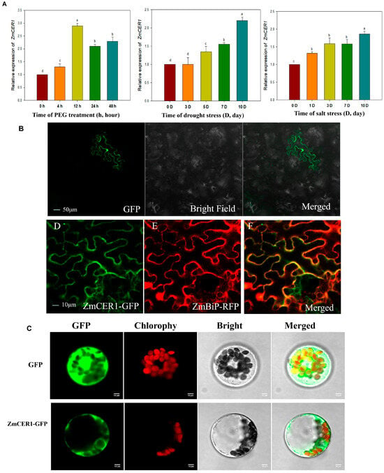
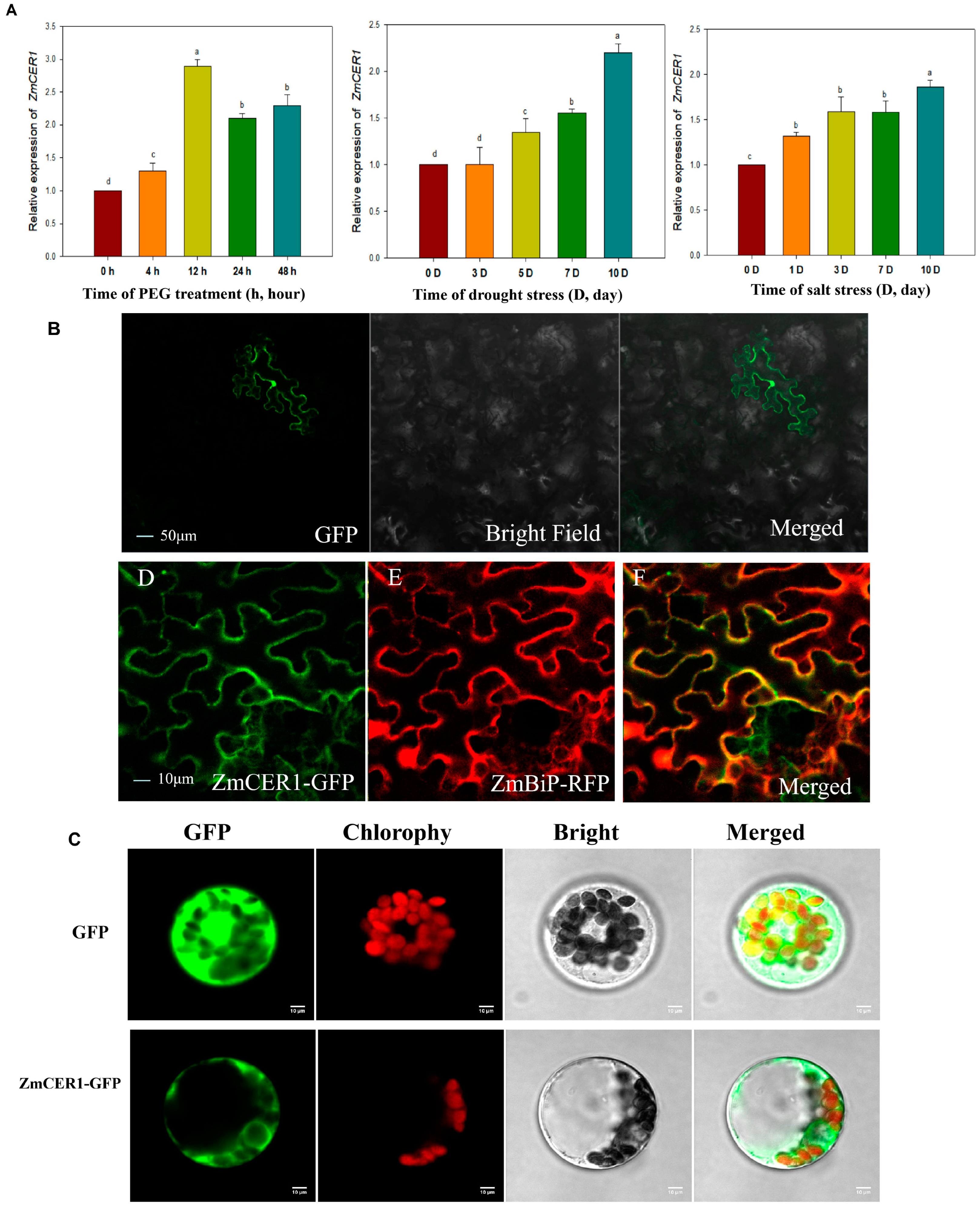
As shown in Figure 1A, the expression of ZmCER1 was obviously induced by drought and salt stress, which implied its potential involvement in maize abiotic adaptive responses. The subcellular localization of ZmCER1 was analyzed in Nicotiana benthamiana leaves and maize protoplasts cells (Figure 1B,C). In Nicotiana benthamiana leaves, the microscopic images showed that the ZmCER1-GFP fluorescence was observed in the endoplasmic reticulum (ER), while the fluorescence of GFP or the ER localization marker control was observed throughout cell or in the ER, respectively. In the maize protoplast cells, the ZmCER1-GFP fluorescence was also observed in the ER.
Figure 1.
Expression pattern analysis and subcellular localization analysis. (A) Relative expression level of ZmCER1 responsive to PEG, drought stress and salt stress. The expression levels of ZmCER1 were normalized to that of maize Actin1. Different lowercase letters indicate the statistically significant differences between Arabidopsis thaliana plants at the p < 0.05 level using Duncan’s multiple-range test. Values are means ± SD of three biological replicates. (B) Subcellular localization of ZmCER1 in tobacco leaves. ZmBiP, endoplasmic reticulum localization marker. GFP, green fluorescent protein. RFP, red fluorescent protein. (C) Subcellular localization of ZmCER1 in maize protoplasts cells. Scale bar = 10 μm.
3.2. ZmCER1 Positively Modulates Salt and Drought Tolerance in Arabidopsis thaliana
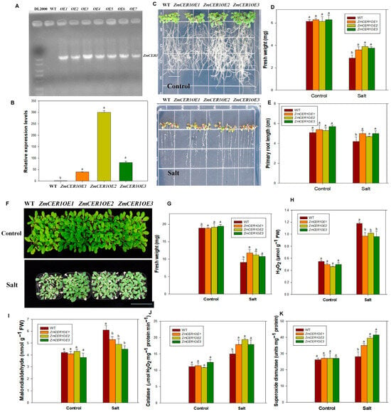
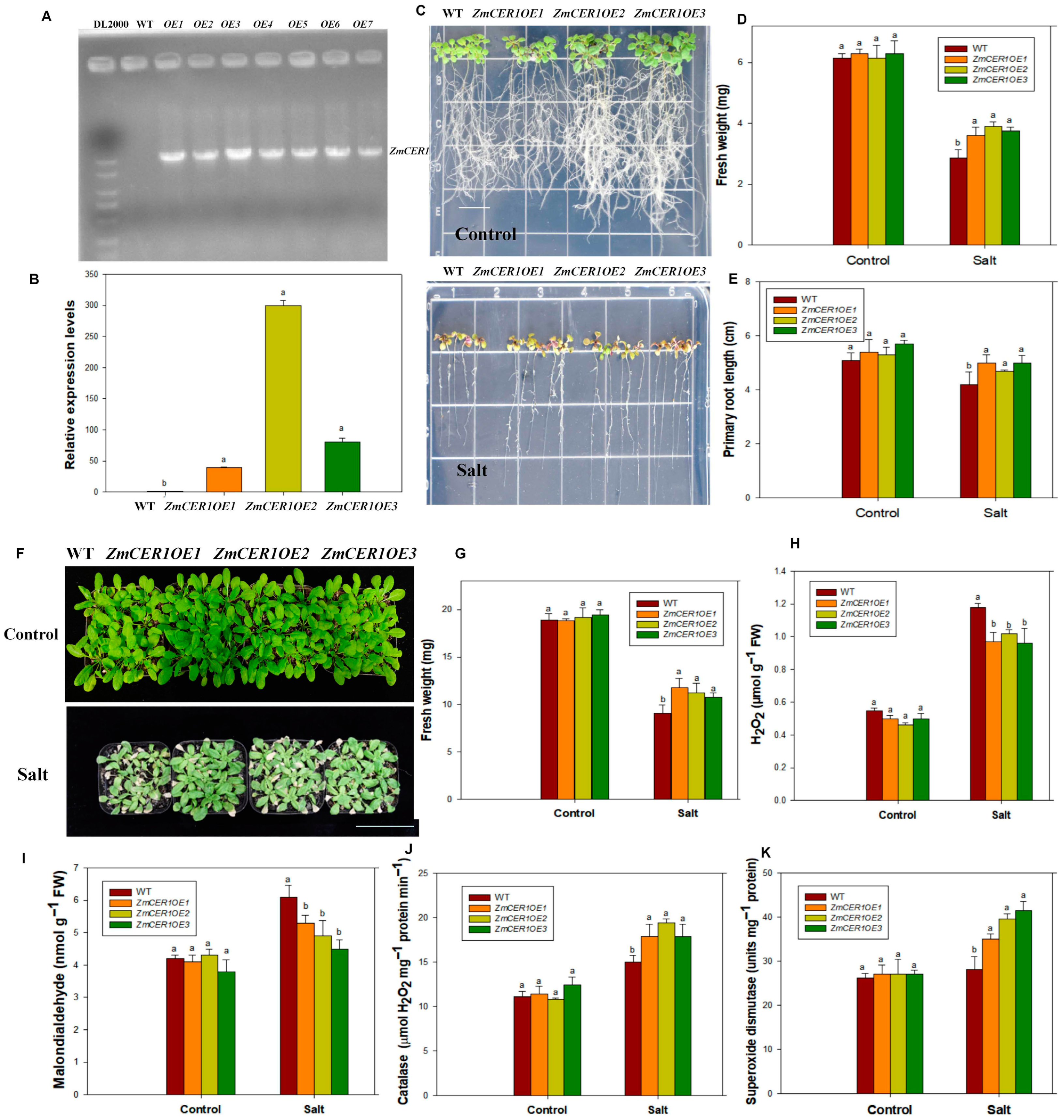
In order to investigate the role of ZmCER1 in regulating abiotic stress tolerance, transgenic Arabidopsis thaliana plants that overexpressed ZmCER1 were generated. A molecular analysis of the transgenic plants was performed. The results indicated the presence of ZmCER1 and the enhancement in the expression levels of ZmCER1 in the transgenic plants (Figure 2A,B). Three independent ZmCER1OE Arabidopsis thaliana lines were chosen to investigate the role of ZmCER1 in detail.
Figure 2.
ZmCER1 positively modulates salt tolerance in Arabidopsis thaliana. (A) Polymerase chain reaction (PCR) analysis of wild type and transgenic Arabidopsis thaliana lines with specific primers for ZmCER1. WT, wild type. (B) Relative expression levels of ZmCER1 in transgenic Arabidopsis thaliana lines. The expression levels of ZmCER1 were normalized to that of AtACT2. (C) Phenotypes of ZmCER1OE and WT Arabidopsis thaliana lines under salt stress on 1/2 MS medium. Scale bar indicates 1 cm. (D) Fresh weight. (E) Primary root length. (F) Phenotypes of ZmCER1OE and WT Arabidopsis thaliana lines under salt stress in soil. Scale bar indicates 15 cm. (G) Fresh weight. (H) Hydrogen peroxide (H2O2) content in leaves. (I) Malondialdehyde (MDA) contents in leaves. (J) Catalase (CAT) activity in leaves. (K) Superoxide dismutases (SOD) activity in leaves. Different lowercase letters indicate the statistically significant differences between Arabidopsis thaliana plants under same conditions at the p < 0.05 level using Duncan’s multiple-range test. Values are means ± SD of three biological replicates.
Cultured on medium for 7 days for salt tolerance analysis, the growth status of the WT and overexpressing lines remained comparable under normal conditions. However, under NaCl treatments, the overexpressing lines exhibited superior shoot and root growth compared to the WT (Figure 2C). The root length and fresh weight of the ZmCER1OE plants were significantly higher than those of the WT (Figure 2D,E). Cultured in soil for further salt tolerance analysis, the Arabidopsis plants were treated with 200 mM NaCl for 10 days, and growth retardation was observed in the stress groups relative to the controls, while the overexpressing lines maintained better growth performance than the WT (Figure 2F). The fresh weights of the overexpressing plants were significantly higher than the WT (Figure 2G). The MDA content and the H2O2 content in the ZmCER1OE plants was significantly lower than that in the WT, while the SOD and CAT activities were significantly higher (Figure 2H–K). These results indicate that ZmCER1 positively modulates salt tolerance in Arabidopsis thaliana.
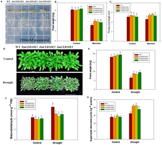
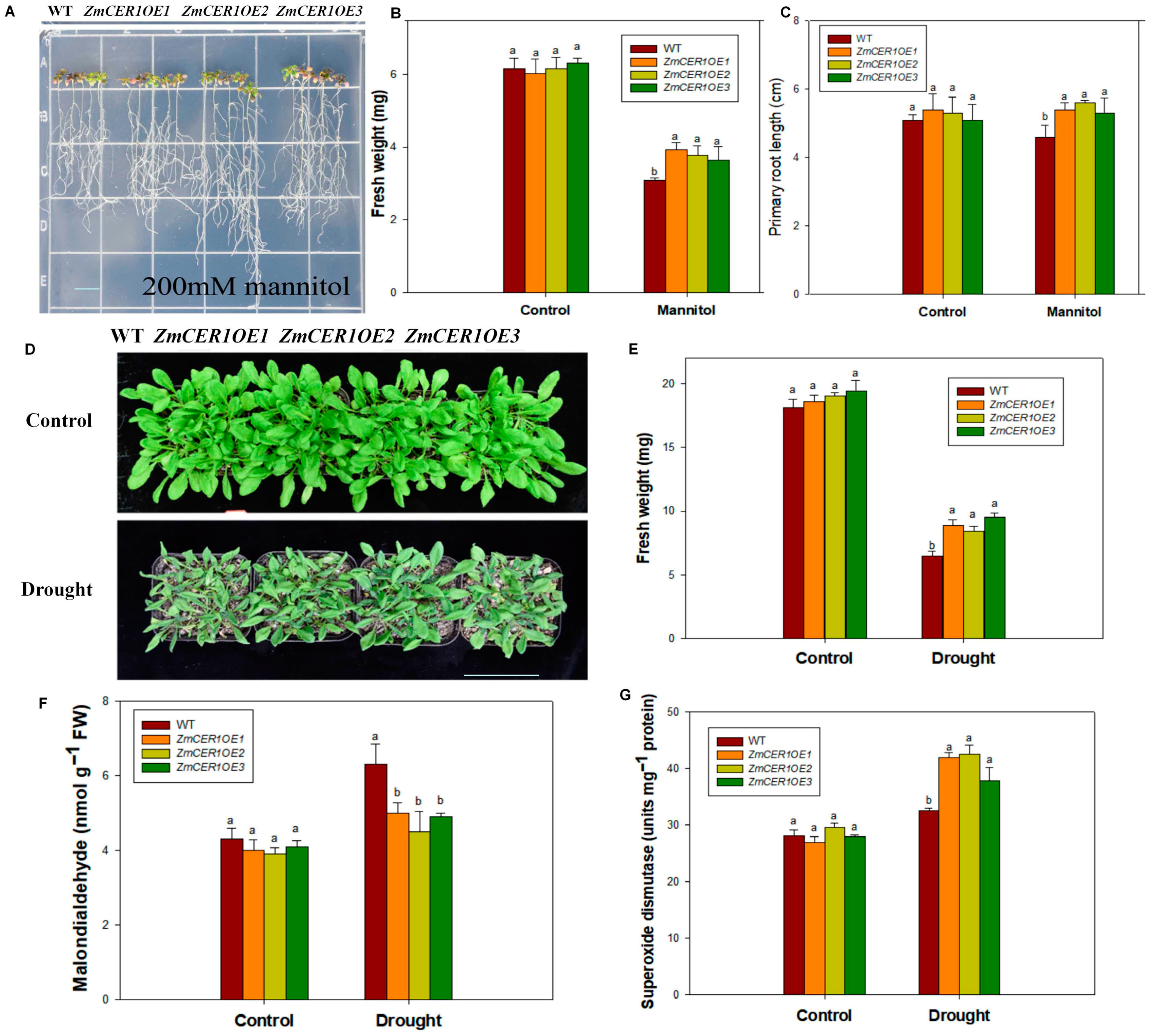
The Arabidopsis plants cultured on 1/2 MS medium for 5 days were then transferred to 1/2 MS medium containing 200 mM mannitol for 7 days. Under osmotic stress conditions, the overexpression lines exhibited superior growth compared to the WT (Figure 3A), with significantly greater fresh weight and root length (Figure 3B,C). Under drought stress in soil for 10 days, the overexpression lines showed better growth than the WT (Figure 3D). Compared to the WT, the treated overexpression lines showed significantly higher biomass and SOD activities and a lower content of MDA (Figure 3E–G). These results indicate that ZmCER1 positively regulates drought tolerance in Arabidopsis thaliana.
Figure 3.
ZmCER1 positively regulated drought tolerance in Arabidopsis thaliana. (A) Phenotypes of ZmCER1OE and WT Arabidopsis thaliana lines under mannitol treatment on 1/2 MS medium. Scale bar indicates 1 cm. (B) Fresh weight on medium. (C) Primary root length. (D) Phenotypes of ZmCER1OE and WT Arabidopsis thaliana lines under drought stress in soil. Scale bar indicates 15 cm. (E) Fresh weight. (F) MDA content in leaves. (G) SOD activity in leaves. Different lowercase letters indicate the statistically significant differences between Arabidopsis thaliana under same conditions at the p < 0.05 level using Duncan’s multiple-range test. Values are means ± SD of three biological replicates.
3.3. ZmCER1 Positively Modulates Salt Tolerance and Drought Tolerance in Maize
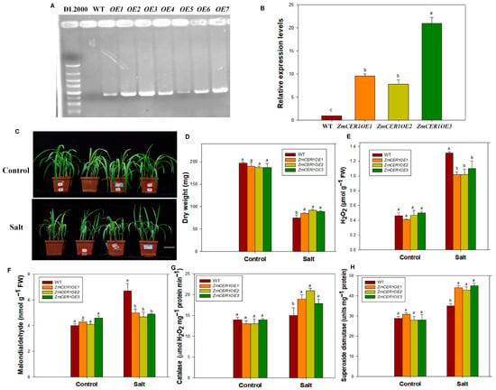
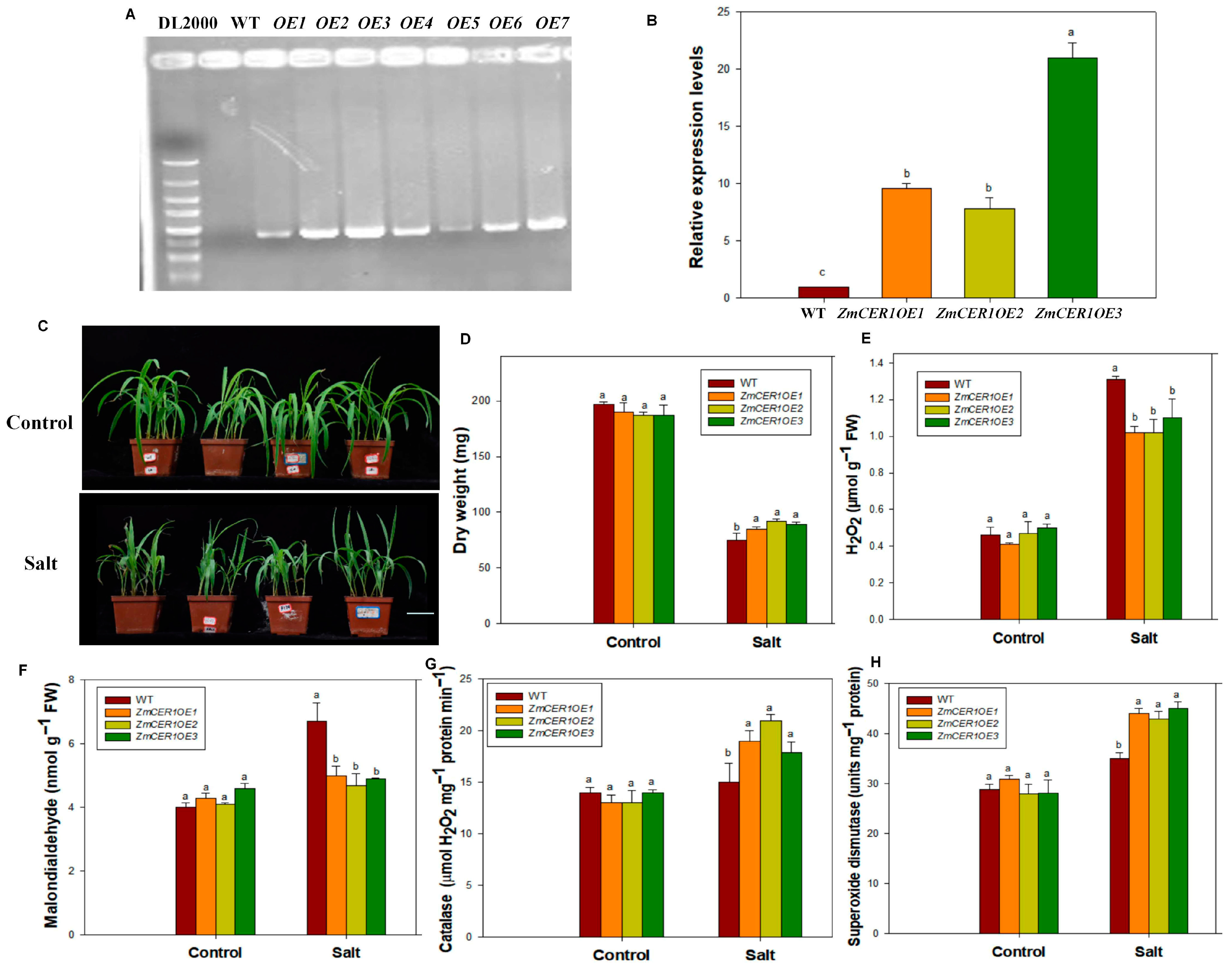
Transgenic maize plants that overexpressed ZmCER1 were generated. A molecular analysis of the transgenic maize plants was performed. The results indicated the presence of ZmCER1 and an enhancement in the expression levels of ZmCER1 in the transgenic maize plants (Figure 4A,B). Three independent ZmCER1OE Arabidopsis thaliana lines were chosen to investigate the role of ZmCER1 in detail.
Figure 4.
ZmCER1 positively regulated salt tolerance in maize. (A) PCR analysis of wild type and transgenic maize with specific primers for ZmCER1. WT, wild type. (B) Relative expression levels of ZmCER1 in transgenic maize lines. The expression levels of ZmCER1 were normalized to that of maize Actin1. The wild type (WT) was set as the reference control. Different lowercase letters indicate the statistically significant differences between maize lines at the p < 0.05 level using Duncan’s multiple-range test. Values are means ± SD of three biological replicates. (C) Phenotypes of ZmCER1OE and WT maize plants under salt stress in soil. Scale bar indicates 10 cm. (D) Dry weight. (E) H2O2 content in leaves. (F) MDA content in leaves. (G) CAT activity in leaves. (H) SOD activity in leaves. Different lowercase letters indicate the statistically significant differences between maize plants under same conditions at the p < 0.05 level using Duncan’s multiple-range test. Values are means ± SD of three biological replicates.
After the maize plants reached the three-leaf stage, they were treated with 200 mM NaCl for 10 days. Under normal conditions, all the lines exhibited relatively uniform growth. Salt stress exposure significantly increased adverse effects on growth across all the lines. The WT plants exhibited more pronounced wilting and desiccation than the ZmCER1OE maize plants (Figure 4C), indicating that the overexpression of ZmCER1 enhances salt stress tolerance in maize. Concurrently, various physiological parameters supported this conclusion. Under salt stress, the dry weight and activity of CAT and SOD were significantly higher in the ZmCER1OE maize lines, while the MDA and H2O2 content was lower than that in the WT (Figure 4D–H). This suggests that the ZmCER1 overexpression lines might confer enhanced salt tolerance through an improved ROS-scavenging capacity.
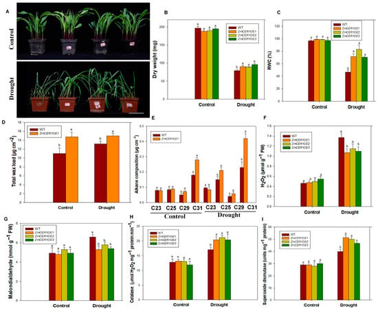
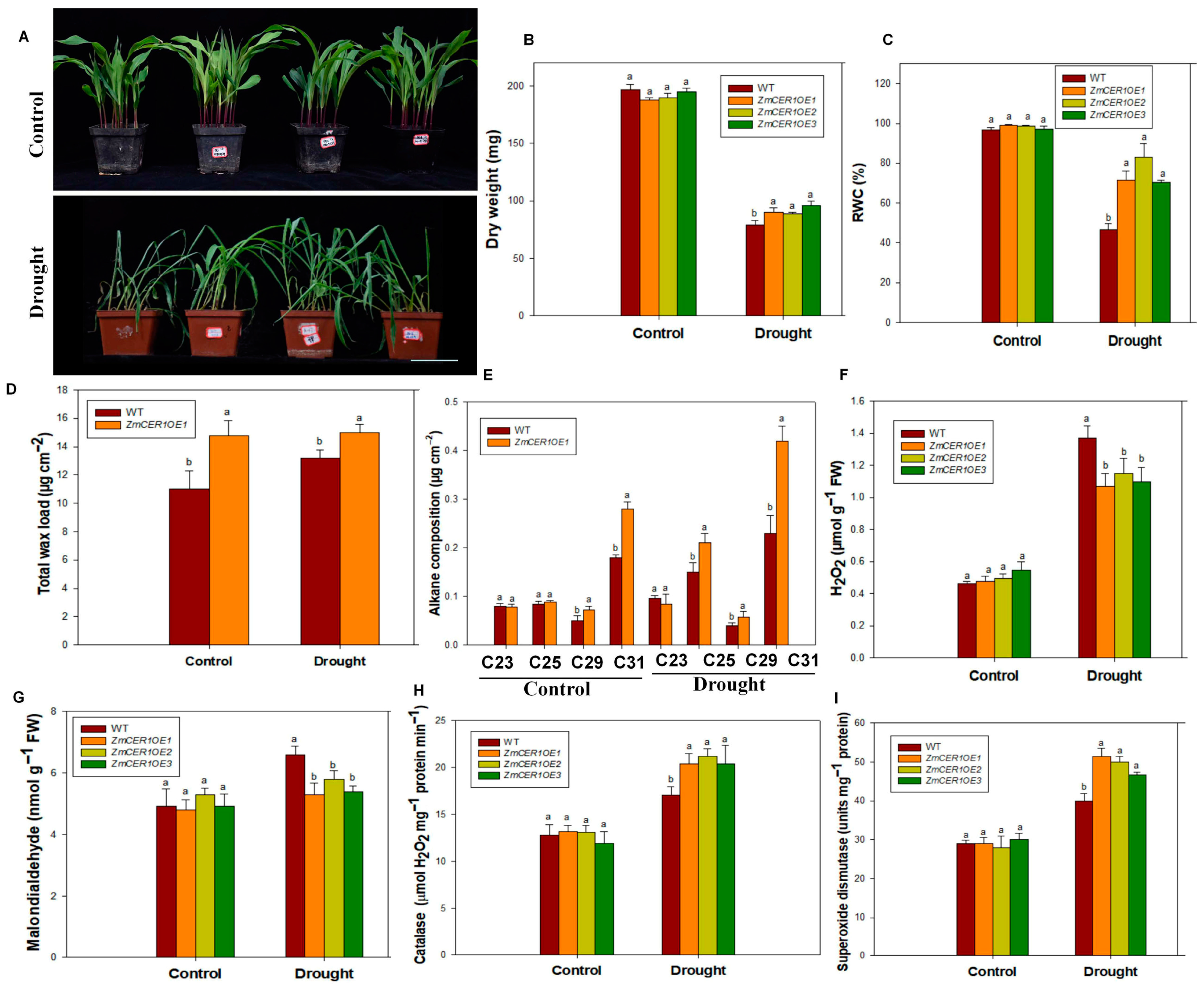
Drought treatment was initiated after the maize reached the three-leaf stage. Under normal conditions, the growth of all the lines was relatively uniform. Under drought treatment for 10 days, the growth of all the plants was significantly impaired. The WT exhibited more severe leaf wilting and plant lodging compared to the ZmCER1OE plants (Figure 5A), indicating that the overexpression of ZmCER1 enhances maize tolerance to drought stress. Concurrently, various physiological indices supported this conclusion. Under drought stress, the dry weight and WRC were significantly higher in the ZmCER1OE lines than those in the WT (Figure 5B,C). Additionally, the amount of total wax and alkane content—especially that of C29 and C31—were significantly increased in the ZmCER1OE lines compared to the WT (Figure 5D,E). Moreover, the CAT and SOD activities were significantly higher in the ZmCER1OE maize lines, while the MDA and H2O2 contents were lower than that in the WT (Figure 5F–I). These results suggest that the overexpression of ZmCER1 might confer enhanced drought tolerance in maize through the improved biosynthesis of cuticular waxes and ROS-scavenging capacity.
Figure 5.
ZmCER1 positively regulated drought tolerance in maize. (A) Phenotypes of ZmCER1OE and WT maize plants under drought stress. Scale bar indicates 10 cm. (B) Dry weight. (C) Relative water content (RWC) in leaves. (D) Total wax loads on maize leaf surfaces. (E) Amount of alkanes on maize leaf surfaces. (F) H2O2 content in leaves. (G) MDA contents in leaves. (H) CAT activity in leaves. (I) SOD activity in leaves. Different lowercase letters indicate the statistically significant differences between maize plants under same conditions at the p < 0.05 level using Duncan’s multiple-range test. Values are means ± SD of three biological replicates.
3.4. Screening of Interacting Proteins for ZmCER1
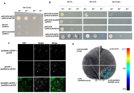
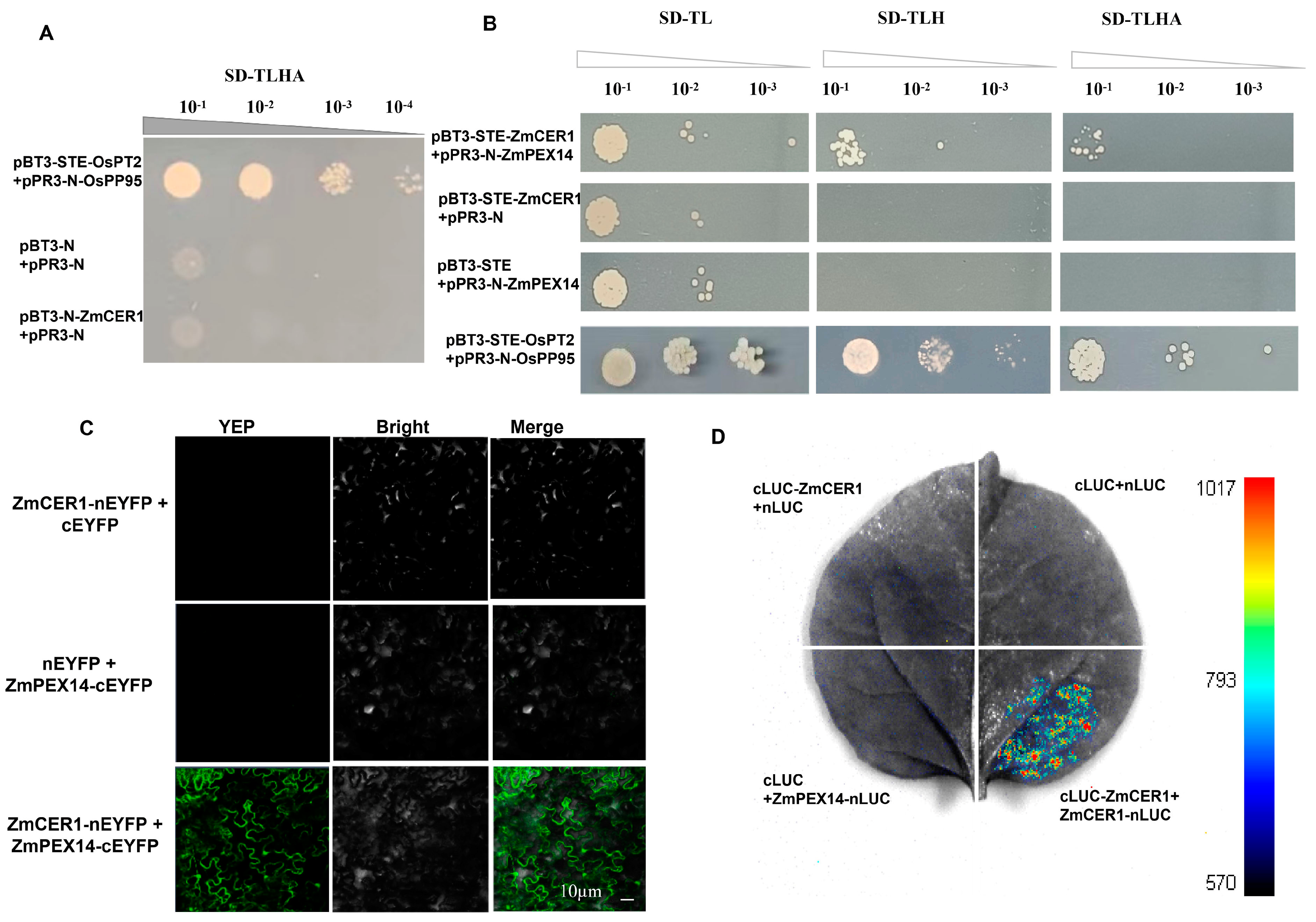
To elucidate the mechanisms by which ZmCER1 confers stress resistance in maize, we employed a Y2H assay to screen for proteins interacting with ZmCER1. First, we tested whether ZmCER1 exhibits autoactivation activity. It was observed that yeast cells carrying the ZmCER1 recombinant vector failed to grow on SD/-Leu/-His/-Trp/-Ade medium (Figure 6A), indicating that the ZmCER1 protein does not exhibit autoactivation activity in yeast, confirming its suitability for subsequent Y2H screening. The Y2H screen was performed to identify proteins interacting with ZmCER1. A total of 123 yeast colonies were obtained, followed by a PCR amplification of these colonies. A total of 15 candidate genes were obtained (Table S2). Among them, the peroxisomal membrane protein ZmPEX14 was selected for further interaction analysis. The results of the Y2H assay showed that ZmPEX14 possibly interacts with ZmCER1 (Figure 6B). The interactions were further confirmed by BiFC and LUC (Figure 6C,D).
Figure 6.
Interaction analysis of ZmCER1 with ZmPEX14. (A) Analysis of the self-activation of ZmCER1. (B) Yeast two-hybrid (Y2H) assay. OsPT2, rice phosphate transporter 2. OsPP95, rice purple acid phosphatase 95. (C) Bimolecular fluorescence complementation (BiFC). (D) Firefly luciferase complementation imaging (LUC).
3.5. ZmPEX14 Positively Modulates Salt Tolerance and Drought Tolerance in Arabidopsis
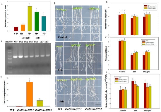
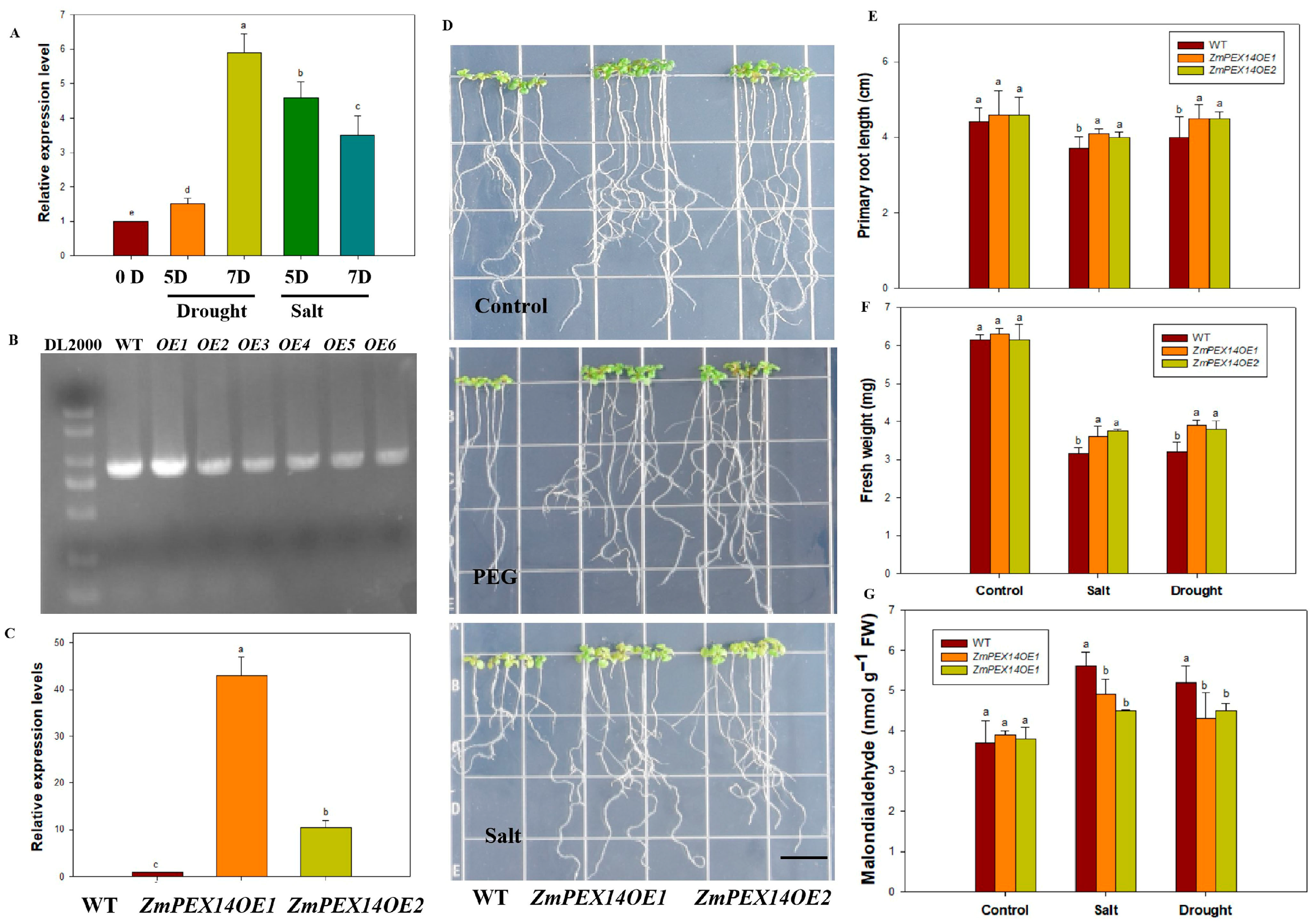
As shown in Figure 7A, the expression of ZmPEX14 was obviously induced by drought and salt stress, which implied its potential involvement in abiotic adaptive responses. The transgenic Arabidopsis plants that overexpressed ZmPEX14 were generated to analyze the role of ZmPEX14 in drought and salt tolerance. The results of the molecular analysis indicated the presence of the ZmPEX14 and the enhancement in the expression levels of ZmPEX14 in transgenic Arabidopsis plants (Figure 7B,C). As shown in Figure 7D, when cultured on a medium supplemented with 5% PEG or 100 mM NaCl for 5 days, the ZmPEX14OE lines showed better growth status than the WT, and the root length and fresh weight of the ZmPEX14OE plants were significantly higher than those of the WT, while the MDA content was lower than that in the WT (Figure 7E–G). Given that the peroxisome has been reported to be associated with the scavenging of reactive oxygen species in plants, which is crucial for abiotic stress tolerance [37,38], these findings suggest that ZmCER1 may regulate maize abiotic stress tolerance by influencing the scavenging of ROS through the interaction with ZmPEX14.
Figure 7.
ZmPEX14 positively modulates drought and salt tolerance in Arabidopsis thaliana. (A) Relative expression levels of ZmPEX14 responsive to drought and salt stress. The expression levels of ZmPEX14 were normalized to that of maize Actin1. Different lowercase letters indicate the statistically significant differences between Arabidopsis thaliana plants at the p < 0.05 level using Duncan’s multiple-range test. Values are means ± SD of three biological replicates. (B) PCR analysis of wild type and transgenic Arabidopsis thaliana lines with specific primers for ZmPEX14. WT, wild type. (C) Relative expression levels of ZmPEX14 in transgenic Arabidopsis thaliana lines. The expression levels of ZmPEX14 were normalized to that of AtACT2. (D) Phenotypes of ZmPEX141OE Arabidopsis thaliana lines and WT under PEG 6000 (10%) and salt stress on 1/2 MS medium. Scale bar indicates 1 cm. (E) Primary root length. (F) Fresh weight. (G) MDA contents in leaves. Different lowercase letters indicate the statistically significant differences between Arabidopsis thaliana plants under same conditions at the p < 0.05 level using Duncan’s multiple-range test. Values are means ± SD of three biological replicates.
4. Discussion
The escalating threat of abiotic stresses, particularly drought and salinity, poses a significant challenge to global food security, with maize (Zea mays L.) being exceptionally vulnerable [39,40]. The plant cuticle, a hydrophobic barrier composed of cutin and cuticular waxes, serves as the first line of defense against environmental adversities [10,12]. Within the cuticular waxes, VLCAs are critical components that significantly reduce epidermal permeability [17]. The enzyme aldehyde decarbonylase, encoded by the CER1 gene in Arabidopsis, is a key catalyst in the alkane-forming pathway [21]. Although many studies collectively establish CER1 orthologs as key regulators of cuticular wax formation and abiotic stress responses in angiosperms, the specific functions of CER1 homologs in major monocot crops such as maize—particularly their potential role in modulating antioxidant defense systems—remain poorly understood. In this study, we functionally characterized ZmCER1, demonstrating its pivotal role in VLCA biosynthesis, its ROS-scavenging ability and its positive regulation of drought and salt tolerance in both model and crop plants.
The induction of ZmCER1 expression by drought and salt stress (Figure 1A) is a hallmark of stress-responsive genes involved in stress adaptive responses. This pattern aligns with observations in other species, for instance, BnCER1-2 in Brassica napus and PpCER1 in Poa pratensis are similarly upregulated by abiotic stress and their overexpression enhances drought tolerance [27,41]. The subcellular localization of ZmCER1 to the ER (Figure 1B,C) is consistent with the established understanding of cuticular wax biosynthesis, where the ER serves as the primary site for the synthesis of VLCFA precursors [11,15]. The most compelling evidence for ZmCER1’s function comes from the phenotypic analysis of the overexpression lines. The heterologous overexpression of ZmCER1 in Arabidopsis thaliana conferred enhanced tolerance to both salt and drought stresses (Figure 2 and Figure 3). Crucially, we validated these findings in maize. The overexpression of ZmCER1 in maize led to a marked improvement in salt and drought tolerance (Figure 4 and Figure 5). The consistent positive effects in both Arabidopsis and maize underscore its potential as a prime target for molecular breeding.
Beyond the morphological advantages, the physiological data point to a robust mechanism underlying the stress tolerance conferred by ZmCER1. In both Arabidopsis and maize overexpression lines, we observed a consistent pattern: under stress conditions, the overexpression lines possessed significantly higher activities of the antioxidant enzymes CAT and SOD, coupled with lower levels of H2O2 and MDA (Figure 4 and Figure 5). MDA is a key marker of lipid peroxidation, and its accumulation indicates severe oxidative damage to cell membranes [9]. The reduced MDA and ROS levels in the ZmCER1-overexpressing maize plants suggest that the cellular membranes are better protected against oxidative damage under salt and drought stress. This finding strongly suggests that ZmCER1 enhances stress tolerance not merely by reinforcing the physical cuticular barrier but also by boosting the cellular ROS-scavenging capacity.
An intriguing question emerged regarding the influence of alkane synthase on the antioxidant system. Our protein interaction screening has now provided a key molecular link. The identification of ZmPEX14, a peroxisomal membrane protein, as a potential interacting partner of ZmCER1 is a finding of significant interest (Figure 6, Table S2). Peroxisomes in plants contain approximately 200 types of proteins and participate in various primary and secondary metabolic pathways, including photorespiration, fatty acid degradation, phytohormone biosynthesis, and ROS detoxification, and play crucial roles in plant growth, development, and stress responses while significantly influencing crop yield and stress resistance [42,43]. Moreover, peroxisomes contain various ROS-scavenging proteins, including CAT and SOD [44,45].
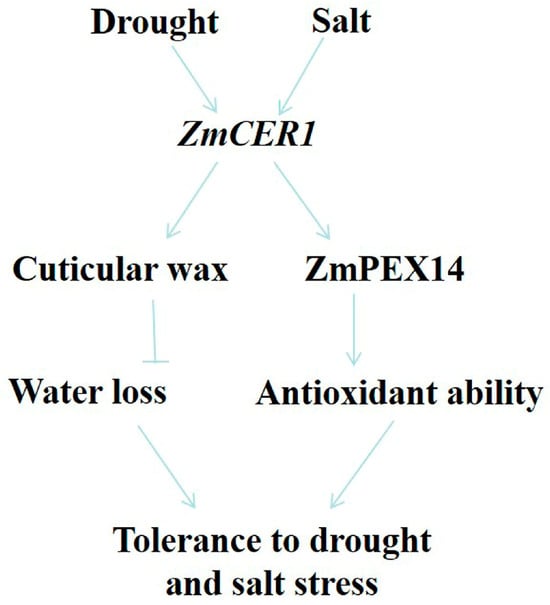
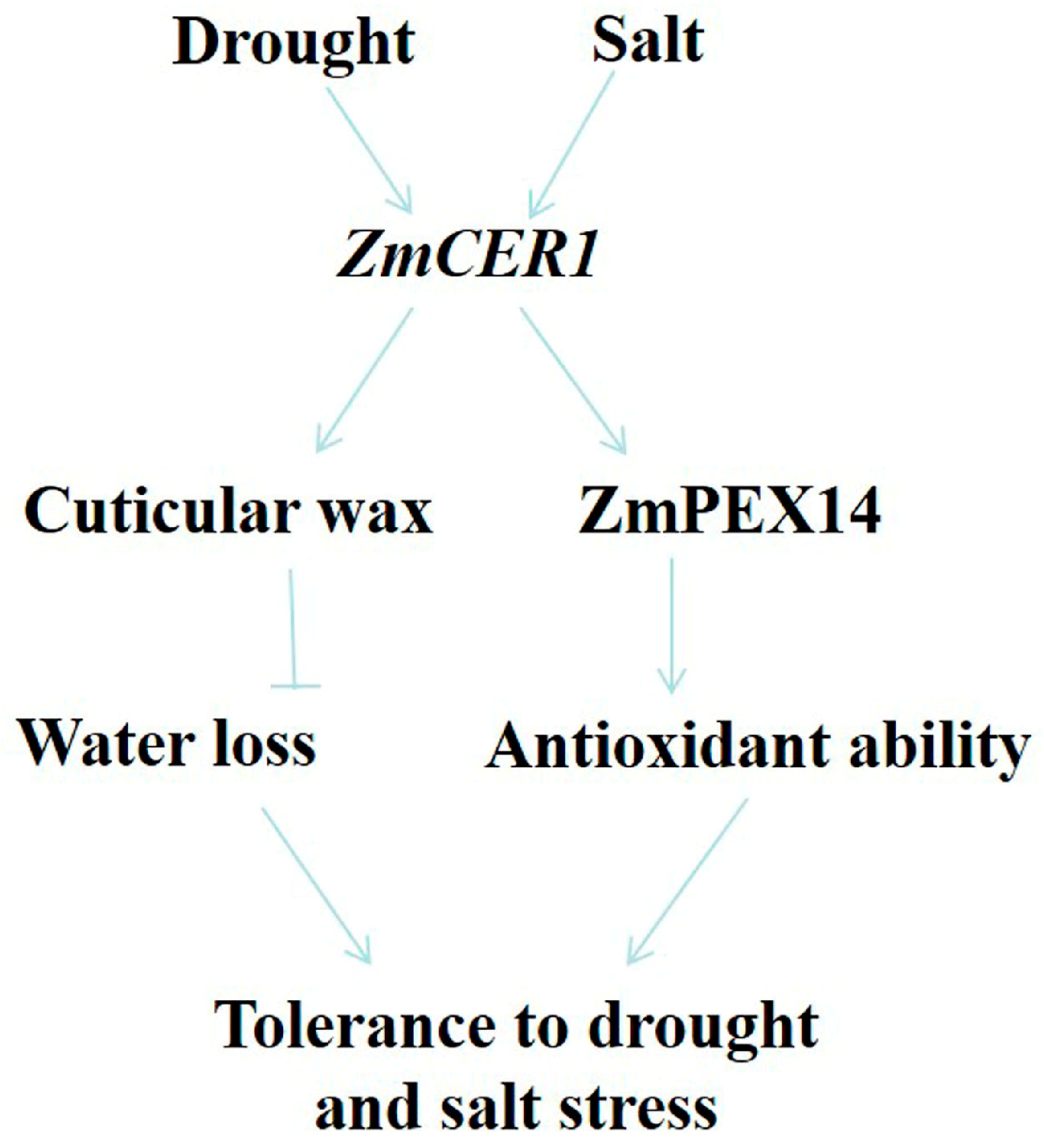
The proteins involved in peroxisome assembly are primarily peroxins (PEX), including the membrane proteins PEX3, PEX16, PEX19, PEX13, PEX14, PEX22, and PEX26; the receptor proteins PEX5 and PEX7; the E2 ubiquitin-conjugating enzyme PEX4; the E3 ubiquitin ligases PEX2, PEX10, and PEX12; and the two AAA ATPases PEX1 and PEX6, among others. These PEX proteins are highly conserved across eukaryotes. Defects in these proteins impair proper peroxisome formation, affecting their number, morphology, and overall functional integrity [46]. Studies have shown that this class of proteins is involved in plant stress response processes. For instance, under H2O2-induced oxidative stress, the expression of AtPEX1, AtPEX5, and AtPEX14 is up-regulated [47]. Under salt stress, the expression of AtPEX10 and AtPEX1 is induced [48]. Under drought stress, AtPEX14 is positively regulated to enhance drought tolerance in Arabidopsis [49]. Under cadmium stress, AtPEX7 is essential for the correct import of peroxisomal matrix proteins and plays a critical role in the Arabidopsis root response to cadmium stress and in scavenging ROS accumulation [50]. In this study, we found that the overexpression of ZmPEX14 obviously improved drought and salt stress in Arabidopsis (Figure 7). Thus, we propose a dual-mode model for ZmCER1-mediated stress tolerance (Figure 8). First, as a direct effector, ZmCER1 enhances the synthesis of VLCFA, fortifying the cuticular layer and reducing non-stomatal water loss, the primary benefit of a robust cuticle [18]. Second, as a signaling or regulatory node, ZmCER1 may influence peroxisomal activity through its interaction with ZmPEX14. This interaction might potentially modulate the import of antioxidant enzymes into peroxisomes or influence peroxisomal metabolism, thereby enhancing the cell’s overall capacity to detoxify ROS. In conclusion, our study unequivocally identifies ZmCER1 as a key genetic determinant for improving abiotic stress tolerance in maize. It functions not only by reinforcing the physical cuticular wax barrier but also, we hypothesize, by potentiating the cellular antioxidant system via an interaction with the peroxisomal protein ZmPEX14. This dual role makes ZmCER1 an exceptionally attractive candidate for precision breeding. Deploying favorable ZmCER1 alleles or editing its promoter region in elite maize cultivars holds great promise for developing new varieties capable of sustaining yield in the face of climate change-induced water scarcity and soil salinization.
Figure 8.
A proposed model for the role of ZmCER1 in regulating drought and salt tolerance. ZmCER1 functions not only by bolstering the physical cuticular wax barrier but also by potentiating the cellular antioxidant system via an interaction with the peroxisomal protein ZmPEX14. Arrows indicate positive regulation, bold blunt-ended bars indicate inhibition.
5. Conclusions
This study confirms that maize ZmCER1, a key gene involved in cuticular wax alkane biosynthesis, serves as a critical regulator of salt and drought tolerance in maize. The core findings demonstrate that ZmCER1 exerts its protective effects through two interconnected mechanisms: reinforcing the cuticular wax barrier and regulating ROS detoxification.
Functionally, ZmCER1 is induced by stress and encodes an ER-localized protein. Both heterologous overexpression in Arabidopsis and homologous overexpression in maize significantly enhance the transgenic plants’ stress tolerance, which is associated with elevated activities of antioxidant enzymes (SOD and CAT), reduced levels of MDA and H2O2, and—specifically in maize—increased contents of cuticular wax alkanes that reduce non-stomatal water loss by strengthening the hydrophobic barrier.
Molecularly, we identified ZmPEX14, a stress-inducible peroxisomal membrane protein, as a direct interacting partner of ZmCER1, which was validated by Y2H, LUC, and BiFC assays. Consistent with its interaction with ZmCER1, the overexpression of ZmPEX14 also improves stress tolerance in Arabidopsis, suggesting a synergistic role of the ZmCER1–ZmPEX14 complex in modulating ROS detoxification.
Collectively, our findings establish that ZmCER1 mediates maize stress tolerance by integrating cuticular wax barrier fortification and peroxisomal ROS detoxification via interaction with ZmPEX14. This validates ZmCER1 as a promising candidate gene for molecular breeding strategies aimed at improving maize stress tolerance and ensuring stable yields under the challenges of climate change.
Supplementary Materials
The following supporting information can be downloaded at: https://www.mdpi.com/article/10.3390/cimb48050509/s1, Table S1: PCR primers used in this study; Table S2: Two-hybrid (Y2H) screen of ZmCER1.
Author Contributions
Writing—original draft, methodology, investigation, Y.Y.; methodology, formal analysis, investigation, M.S.; writing–original draft, methodology, Y.L.; methodology, investigation, X.G.; investigation, funding acquisition, H.L.; writing—review and editing, investigation, funding acquisition, L.P. All authors have read and agreed to the published version of the manuscript.
Funding
This research was funded by the Natural Science Foundation of Shandong Province (grant number: ZR2024MC143), the Shandong Provincial Natural Science Foundation for Distinguished Young Scholars (Class A) (grant number: ZR2025QA35), the Taishan Scholar Young Expert of Shandong Province (grant number: tsqn202312211), and the National Natural Science Foundation of China (grant number: 32272075).
Institutional Review Board Statement
Not applicable.
Informed Consent Statement
Not applicable.
Data Availability Statement
Data supporting the findings of this study are available in the article and its Supplementary Materials.
Conflicts of Interest
The authors declare no conflicts of interest.
Abbreviations
The following abbreviations are used in this manuscript:
| ZmCRE1 | Maize aldehyde decarbonylase 1 gene |
| MS | Murashige and Skoog |
| RT-PCR | Real-time polymerase chain reaction |
| ROS | Reactive oxygen species |
| MDA | Malondialdehyde |
| RWC | Relative water content |
| SOD | Superoxide dismutase |
| CAT | Catalase |
| VLCFAs | Very-long-chain fatty acids |
| Y2H | Yeast two-hybrid |
| LUC | Luciferase complementation |
| BiFC | Bimolecular fluorescence complementation |
References
- Ribeiro, C.; Hennen-Bierwagen, T.A.; Myers, A.M.; Cline, K.; Settles, A.M. Engineering 6-phosphogluconate dehydrogenase improves grain yield in heat-stressed maize. Proc. Natl. Acad. Sci. USA 2020, 117, 33177–33185. [Google Scholar] [CrossRef] [Scilit] [PubMed]
- Zhang, H.; Sun, X.; Dai, M. Improving crop drought resistance with plant growth regulators and rhizobacteria: Mechanisms, applications, and perspectives. Plant Commun. 2021, 3, 100228. [Google Scholar] [CrossRef] [Scilit]
- Campos, H.; Cooper, M.; Habben, J.E.; Edmeades, G.O.; Schussler, J.R. Improving drought tolerance in maize: A view from industry. Field Crop. Res. 2004, 90, 19–34. [Google Scholar] [CrossRef] [Scilit]
- Gupta, A.; Rico-Medina, A.; Caño-Delgado, A.I. The physiology of plant responses to drought. Science 2020, 368, 266–269. [Google Scholar] [CrossRef] [Scilit]
- Munns, R.; Gilliham, M. Salinity tolerance of crops—What is the cost? New Phytol. 2015, 208, 668–673. [Google Scholar] [CrossRef] [Scilit]
- Zhao, C.; Zhang, H.; Song, C.; Zhu, J.K.; Shabala, S. Mechanisms of Plant Responses and Adaptation to Soil Salinity. Innovation 2020, 1, 100017. [Google Scholar] [CrossRef] [Scilit] [PubMed]
- Mittler, R. ROS Are Good. Trends Plant Sci. 2017, 2, 11–19. [Google Scholar] [CrossRef] [Scilit] [PubMed]
- Demidchik, V. Mechanisms of oxidative stress in plants: From classical chemistry to cell biology. Environ. Exp. Bot. 2015, 109, 212–228. [Google Scholar] [CrossRef] [Scilit]
- Gill, S.S.; Tuteja, N. Reactive oxygen species and antioxidant machinery in abiotic stress tolerance in crop plants. Plant Physiol. Biochem. 2010, 48, 909–930. [Google Scholar] [CrossRef] [Scilit]
- Bernard, A.; Joubès, J. Arabidopsis cuticular waxes: Advances in synthesis, export and regulation. Prog. Lipid Res. 2013, 52, 110–129. [Google Scholar] [CrossRef] [Scilit]
- Lee, S.B.; Suh, M.C. Advances in the understanding of cuticular waxes in Arabidopsis thaliana and crop species. Plant Cell Rep. 2015, 34, 557–572. [Google Scholar] [CrossRef] [Scilit] [PubMed]
- Riederer, M.; Schreiber, L. Protecting against water loss: Analysis of the barrier properties of plant cuticles. J. Exp. Bot. 2001, 52, 2023–2032. [Google Scholar] [CrossRef] [Scilit] [PubMed]
- Bowman, J.L.; Kohchi, T.; Yamato, K.T.; Jenkins, J.; Shu, S.; Ishizaki, K.; Yamaoka, S.; Nishihama, R.; Nakamura, Y.; Berger, F.; et al. Insights into land plant evolution garnered from the Marchantia polymorpha genome. Cell 2017, 171, 287–304. [Google Scholar] [CrossRef] [Scilit]
- Nawrath, C. Unraveling the complex network of cuticular structure and function. Curr. Opin. Plant Biol. 2006, 9, 281–287. [Google Scholar] [CrossRef] [Scilit]
- Samuels, L.; Kunst, L.; Jetter, R. Sealing plant surfaces: Cuticular wax formation by epidermal cells. Annu. Rev. Plant Biol. 2008, 59, 683–707. [Google Scholar] [CrossRef] [Scilit]
- Jetter, R.; Kunst, L. Plant surface lipid biosynthetic pathways and their utility for metabolic engineering of waxes and hydrocarbon biofuels. Plant J. 2008, 54, 670–683. [Google Scholar] [CrossRef] [Scilit]
- Bourdenx, B.; Bernard, A.; Domergue, F.; Pascal, S.; Léger, A.; Roby, D.; Pervent, M.; Vile, D.; Haslam, R.P.; Napier, J.A.; et al. Overexpression of Arabidopsis ECERIFERUM1 promotes wax very-long-chain alkane biosynthesis and influences plant response to biotic and abiotic stresses. Plant Physiol. 2011, 156, 29–45. [Google Scholar] [CrossRef] [Scilit] [PubMed]
- Kosma, D.K.; Bourdenx, B.; Bernard, A.; Parsons, E.P.; Lü, S.; Joubès, J.; Jenks, M.A. The impact of water deficiency on leaf cuticle lipids of Arabidopsis. Plant Physiol. 2009, 151, 1918–1929. [Google Scholar] [CrossRef] [Scilit]
- Daryanto, S.; Wang, L.X.; Jacinthe, P.A. Global synthesis of drought effects on maize and wheat production. PLoS ONE 2016, 11, e0156362. [Google Scholar] [CrossRef] [Scilit]
- Tian, T.; Wang, S.H.; Yang, S.P.; Yang, Z.R.; Liu, S.X.; Wang, Y.; Gao, H.; Zhang, S.; Yang, X.; Jiang, C.; et al. Genome assembly and genetic dissection of a prominent drought-resistant maize germplasm. Nat. Genet. 2023, 55, 496–506. [Google Scholar] [CrossRef] [Scilit]
- Aarts, M.G.; Keijzer, C.J.; Stiekema, W.J.; Pereira, A. Molecular characterization of the CER1 gene of Arabidopsis involved in epicuticular wax biosynthesis and pollen fertility. Plant Cell 1995, 7, 2115–2127. [Google Scholar]
- Sakuradani, E.; Zhao, L.F.; Haslam, T.M.; Kunst, L. The CER22 gene required for the synthesis of cuticular wax alkanes in Arabidopsis thaliana is allelic to CER1. Planta 2013, 237, 731–738. [Google Scholar] [CrossRef] [Scilit] [PubMed]
- Bernard, A.; Domergue, F.; Pascal, S.; Jetter, R.; Renne, C.; Faure, J.D.; Haslam, R.P.; Napier, J.A.; Lessire, R.; Joubès, J. Reconstitution of plant alkane biosynthesis in yeast demonstrates that Arabidopsis ECERIFERUM1 and ECERIFERUM3 are core components of a very-long-chain alkane synthesis complex. Plant Cell 2012, 24, 3106–3118. [Google Scholar] [CrossRef] [Scilit] [PubMed]
- Pascal, S.; Bernard, A.; Sorel, M.; Pervent, M.; Vile, D.; Haslam, R.P.; Napier, J.A.; Lessire, R.; Domergue, F.; Joubès, J. The Arabidopsis cer26 mutant, like the cer2 mutant, is specifically affected in the very long chain fatty acid elongation process. Plant J. 2013, 73, 733–746. [Google Scholar] [CrossRef] [Scilit] [PubMed]
- Wang, W.; Zhang, Y.; Xu, C.; Ren, J.J.; Liu, X.F.; Black, K.; Gai, X.; Wang, Q.; Ren, H. Cucumber ECERIFERUM1 (CsCER1), which influences the cuticle properties and drought tolerance of cucumber, plays a key role in VLC alkanes biosynthesis. Plant Mol. Biol. 2015, 87, 219–233. [Google Scholar] [CrossRef] [Scilit]
- Ni, E.; Zhou, L.; Li, J.; Jiang, D.; Wang, Z.; Zheng, S.; Qi, H.; Zhou, Y.; Wang, C.; Xiao, S.; et al. OsCER1 plays a pivotal role in very-long-chain alkane biosynthesis and affects plastid development and programmed cell death of tapetum in rice (Oryza sativa L.). Front. Plant Sci. 2018, 9, 1217. [Google Scholar] [CrossRef] [Scilit]
- Wang, Y.M.; Jin, S.R.; Xu, Y.; Li, S.; Zhang, S.J.; Yuan, Z.; Li, J.N.; Ni, Y. Overexpression of BnKCS1-1, BnKCS1-2, and BnCER1-2 promotes cuticular wax production and increases drought tolerance in Brassica napus. Crop J. 2020, 8, 26–37. [Google Scholar] [CrossRef] [Scilit]
- Wang, D.J.; Ni, Y.; Liao, L.X.; Xiao, Y.; Guo, Y.J. Poa pratensis ECERIFERUM1 (PpCER1) is involved in wax alkane biosynthesis and plant drought tolerance. Plant Physiol. Biochem. 2021, 159, 312–321. [Google Scholar] [CrossRef] [Scilit]
- Wu, H.Q.; Liu, L.; Chen, Y.F.; Liu, T.X.; Jiang, Q.Q.; Wei, Z.Y.; Li, C.; Wang, Z. Tomato SlCER1-1 catalyzes the synthesis of wax alkanes which increases the drought tolerance and fruit storability. Hortic. Res. 2022, 9, uhac004. [Google Scholar] [CrossRef] [Scilit]
- Livak, K.J.; Schmittgen, T.D. Analysis of relative gene expression data using real-time quantitative PCR and the 2(-Delta Delta C(T)) Method. Methods 2001, 25, 402–408. [Google Scholar] [CrossRef] [Scilit]
- Park, S.; Li, J.; Pittman, J.K.; Berkowitz, G.A.; Yang, H.; Undurraga, S.; Morris, J.; Hirschi, K.D.; Gaxiola, R.A. Up-regulation of a H+-pyrophosphatase (H+-PPase) as a strategy to engineer drought resistant crop plants. Proc. Natl. Acad. Sci. USA 2005, 102, 18830–18835. [Google Scholar] [CrossRef] [Scilit]
- Peever, T.L.; Higgins, V.J. Electrolyte leakage, lipoxygenase, and lipid peroxidation induced in tomato leaf tissue by specific and nonspecific elicitors from Cladosporium fulvum. Plant Physiol. 1989, 90, 867–875. [Google Scholar] [CrossRef] [Scilit]
- Giannopolitis, C.N.; Ries, S.K. Superoxide dismutase: I. occurrence in higher plants. Plant Physiol. 1977, 59, 309–314. [Google Scholar] [CrossRef] [Scilit]
- Alia; Kondo, Y.; Sakamoto, A.; Nonaka, H.; Hayashi, H.; Saradhi, P.P.; Chen, T.H.; Murata, N. Enhanced tolerance to light stress of transgenic Arabidopsis plants that express the codA gene for a bacterial choline oxidase. Plant Mol. Biol. 1999, 40, 279–288. [Google Scholar] [CrossRef] [Scilit]
- Sturaro, M.; Hartings, H.; Schmelzer, E.; Velasco, R.; Salamini, F.; Motto, M. Cloning and characterization of GLOSSY1, a maize gene involved in cuticle membrane and wax production. Plant Physiol. 2005, 138, 478–489. [Google Scholar] [CrossRef] [Scilit] [PubMed]
- Chen, W.; Yu, X.H.; Zhang, K.; Shi, J.; Oliveira, S.D.; Schreiber, L.; Shanklin, J.; Zhang, D. Male Sterile2 encodes a plastid-localized fatty acyl carrier protein reductase required for pollen exine development in Arabidopsis. Plant Physiol. 2011, 157, 842–853. [Google Scholar] [CrossRef] [Scilit]
- Theodoulou, F.L.; Bernhardt, K.; Linka, N.; Baker, A. Peroxisome membrane proteins: Multiple trafficking routes and multiple functions? Biochem. J. 2013, 451, 345–352. [Google Scholar] [CrossRef] [Scilit] [PubMed]
- Smith, J.J.; Aitchison, J.D. Peroxisomes take shape. Nat. Rev. Mol. Cell Biol. 2013, 14, 803–817. [Google Scholar] [CrossRef] [Scilit]
- Yang, Z.R.; Cao, Y.B.; Shi, Y.T.; Qin, F.; Jiang, C.F.; Yang, S.H. Genetic and molecular exploration of maize environmental stress resilience: Toward sustainable agriculture. Mol. Plant 2023, 16, 1496–1517. [Google Scholar] [CrossRef] [Scilit] [PubMed]
- Liao, Q.; Gu, S.; Kang, S.; Du, T.; Tong, L.; Wood, J.D.; Ding, R. Mild water and salt stress improve water use efficiency by decreasing stomatal conductance via osmotic adjustment in field maize. Sci. Total Environ. 2022, 805, 150364. [Google Scholar] [CrossRef] [Scilit]
- Jenks, M.A.; Tuttle, H.A.; Eigenbrode, S.D.; Feldmann, K.A. Leaf epicuticular waxes of the eceriferum mutants in Arabidopsis. Plant Physiol. 1995, 108, 369–377. [Google Scholar] [CrossRef] [Scilit]
- Corpas, F.J.; González-Gordo, S.; Cañas, A.; Palma, J.M. Nitric oxide and hydrogen sulfide in plants: Which comes first? J. Exp. Bot. 2020, 70, 4391–4404. [Google Scholar] [CrossRef] [Scilit]
- Pan, R.; Liu, J.; Wang, S.; Hu, J. Peroxisomes: Versatile organelles with diverse roles in plants. New Phytol. 2020, 225, 1410–1427. [Google Scholar] [CrossRef] [Scilit]
- Du, Y.Y.; Wang, P.C.; Chen, J.; Song, C.P. Comprehensive functional analysis of the catalase gene family in Arabidopsis thaliana. J. Integr. Plant Biol. 2008, 50, 1318–1326. [Google Scholar] [CrossRef] [Scilit] [PubMed]
- Frugoli, J.A.; Zhong, H.H.; Nuccio, M.L.; McCourt, P.; McPeek, M.A.; Thomas, T.L.; McClung, C.R. Catalase is encoded by a multigene family in Arabidopsis thaliana (L.) Heynh. Plant Physiol. 1996, 112, 327–336. [Google Scholar] [CrossRef] [Scilit] [PubMed]
- Kao, Y.T.; Gonzalez, K.L.; Bartel, B. Peroxisome function, biogenesis, and dynamics in plants. Plant Physiol. 2018, 176, 162–177. [Google Scholar] [CrossRef] [Scilit] [PubMed]
- Lopez-Huertas, E.; Charlton, W.L.; Johnson, B.; Graham, I.A.; Baker, A. Stress induces peroxisome biogenesis genes. EMBO J. 2000, 19, 6770–6777. [Google Scholar] [CrossRef] [Scilit]
- Charlton, W.L.; Matsui, K.; Johnson, B.; Graham, I.A.; Ohme-Takagi, M.; Baker, A. Salt-induced expression of peroxisome-associated genes requires components of the ethylene, jasmonate and abscisic acid signalling pathways. Plant Cell Environ. 2005, 28, 513–524. [Google Scholar] [CrossRef] [Scilit]
- Shi, H.; Ye, T.; Yang, F.; Chan, Z. Arabidopsis PED2 positively modulates plant drought stress resistance. J. Integr. Plant Biol. 2015, 57, 796–806. [Google Scholar] [CrossRef] [Scilit]
- Piacentini, D.; Della Rovere, F.; Bertoldi, I.; Massimi, L.; Sofo, A.; Altamura, M.M.; Falasca, G. Peroxisomal PEX7 receptor affects cadmium-induced ros and auxin homeostasis in Arabidopsis root system. Antioxidants 2021, 10, 1494. [Google Scholar] [CrossRef] [Scilit]
Disclaimer/Publisher’s Note: The statements, opinions and data contained in all publications are solely those of the individual author(s) and contributor(s) and not of MDPI and/or the editor(s). MDPI and/or the editor(s) disclaim responsibility for any injury to people or property resulting from any ideas, methods, instructions or products referred to in the content. |
© 2026 by the authors. Licensee MDPI, Basel, Switzerland. This article is an open access article distributed under the terms and conditions of the Creative Commons Attribution (CC BY) license.