


Abstract
Androgenetic alopecia (AGA) is a common hair loss disease that is pathologically featured by progressive hair follicle miniaturization. Its molecular pathogenesis is closely linked to aberrant activation of androgen signaling and dysregulation of multiple programmed cell death (PCD) pathways. This review mainly summarizes the regulatory roles of several PCD modalities, including apoptosis, necroptosis, pyroptosis, ferroptosis and autophagy, in the development of AGA. During hair follicle regression, endogenous metabolic disturbances and extrinsic microenvironmental stimuli such as androgen abnormality, oxidative stress and inflammatory signals can aberrantly activate the above-mentioned cell death mechanisms, resulting in dysfunction of dermal papilla cells and disrupted homeostasis of hair follicle stem cells. These events perturb normal hair follicle cycling, ultimately shortening the anagen phase, accelerating catagen progression and prolonging the telogen phase, thereby aggravating hair shedding. Most current studies only address the role of a single PCD subtype in AGA, lacking systematic integration of these five PCD forms. Comprehensive reviews concerning their spatiotemporal distribution, pathway crosstalk and shared regulatory targets during hair follicle miniaturization are still insufficient, which also hinders the interpretation of broad multi-pathway effects observed for most candidate drugs. Herein, we systematically collate molecular evidence supporting the involvement of the five PCD forms in pathological injury of AGA, summarize the molecular characteristics and intervention strategies of each pathway, and dissect interaction patterns and common regulatory nodes across distinct PCD pathways. Meanwhile, we highlight limitations in this field, including insufficient validation of causal relationships, scarcity of pathological evidence from human tissues, shortage of time-series intervention experiments, and obstacles in clinical translation. This review aims to deepen the understanding of the degenerative injury mechanism of hair follicles in AGA and provide theoretical support for developing novel therapeutic strategies for AGA by targeting PCD.
1. Introduction
PCD is a core physiological mechanism that maintains the homeostasis of the internal environment of the organism and regulates tissue development and remodeling. With the continuous in-depth research on the regulatory network of cell death, its pathological roles in the progression of various diseases have been gradually elucidated. Among them, the regulatory effects of such mechanisms in AGA have become a research hotspot in the field of hair disorders. AGA is a common hair loss disorder characterized by the progressive miniaturization of scalp hair follicles. Among several forms of cell death associated with androgenetic alopecia, apoptosis is the most prevalent. Apoptosis is a genetically regulated and ordered process of cell death, characterized by nuclear pyknosis, DNA fragmentation and cell membrane blebbing [1]. In the pathogenesis of AGA, increased nuclear localization of the Androgen Receptor (AR) in Dermal Papilla (DP) cells leads to AR-mediated paracrine signaling that induces apoptosis of microvascular endothelial cells, thereby causing vascular degeneration of the dermal papilla within the DP [2]. Recent single-cell transcriptomic studies have further confirmed that abnormal contraction of the AGA hair follicle sheath induces hair follicle progenitor cell apoptosis via Piezo1 mechanical signals, participating in the early initiation of hair follicle miniaturization [3].
In AGA, hair follicles undergo cyclic changes including the anagen, catagen and telogen phases, and the transition between these cycles relies on the activity of hair follicle stem cells. Studies have shown that the anti-apoptotic protein Bcl-2 is highly expressed in hair follicle stem cells and is critical for maintaining stem cell homeostasis and tissue regeneration. Blockage or loss of Bcl-2 function leads to apoptosis of specific stem cell subpopulations, thereby affecting the cyclic regeneration of hair follicles [4]. Necroptosis usually occurs in the catagen phase. Targeted regulation of key necroptosis proteins such as RIPK1, RIPK3 and TNF-α can modulate the hair follicle cycle, which has potential for targeted intervention of AGA [5]. The androgen receptor can mediate NLRP3 inflammasome activation and regulate the expression of proteins such as Caspase-1, IL-1β and GSDMD-N, suggesting that androgenetic alopecia is associated with pyroptosis [6,7]. The key molecule of ferroptosis, NRF2, is widely expressed in hair follicles. Activation of NRF2 can alleviate hair follicle damage caused by oxidative stress and reverse the inhibitory effect induced by DHT [8,9]. In addition, ferroptosis factors MBOAT1/2 can be upregulated by the androgen receptor AR. Imbalanced iron metabolism in the body is closely associated with hair loss. Synergistic intervention targeting ferroptosis and the androgen signaling axis can effectively ameliorate the pathological phenotype of AGA [10]. Existing studies have found that DHT-induced apoptosis of DPCs is also associated with the autophagy process. DHT treatment reduces the activity of mTORC1, a key autophagy molecule. Activating the activity of this molecule can reverse DHT-induced cellular autophagy and apoptosis, thereby protecting HDPCs and ameliorating androgenetic alopecia [11].
Existing basic experiments have confirmed that the abnormal activation of programmed cell death is a key factor inducing degenerative damage of hair follicles in AGA. Existing reviews on androgenetic alopecia mostly focus on the regulatory mechanisms of a single cell death mode, among which the summaries related to follicular cell apoptosis are the most comprehensive [12]. Other studies have focused on tissue engineering repair strategies and hair follicle regeneration techniques for androgenetic alopecia, summarizing intervention methods for hair follicle miniaturization from a therapeutic perspective [13]. However, current reviews in this field mostly focus on the association between a single type of programmed cell death and AGA, lacking systematic integration and sorting of five types of programmed cell death, namely apoptosis, necroptosis, pyroptosis, ferroptosis and autophagy. There is a lack of specific reviews on the temporal distribution patterns of different PCDs during hair follicle miniaturization, the crosstalk between pathways and shared regulatory targets. This makes it difficult to explain the broad-spectrum multi-pathway effects shown by most candidate drugs and also restricts the development of novel therapeutic strategies for AGA targeting programmed cell death. To this end, this paper systematically summarizes the molecular evidence for the involvement of five types of programmed cell death in the pathogenesis of AGA, analyzes the functional characteristics and research limitations of each pathway, sorts out the spatiotemporal associations and interaction mechanisms among these pathways, and summarizes potential intervention strategies targeting PCD, so as to provide references for the mechanistic elucidation and clinical translational research of AGA.
2. Main Pathogenesis of AGA
The pathogenesis of AGA is driven primarily by the abnormal activation of the androgen signaling pathway, which is mainly accompanied by two typical pathological features: progressive miniaturization of hair follicles and shortening of the anagen phase of hair follicles. There are three types of 5α-reductase in the human body, namely type I, type II and type III. Among them, type II 5α-reductase is highly expressed in dermal papilla cells of AGA patients, mostly distributed in hair follicles and their surrounding tissues, and is closely associated with the pathogenesis of AGA. 5α-reductase converts testosterone, the primary androgen in the body, into dihydrotestosterone (DHT). After DHT binds to the intracellular AR to form a homomeric complex, nuclear translocation occurs, which further extensively regulates the transcription and translation processes of downstream target genes. Multiple studies have confirmed that the expression level of AR in dermal papilla cells from the balding area and beard region of AGA patients is significantly upregulated, which can markedly increase the cellular responsiveness to androgens and amplify the damaging effects of androgens [14]. As a nuclear transcription factor, activated AR can induce dermal papilla cells to secrete large amounts of multiple paracrine factors that negatively regulate hair growth, such as DKK1, IL-6 and TGF-β, ultimately inducing atrophy and degenerative changes in hair follicle structures and promoting the progression of alopecia. Androgens are steroid hormones that regulate the proliferation, differentiation and physiological functions of hair follicle cells through dual modes: genomic pathways and rapid non-genomic pathways. In the classical genomic regulation mode, androgens cross the cell membrane to enter the cytoplasm and bind to and activate AR. The activated receptors can specifically recognize response elements in the promoter regions of target genes, and positively or negatively regulate gene transcription and protein synthesis. This process takes effect slowly [15]; meanwhile, the non-genomic effects mediated by androgens act rapidly, triggering biological responses within seconds to minutes. This process is entirely independent of gene transcription and protein synthesis and is mainly achieved through cell membrane-associated signaling pathways. Androgens can increase the activity of Ca2+-ATPase and interact with membrane phospholipids to affect membrane fluidity, further participating in the regulation of the physiological state of hair follicle cells. Both types of regulatory modes rely on AR signaling for completion.
Collectively, the androgen–AR signaling axis constitutes the core driver of AGA pathogenesis. The pathological cascade triggered by 5α-reductase, DHT-AR activation and downstream paracrine factors leading to hair follicle miniaturization is summarized in (Figure 1).
Figure 1.
Visual framework illustrating the core pathogenesis of androgenetic alopecia, covering androgen–AR axis, downstream programmed cell death and hair follicle miniaturization. Created in BioRender. Adnan, N. (2026) http://BioRender.com/45zmjlr. Note: Colored background boxes are used only to distinguish the four functional modules of AGA pathogenesis; all black arrows represent signal transduction.
3. Cell Death Signaling Pathways and Their Roles in AGA
3.1. Apoptosis and AGA
Apoptosis is mainly initiated through two major pathways: the intrinsic mitochondrial pathway and the extrinsic death receptor pathway. Intrinsic apoptosis is mostly induced by intracellular homeostatic imbalance triggered by DNA damage and toxic stimuli. This pathway is primarily regulated by the Bcl-2 family protein network. After oligomerization, pro-apoptotic proteins BAK and BAX localize to the outer mitochondrial membrane and trigger mitochondrial outer membrane permeabilization (MOMP), promoting the release of factors such as cytochrome c, Smac and HTRA2 into the cytoplasm [16]. Among them, cytochrome c can bind to Apaf-1 and procaspase-9, and assemble to form apoptosomes with the participation of dATP, thereby activating caspase-9. The intracellular IAP family protein XIAP can inhibit the activation of caspase-3 and thereby block the apoptotic process, while the simultaneously released Smac and HTRA2 can relieve the inhibitory effect of XIAP and ultimately promote the progression of the apoptotic program [1]. The extrinsic apoptotic pathway is initiated by the activation of death receptors on the cell membrane surface. After binding to their corresponding ligands, death receptors undergo oligomerization to assemble signaling platforms [1], and further recruit related adaptor molecules to form the death-inducing signaling complex (DISC), which sequentially activates caspase-8 and caspase-10. The two apoptotic pathways, the extrinsic and intrinsic pathways, ultimately converge on the caspase cascade, jointly mediating the process of programmed cell death. In the pathological process of AGA, the aforementioned apoptotic pathways are abnormally regulated by the DHT-AR signal.
Apoptosis is closely associated with the hair follicle growth cycle and plays a dominant regulatory role in the catagen phase of hair follicles. There are significant differences in the sensitivity of different hair follicle cells to apoptotic stimuli: melanocytes and follicular epithelial cells are prone to apoptosis, while dermal papilla fibroblasts and some keratinocytes possess strong anti-apoptotic capabilities [17]. Apoptosis can significantly reduce the adhesion between keratinocytes and effectively drive hair follicles into the catagen phase. Based on morphological and functional characteristics, the catagen phase of hair follicles can be further divided into eight sub-stages, with significant differences in the patterns of cellular apoptosis among each stage. TUNEL staining results related to the hair follicle cycle in mice confirmed that apoptotic cells first appear in the melanogenic region of hair follicles in the late anagen phase, and then sequentially distribute in the hair matrix, inner and outer root sheaths and epithelial tissues [18]. Obvious apoptotic signals can also be detected in the hair follicle bulge structure and hair shaft tissues by the late catagen phase.
In the pathological progression of AGA, androgens bind to AR to initiate a cascade of molecular events, ultimately inducing hair follicle atrophy and excessive apoptosis of hair follicle cells. Clinical sample studies have found that the nuclear localization level of AR in dermal papilla cells in the balding area of AGA patients is significantly upregulated. Transcriptomic analysis confirms obvious microvascular structural abnormalities in the dermal papilla tissue of the balding area, verifying that hair follicle miniaturization is accompanied by regressive vascular changes in the dermal papilla at an early stage. Further mechanistic studies show that AR-dependent paracrine signals, especially the TGF-β pathway, can induce apoptosis of microvascular endothelial cells in the dermal papilla, ultimately leading to the decline in blood supply to hair follicles [2]. DHT can regulate the endogenous apoptotic pathways of human dermal papilla cells through multiple pathways and mediate the functional decline in hair follicles. Studies have found that circular RNA CircAGK can mediate the apoptosis of dermal papilla cells stimulated by high concentrations of DHT by targeting the miR-3180-5p/BAX signaling axis, clarifying the regulatory role of non-coding RNAs in AGA cell apoptosis [19]. Mitochondrial dysfunction is a critical upstream event in DHT-induced DPC injury. Loss of ACOD1 can exacerbate mitochondrial dysfunction in dermal papilla cells by upregulating the methylation level of DDX1, ultimately inducing the coordinated occurrence of cellular senescence and apoptosis and accelerating follicular damage and degeneration [20]. At the therapeutic level, human umbilical cord mesenchymal stem cell exosomes can deliver Let-7b/7f-5p to target dermal papilla cells. By downregulating androgen receptor expression and inhibiting the activation of apoptotic pathways, they effectively alleviate DHT-induced DPC injury, providing experimental evidence for the targeted treatment of AGA via apoptotic pathways [21,22]. This confirms that abnormal apoptosis is a core and key link in the pathogenesis of AGA, and also provides a theoretical basis for targeted anti-apoptotic intervention to treat hair follicle atrophy [23].
Numerous in vivo and in vitro experiments have confirmed that interventions designed based on the apoptotic pathway can effectively ameliorate the phenotype of androgenetic alopecia. The ethanol extract of the tropical plant Connarus semidecandrus Jack (Cs-EE) can target and regulate the endogenous mitochondrial apoptotic pathway [24]. On the one hand, it downregulates AR expression and inhibits 5α-reductase activity to attenuate the pathogenic effects of androgens; on the other hand, it upregulates the expression of anti-apoptotic protein Bcl-2, downregulates the transcriptional levels of Bax and caspase-9, suppresses mitochondrial membrane permeabilization, and thereby blocks the apoptosis of hair follicle cells. Studies based on the AGA model of C57BL/6 mice show that cell-free fat extract (CEFFE) can reduce DHT accumulation in dermal papilla cells and downregulate AR expression. It inhibits cell apoptosis by increasing the proportion of CD31- and Ki67-positive cells, thereby repairing the physiological function of hair follicles [25]. Studies at the genetic level have found that p53 participates in hair follicle cell apoptosis by regulating the Bcl-2/Bax balance. p53 is highly expressed in hair follicles during the catagen phase and co-localizes with apoptotic markers. Targeted inhibition of p53 can effectively reduce hair follicle cell apoptosis and has potential for AGA treatment [26].
The classic clinical drug minoxidil can exert a significant anti-apoptotic effect by upregulating the Bcl-2/Bax ratio, prolong the anagen phase of hair follicles and promote hair growth [27]. The polyphenolic active ingredient EGCG can activate the ERK and AKT pathways, simultaneously upregulate the Bcl-2/Bax expression ratio, and promote the proliferation of human dermal papilla cells. Human umbilical cord mesenchymal stem cells can upregulate endogenous anti-apoptotic genes by activating the Wnt/β-catenin pathway, reduce dermal papilla cell apoptosis and promote hair regeneration. Inhibition of β-catenin degradation will aggravate apoptotic damage to dermal papilla cells [22,28]. In addition, the functional microneedle delivery system constructed based on tremella polysaccharide and hyaluronic acid contains magnetic intracellular vesicles (Mag-IV), which exhibit stronger pro-angiogenic effects and a greater capacity to protect HHDPC from apoptosis compared with other vesicles. It can effectively antagonize the apoptosis of dermal papilla cells and accelerate hair repair in AGA model mice [29].
Existing basic experiments have confirmed that DHT can directly induce the apoptosis of dermal papilla cells, and AR nuclear translocation can positively activate TGF-β-mediated apoptotic signals, indicating that apoptosis is a direct downstream effect of abnormal androgen signals. At this stage, most anti-apoptotic interventions can simultaneously optimize the hair follicle microenvironment and reduce local androgen levels. It is difficult to accurately distinguish whether their therapeutic effects directly target the apoptotic pathway or are achieved indirectly by improving the microenvironment.
DHT-AR signaling triggers both intrinsic mitochondrial and extrinsic death-receptor-mediated apoptotic cascades in human dermal papilla cells. The key molecular regulatory network is depicted in (Figure 2).
Figure 2.
Schematic diagram of the intrinsic mitochondrial and extrinsic death receptor-mediated apoptotic pathways in dermal papilla cells under DHT stimulation. Created in BioRender. Adnan, N. (2026) http://BioRender.com/45zmjlr. Note: Colored backgrounds separate exogenous and endogenous apoptotic pathways. Arrows denote signal transduction; ↑ = upregulation, ↓ = downregulation.
3.2. Necroptosis and AGA
Necroptosis is another type of programmed cell death distinct from apoptosis. This form of cell death does not rely on core apoptosis-related molecules and is mostly induced by stress conditions such as inflammatory stimulation and external physical and chemical injuries. It is widely involved in the injury processes of various chronic diseases [30]. At the morphological level, necroptosis is typically characterized by the rupture of cell membrane integrity, which clearly distinguishes it from apoptosis that maintains intact cell membrane structure. Although the two share some common activating triggers, there are significant differences in their downstream signal regulatory pathways [31]. The initiation of necroptosis is closely associated with the tumor necrosis factor receptor family, and the receptor-interacting protein kinase family (RIPKs) serves as the core mediator regulating this process. After the activation of death domain receptors such as Fas and TNFR, as well as TLR3/4 pattern recognition receptors [32] and adaptor proteins such as FADD and TRIF that can be recruited to interact with caspase-8/10 [33], it further mediates the deubiquitination modification of RIPK1 and recruits and phosphorylates RIPK3 to form a functional complex [34]. The complex further phosphorylates the downstream MLKL protein and induces its oligomerization. Oligomerized MLKL localizes to the cell membrane and triggers pore formation, resulting in massive leakage of intracellular contents and ultimately completing the necroptosis process [35]. Direct experimental evidence for necroptosis of hair follicles in AGA remains relatively scarce, and most existing mechanisms related to hair follicle necroptosis are derived from studies using alopecia areata models. Studies have confirmed that stress-induced excessive activation of the sympathetic nervous system can trigger necroptosis in hair follicle cells, leading to autoimmune injury and destruction of hair follicles [36]. It should be clarified that the conclusions of this study are based on the alopecia areata disease model rather than the androgenetic alopecia model. Whether the revealed regulatory mechanism of necroptosis can be applied to DHT-induced AGA hair follicle damage still needs further verification in human dermal papilla cells and AGA animal models.
In the pathological context of AGA, abnormal accumulation of DHT and local chronic microinflammation in hair follicles can aberrantly activate the necroptosis signaling pathway, resulting in disrupted homeostasis and loss of function of hair follicle stem cells. The inflammatory microenvironment can markedly increase the responsiveness of hair follicle cells to pro-inflammatory factors such as TNF-α. Persistently abnormal inflammatory signals further amplify the cell death effect and accelerate the functional decline in hair follicles [37]. In vitro experiments have confirmed that high concentrations of TNF-α can significantly inhibit the normal growth and development of hair follicles. The novel microneedle drug delivery system developed in existing studies can downregulate the levels of pro-inflammatory factors such as TNF-α and IL-1β in hair follicle tissues by delivering marine collagen peptides, effectively alleviating inflammatory damage to hair follicles and promoting hair repair and regeneration in AGA model mice [38].
Targeted regulation of the necroptosis pathway has become a potential direction for regulating the hair follicle growth cycle and developing treatments for hair loss [5,39]. Nec-1, a specific inhibitor of RIPK1, was first applied to the intervention of neurological diseases and exerted a remarkable anti-necroptosis effect [40]. Experiments on hair follicle organ culture confirmed that RIPK1 and RIPK3 are highly expressed in the outer root sheath tissue of hair follicles. Moreover, necroptosis mainly occurs during the catagen and telogen phases of hair follicles, with the highest expression activity in the catagen phase. Nec-1 can effectively remodel the hair follicle growth cycle by targeting and inhibiting the activity of the RIPK1/RIPK3 complex [5]. Another study shows that astragaloside IV can inhibit procaspase-8 and regulate the expression of TNF-α, and its therapeutic effect on hair loss is related to blocking the Fas/Fas L-mediated apoptotic pathway [41]. Population samples and animal experiment results show that the necroptosis effector protein MLKL is significantly upregulated in the hair follicle tissue of AGA patients in alopecia areas. It is also highly expressed in mouse hair follicle outer root sheath cells, with its expression peaking during the catagen phase. In vitro and in vivo experiments have confirmed that knocking down MLKL expression or using the MLKL-specific inhibitor necrosulfonamide (NSA) can block the necroptosis process of outer root sheath cells, activate the Wnt pathway in dermal papilla cells, and effectively ameliorate androgen-induced hair follicle damage [42].
Multiple basic studies have now confirmed that necroptosis is involved in the pathological injury process of AGA. From the perspective of the hair follicle cycle timeline, apoptosis is mostly initiated in the early catagen phase, mainly acting on dermal papilla cells and microvascular endothelial cells; necroptosis activity peaks in the outer root sheath tissue during the catagen phase. The two processes show obvious temporal overlap in the catagen phase, creating potential conditions for synergistic regulation of hair follicle regression. RIPK1, a key converging molecule linking apoptosis and necroptosis, has its activity regulated by caspase-8; when the apoptotic pathway is inhibited, the cell death program can shift toward necroptosis. At present, academia has only confirmed that there is a temporal synergistic effect between the two. However, systematic comparative studies are still lacking regarding the effector cells, initiation timing and functional division of the two death modes within hair follicles.
Combined DHT and TNF-α stimulation initiates RIPK1-RIPK3-MLKL-dependent necroptosis and aggravates outer root-sheath injury. The core signaling cascade is shown in (Figure 3).
Figure 3.
Schematic diagram of TNF-α/RIPK1-RIPK3-MLKL-mediated necroptosis and its role in androgenetic alopecia. Created in BioRender. Adnan, N. (2026) http://BioRender.com/45zmjlr. Note: Colored boxes are used to separate functional modules. Black arrows represent signal transduction; the cross symbol indicate inhibition or blockade; upward arrows (↑) represent upregulation. Ellipsis (…) indicates that additional types of DAMPs exist besides the representative molecules listed here.
3.3. Pyroptosis and AGA
Pyroptosis is a pro-inflammatory programmed cell death mediated by the Gasdermin family, which is distinct from non-inflammatory apoptosis and necroptosis. It was first discovered in macrophages infected with Shigella and Salmonella. Early studies misidentified it as apoptosis, yet the activating triggers and core regulatory pathways of these two types of cell death modes are fundamentally different [43]. Pyroptosis is characterized by rapid membrane damage. Core events such as membrane structure disruption and the release of cellular contents can be initiated within minutes, accompanied by phosphatidylserine (PtdSer) externalization, nuclear pyknosis and DNA damage, ultimately triggering a robust local inflammatory response [44]. According to the molecular activation patterns, pyroptosis can be divided into the caspase-1-dependent canonical pathway and the caspase-3/11-mediated non-canonical pathway. In the canonical pathway, intracellular PAMP and DAMP signals can activate the NLRP3 inflammasome, recruit the ASC adaptor protein and activate pro-caspase-1 [45]. Activated caspase-1 specifically cleaves the pyroptosis core protein GSDMD, releasing the GSDMD N-terminal domain with membrane pore-forming activity; this fragment undergoes oligomerization on the cell membrane to form membrane pores, mediating potassium ion efflux and promoting massive release of pro-inflammatory factors such as mature IL-1β and IL-18, which recruit immune cells and amplify local inflammatory responses [46]. In addition, caspase-11 can also directly cleave GSDMD to initiate non-canonical pyroptosis [47]. In addition to the GSDMD pathway, activation of death receptors or mitochondrial dysfunction-induced caspase-3 activation can specifically cleave GSDME, mediating cell swelling and lysis through the GSDME membrane pore-forming mode, which represents a GSDME-dependent subtype of pyroptosis.
Mononuclear transcriptomic data from patients with androgenetic alopecia shows enrichment of NK cells and CD8+ T cells in lesional scalp tissues, suggesting robust inflammatory activation within the hair follicle microenvironment of AGA [48]. DKK3-CKAP4 signaling mediates fibroblast-immune remodeling in AGA, further amplifying local inflammatory signals, which may initiate inflammasome activation and create a cellular microenvironment conducive to pyroptosis [49]. Existing studies have confirmed that the androgen–AR signaling axis is an important upstream pathway regulating pyroptosis, which provides a crucial indirect theoretical basis for the pyroptosis mechanism of hair follicles in AGA. AR participates in the regulation of pyroptosis in multiple tissues by modulating caspase-1 activity [50]. In 8-week-old male Sprague–Dawley rats, low-dose androgen intervention can significantly upregulate pyroptosis markers such as NLRP3, GSDMD-N and IL-1β, and induce pyroptotic injury of tissue cells [51]. Disruption of androgen signaling can significantly alter pyroptosis sensitivity of cells, and intervention in the androgen pathway can effectively regulate the activity of core molecules such as caspase-1 and GSDMD [52]. At the molecular mechanism level, AR can mediate KAT2B to regulate the acetylation modification of NLRP3, thereby targetedly controlling the activation state of the NLRP3 inflammasome; epigenetic factors such as circRNA can also amplify inflammatory and pyroptotic responses via the AR-NLRP3 axis [53]. Molecular modeling shows that DHT may bind to AR and NLRP3. The expression of AR and NLRP3 is inhibited after the application of mitochondria-targeted antioxidants, and siRNA transfection indicates a chain reaction between them [54]. Testosterone intervention can activate the NLRP3-caspase-1 pyroptosis pathway by increasing the mitochondrial ROS levels, inducing abnormal tissue function. NLRP3 gene knockout can effectively reverse androgen-mediated damage effects, confirming that NLRP3 is the core hub for androgens to regulate pyroptosis [55].
In hair follicle-specific studies, mutations in the Gasdermin family can directly mediate the imbalance of hair follicle homeostasis and the hair loss phenotype [56]. Gain-of-function mutation of Gsdma3 in hair follicles can induce aberrant pyroptosis of hair follicle cells by activating the GSDM N-terminal domain, disrupt the rhythmicity of the hair follicle cycle, cause collapse of hair follicle structure and abnormal activation of hair follicle stem cells, and trigger premature hair loss and disordered hair regeneration [57]. Follicular microenvironmental factors can effectively antagonize pyroptotic injury. Hair follicle mesenchymal stem cells and their exosomes can significantly downregulate the expression of pyroptosis-related proteins and maintain the viability of hair follicle cells, further confirming that pyroptosis imbalance is involved in the pathological damage of hair follicles [58].
Preliminary evidence has suggested that the AR–NLRP3–pyroptosis signaling axis is involved in the pathological progression of AGA, yet the overall research is still in its infancy. Most existing evidence is based on cell models and animal experiments, and there is still a lack of direct clinical verification for key pathological events such as GSDMD activation, inflammasome assembly, and localization of follicular pyroptosis in hair follicle tissues of AGA patients. At the mechanistic level, androgens can either directly mediate NLRP3 transcriptional modification and initiate the pyroptosis program via AR, or indirectly activate the pyroptosis pathway by inducing oxidative stress and microinflammation in hair follicles. At the current stage, it remains unclear whether pyroptosis is a direct downstream event of the DHT-AR signaling or a secondary pathological response triggered by the disordered hair follicle microenvironment, which constitutes a core issue that is yet to be elucidated in this field.
The AR-NLRP3 inflammasome axis is a major contributor to inflammatory pyroptotic damage in hair follicles, and the corresponding molecular pathway is illustrated in (Figure 4).
Figure 4.
Schematic diagram of DHT-AR-NLRP3 inflammasome-mediated pyroptosis in hair follicle cells and its relevance to AGA. Created in BioRender. Adnan, N. (2026) http://BioRender.com/45zmjlr. Note: Colored boxes distinguish the NLRP3 inflammasome activation pathway and clinical correlation modules. Black arrows represent signal transduction; upward arrows (↑) indicate upregulation. Lightning and flame icons are visual indicators for gene mutation and aggravated inflammation, respectively.
3.4. Ferroptosis and AGA
Ferroptosis is an iron-dependent form of regulated cell death mediated by membrane damage triggered by lipid peroxidation [59]. DHT stimulation can significantly induce disorders of iron metabolism in human dermal papilla cells, resulting in intracellular iron ion accumulation, abnormal activation of ACSL4 and accumulation of lipid peroxidation product 4-HNE, ultimately triggering ferroptosis of DPCs, inhibiting the hair follicle growth cycle and promoting hair follicle miniaturization. Studies have confirmed that sinapic acid and glabridin can effectively reverse DHT-induced ferroptosis of DPCs by stabilizing β-catenin and inhibiting its ubiquitination and degradation, alleviate follicular degenerative damage in AGA model mice, and clarify the core role of ferroptosis in the pathogenesis of AGA [60]. The process of ferroptosis is jointly regulated by cellular signaling pathways and external environmental stress, and it affects ferroptosis by modulating redox homeostasis, lipid and glucose metabolism [61]. Morphologically, cells undergoing ferroptosis are mainly characterized by disrupted cell membrane integrity, swelling of the cytoplasm and organelles, often accompanied by an increased number of intracellular autophagosomes [59]. Studies have shown that ferroptotic signals can be rapidly transmitted between adjacent cells. Significant pathological changes in mitochondrial morphology can be observed under ultrastructural examination, and mitochondria-mediated reactive oxygen species production is an important trigger driving the initiation and progression of ferroptosis [62,63]. In the classical regulatory pathway, the System Xc−-GSH-GPX axis is the core signaling axis that regulates ferroptosis. System Xc− consists of SLC3A2 and SLC7A11 subunits, which mediate the transmembrane exchange of extracellular cystine and intracellular glutamate; SLC7A11 promotes the synthesis of glutathione (GSH) from intracellular cystine. Once the cystine–glutamate transport function is impaired, the blocked intracellular GSH synthesis will directly lead to the loss of activity of glutathione peroxidase 4 (GPX4). Massive lipid reactive oxygen species keep accumulating, ultimately inducing lipid peroxidation and initiating the ferroptosis program [64]. The anabolic process of GSH is tightly regulated by the transcription factor NRF2, which also participates in regulating the expression of key iron metabolism proteins FTL and FTH1 [65]. After NRF2 undergoes phosphorylation modification, it translocates into the nucleus. Once inside the nucleus, it can positively activate the transcription of downstream target genes such as HO-1 [66]. HO-1 can catalyze the decomposition of heme, releasing iron ions, carbon monoxide and biliverdin. It also functions to scavenge intracellular reactive oxygen species and antagonize peroxide damage [67]. Activation of the NRF2-HO-1 pathway can significantly enhance the cellular antioxidant stress capacity, upregulate GPX4 expression and reduce intracellular ROS levels, thereby effectively inhibiting ferroptosis [64].
Multiple studies have confirmed that iron metabolic homeostasis is critical for maintaining the normal hair follicle growth cycle. The level of iron ion release by macrophages in the hair follicle microenvironment can directly regulate the physiological activities of hair follicles. Loss of function of iron transport-related proteins disrupts the hair follicle growth rhythm and ultimately induces hair loss [68]. Clinical sample analysis revealed that serum ferritin levels in patients with AGA were significantly lower than those in healthy individuals [69]. Animal experiments also verified that knockout of Tmprss6, a key regulatory gene for iron homeostasis, results in concurrent anemia and significant hair loss phenotypes in mice [70]. Pharmacological intervention experiments further confirm that ferroptosis is involved in DHT-mediated hair follicle damage. The natural active ingredient ellagic acid can reverse DHT-induced ferroptosis in hair follicle cells by activating the Wnt/β-catenin pathway, correcting iron accumulation, lipid peroxidation disorders and mitochondrial dysfunction in hair follicles, and promoting hair regeneration in AGA model mice [71].
Recent studies have confirmed that ferroptosis negative regulatory factors MBOAT1/2 can block the ferroptosis process by remodeling intracellular phospholipid components. Moreover, their transcriptional expression is targetedly regulated by the estrogen receptor (ER) and androgen receptor (AR), respectively, suggesting that androgen signaling can directly participate in the transcriptional regulation of the ferroptosis pathway [72]. Schizochytrium extract (SEL) can exert anti-hair loss and hair care effects by inhibiting the ferroptosis process and enhancing the antioxidant capacity of dermal hair follicle cells. The extract can effectively alleviate oxidative stress damage in dermal papilla cells, promote cell proliferation and reduce intracellular reactive oxygen species levels. Ultimately, by targeted inhibition of the ferroptosis pathway, it shortens the telogen phase of hair follicles, facilitates hair follicle morphological reconstruction and hair elongation, and improves the alopecia phenotype [73]. Transcription factor NRF2 is widely expressed in hair follicle tissues. Targeted activation of the NRF2 signaling can effectively counteract oxidative stress-mediated hair follicle damage and delay the process of abnormal hair loss [8,74]. Studies have shown that DHT can induce oxidative stress in KGN cells, and activating the NRF2 and HO-1 signaling pathways can reverse the adverse effects mediated by DHT [9]. GPX4 also plays an important role in hair growth in mice. In GPX4 knockout mice, the expression of GPX and thioredoxin reductase 1 (TR1) is upregulated. Perinatal mice exhibit dysplastic hair follicles and alopecia, accompanied by epidermal hyperplasia and inflammatory infiltration [75].
In summary, existing studies have confirmed that ferroptosis is widely involved in the degenerative pathological process of hair follicles. Its core regulatory molecules NRF2 and GPX4 are both stably expressed in hair follicle tissues. However, the spatiotemporal expression patterns and cellular localization characteristics of the two molecules in AGA-affected hair follicles have not been elucidated. The activation level of ferroptosis in the outer root sheath of catagen hair follicles, as well as the interactive regulatory mechanism between ferroptosis and the autophagy pathway, remain unclear.
DHT-driven iron overload, impairment of the System Xc−-GSH-GPX4 axis and lipid peroxidation collectively trigger ferroptosis in dermal papilla cells. Major regulatory steps are summarized in (Figure 5).
Figure 5.
Schematic diagram of ferroptosis regulatory pathways in AGA, including ferroptosis-promoting axis and NRF2-mediated protective axis. Created in BioRender. Adnan, N. (2026) http://BioRender.com/45zmjlr. Note: Colored boxes distinguish the ferroptosis-promoting and anti-ferroptosis axes. Solid black arrows represent signal transduction; dashed black arrows indicate indirect transformation or regulation. Upward arrows (↑) denote upregulation, downward arrows (↓) denote downregulation; the cross symbol and represent inhibition.
3.5. Autophagy and AGA
Autophagy is a physiological process in which cells enclose intracellular components in autophagic vesicles, which then fuse with lysosomes to degrade their contents and recycle substances [76,77]. Autophagy is a key protective mechanism that maintains the homeostasis of hair follicle cells and counteracts oxidative stress damage. Imbalanced autophagy function serves as an important inducer that exacerbates hair follicle damage in AGA. Single-cell transcriptomic analysis reveals that there is significant fibro-immune remodeling in the hair follicle tissue of AGA patients. CXCL12-mediated microenvironment disorder is accompanied by abnormal autophagy levels and oxidative stress imbalance in hair follicle cells, disrupting the normal cellular homeostatic regulatory system [78]. Moderate autophagy can clear damaged organelles and alleviate lipid peroxidation and oxidative damage. In contrast, abnormally activated or inhibited autophagy loses its protective effect, further synergizing with oxidative stress and inflammatory responses to aggravate functional damage of dermal papilla cells, induce abnormal activation of multiple programmed cell death pathways, and participate in the progression of AGA. When cells are exposed to external stress stimuli, they can initiate the autophagy program through the AMPK and mTOR signaling pathways. Under physiological conditions, mTOR binds to and inhibits the ULK1 complex. Upon stress occurrence, the two dissociate. The activated ULK1 complex mediates the elongation and closure of the isolation membrane to form mature autophagosomes. Autophagosomes further recruit and fuse with lysosomes to complete the degradation, recycling and metabolism of intracellular substances [79]. Autophagy exerts bidirectional biological effects in the progression of diseases. Taking tumor lesions as an example, in the early stage of the disease, autophagy can block lesion progression by regulating oxidative stress and inhibiting inflammatory responses. In the advanced stage of tumor development, cancer cells can obtain nutrients through autophagy to achieve proliferation and escape [80,81]. The two major roles of autophagy in cell death can be classified into autophagy-dependent cell death (ADCD) and autophagy-mediated cell death (AMCD) [79]. Among them, ADCD is characterized by high autophagic flux, which is synergistically mediated by multiple autophagy-related molecules. It can function independently of other programmed cell death modes such as apoptosis and ferroptosis, and mainly includes subtypes such as mitophagy and reticulophagy. In contrast, AMCD does not directly induce cell death; instead, it acts as an upstream regulatory signal to mediate the activation of multiple death programs including apoptosis, necroptosis and ferroptosis [82,83].
The skin serves as the first barrier against pathogen invasion. External stresses such as ultraviolet radiation exposure and pathogen invasion can activate autophagy in skin cells, which maintains the homeostasis of the skin microenvironment by clearing intracellular foreign substances [84]. Moderate activation of autophagy can advance hair follicles from the telogen phase into the anagen phase, while inhibition of autophagy accelerates hair follicles entering the catagen phase, which demonstrates the vital role of autophagy in hair growth [85,86]. At the mechanistic level, autophagy can also activate hair follicle stem cells by regulating glycolytic metabolic reprogramming. Autophagic activity within hair follicle stem cells peaks during the transition from the telogen to the anagen phase, which can upregulate the expression and activity of lactate dehydrogenase, thereby achieving the activation of hair follicle stem cells and the regeneration of hair follicle tissue [87]. Mitophagy can promote the proliferation of hair germ (HG) cells by regulating glutathione (GSH) metabolism [88].
In addition to classic cycle regulation, multiple signaling molecules and active substances can participate in the pathological process of AGA by regulating autophagy. Adhesion protein JAM-A can alleviate hair follicle damage caused by DHT and macrophage infiltration and facilitate hair regeneration. Upregulating JAM-A expression through genetic engineering also provides a novel intervention strategy for alopecia disorders with abnormal follicular adhesion function [87]. Transcriptomic analysis confirms that aberrant activation of the AMPK-mediated PPAR signaling pathway is also involved in the progression of AGA lesions. Activation of this pathway inhibits the normal proliferation of dermal cells and keratinocytes and disrupts the balance of the hair follicle microenvironment [89]. Androgen signaling can directly regulate the autophagy level of hair follicle cells through the mTORC1 pathway. In vitro experiments confirmed that DHT can downregulate mTORC1 activity in human dermal papilla cells and thereby activate the autophagy pathway; peptide AC2 can enhance the binding capacity of mTORC1 to the raptor protein, effectively reversing DHT-induced abnormal autophagy activation and cell apoptosis to achieve a protective effect on hair follicle cells [11]. Isoquercitrin can synergistically activate autophagy and angiogenesis pathways by regulating the AMPK-mTOR-ULK1 signaling axis, thereby promoting hair regeneration [90,91]. 5-bromo-3,4-dihydroxybenzaldehyde can upregulate the expression of autophagy marker molecules such as ATG7 and LC3B, increase the number of autophagic vesicles, and promote the proliferation of dermal papilla cells [92]. Studies have shown that there is a basal level of expression of the LC3 autophagy marker in normal dermal tissue. Excessively activated cellular autophagy can downregulate the activity of the Wnt/β-catenin pathway and accelerate hair follicles entering the catagen phase. Ginsenoside Re can moderately regulate the autophagy intensity of hair follicle cells, correct hair follicle damage caused by abnormal autophagy, and delay the process of hair loss [93].
Autophagy runs through the entire hair follicle growth cycle, exerting dual effects of maintaining physiological homeostasis and mediating pathological damage. DHT-mediated androgen disorder can disrupt the autophagy level of hair follicle cells and break the balance of the hair follicle cycle through the core AMPK-mTOR-ULK1 axis. However, there is still no consensus on whether autophagy plays a protective or damaging role in AGA, and the linkage mechanism between autophagy and other cell death pathways remains unclear.
DHT perturbs hair-follicle autophagy homeostasis mainly through the AMPK-mTOR-ULK1 axis, which exerts context-dependent dual biological functions. This regulatory network is presented in (Figure 6).
Figure 6.
Schematic diagram of AMPK/mTOR-regulated autophagy, DHT-induced autophagy dysregulation and therapeutic strategies targeting autophagy in AGA. Created in BioRender. Adnan, N. (2026) http://BioRender.com/45zmjlr. Note: Colored boxes separate the three functional modules. Solid black arrows represent signal transduction; dashed black arrows indicate fusion and degradation processes. Upward arrows (↑) denote upregulation, downward arrows (↓) denote downregulation.
The foregoing has separately elaborated the independent roles of apoptosis, necroptosis, pyroptosis, ferroptosis and autophagy in androgenetic alopecia. To facilitate an intuitive comparison of the key features of these five types of programmed cell death (PCD), this paper summarizes the core effector molecules, morphological characteristics, main occurrence stages in hair follicles, and biological functions in AGA for each type of PCD (Table 1). Meanwhile, it generalizes the upstream signaling pathways, key regulatory molecules, DHT effects and potential intervention targets of androgen-mediated PCDs (Table 2).
Table 1.
Comparison of core characteristics of five types of programmed cell death.
Table 2.
Programmed cell death signaling and potential therapeutic targets in androgenetic alopecia
4. Spatiotemporal Characteristics, Crosstalk and Target Intersection of Programmed Cell Death in AGA
Existing studies have shown that the five types of programmed cell death do not independently regulate the progression of AGA; instead, they have complex crosstalk relationships. Reactive oxygen species serve as the core upstream hub mediating the cross-regulation of multiple pathways. Endogenous ROS in hair follicles not only regulates hair follicle cycle turnover and DNA damage repair, but also mediates hair follicle cell apoptosis, oxidative stress injury and immune cell polarization simultaneously, acting as a key signaling molecule that initiates multiple forms of programmed cell death [94,95]. Abnormal accumulation of ROS can disrupt cellular homeostasis and simultaneously activate multiple PCD pathways such as apoptosis, ferroptosis and pyroptosis, resulting in a synergistic damage effect via multiple pathways. Notably, direct molecular evidence for the crosstalk of multi-pathway PCD in human AGA dermal papilla cells remains relatively limited.
Single-cell transcriptome research further confirms that distinct cell subpopulations in the scalp hair follicles of male AGA patients exhibit significant heterogeneity in their sensitivity to injury signals, and various types of programmed cell death show obvious spatiotemporal expression differences during the hair follicle miniaturization process [3]. Based on existing experimental evidence, this paper proposes a PCD cascade hypothesis for AGA follicular miniaturization: DHT-induced oxidative stress and remodeling of the follicular immune microenvironment can sequentially activate different types of programmed cell death. The activation of PCD in various follicular cells exhibits spatiotemporal specificity, and the crosstalk of multiple signaling pathways ultimately mediates progressive follicular miniaturization, hair follicle cycle disruption and the phenotype of hair loss.
4.1. Spatiotemporal Pattern of PCD
Apoptosis is an early key event that initiates the catagen phase of hair follicles. Excessive activation of the DHT-AR signaling pathway amplifies apoptotic signals in dermal papilla cells and outer root sheath cells, prompting hair follicles to enter the catagen phase prematurely and shortening the duration of the anagen phase [27,96]. Necroptosis and pyroptosis are mostly secondary to the chronic inflammatory microenvironment of hair follicles. After initial apoptosis occurs, the leakage of cellular contents recruits inflammatory cell infiltration; DHT acts synergistically with inflammatory mediators such as TNF α and IL-1β to further trigger necroptosis and pyroptosis [5,55]. It is worth noting that when oxidative and inflammatory stimuli are intense, necroptosis and pyroptosis can also be directly activated independently of apoptosis, and they are not inevitable downstream events of apoptosis.
Ferroptosis is mainly driven by excessive iron accumulation in hair follicles and continuous buildup of lipid peroxidation. It mostly occurs in the middle and advanced stages of AGA, aggravating cellular damage and promoting functional exhaustion of miniaturized hair follicles [71,73]. Autophagy exhibits bidirectional regulatory characteristics throughout the disease progression. In the early stage of AGA, DHT-induced autophagy is a stress compensatory response that attempts to alleviate hair follicle damage. As pathological stimulation persists, autophagy homeostasis is disrupted, shifting toward autophagy-dependent cell death and further aggravating hair follicle degeneration [11,93].
In summary, there is no absolutely fixed sequential activation order for the five types of programmed cell death in AGA. Apoptosis is usually activated preferentially in the early stage of lesions; necroptosis and pyroptosis emerge along with amplified inflammation; ferroptosis is more commonly seen in middle and late-stage lesions with continuous accumulation of oxidative damage; and autophagy runs through the whole process, and its function shifts with the degree of injury. Different modes of cell death often overlap in time and become activated in parallel; when the intensities of androgens, oxidative stress and inflammatory stimuli change, the activation sequence of various types of PCD can also be altered accordingly.
4.2. Crosstalk Between Different PCD Pathways
Numerous basic studies have confirmed that there is extensive crosstalk among various programmed cell death pathways, and one mode of cell death can induce or inhibit another. However, it should be emphasized that most mechanistic evidence comes from non-follicular tissues (tumors, nerves, liver, etc.). There is a severe lack of direct genetic intervention evidence in the in vivo environment of AGA hair follicles, and the application of relevant inferences to hair follicles remains a reasonable speculation [50].
The crosstalk between apoptosis and necroptosis is mainly regulated by Caspase 8. When Caspase-8 is fully activated, it can inhibit the assembly of the RIPK1-RIPK3 complex and block necroptosis. Once Caspase-8 activity is suppressed, damage signals can switch from apoptosis to necroptosis, exacerbating outer root sheath cell injury [42]. In the crosstalk between apoptosis and pyroptosis, the apoptosis execution molecule Caspase 3 can cleave and inactivate GSDMD to inhibit the occurrence of pyroptosis, while Caspase 1 produced by the activation of the NLRP3 inflammasome can promote apoptotic signals. A large number of pro-inflammatory factors released during pyroptosis can also induce apoptosis of adjacent dermal papilla cells in a paracrine manner, forming a positive feedback loop of injury [6,55].
The autophagy–ferroptosis axis represents another critical regulatory module. Autophagy mediates ferritin degradation, increases the intracellular labile iron pool and promotes the onset of ferroptosis; conversely, oxidative stress induced by lipid peroxidation can activate the AMPK-mTOR pathway and further enhance autophagic flux [73]. Autophagy exerts a bidirectional effect on apoptosis: moderate autophagy removes damaged mitochondria and alleviates mitochondria-mediated apoptosis; persistently overactivated autophagy aggravates mitochondrial damage and synergistically amplifies the apoptotic response. In addition, lipid peroxides generated by ferroptosis can activate the NLRP3 inflammasome and RIPK3, thereby promoting pyroptosis and necroptosis.
Based on the above evidence, this paper proposes a programmed cell death cascade hypothesis for AGA: initial damage mediated by DHT-AR activates a certain type of programmed cell death, which subsequently triggers other forms of cell death through protein interactions, oxidative stress, and pro-inflammatory mediators, collectively driving follicular miniaturization. It should be highlighted that this cascade hypothesis is primarily inferred from pre-clinical cellular and animal data. This cascade hypothesis remains to be further verified by hair follicle-specific gene intervention experiments.
4.3. Common Targets of Different PCD Pathways
Various programmed cell death pathways are not completely independent of each other, and there are multiple shared regulatory hubs. These molecules serve as the material basis for pathway crosstalk and also provide ideas for therapeutic intervention.
The NLRP3 inflammasome is the core effector of pyroptosis; its downstream TNF-α can initiate necroptosis via RIPK1 and RIPK3, and can also trigger death receptor-mediated apoptosis [7,41]. Therefore, NLRP3 and TNF-α are the most important upstream nodes linking apoptosis, necroptosis and pyroptosis. Elevated ROS serves as a common upstream stimulus for apoptosis, necroptosis, pyroptosis and ferroptosis. NRF2 mediates cellular antioxidant defense and antagonizes the aforementioned four types of cell death; meanwhile, NRF2 directly regulates SLC7A11 to inhibit ferroptosis and maintains mitochondrial homeostasis to suppress apoptosis indirectly [71]. The AMPK-mTOR signaling axis not only regulates autophagy initiation, but also affects mitochondrial function to modulate mitochondrial apoptosis. Meanwhile, it participates in regulating SLC7A11-mediated cystine transport and is involved in ferroptosis regulation [11]. The Bcl-2 family have traditionally been regarded as core molecules of apoptosis. They can also regulate the permeability of the outer mitochondrial membrane, alter the level of ROS release and thereby affect ferroptosis. In addition, they can indirectly influence inflammasome activity by modulating mitochondrial stress and interfere with the process of pyroptosis.
Precisely because of the above shared targets, most drugs and active ingredients exhibit broad-spectrum regulation through multiple pathways, instead of specifically blocking only a single type of programmed cell death. For example, astragaloside IV can inhibit both apoptosis and necroptosis; antioxidants can simultaneously suppress ferroptosis, pyroptosis and apoptosis. This also poses challenges for the interpretation of current preclinical studies: improvements in hair phenotype after drug treatment cannot simply be attributed to the blockade of one specific type of cell death. It may also result from acting on common targets to alleviate cellular stress injury as a whole.
By combining the spatiotemporal characteristics, pathway crosstalk and target intersections of programmed cell death, we can summarize the research insights and existing shortcomings in the current AGA field. First, most existing studies investigate each type of programmed cell death as an independent pathogenic factor. However, considering the damage cascade crosstalk between pathways, simply inhibiting one type of cell death may shift cellular damage stress to other death modes, resulting in unsatisfactory therapeutic effects. This can partly explain the limited efficacy of some single-target interventions in animal models. Second, compared with downstream effector molecules for cell death, upstream shared hub molecules such as NRF2, NLRP3, AMPK-mTOR have better intervention potential. However, broad-spectrum inhibition of programmed cell death also carries potential risks. Physiological levels of programmed cell death are essential for maintaining homeostasis of the normal hair follicle cycle, and complete blockade of PCD will interfere with normal hair follicle regeneration. Third, most evidence of pathway crosstalk comes from in vitro cell experiments, and in vivo genetic evidence specific to hair follicles is relatively scarce. It remains unclear whether these crosstalk phenomena truly occur in vivo or are merely experimental artifacts under in vitro culture conditions. Meanwhile, there is also a lack of analysis on whether the crosstalk patterns of programmed cell death differ among various hair follicle cells such as dermal papilla cells, outer root sheath cells, and hair follicle stem cells. Fourth, there is a severe lack of evidence at the clinical level. Most existing conclusions are based on cell and animal models. Scalp tissue samples from AGA patients at different disease stages are lacking to analyze the temporal expression of various PCD proteins. Therefore, the spatiotemporal pattern and cascade hypothesis described above are mainly inferred from preclinical data and still need to be verified using human clinical tissue samples.
Based on the above analysis of PCD pathways and shared regulatory targets, a variety of natural active molecules, peptides, stem cell preparations and small-molecule inhibitors have been used for AGA intervention in cellular or animal models, exerting hair-protective effects by targeting different programmed cell death pathways. The reported candidate intervention substances targeting PCD, their mechanisms of action and experimental models are summarized below (Table 3). From a translational research perspective, these candidate therapeutic agents can be categorized into natural extracts, bioactive peptides, stem-cell-derived preparations and small-molecule PCD inhibitors, each with distinct advantages and limitations. Natural compounds commonly produce broad-spectrum regulation over multiple shared PCD hubs such as the NRF2, NLRP3 and Bcl-2 families, yet they generally possess low molecular target specificity. Peptide agents have relatively definite molecular targets, while poor skin-penetration capacity constitutes a major bottleneck limiting topical scalp application. Small-molecule PCD inhibitors exhibit high pathway selectivity; however, they raise potential risks of compensatory cell-death-subtype switching. Exosomes and cell-derived preparations secreted by stem cells confer multi-target protective benefits, whereas batch-to-batch consistency and large-scale manufacturing remain practical obstacles. Collectively, combinatorial strategies targeting multiple shared PCD hubs may represent a promising direction for future AGA therapeutic development.
Table 3.
Potential drugs targeting cell death pathways in Androgenetic alopecia.
5. Conclusions and Foresight
AGA is a common hair disorder characterized by progressive follicular miniaturization, and the androgen–AR signaling axis is the core upstream factor driving disease progression. Abnormal activation of multiple PCD pathways such as apoptosis, necroptosis, pyroptosis, ferroptosis and autophagy in the hair follicle microenvironment jointly participates in hair follicle degenerative injury. Androgen signaling can either directly activate some PCD pathways or indirectly amplify cell death effects by inducing oxidative stress and local chronic inflammation, ultimately impairing dermal papilla cell function, disrupting the homeostasis of hair follicle stem cells, and driving hair follicle cycle disorder and miniaturization. Based on the existing evidence from cell culture, ex vivo hair follicle organ culture and animal models, this paper proposes that various types of PCD are not activated independently or sequentially during the process of hair follicle miniaturization. Apoptosis mostly occurs in the early stage of lesions and initiates the follicular regression process; necroptosis and pyroptosis are activated along with amplified inflammation; ferroptosis accumulates more in the middle and late stages of diseases with progressively worsening oxidative stress; and autophagy runs through the whole course of the disease and exhibits a bidirectional feature shifting from compensatory protection to pathological damage. Extensive crosstalk exists between different cell death pathways. Molecules such as NLRP3-TNF-α, ROS-NRF2, AMPK-mTOR, and the Bcl-2 family serve as shared regulatory hubs that mediate the interaction of various PCD pathways. This also explains why most candidate drugs exhibit broad-spectrum regulation of multiple pathways. It should be noted that this cascade hypothesis is mainly derived from inferences of preclinical studies, and there is still a lack of direct evidence from hair follicle-specific gene intervention and human scalp tissue to confirm it. There are still multiple urgent problems to be addressed in this field, which can be summarized from three aspects: mechanism analysis, experimental techniques and clinical translation. At the mechanistic level, it remains difficult to clarify the causal hierarchy between DHT-AR signaling and PCD abnormalities: it is still inconclusive whether the disruption of the PCD pathway is a direct downstream event of androgen signaling or a secondary pathological outcome following the imbalance of the follicular microenvironment. Differentiation trends have emerged in the cellular selectivity and time window of different PCDs in dermal papilla cells, outer root sheath cells and hair follicle stem cells. However, direct experimental evidence is still lacking for the synergistic damage mechanism where multiple PCDs coexist within the same hair follicle and the products of one death mode trigger another. At the experimental technical level, most existing studies rely on in vitro cells, rat and mouse AGA models. There is a lack of pathological evidence from scalp tissues of AGA patients. The organotypic hair follicle culture model can recapitulate DHT-induced hair follicle miniaturization and PCD activation, providing a powerful tool for time-series intervention experiments [97]; there is a lack of time-series intervention experiments that block androgen signals and downstream PCD pathways separately at different stages of follicular miniaturization, as well as tracking data on the dynamic changes in PCD in various hair follicle cells at single-cell resolution, which limits the verification of the temporal crosstalk hypothesis. It should be emphasized that considerable discrepancies exist in therapeutic efficacy and target responsiveness across distinct experimental platforms. In vitro dermal papilla cell models facilitate molecular dissection of PCD cascades but lack native three-dimensional follicle architecture, immune cell components and physiological intercellular crosstalk. Rodent AGA models can recapitulate partial follicle miniaturization phenotypes; nevertheless, mouse hair-cycle rhythm and androgen sensitivity differ markedly from human scalp follicles. Even though ex vivo human hair-follicle organ culture partially bridges this gap, it cannot mimic the chronic long-term pathological progression observed in AGA patients. Accordingly, candidate therapeutic targets validated in pre-clinical systems, including small-molecule inhibitors targeting RIPK1, MLKL and NLRP3, may exhibit divergent therapeutic effects, skin-penetration performance and off-target risks when translated to human scalp tissues. Spatial transcriptomics combined with scalp biopsy samples collected from AGA patients at different disease stages are urgently required to validate these pre-clinical findings. At the clinical translation level, most current interventions targeting PCD remain in the basic research stage. These interventions are mostly natural products, small-molecule inhibitors and nano-delivery systems, which generally suffer from insufficient pathway selectivity, making it difficult to achieve precise regulation of a single type of PCD. Under physiological conditions, PCD is an essential physiological process for maintaining the normal hair follicle cycle. How to inhibit pathological PCD damage while preserving its physiological functions is a key challenge that needs to be balanced in subsequent translational research. In addition to the above five main types of PCD, lysosome-mediated cell death and abnormal trace element homeostasis are also involved in the regulation of the hair cycle. The deletion of lysosomal cysteine protease cathepsin L (CTSL) causes abnormal hair cycles and aberrant hair loss in mice [98]. Deficiencies in micronutrients such as Se, Cu and Zn are also associated with the pathogenesis of AGA [99]. Nano-composite microneedle (ZCQ/MN) patches containing copper/zinc co-doped mesoporous silica nanoparticles loaded with quercetin (ZCQ) can release copper ions and zinc ions, synergistically promoting hair regeneration for the treatment of AGA [100].
In summary, the five types of programmed cell death, namely apoptosis, necroptosis, pyroptosis, ferroptosis and autophagy, are all involved in follicular degenerative lesions in androgenetic alopecia. Each pathway functions independently and forms a complex regulatory network through core nodes such as ROS and inflammatory signals, collectively driving the process of hair follicle miniaturization. The specific temporal activation mechanisms, cell-specific regulatory patterns and precise crosstalk networks of various PCDs in AGA have not been fully elucidated, and the relevant mechanisms require further verification. Hair follicle organoid models can highly mimic the physiological structure and pathological characteristics of human hair follicles, serving as an ideal experimental platform for in vitro verification of the PCD cascade hypothesis and exploration of the pathological mechanism of AGA [101]. Combining single-cell and spatial transcriptomics technologies [94], it can accurately analyze the spatiotemporal expression characteristics of PCD in different hair follicle cell subpopulations, further improve the regulatory mechanism of AGA progression mediated by multi-pathway programmed cell death, and provide brand new theoretical support and an experimental basis for precision targeted therapy of AGA.
The Influence of Sleep-Related Stress on Hair Follicle Homeostasis in AGA
Genetic predisposition and the androgen–DHT–AR signaling axis serve as the intrinsic primary drivers of androgenetic alopecia, whereas modifiable lifestyle factors cannot independently trigger AGA but are capable of accelerating disease progression and aggravating pathological phenotypes [102]. Disturbed sleep patterns, including chronic sleep deprivation and persistent insomnia, represent one such important lifestyle-related risk factor affecting follicular health.
Although sleep disturbance itself cannot initiate androgenetic alopecia, insufficient nocturnal rest markedly exacerbates hair loss severity and promotes follicular miniaturization in individuals with genetic DHT sensitivity. Sleep loss elevates circulating cortisol, the major stress-responsive hormone. Excess cortisol impairs follicular structure integrity, shortens the duration of the anagen growth phase, and disturbs local and systemic hormonal homeostasis [102]. With the background of inherent follicular susceptibility to dihydrotestosterone, high cortisol further amplifies DHT-provoked follicular degenerative injury.
Mechanistically, cortisol-mediated stress aggravates perifollicular oxidative stress and local micro-inflammatory responses. As discussed in previous sections, oxidative stress and inflammatory mediators act as common upstream triggers for apoptosis, necroptosis, pyroptosis and ferroptosis. Therefore, sleep disturbance may indirectly promote aberrant activation of multiple programmed cell death pathways, consequently worsening dermal papilla cell dysfunction and disrupting hair follicle stem-cell homeostasis.
From a clinical perspective, optimizing sleep hygiene can be considered a promising adjuvant non-pharmacological strategy for AGA management, complementary to pharmacological interventions targeting androgen signaling or programmed cell death. Nevertheless, direct clinical cohort evidence specifically deciphering the molecular links among the sleep–cortisol axis, follicular PCD activation and AGA progression remains scarce. Further human-based observational and interventional studies are required to validate the above-mentioned mechanistic associations [102].
Author Contributions
Investigation, H.Z. and Z.L.; writing—original draft preparation, Z.Q., W.Z. and Q.T.; writing—review and editing, Z.Q.; L.H., S.X., J.A. and Z.H. visualization, J.L.; supervision, H.Z. and Z.L. funding acquisition, Z.L. All authors have read and agreed to the published version of the manuscript.
Funding
This work was supported by National Natural Science Foundation of China (Grant Nos. 82060682, 82304649); Natural Science and Technology Foundation of Guizhou Province [(2020) 4Y095].
Institutional Review Board Statement
Not applicable.
Informed Consent Statement
Not applicable.
Data Availability Statement
No new data were created or analyzed in this study. Data sharing is not applicable to this article.
Acknowledgments
We appreciate BioRender for figure preparation.
Conflicts of Interest
The authors declare no conflicts of interest.
References
- Bertheloot, D.; Latz, E.; Franklin, B.S. Necroptosis, pyroptosis and apoptosis: An intricate game of cell death. Cell. Mol. Immunol. 2021, 18, 1106–1121. [Google Scholar] [CrossRef] [Scilit]
- Deng, Z.; Chen, M.; Liu, F.; Wang, Y.; Xu, S.; Sha, K.; Peng, Q.; Wu, Z.; Xiao, W.; Liu, T.; et al. Androgen Receptor-Mediated Paracrine Signaling Induces Regression of Blood Vessels in the Dermal Papilla in Androgenetic Alopecia. J. Investig. Dermatol. 2022, 142, 2088–2099.e9. [Google Scholar] [CrossRef] [Scilit]
- Li, G.; Yang, L.; Duan, S.; Chen, M.; Zhang, Y.; Liu, F.; Wang, Y.; Li, J.; Xu, S.; Wu, Z.; et al. Single-cell transcriptomics reveals hair growth retardation mediated by aberrant connective tissue sheath contraction in male androgenetic alopecia. Nat. Commun. 2026, 17, 3252. [Google Scholar] [CrossRef] [Scilit]
- Geueke, A.; Mantellato, G.; Kuester, F.; Schettina, P.; Nelles, M.; Seeger, J.M.; Kashkar, H.; Niemann, C. The anti-apoptotic Bcl-2 protein regulates hair follicle stem cell function. EMBO Rep. 2021, 22, e52301. [Google Scholar] [CrossRef] [Scilit]
- Zheng, M.; Choi, N.; Jang, Y.; Kwak, D.E.; Kim, Y.; Kim, W.S.; Oh, S.H.; Sung, J.H. Hair growth promotion by necrostatin-1s. Sci. Rep. 2020, 10, 17622, Erratum in: Sci. Rep. 2021, 11, 9082. https://doi.org/10.1038/s41598-021-88759-7. [Google Scholar] [CrossRef] [Scilit]
- Alves, J.V.; da Costa, R.M.; Pereira, C.A.; Fedoce, A.G.; Silva, C.A.A.; Carneiro, F.S.; Lobato, N.S.; Tostes, R.C. Supraphysiological Levels of Testosterone Induce Vascular Dysfunction via Activation of the NLRP3 Inflammasome. Front. Immunol. 2020, 11, 1647. [Google Scholar] [CrossRef] [Scilit]
- Wang, D.; Weng, Y.; Zhang, Y.; Wang, R.; Wang, T.; Zhou, J.; Shen, S.; Wang, H.; Wang, Y. Exposure to hyperandrogen drives ovarian dysfunction and fibrosis by activating the NLRP3 inflammasome in mice. Sci. Total Environ. 2020, 745, 141049. [Google Scholar] [CrossRef] [Scilit]
- Jadkauskaite, L.; Coulombe, P.A.; Schäfer, M.; Dinkova-Kostova, A.T.; Paus, R.; Haslam, I.S. Oxidative stress management in the hair follicle: Could targeting NRF2 counter age-related hair disorders and beyond? Bioessays 2017, 39, 1700029. [Google Scholar] [CrossRef] [Scilit]
- Ji, R.; Jia, F.Y.; Chen, X.; Wang, Z.H.; Jin, W.Y.; Yang, J. Salidroside alleviates oxidative stress and apoptosis via AMPK/Nrf2 pathway in DHT-induced human granulosa cell line KGN. Arch. Biochem. Biophys. 2022, 715, 109094. [Google Scholar] [CrossRef] [Scilit]
- Ruksiriwanich, W.; Linsaenkart, P.; Khantham, C.; Muangsanguan, A.; Sringarm, K.; Jantrawut, P.; Prom, U.T.C.; Jamjod, S.; Yamuangmorn, S.; Arjin, C.; et al. Regulatory Effects of Thai Rice By-Product Extracts from Oryza sativa L. cv. Bue Bang 3 CMU and Bue Bang 4 CMU on Melanin Production, Nitric Oxide Secretion, and Steroid 5α-Reductase Inhibition. Plants 2023, 12, 653. [Google Scholar] [CrossRef] [Scilit]
- Nam, G.H.; Jo, K.J.; Park, Y.S.; Kawk, H.W.; Yoo, J.G.; Jang, J.D.; Kang, S.M.; Kim, S.Y.; Kim, Y.M. The peptide AC 2 isolated from Bacillus-treated Trapa japonica fruit extract rescues DHT (dihydrotestosterone)-treated human dermal papilla cells and mediates mTORC1 signaling for autophagy and apoptosis suppression. Sci. Rep. 2019, 9, 16903, Erratum in: Sci. Rep. 2020, 10, 8571. https://doi.org/10.1038/s41598-020-65542-8. [Google Scholar] [CrossRef] [Scilit]
- Wang, W.; Wang, H.; Long, Y.; Li, Z.; Li, J. Controlling Hair Loss by Regulating Apoptosis in Hair Follicles: A Comprehensive Overview. Biomolecules 2023, 14, 20. [Google Scholar] [CrossRef] [Scilit]
- Llamas-Molina, J.M.; Carrero-Castaño, A.; Ruiz-Villaverde, R.; Campos, A. Tissue Engineering and Regeneration of the Human Hair Follicle in Androgenetic Alopecia: Literature Review. Life 2022, 12, 117. [Google Scholar] [CrossRef] [Scilit]
- Inui, S.; Itami, S. Molecular basis of androgenetic alopecia: From androgen to paracrine mediators through dermal papilla. J. Dermatol. Sci. 2011, 61, 1–6. [Google Scholar] [CrossRef] [Scilit]
- Foradori, C.D.; Weiser, M.J.; Handa, R.J. Non-genomic actions of androgens. Front. Neuroendocrinol. 2008, 29, 169–181. [Google Scholar] [CrossRef] [Scilit]
- Kim, H.; Tu, H.C.; Ren, D.; Takeuchi, O.; Jeffers, J.R.; Zambetti, G.P.; Hsieh, J.J.; Cheng, E.H. Stepwise activation of BAX and BAK by tBID, BIM, and PUMA initiates mitochondrial apoptosis. Mol. Cell 2009, 36, 487–499. [Google Scholar] [CrossRef] [Scilit]
- Botchkareva, N.V.; Ahluwalia, G.; Shander, D. Apoptosis in the hair follicle. J. Investig. Dermatol. 2006, 126, 258–264. [Google Scholar] [CrossRef] [Scilit]
- Trüeb, R.M. Molecular mechanisms of androgenetic alopecia. Exp. Gerontol. 2002, 37, 981–990. [Google Scholar] [CrossRef] [Scilit]
- Wei, H.; Yang, S.; Yi, T.; Xu, X.; Liu, C.; Shen, C.; Guo, Y.; Li, Q.; Jin, P. CircAGK regulates high dihydrotestosterone-induced apoptosis in DPCs through the miR-3180-5p/BAX axis. FASEB J. 2023, 37, e22728. [Google Scholar] [CrossRef] [Scilit]
- Zhao, M.; Wu, Q.; Ding, Y.; Lu, C.; Du, Y.; Lin, X.; Bi, L.; Wang, C.; Ji, J.; Sun, W.; et al. ACOD1 deficiency promotes DDX1 methylation-mediated mitochondrial dysfunction and dermal papilla cell senescence in androgenetic alopecia. BMC Med. 2026, 24, 417. [Google Scholar] [CrossRef] [Scilit]
- Winiarska, A.; Mandt, N.; Kamp, H.; Hossini, A.; Seltmann, H.; Zouboulis, C.C.; Blume-Peytavi, U. Effect of 5alpha-dihydrotestosterone and testosterone on apoptosis in human dermal papilla cells. Ski. Pharmacol. Physiol. 2006, 19, 311–321. [Google Scholar] [CrossRef] [Scilit]
- Chen, Q.; Wang, X.; Zhang, Y.; Liang, F.; Zhang, H.; Guo, C.; Huang, M.; Yang, J.; Wang, X.; Miao, Y.; et al. hUC-MSC-exosomes deliver Let-7b/7f-5p to dermal papilla cells for a multi-target synergistic treatment of androgenetic alopecia. J. Nanobiotechnology 2026, 24, 839. [Google Scholar] [CrossRef] [Scilit]
- Chang, R.; Hao, J.; Huang, Z.; Zhang, X.; Li, Z.; Wu, Q.; Tang, X.; Li, Y.; Xia, H.; Deng, J.; et al. Recombinant Filaggrin-2 microneedles reverse androgenetic alopecia by rescuing mitochondrial dysfunction in dermal papilla cells. Int. J. Biol. Macromol. 2026, 357, 151522. [Google Scholar] [CrossRef] [Scilit]
- Jang, W.Y.; Kim, D.S.; Park, S.H.; Yoon, J.H.; Shin, C.Y.; Huang, L.; Nang, K.; Kry, M.; Byun, H.W.; Lee, B.H.; et al. Connarus semidecandrus Jack Exerts Anti-Alopecia Effects by Targeting 5α-Reductase Activity and an Intrinsic Apoptotic Pathway. Molecules 2022, 27, 4086. [Google Scholar] [CrossRef] [Scilit]
- Cai, Y.; Jia, Z.; Zhang, Y.; Kang, B.; Chen, C.; Liu, W.; Li, W.; Zhang, W. Cell-free fat extract restores hair loss: A novel therapeutic strategy for androgenetic alopecia. Stem Cell Res. Ther. 2023, 14, 219. [Google Scholar] [CrossRef] [Scilit]
- Botchkarev, V.A.; Komarova, E.A.; Siebenhaar, F.; Botchkareva, N.V.; Sharov, A.A.; Komarov, P.G.; Maurer, M.; Gudkov, A.V.; Gilchrest, B.A. p53 Involvement in the control of murine hair follicle regression. Am. J. Pathol. 2001, 158, 1913–1919. [Google Scholar] [CrossRef] [Scilit]
- Han, J.H.; Kwon, O.S.; Chung, J.H.; Cho, K.H.; Eun, H.C.; Kim, K.H. Effect of minoxidil on proliferation and apoptosis in dermal papilla cells of human hair follicle. J. Dermatol. Sci. 2004, 34, 91–98. [Google Scholar] [CrossRef] [Scilit]
- Yan, W.; Liu, J.; Xie, X.; Jin, Q.; Yang, Y.; Pan, Y.; Zhang, Y.; Zhang, F.; Wang, Y.; Liu, J.; et al. Restoration of follicular β-catenin signaling by mesenchymal stem cells promotes hair growth in mice with androgenetic alopecia. Stem Cell Res. Ther. 2024, 15, 439. [Google Scholar] [CrossRef] [Scilit]
- Liu, Y.; Zhou, H.; Pang, P.; Liu, Y.; Situ, J.; Wang, J.; He, Y.; Ye, Q. Tremella polysaccharide microneedles loaded with magnetic dental pulp stem cell intracellular vesicles used for androgenic alopecia. Stem Cell Res. Ther. 2025, 16, 161. [Google Scholar] [CrossRef] [Scilit]
- Khoury, M.K.; Gupta, K.; Franco, S.R.; Liu, B. Necroptosis in the Pathophysiology of Disease. Am. J. Pathol. 2020, 190, 272–285. [Google Scholar] [CrossRef] [Scilit]
- Gupta, K.; Phan, N.; Wang, Q.; Liu, B. Necroptosis in cardiovascular disease—A new therapeutic target. J. Mol. Cell. Cardiol. 2018, 118, 26–35. [Google Scholar] [CrossRef] [Scilit]
- Holler, N.; Zaru, R.; Micheau, O.; Thome, M.; Attinger, A.; Valitutti, S.; Bodmer, J.L.; Schneider, P.; Seed, B.; Tschopp, J. Fas triggers an alternative, caspase-8-independent cell death pathway using the kinase RIP as effector molecule. Nat. Immunol. 2000, 1, 489–495. [Google Scholar] [CrossRef] [Scilit]
- Tang, D.; Kang, R.; Berghe, T.V.; Vandenabeele, P.; Kroemer, G. The molecular machinery of regulated cell death. Cell Res. 2019, 29, 347–364. [Google Scholar] [CrossRef] [Scilit]
- Feoktistova, M.; Geserick, P.; Kellert, B.; Dimitrova, D.P.; Langlais, C.; Hupe, M.; Cain, K.; MacFarlane, M.; Häcker, G.; Leverkus, M. cIAPs block Ripoptosome formation, a RIP1/caspase-8 containing intracellular cell death complex differentially regulated by cFLIP isoforms. Mol. Cell 2011, 43, 449–463. [Google Scholar] [CrossRef] [Scilit]
- Wang, H.; Sun, L.; Su, L.; Rizo, J.; Liu, L.; Wang, L.F.; Wang, F.S.; Wang, X. Mixed lineage kinase domain-like protein MLKL causes necrotic membrane disruption upon phosphorylation by RIP3. Mol. Cell 2014, 54, 133–146. [Google Scholar] [CrossRef] [Scilit]
- Scott-Solomon, E.; Brielle, S.; Mann, A.O.; Khoury, M.J.; Peng, J.; Tellez, L.; Harrigan, M.; Ziogas, M.; Pasolli, H.A.; Cassandras, M.; et al. Stress-induced sympathetic hyperactivation drives hair follicle necrosis to trigger autoimmunity. Cell 2026, 189, 252–271.e19. [Google Scholar] [CrossRef] [Scilit]
- Xie, Y.; Chen, D.; Jiang, K.; Song, L.; Qian, N.; Du, Y.; Yang, Y.; Wang, F.; Chen, T. Hair shaft miniaturization causes stem cell depletion through mechanosensory signals mediated by a Piezo1-calcium-TNF-α axis. Cell Stem Cell 2022, 29, 70–85.e6. [Google Scholar] [CrossRef] [Scilit]
- Zheng, W.; Wang, F.; Tao, N.; Wang, X.; Jin, X.; Zhang, C.; Xu, C. An androgenetic alopecia remedy based on marine collagen peptide-incorporated dissolving microneedles. Int. J. Pharm. 2024, 650, 123629. [Google Scholar] [CrossRef] [Scilit]
- Hou, C.; Wang, S.; Li, Z.; Huang, Q.; Jiang, Y.; Zhou, X.; Ou, R.; Li, D.; Xu, Y. Curcumin-primed milk-derived extracellular vesicles remodel hair follicle microenvironment for the treatment of androgenetic alopecia. Regen. Biomater. 2025, 12, rbaf051. [Google Scholar] [CrossRef] [Scilit]
- Degterev, A.; Ofengeim, D.; Yuan, J. Targeting RIPK1 for the treatment of human diseases. Proc. Natl. Acad. Sci. USA 2019, 116, 9714–9722. [Google Scholar] [CrossRef] [Scilit]
- Kim, M.H.; Kim, S.H.; Yang, W.M. Beneficial effects of Astragaloside IV for hair loss via inhibition of Fas/Fas L-mediated apoptotic signaling. PLoS ONE 2014, 9, e92984. [Google Scholar] [CrossRef] [Scilit]
- Liu, Y.; Yang, S.; Tan, L.; Li, X.; Long, D.; Lu, J.; Wang, D. Necrosulfonamide promotes hair growth and ameliorates DHT-induced hair growth inhibition. J. Dermatol. Sci. 2024, 115, 64–74. [Google Scholar] [CrossRef] [Scilit]
- Monack, D.M.; Raupach, B.; Hromockyj, A.E.; Falkow, S. Salmonella typhimurium invasion induces apoptosis in infected macrophages. Proc. Natl. Acad. Sci. USA 1996, 93, 9833–9838. [Google Scholar] [CrossRef] [Scilit]
- de Vasconcelos, N.M.; Van Opdenbosch, N.; Van Gorp, H.; Parthoens, E.; Lamkanfi, M. Single-cell analysis of pyroptosis dynamics reveals conserved GSDMD-mediated subcellular events that precede plasma membrane rupture. Cell Death Differ. 2019, 26, 146–161. [Google Scholar] [CrossRef] [Scilit]
- Ketelut-Carneiro, N.; Fitzgerald, K.A. Apoptosis, Pyroptosis, and Necroptosis-Oh My! The Many Ways a Cell Can Die. J. Mol. Biol. 2022, 434, 167378. [Google Scholar] [CrossRef] [Scilit]
- Liu, X.; Zhang, Z.; Ruan, J.; Pan, Y.; Magupalli, V.G.; Wu, H.; Lieberman, J. Inflammasome-activated gasdermin D causes pyroptosis by forming membrane pores. Nature 2016, 535, 153–158. [Google Scholar] [CrossRef] [Scilit]
- Aglietti, R.A.; Estevez, A.; Gupta, A.; Ramirez, M.G.; Liu, P.S.; Kayagaki, N.; Ciferri, C.; Dixit, V.M.; Dueber, E.C. GsdmD p30 elicited by caspase-11 during pyroptosis forms pores in membranes. Proc. Natl. Acad. Sci. USA 2016, 113, 7858–7863. [Google Scholar] [CrossRef] [Scilit]
- Fu, H.; Zhao, W.; Jiang, L.; Shan, S. Single-Nucleus and Bulk RNA Sequencing Reveals the Involvement of Natural Killer and CD8(+) T Cells in the Progression of Androgenetic Alopecia. J. Inflamm. Res. 2025, 18, 7033–7046. [Google Scholar] [CrossRef] [Scilit]
- An, S.; Kim, H.; Zheng, M.; Park, I.G.; Song, L.; Kim, J.; Noh, M.; Sung, J.H. DKK3-CKAP4 signaling drives fibroimmune remodeling and hair follicle miniaturization in androgenetic alopecia. Theranostics 2026, 16, 7124–7138. [Google Scholar] [CrossRef] [Scilit]
- Shi, P.; Zhao, X.D.; Shi, K.H.; Ding, X.S.; Tao, H. MiR-21-3p triggers cardiac fibroblasts pyroptosis in diabetic cardiac fibrosis via inhibiting androgen receptor. Exp. Cell Res. 2021, 399, 112464. [Google Scholar] [CrossRef] [Scilit]
- Chen, Z.B.; Li, G.; Lin, H.; Jiang, J.; Jiang, R. Low androgen status inhibits erectile function by increasing pyroptosis in rat corpus cavernosum. Andrology 2021, 9, 1264–1274. [Google Scholar] [CrossRef] [Scilit]
- Song, J.L.; Zhang, G.L. Deoxynivalenol and Zearalenone: Different Mycotoxins with Different Toxic Effects in the Sertoli Cells of Equus asinus. Cells 2021, 10, 1898. [Google Scholar] [CrossRef] [Scilit]
- Zhao, A.N.; Yang, Z.; Wang, D.D.; Shi, B.; Zhang, H.; Bai, Y.; Yan, B.W.; Zhang, Y.; Wen, J.K.; Wang, X.L.; et al. Disturbing NLRP3 acetylation and inflammasome assembly inhibits androgen receptor-promoted inflammatory responses and prostate cancer progression. FASEB J. 2022, 36, e22602. [Google Scholar] [CrossRef] [Scilit]
- Jin, B.R.; Lim, C.Y.; Kim, H.J.; Lee, M.; An, H.J. Antioxidant mitoquinone suppresses benign prostatic hyperplasia by regulating the AR-NLRP3 pathway. Redox Biol. 2023, 65, 102816. [Google Scholar] [CrossRef] [Scilit]
- Ma, X.; Zhou, Y.; Qiao, B.; Jiang, S.; Shen, Q.; Han, Y.; Liu, A.; Chen, X.; Wei, L.; Zhou, L.; et al. Androgen aggravates liver fibrosis by activation of NLRP3 inflammasome in CCl(4)-induced liver injury mouse model. Am. J. Physiol. Endocrinol. Metab. 2020, 318, E817–E829. [Google Scholar] [CrossRef] [Scilit]
- Shi, J.; Zhao, Y.; Wang, K.; Shi, X.; Wang, Y.; Huang, H.; Zhuang, Y.; Cai, T.; Wang, F.; Shao, F. Cleavage of GSDMD by inflammatory caspases determines pyroptotic cell death. Nature 2015, 526, 660–665. [Google Scholar] [CrossRef] [Scilit]
- Li, S.T.; Suen, W.J.; Kao, C.H.; Yang, M.K.; Yang, L.T. Gasdermin A3-Mediated Cell Death Causes Niche Collapse and Precocious Activation of Hair Follicle Stem Cells. J. Investig. Dermatol. 2020, 140, 2117–2128. [Google Scholar] [CrossRef] [Scilit]
- Chang, Y.; Zhang, Y.; Jiang, Y.; Zhao, L.; Lv, C.; Huang, Q.; Guan, J.; Jin, S. From Hair to Colon: Hair Follicle-Derived MSCs Alleviate Pyroptosis in DSS-Induced Ulcerative Colitis by Releasing Exosomes in a Paracrine Manner. Oxid. Med. Cell. Longev. 2022, 2022, 9097530. [Google Scholar] [CrossRef] [Scilit]
- Tang, D.; Chen, X.; Kang, R.; Kroemer, G. Ferroptosis: Molecular mechanisms and health implications. Cell Res. 2021, 31, 107–125. [Google Scholar] [CrossRef] [Scilit]
- Huang, Z.; Weng, Z.; Yuan, J.; Liu, Y.; Fu, H.; Xie, Y.; Li, Y.; Feng, S.; Mao, Q.; Luo, K.; et al. Sinapic acid and glabridin synergistically mitigate androgenetic alopecia by modulating ferroptosis through stabilization of β-catenin against ubiquitin-dependent degradation. Biochem. Pharmacol. 2025, 241, 117171. [Google Scholar] [CrossRef] [Scilit]
- Jiang, X.; Stockwell, B.R.; Conrad, M. Ferroptosis: Mechanisms, biology and role in disease. Nat. Rev. Mol. Cell Biol. 2021, 22, 266–282. [Google Scholar] [CrossRef] [Scilit]
- Gao, M.; Yi, J.; Zhu, J.; Minikes, A.M.; Monian, P.; Thompson, C.B.; Jiang, X. Role of Mitochondria in Ferroptosis. Mol. Cell 2019, 73, 354–363.e353. [Google Scholar] [CrossRef] [Scilit]
- Riegman, M.; Sagie, L.; Galed, C.; Levin, T.; Steinberg, N.; Dixon, S.J.; Wiesner, U.; Bradbury, M.S.; Niethammer, P.; Zaritsky, A.; et al. Ferroptosis occurs through an osmotic mechanism and propagates independently of cell rupture. Nat. Cell Biol. 2020, 22, 1042–1048. [Google Scholar] [CrossRef] [Scilit]
- Deng, L.; He, S.; Guo, N.; Tian, W.; Zhang, W.; Luo, L. Molecular mechanisms of ferroptosis and relevance to inflammation. Inflamm. Res. 2023, 72, 281–299. [Google Scholar] [CrossRef] [Scilit]
- Campbell, M.R.; Karaca, M.; Adamski, K.N.; Chorley, B.N.; Wang, X.; Bell, D.A. Novel hematopoietic target genes in the NRF2-mediated transcriptional pathway. Oxid. Med. Cell. Longev. 2013, 2013, 120305. [Google Scholar] [CrossRef] [Scilit]
- Wei, R.; Zhao, Y.; Wang, J.; Yang, X.; Li, S.; Wang, Y.; Yang, X.; Fei, J.; Hao, X.; Zhao, Y.; et al. Tagitinin C induces ferroptosis through PERK-Nrf2-HO-1 signaling pathway in colorectal cancer cells. Int. J. Biol. Sci. 2021, 17, 2703–2717. [Google Scholar] [CrossRef] [Scilit]
- Schipper, H.M.; Song, W.; Tavitian, A.; Cressatti, M. The sinister face of heme oxygenase-1 in brain aging and disease. Prog. Neurobiol. 2019, 172, 40–70. [Google Scholar] [CrossRef] [Scilit]
- Recalcati, S.; Gammella, E.; Buratti, P.; Doni, A.; Anselmo, A.; Locati, M.; Cairo, G. Macrophage ferroportin is essential for stromal cell proliferation in wound healing. Haematologica 2019, 104, 47–58. [Google Scholar] [CrossRef] [Scilit]
- Kantor, J.; Kessler, L.J.; Brooks, D.G.; Cotsarelis, G. Decreased serum ferritin is associated with alopecia in women. J. Investig. Dermatol. 2003, 121, 985–988. [Google Scholar] [CrossRef] [Scilit]
- Folgueras, A.R.; de Lara, F.M.; Pendás, A.M.; Garabaya, C.; Rodríguez, F.; Astudillo, A.; Bernal, T.; Cabanillas, R.; López-Otín, C.; Velasco, G. Membrane-bound serine protease matriptase-2 (Tmprss6) is an essential regulator of iron homeostasis. Blood 2008, 112, 2539–2545. [Google Scholar] [CrossRef] [Scilit]
- Fu, H.; Li, W.; Liu, J.; Tang, Q.; Weng, Z.; Zhu, L.; Ding, B. Ellagic acid inhibits dihydrotestosterone-induced ferroptosis and promotes hair regeneration by activating the wnt/β-catenin signaling pathway. J. Ethnopharmacol. 2024, 330, 118227. [Google Scholar] [CrossRef] [Scilit]
- Liang, D.; Feng, Y.; Zandkarimi, F.; Wang, H.; Zhang, Z.; Kim, J.; Cai, Y.; Gu, W.; Stockwell, B.R.; Jiang, X. Ferroptosis surveillance independent of GPX4 and differentially regulated by sex hormones. Cell 2023, 186, 2748–2764.e22. [Google Scholar] [CrossRef] [Scilit]
- Zeng, Z.; Wang, B.; Ibrar, M.; Ying, M.; Li, S.; Yang, X. Schizochytrium sp. Extracted Lipids Prevent Alopecia by Enhancing Antioxidation and Inhibiting Ferroptosis of Dermal Papilla Cells. Antioxidants 2023, 12, 1332. [Google Scholar] [CrossRef] [Scilit]
- Yuan, A.; Xia, F.; Bian, Q.; Wu, H.; Gu, Y.; Wang, T.; Wang, R.; Huang, L.; Huang, Q.; Rao, Y.; et al. Ceria Nanozyme-Integrated Microneedles Reshape the Perifollicular Microenvironment for Androgenetic Alopecia Treatment. ACS Nano 2021, 15, 13759–13769. [Google Scholar] [CrossRef] [Scilit]
- Sengupta, A.; Lichti, U.F.; Carlson, B.A.; Cataisson, C.; Ryscavage, A.O.; Mikulec, C.; Conrad, M.; Fischer, S.M.; Hatfield, D.L.; Yuspa, S.H. Targeted disruption of glutathione peroxidase 4 in mouse skin epithelial cells impairs postnatal hair follicle morphogenesis that is partially rescued through inhibition of COX-2. J. Investig. Dermatol. 2013, 133, 1731–1741. [Google Scholar] [CrossRef] [Scilit]
- Dikic, I.; Elazar, Z. Mechanism and medical implications of mammalian autophagy. Nat. Rev. Mol. Cell Biol. 2018, 19, 349–364. [Google Scholar] [CrossRef] [Scilit]
- Tsukada, M.; Ohsumi, Y. Isolation and characterization of autophagy-defective mutants of Saccharomyces cerevisiae. FEBS Lett. 1993, 333, 169–174. [Google Scholar] [CrossRef] [Scilit]
- An, S.; Zheng, M.; Park, I.G.; Song, L.; Kim, J.; Noh, M.; Sung, J.H. CXCL12 Drives Reversible Fibroimmune Remodeling in Androgenetic Alopecia Revealed by Single-Cell RNA Sequencing. Int. J. Mol. Sci. 2025, 26, 6568. [Google Scholar] [CrossRef] [Scilit]
- Liu, S.; Yao, S.; Yang, H.; Liu, S.; Wang, Y. Autophagy: Regulator of cell death. Cell Death Dis. 2023, 14, 648. [Google Scholar] [CrossRef] [Scilit]
- Barnard, R.A.; Regan, D.P.; Hansen, R.J.; Maycotte, P.; Thorburn, A.; Gustafson, D.L. Autophagy Inhibition Delays Early but Not Late-Stage Metastatic Disease. J. Pharmacol. Exp. Ther. 2016, 358, 282–293. [Google Scholar] [CrossRef] [Scilit]
- Yamamoto, K.; Venida, A.; Yano, J.; Biancur, D.E.; Kakiuchi, M.; Gupta, S.; Sohn, A.S.W.; Mukhopadhyay, S.; Lin, E.Y.; Parker, S.J.; et al. Autophagy promotes immune evasion of pancreatic cancer by degrading MHC-I. Nature 2020, 581, 100–105. [Google Scholar] [CrossRef] [Scilit]
- Jung, S.; Jeong, H.; Yu, S.W. Autophagy as a decisive process for cell death. Exp. Mol. Med. 2020, 52, 921–930. [Google Scholar] [CrossRef] [Scilit]
- Young, M.M.; Takahashi, Y.; Khan, O.; Park, S.; Hori, T.; Yun, J.; Sharma, A.K.; Amin, S.; Hu, C.D.; Zhang, J.; et al. Autophagosomal membrane serves as platform for intracellular death-inducing signaling complex (iDISC)-mediated caspase-8 activation and apoptosis. J. Biol. Chem. 2012, 287, 12455–12468. [Google Scholar] [CrossRef] [Scilit]
- Kim, H.J.; Park, J.; Kim, S.K.; Park, H.; Kim, J.E.; Lee, S. Autophagy: Guardian of Skin Barrier. Biomedicines 2022, 10, 1817. [Google Scholar] [CrossRef] [Scilit]
- Chai, M.; Jiang, M.; Vergnes, L.; Fu, X.; de Barros, S.C.; Doan, N.B.; Huang, W.; Chu, J.; Jiao, J.; Herschman, H.; et al. Stimulation of Hair Growth by Small Molecules that Activate Autophagy. Cell Rep. 2019, 27, 3413–3421.e3413. [Google Scholar] [CrossRef] [Scilit]
- Parodi, C.; Hardman, J.A.; Allavena, G.; Marotta, R.; Catelani, T.; Bertolini, M.; Paus, R.; Grimaldi, B. Autophagy is essential for maintaining the growth of a human (mini-)organ: Evidence from scalp hair follicle organ culture. PLoS Biol. 2018, 16, e2002864. [Google Scholar] [CrossRef] [Scilit]
- Xiong, J.; Liu, Z.; Jia, L.; Sun, Y.; Guo, R.; Xi, T.; Li, Z.; Wu, M.; Jiang, H.; Li, Y. Bioinspired engineering ADSC nanovesicles thermosensitive hydrogel enhance autophagy of dermal papilla cells for androgenetic alopecia treatment. Bioact. Mater. 2024, 36, 112–125. [Google Scholar] [CrossRef] [Scilit]
- Wang, D.; Jiang, J.; Wang, M.; Li, K.; Liang, H.; Wang, N.; Liu, W.; Wang, M.; Zhou, S.; Zhang, M.; et al. Mitophagy Promotes Hair Regeneration by Activating Glutathione Metabolism. Research 2024, 7, 0433. [Google Scholar] [CrossRef] [Scilit]
- Ho, B.S.; Vaz, C.; Ramasamy, S.; Chew, E.G.Y.; Mohamed, J.S.; Jaffar, H.; Hillmer, A.; Tanavde, V.; Bigliardi-Qi, M.; Bigliardi, P.L. Progressive expression of PPARGC1α is associated with hair miniaturization in androgenetic alopecia. Sci. Rep. 2019, 9, 8771. [Google Scholar] [CrossRef] [Scilit]
- Manzoor, M.; Chen, D.; Lin, J.; Wang, Y.; Xiang, L.; Qi, J. Isoquercitrin promotes hair growth through induction of autophagy and angiogenesis by targeting AMPK and IGF-1R. Phytomedicine 2025, 136, 156289. [Google Scholar] [CrossRef] [Scilit]
- Chen, Y.; Zhu, Q.; Zhou, Y.; Zhou, W.; Chen, Y. Multifunctional nanomedicine targeting the ‘seed-and-soil’ of hair follicles via simultaneous alleviation of oxidative stress and activation of autophagy for androgenetic alopecia therapy. Mater. Today. Bio 2025, 34, 102145. [Google Scholar] [CrossRef] [Scilit]
- Kang, J.I.; Choi, Y.K.; Han, S.C.; Nam, H.; Lee, G.; Kang, J.H.; Koh, Y.S.; Hyun, J.W.; Yoo, E.S.; Kang, H.K. 5-Bromo-3,4-dihydroxybenzaldehyde Promotes Hair Growth through Activation of Wnt/β-Catenin and Autophagy Pathways and Inhibition of TGF-β Pathways in Dermal Papilla Cells. Molecules 2022, 27, 2176. [Google Scholar] [CrossRef] [Scilit]
- Jeong, G.; Shin, S.H.; Kim, S.N.; Na, Y.; Park, B.C.; Cho, J.H.; Park, W.S.; Kim, H.J. Ginsenoside Re prevents 3-methyladenine-induced catagen phase acceleration by regulating Wnt/β-catenin signaling in human dermal papilla cells. J. Ginseng Res. 2023, 47, 440–447. [Google Scholar] [CrossRef] [Scilit]
- Liu, M.; Liu, X.; Wang, Y.; Sui, Y.; Liu, F.; Liu, Z.; Zou, F.; Zuo, K.; Wang, Z.; Sun, W.; et al. Intrinsic ROS Drive Hair Follicle Cycle Progression by Modulating DNA Damage and Repair and Subsequently Hair Follicle Apoptosis and Macrophage Polarization. Oxid. Med. Cell. Longev. 2022, 2022, 8279269. [Google Scholar] [CrossRef] [Scilit]
- Hariom, C.; Champesh, K.; Jyoti, B.; Neeraj, V. Molecular Mechanisms and Targeted Therapeutics in Androgenetic Alopecia: From Signaling Pathways to Regenerative Approaches. Prem. J. Sci. 2026, 20, 100278. [Google Scholar] [CrossRef] [Scilit]
- Garrido, C.; Galluzzi, L.; Brunet, M.; Puig, P.E.; Didelot, C.; Kroemer, G. Mechanisms of cytochrome c release from mitochondria. Cell Death Differ. 2006, 13, 1423–1433. [Google Scholar] [CrossRef] [Scilit]
- Zhang, Y.; Huang, J.; Fu, D.; Liu, Z.; Wang, H.; Wang, J.; Qu, Q.; Li, K.; Fan, Z.; Hu, Z.; et al. Transcriptome Analysis Reveals an Inhibitory Effect of Dihydrotestosterone-Treated 2D- and 3D-Cultured Dermal Papilla Cells on Hair Follicle Growth. Front. Cell Dev. Biol. 2021, 9, 724310. [Google Scholar] [CrossRef] [Scilit]
- Tobin, D.J.; Foitzik, K.; Reinheckel, T.; Mecklenburg, L.; Botchkarev, V.A.; Peters, C.; Paus, R. The lysosomal protease cathepsin L is an important regulator of keratinocyte and melanocyte differentiation during hair follicle morphogenesis and cycling. Am. J. Pathol. 2002, 160, 1807–1821. [Google Scholar] [CrossRef] [Scilit]
- Wang, R.; Lin, J.; Liu, Q.; Wu, W.; Wu, J.; Liu, X. Micronutrients and Androgenetic Alopecia: A Systematic Review. Mol. Nutr. Food Res. 2024, 68, e2400652. [Google Scholar] [CrossRef] [Scilit]
- Zhang, Z.; Li, W.; Chang, D.; Wei, Z.; Wang, E.; Yu, J.; Xu, Y.; Que, Y.; Chen, Y.; Fan, C.; et al. A combination therapy for androgenic alopecia based on quercetin and zinc/copper dual-doped mesoporous silica nanocomposite microneedle patch. Bioact. Mater. 2023, 24, 81–95. [Google Scholar] [CrossRef] [Scilit]
- Roets, B. Potential application of PBM use in hair follicle organoid culture for the treatment of androgenic alopecia. Mater. Today Bio 2023, 23, 100851. [Google Scholar] [CrossRef] [Scilit]
- Huang, F.; Tang, L.; Zhou, X.; Fu, Q.; Lu, Y. Lifestyle factors affecting the pathogenesis of androgenetic alopecia: A literature review. Front. Public Health 2026, 14, 1739298. [Google Scholar] [CrossRef] [Scilit]
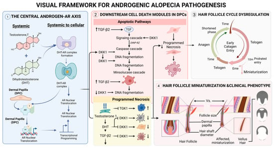
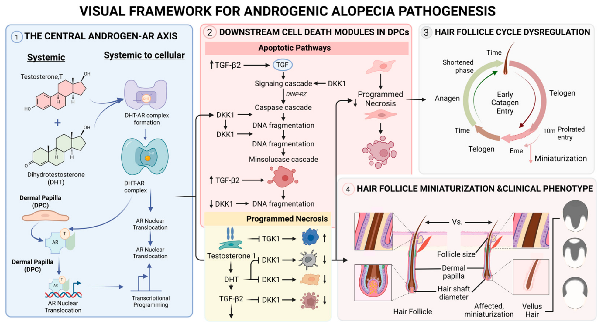
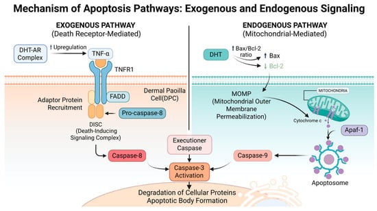
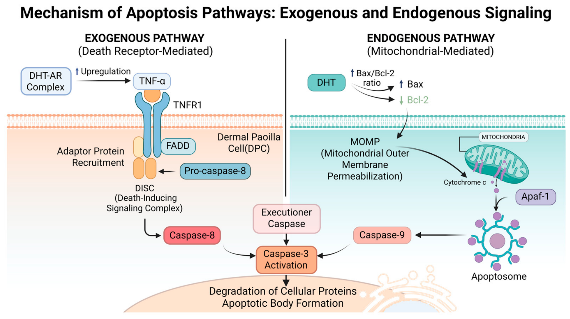
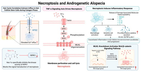
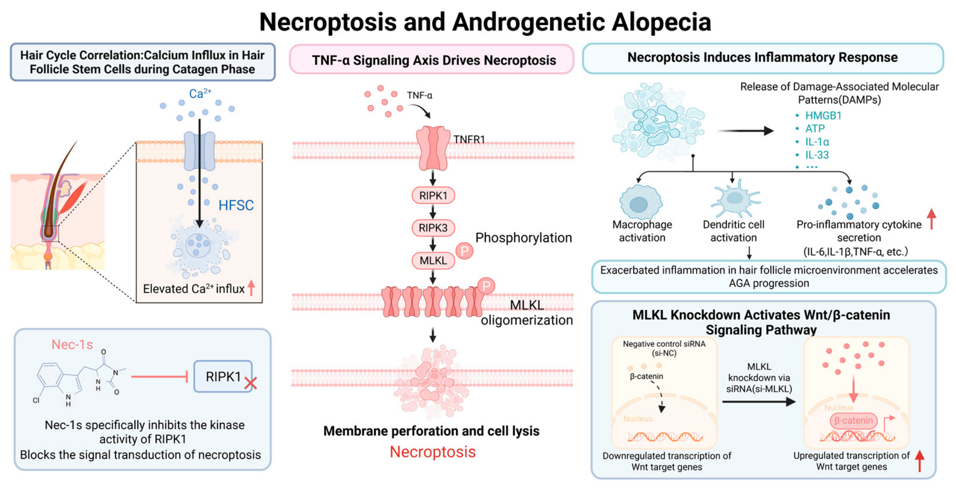
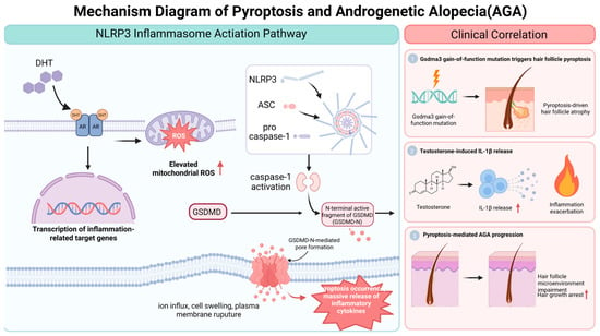
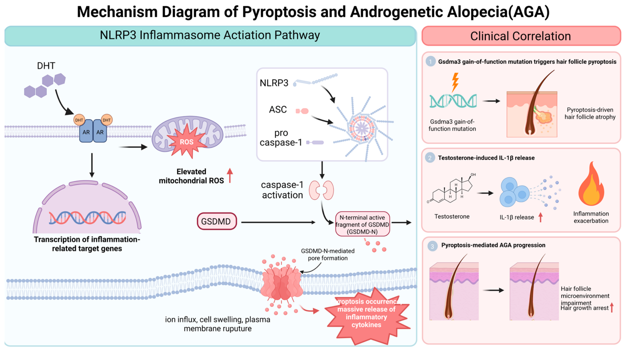
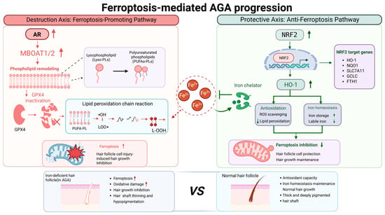
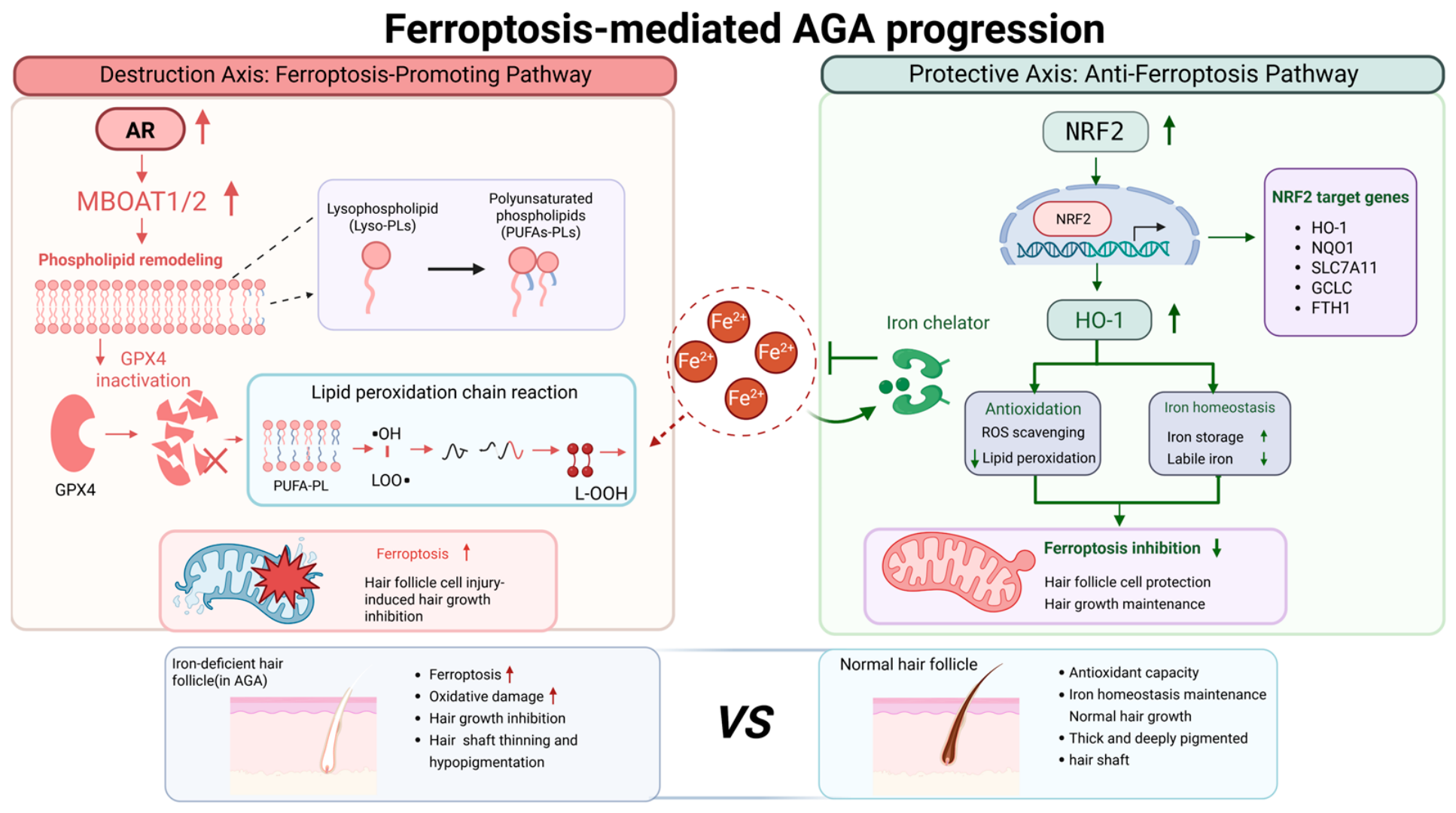
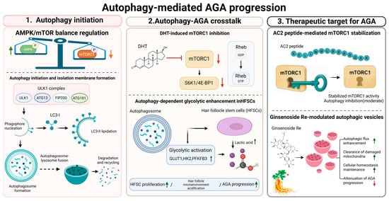
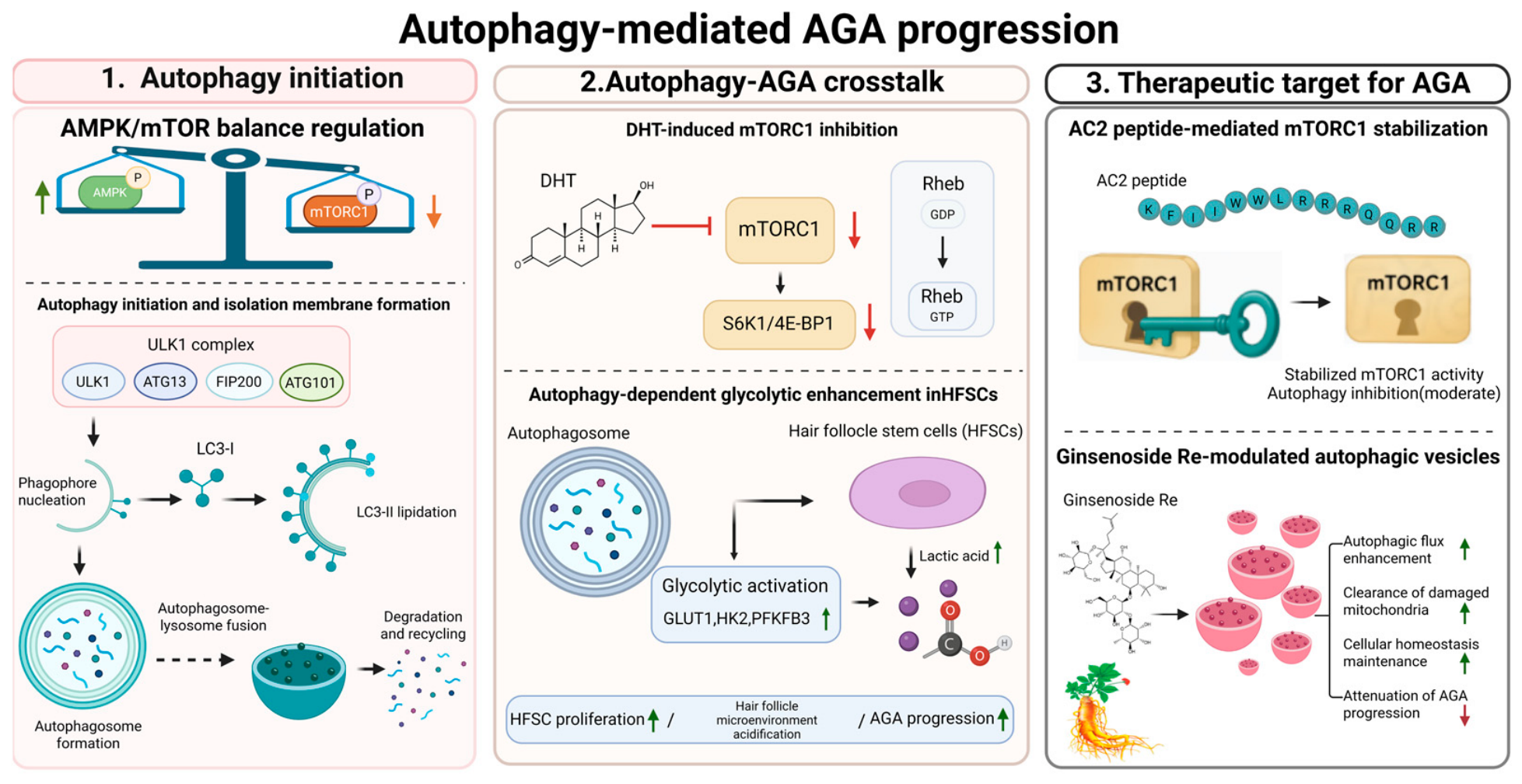

Disclaimer/Publisher’s Note: The statements, opinions and data contained in all publications are solely those of the individual author(s) and contributor(s) and not of MDPI and/or the editor(s). MDPI and/or the editor(s) disclaim responsibility for any injury to people or property resulting from any ideas, methods, instructions or products referred to in the content. |
© 2026 by the authors. Licensee MDPI, Basel, Switzerland. This article is an open access article distributed under the terms and conditions of the Creative Commons Attribution (CC BY) license.