


Abstract
Glycation contributes to skin yellowing and deterioration of the dermal collagen matrix, highlighting the potential of antiglycation strategies to address these features of skin aging. This study investigated the protective effects of rhamnose acetamide (RA) against glycation-associated changes in human dermal fibroblasts and methylglyoxal (MGO)-treated human skin explants. Longitudinal facial colour measurements in 71 Korean women showed increased skin yellowness over approximately three years, underscoring the relevance of this visible skin change. In skin explants, topical RA partially attenuated MGO-induced yellowness and preserved the dermal collagen-positive area. The mean collagen-positive area was approximately 41% in RA-treated explants, compared with 28% in MGO-exposed vehicle explants and 51% in untreated explants. In fibroblasts, RA increased glyoxalase 1 (GLO1) expression and enzymatic activity, reduced high-glucose-induced advanced glycation end product (AGE)-associated immunofluorescence, and decreased the proportion of senescence-associated β-galactosidase-positive cells. Under MGO stress, RA partially restored intracellular ATP levels and increased sirtuin 1 activity at 200 μg/mL. RA also reduced reactive oxygen species and lipofuscin-associated accumulation under repeated ultraviolet A irradiation with MGO exposure between irradiations. Together, these findings demonstrate protective effects of RA against glycation-associated cellular and tissue changes in the experimental models studied, encompassing skin yellowness, dermal collagen preservation, and cellular senescence-related responses. These results highlight RA as a promising candidate for targeting glycation-associated features of skin aging.
1. Introduction
Skin aging is a progressive loss of cutaneous homeostasis caused by interacting intrinsic and extrinsic processes. In addition to wrinkles, laxity, and impaired barrier function, aging is accompanied by changes in skin colour and colour heterogeneity, including uneven pigmentation, focal dark spots, dullness, and increased yellow or brown tones [1,2,3,4]. These chromatic changes are clinically important because they strongly influence perceived age. Although epidermal melanin is a major determinant of focal pigmentation, non-melanin chromophores, including bilirubin and yellow advanced glycation end products (AGEs), may also contribute to age-associated skin yellowness [4,5,6].
AGEs are heterogeneous, largely irreversible adducts generated through non-enzymatic reactions between carbonyl groups and amino groups in proteins, lipids, and nucleic acids [7,8]. Reactive dicarbonyls, particularly methylglyoxal (MGO), accelerate AGE formation and carbonyl stress. MGO is generated mainly as a by-product of glycolysis, and its burden can increase when metabolic flux, oxidative stress, mitochondrial function, or detoxification capacity becomes unbalanced [8,9,10]. In skin, long-lived extracellular matrix proteins such as collagen are especially susceptible to glycation. AGE accumulation and AGE-mediated cross-linking can also increase matrix stiffness, impair fibroblast function, and amplify oxidative and inflammatory signalling through the receptor for AGEs [8,11]. Thus, glycation provides a plausible biochemical link between metabolic aging, dermal deterioration, and age-associated changes in skin colour. Glycation-associated matrix damage is therefore relevant not only to yellowing but also to collagen preservation, making the two outcomes complementary targets for an antiglycation intervention [8].
The glyoxalase system is the principal cellular defence against MGO. Glyoxalase 1 (GLO1), together with reduced glutathione, catalyses the conversion of the spontaneously formed MGO-glutathione hemithioacetal to S-D-lactoylglutathione, which is subsequently converted to D-lactate by glyoxalase 2 [12]. Reduced GLO1 expression or activity increases dicarbonyl stress and MGO-derived protein modification, whereas increased GLO1 expression or activity can strengthen endogenous antiglycation capacity [11]. GLO1 is therefore an attractive intervention point because it acts upstream of irreversible AGE formation rather than merely neutralizing its downstream consequences. An increase in GLO1 expression and activity may consequently provide a cellular basis for reducing glycation-associated damage and supporting dermal matrix maintenance.
Cellular energy status and stress-response regulation provide additional functional readouts of protection against carbonyl stress. ATP availability reflects cellular energy status, whereas sirtuin 1 (SIRT1), an NAD+-dependent deacetylase, participates in metabolic and stress-response regulation [9,13]. Examining ATP and SIRT1 activity alongside measurements of GLO1 expression, GLO1 activity, and AGE-associated immunofluorescence can therefore help assess whether an antiglycation candidate also preserves fibroblast functions that may support dermal homeostasis.
Carbonyl and photo-oxidative stress may also converge on lipofuscin accumulation. Lipofuscin is an autofluorescent, yellow-brown aggregate of oxidized and cross-linked proteins and lipids that accumulates in lysosomes when damaged macromolecules are generated faster than they can be degraded [14,15]. Chronic UVA exposure promotes ROS-dependent photo-oxidative stress and impairs lysosomal proteolysis in dermal fibroblasts, thereby facilitating the accumulation of lipofuscin-like autofluorescent material [16,17]. Because lipofuscin increases with cellular aging and can further disrupt proteostasis and redox balance, simultaneous control of dicarbonyl stress and lipofuscin formation may provide a mechanistically distinct approach to age-related skin colour changes.
Glycation can affect both skin colour and the structural integrity of the dermal matrix. Long-lived collagen is particularly susceptible to glycation, which can alter fibrillar organisation and matrix remodelling. Collagen glycation has been associated with altered dermal structure and increased matrix metalloproteinase production in reconstructed human skin, while MGO-induced glycation has been linked to reduced collagen density and disrupted collagen organisation in three-dimensional skin models [18,19]. These findings suggest that limiting glycation may help preserve dermal collagen while also reducing the accumulation of glycation-associated chromophores. Antiglycation interventions therefore warrant investigation for their potential to address both skin yellowness and dermal collagen deterioration.
Rhamnose acetamide (RA) is a newly synthesized rhamnose derivative. To our knowledge, the present study is the first to report the biological efficacy of RA. Whereas previous studies have described the relevance of glycation to skin aging and dermal collagen alteration and have evaluated antiglycation effects of other compounds in skin models, the effects of RA under glycation- and carbonyl-stress conditions have not previously been reported. Following an initial screen of three rhamnose derivatives for effects on GLO1 activity, RA was selected for further investigation in human dermal fibroblasts and MGO-treated skin explants. Cellular assessments examined GLO1 expression and activity, AGE-associated immunofluorescence, cellular senescence, ATP and SIRT1 activity, and ROS and lipofuscin-associated accumulation. In addition, the skin explant experiments explored whether topical RA was associated with attenuation of yellowness and maintenance of collagen-positive area under MGO stress. Thus, this study provides an initial exploratory evaluation of the antiglycation and skin-related effects of RA across complementary cellular and tissue-level models.
2. Materials and Methods
2.1. Subject Recruitment and Skin Chromaticity Measurements
A total of 71 healthy Korean women were enrolled in a longitudinal study conducted over an approximately three-year interval. Participants visited the LG H&H Research Center on two occasions between 2020 and 2024. At each visit, participants washed their faces with water and acclimatized for 15 min under controlled environmental conditions (20–25 °C and 40–60% relative humidity). Facial skin colour was assessed using a 24.2-megapixel imaging system (Janus III; PIE Inc., Suwon, Republic of Korea). Skin brightness (LAB_L), yellowness (LAB_B), and redness (LAB_A) were obtained as L*, b*, and a* values, respectively, using the manufacturer’s algorithm. Changes between the two visits were evaluated using the Wilcoxon signed-rank test. All participants provided written informed consent. The study protocols were approved by the Institutional Review Board of the LG H&H Research Center (LGHH-20200731-AB-01, LGHH-20201015-AB-01, LGHH-20231102-AB-03-01, LGHH-20240201-AB-03-01, and LGHH-20240321-AB-05-01) and were conducted in accordance with applicable institutional and ethical guidelines.
2.2. Materials
Rhamnose acetic acid (RAA), disodium malonate rhamnoside (DMR), and rhamnose acetamide (RA) were chemically synthesized. Their chemical structures and relevant information are provided in the Supplementary Materials (Figure S1). Additionally, the synthesis procedure, batch information, and analytical characterization of RA are described in the Supplementary Materials (Figure S2).
2.3. Cell Culture
Hs68 human dermal fibroblasts (CRL-1635) and primary normal human dermal fibroblasts (NHDFs; PCS-201-012) were obtained from the American Type Culture Collection (ATCC, Manassas, VA, USA). Both cell types were maintained in Dulbecco’s modified Eagle’s medium (DMEM) supplemented with 10% fetal bovine serum and penicillin–streptomycin at 37 °C in a humidified atmosphere containing 5% CO2.
All cell-based assays were conducted once using Hs68 cells or primary NHDFs at passages not exceeding 20. Unless otherwise indicated, each treatment condition was assessed in three wells. Wells were considered technical replicates and not independent biological replicates. For image-based assays, 3–5 microscopic fields were acquired per well; these fields were treated as subsamples rather than independent observations.
2.4. Immunofluorescence Analysis
Hs68 cells were seeded in 24-well plates at 2 × 10⁵ cells/mL in low-glucose DMEM (5.6 mM glucose). On the following day, the medium was replaced with fresh low-glucose medium, high-glucose medium (75 mM glucose), or high-glucose medium containing RA at the indicated concentrations. After 6 days, cells were fixed with 4% paraformaldehyde for 10 min, permeabilized with 0.5% Triton X-100 (Sigma-Aldrich, St. Louis, USA) for 5 min, and blocked with 2% bovine serum albumin in phosphate-buffered saline (PBS) for 2 h at room temperature. Cells were incubated overnight at 4 °C with an anti-AGE antibody (TransGenic Inc., Tokyo, Japan, cat. KH011) diluted 1:200, followed by an Alexa Fluor 594-conjugated secondary antibody for 1 h at room temperature. Nuclei were counterstained using VECTASHIELD Antifade Mounting Medium with DAPI. Images were acquired using an EVOS FL Auto 2 imaging system (Thermo Fisher Scientific, Waltham, MA, USA), and fluorescence intensity was quantified using ImageJ version 1.53e.
2.5. Senescence-Associated β-Galactosidase (SA-β-Gal) Staining
Senescence under high-glucose conditions was evaluated using an X-gal-based Senescence β-Galactosidase Staining Kit (Cell Signaling Technology, Danvers, MA, USA) according to the manufacturer’s instructions. Cells were fixed with 1× fixative solution for 10 min and washed with PBS. Staining solution containing 1 mg/mL X-gal was added, and the sealed plates were incubated for 16 h at 37 °C in a CO2-free incubator. After washing, blue SA-β-gal-positive cells were imaged using the EVOS FL Auto 2 system and quantified as a percentage of the total cell number.
2.6. GLO1 Activity Assay
GLO1 activity was measured using the QuantiChrom GLO1 Assay Kit (BioAssay Systems, Hayward, CA, USA) according to the manufacturer’s instructions. Cell lysates were prepared using M-PER Mammalian Protein Extraction Reagent (Thermo Fisher Scientific), and protein concentrations were determined using a bicinchoninic acid assay (Biomax, Guri, Republic of Korea). Lysate samples (40 μL) were mixed with 160 μL of working reagent and incubated for 2 h at room temperature. Perchloric acid (70 μL, 4 M) was then added, and samples were vortexed, chilled on ice for 15 min, and centrifuged at 13,000 rpm for 10 min. The supernatant (200 μL) was transferred to a UV-transparent 96-well plate, and absorbance at 240 nm was measured using a BioTek Synergy H1 microplate reader (Agilent, Santa Clara, CA, USA).
2.7. Isolation of Total RNA and Quantitative Real-Time PCR (qRT-PCR)
Hs68 cells were seeded at 1 × 10⁵ cells per well in 6-well plates and cultured for 24 h. The medium was then replaced with DMEM containing 0.5% fetal bovine serum, and cells were treated with RA at 10, 20, 50, 100, or 200 μg/mL for 24 h. Total RNA was isolated using the AccuPrep Universal RNA Extraction Kit (Bioneer, Daejeon, Republic of Korea), and RNA concentration and purity were measured using a NanoDrop spectrophotometer (Thermo Fisher Scientific). Complementary DNA was synthesized from 1 μg of total RNA using AccuPower RocketScript Cycle RT PreMix (Bioneer). Quantitative real-time PCR was performed using a StepOnePlus Real-Time PCR System and TaqMan Universal PCR Master Mix (Applied Biosystems, Foster City, CA, USA) with TaqMan assays for GLO1 (Hs00198702_m1) and ACTB (Hs01060665_g1). Relative GLO1 expression was normalized to ACTB and calculated using the 2−ΔΔCt method.
2.8. Measurement of Intracellular ATP
Hs68 cells were exposed to 400 μM MGO for 24 h in the presence or absence of RA at the indicated concentrations; resveratrol (10 μg/mL) was included as a comparator. Intracellular ATP was measured using the PicoSens ATP Assay Kit (Biomax, Guri, Republic of Korea) according to the manufacturer’s instructions. Cells were harvested and lysed in 100 μL of assay buffer, and lysates were centrifuged at 13,000 rpm for 10 min. Supernatant (50 μL) was transferred to a 96-well plate and mixed with 50 μL of reaction mixture. After incubation for 2 h at room temperature, fluorescence was measured at excitation/emission wavelengths of 535/590 nm.
2.9. SIRT1 Activity
SIRT1 activity was measured in lysates from MGO-exposed cells using a SIRT1 Fluorogenic Assay Kit (BPS Bioscience, San Diego, CA, USA) with minor modifications. A master mixture containing the fluorogenic substrate, NAD+, bovine serum albumin, and assay buffer was prepared according to the manufacturer’s instructions. Cells were lysed using M-PER, and equal amounts of protein were combined with the reaction mixture and incubated at 37 °C for 30 min. Developer solution was then added, followed by incubation for 15 min at room temperature. Fluorescence was measured at excitation/emission wavelengths of 360/450 nm and normalized to protein content.
2.10. Cellular ROS Assay
Intracellular ROS was assessed in primary NHDFs using a DCFDA Cellular ROS Assay Kit (Abcam, Cambridge, UK). Cells received UVA irradiation at 3 J/cm2 once every 24 h for a total of three irradiations. MGO (40 μM) and RA (50, 100, or 200 μg/mL) were applied only during the intervals between UVA exposures. Before each UVA exposure, the MGO- and RA-containing medium was removed and the cells were washed; thus, MGO and RA were not present during irradiation. After completion of the exposure schedule, cells were washed and incubated with DCFDA solution for 45 min at 37 °C in the dark. The probe solution was then replaced with phenol-red-free DMEM containing 10% fetal bovine serum. Fluorescence images were acquired using the EVOS FL Auto 2 system under identical acquisition settings and quantified using ImageJ.
2.11. Lipofuscin Accumulation
Primary NHDFs were seeded in 96-well plates at 5 × 10³ cells per well. Cells were irradiated with UVA at 3 J/cm2 once every 24 h for a total of three exposures. MGO (40 μM) and RA (50, 100, or 200 μg/mL) were applied during the non-irradiation intervals only. Before each UVA exposure, the treatment medium was removed and the cells were washed; neither MGO nor RA was present during UVA irradiation. Following irradiation, the cells were returned to medium containing the designated MGO and RA treatments for the subsequent inter-irradiation interval. Intrinsic cellular autofluorescence was imaged 24 h after the third UVA exposure using the EVOS FL Auto 2 system under identical imaging settings and quantified using ImageJ software (NIH, USA). The mean gray value of the cellular regions was measured, and background fluorescence was corrected by subtracting the mean gray value of cell-free regions in each image. The background-corrected fluorescence intensities were then normalized to the mean value of the untreated control group, which was set to 100%, and expressed as relative fluorescence intensity (% of control). In addition, Sudan Black B staining was performed as an orthogonal histochemical assessment of lipofuscin-associated material. Cells were washed with PBS, fixed in 4% paraformaldehyde, and incubated with 0.1% Sudan Black B solution for 10 min. After washing with 50% ethanol and then three times with PBS, bright-field images were acquired using the EVOS FL Auto 2 system and analysed using ImageJ.
2.12. Human Skin
An abdominal skin sample (phototype III) from a healthy 65-year-old Caucasian female donor was obtained through Biopredic International (Rennes, France). Tissues were collected from surgical residues after plastic surgery in accordance with French Law L.1245-2 CSP. After removal of adipose tissue, full-thickness skin was punched into 10-mm discs and maintained at 37 °C and 5% CO2 with the dermal side immersed in long-term skin culture medium. Twelve explants from a single donor were randomly assigned to three experimental groups (n = 4 explants per group). To establish an accelerated ex vivo glycation-stress model, explants were exposed to 500 mM MGO in the culture medium and topically treated with vehicle serum or serum containing 5% RA at 6.37 μL/cm2. This concentration was selected based on a previously reported 3D human skin model in which 500 mM MGO induced measurable AGE accumulation and glycation-associated alterations in collagen organization and skin structure [19]. After 48 h, skin-colour parameters were measured using a CM-700d chromameter (Konica Minolta, Tokyo, Japan), and tissues were fixed in 4% paraformaldehyde. Fixed tissues were embedded in OCT compound (Tissue-Tek; Sakura Finetek, Tokyo, Japan), sectioned, and stained with Masson’s trichrome. Histological images were acquired using the EVOS FL Auto 2 system. For histological analysis, four sections were analysed per explant, and 3–6 microscopic fields were acquired per section. For each image, the entire dermal region visible in the field was selected as the region of interest. Images were converted to 8-bit grayscale in ImageJ, and a fixed threshold value of 206 was applied. Pixels meeting the predefined threshold criterion within the dermal region of interest were identified as collagen-positive staining, and the collagen-positive area was quantified. Collagen-positive area was calculated as the blue-stained area divided by the total dermal region-of-interest area × 100. Fields and sections were treated as subsamples and averaged to obtain one collagen-positive area value per explant. Collagen-image quantification was performed without blinding.
2.13. Statistical Analysis
Data are presented as mean ± SD. Differences among three or more groups were analysed using one-way analysis of variance followed by Tukey’s multiple-comparison test. Comparisons between two independent groups were performed using an unpaired Student’s t-test, whereas paired longitudinal skin-colour measurements were analysed using the Wilcoxon signed-rank test. A p value < 0.05 was considered statistically significant.
The ex vivo data were presented descriptively.
2.14. Use of Generative Artificial Intelligence
ChatGPT-6 (OpenAI) was used to assist with drafting and revising the manuscript text. It was not used to generate the experimental data reported in this study.
3. Results
3.1. Longitudinal Changes in Facial Skin Chromaticity over Three Years
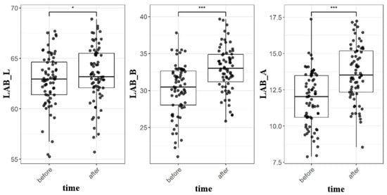
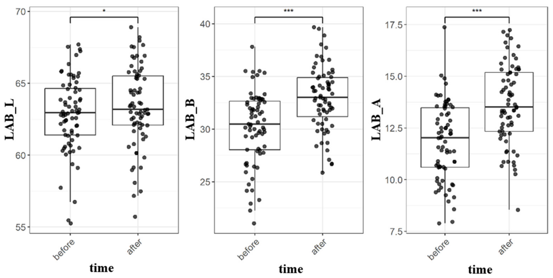
To characterize age-associated changes in facial skin colour within the same individuals, L*, a*, and b* values were compared at baseline and approximately three years later in 71 Korean women. Mean L* increased modestly from 62.9 ± 2.6 to 63.4 ± 2.8 (p = 0.022; Figure 1a). In contrast, b* showed a larger increase, from 30.2 ± 1.9 to 33.0 ± 3.0 (p = 1.4 × 10⁻¹²; Figure 1b), indicating a pronounced shift toward greater facial yellowness. Mean a* also increased from 11.9 ± 1.9 to 13.7 ± 1.9 (p = 7.4 × 10⁻¹²; Figure 1c). Thus, over the observation period, increases in yellowness and redness occurred despite a small increase in brightness. These findings indicate that longitudinal aging-related colour change cannot be represented by L* alone and provide descriptive context for investigating biological processes that may be associated with increasing skin yellowness. Because yellow AGEs have been identified in human skin [6], glycation-related mechanisms were examined in the subsequent experimental models.
Figure 1.
Comparison of skin colour parameters before and after 3 years. Boxplots show the distribution of (a) skin brightness (LAB_L), (b) yellowness (LAB_B), and (c) redness (LAB_A) measured in 71 Korean women at two time points: three years prior (“before”) and three years later (“after”). Statistical significance between paired measurements was assessed using the Wilcoxon signed-rank test. * p < 0.05 and *** p < 0.001.
3.2. Rhamnose Acetamide Increases GLO1 Expression and Enzymatic Activity in Human Dermal Fibroblasts
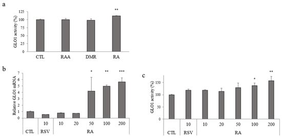
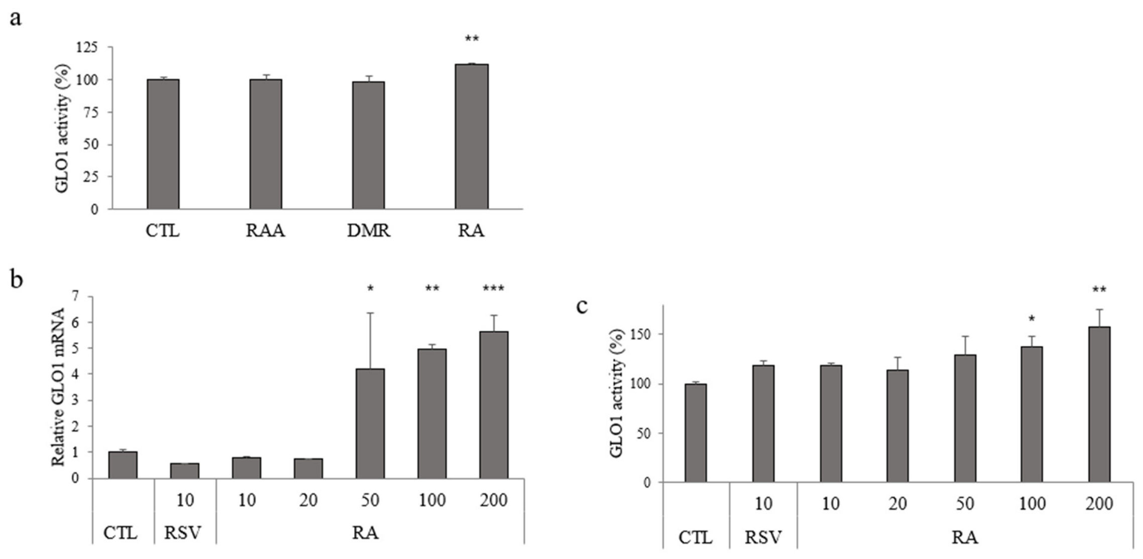
Three rhamnose derivatives were screened at 200 μg/mL for their effects on GLO1 activity in Hs68 human dermal fibroblasts. RA increased GLO1 activity relative to the untreated control, whereas RAA and DMR did not produce a detectable increase under the same conditions (Figure 2a). RA was therefore selected for concentration-response analysis.
Figure 2.
Screening of rhamnose derivatives for their effects on GLO1 expression and activity in Hs68 cells. (a) GLO1 enzymatic activity expressed as a percentage of the untreated control (CTL). Hs68 cells were treated with 200 μg/mL of RAA, DMR, or RA for 48 h. (b) Concentration-dependent effect of RA on GLO1 mRNA expression after 24 h of treatment, as determined by quantitative real-time PCR. (c) Concentration-dependent effect of RA on GLO1 enzymatic activity after 48 h treatment. Data are presented as mean ± SD (n = 3 technical replicate wells). * p < 0.05, ** p < 0.01, and *** p < 0.001 vs. CTL (one-way ANOVA followed by Tukey’s post hoc test).
RA increased GLO1 mRNA expression at 50–200 μg/mL, with the largest response observed at 200 μg/mL (Figure 2b). GLO1 enzymatic activity also increased with RA concentration and was significantly elevated at 100 and 200 μg/mL (Figure 2c). Resveratrol, included as a comparator, produced only a modest change in GLO1 activity and did not increase GLO1 mRNA under the tested conditions. Based on the initial GLO1 activity screen at 200 μg/mL, RA was selected for subsequent concentration-response and phenotypic studies. In RA-treated fibroblasts, GLO1 mRNA expression and enzymatic activity increased under the tested conditions.
3.3. Rhamnose Acetamide Attenuates High-Glucose-Induced Increases in AGE-Associated Immunofluorescence and Cellular Senescence
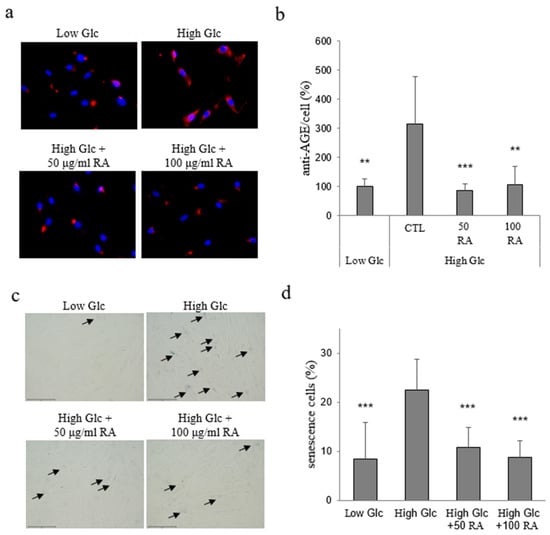
Hs68 fibroblasts were maintained for 6 days under low- or high-glucose conditions to model sustained glycation stress. High glucose markedly increased intracellular AGE-associated immunofluorescence compared with low glucose (Figure 3a,b). Co-treatment with RA at 50 or 100 μg/mL reduced AGE-associated immunofluorescence to near the low-glucose level. The signal was normalized to the number of DAPI-positive nuclei, indicating that the reduction cannot be attributable solely to differences in cell number.
Figure 3.
RA attenuates high-glucose-induced increases in AGE-associated immunofluorescence and cellular senescence. Hs68 cells were cultured for 6 days in low-glucose medium (Low Glc), high-glucose medium (High Glc), or High Glc supplemented with RA (50 or 100 μg/mL). (a) Representative fluorescence images of Hs68 cells stained with an anti-AGE antibody (red) and counterstained with DAPI (blue). (b) AGE-associated immunofluorescence intensity per cell. Total AGE-associated immunofluorescence (red) was normalized to the number of DAPI-positive nuclei and expressed relative to Low Glc. (c) Representative images of SA-β-gal staining; arrows indicate SA-β-gal-positive cells (blue). Scale bar = 75 μm. (d) Percentage of SA-β-gal-positive cells. Images were analysed using ImageJ. Data are presented as mean ± SD (n = 3 technical replicate wells). Statistical significance was determined by one-way ANOVA (** p < 0.01 and *** p < 0.001 vs. High Glc).
High-glucose culture also increased the proportion of SA-β-gal-positive cells from approximately 8% under low-glucose conditions to approximately 22% under high-glucose conditions (Figure 3c,d). RA at 50 and 100 μg/mL reduced the proportion of SA-β-gal-positive cells to approximately 10% and 8%, respectively. Thus, RA reduced AGE-associated immunofluorescence and attenuated the associated senescence phenotype during prolonged high-glucose exposure.
3.4. Rhamnose Acetamide Partially Restores ATP and SIRT1 Activity Under MGO Stress
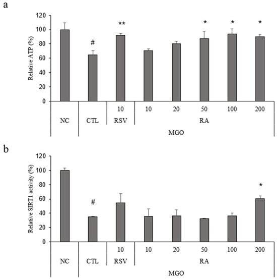
To determine whether RA protected cellular function during carbonyl stress, Hs68 fibroblasts were exposed to 400 μM MGO. MGO reduced intracellular ATP to approximately 65% of the untreated control (Figure 4a). RA significantly increased ATP levels at 50, 100, and 200 μg/mL, reaching approximately 87–94% of the untreated-control level. Resveratrol (10 μg/mL) produced a comparable recovery.
Figure 4.
RA partially restores ATP and SIRT1 activity under methylglyoxal (MGO) stress. Hs68 cells were exposed to 400 μM MGO for 24 h in the presence or absence of RA; resveratrol (10 μg/mL) was used as a comparator. (a) Relative intracellular ATP levels. MGO reduced ATP compared with the untreated control (NC), whereas RA restored ATP in a concentration-related manner. (b) SIRT1 activity normalized to protein content. MGO decreased SIRT1 activity, whereas a significant increase was observed with RA at 200 μg/mL. Data are presented as mean ± SD (n = 3 technical replicate wells). Statistical significance was determined by one-way ANOVA followed by Tukey’s post hoc test (* p < 0.05 and ** p < 0.01 vs. CTL; # p < 0.001 vs. NC). NC, untreated control; CTL, MGO-treated control; RSV, resveratrol (10 μg/mL).
MGO also reduced SIRT1 activity to approximately 35% of the untreated control (Figure 4b). RA at 200 μg/mL significantly increased SIRT1 activity to approximately 60%, whereas lower concentrations did not produce statistically supported recovery. Resveratrol showed a non-significant intermediate response. These findings indicate that RA partially preserved cellular ATP and SIRT1-associated activity during acute MGO exposure, although significant restoration of SIRT1 activity was observed only at the highest tested concentration.
3.5. Rhamnose Acetamide Reduces ROS and Lipofuscin Accumulation Induced by Repeated UVA and Inter-Irradiation MGO Exposure
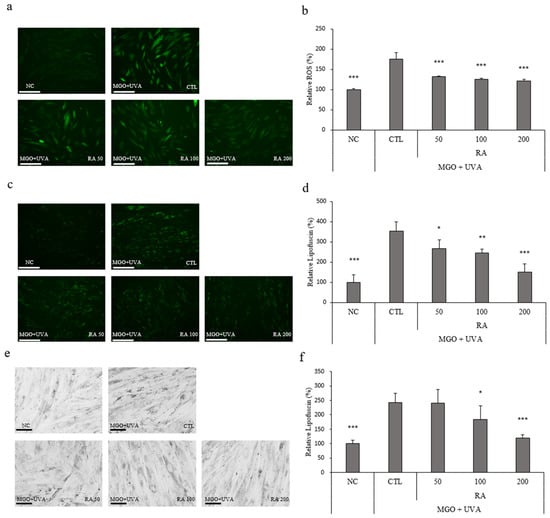
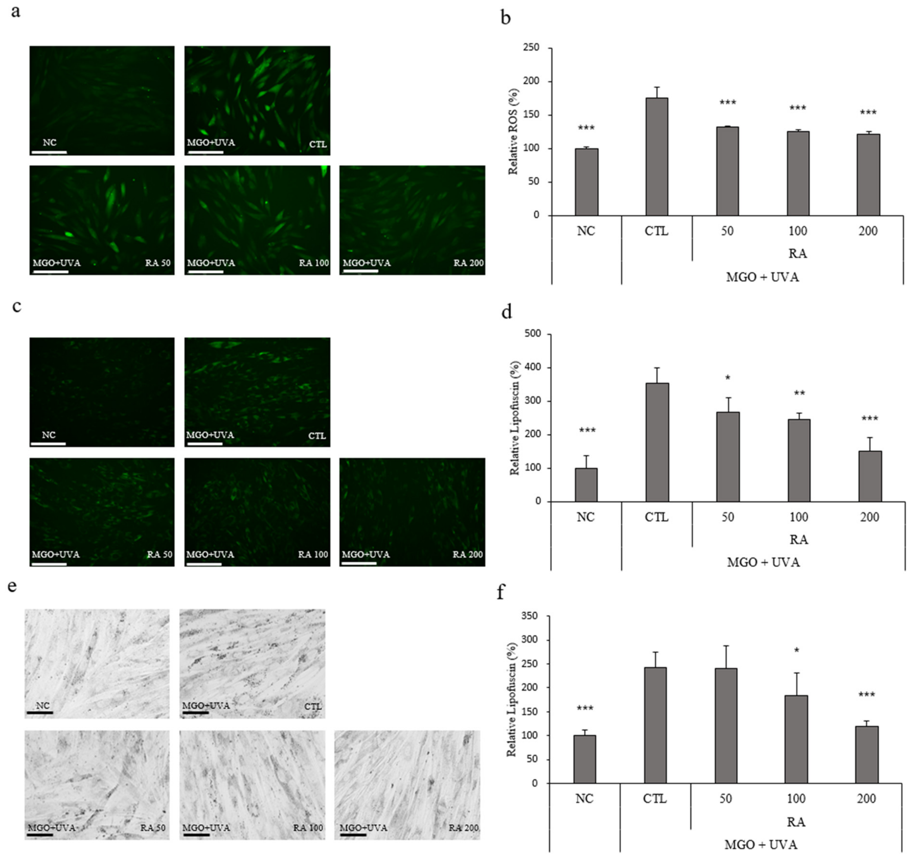
Primary NHDFs were irradiated with UVA at 3 J/cm2 once every 24 h for a total of three exposures, while 40 μM MGO was applied only during the intervals between irradiations and was absent during UVA exposure. This sequential regimen increased intracellular ROS-associated fluorescence to 176% of the untreated control (Figure 5a,b). RA significantly reduced the ROS signal to 132%, 125%, and 121% at 50, 100, and 200 μg/mL, respectively.
Figure 5.
Rhamnose acetamide reduces ROS and lipofuscin-associated accumulation induced by repeated UVA irradiation and inter-irradiation MGO exposure in primary human dermal fibroblasts. Cells received UVA irradiation at 3 J/cm2 once every 24 h for a total of three exposures. MGO (40 μM) and RA (50, 100, or 200 μg/mL) were present only during the intervals between UVA exposures and were absent during irradiation. (a) Representative images of intracellular ROS-associated fluorescence. (b) Quantification of relative ROS fluorescence. (c) Representative images of intrinsic green autofluorescence associated with lipofuscin-like material. (d) Quantification of relative autofluorescence. (e) Representative bright-field images of Sudan Black B-stained cells. (f) Quantification of Sudan Black B-positive material. Values are normalized to the untreated control (NC) and presented as mean ± SD (n = 3 technical replicate wells). Scale bars: 150 μm in (a,c) and 50 μm in (e). * p < 0.05, ** p < 0.01, and *** p < 0.001 vs. CTL.
The repeated-UVA/inter-irradiation-MGO regimen also increased intrinsic green autofluorescence to 353% of the untreated control (Figure 5c,d). RA reduced the autofluorescent signal in a concentration-related manner to approximately 266%, 243%, and 151% at 50, 100, and 200 μg/mL, respectively. The reduction was statistically significant at each tested concentration, with the largest effect observed at 200 μg/mL.
Sudan Black B staining provided an independent histochemical assessment of lipofuscin-associated material (Figure 5e,f). The sequential stress regimen markedly increased Sudan Black B-positive material compared with untreated cells. RA produced a concentration-related reduction in the Sudan Black B signal; a clear reduction was observed at 100 μg/mL, and treatment with 200 μg/mL significantly reduced the signal toward the untreated-control level. The concordant changes in intrinsic autofluorescence and Sudan Black B staining support the conclusion that RA limits lipofuscin-associated accumulation under combined, temporally separated carbonyl and photo-oxidative stress.
3.6. Rhamnose Acetamide Attenuates MGO-Induced Yellowness and Is Associated with Maintenance of Collagen-Positive Area in a Human Skin Explant Model
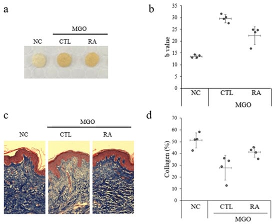
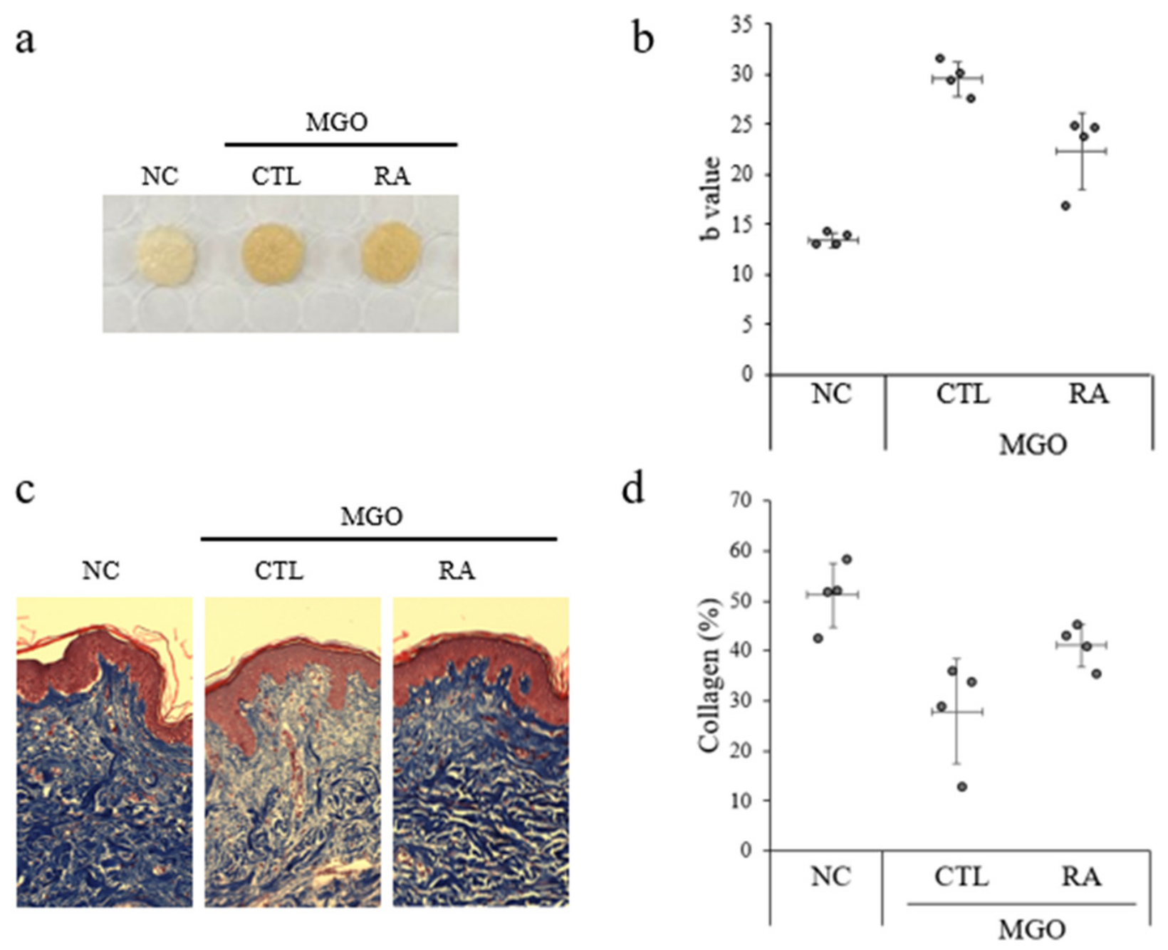
To explore the tissue-level effects of RA under glycation stress, skin explants obtained from one donor were exposed to MGO and topically treated with vehicle or serum containing 5% RA for 48 h. MGO exposure was associated with visible yellow-brown discoloration of the skin surface (Figure 6a). The mean b* value increased from approximately 13 in untreated explants to approximately 29 in MGO-exposed, vehicle-treated explants (Figure 6b). In this single-donor explant model, the mean b* value was lower in RA-treated explants, at approximately 22, than in MGO-plus-vehicle explants, although it remained above that of the untreated condition.
Figure 6.
Descriptive effects of rhamnose acetamide on skin yellowness and dermal collagen-positive area in MGO-treated explants from a single human skin donor. Human skin explants from one 65-year-old female donor were exposed to 500 mM MGO and topically treated with vehicle serum or serum containing 5% RA for 48 h. Experimental groups comprised untreated control (NC), MGO plus vehicle (CTL), and MGO plus serum containing 5% RA (RA). (a) Representative images of human skin explants. (b) Skin b* values measured using a chromameter. (c) Representative Masson’s trichrome-stained skin sections showing collagen-positive staining in the dermis. (d) Collagen-positive area in the dermis. Each point represents one explant. For collagen-positive area, values from four histological sections per explant were averaged to obtain one explant-level value. Horizontal bars indicate group means, and vertical error bars indicate mean ± SD across explants. Twelve explants from a single donor were randomly assigned to three experimental groups (n = 4 explants per group). Data are presented descriptively; no donor-level inferential statistical testing was performed.
Masson’s trichrome staining showed a lower mean dermal collagen-positive area in MGO-exposed, vehicle-treated explants than in untreated explants, decreasing from 51% to 28% (Figure 6c,d). RA-treated explants showed a higher mean collagen-positive area of 41%, approaching the value observed in untreated explants.
4. Discussion
This study demonstrates the effects of RA on attenuating glycation-associated skin yellowness and preserving dermal collagen-positive area in human skin explants. Under MGO exposure, RA partially attenuated the increase in skin yellowness and preserved dermal collagen-positive area, supporting its potential to address two distinct manifestations of glycation-associated skin aging. In separate fibroblast assays, RA treatment was associated with increased GLO1 expression and activity, reduced AGE-associated immunofluorescence and senescence-associated staining, partial recovery of ATP and SIRT1 activity, and reduced ROS and lipofuscin-associated accumulation. Together, these findings support further investigation of RA as an antiglycation candidate for mitigating skin discoloration and preserving the dermal collagen matrix.
The longitudinal analysis in 71 Korean women provides descriptive observational context for the experimental work. Previous studies have linked aging and dermal carbonyl modification to increased skin yellowness [4,20], and yellow AGEs have been identified in human skin [6]. In the present cohort, mean b* increased from 30.2 to 33.0 over approximately three years, despite a small increase in L*. However, b* is not specific to glycation, and AGEs were not measured in these participants. Therefore, the longitudinal data do not demonstrate increased tissue AGE content or establish glycation as the cause of the observed increase in facial yellowness. Nevertheless, the age-associated increase in facial yellowness observed in this cohort is consistent with previous reports linking skin aging and dermal carbonyl modification with skin yellowness [4,20]. These descriptive longitudinal observations provide a clinical context for investigating whether glycation-related processes contribute to age-associated skin colour changes. The subsequent experimental models were used to examine glycation-related cellular responses; however, direct measurement of skin AGEs in human participants will be required to determine their relationship with facial yellowness.
RA treatment was associated with increased GLO1 mRNA expression and enzymatic activity in fibroblasts. Because GLO1 detoxifies MGO upstream of AGE formation, these findings are consistent with a potential role of the glyoxalase system in the cellular response to RA [11,12]. RA treatment was also associated with reduced AGE-associated immunofluorescence under high-glucose conditions. However, the present experiments were not designed to determine whether the increase in GLO1 expression or activity caused or mediated the effects of RA on AGE-associated immunofluorescence, cellular function, skin yellowness, or collagen-positive area. GLO1 inhibition or knockdown experiments will be required to establish such a causal role. Although RA treatment markedly increased GLO1 mRNA expression, the corresponding increase in enzymatic activity was relatively modest. Similar discrepancies between mRNA expression and enzymatic activity have been reported in mammalian cells [21,22], suggesting that transcriptional induction does not necessarily translate into proportional changes in enzyme activity. This difference may reflect additional post-transcriptional and post-translational regulatory mechanisms, although the specific mechanisms involved remain to be elucidated.
High-glucose exposure simultaneously increased AGE-associated immunofluorescence and X-gal-positive SA-β-gal staining, whereas RA reduced both endpoints. This finding is consistent with the reciprocal relationship between carbonyl stress and cellular senescence: AGE–RAGE-dependent oxidative signalling can promote senescence, while senescence-associated metabolic and proteostatic dysfunction can facilitate further accumulation of damaged macromolecules [11,21]. The coordinated reduction in these endpoints therefore supports a broader protective effect of RA against glycation-associated cellular dysfunction.
The ATP and SIRT1 measurements in Figure 4 extend this mechanistic interpretation from AGE-associated immunofluorescence to cellular function. MGO reduced ATP to approximately 65% and SIRT1 activity to approximately 35% of untreated-control levels. RA restored ATP to approximately 87–94% at 50–200 μg/mL, whereas significant recovery of SIRT1 activity, to approximately 60%, was observed only at 200 μg/mL. ATP recovery is consistent with improved cellular energy status, and preservation of SIRT1 activity may support metabolic and stress-response regulation [9,13]. Together with the increases in GLO1 expression and activity and the reduction in AGE-associated immunofluorescence, these findings support a working model in which RA limits glycation-associated cellular dysfunction while maintaining functions relevant to fibroblast homeostasis. Such protection could contribute to a dermal environment that better supports collagen maintenance. The experiments do not establish a sequential GLO1–SIRT1 pathway or show that ATP or SIRT1 recovery directly mediates collagen preservation.
The repeated-UVA/inter-irradiation-MGO model combined a major environmental stressor with carbonyl stress while avoiding simultaneous exposure during irradiation. UVA can generate ROS, oxidize proteins and lipids, impair lysosomal proteases, and promote the accumulation of autofluorescent lipofuscin-like material in fibroblasts [16,17]. RA reduced both ROS-associated fluorescence and intrinsic cellular autofluorescence, and the latter was supported by a concordant reduction in Sudan Black B-positive material. These complementary readouts strengthen the interpretation that RA limits lipofuscin-associated accumulation under combined photo-oxidative and carbonyl stress, although chemical characterization would be required to define the accumulated material precisely.
The ex vivo collagen result is particularly relevant to the proposed tissue-protective activity of RA. MGO reduced collagen-positive dermal area from 51% to 28%, whereas RA-treated explants retained an area of 41%, close to the untreated value. Alongside the partial reduction in b*, this finding shows that the effects of RA extend to preservation of the dermal matrix in the same tissue model. The result is consistent with the susceptibility of long-lived collagen to glycation and with reported glycation-associated changes in collagen structure and density [18,19]. Because RA was applied during MGO exposure, the finding supports preservation of collagen-positive area under glycation stress.
Glycation may compromise dermal collagen through changes in matrix organisation and remodelling. In reconstructed human skin, collagen glycation altered dermal fibrillar structure and increased MMP1, MMP2 and MMP9 production [18]. MGO-induced glycation in a full-thickness 3D human skin model was also associated with reduced collagen density and disruption of collagen organisation in the upper dermis [19]. In the present study, RA attenuated the MGO-induced reduction in collagen-positive area, supporting preservation of dermal collagen-positive area under glycation stress. By preserving the collagen matrix that contributes to dermal mechanical support, RA may help prevent glycation-associated deterioration of skin elasticity. This potential functional benefit remains to be confirmed by direct biomechanical measurements, as skin elasticity was not assessed in the present study.
This study has several limitations. The longitudinal analysis provides descriptive observational evidence of age-associated changes in facial yellowness, whereas the experimental models were used to examine glycation- and lipofuscin-associated cellular responses. Because AGEs were not measured in the longitudinal cohort, the human observations do not establish a relationship between glycation and facial yellowness. In addition, GLO1 dependence was not directly examined. GLO1 inhibition or knockdown experiments will be required to determine whether GLO1 mediates any of the observed effects of RA. The lipofuscin-associated material was identified by intrinsic autofluorescence and Sudan Black B staining rather than chemical characterization. In the ex vivo experiment, 500 mM MGO was used as an accelerated glycation-stress model, based on a previously reported 3D human skin model [19], rather than to reproduce physiological MGO exposure. Tissue viability was not directly measured; therefore, non-specific tissue toxicity cannot be completely excluded. Together with the use of skin from a single donor, these factors limit the physiological and population-level generalizability of the explant findings. Collagen-positive area is a histological endpoint rather than a direct measure of collagen turnover or skin elasticity. Clinical studies will be required to determine whether topical RA improves age-associated skin discoloration or biomechanical function in vivo.
5. Conclusions
RA treatment partially attenuated MGO-induced skin yellowness and maintained the dermal collagen-positive area in a human skin explant model. Together with the cellular findings, these results support the potential of RA to mitigate glycation-associated changes in skin coloration and dermal collagen and provide a basis for further evaluation of RA as an antiglycation ingredient for skin care.
Supplementary Materials
The following supporting information can be downloaded at: https://www.mdpi.com/article/10.3390/cimb48111211/s1.
Author Contributions
Conceptualization, D.K., H.C., J.Y.S. and S.-H.J.; Methodology, D.K., H.C. and J.Y.S.; Investigation, D.K., H.C., J.P. and Y.O.; Formal Analysis, D.K., H.C., J.P. and Y.O.; Data Curation, D.K. and H.C.; Writing—Original Draft, D.K. and H.C.; Writing—Review and Editing, D.K., H.C., J.P., Y.O., J.Y.S., Y.K., S.-H.J. and E.T.J.; Supervision, E.T.J. and N.-G.K.; Project Administration, E.T.J. and N.-G.K. All authors have read and agreed to the published version of the manuscript.
Funding
The research presented in this article was exclusively funded by LG H&H, without any external financial support. The funder was not involved in the study design, collection, analysis, interpretation of data, the writing of this article or the decision to submit it for publication.
Institutional Review Board Statement
The longitudinal human study was conducted according to the guidelines of the Declaration of Helsinki, and was approved by the Institutional Review Board of the LG H&H Research Center (LGHH-20200731-AB-01, LGHH-20201015-AB-01, LGHH-20231102-AB-03-01, LGHH-20240201-AB-03-01, and LGHH-20240321-AB-05-01).
Informed Consent Statement
Written informed consent for participation was obtained from all participants in the longitudinal human study.
Data Availability Statement
The data that support the findings of this study are available from the corresponding author upon reasonable request.
Acknowledgments
During the preparation of this manuscript, the authors used ChatGPT-6 (OpenAI) to assist with drafting and revising the manuscript text. The authors reviewed and edited the output and take full responsibility for the content of this publication.
Conflicts of Interest
All authors are employees of LG Household & Health Care (LG H&H). LG H&H funded this study and is the manufacturer of the rhamnose acetamide (RA) used in this study. The authors were involved in the study design, data collection, analysis, interpretation of the results, and preparation of the manuscript. The authors declare that these institutional and employment relationships represent potential competing interests.
References
- Naharro-Rodriguez, J.; Bacci, S.; Hernandez-Bule, M.L.; Perez-Gonzalez, A.; Fernandez-Guarino, M. Decoding Skin Aging: A Review of Mechanisms, Markers, and Modern Therapies. Cosmetics 2025, 12, 144. [Google Scholar] [CrossRef] [Scilit]
- Hussein, R.S.; Bin Dayel, S.; Abahussein, O.; El-Sherbiny, A.A. Influences on Skin and Intrinsic Aging: Biological, Environmental, and Therapeutic Insights. J. Cosmet. Dermatol. 2025, 24, e16688. [Google Scholar] [CrossRef] [Scilit] [PubMed]
- Krutmann, J.; Schikowski, T.; Morita, A.; Berneburg, M. Environmentally-Induced (Extrinsic) Skin Aging: Exposomal Factors and Underlying Mechanisms. J. Investig. Dermatol. 2021, 141, 1096–1103. [Google Scholar] [CrossRef] [Scilit] [PubMed]
- De Rigal, J.; Des Mazis, I.; Diridollou, S.; Querleux, B.; Yang, G.; Leroy, F.; Barbosa, V.H. The effect of age on skin color and color heterogeneity in four ethnic groups. Skin Res. Technol. 2010, 16, 168–178. [Google Scholar] [CrossRef] [Scilit] [PubMed]
- Fang, B.; Card, P.D.; Chen, J.; Li, L.; Laughlin, T.; Jarrold, B.; Zhao, W.; Benham, A.M.; Maatta, A.T.; Hawkins, T.J.; et al. A Potential Role of Keratinocyte-Derived Bilirubin in Human Skin Yellowness and Its Amelioration by Sucrose Laurate/Dilaurate. Int. J. Mol. Sci. 2022, 23, 5884. [Google Scholar] [CrossRef] [Scilit] [PubMed]
- Fang, B.; Li, L.; Winget, J.; Laughlin, T.; Hakozaki, T. Identification of Yellow Advanced Glycation End Products in Human Skin. Int. J. Mol. Sci. 2024, 25, 5596. [Google Scholar] [CrossRef] [Scilit] [PubMed]
- Singh, V.P.; Bali, A.; Singh, N.; Jaggi, A.S. Advanced glycation end products and diabetic complications. Korean J. Physiol. Pharmacol. 2014, 18, 1–14. [Google Scholar] [CrossRef] [Scilit] [PubMed]
- Chen, C.-y.; Zhang, J.-Q.; Li, L.; Guo, M.-m.; He, Y.-f.; Dong, Y.-m.; Meng, H.; Yi, F. Advanced Glycation End Products in the Skin: Molecular Mechanisms, Methods of Measurement, and Inhibitory Pathways. Front. Med. 2022, 9, 837222. [Google Scholar] [CrossRef] [Scilit] [PubMed]
- Miwa, S.; Kashyap, S.; Chini, E.; von Zglinicki, T. Mitochondrial dysfunction in cell senescence and aging. J. Clin. Investig. 2022, 132, e158447. [Google Scholar] [CrossRef] [Scilit] [PubMed]
- Oliveira, A.L.; de Oliveira, M.G.; Mónica, F.Z.; Antunes, E. Methylglyoxal and Advanced Glycation End Products (AGEs): Targets for the Prevention and Treatment of Diabetes-Associated Bladder Dysfunction? Biomedicines 2024, 12, 939. [Google Scholar] [CrossRef] [Scilit] [PubMed]
- Nigro, C.; Leone, A.; Fiory, F.; Prevenzano, I.; Nicolò, A.; Mirra, P.; Beguinot, F.; Miele, C. Dicarbonyl Stress at the Crossroads of Healthy and Unhealthy Aging. Cells 2019, 8, 749. [Google Scholar] [CrossRef] [Scilit] [PubMed]
- Saeed, M.; Kausar, M.A.; Singh, R.; Siddiqui, A.J.; Akhter, A. The Role of Glyoxalase in Glycation and Carbonyl Stress Induced Metabolic Disorders. Curr. Protein Pept. Sci. 2020, 21, 846–859. [Google Scholar] [CrossRef] [Scilit] [PubMed]
- Canto, C.; Auwerx, J. Targeting sirtuin 1 to improve metabolism: All you need is NAD(+)? Pharmacol. Rev. 2012, 64, 166–187. [Google Scholar] [CrossRef] [Scilit] [PubMed]
- Terman, A.; Brunk, U.T. Lipofuscin: Mechanisms of formation and increase with age. APMIS 1998, 106, 265–276. [Google Scholar] [CrossRef] [Scilit] [PubMed]
- Hohn, A.; Konig, J.; Grune, T. Protein oxidation in aging and the removal of oxidized proteins. J. Proteom. 2013, 92, 132–159. [Google Scholar] [CrossRef] [Scilit] [PubMed]
- Lamore, S.D.; Qiao, S.; Horn, D.; Wondrak, G.T. Proteomic identification of cathepsin B and nucleophosmin as novel UVA-targets in human skin fibroblasts. Photochem. Photobiol. 2010, 86, 1307–1317. [Google Scholar] [CrossRef] [Scilit] [PubMed]
- Lamore, S.D.; Wondrak, G.T. UVA causes dual inactivation of cathepsin B and L underlying lysosomal dysfunction in human dermal fibroblasts. J. Photochem. Photobiol. B 2013, 123, 1–12. [Google Scholar] [CrossRef] [Scilit] [PubMed]
- Pageon, H.; Bakala, H.; Monnier, V.; Asselineau, D. Collagen glycation triggers the formation of aged skin in vitro. Eur. J. Dermatol. 2007, 17, 12–20. [Google Scholar] [PubMed]
- Markiewicz, E.; Jerome, J.; Mammone, T.; Idowu, O.C. Anti-Glycation and Anti-Aging Properties of Resveratrol Derivatives in the in-vitro 3D Models of Human Skin. Clin. Cosmet. Investig. Dermatol. 2022, 15, 911–927. [Google Scholar] [CrossRef] [Scilit] [PubMed]
- Ogura, Y.; Kuwahara, T.; Akiyama, M.; Tajima, S.; Hattori, K.; Okamoto, K.; Okawa, S.; Yamada, Y.; Tagami, H.; Takahashi, M.; et al. Dermal carbonyl modification is related to the yellowish color change of photo-aged Japanese facial skin. J. Dermatol. Sci. 2011, 64, 45–52. [Google Scholar] [CrossRef] [Scilit] [PubMed]
- Wagner, A.E.; Hug, H.; Gössl, R.; Riss, G.; Mussler, B.; Elste, V.; Rimbach, G.; Barella, L. The natural compound ascorbigen modulates NADPH-quinone oxidoreductase (NQO1) mRNA and enzyme activity levels in cultured liver cells and in laboratory rats. Ann. Nutr. Metab. 2008, 53, 122–128. [Google Scholar] [CrossRef] [Scilit] [PubMed]
- Geiger, J.; Doelker, R.; Salö, S.; Roitsch, T.; Dalgaard, L.T. Physiological phenotyping of mammalian cell lines by enzymatic activity fingerprinting of key carbohydrate metabolic enzymes: A pilot and feasibility study. BMC Res. Notes 2019, 12, 682. [Google Scholar] [CrossRef] [Scilit] [PubMed]
Disclaimer/Publisher’s Note: The statements, opinions and data contained in all publications are solely those of the individual author(s) and contributor(s) and not of MDPI and/or the editor(s). MDPI and/or the editor(s) disclaim responsibility for any injury to people or property resulting from any ideas, methods, instructions or products referred to in the content. |
© 2026 by the authors. Licensee MDPI, Basel, Switzerland. This article is an open access article distributed under the terms and conditions of the Creative Commons Attribution (CC BY) license.