


Abstract
Marine microorganisms, particularly members of the marine Roseobacter-group, participate in chemically mediated associations with phytoplankton, macroalgae, and marine invertebrates. This review examines structurally characterized, host-associated metabolites produced by Roseobacter-group bacteria within this ecological framework. These metabolites include sulfur-containing compounds, nitrogen-containing and hybrid scaffolds, pigments, lipids, and signaling molecules, exemplified by the antibacterial tropodithietic acid and the algaecidal roseobacticides. We discuss biosynthetic connections between phenylacetate and sulfur metabolism that give rise to tropodithietic acid and roseobacticides, as well as tryptophan–sinapic-acid-derived hybrid pathways. By integrating genomic potential, host-derived elicitors, metabolomics, structure elucidation, and experimentally demonstrated activities, this review distinguishes chemically characterized products from bioinformatically predicted pathways and proposed applications.
1. Introduction
Marine microorganisms produce chemically diverse metabolites with ecological and biological activities. Host-associated bacteria occupy nutrient-rich surfaces where competition and chemical exchange influence metabolite production [1,2,3]. In the Roseobacter-group, characterized compounds include antibiotics, signaling molecules, siderophores, pigments, and membrane lipids that participate in interactions ranging from mutualism to antagonism [1,2,3].
Among marine bacterial lineages, the Roseobacter-group is one of the most metabolically versatile and ecologically important groups within the Alphaproteobacteria. Members of the Roseobacter-group are widely distributed from coastal waters to the open ocean. They can exist as free-living planktonic cells, attached to floating particles, or as part of microbial communities associated with phytoplankton, macroalgae, corals, sponges, and other marine animals [1,2,3]. Their ecological relationships are linked to their ability to use various substrates released by marine primary producers, including amino acids, aromatic compounds, polyamines, and organosulfur metabolites like dimethylsulfoniopropionate (DMSP) [4,5]. Furthermore, they respond flexibly to nutrient-rich microenvironments such as the phycosphere [4,5]. These close interactions offer opportunities for mutual chemical exchange. Host-derived metabolites can serve as nutrients, biosynthetic precursors, or environmental signals. In return, bacterial metabolites can influence host growth, microbial competition, surface colonization, and the overall health of the association. For example, Phaeobacter inhibens can convert algal-derived tryptophan into indole-3-acetic acid (IAA), a plant hormone that supports interactions with the coccolithophore Emiliania huxleyi [5]. In contrast, algal phenylpropanoids released under different conditions can trigger the production of algicidal metabolites, such as roseobacticides and other hidden natural products [6,7,8]. Similarly, quorum-sensing molecules, tropodithietic acid (TDA), siderophores, and membrane-associated lipids illustrate how specialized metabolism can be integrated with cell-density sensing, iron availability, oxidative stress, and surface-associated growth [9,10,11,12].
Although earlier reviews established the ecological and genomic importance of the Roseobacter-group [1,2,3,13], they predate recent advances in host-cue-based elicitation, genetic linkage of pathways to products, and structure elucidation from trace quantities. This review therefore focuses on structurally characterized metabolites recovered from host-associated settings or induced by host-derived cues. It integrates taxonomic and genomic context with host-association frameworks, biosynthetic origins, experimentally demonstrated ecological functions, and reported bioactivities. In particular, the phenylacetate/sulfur-metabolism network connecting tropodithietic acid and roseobacticide production and the tryptophan–sinapic-acid-derived hybrid pathways illustrate how host chemistry can shape specialized metabolism. Nevertheless, many predicted products remain chemically uncharacterized, and sequence-based predictions alone do not establish metabolite production under a particular culture or environmental condition [3,13].
2. Taxonomic and Genomic Overview of the Marine Roseobacter-Group
2.1. Taxonomic Position and Representative Genera
The marine Roseobacter-group belongs to the class Alphaproteobacteria within the phylum Pseudomonadota and has historically been treated as an informal lineage within the former Rhodobacteraceae assemblage. Classifications based on 16S rRNA sequences left several genera polyphyletic or paraphyletic. A genome-based analysis of 331 type strains representing 119 genera used core-genome phylogeny, genomic similarity, and phenotypic data to propose separation of the predominantly marine lineage as Roseobacteraceae [14]. A 2023 phylotaxonomic assessment of Roseobacteraceae and Paracoccaceae further refined the framework for genus-level delimitation [15]. The nomenclatural framework used here follows LPSN [16]. In GTDB release R11-RS232, Roseobacter and Paracoccus—the type genera of the LPSN families Roseobacteraceae and Paracoccaceae, respectively—are both classified within Rhodobacteraceae [17,18]. This reflects a broader genome-based family circumscription than LPSN rather than a formal nomenclatural synonymy. Here, “Roseobacter-group” is retained as an ecological and evolutionary term.
Cultured members of the Roseobacter-group include Phaeobacter, Ruegeria, Sulfitobacter, Dinoroseobacter, Roseovarius, Leisingera, Marinovum, and Planktomarina [1,3]. The type genus Roseobacter should be distinguished from this broader ecological group, which encompasses numerous phylogenetically and physiologically diverse genera [14,15]. Among these members, Ruegeria pomeroyi DSS-3 (formerly Silicibacter pomeroyi) is a key model for marine heterotrophy and transformation of phytoplankton-derived DMSP [4]. P. inhibens is widely studied for surface attachment and chemically mediated algal interactions [5]. Dinoroseobacter shibae, an aerobic anoxygenic phototroph isolated from dinoflagellate cultures, is a model for Roseobacter-group–dinoflagellate associations [19]. The genus Sulfitobacter includes phytoplankton-associated strains such as SA11, identified with the diatom Pseudo-nitzschia multiseries [20]. Cultured strains nevertheless represent only a fraction of the group’s natural diversity; several numerically important pelagic lineages remain largely uncultivated or have few representative isolates [1,3,21].
2.2. Genomic Architecture and Biosynthetic Potential
Genome architecture varies considerably across the Roseobacter-group. Among cultivated representatives, genome sizes range from approximately 2.2 Mbp to over 5.0 Mbp. Many extensively studied coastal and host-associated isolates possess relatively large, G + C-rich genomes that are often divided between a primary chromosome and several extrachromosomal replicons. However, this genomic pattern is not shared by all members of the Roseobacter-group. Several abundant pelagic lineages, including SAG-O19 and RCA-related populations, possess smaller genomes, lower G + C contents, and reduced regulatory capacity. Genome reduction is not universal even among free-living lineages, however, as the uncultivated CHAB-I-5 lineage was estimated to have a genome of approximately 4.1–4.4 Mbp. Consequently, the Roseobacter-group cannot be adequately represented by a single genomic model [3,13].
Comparative genomic analyses reveal a surprisingly small core genome relative to a vast accessory gene pool across the Roseobacter-group. Early studies demonstrated that key metabolic pathways, including carbon monoxide oxidation, sulfur oxidation, denitrification, and DMSP demethylation, are present in only a subset of strains, indicating that metabolic capabilities should not be assumed to coexist in every member. Broader comparisons of isolate genomes confirm that trophic strategy accounts for the largest proportion of variation in gene content, though genome composition is ultimately shaped by the combined interplay of evolutionary lineage and adaptation to specific ecological niches. Much of this genomic variation resides in genes encoding substrate transport, complex carbon/organosulfur utilization, and light-harvesting mechanisms [13]. For instance, RepA-I-type megaplasmids in P. inhibens and other members of the Roseobacter-group carry conserved gene clusters for exopolysaccharide biosynthesis, directly modulating the transition between swimming motility and surface-attached biofilm formation [22]. Overall, an individual strain’s metabolic responsiveness and lifestyle are defined by its unique accessory gene repertoire and plasmid composition [13].
Roseobacter-group genomes contain candidate pathways for specialized-metabolite production [13]. In a PCR-based screen of 22 strains, NRPS-like and PKS-like gene fragments were detected in seven and five strains, respectively; because this targeted survey did not reconstruct complete biosynthetic gene clusters or test pathway-to-product relationships, it cannot provide a numerical count of product-linked clusters [23]. Thus, these data establish biosynthetic potential, not direct pathway-to-product assignments. In this review, the igi locus for indigoidine biosynthesis is discussed separately as a genetically validated NRPS-like example and not as a product assignment generated by that screening dataset [24]. Genomic and genetic studies of Phaeobacter have also identified loci associated with TDA, AHL signaling, and siderophore production [9,10,11]. Sequence-based predictions alone do not establish metabolite production under a particular culture or environmental condition [13].
Pigmentation is another ecologically relevant trait in some Roseobacter-group bacteria. Several aerobic anoxygenic phototrophic members display pink-to-orange pigmentation because they contain carotenoids such as spheroidenone. D. shibae, a dinoflagellate-associated species, contains spheroidenone as its only reported carotenoid [19]. These pigments contribute to light harvesting and protection from photooxidative stress. In phytodetritus-associated conditions, spheroidenone reduced singlet-oxygen transfer and oxidative-stress responses in D. shibae, supporting bacterial persistence near senescent phytoplankton; this evidence does not, however, establish a direct benefit to the host [25].
2.3. Distribution in Global Ocean Microbiomes
Members of the Roseobacter-group occur throughout the major ocean basins, from tropical to polar waters and from coastal environments to the open ocean. Earlier surveys reported that members of the Roseobacter-group commonly account for approximately 5–20% of marine bacterioplankton and occasionally exceed 20% in coastal, temperate, and polar waters. A recent analysis of 745 metagenomic samples found that Roseobacter-group-affiliated reads ranged from <2% to 24% of the prokaryotic community in the epi- and mesopelagic zones and from <2% to 16% in the bathypelagic zone. The highest epipelagic values, ranging from 15% to 22%, occurred mainly in coastal, temperate, and polar regions. Roseobacter-group populations also respond to seasonal changes in organic-matter supply. During spring phytoplankton blooms in the southern North Sea, different Roseobacter-group taxa appeared at successive stages of the bloom rather than increasing as a single uniform group [1,21].
Culture-independent surveys demonstrate that the Roseobacter-group lineages most frequently maintained in culture do not represent those dominating natural seawater. Early surveys showed that largely uncultivated pelagic lineages, such as SAG-O19, NAC11-7, CHAB-I-5, and DC5-80-3/RCA, collectively account for roughly two-thirds of pelagic members of the Roseobacter-group, displaying clear latitudinal partitioning from tropical to polar waters. A more recent global study delineating 610 high-quality metagenome-assembled genomes (MAGs) and 43 single-cell amplified genomes (SAGs) resolved six major pelagic clusters (RCA, TCR, AAPR, AAR, COR, and LUX) that comprised approximately 70% of epipelagic Roseobacter-group assemblies. Their distributions were strongly structured by latitude: RCA, TCR, AAPR, and AAR predominated in temperate and polar regions, whereas COR and LUX were most prominent in tropical and subtropical waters, a pattern often accompanied by lineage-specific traits like genome streamlining and distinct light-harvesting mechanisms [21].
However, these open-ocean clusters decrease significantly below the epipelagic zone. They accounted for only about 21% of Roseobacter-group MAGs and SAGs in the mesopelagic layer, while bathypelagic samples were mostly limited to Sulfitobacter- and Marinovum-related lineages. Read-mapping analyses also confirmed that different Roseobacter-group genera become more dominant with increasing water depth. Overall, the distribution of the Roseobacter-group is shaped by a combination of latitude, depth, nutrient levels, and lifestyle, rather than forming a single, uniform global population. This environmental partitioning provides the ecological context for the host-associated relationships discussed in the next section [21].
3. Ecological Associations of the Marine Roseobacter-Group
3.1. Phytoplankton-Associated Members of the Roseobacter-Group
Ecological interactions between members of the Roseobacter-group and phytoplankton are among the most thoroughly studied bacterial–algal relationships in the ocean [1,3]. Much of this interaction takes place within the phycosphere, the microscale region surrounding phytoplankton cells where released metabolites create chemical gradients distinct from the surrounding seawater [5]. Members of the Roseobacter-group are exceptionally well-adapted to this niche, largely due to their ability to utilize a wide array of algal-derived compounds as nutrients or signaling cues [1,3]. DMSP is particularly central to this dynamic, serving as a primary source of carbon and reduced sulfur for many group members and thereby linking local bacterial–algal interactions to broader marine sulfur cycling [4]. The ability to respond to these chemical gradients enables members of the Roseobacter-group to maintain close contact with phytoplankton [1,3]. For example, Ruegeria sp. TM1040 (formerly Silicibacter sp. TM1040) is strongly attracted to DMSP, several of its degradation products, and specific amino acids exuded by or associated with its dinoflagellate host [4]. This chemotactic response acts as a direct mechanism allowing motile members of the Roseobacter-group to find nutrient-rich niches around algae and form close host-associated relationships [1,3].
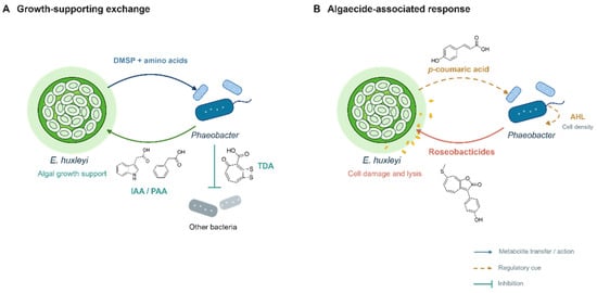
The outcome of these associations can change as the physiological state of the host changes. This has been studied extensively in interactions between Phaeobacter species and the coccolithophore E. huxleyi. In the P. inhibens–E. huxleyi model, algal cells release tryptophan, which increases bacterial attachment and stimulates production of the IAA [5]. Co-cultures initially show enhanced algal growth and later undergo algal decline; these observations should be distinguished from the effects of experimentally added IAA [5]. Other metabolites associated with the beneficial phase of Phaeobacter–algal interactions include phenylacetic acid (PAA), which has auxin-like activity, and the antibiotic TDA, which may reduce competition from neighboring bacteria [10]. Separate studies have linked algal phenylpropanoids to induction of bacterial algaecides [6,7]. Aging E. huxleyi releases p-coumaric acid (pCA), which alters secondary metabolism of P. inhibens and induces the production of algicidal roseobacticides [6,11]. Beyond triggering roseobacticide production, pCA evokes oxidative stress and alters other metabolic pathways in P. inhibens [11]. This shift from a beneficial partnership to active host killing exemplifies the “Jekyll-and-Hyde” dynamic characteristic of this interaction (Figure 1) [6,11]. A biphasic interaction is also observed between D. shibae and the dinoflagellate Prorocentrum minimum. In this system, however, the underlying mechanism differs fundamentally from the roseobacticide-mediated model described above. Functional flagella and the CtrA phosphorelay system are required for the early growth-promoting effect of D. shibae, whereas the subsequent algal-killing phenotype depends on a 191-kb extrachromosomal plasmid. The specific factor encoded by this plasmid that ultimately induces host cell death remains to be identified [19].
Figure 1.
Chemical regulation of the mutualistic-to-algicidal transition in the E. huxleyi–Phaeobacter interaction. (A) Growth-supporting exchange: E. huxleyi supplies dimethylsulfoniopropionate (DMSP) and amino acids to Phaeobacter, while bacterial indole-3-acetic acid (IAA) and phenylacetic acid (PAA) support algal growth. Tropodithietic acid (TDA) inhibits other bacteria; (B) Algaecide-associated response: algal p-coumaric acid acts as a regulatory cue for bacterial roseobacticide production, leading to algal cell damage and lysis. Acyl-homoserine lactone (AHL)-mediated cell-density signaling is also indicated. Solid arrows represent metabolite transfer or action, dashed arrows indicate regulatory cues, and the T-shaped line denotes inhibition.
In the Sulfitobacter D7–E. huxleyi system, DMSP promotes pathogenicity, whereas benzoate can counteract this response and support coexistence [26]. Exposure to stationary-phase algal exudates increased expression of nutrient transporters and motility genes, consistent with exploitation of declining algal cells and enhanced dispersal capacity [26]. The proposed “eat-and-run” strategy is an ecological interpretation of these transcriptional and phenotypic responses, rather than a direct observation of migration between hosts.
These examples demonstrate that phytoplankton-associated Roseobacter-group bacteria are not obligate mutualists or dedicated pathogens; rather, their behavioral modes shift dynamically in response to host physiology and the metabolite composition of the phycosphere [5,6]. The production and effects of IAA, TDA, and roseobacticides depend on the strain, host condition, and available chemical substrates [5,6,10].
3.2. Macroalgae-Associated Members of the Roseobacter-Group
Beyond phytoplankton, members of the Roseobacter-group are key residents of macroalgal surface communities. These surfaces provide a stable habitat for biofilm formation and intense competition for space and nutrients [27]. Indeed, Roseobacter-group strains isolated from Ulva australis display robust surface colonization and competitive dominance within the macroalgal epiphytic microbiome [28]. Bacterial regulation of host development is exceptionally well characterized in Ulva mutabilis. Axenic U. mutabilis gametes fail to develop into normal thalli, instead forming poorly differentiated, callus-like cellular clumps. Reintroducing specific bacterial partners, however, fully restores distinct aspects of normal morphogenesis. Roseovarius sp. MS2 (formerly Roseobacter sp. MS2) releases a yet-uncharacterized factor with cytokinin-like activity that promotes cell division and partial tissue differentiation. Interestingly, strain MS2 alone is insufficient for complete morphological recovery. Full morphogenesis requires a complementary morphogenetic signal from Maribacter sp. MS6 (formerly Cytophaga sp. MS6), demonstrating complementary bacterial effects in this defined laboratory association [27].
Bacterial signaling can also exert adverse effects on early algal development. Sulfitobacter sp. strain 376, isolated from Ulva linza, produces N-acyl homoserine lactones (AHLs) for bacterial quorum sensing. When AHL signaling was quenched via the expression of the AHL-degrading lactonase AiiA, zoospore germination and early germling growth were markedly improved compared to treatments with the wild-type AHL-producing strain. Experiments utilizing synthetic AHLs provided further confirmation that these signaling molecules directly impair early Ulva development. Thus, this system demonstrates how a metabolite utilized for bacterial cell-to-cell communication can concurrently affect a eukaryotic host, illustrating cross-kingdom chemical signaling [9]. Macroalgal surface ecology also exhibits notable community-level dynamics, as seen in U. australis. The taxonomic structure of epiphytic bacteria fluctuates greatly across individual algae, lacking a defined species-level core. This supports a competitive lottery model where diverse bacterial species fill equivalent surface niches. Indeed, metagenomic analysis showed that while phylogenetic similarity was only ~15%, functional gene profiles shared ~70% similarity across samples. This decoupling suggests that taxonomically distinct communities maintain equivalent ecological roles through functional redundancy. Importantly, these findings characterize the broader epiphytic microbiome of U. australis, rather than being unique to Roseobacter-group bacteria [28].
Macroalgal associations illustrate several functions of Roseobacter-group-derived chemical signals. While some bacterial signaling molecules directly influence host morphogenesis, others participate in interspecies communication or surface competition, and several remain chemically uncharacterized. These ecological interactions provide a valuable framework for understanding the natural products discussed in the following section, without assuming that every observed host phenotype can currently be attributed to a structurally characterized Roseobacter-group-derived metabolite [9,27].
3.3. Metazoan-Associated Members of the Roseobacter-Group
Members of the Roseobacter-group associate not only with micro- and macroalgae but also with a diverse array of marine invertebrates, including corals, sponges, and mollusks such as cephalopods. However, many host associations are supported primarily by descriptive microbiome surveys rather than mechanistic insights. The presence of Roseobacter-group bacteria within a host microbiome does not by itself demonstrate a beneficial function, and direct links between individual strains, their metabolites, and host outcomes remain limited.
Some of the clearest evidence for a potential protective role comes from coral-associated bacteria. A Roseobacter-group strain isolated from the surface mucus of Oculina patagonica inhibited several marine bacteria, including the coral pathogens Vibrio coralliilyticus and Thalassomonas loyana. Furthermore, when Vibrio shiloi was introduced to a pre-established biofilm of this Roseobacter-group isolate, its viable population declined by approximately five orders of magnitude within 3 h. These findings are compatible with the coral probiotic hypothesis, which proposes that dynamic shifts in the resident microbiome can enhance the resilience of the coral holobiont under changing environmental conditions. Importantly, this antimicrobial phenotype was demonstrated for a specific isolate and should not be generalized to the Roseobacter-group as a whole [29].
Roseobacter-group strains have also been isolated from marine sponges. For example, Ruegeria sp. KLH11, isolated from the sponge Mycale laxissima, harbors a complex LuxR–LuxI-type quorum-sensing network mediated by AHLs. Genetic analyses revealed that this AHL-based system enhances flagellar motility while suppressing biofilm formation, with the CckA–ChpT–CtrA phosphorelay system directly linking quorum sensing to flagellar gene regulation. These findings provide a detailed mechanistic picture of cell-density-dependent gene regulation in a sponge-associated Roseobacter-group bacterium, although the ecological consequences of this signaling network for the sponge host remain to be elucidated [9].
A well-defined animal-microbe symbiosis is found in the Hawaiian bobtail squid, Euprymna scolopes. Sexually mature females harbor dense bacterial communities inside the epithelial tubules of their accessory nidamental gland (ANG). This ANG microbiota is dominated by Alphaproteobacteria—specifically Rhodobacterales affiliated with the Roseobacter-group, including prominent Phaeobacter species—which are spatially partitioned across individual tubules. These symbiotic bacteria, including Roseobacter-group bacteria, are transferred to the jelly coat of freshly laid eggs during oviposition [30]. Comparative genomics of ANG isolates identified diverse Roseobacter-group lineages, including Leisingera, Ruegeria, and Tateyamaria [31]. Subsequent functional studies demonstrated the protective role of this symbiotic consortium. Antibiotic clearance of the egg-associated microbiota led to heavy fungal biofouling by Fusarium keratoplasticum and significantly reduced egg hatching rates. Culture-based screening showed that 87.5% of ANG and egg-jelly isolates inhibited F. keratoplasticum, with extracts from 72.7% showing activity against F. keratoplasticum and/or Candida albicans. These findings directly link the egg-jelly microbiome to antifungal defense, though this protective trait represents a consortium-level function rather than one exclusive to Roseobacter-group bacteria [30].
Individual Roseobacter-group isolates from this system nevertheless have considerable secondary-metabolic potential [32]. Leisingera sp. JC1, obtained from the egg jelly coat, produces the blue bis-pyridone pigment indigoidine, along with siderophores and AHL signaling molecules [30,32]. Extracts and whole cells of JC1 displayed differential antibacterial activity against diverse marine Vibrio pathogens. While indigoidine contributes to this inhibition, it does not fully explain the antimicrobial spectrum, implying the presence of other bioactive secondary metabolites. Similarly, genomic screening of the ANG isolate Leisingera sp. ANG59 confirmed the presence of a homologous indigoidine biosynthetic gene cluster [32]. Comparative analysis of 12 ANG-derived Rhodobacterales genomes identified loci associated with AHL signaling, siderophores, secretion systems, and secondary metabolism. AHL production was detected experimentally, while siderophore production varied among isolates [31]. The functions of many predicted pathways within the squid host remain unresolved.
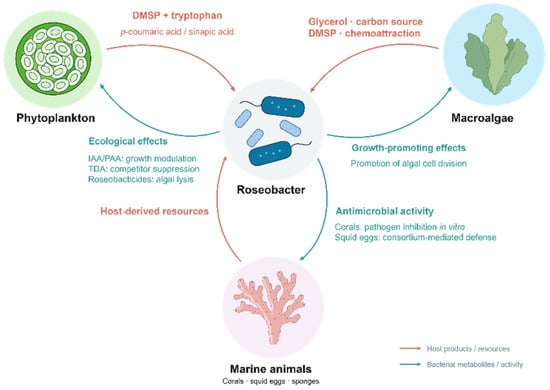
Studies of animal-associated Roseobacter-group bacteria connect bacterial secondary metabolism with host-associated competition and, in some cases, protection [29,30,32]. The squid ANG system links comparative genomics and isolate-level activities with experiments demonstrating consortium-mediated egg protection [30,31,32]. Across phytoplankton, macroalgae, and marine animals, host-associated compounds and bacterial metabolites contribute to different forms of chemical exchange (Figure 2) [5,27,30]. The following section examines the structures, biosynthetic pathways, and activities of characterized metabolites.
Figure 2.
Host-derived resources and Roseobacter-mediated ecological functions in marine host associations.
4. Host-Associated Metabolites of the Marine Roseobacter-Group
4.1. Sulfur-Containing Metabolites
Sulfur-containing metabolites represent some of the most structurally and functionally distinctive natural products produced by the marine Roseobacter-group. Members of this lineage actively utilize algal-derived organosulfur compounds, particularly DMSP and 2,3-dihydroxypropanesulfonate, as important carbon and sulfur sources. Sulfur obtained through these pathways can enter cysteine, methanethiol, and other reduced-sulfur intermediates, which are subsequently incorporated into diverse functional natural products, including antibiotics, algaecides, aromatic thiomethyl compounds, sulfonolipids, and volatile sulfur compounds. Thus, sulfur metabolism within the Roseobacter-group links nutrient acquisition to microbial competition, host interactions, membrane organization, and marine biogeochemical cycling [4,10].
4.1.1. Tropodithietic Acid and Related Metabolites
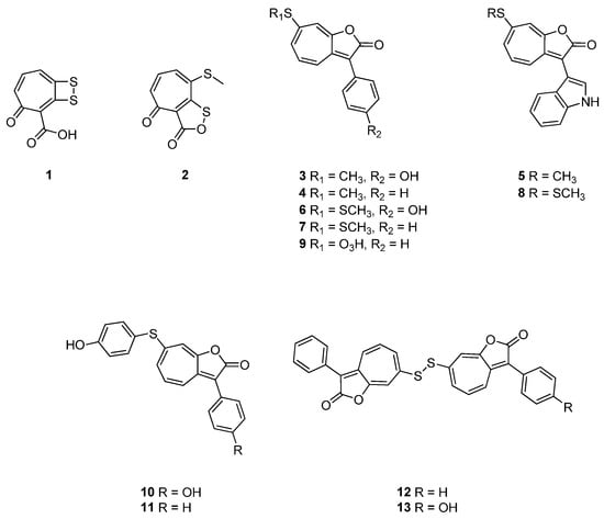
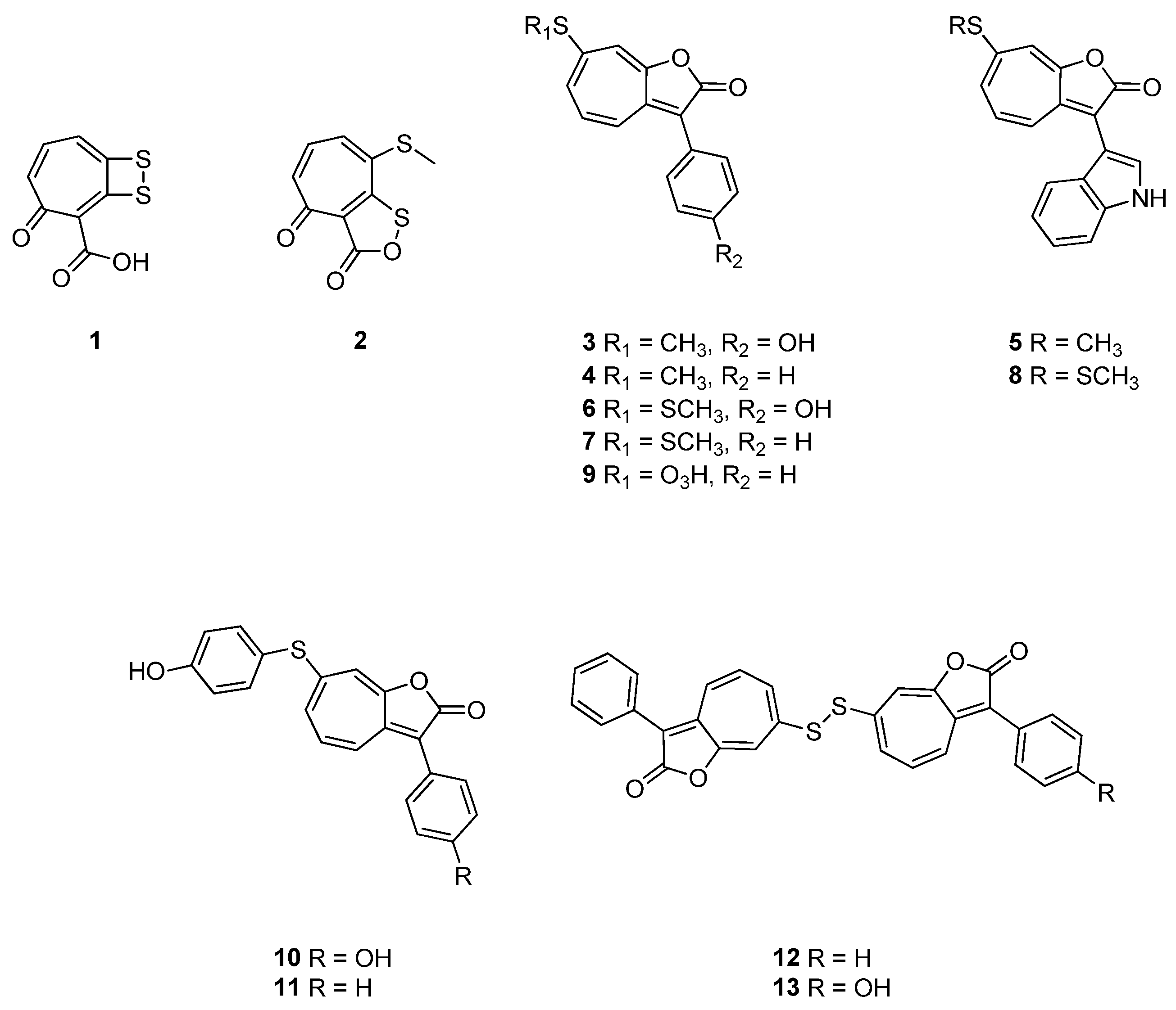
Tropodithietic acid (1, TDA) is the most extensively investigated natural product within the marine Roseobacter-group (Figure 3). It has been reported from several strains of Phaeobacter, Tritonibacter, and related marine Roseobacteraceae. Structurally, TDA possesses an unusual sulfur-rich tropone-derived scaffold and exhibits broad antibacterial activity against Gram-positive and Gram-negative bacteria, including major aquaculture pathogens [10]. For example, purified TDA showed minimum inhibitory concentrations (MICs) of 1.25 μg/mL against Vibrio anguillarum NB10Sm, 5 μg/mL against Roseovarius crassostreae CV919Sm, and 6.25 μg/mL against Vibrio tubiashii RE22Sm (Table 1) [10]. Its antibacterial activity is mainly attributed to dissipation of the proton motive force across the target-cell membrane, which interferes with adenosine triphosphate (ATP) production, solute transport, and flagellar motility [33]. TDA also exhibits weak iron-binding activity, although membrane-associated proton translocation appears to be the principal mechanism underlying its antibacterial effect [10,33]. Owing to these antimicrobial properties, TDA-producing Phaeobacter strains have attracted considerable interest as biocontrol agents and probiotics for fish larvae, shellfish, live feeds, and microalgal cultures. TDA also regulates the physiology of the producer. A TDA-deficient mutant of P. piscinae S26 exhibited enhanced motility, altered cell morphology, and changes in systems associated with horizontal gene transfer, including a prophage, a type IV secretion system, and a gene-transfer agent [10]. These findings suggest that TDA accumulation may contribute to the transition from a motile planktonic state to a persistent surface-associated lifestyle [10].
Figure 3.
Chemical structures of compounds 1–13.
Table 1.
Antibacterial/antimicrobial activities of Roseobacter-group-derived compounds.
Genetic studies link TDA production to phenylacetate catabolism and the tdaABCDEF locus [38]. Subsequent biochemical work showed that TdaE acts on a CoA-bound shunt product of phenylacetate catabolism and catalyzes dehydrogenation, CoA-ester oxygenolysis, and ring epoxidation, redirecting this intermediate toward tropone biosynthesis [39]. Sulfur-metabolism enzymes also contribute to TDA formation; PatB has been proposed to supply reactive sulfur through formation of S-thiocysteine from cystine [10].
Methyl troposulfenin (2) is an S-methylated TDA analogue isolated from P. inhibens DSM 17395 and structurally characterized by nuclear magnetic resonance (NMR) spectroscopy and high-resolution mass spectrometry (HRMS) (Figure 3) [34]. Biological evaluation revealed that methyl troposulfenin exhibits approximately 4- to 100-fold weaker inhibitory activity against V. anguillarum compared with TDA. Mechanistically, S-methylation abolishes the acidic proton essential for maintaining the ionizable sulfur-containing system [34]. This marked reduction in antibacterial potency upon methylation strongly supports the proposed protonophore-like mode of action for TDA [34].
Beyond aquaculture, TDA has shown broad antiproliferative and cytotoxic activity in mammalian cell models [33,40]. In the NCI-60 cancer-cell-line screen, 10 μM TDA produced 91%, 76%, and 76% lethality in U251 central nervous system (CNS), SNB-19 CNS, and A498 renal cancer cells, respectively; dose–response experiments yielded LC50 values of 5.7, 7.5, and 6.3 μM in these three cell lines, while growth-inhibitory IC50 values across multiple cancer cell lines were generally in the 1–3 μM range [33,40]. By comparison, non-tumorigenic MCF-10A human mammary epithelial cells showed an IC50 of 19.5 μM (Table 2) [33]. TDA is also cytotoxic to rat oligodendroglial OLN-93 and mouse neuroblastoma-derived N2a clonal cell lines, which should not be treated collectively as primary normal-cell models. These cells displayed marked morphological and viability changes at 0.3–0.5 μg/mL (1.4–2.4 μM), with only approximately 10% and 20% viability, respectively, after 24 h exposure to 0.5 μg/mL. These findings support further mechanistic investigation of TDA but also indicate that therapeutic selectivity and safety require careful evaluation [33,40].
Table 2.
Cytotoxic/Anticancer activities of Roseobacter-group-derived compounds.
4.1.2. Roseobacticides
Roseobacticides A–K (3–13) represent a family of sulfur-containing algaecidal metabolites produced by specific Phaeobacter strains in response to algal phenylpropanoids (Figure 3). Roseobacticides A (3) and B (4) were initially isolated and structurally characterized from P. gallaeciensis BS107 after supplementation of the liquid culture with pCA. Subsequent elicitation studies using an expanded panel of phenylpropanoids, including pCA, sinapic acid, ferulic acid, and cinnamic acid, led to the purification and structural identification of roseobacticides C–K (5–13) [6,7]. Structurally, these compounds share an unusual sulfur-substituted 1-oxaazulan-2-one or tropolactone-related core. Their chemical diversity stems primarily from structural variations at the C-3 and C-7 positions. Roseobacticides A, D, and H contain phenolic aromatic groups; B, E, G, and I possess phenyl-derived substituents; C and F contain indole-derived units; and J and K represent more complex sulfur-linked congeners. Although roseobacticides C and F additionally contain nitrogen, they are grouped within this section because the sulfur-bearing tropolactone scaffold serves as the defining structural hallmark of this natural product family [6,7].
Roseobacticides are hybrid metabolites constructed from molecular building blocks contributed by both the bacterial producer and its algal partner. Isotope-labeling experiments revealed that their chemical structures incorporate bacterial phenylacetate pathway intermediates, algal phenylpropanoids, and sulfur derived from host-associated sulfur metabolism. Notably, metabolites associated with mutualistic interactions are repurposed during the transition to parasitism, as bacterial PAA (an algal growth promoter) and host-derived DMSP (a bacterial carbon and sulfur source) are converted into precursors for destructive algaecidal agents. Genes required for TDA production, including the tda operon and the phenylacetate catabolic genes, are also required for roseobacticide biosynthesis. Thus, a shared metabolic system can generate two structurally and biologically distinct natural-product families. Under mutualistic conditions, the pathway produces the protective antibiotic TDA, whereas exposure to phenylpropanoids released by stressed or senescent algae redirects metabolism toward roseobacticides [7].
The production of roseobacticides is additionally regulated by quorum sensing. Specifically, the AHL signal N-(3-hydroxydecanoyl)-L-homoserine lactone (3-OH-C10-HSL) acts synergistically with algal phenylpropanoid elicitors, ensuring that algaecide synthesis is triggered only when the bacterial population achieves a threshold density in the presence of a physiologically compromised host. This dual-regulatory mechanism provides a highly coordinated switch from a mutualistic to a parasitic lifestyle. The total yield and congener profile of roseobacticides vary depending on both the specific elicitor and the bacterial producer strain. In P. gallaeciensis BS107, supplementation with sinapic acid generally induced the highest overall yield and the greatest structural diversity of roseobacticides, whereas a distinct response profile was observed in strain 2.10. These strain-specific production profiles likely reflect evolutionary adaptation to distinct algal hosts and their characteristic phenylpropanoid signatures [7].
Roseobacticides showed little antibacterial activity but potent and selective effects against microalgae. Roseobacticide A inhibited E. huxleyi and Rhodomonas salina with IC50 values of approximately 2.2 and 0.10 μM, respectively, whereas roseobacticide B inhibited E. huxleyi with an IC50 of approximately 0.19 μM. Treatment produced chloroplast loss, cellular damage, and algal lysis (Table 3). These compounds are therefore considered chemical mediators of the parasitic phase of the Phaeobacter–algal interaction. Their unusual sulfur-rich architectures and selective toxicity toward particular eukaryotic cells also make them attractive for mechanistic and structure–activity investigations [6]. Nevertheless, their effects on mammalian cells and molecular targets remain largely unknown, and their biomedical relevance remains less established than their ecological functions [6,7].
Table 3.
Antialgal/Algaecidal activities of Roseobacter-group-derived compounds.
4.1.3. Roseochelin B
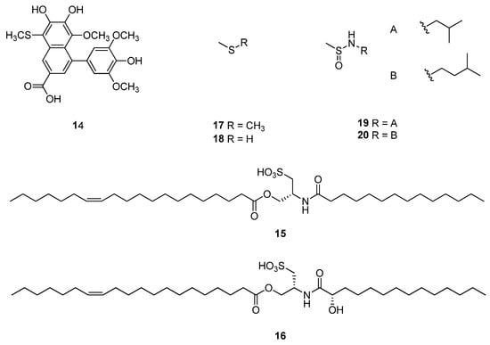
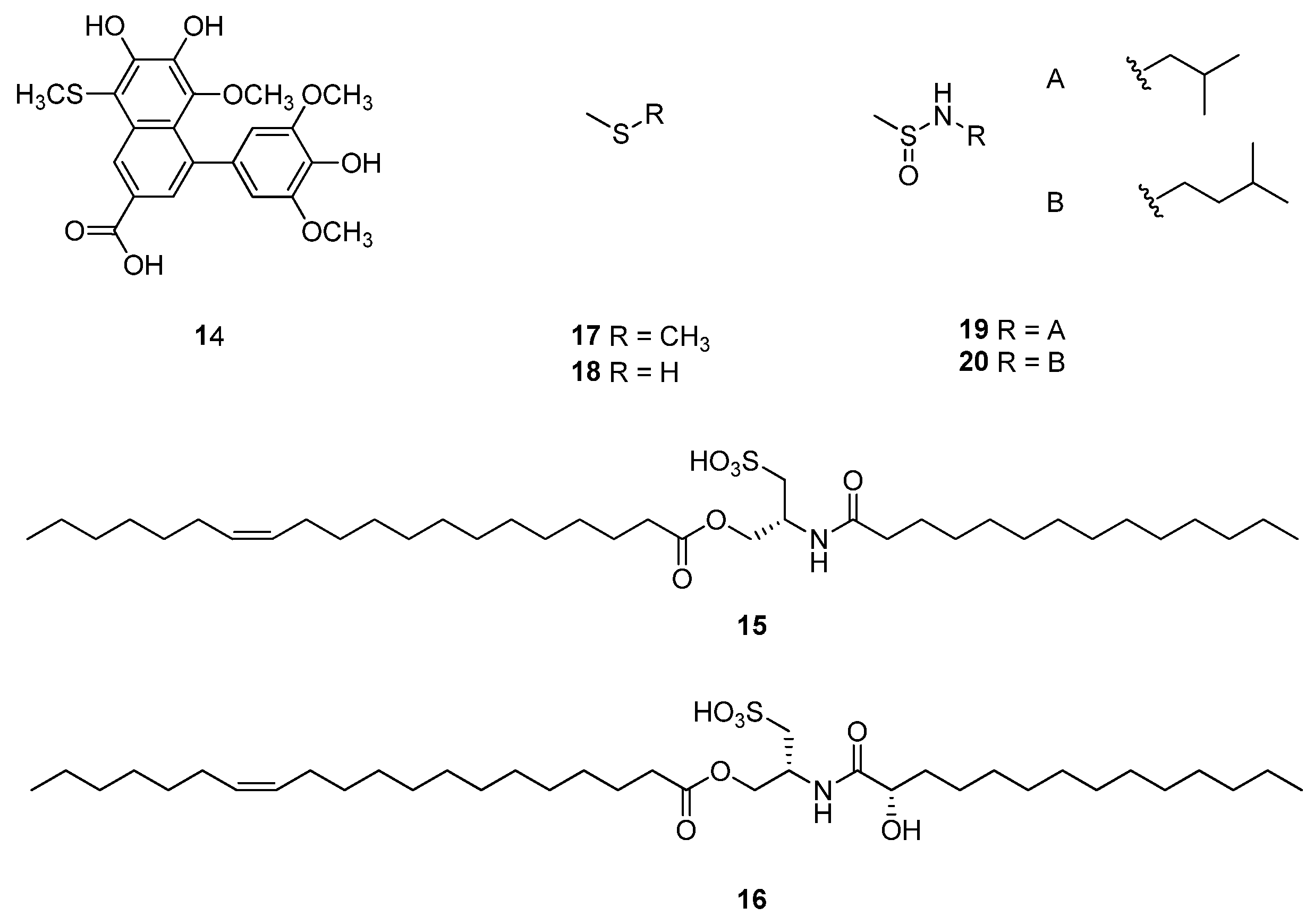
Roseochelin B (14) is a sulfur-containing biaryl naphthoic acid derivative produced by P. inhibens upon exposure to algal sinapic acid (Figure 4). It features a thiomethyl group and a 6,7-dihydroxy catechol-like motif, distinguishing it from its sulfur-free analogues, roseochelins A, C, and D. The biosynthesis of roseochelin B involves non-enzymatic reaction steps and enzyme-catalyzed transformations. Initially, two molecules of sinapic acid undergo spontaneous coupling and subsequent rearrangement to form the sulfur-free intermediate roseochelin A. P. inhibens modifies this scaffold via demethylation and thiomethylation to yield roseochelin B. Exogenous roseochelin A is converted into roseochelin B only when the bacterial culture is co-supplemented with sinapic acid. This requirement demonstrates that sinapic acid serves a dual role in P. inhibens, functioning both as a chemical precursor and as an essential elicitor that induces the necessary enzymatic machinery [41].
Figure 4.
Chemical structures of compounds 14–20.
Roseochelin B demonstrated iron-chelating capability in the chrome azurol S (CAS) assay, consistent with the presence of its catechol-like moiety. However, because production is induced by sinapic acid under both iron-limited and iron-replete conditions, roseochelin B is more accurately defined as an iron-binding natural product rather than a dedicated siderophore. Roseochelin B exhibits minimal antibacterial activity but showed algaecidal activity against E. huxleyi; an IC50 value was not reported (Table 3) [41]. Its dual iron-binding and algaecidal properties may provide complementary ecological advantages during the parasitic phase, but this proposed role remains to be validated under natural environmental conditions.
4.1.4. Sulfur Amino Lipids and Cysteinolides
Sulfur amino lipid-656 (15, SAL-656) and sulfur amino lipid-672 (16, SAL-672) were originally detected as related sulfonolipid families in R. pomeroyi and P. inhibens. LC–MS/MS and molecular networking revealed lipoforms with different fatty-acyl compositions [43]. Representative constituents were subsequently isolated from R. pomeroyi and Ruegeria meonggei and structurally revised as cysteinolides A and B, respectively (Figure 4) [44].
Roman et al. revised the original head-group and acyl-linkage assignments using isolation, NMR spectroscopy, degradation studies, and total synthesis. Cysteinolides contain an N,O-diacylated cysteinolic acid head group, and the absolute structures of representative cysteinolides A and B were established [44]. This structural assignment applies to the characterized compounds; the full range of acyl-chain variants detected by mass spectrometry should not be assumed to have been individually established.
Deletion and complementation experiments support a role for salA in SAL production, and mutant phenotypes connect these lipids with biofilm formation [43]. The revised structures provide a basis for investigating the enzymatic steps of their biosynthesis. Cysteinolides were tested in a cell-free 5-lipoxygenase (5-LOX) enzyme assay. At 30 μM, cysteinolide B showed greater inhibition than cysteinolide A; an IC50 value was not reported (Table 4) [44]. These cell-free enzyme data do not establish anti-inflammatory efficacy in vivo.
Table 4.
Anti-inflammatory, Anti-allergic and Immunomodulatory Activities of Roseobacter-group-derived Compounds.
4.1.5. Volatile and Low-Molecular-Weight Sulfur Metabolites
Marine Roseobacter-group bacteria transform algal organosulfur substrates into several low-molecular-weight sulfur metabolites. Dimethyl sulfide (17, DMS) is produced via enzymatic cleavage of DMSP, whereas methanethiol (18, MeSH) is generated through the alternative demethylation pathway. DMS volatilizes from seawater into the atmosphere, contributing to marine–atmospheric sulfur flux, whereas MeSH-derived sulfur is retained more efficiently within bacterial cells for biomass production and downstream metabolic processes [4]. Although DMSP is commonly supplied by phytoplankton in marine algal–bacterial interactions, some marine heterotrophic bacteria can also synthesize DMSP through a dsyB-dependent pathway [46]. DMSP is not assigned a compound number here because it is treated as a widespread primary osmolyte and central sulfur metabolite rather than a structurally characterized specialized metabolite produced by the Roseobacter-group.
N-Isobutylmethanesulfinamide (19) and N-isopentylmethanesulfinamide (20) belong to the nitrogen- and sulfur-containing volatiles identified from Roseobacter-group bacteria (Figure 4). Their identities were investigated by GC–MS and comparison with synthetic standards [47]. Amino-acid-derived alkylamines provide plausible precursors, but their complete biosynthetic pathways and ecological functions remain uncharacterized [47].
4.2. Nitrogen-Containing Metabolites
Nitrogen-containing Roseobacter-group metabolites have several biosynthetic origins. Algal-derived tryptophan can support bacterial IAA production [5], while tryptophan and sinapic acid jointly induce hybrid algaecides [8]. Indigoidine derives from glutamine-dependent biosynthesis [24], whereas N-acylated amino-acid methyl esters combine amino-acid-derived head groups with fatty-acyl chains [35]. Other nitrogen-containing metabolites include siderophores involved in iron acquisition [11] and ceramide lipids associated with membrane remodeling [12].
4.2.1. Sinacidins, Sinatryptins, and Sinamicins
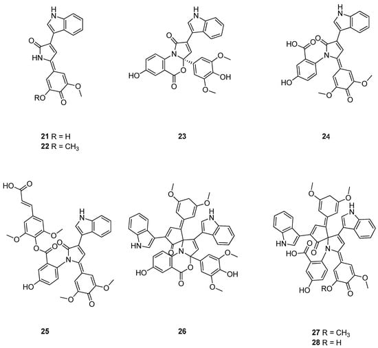
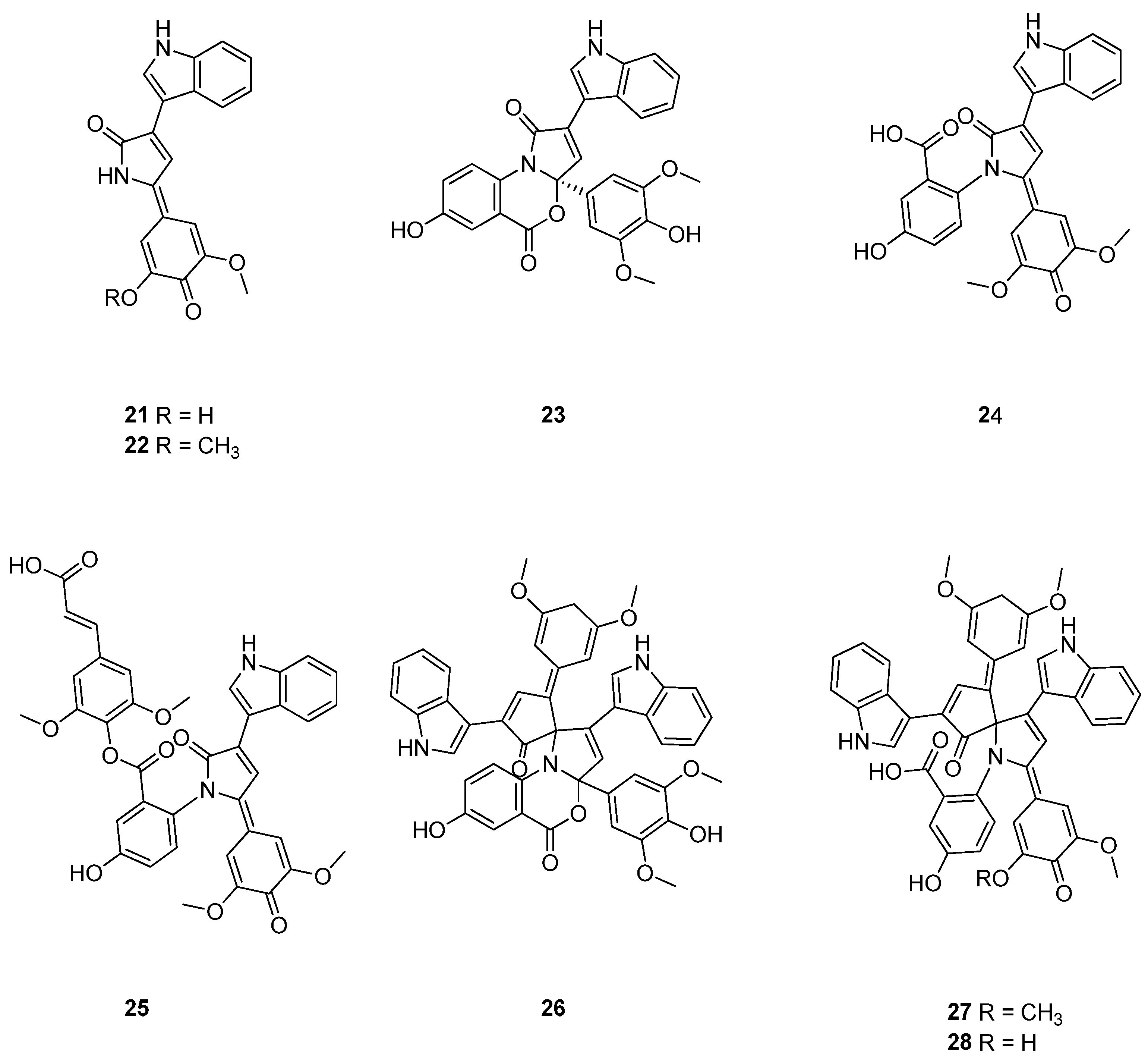
Sinacidins A and B (21 and 22), sinatryptins A–C (23–25), and sinamicins A–C (26–28) constitute a group of eight structurally unique metabolites produced by P. inhibens upon co-treatment with algal-derived sinapic acid and tryptophan (Figure 5). Their production was substantially lower when either precursor was supplied individually, demonstrating that phenylpropanoid and amino acid signals act cooperatively to elicit this cryptic chemical response. Sinacidins A and B contain an indole-substituted 3-pyrrolin-2-one linked to differently methoxylated para-quinone-methide-related units. Sinatryptins A–C possess a related heterocyclic system but additionally incorporate a tryptophan-derived 3-hydroxybenzoic-acid portion; sinatryptin C contains a further sinapic-acid-derived ester. Sinatryptin B was produced at such a low level that its structure was determined by microcrystal electron diffraction (MicroED) rather than by conventional NMR spectroscopy. Sinamicin A (26) is a heterodimer containing an unusual spiro-3-pyrroline junction and was reported as a racemic mixture. Sinamicin B (27) is the methylated ring-opened congener, whereas sinamicin C (28) is the corresponding hydroxy analogue. Sinamicins B and C were obtained as a minute co-crystallized mixture, and both structures were solved simultaneously by MicroED. Isotope-feeding experiments utilizing ring-deuterated tryptophan yielded diagnostic mass shifts, confirming the direct incorporation of tryptophan-derived indole and hydroxybenzoic acid units. The shared 3,5-dimethoxyphenolic moieties indicate that sinapic acid also contributes directly to their chemical structures. These compounds therefore represent hybrid natural products assembled largely from metabolites that could be supplied by an algal host [8].
Figure 5.
Chemical structures of compounds 21–28.
Sinacidins A (21) and B (22), sinatryptin A (23), and sinamicin A (26) inhibited E. huxleyi with IC50 values of 7.1 ± 0.6, 5.7 ± 0.3, 6.9 ± 1.6, and 8.2 ± 0.7 μM, respectively. Sinatryptins B (24) and C (25) had IC50 values above 100 μM (Table 3). Sinatryptin A also inhibited N. gonorrhoeae with an MIC of 8 μg/mL (Table 1) [8]. In a subsequent study of differentiated Th2 cells, sinatryptin A suppressed IL-4, IL-5, and IL-13 responses, reduced STAT6 phosphorylation, and decreased GATA3 and IRF4 expression. The reported IC50 for inhibition of IL-13 production was 2.44 μM (Table 4) [45]. These findings establish activity in a cellular model; efficacy and tolerability in animals remain to be determined.
4.2.2. Roseobactin
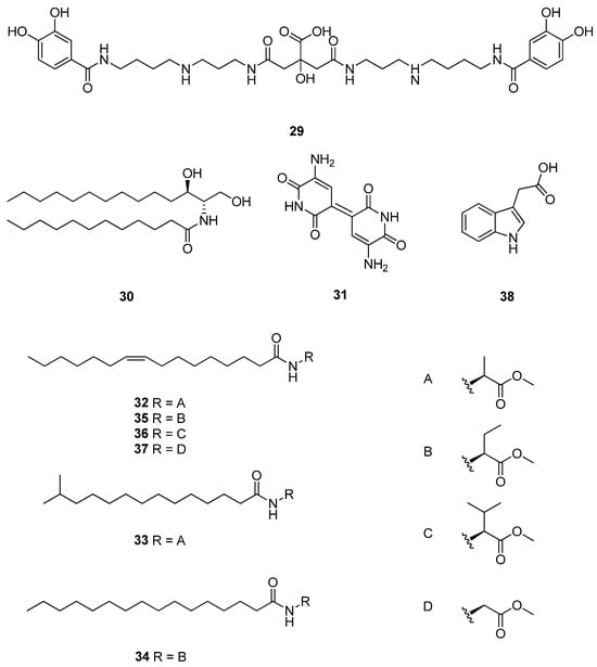
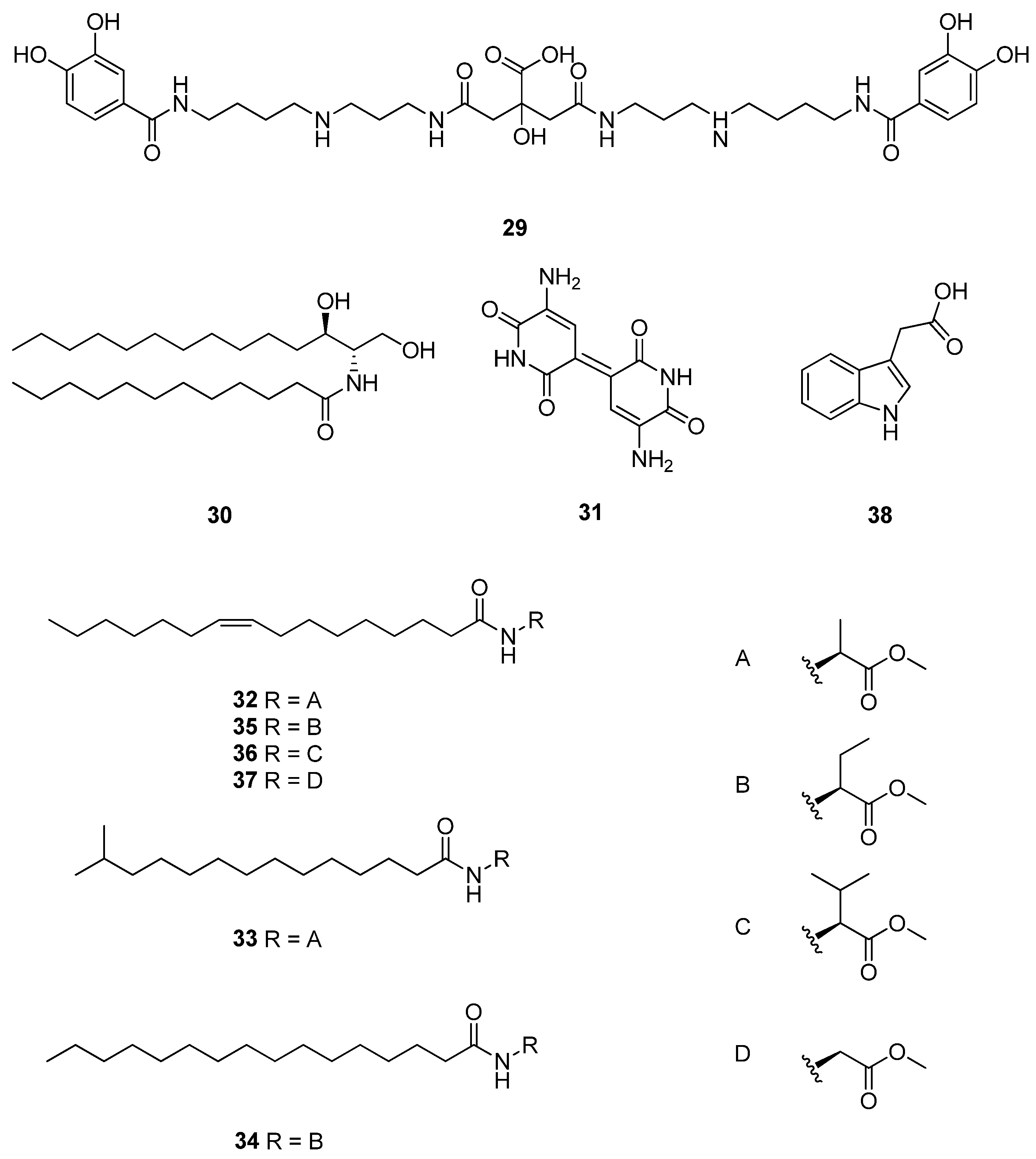
Roseobactin (29) is a catecholate-containing siderophore isolated from P. inhibens 2.10 (Figure 6) [11]. Its discovery was driven by transcriptomic profiling, which revealed that exposure to pCA induced oxidative-stress response genes and dramatically upregulated a previously uncharacterized biosynthetic locus, designated the rbt cluster. Targeted deletion of rbtB completely abolished both the corresponding LC–MS signal and iron-chelating activity, directly linking this gene cluster to roseobactin production. Although its molecular weight initially suggested structural similarity to the known siderophore petrobactin, detailed MS/MS and spectroscopic characterizations demonstrated a distinct architecture comprising catecholate, polyamine, and central oxygen-rich moieties. Thus, petrobactin represents an external structural analogue rather than a Roseobacter-group natural product [11].
Figure 6.
Chemical structures of compounds 29–38.
The co-induction of roseobactin and algaecidal metabolism by phenylpropanoids links iron acquisition with the bacterial response to algal metabolites [11]. Capture of iron released from damaged algae is a plausible ecological function, but direct iron transfer through this route has not been demonstrated in microbial communities.
4.2.3. Roseoceramide A
Roseoceramide A (30) is a bacterial ceramide lipid isolated from Roseovarius tolerans (Figure 6) [12]. It was uncovered through an ecological elicitor screen in which the iron-chelating algal flavonoid myricetin induced a previously unrecognized lipid feature. The associated rcc locus contains genes consistent with bacterial ceramide biosynthesis, together with a transporter and a putative phosphodiesterase. The serine palmitoyltransferase RccD was biochemically validated, supporting condensation of serine with a fatty-acyl substrate as an early step. Despite a strong increase in roseoceramide A production, transcription of core biosynthetic genes changed only moderately, suggesting substantial control by substrate availability or post-transcriptional regulation [12].
Myricetin-induced roseoceramide production is consistent with iron chelation: added siderophores also stimulated production, whereas iron supplementation attenuated it. Hypoxia, elevated salinity, and alkaline pH likewise enhanced production. Roseoceramide A contains relatively saturated acyl chains, and its accumulation was associated with reduced membrane fluidity and permeability. Production was detected in Roseovarius mucosus, which carries a homologous rcc cluster, but not in cluster-deficient Roseovarius nubinhibens [12]. These observations support a bacterial membrane-stress response; they do not establish mammalian signaling or pharmacological activity.
4.2.4. Indigoidine
Indigoidine (31) is a blue nitrogen-containing pigment isolated from Phaeobacter sp. Y4I, and its structure was confirmed by high-resolution mass spectrometry and 1H NMR spectroscopy (Figure 6). Indigoidine is produced through the igiBCDFE locus, in which the NRPS-like enzyme IgiD activates glutamine-derived precursors and directs formation of the bis-pyridone scaffold. Although an NRPS enzyme is involved, the final compound is a heterocyclic pigment rather than a peptide [24].
Genetic knockouts that abolished indigoidine biosynthesis concurrently eliminated antimicrobial activity against V. fischeri. In competitive co-culture assays, indigoidine-producing Phaeobacter sp. Y4I cells effectively suppressed surface colonization by V. fischeri, demonstrating a key biological role for indigoidine in interference competition within marine biofilms. Indigoidine also influences producer physiology. An igiD mutant displayed altered hydrogen-peroxide resistance, reduced swimming motility, and a different rate of surface attachment. Expression of igiD increased during surface-associated growth, suggesting that indigoidine coordinates oxidative physiology, motility, and competitive colonization rather than functioning solely as an extracellular pigment [24].
4.2.5. N-Acylated Amino-Acid Methyl Esters
N-Acylated amino-acid methyl esters are lipid-like metabolites composed of a fatty-acyl chain linked through an amide bond to an amino-acid methyl ester. Four subclasses have been reported from Roseobacter-group bacteria: N-acylalanine methyl esters (NAMEs), N-acyl-2-aminobutyric acid methyl esters (NABMEs), N-acylvaline methyl esters (NAVMEs), and N-acylglycine methyl esters (NAGMEs). Representative compounds discussed here include (Z)-N-(hexadec-9-enoyl)alanine methyl ester (Z9-C16:1-NAME, 32), N-(13-methyltetradecanoyl)alanine methyl ester (iso-C15:0-NAME, 33), N-hexadecanoyl-2-aminobutyric acid methyl ester (C16:0-NABME, 34), N-[(Z)-hexadec-9-enoyl]-2-aminobutyric acid methyl ester (Z9-C16:1-NABME, 35), N-[(Z)-hexadec-9-enoyl]valine methyl ester (Z9-C16:1-NAVME, 36), and N-[(Z)-hexadec-9-enoyl]glycine methyl ester (Z9-C16:1-NAGME, 37) (Figure 6). Their structural assignments were established through characteristic mass-spectrometric fragmentation, chemical derivatization, retention-index matching, high-resolution mass spectrometry, and comparison with synthesized standards. Their biosynthetic gene clusters remain unidentified, although formation is expected to involve fatty-acyl activation, amide-bond formation with the corresponding amino-acid methyl ester, and acyl-chain tailoring. Despite their structural similarity to AHLs, tested NAMEs neither activated nor antagonized the Roseobacter-group AHL receptors examined [35].
Biological assays indicate that activity varies substantially with both the amino-acid head group and the acyl chain [35]. Z9-C16:1-NAME has been reported to show moderate antialgal activity (Table 3) [42]. In the N-acylated amino-acid methyl ester series, the glycine derivative Z9-C16:1-NAGME (37) showed activity against the Gram-positive bacteria Bacillus subtilis, Staphylococcus aureus, and Micrococcus luteus and against the filamentous fungus Mucor hiemalis, together with moderate activity against Mycobacterium smegmatis and an efflux-deficient E. coli TolC strain. The saturated NABME C16:0-NABME (34) was active against M. hiemalis, M. luteus, and S. aureus, whereas Z9-C16:1-NABME (35) was mainly active against E. coli TolC and showed cytotoxicity against HCT-116 human colon carcinoma cells with an IC50 of 15.8 μg/mL (Table 1 and Table 2). By contrast, the valine derivative Z9-C16:1-NAVME (36) was nearly inactive. These results support compound-dependent antimicrobial and antialgal effects, while their physiological functions and biomedical relevance remain to be established [35].
4.2.6. Indole-3-Acetic Acid
Indole-3-acetic acid (38, IAA) is an auxin produced by P. inhibens from algal-derived tryptophan (Figure 6). In P. inhibens pure cultures, IAA was detected at approximately 0.4 nM, and its identity was confirmed by comparison of retention time, accurate mass, and targeted tandem mass spectrometry with an authentic IAA standard. In co-culture experiments, tryptophan exuded by E. huxleyi significantly increased both bacterial IAA production and attachment to algal cells, providing direct experimental support for including IAA among Roseobacter-group-derived metabolites involved in algal–bacterial chemical exchange [5].
Experiments with exogenous IAA showed concentration-dependent effects on algal growth, including growth promotion at lower concentrations and adverse effects at higher concentrations. Bleaching, oxidative-stress responses, and DNA fragmentation were also observed during bacterial–algal co-culture, but these observations do not demonstrate that an increase in locally produced IAA alone causes the entire sequence of host-cell damage [5].
Tryptophan supplementation increased bacterial attachment and accelerated algal decline in co-culture [5]. These results connect substrate availability with bacterial behavior but do not provide a direct measurement of IAA concentration at the algal surface or exclude contributions from other bacterial factors.
IAA therefore provides an example of metabolite exchange whose biological outcome depends on concentration and experimental context [5].
4.3. Phenolic Metabolites
Phenolic metabolites within the marine Roseobacter-group can originate either through direct enzymatic modification of algal phenylpropanoids or via spontaneous extracellular oxidative coupling. Sinapic acid is particularly reactive because its phenolic and conjugated side-chain system can generate oxidized intermediates that dimerize, cyclize, and rearrange. Subsequently, bacterial enzymes may further tailor these non-enzymatically generated molecular scaffolds through oxidation, reduction, demethylation, or sulfur incorporation [41,48].
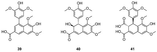
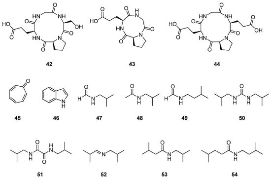
Roseochelin A (39) was isolated alongside roseochelin B from sinapic-acid-supplemented P. inhibens cultures and can form through nonenzymatic reactions in cell-free medium [41]. Subsequent LC–MS/UV-guided fractionation yielded roseochelins C (40) and D (41) (Figure 7). The structures of these new congeners were investigated by NMR spectroscopy and electronic circular dichroism analysis; roseochelin D was assigned the absolute configuration (3R,4S) [45]. These C/D assignments derive from the later study, distinct from the original roseochelin A/B report.
Figure 7.
Chemical structures of compounds 39–41.
Recent biomimetic total syntheses of roseochelins A and B provided chemical support for a revised formation pathway. In that study, roseochelin A was generated by spontaneous [4 + 2] dimerization of sinapic acid to thomasidioic acid, followed by metal-catalyzed oxidative decarboxylation. Roseochelin B was accessed through regioselective catechol formation, ortho-quinone-mediated thiomethylation, and a late-stage metal- or photocatalyzed oxidative decarboxylation. These results identify oxidative decarboxylation as an important chemical step in construction of the roseochelin scaffold and refine earlier biosynthetic proposals [48].
Roseochelin A showed negligible iron-chelating, antibacterial, or algaecidal activity in the original study [41]. Roseochelins A, C, and D subsequently inhibited IL-13 secretion in differentiated Th2 cells without detectable cytotoxicity over the tested concentration range (Table 4) [45]. Their ecological functions and the molecular basis of the observed cellular activity remain unresolved.
4.4. Other Metabolites
The metabolites grouped in this section are classified primarily by their distinctive structural and functional features rather than by elemental composition. Their biosynthetic origins are correspondingly diverse: cyclic peptides arise through peptide-bond formation and macrocyclization, volatile organic compounds are often generated through transformations of aromatic or amino-acid-derived intermediates, and AHLs are synthesized as cell-density-dependent signaling molecules. Together, these compounds contribute to chemical defense, surface colonization, biofilm development, motility, cell differentiation, and interspecies communication [9,36,49].
4.4.1. Cyclic Peptides
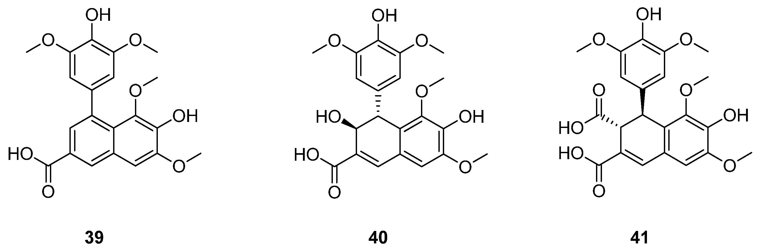
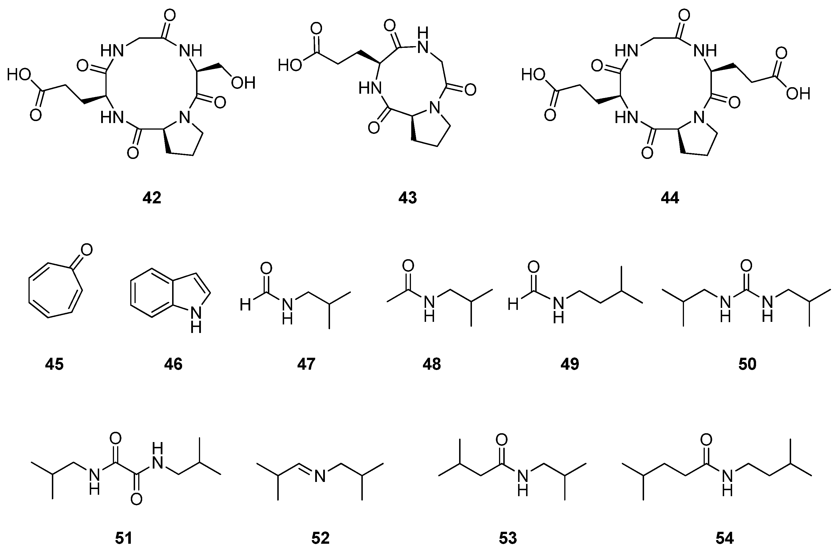
A Ruegeria strain designated SDC-1, isolated from cell cultures of the marine sponge Suberites domuncula, produced several cyclic peptides. The study could not establish conclusively whether SDC-1 was an obligate sponge symbiont or a persistent environmental bacterium associated with the sponge culture. Cyclo-(glycyl-L-seryl-L-prolyl-L-glutamyl) (42) (Figure 8) is a cyclic tetrapeptide containing glycine, serine, proline, and glutamic acid. Cyclo-(glycyl-L-prolyl-L-glutamyl) (43) was originally assigned as a related cyclic tripeptide lacking serine. The original sequences and stereochemical assignments were based on evidence obtained through multidimensional NMR spectroscopy, HRMS, fragmentation analysis, and chiral HPLC of acid hydrolysates. Cyclo-(L-prolyl-L-glutamyl)2 (44) is a symmetrical cyclic tetrapeptide composed of two Pro–Glu units [36]. Its molecular symmetry resulted in observation of only half of the expected NMR signals [36]. A later synthetic study questioned the tripeptide assignment of 43 and suggested that the natural product could instead be the cyclic hexapeptide cyclo-(Gly-Pro-Glu)2 [37]. This remains a structural-assignment issue requiring direct comparison with the natural material, rather than a definitive reassignment adopted here.
Figure 8.
Chemical structures of compounds 42–54.
Compounds 42 and 43 showed moderate antibacterial activity against B. subtilis, with MIC values of 25 and 50 μg/mL, respectively (Table 1), but were inactive against E. coli and Saccharomyces cerevisiae. Compound 44 was inactive in the same assays. These results indicate selective but moderate activity against a Gram-positive bacterium. However, the original study did not establish their natural concentrations, molecular targets, biosynthetic genes, or toxicity toward mammalian cells; consequently, their ecological functions and biomedical relevance remain unresolved [36].
4.4.2. Volatile Organic Compounds
Tropone (45) and indole (46) occur among Roseobacter-group-associated volatile metabolites (Figure 8) [42,49]. Studies of tropone biosynthesis connect this chemistry with phenylacetate catabolism; subsequent biochemical work identified TdaE-mediated dehydrogenation, CoA-ester oxygenolysis, and ring epoxidation [39,49]. This enzymatic work describes a route into tropone-derived natural-product biosynthesis but does not by itself assign every volatile detected in headspace analyses to that route.
Nitrogen-containing volatile compounds identified from Roseobacter-group strains include N-isobutylformamide (47), N-isobutylacetamide (48), N-isopentylformamide (49), N,N′-diisobutylurea (50), N,N′-diisobutyloxalamide (51), (E)-N-isobutyl-2-methylpropan-1-imine (52), N-isobutyl-3-methylbutanamide (53), and N-isopentyl-4-methylpentanamide (54) (Figure 8). These metabolites were predominantly detected in R. pelophilus, with overlapping subsets identified in Pseudoruegeria sp. and P. gallaeciensis. Their chemical structures were determined utilizing GC–MS fragmentation, gas-chromatographic retention behavior, infrared analysis, and comparison with synthesized standards [47].
The identified amides, imines, ureas, and oxalamides expand the known range of bacterial nitrogen-containing volatiles. Amino-acid-derived amines are plausible building blocks, and synthetic comparisons supported their structural assignments [47]. Their detection does not establish signaling functions: receptors, ecologically relevant exposure levels, and responses in recipient organisms remain to be determined.
4.4.3. N-Acylhomoserine Lactones
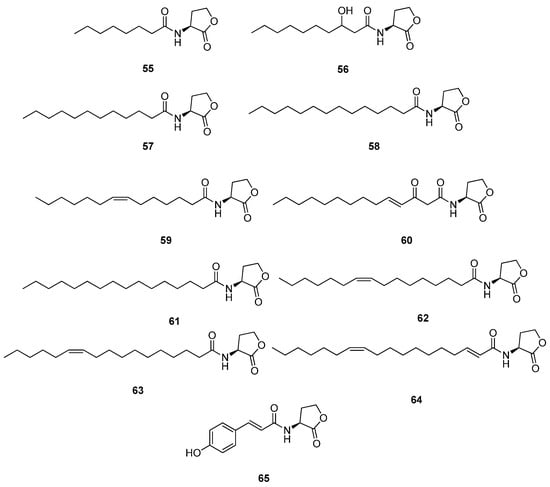
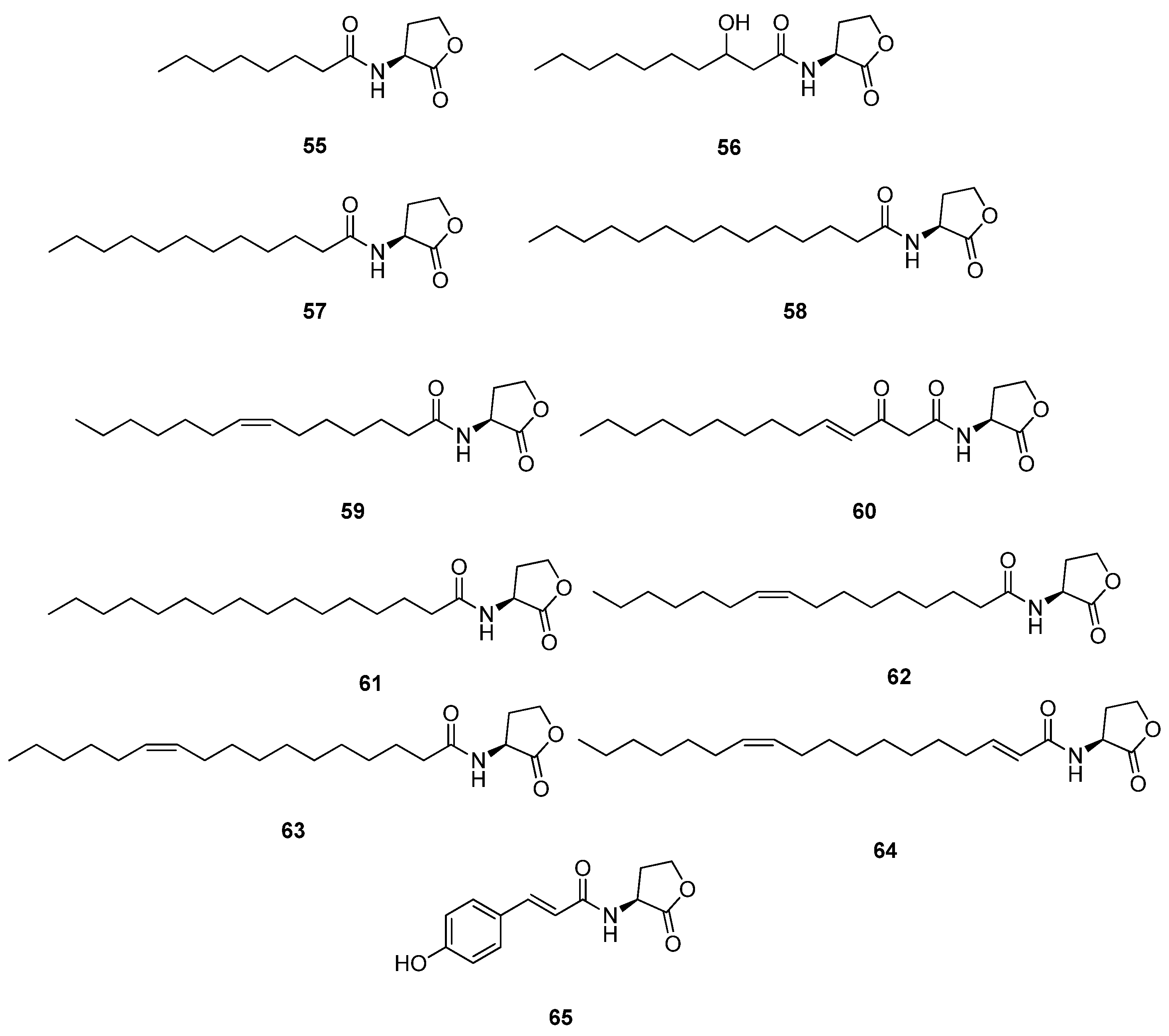
N-Acylhomoserine lactones (AHLs) are the principal quorum-sensing signal molecules produced by members of the marine Roseobacter-group. Structurally, these autoinducers contain a homoserine-lactone head group linked through an amide bond to an acyl side chain that varies in chain length, degree of unsaturation, and oxidation state. LuxI-type synthases generally use S-adenosylmethionine to generate the lactone portion and activated fatty-acyl substrates to provide the acyl chain [9]. Representative compounds include C8:0-HSL (55), 3-OH-C10:0-HSL (56), C12:0-HSL (57), C14:0-HSL (58), Z7-C14:1-HSL (59), 3-oxo-C14:1-HSL (60), C16:0-HSL (61), C16:1-HSL (62), (11Z)-C18:1-HSL (63), (2E,11Z)-C18:2-HSL (64), and p-coumaroyl-HSL (65) (Figure 9) [9,50]. Additional C13, C15, C16, and C18 congeners have been identified as major or trace metabolites from various Roseobacter-group strains [9].
Figure 9.
Chemical structures of compounds 55–65.
C8:0-HSL was identified from Roseobacter denitrificans, whereas 3-OH-C10:0-HSL is characteristic of several Phaeobacter strains [9]. Long-chain AHLs occur frequently in algal-associated species: D. shibae produces (11Z)-C18:1-HSL and (2E,11Z)-C18:2-HSL, while Roseovarius and Jannaschia strains produce diverse C14–C18 congeners [9]. A comprehensive screening of 24 macroalga-associated Roseobacter-group isolates revealed AHL production in 19 strains. However, no clear correlation was observed among specific AHL structural profiles, bacterial phylogeny, and isolation habitats [42]. In P. inhibens, 3-OH-C10:0-HSL regulates TDA production and is required together with algal phenylpropanoids for roseobacticide biosynthesis [7,9]. Quorum sensing thus enables the bacterium to integrate its own population density with chemical cues reflecting the physiological status of the algal host [9].
The unusual aromatic signal p-coumaroyl-HSL (65) is produced by R. pomeroyi when pCA is available [50]. In this case, an environmental or host-derived aromatic compound is incorporated directly into a bacterial signaling molecule, demonstrating that quorum-sensing chemistry can change according to substrates supplied by neighboring organisms. AHL signaling regulates antibiotic production, motility, biofilm development, and cell differentiation in different Roseobacter-group bacteria [9]. Long-chain AHLs also exhibited direct antialgal activity against Skeletonema costatum, indicating that they may act both as bacterial communication molecules and as interference metabolites when present at sufficiently high local concentrations (Table 3) [42]. The biomedical significance of Roseobacter-group AHLs lies primarily in their value as models for quorum-sensing and quorum-quenching research. The specific compounds described here have not been developed as therapeutic agents, but their synthases and receptors may provide tools for studying bacterial communication and for designing strategies that interfere with surface colonization or virulence-related signaling [9].
5. Conclusions
Marine bacteria in the Roseobacter-group occupy planktonic and host-associated habitats, and their metabolites contribute to competition, signaling, and interactions with marine eukaryotes [1,3]. Studies of Phaeobacter–algal associations show that the outcome can shift with host condition and available substrates, but the mechanisms differ among bacterial–host pairs [5,6,7,19].
The characterized products arise from several distinct pathways. TDA and roseobacticides connect phenylacetate and sulfur metabolism [7,10,39], while sinacidins, sinatryptins, and sinamicins incorporate building blocks associated with tryptophan and sinapic acid [8]. Indigoidine instead derives from glutamine-dependent biosynthesis [24], and N-acylated amino-acid methyl esters combine amino-acid and fatty-acyl components [35]. Sulfonolipids and ceramides contribute to membrane-associated physiology [12,43]. These pathways should not be treated as uniformly dependent on the same host-derived precursors.
A central unresolved problem is the connection between predicted biosynthetic loci and structurally characterized products [13,23]. Ecological elicitation has revealed compounds missed under standard culture conditions, while MicroED has enabled structure determination from small samples [8]. Future studies could combine controlled co-cultures, metabolite profiling, and targeted genetic perturbation to test how individual pathways affect host interactions. Such approaches should distinguish metabolite induction from evidence that the compound performs a specific ecological function.
Applications are best evaluated against the available experimental endpoints. TDA-producing strains have been studied for aquaculture biocontrol [10], whereas the anti-allergic effects of sinatryptin A and roseochelins currently rest on cellular assays [45]. Establishing useful applications will require compound-specific evidence of selectivity, exposure, and activity in the intended biological setting.
Author Contributions
Conceptualization, S.R.L.; formal analysis, T.G.K. and C.H.; investigation, Y.N., E.S.J., Y.J.L., H.-J.H. and S.P.; writing—original draft preparation, T.G.K., C.H. and S.R.L.; writing—review and editing, S.R.L.; visualization, T.G.K., C.H., P.M.S., E.L.K., K.M. and S.R.L.; supervision, S.R.L.; project administration, S.R.L.; funding acquisition, S.R.L. All authors have read and agreed to the published version of the manuscript.
Funding
This work was supported by a 2-Year Research Grant of Pusan National University.
Institutional Review Board Statement
Not applicable.
Informed Consent Statement
Not applicable.
Data Availability Statement
No new data were created or analyzed in this study. Data sharing is not applicable to this article.
Conflicts of Interest
The authors declare no conflicts of interest.
References
- Buchan, A.; González, J.M.; Moran, M.A. Overview of the marine Roseobacter lineage. Appl. Environ. Microbiol. 2005, 71, 5665–5677. [Google Scholar] [CrossRef] [Scilit]
- Wagner-Döbler, I.; Biebl, H. Environmental biology of the marine Roseobacter lineage. Annu. Rev. Microbiol. 2006, 60, 255–280. [Google Scholar] [CrossRef] [Scilit]
- Luo, H.; Moran, M.A. Evolutionary ecology of the marine Roseobacter clade. Microbiol. Mol. Biol. Rev. 2014, 78, 573–587. [Google Scholar] [CrossRef] [Scilit]
- Reisch, C.R.; Moran, M.A.; Whitman, W.B. Bacterial catabolism of dimethylsulfoniopropionate (DMSP). Front. Microbiol. 2011, 2, 172. [Google Scholar] [CrossRef] [Scilit]
- Segev, E.; Wyche, T.P.; Kim, K.H.; Petersen, J.; Ellebrandt, C.; Vlamakis, H.; Barteneva, N.; Paulson, J.N.; Chai, L.; Clardy, J.; et al. Dynamic metabolic exchange governs a marine algal–bacterial interaction. eLife 2016, 5, e17473. [Google Scholar] [CrossRef]
- Seyedsayamdost, M.R.; Case, R.J.; Kolter, R.; Clardy, J. The jekyll-and-hyde chemistry of Phaeobacter gallaeciensis. Nat. Chem. 2011, 3, 331–335. [Google Scholar] [CrossRef] [Scilit]
- Wang, R.; Gallant, É.; Seyedsayamdost, M.R. Investigation of the genetics and biochemistry of roseobacticide production in the Roseobacter clade bacterium Phaeobacter inhibens. mBio 2016, 7, e02118-15. [Google Scholar] [CrossRef] [Scilit]
- Park, J.-D.; Li, Y.; Moon, K.; Han, E.J.; Lee, S.R.; Seyedsayamdost, M.R. Structural elucidation of cryptic algaecides in marine algal–bacterial symbioses by NMR spectroscopy and microED. Angew. Chem. Int. Ed. 2022, 61, e202114022. [Google Scholar] [CrossRef] [Scilit]
- Zan, J.; Liu, Y.; Fuqua, C.; Hill, R.T. Acyl-homoserine lactone quorum sensing in the Roseobacter clade. Int. J. Mol. Sci. 2014, 15, 654–669. [Google Scholar] [CrossRef] [Scilit]
- Henriksen, N.N.S.E.; Lindqvist, L.L.; Wibowo, M.; Sonnenschein, E.C.; Bentzon-Tilia, M.; Gram, L. Role is in the eye of the beholder—The multiple functions of the antibacterial compound tropodithietic acid produced by marine Rhodobacteraceae. FEMS Microbiol. Rev. 2022, 46, fuac007. [Google Scholar] [CrossRef] [Scilit]
- Wang, R.; Gallant, É.; Wilson, M.Z.; Wu, Y.; Li, A.; Gitai, Z.; Seyedsayamdost, M.R. Algal p-Coumaric acid induces oxidative stress and siderophore biosynthesis in the bacterial symbiont Phaeobacter inhibens. Cell Chem. Biol. 2022, 29, 670–679.e5. [Google Scholar] [CrossRef] [Scilit]
- Ganley, J.G.; Seyedsayamdost, M.R. Iron limitation triggers roseoceramide biosynthesis and membrane remodeling in marine Roseobacter. Proc. Natl. Acad. Sci. USA 2025, 122, e2414434122, Erratum in Proc. Natl. Acad. Sci. USA 2025, 122, e2502118122. [Google Scholar] [CrossRef] [Scilit]
- Newton, R.J.; Griffin, L.E.; Bowles, K.M.; Meile, C.; Gifford, S.; Givens, C.E.; Howard, E.C.; King, E.; Oakley, C.A.; Reisch, C.R.; et al. Genome characteristics of a generalist marine bacterial lineage. ISME J. 2010, 4, 784–798. [Google Scholar] [CrossRef] [Scilit]
- Liang, K.Y.H.; Orata, F.D.; Boucher, Y.F.; Case, R.J. Roseobacters in a sea of poly- and paraphyly: Whole genome-based taxonomy of the family Rhodobacteraceae and the proposal for the split of the “Roseobacter clade” into a novel family, Roseobacteraceae fam. nov. Front. Microbiol. 2021, 12, 683109. [Google Scholar] [CrossRef] [Scilit]
- Zhang, D.-F.; He, W.; Shao, Z.; Ahmed, I.; Zhang, Y.; Li, W.-J.; Zhao, Z. Phylotaxonomic assessment based on four core gene sets and proposal of a genus definition among the families Paracoccaceae and Roseobacteraceae. Int. J. Syst. Evol. Microbiol. 2023, 73, 006156, Erratum in Int. J. Syst. Evol. Microbiol, 2023, 73, 006207. [Google Scholar] [CrossRef] [Scilit]
- Parte, A.C.; Sardà Carbasse, J.; Meier-Kolthoff, J.P.; Reimer, L.C.; Göker, M. List of Prokaryotic names with Standing in Nomenclature (LPSN) moves to the DSMZ. Int. J. Syst. Evol. Microbiol. 2020, 70, 5607–5612. [Google Scholar] [CrossRef] [Scilit]
- Parks, D.H.; Chuvochina, M.; Chaumeil, P.-A.; Rinke, C.; Mussig, A.J.; Hugenholtz, P. A complete domain-to-species taxonomy for Bacteria and Archaea. Nat. Biotechnol. 2020, 38, 1079–1086, Erratum in Nat. Biotechnol 2020, 38, 1098. [Google Scholar] [CrossRef] [Scilit]
- Genome Taxonomy Database (GTDB). Release R11-RS232. Available online: https://gtdb.ecogenomic.org/ (accessed on 30 October 2026).
- Wang, H.; Tomasch, J.; Michael, V.; Bhuju, S.; Jarek, M.; Petersen, J.; Wagner-Döbler, I. Identification of genetic modules mediating the jekyll and hyde interaction of Dinoroseobacter shibae with the dinoflagellate Prorocentrum minimum. Front. Microbiol. 2015, 6, 1262. [Google Scholar] [CrossRef] [Scilit]
- Amin, S.A.; Hmelo, L.R.; van Tol, H.M.; Durham, B.P.; Carlson, L.T.; Heal, K.R.; Morales, R.L.; Berthiaume, C.T.; Parker, M.S.; Djunaedi, B.; et al. Interaction and signalling between a cosmopolitan phytoplankton and associated bacteria. Nature 2015, 522, 98–101. [Google Scholar] [CrossRef] [Scilit]
- Liu, Y.; Brinkhoff, T.; Simon, M. Ecogenomics and functional biogeography of the Roseobacter group in the global oceans based on 653 MAGs and SAGs. Microbiome 2025, 13, 247. [Google Scholar] [CrossRef] [Scilit]
- Michael, V.; Frank, O.; Bartling, P.; Scheuner, C.; Göker, M.; Brinkmann, H.; Petersen, J. Biofilm plasmids with a rhamnose operon are widely distributed determinants of the “swim-or-stick” lifestyle in Roseobacters. ISME J. 2016, 10, 2498–2513. [Google Scholar] [CrossRef] [Scilit]
- Martens, T.; Gram, L.; Grossart, H.-P.; Kessler, D.; Müller, R.; Simon, M.; Wenzel, S.C.; Brinkhoff, T. Bacteria of the Roseobacter clade show potential for secondary metabolite production. Microb. Ecol. 2007, 54, 31–42. [Google Scholar] [CrossRef] [Scilit]
- Cude, W.N.; Mooney, J.; Tavanaei, A.A.; Hadden, M.K.; Frank, A.M.; Gulvik, C.A.; May, A.L.; Buchan, A. Production of the antimicrobial secondary metabolite indigoidine contributes to competitive surface colonization by the marine Roseobacter Phaeobacter sp. strain Y4I. Appl. Environ. Microbiol. 2012, 78, 4771–4780. [Google Scholar] [CrossRef] [Scilit]
- Simon, G.; Casalot, L.; Valette, C.; Burot, C.; Rontani, J.-F.; Bonin, P. Do carotenoids protect phytodetritus-associated bacteria from oxidative stress? Environ. Sci. Pollut. Res. 2025, 32, 11167–11178. [Google Scholar] [CrossRef] [Scilit]
- Barak-Gavish, N.; Dassa, B.; Kuhlisch, C.; Nussbaum, I.; Brandis, A.; Rosenberg, G.; Avraham, R.; Vardi, A. Bacterial lifestyle switch in response to algal metabolites. eLife 2023, 12, e84400. [Google Scholar] [CrossRef] [Scilit]
- Grueneberg, J.; Engelen, A.H.; Costa, R.; Wichard, T. Macroalgal morphogenesis induced by waterborne compounds and bacteria in coastal seawater. PLoS ONE 2016, 11, e0146307. [Google Scholar] [CrossRef] [Scilit]
- Burke, C.; Steinberg, P.; Rusch, D.; Kjelleberg, S.; Thomas, T. Bacterial community assembly based on functional genes rather than species. Proc. Natl. Acad. Sci. USA 2011, 108, 14288–14293. [Google Scholar] [CrossRef] [Scilit]
- Nissimov, J.; Rosenberg, E.; Munn, C.B. Antimicrobial properties of resident coral mucus bacteria of Oculina patagonica. FEMS Microbiol. Lett. 2009, 292, 210–215. [Google Scholar] [CrossRef] [Scilit]
- Kerwin, A.H.; Gromek, S.M.; Suria, A.M.; Samples, R.M.; Deoss, D.J.; O’Donnell, K.; Frasca, S., Jr.; Sutton, D.A.; Wiederhold, N.P.; Balunas, M.J.; et al. Shielding the next generation: Symbiotic bacteria from a reproductive organ protect bobtail squid eggs from fungal fouling. mBio 2019, 10, e02376-19. [Google Scholar] [CrossRef] [Scilit]
- Collins, A.J.; Fullmer, M.S.; Gogarten, J.P.; Nyholm, S.V. Comparative genomics of Roseobacter clade bacteria isolated from the accessory nidamental gland of Euprymna scolopes. Front. Microbiol. 2015, 6, 123. [Google Scholar] [CrossRef] [Scilit]
- Gromek, S.M.; Suria, A.M.; Fullmer, M.S.; Garcia, J.L.; Gogarten, J.P.; Nyholm, S.V.; Balunas, M.J. Leisingera sp. JC1, a bacterial isolate from Hawaiian bobtail squid eggs, produces indigoidine and differentially inhibits Vibrios. Front. Microbiol. 2016, 7, 1342. [Google Scholar] [CrossRef] [Scilit]
- Wilson, M.Z.; Wang, R.; Gitai, Z.; Seyedsayamdost, M.R. Mode of action and resistance studies unveil new roles for tropodithietic acid as an anticancer agent and the γ-glutamyl cycle as a proton sink. Proc. Natl. Acad. Sci. USA 2016, 113, 1630–1635. [Google Scholar] [CrossRef] [Scilit]
- Phippen, C.B.W.; Jørgensen, C.M.; Bentzon-Tilia, M.; Gotfredsen, C.H.; Larsen, T.O.; Gram, L.; Sonnenschein, E.C. Isolation of methyl troposulfenin from Phaeobacter inhibens. J. Nat. Prod. 2019, 82, 1387–1390. [Google Scholar] [CrossRef] [Scilit]
- Bruns, H.; Ziesche, L.; Khakin Taniwal, N.; Wolter, L.; Brinkhoff, T.; Herrmann, J.; Müller, R.; Schulz, S. N-Acylated amino acid methyl esters from marine Roseobacter group bacteria. Beilstein J. Org. Chem. 2018, 14, 2964–2973. [Google Scholar] [CrossRef] [Scilit]
- Mitova, M.; Popov, S.; De Rosa, S. Cyclic peptides from a Ruegeria strain of bacteria associated with the sponge Suberites domuncula. J. Nat. Prod. 2004, 67, 1178–1181. [Google Scholar] [CrossRef] [Scilit]
- Lin, C.-Y.; Chakraborty, S.; Wong, C.-W.; Tai, D.F. Controversy of peptide cyclization from tripeptide. Molecules 2021, 26, 389. [Google Scholar] [CrossRef] [Scilit]
- Geng, H.; Bruhn, J.B.; Nielsen, K.F.; Gram, L.; Belas, R. Genetic dissection of tropodithietic acid biosynthesis by marine Roseobacters. Appl. Environ. Microbiol. 2008, 74, 1535–1545. [Google Scholar] [CrossRef] [Scilit]
- Duan, Y.; Toplak, M.; Hou, A.; Brock, N.L.; Dickschat, J.S.; Teufel, R. A flavoprotein dioxygenase steers bacterial tropone biosynthesis via coenzyme A-ester oxygenolysis and ring epoxidation. J. Am. Chem. Soc. 2021, 143, 10413–10421. [Google Scholar] [CrossRef] [Scilit]
- Wichmann, H.; Vocke, F.; Brinkhoff, T.; Simon, M.; Richter-Landsberg, C. Cytotoxic effects of tropodithietic acid on mammalian clonal cell lines of neuronal and glial origin. Mar. Drugs 2015, 13, 7113–7123. [Google Scholar] [CrossRef] [Scilit]
- Wang, R.; Seyedsayamdost, M.R. Roseochelin B, an algaecidal natural product synthesized by the Roseobacter Phaeobacter inhibens in response to algal sinapic acid. Org. Lett. 2017, 19, 5138–5141. [Google Scholar] [CrossRef] [Scilit]
- Ziesche, L.; Bruns, H.; Dogs, M.; Wolter, L.; Mann, F.; Wagner-Döbler, I.; Brinkhoff, T.; Schulz, S. Homoserine lactones, methyl oligohydroxybutyrates, and other extracellular metabolites of macroalgae-associated bacteria of the Roseobacter clade: Identification and functions. ChemBioChem 2015, 16, 2094–2107. [Google Scholar] [CrossRef] [Scilit]
- Smith, A.F.; Silvano, E.; Päuker, O.; Guillonneau, R.; Quareshy, M.; Murphy, A.; Mausz, M.A.; Stirrup, R.; Rihtman, B.; Aguilo-Ferretjans, M.; et al. A novel class of sulfur-containing aminolipids widespread in marine Roseobacters. ISME J. 2021, 15, 2440–2453. [Google Scholar] [CrossRef] [Scilit]
- Roman, D.; Meisinger, P.; Guillonneau, R.; Peng, C.C.; Peltner, L.K.; Jordan, P.M.; Haensch, V.; Götze, S.; Werz, O.; Hertweck, C.; et al. Structure revision of a widespread marine sulfonolipid class based on isolation and total synthesis. Angew. Chem. Int. Ed. 2024, 63, e202401195. [Google Scholar] [CrossRef] [Scilit]
- Kim, J.; Park, S.-J.; Lee, Y.J.; Hwang, H.J.; Kim, E.L.; Nam, Y.; Park, J.D.; Seyedsayamdost, M.R.; Yun, H.; Lee, S.R. Algal–bacterial interaction-driven secondary metabolites from Phaeobacter inhibens and their anti-allergic effects on Th2 cell immune response. Arch. Pharm. 2026, 359, e70192. [Google Scholar] [CrossRef] [Scilit]
- Curson, A.R.J.; Liu, J.; Bermejo Martínez, A.; Green, R.T.; Chan, Y.; Carrión, O.; Williams, B.T.; Zhang, S.-H.; Yang, G.-P.; Bulman Page, P.C.; et al. Dimethylsulfoniopropionate biosynthesis in marine bacteria and identification of the key gene in this process. Nat. Microbiol. 2017, 2, 17009. [Google Scholar] [CrossRef] [Scilit]
- Harig, T.; Schlawis, C.; Ziesche, L.; Pohlner, M.; Engelen, B.; Schulz, S. Nitrogen-containing volatiles from marine Salinispora pacifica and Roseobacter-group bacteria. J. Nat. Prod. 2017, 80, 3289–3295. [Google Scholar] [CrossRef] [Scilit]
- Allas, M.J.; Burchill, L.; Stewart, A.W.E.; Courtney, M.; White, J.M.; Williams, S.J. Total syntheses of Roseochelins A and B using an oxidative decarboxylation strategy. Org. Lett. 2026, 28, 4195–4200. [Google Scholar] [CrossRef] [Scilit]
- Thiel, V.; Brinkhoff, T.; Dickschat, J.S.; Wickel, S.; Grunenberg, J.; Wagner-Döbler, I.; Simon, M.; Schulz, S. Identification and biosynthesis of tropone derivatives and sulfur volatiles produced by bacteria of the marine Roseobacter clade. Org. Biomol. Chem. 2010, 8, 234–246. [Google Scholar] [CrossRef] [Scilit]
- Schaefer, A.L.; Greenberg, E.P.; Oliver, C.M.; Oda, Y.; Huang, J.J.; Bittan-Banin, G.; Peres, C.M.; Schmidt, S.; Juhaszova, K.; Sufrin, J.R.; et al. A new class of homoserine lactone quorum-sensing signals. Nature 2008, 454, 595–599. [Google Scholar] [CrossRef] [Scilit]

Disclaimer/Publisher’s Note: The statements, opinions and data contained in all publications are solely those of the individual author(s) and contributor(s) and not of MDPI and/or the editor(s). MDPI and/or the editor(s) disclaim responsibility for any injury to people or property resulting from any ideas, methods, instructions or products referred to in the content. |
© 2026 by the authors. Licensee MDPI, Basel, Switzerland. This article is an open access article distributed under the terms and conditions of the Creative Commons Attribution (CC BY) license.