


Abstract
Mitochondrial genomes are valuable molecular markers for species identification and phylogenetic reconstruction among insects, yet mitogenomic resources are still scarce for ants (Formicidae). Here, we assembled and annotated the new complete mitochondrial genome of Myrmica afghanica. The circular mitogenome is 15,436 bp in length, containing the conserved ancestral hymenopteran complement of 37 mitochondrial genes without large-scale structural rearrangement. It exhibits a high AT content (82.29%) and heterogeneous nucleotide skews, consistent with lineage-specific mitogenomic features within Myrmicinae. The protein-coding genes exhibit AT-biased codon usage and enrichment of hydrophobic amino acids, and the trnS1-tct gene lacks a complete DHU stem–loop structure. Phylogenetic analyses supports M. afghanica as the sister species of M. scabrinodis. This study presents a useful new mitochondrial genome of M. afghanica, clarifying its phylogenetic placement. Our findings provide the essential molecular data and improve the phylogenetic resolution for future systematic studies of the genus Myrmica.
1. Introduction
Ants (Hymenoptera: Formicidae) are among the most diverse insect groups, with over 14,000 described species globally and striking morphological and evolutionary variations [1,2]. Despite major advances in ant systematics, many generic and species-level phylogenetic relationships and taxonomic boundaries remain poorly resolved, largely due to morphological conservatism and intraspecific phenotypic plasticity [3,4]. Mitogenomic data have emerged as a powerful molecular resource to refine insect taxonomy and resolve ambiguous phylogenetic relationships across taxonomic levels [5,6].
Mitochondrial genomes feature rapid evolutionary rates, maternal inheritance and limited recombination, properties well-suited for species delimitation and phylogenetic reconstruction in ants [7,8,9]. Comparative mitogenomics has helped uncover cryptic species, revise controversial taxonomic statuses, and improve the resolution of intra- and intergeneric ant phylogenies [10,11,12]. Therefore, expanding mitogenomic sampling for understudied ant taxa is valuable in strengthening the phylogenetic framework of Formicidae.
The genus Myrmica belongs to the largest ant subfamily, Myrmicinae, and is widely distributed in temperate zones and the high mountains of Southeast Asia [13]. High morphological similarity and intraspecific polymorphism complicate conventional morphological identification, resulting in persistent uncertainties in species boundaries and phylogenetic relationships within Myrmica [14]. Reliable mitogenomic data are thus required to test phylogenetic relationships and stabilize the taxonomy of this genus.
Myrmica afghanica (Radchenko & Elmes, 2003) is a poorly documented Myrmica species originally described from Afghanistan, with subsequent records from high-altitude regions of western China [15,16,17,18,19]. Its taxonomic delimitation and phylogenetic placement have long remained unresolved [15,20]. Because the male morphology of M. afghanica has not yet been described, this species cannot be confidently assigned to any established Myrmica species group based solely on female morphological traits [20]. Morphological evidence from workers and queens supports two competing hypotheses: M. afghanica may belong either to the rubra-group (specifically the Central Asian dshungarica-complex) or the smythiesii-group. The dshungarica-complex comprises M. dshungarica, M. juglandeti, M. ferganensis and M. kryzhanovskii from the Tien-Shan and Pamir Mountains. Among these taxa, M. afghanica most closely resembles M. kryzhanovskii but differs in clypeal shape, as well as possessing shorter propodeal spines and coarser cephalic rugosity. It can also be distinguished from other members of this complex by head shape, petiole morphology, and pilosity. Previous classification of M. tibetana within the rubra-group has been rejected; this species is now recognized as forming its own species group with affinities to the scabrinodis-group [21], and M. afghanica is readily separable from M. tibetana. Critically, no complete mitochondrial genome or sufficient molecular data have been available for M. afghanica, leaving its phylogenetic position within Myrmica untested and preventing rigorous molecular validation of these competing morphological hypotheses. This data gap restricts further systematic and biogeographic research on montane Myrmica.
In this study, we newly sequenced and annotated the complete mitochondrial genome of M. afghanica. We systematically characterized its genomic organization, nucleotide composition, and codon usage patterns, and performed phylogenetic analyses using mitogenomes from 36 ant species. Our main objective was to generate new mitochondrial genome for M. afghanica and clarify its phylogenetic position within the genus Myrmica. This work fills an important data gap and provides fundamental molecular resources to support future systematic and phylogenetic studies of Myrmica ants.
2. Materials and Methods
2.1. Sample Collection and DNA Extraction
Specimens of Myrmica afghanica (voucher No. C23845) were collected from Zheduohehe Village, Yulin Subdistrict, Kangding City, Sichuan Province, China (30°02′26.23″ N, 100°50′12.89″ E), on 19 July 2023. Thirty-two worker individuals were collected in the field. Twelve nestmate workers, all morphologically identified as M. afghanica, were pooled for DNA extraction to obtain sufficient DNA for library construction and mitochondrial genome sequencing. Morphological identification was performed by Prof. Zheng-Hui Xu (Southwest Forestry University). Workers were preserved immediately after collection in 95% anhydrous ethanol and stored long-term at −20 °C prior to nucleic acid extraction. The voucher specimens were permanently deposited in the Entomological Collection Repository of Southwest Forestry University, Kunming, Yunnan, China, with voucher code C23845 available for future reference. The intact mitochondria were isolated from ant tissue using the Minute™ (Invent Bioltechnologies, Plymouth, MN, USA) Muscle Tissue and Cell Mitochondria Isolation Kit. Tissue was minced and homogenized with tissue dissociation beads in Buffer A on a filter column, followed by a series of differential centrifugation steps (16,000× g for 30 s; 1000× g for 5 min; 11,000× g for 20 min; 11,000× g for 10 min; 6000× g for 20 min). All reagents and consumables were pre-cooled at −20 °C, and samples were maintained at low temperature throughout the procedure.
The extracted DNA was qualified using Nanodrop spectrophotometer (Thermo Fisher Scientific, Wilmington, DE, USA), Qubit fluorometer (Thermo Fisher Scientific, Wilmington, DE, USA) and agarose gel electrophoresis to evaluate purity, concentration and integrity; qualified DNA had fragment sizes ≥ 15 kb and showed no obvious degradation. Mitochondrial DNA enrichment was further performed with the NEBNext® Microbiome DNA Enrichment Kit. Briefly, MBD2-Fc protein was pre-bound to Protein A magnetic beads, followed by two washes with ice-cold 1 × Bind/Wash buffer. The qualified genomic DNA was incubated with the prepared MBD2-Fc-bead complex at room temperature for 15 min with rotation. The supernatant containing enriched mitochondrial DNA was collected, purified with AMPure XP magnetic beads (1.8× ratio), washed twice with 80% ethanol, air-dried, and finally eluted in 50 μL 1 × TE buffer for library construction.
2.2. Library Construction and Mitogenome Assembly
Paired-end genomic libraries were built with the Illumina TruSeq™ Nano DNA Sample Prep Kit (Illumina, San Diego, CA, USA) following the manufacturer’s standard protocols. Validated libraries were sequenced on the Illumina NovaSeq 6000 platform (Illumina, San Diego, CA, USA) to generate 150 bp paired-end raw sequencing reads. A total of 30,221,992 raw reads were obtained, with a raw Q30 value of 98.06%. After quality filtering, 29,989,460 clean reads were retained, corresponding to 4,533,298,800 clean bases. The read mapping rate to the final mitogenome assembly was 0.19% (the number of reads mapped to animal mitochondrial sequences divided by total clean reads), with an average sequencing depth of 208.73×. Raw reads were quality-filtered and trimmed using Trimmomatic v0.39 to remove adapters, low-quality bases (Phred Q < 20), and reads shorter than 50 bp. Clean reads were used for de novo mitochondrial genome assembly with GetOrganelle v1.7.7.5. The mitochondrial genome was further reconstructed by combining de novo assembly (MitoZ v2.3) and reference-guided approaches. The assembly circularity was verified with MUMmer 3.23. The final assembly formed a single contig with overlapping terminal sequences and was closed into a complete circular mitogenome of 15,436 bp. The sequencing depth was calculated as the total aligned bases divided by the genome length. Nuclear mitochondrial insertions (NUMTs) were ruled out by confirming that all 13 protein-coding genes were intact without internal stop codons or frameshifts, that gene lengths and order matched those of related Myrmica mitogenomes, and that the sequence formed a closed circle—features inconsistent with nuclear pseudogenes. The finalized full-length mitogenome sequence of Myrmica afghanica was deposited in NCBI GenBank under accession number PZ530274.
2.3. Gene Annotation, Sequence Characterization
Initial structural and functional gene annotation of the circular mitogenome was implemented using the MITOS online server (Galaxy Version 2.1.10+galaxy0) with default parameters to predict protein-coding genes (PCGs), tRNAs, and rRNAs. The invertebrate mitochondrial genetic code was used; start codons (ATG/ATT/ATA) and stop codons (TAA/TAG/truncated T) were manually checked in SnapGene Viewer v1.1.3 against closely related Myrmica mitogenomes. tRNA genes predicted by MITOS were confirmed for secondary structures, and the boundaries of rRNA (rrnL and rrnS) were determined by BLAST v2.2.26 alignment to reference mitogenomes. The gene content and order were compared with published Myrmica mitogenomes. Strand nucleotide asymmetry was estimated by calculating AT-skew = (A − T)/(A + T) and GC-skew = (G − C)/(G + C) [22]. Codon usage and relative synonymous codon usage (RSCU) values for unique protein-coding genes (PCGs) were calculated using CodonW v1.3 software.
2.4. Phylogenetic Analysis
Phylogenetic analyses for the Myrmica afghanica mitogenome and other ant mitogenomes were conducted using Apis mellifera ligustica (NC001566) as the outgroup (Table S1) to confirm the phylogenetic position of M. afghanica in Formicidae. Nucleotide sequences of the thirteen mitochondrial protein-coding genes were aligned using MAFFT v7.450 [23], and Maximum Likelihood (ML) phylogeny was inferred using IQ-TREE v3.0.1 [24]. ModelFinder was applied to select the optimal model under the Bayesian information criterion (BIC), and GTR+R6+F was identified as the best-fit model for the dataset. Branch support was assessed with 5000 ultrafast bootstraps [25]. Bayesian Inference (BI) was performed in MrBayes v3.2.6 using two independent MCMC runs, each with one cold and one heated chain. The Markov chain Monte Carlo (MCMC) simulation was run for 2,000,000 generations, with trees sampled every 1000 generations. Convergence and stationarity of posterior probabilities were checked to ensure sufficient chain mixing. The first 25% of sampled trees were discarded as burn-in, and the remaining post-burn-in trees were summarized to generate the final consensus phylogenetic topology. The model selection was also performed with ModelFinder under BIC, with GTR+F+I+G4 selected as the optimal model.
3. Results
3.1. General Features of the Complete Mitochondrial Genome of Myrmica afghanica
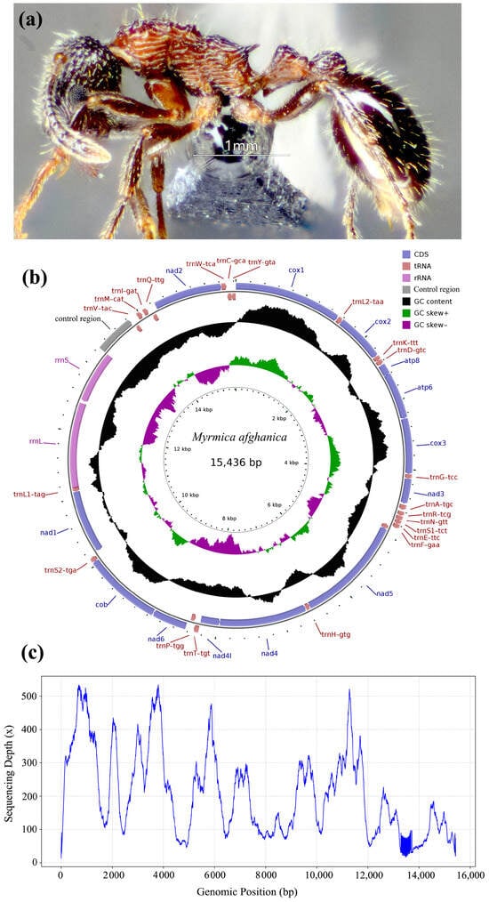
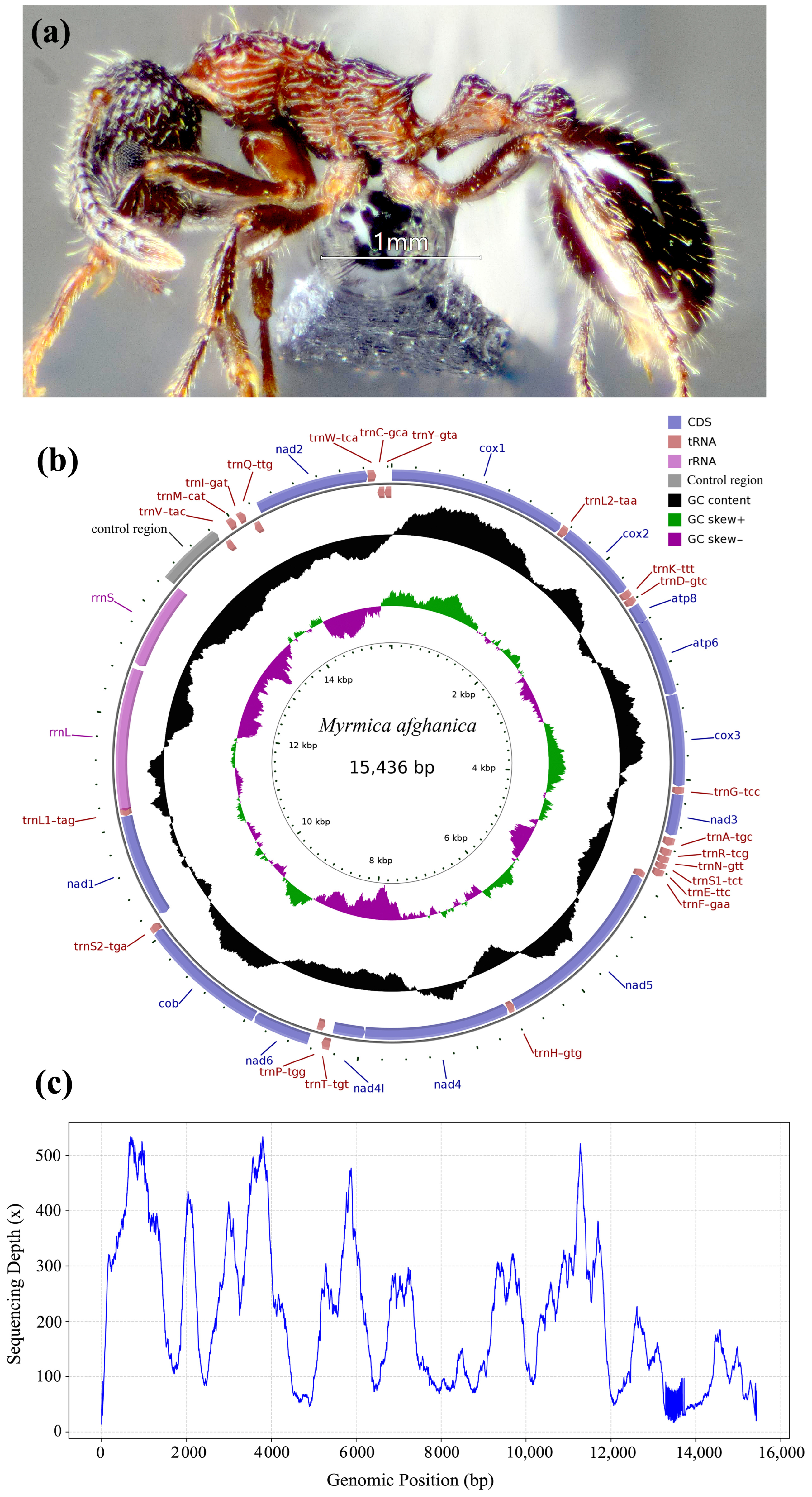
The complete mitochondrial genome of M. afghanica was successfully assembled and annotated (Figure 1). The circular mitogenome was 15,436 bp in length and encoded the typical complement of 37 mitochondrial genes: 13 protein-coding genes (PCGs), 22 transfer RNA (tRNA) genes, 2 ribosomal RNA genes (rrnS and rrnL), and one non-coding AT-rich control region. The overall nucleotide composition was strongly biased toward adenine and thymine, with a total AT content of 82.29%. The whole mitogenome exhibited an AT-skew of −0.02783 and a GC-skew of −0.29644. The base composition varied across different genomic regions (Table 1). The protein-coding region (10,999 bp) had an AT content of 80.55%, with negative AT-skew (−0.14115) and slightly negative GC-skew (−0.05193). The tRNA genes (1484 bp) and rRNA genes (2086 bp) showed high AT contents of 86.25% and 84.47%, respectively; both regions displayed positive AT-skew and positive GC-skew. The control region was 571 bp long and possessed the highest AT content (91.77%) among all regions, with negative AT-skew (−0.02735) and strongly negative GC-skew (−0.44593) (Table 1).
Figure 1.
Morphological and mitogenomic features of Myrmica afghanica. (a) The image of a worker of M. afghanica, lateral view; (b) the circular map of M. afghanica mitochondrial genome; (c) coverage depth of the complete mitochondrial genome (mean coverage = 208.73×).
Table 1.
Base composition of the mitochondrial genome of Myrmica afghanica.
Gene organization and structural features of the M. afghanica mitogenome are summarized in Table 2. Among the 37 annotated genes, 23 were located on the forward strand and 14 on the reverse strand. PCGs length varied substantially, ranging from 162 bp (atp8) to 1659 bp (nad5). The tRNA genes ranged from 58 bp (trnS1-tct) to 76 bp (trnT-tgt), while the large and small ribosomal RNA genes were 1300 bp (rrnL) and 786 bp (rrnS), respectively. Intergenic spacers and gene overlaps were distributed across the circular genome. Intergenic spacer lengths varied from 1 bp to 571 bp, the longest spacer located between trnV-tac and trnM-cat, corresponded to the AT-rich control region. Small gene overlaps ranged from −1 bp to −26 bp; notable overlapping events included trnK-ttt/trnD-gtc (−1 bp), atp8/atp6 (−7 bp), and trnL1-tag/rrnL (−26 bp) (Table 2).
Table 2.
The organization of the mitochondrial genome of Myrmica afghanica.
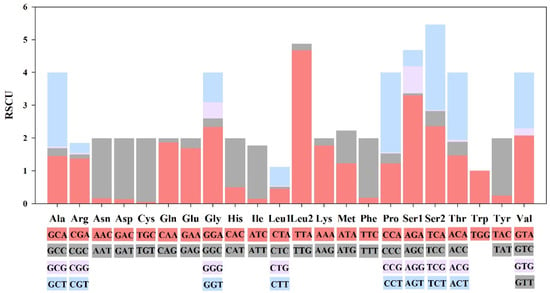
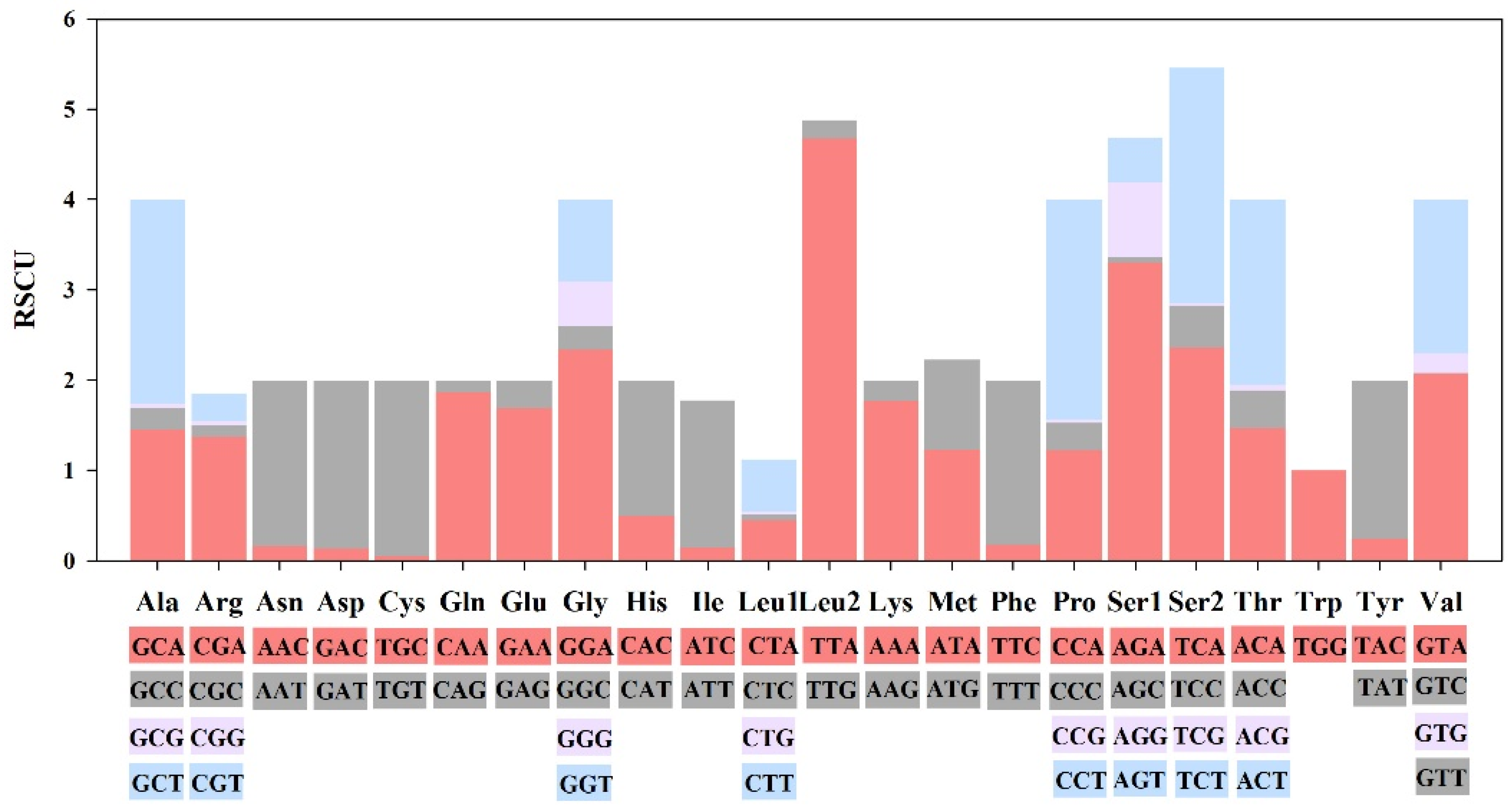
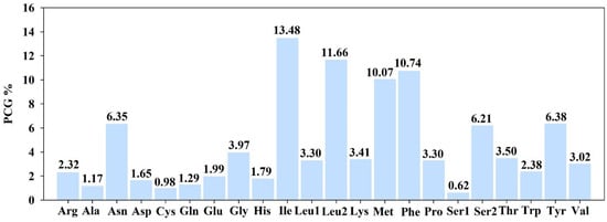
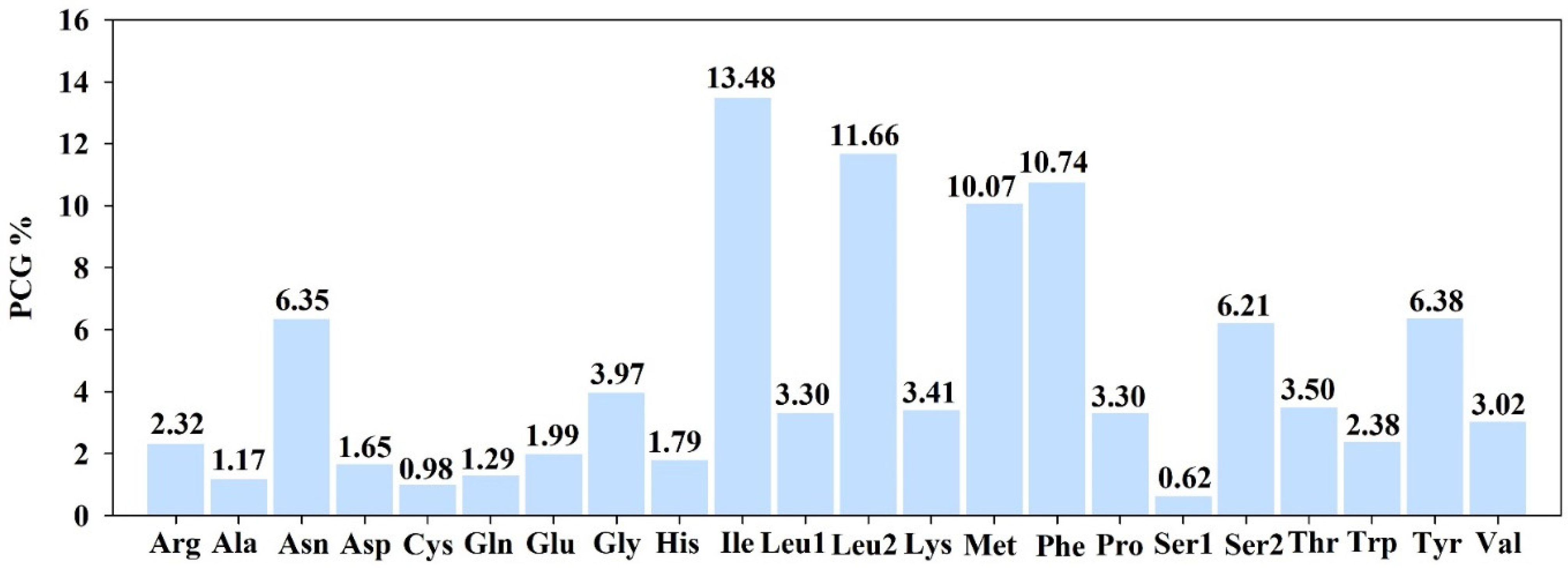
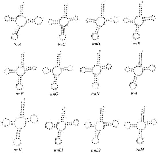
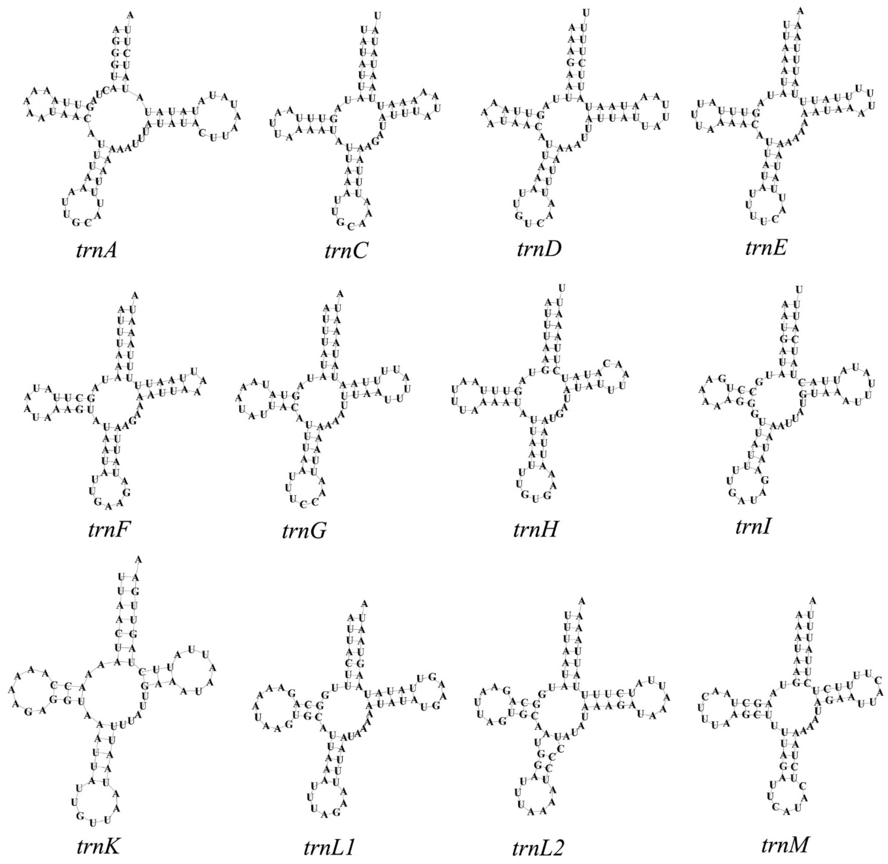
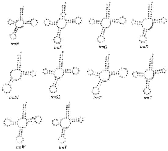
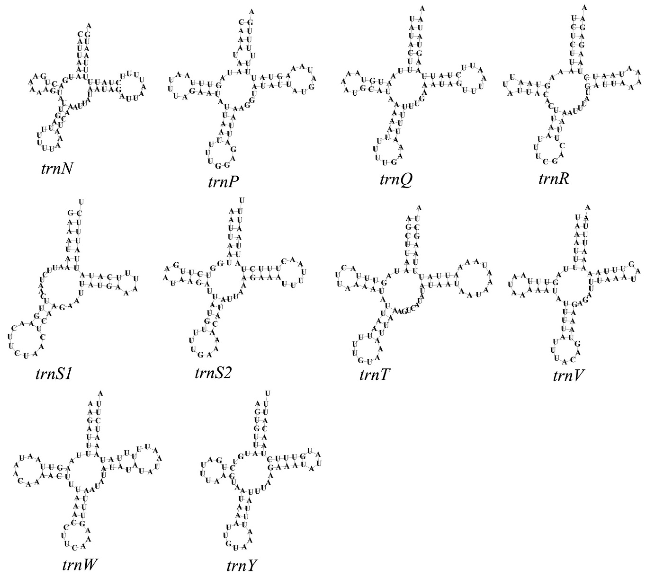
All PCGs utilized canonical invertebrate mitochondrial start codons (ATG, ATT, and ATA). Most PCGs terminated with complete TAA or TAG stop codons, while only nad2 used an incomplete single-T stop codon (Table 2). Relative synonymous codon usage (RSCU) analysis revealed strong AT-biased codon preference in M. afghanica (Figure 2). Codons ending in A or T are more abundant than those ending in G or C. Amino acid abundance analysis showed highly heterogeneous residue composition. Hydrophobic amino acids dominated the mitochondrial proteome: Ile (13.48%), Leu2 (11.66%), Phe (10.74%), and Met (10.07%) were the most abundant residues, collectively accounting for over 45% of all encoded amino acids (Figure 3). In comparison, Ser1 (0.62%) and Cys (0.98%) exhibited the lowest frequencies. Secondary structures of all 22 mitochondrial tRNA genes of M. afghanica were predicted and visualized (Figure 4 and Figure 5). Most tRNAs folded into canonical cloverleaf secondary structures composed of acceptor arms, DHU arms, anticodon arms, TΨC arms, and associated loops; only trnS1-tct lacked a stable DHU stem–loop.
Figure 2.
The relative synonymous codon usage (RSCU) in Myrmica afghanica mitogenome. Codons ending with A or T (red and blue columns); codons ending with G or C (pink and gray columns).
Figure 3.
The codon distribution in Myrmica afghanica mitogenome.
Figure 4.
Putative secondary structures for trnA, trnC, trnD, trnE, trnF, trnG, trnH, trnI, trnK, trnL1, trnL2, and trnM of Myrmica afghanica mitogenome.
Figure 5.
Putative secondary structures for trnN, trnP, trnQ, trnR, trnS1, trnS2, trnT, trnV, trnW, and trnY of Myrmica afghanica mitogenome.
3.2. Phylogenetic Topological Relationships
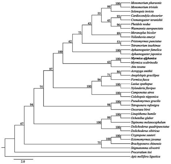
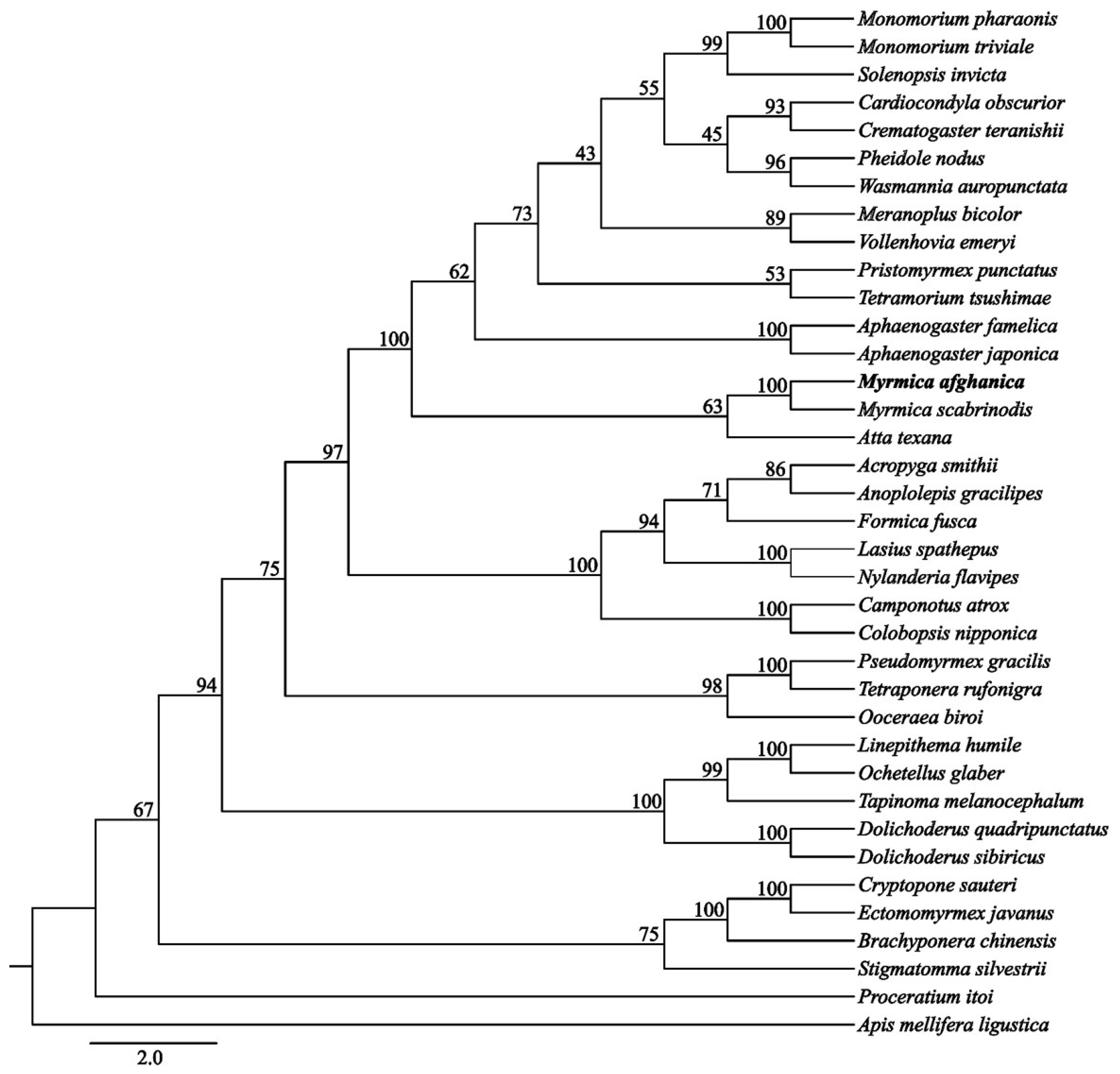
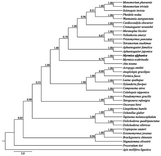
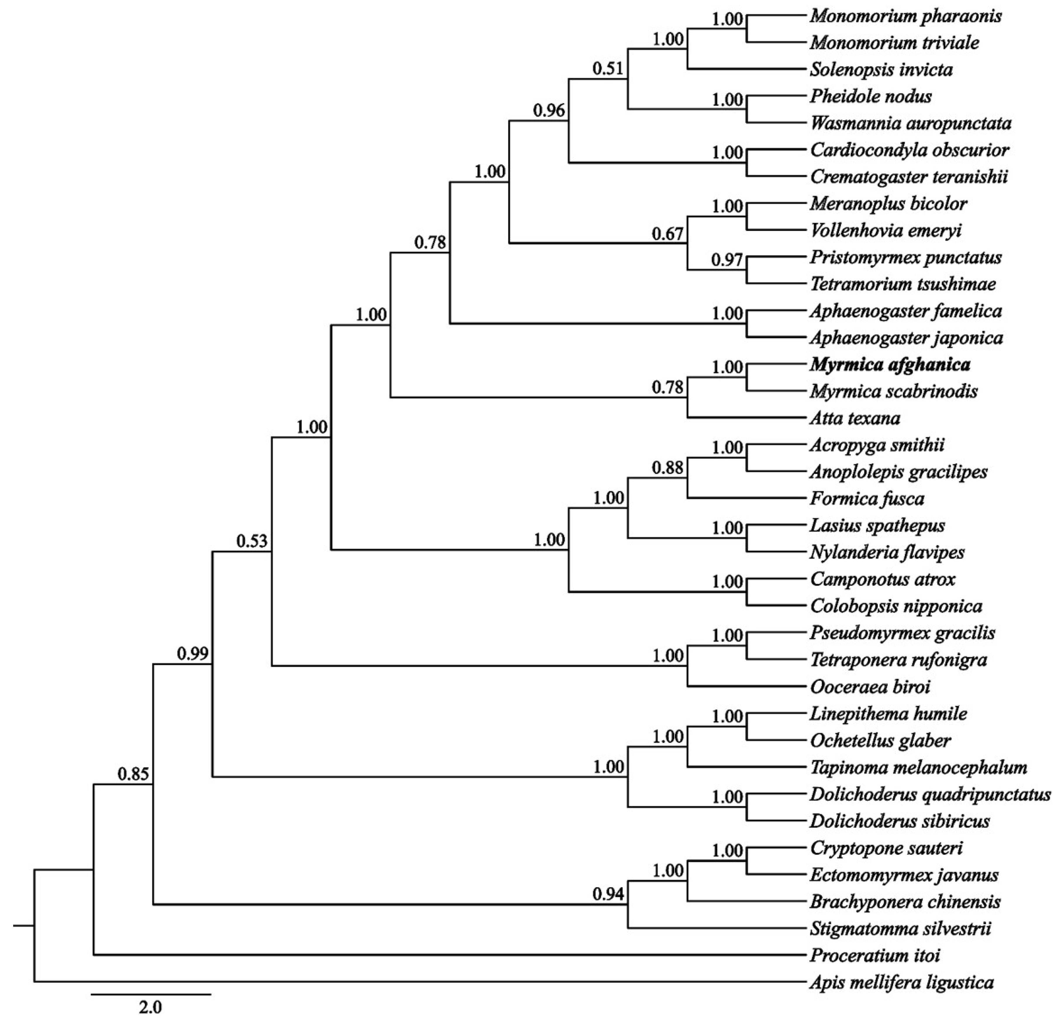
Phylogenetic relationships among Myrmica afghanica and 35 other ant species were inferred using 13 mitochondrial protein-coding genes. Both the Maximum Likelihood (ML) (Figure 6) and Bayesian inference (BI) (Figure 7) recovered all sampled ant species within a well-supported monophyletic clade. M. afghanica was supported as the sister species of M. scabrinodis, with high support from both methods: a bootstrap value (BS) of 100% in the ML tree and a posterior probability (PP) of 1.00 in the BI tree.
Figure 6.
Phylogenetic position of Myrmica afghanica inferred by Maximum Likelihood (ML) based on the 13 mitochondrial protein-coding gene sequences.
Figure 7.
Phylogenetic position of Myrmica afghanica inferred by Bayesian inference (BI) based on the 13 mitochondrial protein-coding gene sequences. Bootstrap values are shown next to the nodes.
4. Discussion
4.1. Genomic Architecture of the Myrmica afghanica Mitochondrial Genome
The newly assembled mitochondrial genome of M. afghanica retains the ancestral hymenopteran gene set (37 genes: 13 protein-coding genes, 22 transfer RNA genes, 2 ribosomal RNA genes and one AT-rich control region) without large-scale rearrangement, consistent with the conserved mitogenome organization observed in Myrmicinae [26,27,28,29]. Animal mitochondrial genomes typically exhibit compact organization, high AT content, and limited gene rearrangement within closely related taxonomic groups [5].
Nucleotide composition analysis revealed an extremely high AT content in M. afghanica, which is consistent with the universal AT bias characteristic of insect mitochondrial genomes [5,8,28,30,31]. Its total length (15,436 bp) and AT content (82.29%) are highly similar to those of its sister species D. scabrinodis (15,310 bp, AT = 82.2%). This close similarity in genome size and AT composition supports their close phylogenetic affinity, consistent with our tree topology. Codon usage and amino acid composition analyses demonstrated pronounced AT-preferred codon bias in M. afghanica mitochondrial PCGs. Nearly all high frequency synonymous codons terminate with A/T, while GC-rich codons are severely suppressed, which is tightly coupled with the genome-wide high AT background [8,28]. Notably, significant divergence between UUR-type and CUN-type leucine, as well as between UCN-type and AGN-type serine, suggests that codon usage shapes amino acid abundance patterns. Minor structural variation was limited to tRNA secondary structures, as commonly observed in other Myrmicinae ants: among all predicted tRNAs of M. afghanica, only trnS1-tct lacks a complete DHU stem–loop [26,28], a common deviation from the canonical cloverleaf tRNA structure in Hymenoptera mitogenomes [5].
4.2. Phylogenetic Relationships
Phylogenetic analyses focused on Myrmica, and Myrmicinae taxa recovered the monophyly of the genus Myrmica. Myrmica afghanica was strongly supported as the sister species of M. scabrinodis, providing mitochondrial evidence of its long-debated taxonomic placement within Myrmica [14,15]. Original morphological taxonomic work highlighted substantial uncertainty for M. afghanica: male diagnostic specimens were unavailable at the time of species description, so it is impossible to place it, with any certainty, in known Myrmica species groups relying only on worker and queen characters [20]. Based purely on female-caste morphology, two competing hypotheses were proposed: M. afghanica might belong either to the rubra-group (specifically the Central Asian dshungarica-complex including M. kryzhanovskii, M. dshungarica, M. juglandeti and M. ferganensis) or alternatively to the smythiesii-group [15]. Morphological comparisons noted that M. afghanica shares key traits of both groups, such as a long, slender antennal scape gently curved at base, short nearly straight frontal carinae, wide frons and narrow frontal lobes [15,20].
Morphological diagnoses also explicitly excluded close affinity with M. tibetana, which represents its own separate species group, with morphological affinities toward the scabrinodis-group rather than rubra- or smythiesii-group taxa [20]. Detailed morphological contrasts separate M. afghanica from members of the dshungarica-complex by features including clypeal shape, propodeal-spine length, rugosity of the head capsule, and petiole morphology [15]. Against this morphological background, our mitogenomic placement of M. afghanica as sister to M. scabrinodis of the scabrinodis-group offers new molecular evidence that contradicts the previous morphological hypotheses of placement within rubra-group or smythiesii-group, demonstrating the value of mitogenomic data for resolving ambiguous Myrmica taxonomy within Myrmica. M. afghanica is recorded from relatively high altitudes [16,17,18,19] and is speculated to primarily inhabit steppe and forest–steppe habitats associated with river-valley microenvironments [15]. This habitat association aligns with the known ecological profile of the scabrinodis species group, the clade where M. afghanica is recovered. Its close congener M. scabrinodis commonly occurs in cool, humid microhabitats such as moist meadows and riparian zones, suggesting overlapping microhabitat associations within this lineage [14].
5. Conclusions
This study sequenced and annotated the complete mitochondrial genome of Myrmica afghanica, providing a new mitogenomic resource for this taxonomically poorly resolved species. The 15,436 bp mitogenome exhibits the conserved Hymenoptera mitochondrial gene arrangement and contains the typical set of 37 mitochondrial genes. The key finding of this work is the mitochondrial phylogenetic result supporting M. afghanica as the sister species of M. scabrinodis. This molecular placement helps address the long-standing taxonomic uncertainty of M. afghanica. This newly generated mitogenome provides baseline molecular data to support future systematic classification and comparative mitogenomic investigations of Myrmica ants.
Supplementary Materials
The following supporting information can be downloaded at: https://www.mdpi.com/article/10.3390/cimb48111167/s1.
Author Contributions
X.Z. conceived and designed the study. X.Z., L.Z. and Z.X. performed data analysis. X.Z. and L.Z. wrote and revised the original manuscript. Z.X. revised the manuscript. All authors have read and agreed to the published version of the manuscript.
Funding
This study was supported by Yunnan Provincial Science and Technology Department-Applied Basic Research Joint Special Funds of Agriculture (202301BD070001-151) and the Open Fund Key Project of Forestry Discipline and Key Laboratory of Southwest Forestry University (Grant Number: LXXK-2025D14).
Institutional Review Board Statement
Not applicable.
Informed Consent Statement
Not applicable.
Data Availability Statement
The Illumina sequencing data and the assembled mitogenome sequence have been deposited in NCBI (https://www.ncbi.nlm.nih.gov/) accessed on 17 June 2026, with accession number PZ530274.
Acknowledgments
We thank F.P. Zhang for assistance with uploading the mitochondrial genome sequence to GenBank.
Conflicts of Interest
The authors declare no conflicts of interest.
References
- Wilson, E.O.; Hölldobler, B. The Rise of the Ants: A Phylogenetic and Ecological Explanation. Proc. Natl. Acad. Sci. USA 2005, 102, 7411–7414. [Google Scholar] [CrossRef] [Scilit]
- Bolton, B. An Online Catalog of the Ants of the World. Available online: http://www.antcat.org/ (accessed on 7 July 2026).
- Ward, P.S.; Brady, S.G.; Fisher, B.L.; Schultz, T.R. The Evolution of Myrmicine Ants: Phylogeny and Biogeography of a Hyperdiverse Ant Clade. Syst. Entomol. 2015, 40, 61–81. [Google Scholar] [CrossRef] [Scilit]
- Schultheiss, P.; Nooten, S.S.; Wang, R.X.; Wong, M.K.L.; Brassard, F.; Guénard, B. The Abundance, Biomass, and Distribution of Ants on Earth. Proc. Natl. Acad. Sci. USA 2022, 119, e2201550119. [Google Scholar] [CrossRef] [Scilit]
- Cameron, S.L. Insect Mitochondrial Genomics: Implications for Evolution and Phylogeny. Annu. Rev. Entomol. 2014, 59, 95–117. [Google Scholar] [CrossRef] [Scilit] [PubMed]
- Barrera, C.A.; Sosa-Calvo, J.; Schultz, T.R.; Rabeling, C.; Bacci, M. Phylogenomic Reconstruction Reveals New Insights into the Evolution and Biogeography of Atta Leaf-Cutting Ants (Hymenoptera: Formicidae). Syst. Entomol. 2022, 47, 13–35. [Google Scholar] [CrossRef] [Scilit]
- Ballard, J.W.O.; Whitlock, M.C. The Incomplete Natural History of Mitochondria. Mol. Ecol. 2004, 13, 729–744. [Google Scholar] [CrossRef] [Scilit]
- Gotzek, D.; Clarke, J.; Shoemaker, D. Mitochondrial Genome Evolution in Fire Ants (Hymenoptera: Formicidae). BMC Evol. Biol. 2010, 10, 300. [Google Scholar] [CrossRef] [Scilit]
- Barbosa, J.T.V.; Barbosa, M.S.; Morais, S.; Santana, A.E.G.; Almeida, C. Mitochondrial Genomes of Genus Atta (Formicidae: Myrmicinae) Reveal High Gene Organization and Giant Intergenic Spacers. Genet. Mol. Biol. 2019, 42, e20180055. [Google Scholar] [CrossRef] [Scilit]
- Janda, M.; Folková, D.; Zrzavý, J. Phylogeny of Lasius Ants Based on Mitochondrial DNA and Morphology, and the Evolution of Social Parasitism in the Lasiini (Hymenoptera: Formicidae). Mol. Phylogenet. Evol. 2004, 33, 595–614. [Google Scholar] [CrossRef] [Scilit]
- Babbucci, M.; Basso, A.; Scupola, A.; Patarnello, T.; Negrisolo, E. Is It an Ant or a Butterfly? Convergent Evolution in the Mitochondrial Gene Order of Hymenoptera and Lepidoptera. Genome Biol. Evol. 2014, 6, 3326–3343. [Google Scholar] [CrossRef] [Scilit]
- Li, T.; Xu, Z.; Liu, X.; Xiong, Z.; Zhang, X. Characterization and Phylogenetic Implication of Complete Mitochondrial Genome of Cataglyphis aenescens (Hymenoptera: Formicidae): Genomic Comparisons in Formicinae. Entomol. Res. 2023, 53, 45–54. [Google Scholar] [CrossRef] [Scilit]
- Jansen, G.; Savolainen, R.; Vepsäläinen, K. Phylogeny, Divergence-Time Estimation, Biogeography and Social Parasite-Host Relationships of the Holarctic Ant Genus Myrmica (Hymenoptera: Formicidae). Mol. Phylogenet. Evol. 2010, 56, 294–304. [Google Scholar] [CrossRef] [Scilit]
- Ebsen, J.R.; Boomsma, J.J.; Nash, D.R. Phylogeography and Cryptic Speciation in the Myrmica scabrinodis Nylander, 1846 Species Complex (Hymenoptera: Formicidae), and Their Conservation Implications. Insect Conserv. Divers. 2019, 12, 467–480. [Google Scholar] [CrossRef] [Scilit]
- Radchenko, A.; Elmes, G.W. Myrmica afghanica (Hymenoptera: Formicidae), a New Ant Species from Afghanistan. Zootaxa 2003, 375, 1–8. [Google Scholar] [CrossRef] [Scilit]
- Chen, C.; Xiong, Z.P.; Xu, Z.H.; Liu, X.; Qian, Y.H.; Qi, B.; Qian, Y.S.; Guo, N.Y. Distribution Pattern of Ant Species on the Northeast Slope of Qinghai-Tibet Plateau. For. Res. 2022, 35, 131–140. [Google Scholar] [CrossRef]
- Li, Z.Y.; Xu, Z.H.; Zhang, X.M.; Li, T.; Yin, X.D.; Duan, J.H. Ant Species Diversity in the Central and Northern Parts of the Western Sichuan Plateau in China. Diversity 2023, 15, 935. [Google Scholar] [CrossRef] [Scilit]
- Nong, Z.G.; Xiong, Z.P.; Xu, Z.H.; Wang, X.B.; Liu, X. Ant Diversity along Gradient in the Middle-Western Section of Tianshan Mountains in Xinjiang. J. Zhejiang A F Univ. 2025, 42, 143–152. [Google Scholar] [CrossRef]
- Li, B.; Xiong, Z.P.; Xu, Z.H.; Zhai, J.; Zhou, X.Y.; Xu, G.L. Distribution Patterns of Ant Species from Upper Yarlung Zangbo Valley and Southwest Slope of Qinghai-Tibet Plateau. J. Zhejiang A F Univ. 2022, 39, 590–597. [Google Scholar] [CrossRef]
- Radchenko, A.G.; Elmes, G.W. A Taxonomic Revision of the Ant Genus Myrmica Latreille (Hymenoptera, Formicidae) from the Himalaya. Entomol. Basil. 2001, 23, 237–276. [Google Scholar]
- Radchenko, A.G. Taxonomic Structure of the Genus Myrmica (Hymenoptera, Formicidae) of Eurasia. Note 1. Russ. J. Zool. 1994, 73, 39–51. [Google Scholar]
- Perna, N.T.; Kocher, T.D. Patterns of nucleotide composition at fourfold degenerate sites of animal mitochondrial genomes. J. Mol. Evol. 1995, 41, 353–358. [Google Scholar] [CrossRef] [Scilit] [PubMed]
- Katoh, K.; Standley, D.M. MAFFT multiple sequence alignment software version 7: Improvements in performance and usability. Mol. Biol. Evol. 2013, 30, 772–780. [Google Scholar] [CrossRef] [Scilit] [PubMed]
- Nguyen, L.T.; Schmidt, H.A.; von Haeseler, A.; Minh, B.Q. IQ-TREE: A fast and effective stochastic algorithm for estimating maximum-likelihood phylogenies. Mol. Biol. Evol. 2015, 32, 268–274. [Google Scholar] [CrossRef] [Scilit] [PubMed]
- Minh, B.Q.; Nguyen, M.A.T.; von Haeseler, A. Ultrafast approximation for phylogenetic bootstrap. Mol. Biol. Evol. 2013, 30, 1188–1195. [Google Scholar] [CrossRef] [Scilit] [PubMed]
- Zhang, X.M.; Li, T.; Liu, X.; Xu, Z.H. Characterization and Phylogenetic Implication of Complete Mitochondrial Genome of the Medicinal Ant Formica sinae (Hymenoptera: Formicidae): Genomic Comparisons in Formicidae. J. Med. Entomol. 2022, 59, 1971–1979. [Google Scholar] [CrossRef] [Scilit]
- Kolondam, B.J.; Tallei, T.E.; Koneri, R.; Abas, A.H.; Mamahit, J.M.E. A Review on Mitochondrial Genome of Ants (Hymenoptera: Formicidae). Heca J. Appl. Sci. 2023, 1, 48–53. [Google Scholar] [CrossRef] [Scilit]
- Lin, X.; Song, N. The First Complete Mitochondrial Genome of the Genus Pachycondyla (Formicidae, Ponerinae) and Insights into the Phylogeny of Ants. Genes 2023, 14, 1528. [Google Scholar] [CrossRef] [Scilit]
- Cardoso, D.C.; de Lima Baldez, B.C.; Vasconcelos, H.R.; Kalapothakis, E.; Cristiano, M.P. Mitogenomic Architecture of Mycetophylax conformis (Hymenoptera: Formicidae): Evidence for Conserved Gene Order and Strand Asymmetry in Myrmicinae. Biochem. Genet. 2026, 64, 5130–5146. [Google Scholar] [CrossRef] [Scilit]
- Sun, Z.; Wan, D.G.; Murphy, R.W.; Ma, L.; Zhang, X.S.; Huang, D.W. Comparison of Base Composition and Codon Usage in Insect Mitochondrial Genomes. Genes Genom. 2009, 31, 65–71. [Google Scholar] [CrossRef] [Scilit]
- Song, N.; Zhang, H.; Zhao, T. Insights into the Phylogeny of Hemiptera from Increased Mitogenomic Taxon Sampling. Mol. Phylogenet. Evol. 2019, 137, 236–249. [Google Scholar] [CrossRef] [Scilit]

Disclaimer/Publisher’s Note: The statements, opinions and data contained in all publications are solely those of the individual author(s) and contributor(s) and not of MDPI and/or the editor(s). MDPI and/or the editor(s) disclaim responsibility for any injury to people or property resulting from any ideas, methods, instructions or products referred to in the content. |
© 2026 by the authors. Licensee MDPI, Basel, Switzerland. This article is an open access article distributed under the terms and conditions of the Creative Commons Attribution (CC BY) license.