


Abstract
Claudin 18.2 (CLDN18.2) is an emerging therapeutic biomarker in gastric adenocarcinoma. This study evaluated the concordance of CLDN18.2 expression between pretreatment biopsies and corresponding resection specimens and explored its associations with clinicopathological features and survival outcomes. This retrospective single-center study included 71 patients, 58 of whom received neoadjuvant chemotherapy. CLDN18.2 positivity was defined as moderate-to-strong (2+/3+) membranous staining in ≥75% of tumor cells. Concordance was assessed using Cohen’s kappa, positive percent agreement (PPA), negative percent agreement (NPA), and intraclass correlation coefficient (ICC). CLDN18.2 positivity was identified in 26.8% of biopsies and 21.1% of resection specimens. Categorical status was concordant in 65 of 71 patients (91.5%; κ = 0.769; p < 0.001), with a PPA of 73.7% and an NPA of 98.1%. Continuous analysis showed a high ICC (0.817; 95% confidence interval (CI), 0.722–0.881; p < 0.001). Six patients had discordant categorical results, all of whom had received neoadjuvant therapy. Exploratory survival analyses did not identify a statistically significant association between pretreatment biopsy CLDN18.2 status and overall survival (OS) or recurrence-free survival (RFS); however, the limited sample size and statistical precision preclude definitive conclusions regarding its prognostic significance. Overall, CLDN18.2 showed substantial biopsy–resection concordance, although positive agreement was lower than negative agreement. Discordant findings should be interpreted cautiously because treatment-related effects cannot be distinguished from spatial heterogeneity and sampling variation.
1. Introduction
Stomach cancer remains a major global health burden. With approximately 1.09 million new cases and 769,000 deaths reported annually worldwide, it ranks fifth in cancer incidence and fourth in cancer-related mortality [1]. The high mortality associated with gastric cancer is largely attributable to its frequent diagnosis at an advanced stage and its aggressive biological behavior. Consequently, improving early detection and developing more effective treatment strategies remain important clinical priorities.
In recent years, perioperative and neoadjuvant chemotherapy (NAC) have become integral components of the treatment of locally advanced gastric cancer. The FLOT4-AIO trial demonstrated that perioperative treatment with FLOT (5-fluorouracil, leucovorin, oxaliplatin, and docetaxel) significantly improved overall survival (OS) compared with the previously used cisplatin–epirubicin–fluorouracil (ECF) and capecitabine–cisplatin–epirubicin (ECX) regimen [2]. Based on these findings, FLOT has become a commonly preferred perioperative chemotherapy regimen for patients with resectable, locally advanced gastric and gastroesophageal junction (GEJ) adenocarcinomas [3,4]. Although NAC can reduce tumor burden, treat occult micrometastatic disease, and increase the likelihood of curative resection, treatment response remains heterogeneous, and a substantial proportion of patients derive limited benefit [5,6].
The identification of reliable molecular biomarkers is therefore important for improving patient selection and guiding personalized treatment strategies in gastric cancer. Biomarkers including human epidermal growth factor receptor 2 (HER2), programmed death-ligand 1 (PD-L1), microsatellite instability (MSI), and mismatch repair (MMR) status are now routinely evaluated in clinical practice and may influence therapeutic decision making [7,8]. In addition to these established biomarkers, claudin 18.2 (CLDN18.2) has emerged as a promising therapeutic target in gastric cancer in recent years [9,10,11].
CLDN18.2 is a member of the claudin family of tight junction proteins and plays an important role in maintaining cell–cell adhesion and epithelial barrier integrity [12]. The CLDN18 gene encodes two major isoforms: CLDN18.1, which is predominantly expressed in the respiratory epithelium, and CLDN18.2, which is primarily expressed in the gastric mucosa. Under physiological conditions, CLDN18.2 is localized mainly to the apical surface of gastric epithelial cells, where it is relatively inaccessible to circulating antibodies. During malignant transformation, however, disruption of epithelial polarity and loss of normal cell–cell junctions result in exposure of CLDN18.2 over the cell surface, making it more accessible to the immune system and therapeutic antibodies [13,14]. This distinctive expression pattern has led to considerable interest in CLDN18.2 as a potential therapeutic target in gastric cancer.
The reported prevalence of CLDN18.2 expression in gastric cancer varies considerably, ranging from approximately 30% to 60% across different studies. This variability may be partly attributable to differences in antibody clones, immunohistochemical (IHC) scoring systems, and the cutoff values used to define positivity [15,16,17]. In particular, the use of different thresholds, including 50% and 75% of tumor cells showing membranous staining, can substantially affect the reported prevalence and complicate comparisons between studies [15,16,17,18].
Among the therapeutic strategies targeting CLDN18.2, zolbetuximab has demonstrated clinical efficacy in two pivotal phase III randomized controlled trials. The SPOTLIGHT trial evaluated zolbetuximab combined with mFOLFOX6, whereas the GLOW trial investigated its combination with capecitabine and oxaliplatin (CAPOX). Both trials enrolled previously untreated patients with CLDN18.2-positive, HER2-negative, locally advanced unresectable or metastatic gastric or gastroesophageal junction adenocarcinoma. CLDN18.2 positivity was defined as moderate-to-strong membranous staining in ≥75% of tumor cells. Both trials demonstrated significant improvements in progression-free survival and overall survival with the addition of zolbetuximab to first-line chemotherapy [19,20]. These findings established the clinical relevance of CLDN18.2 assessment in advanced disease and provided the rationale for using the ≥75% positivity threshold in the present study, although identical numerical thresholds do not establish analytical equivalence between immunohistochemical assays.
An important consideration in the clinical implementation of CLDN18.2 testing is the stability of its expression following systemic therapy. NAC may induce substantial changes in the tumor microenvironment and tumor cell phenotype, potentially affecting the expression of molecular biomarkers [21,22,23]. Consequently, if CLDN18.2 expression is altered by neoadjuvant treatment, the biomarker status determined from a pretreatment diagnostic biopsy may not accurately represent the CLDN18.2 status of the residual tumor at the time of surgery. Such discordance may have important implications for subsequent treatment decisions, particularly when considering CLDN18.2-directed therapies in the postoperative or recurrent disease setting.
The prognostic significance of CLDN18.2 in gastric cancer remains uncertain, with conflicting findings reported across the literature. While some studies have associated CLDN18.2 positivity with more favorable clinical outcomes [24], others have found no significant association with prognosis [15,25], whereas some have reported an unfavorable prognostic impact [26,27]. These discrepancies may reflect differences in patient populations, disease stages, treatment strategies, IHC assessment methods, and cutoff values used to define CLDN18.2 positivity.
Previous paired-specimen studies, including that of Nagano et al., have highlighted the potential influence of intratumoral heterogeneity and neoadjuvant chemotherapy on CLDN18.2 expression [23]. Building on these findings, we aimed to characterize biopsy–resection concordance in a predominantly neoadjuvant-treated cohort using both directional categorical agreement measures and continuous agreement analysis of staining percentages, while exploring associations with clinicopathological characteristics and survival outcomes.
2. Materials and Methods
2.1. Patient Selection and Study Design
This retrospective study included patients who were diagnosed with gastric adenocarcinoma at the Department of Pathology Çukurova University Faculty of Medicine between May 2016 and May 2026. The inclusion criteria were: (1) histopathologically confirmed gastric adenocarcinoma; (2) receipt of NAC followed by surgical resection or upfront surgical resection; (3) availability of both diagnostic biopsy and surgical resection specimens; and (4) availability of sufficient archived tissue for histopathological and IHC evaluation. Patients were excluded if they had any of the following: (1) insufficient tissue material; (2) incomplete clinical or pathological data; or (3) histological diagnoses other than gastric adenocarcinoma, including gastrointestinal stromal tumor, lymphoma, and neuroendocrine tumor.
The initial screening cohort was identified through a retrospective, chronological review of consecutive patients with gastric neoplasms recorded in the institutional pathology database between May 2016 and May 2026, rather than through selective archival sampling. A total of 237 patients were initially screened. Of these, 113 patients with gastrointestinal stromal tumors, 20 with lymphoma, and 25 with neuroendocrine tumors were excluded, leaving 79 patients with gastric adenocarcinoma. Among these 79 patients, four were excluded due to insufficient tissue material and two were excluded due to incomplete clinical or pathological data. Consequently, 73 patients were initially evaluated for paired biopsy–resection assessment.
To ensure anatomical comparability between paired specimens, the presence of viable primary gastric carcinoma in both the pretreatment biopsy and the corresponding surgical resection specimen was required for inclusion in the primary analysis. Among the 73 eligible patients, two were excluded because no residual viable carcinoma was identified at the primary gastric tumor site following neoadjuvant chemotherapy (ypT0), although viable metastatic carcinoma remained in regional lymph nodes. These patients were not considered complete pathological responders because of persistent nodal disease. Comparison of pretreatment primary tumor biopsies with postoperative lymph node metastases would have introduced an anatomical compartment mismatch; therefore, both patients were excluded from the primary analytical cohort.
The final study cohort comprised 71 patients, including 58 who received neoadjuvant chemotherapy and 13 who underwent upfront surgery. The patient selection process is summarized in Supplementary Figure S1.
The study protocol was approved by the Ethics Committee of the Faculty of Medicine at Çukurova University (Decision No. 70, dated 10 July 2026) and the study was conducted in accordance with the principles of the Declaration of Helsinki.
Demographic and clinicopathological data, including age, sex, tumor location, clinical stage, NAC regimen, type of surgical procedure, pathological stage, and survival outcomes, were retrospectively collected from electronic medical records and pathology reports. Tumor location was classified as corpus, cardia, antrum, or GEJ. Histological subtypes were classified as tubular adenocarcinoma, signet-ring cell carcinoma, mixed adenocarcinoma, poorly cohesive carcinoma, or mucinous adenocarcinoma according to the World Health Organization (WHO) 5th edition classification [28]. Tumors were categorized according to the Lauren classification as intestinal, diffuse, or mixed type [29]. Pathological T, N, and M categories were determined according to the eighth edition of the American Joint Committee on Cancer (AJCC) TNM staging system [30]. For patients who underwent upfront surgery (n = 13), pathological stage groups were assigned using the AJCC eighth-edition pTNM prognostic stage-group definitions. For patients who received neoadjuvant chemotherapy (n = 58), post-treatment pathological stage groups were assigned using the distinct AJCC eighth-edition ypTNM prognostic stage-group definitions. Because these staging systems use different prognostic stage-group assignments, stage groups were evaluated separately according to treatment status and were not pooled across the two cohorts. According to the eighth edition of the AJCC staging system and the College of American Pathologists (CAP) protocol for gastric carcinoma, tumors involving the gastroesophageal junction (GEJ) whose midpoint is located more than 2 cm distal to the junction within the proximal stomach are classified and staged as gastric carcinomas. The five GEJ tumors included in the present cohort fulfilled this anatomical criterion and were therefore staged according to the same gastric cancer TNM system applied to the remaining cases [30,31].
2.2. Histopathological Evaluation
Hematoxylin and eosin (H&E)-stained slides from all cases were reevaluated by a pathologist experienced in gastrointestinal pathology. Diagnostic biopsy specimens containing at least three tissue fragments were included in the histopathological assessment. For each patient, one representative formalin-fixed, paraffin-embedded (FFPE) block from the pretreatment biopsy and one representative block from the corresponding surgical resection specimen were selected to ensure adequate representation of viable primary tumor tissue. Each selected specimen was required to contain at least 100 viable tumor cells. A single 4-µm-thick section from each selected block was stained for CLDN18.2 and evaluated. All viable tumor components within the stained section were assessed, including areas showing heterogeneous staining. As only one section from each specimen was evaluated, scores were not averaged or combined across multiple blocks or sections.
In patients who received neoadjuvant chemotherapy, the pathological response to treatment was assessed using the tumor regression scoring system recommended by the College of American Pathologists (CAP) [31]. Tumor regression was graded as follows: Score 0: no viable cancer cells (complete response); Score 1: single cells or rare small groups of cancer cells (near-complete response); Score 2: residual cancer with evident tumor regression, but involving more than single cells or rare small groups of cancer cells (partial response); and Score 3: extensive residual cancer with no evident tumor regression (poor or no response). Sizable pools of acellular mucin, when present, were not considered residual tumor.
2.3. Immunohistochemical Staining and Scoring
CLDN18.2 IHC was performed on 4-µm-thick formalin-fixed, paraffin-embedded (FFPE) tissue sections using the Ventana BenchMark ULTRA automated staining platform (Roche Diagnostics /Ventana Medical Systems, Inc., Tucson, AZ, USA). Biopsy specimens were fixed in 10% neutral phosphate-buffered formalin for at least 6 h, whereas surgical resection specimens were fixed for at least 24 h. Deparaffinization and heat-induced epitope retrieval were performed on the automated platform using ULTRA CC1. A mouse monoclonal anti-Claudin 18 antibody (clone 43-14A; Abcam Cambridge, UK; catalog no. ab314690; stock concentration, 1.046 mg/mL) was used at a 1:5000 dilution with a 60-min incubation. Immunodetection was performed using the Ventana ultraView Universal DAB Detection Kit (Ventana Medical Systems, Inc., Tucson, AZ, USA), followed by hematoxylin counterstaining and bluing. Matched biopsy and resection sections were stained concurrently in the same staining run using identical antigen retrieval, antibody incubation, immunodetection, and staining conditions. The laboratory-developed staining protocol used in this study was not analytically cross-validated against the reference VENTANA CLDN18 assay.
Normal gastric foveolar epithelium served as the positive control. In surgical specimens, CLDN18.2 assessment was restricted to residual viable primary gastric tumor tissue. Regional lymph node metastases were not used as substitutes for primary tumor tissue in paired biopsy–resection comparisons.
All CLDN18.2-stained sections were evaluated separately by a single pathologist experienced in gastrointestinal pathology, who was fully blinded to the corresponding paired specimen results, neoadjuvant treatment information, and clinical outcomes at the time of scoring. Pretreatment biopsy and surgical resection specimens were assessed using identical predefined staining intensity and percentage criteria. No independent reassessment by a second pathologist was performed. Only specimens containing at least 100 viable tumor cells were considered evaluable. Staining intensity was graded on a four-tiered scale: 0, no staining; 1+, weak membranous staining detectable at ×20 objective magnification and generally requiring ×40 objective magnification for confirmation; 2+, moderate membranous staining readily visible at ×10 objective magnification, with ×20 objective magnification used when necessary to confirm specific membranous staining; and 3+, strong, distinct brown membranous staining in a characteristic “chicken-wire” pattern readily appreciable at low magnification (×5 objective). These magnification levels were used as visual aids to standardize staining intensity assessment and did not imply that different tumor areas were evaluated according to different scoring criteria.
The overall staining pattern and degree of heterogeneity were initially assessed at low magnification (×5 objective). In tumors showing heterogeneous staining, all viable tumor components present in the stained section were comprehensively evaluated. The proportions of tumor cells showing each staining intensity (0, 1+, 2+, and 3+) were rec-orded to accurately determine the proportion exhibiting moderate-to-strong (2+/3+) membranous staining. Areas of dysplasia were excluded from the assessment. Only membranous staining was considered for CLDN18.2 evaluation. The percentage of tumor cells showing membranous staining with an intensity of ≥2+ was recorded [18]. Using the numerical positivity threshold adopted in the SPOTLIGHT and GLOW clinical trials, tumors demonstrating moderate-to-strong (2+/3+) membranous staining in ≥75% of viable tumor cells were classified as CLDN18.2-positive [19,20]. For patient-level analyses of clinicopathological characteristics, recurrence, and survival, CLDN18.2 status was determined exclusively from the pretreatment diagnostic biopsy. Patients were classified as CLDN18.2-positive if the biopsy met the ≥75% positivity criterion, and as CLDN18.2-negative otherwise. Resection specimen results were used to assess paired biopsy–resection concordance and were not combined with biopsy results to assign patient-level CLDN18.2 status.
2.4. Statistical Analysis
Categorical variables were summarized as frequencies (n) and percentages (%), while continuous variables were expressed as mean ± standard deviation or median (interquartile range [IQR]/minimum–maximum), as appropriate. The normality of the distribution of continuous data was evaluated using the Shapiro–Wilk test. Continuous variables were compared between groups using Student’s t-test when the assumptions of normality were met, and the Mann–Whitney U test when they were not. To compare categorical variables between groups, the Pearson chi-square test was used; Fisher’s exact test was preferred when expected cell counts were insufficient. Cohen’s kappa (κ) coefficient was used to assess concordance between pretreatment biopsies and matched resection specimens. To characterize directional asymmetry in categorical agreement, the pretreatment biopsy served as the reference specimen; positive percent agreement (PPA) and negative percent agreement (NPA) were calculated, respectively, as the proportions of biopsy-positive and biopsy-negative cases, that retained the same categorical status in the corresponding surgical specimen. To account for sample size constraints and small denominators, exact 95% binomial confidence intervals (CIs) were computed for overall agreement, PPA, and NPA using the Clopper–Pearson method. For subgroups with no observed discordance (e.g., the untreated cohort), exact Clopper–Pearson binomial confidence intervals were presented to reflect sampling uncertainty rather than asymptotic estimates. PPA and NPA were interpreted strictly as directional agreement proportions rather than as diagnostic sensitivity or specificity against an absolute gold standard. Inter-specimen reliability of tumor-cell staining percentages was evaluated using intraclass correlation coefficients (ICC) based on a two-way random-effects, single-rating, absolute agreement model ICC(A,1). Continuous agreement was further illustrated using a paired scatterplot featuring the line of identity (y = x) and the 75% positivity threshold. Paired differences in CLDN18.2 staining percentages were calculated by subtracting the pretreatment biopsy percentage from the corresponding surgical resection percentage (resection minus biopsy) and were summarized using the median and interquartile range (IQR). The associations between pretreatment biopsy CLDN18.2 status and clinicopathological characteristics were evaluated. Overall survival (OS) was defined as the interval from the date of initial diagnosis to death from any cause. Patients who were alive were censored on the date of their last known follow-up. Recurrence-free survival (RFS) was defined as the interval from the date of surgical resection to the first documented disease recurrence or death from any cause, whichever occurred first. Patients who remained alive without documented recurrence were censored at the date of their last disease-free follow-up. Microscopically negative surgical margins were documented at the primary gastric tumor resection site in all 71 patients. This finding referred specifically to the resection margins of the primary tumor and did not imply complete clearance of distant metastatic disease in patients with M1 status. Therefore, the 12 patients with M1 disease were excluded from the recurrence-free survival analysis, which was restricted to 59 patients with M0 disease and microscopically negative primary-tumor resection margins. The administrative cutoff date for survival follow-up was 30 May 2026. OS and RFS were estimated using the Kaplan–Meier method, and survival curves were compared using the log-rank test. Median follow-up duration was calculated using the reverse Kaplan–Meier method. Cox proportional hazards regression analysis was performed to evaluate factors associated with OS. The proportional hazards assumption was formally evaluated by introducing time-dependent covariate interaction terms (time-by-covariate interaction analysis) into the model. Given the limited number of death events (n = 38), the multivariable Cox regression model was deliberately restricted to a clinically justified set of prognostic variables rather than constructed by selecting covariates solely on the basis of univariable statistical significance. The model included pretreatment biopsy CLDN18.2 status as the primary biomarker of interest, pathological nodal category (pN/ypN) as an indicator of regional disease burden, histological grade as a measure of tumor differentiation, and Lauren classification as a means to account for differences in histological phenotype. The multivariable analysis included 71 patients with 38 death events and was considered exploratory because the number of events was limited relative to the number of model parameters.
Tumor location was analyzed as a nominal categorical variable using indicator (dummy) coding, with the antrum as the reference category. The Lauren classification was similarly modeled using the intestinal type as the reference category. Histological grade, pN/ypN category, and CAP primary tumor regression grade (TRG) were modeled as ordinal variables, with each hazard ratio representing the relative change in mortality hazard associated with a one-category increase. This parameterization assumes a constant multiplicative change in hazard across successive categories. TRG was evaluated separately in a univariable Cox regression analysis restricted to the 58 patients who received neoadjuvant chemotherapy, among whom 31 deaths occurred. TRG was not included in the multivariable model. Recurrence and receipt of adjuvant chemotherapy were not included in the Cox regression analysis because they represent post-surgical events or exposures and were not modeled as time-dependent covariates. Hazard ratios (HRs) were presented with their corresponding 95% confidence intervals (CIs). All statistical analyses were performed using IBM SPSS 20 (IBM Corp., Armonk, NY, USA). A two-sided p-value of <0.05 was considered statistically significant.
3. Results
3.1. Clinicopathological Characteristics
A total of 71 patients were included in the study. The mean age was 58.6 ± 10.1 years (range, 34–82 years), and males comprised the majority of the cohort (n = 49, 69.0%), corresponding to a male-to-female ratio of approximately 2.2:1.
Tumors most frequently arose in the gastric corpus (42.3%), followed by the cardia (26.8%) and the antrum (23.9%), whereas GEJ tumors were uncommon (7%). Histologically, tubular adenocarcinoma was the predominant subtype (47.9%); according to the Lauren classification, intestinal-type tumors slightly outnumbered diffuse-type tumors (52.1% vs. 40.8%). More than half (54.9%) of the tumors were poorly differentiated (grade 3). Peritoneal metastases were identified in 8 patients (11.3%).
Using a predefined cutoff of membranous staining in ≥75% of tumor cells, CLDN18.2 positivity was detected in 19 pretreatment biopsy specimens (26.8%) and in 15 corresponding resection specimens (21.1%). The baseline clinicopathological characteristics of the study cohort are summarized in Table 1.
Table 1.
Clinicopathological characteristics of the study cohort (n = 71).
3.2. Treatment Characteristics and Clinical Outcomes
Neoadjuvant chemotherapy was administered to 58 patients (81.7%), while 13 (18.3%) underwent upfront surgery. Among the 58 patients who received neoadjuvant treatment, FLOT was the most frequently administered regimen (63.8%), followed by DCF (15.5%), FOLFIRINOX (10.3%), and FOLFOX (3.4%). Four additional patients (6.9%) received combination regimens incorporating immune checkpoint inhibitors or targeted agents.
Total gastrectomy was performed on 54 (76.1%) patients. R0 resection was achieved in all 71 patients (100%), with no R1 or R2 resections recorded. Pathological examination demonstrated predominantly advanced disease, with T3–T4 pathological categories (pT/ypT) accounting for 83.1% of cases. Lymph node metastasis was identified in 48 patients (67.6%), while pathological M1 disease (pM1/ypM1) was present in 12 patients (16.9%). According to the CAP primary tumor regression grading system, TRG 1 (near-complete response) was observed in 5 patients (8.6%), TRG 2 (partial response) in 20 patients (34.5%), and TRG 3 (poor or no response) in 33 patients (56.9%). The median number of completed neoadjuvant treatment cycles was 4 (mean, 4.3 ± 2.1).
Following surgery, 46 patients received adjuvant chemotherapy, most commonly with FLOT (n = 27) or capecitabine (n = 8). The median number of completed adjuvant treatment cycles was 4 (mean, 3.8 ± 1.2). The pathological staging, treatment characteristics, and clinical outcomes are summarized in Table 2. The mean interval between pretreatment biopsy and surgical resection was 117.8 days.
Table 2.
Pathological staging, treatment characteristics, and clinical outcomes.
3.3. Follow-Up and Recurrence Patterns
During follow-up, postoperative disease recurrence was documented in 14 of the 59 patients with nonmetastatic (M0) disease (23.7%). The 12 patients with M1 disease were excluded from the recurrence-free survival analysis, as progression of pre-existing metastatic disease was not considered recurrence following disease-free resection. In the overall cohort of 71 patients, 38 patients (53.5%) died, whereas 33 patients (46.5%) were alive at the last follow-up.
Among patients with documented recurrence, the peritoneum was the most frequent site of recurrence, occurring in 9 of 14 patients (64.3%). Peritoneal recurrence occurred in isolation in 2 patients and in combination with other metastatic sites in 7 patients. Pulmonary and locoregional recurrences were the next most frequent sites, each occurring in 5 patients (35.7%), followed by hepatic recurrence in 4 patients (28.6%) and bone recurrence in 1 patient (7.1%). 5 patients (35.7%) developed recurrence at a single anatomical site, whereas 9 patients (64.3%) exhibited multifocal recurrence involving two or more sites, most commonly with concomitant peritoneal involvement.
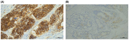
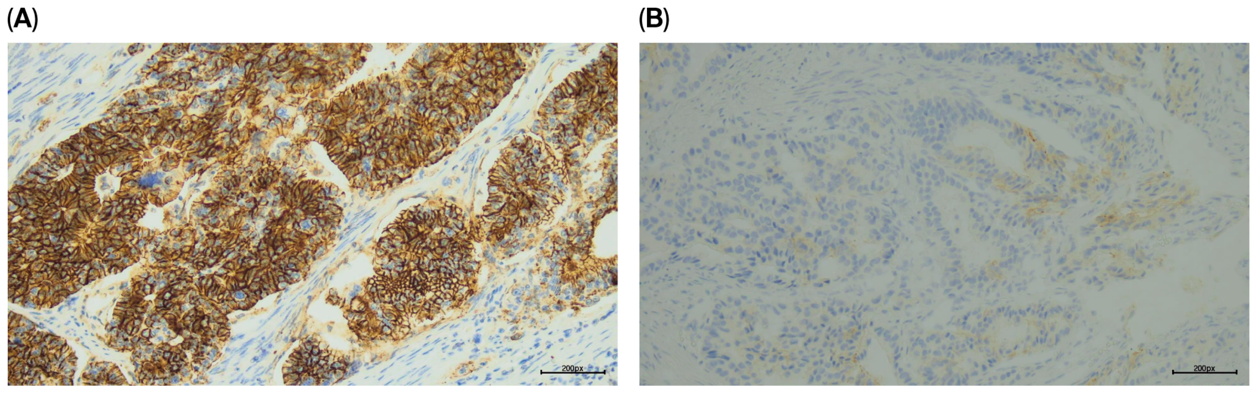
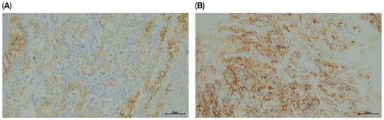
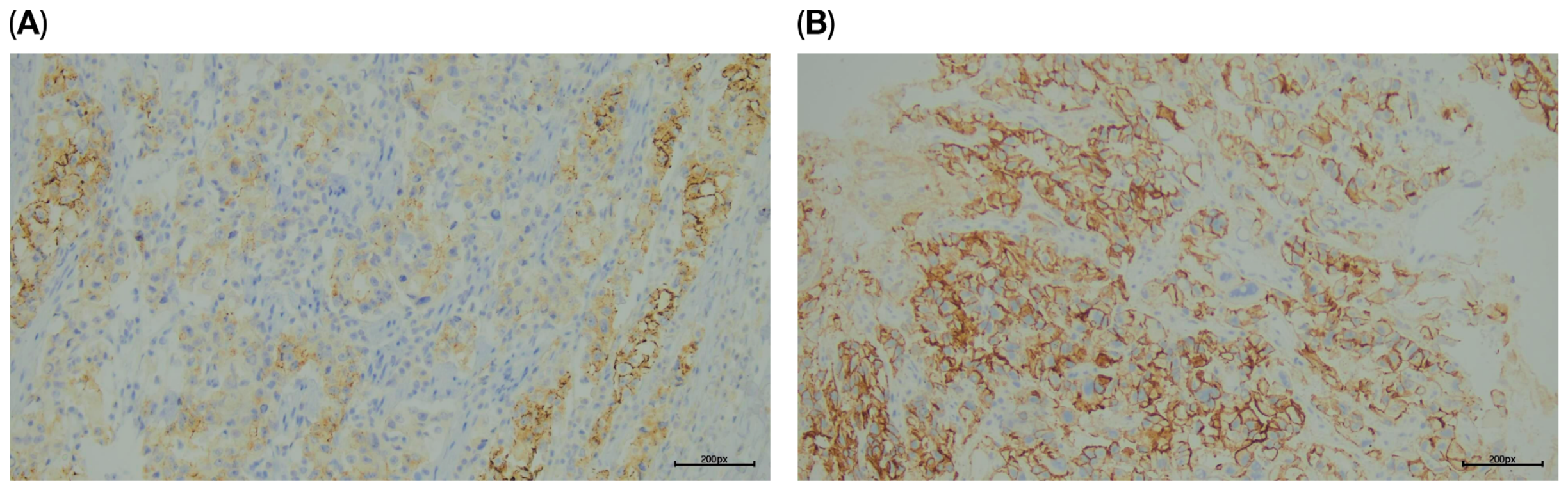
The concordance between CLDN18.2 expression in pretreatment biopsy specimens and corresponding surgical specimens was assessed in all 71 patients. CLDN18.2 status was concordant in 65 of 71 cases (91.5%); 51 patients were negative and 14 were positive in both specimens. Discordance was observed in 6 patients (8.5%), all of whom had received neoadjuvant chemotherapy. The discordance rate was 10.3% (6/58) among patients who received neoadjuvant therapy and 0% (0/13) among those who underwent upfront surgery; however, this difference was not statistically significant (Fisher’s exact test, p = 0.584). Among these 6 discordant cases, 5 were CLDN18.2-positive in the pretreatment biopsy but negative in the matched resection specimen (Figure 1), while 1 showed the opposite pattern (Figure 2). Among the 5 positive-to-negative cases, three patients had received FLOT, one patient had received FOLFIRINOX, and one patient had received FLOT combined with durvalumab. The single patient who showed a negative-to-positive categorical change had received FLOT.
Figure 1.
Representative images of discordant CLDN18.2 expression in matched pretreatment biopsy and surgical resection specimens. (A) Pretreatment biopsy showing strong membranous CLDN18.2 staining (3+) in approximately 90% of viable tumor cells (2+/3+ in 90%). (B) The corresponding resection specimen shows only weak (1+) membranous staining in approximately 10% of viable tumor cells, with no moderate-to-strong (2+/3+) staining (0%). The reported percentages reflect the assessment of all viable tumor cells within the selected CLDN18.2-stained section of each specimen, rather than only in the illustrated microscopic field. Representative IHC images, ×20 magnification.
Figure 2.
Representative images of discordant CLDN18.2 expression in matched pretreatment biopsy and surgical resection specimens. (A) Pretreatment biopsy demonstrating weak (1+) membranous staining in 30% and moderate (2+) membranous staining in 10% of viable tumor cells. Thus, the proportion of tumor cells exhibiting moderate-to-strong (2+/3+) staining was 10%, which is below the predefined 75% positivity threshold. (B) Corresponding resection specimen showing moderate (2+) membranous CLDN18.2 staining in approximately 80% of viable tumor cells, meeting the positivity criterion. All percentages were determined from the entire evaluated CLDN18.2-stained section of each selected specimen, rather than solely from the illustrated microscopic fields. Representative IHC images, ×20 magnification.
Overall, the agreement between pretreatment biopsy and surgical resection specimens in the whole cohort was 91.5% (65/71; exact 95% CI, 82.5–96.8%) when assessed categorically, demonstrating substantial inter-specimen concordance (Cohen’s κ = 0.769; 95% CI, 0.595–0.943; p < 0.001). Directional agreement was asymmetric: the positive percent agreement (PPA) was 73.7% (14/19; exact 95% CI, 48.8–90.9%) and the negative percent agreement (NPA) was 98.1% (51/52; exact 95% CI, 89.7–100.0%). Overall agreement was 89.7% (52/58; exact 95% CI, 78.8–96.1%; Cohen’s κ = 0.705; 95% CI, 0.487–0.922; p < 0.001) among patients treated with neoadjuvant therapy (n = 58). In this subgroup, the PPA was 66.7% (10/15; exact 95% CI, 38.4–88.2%) and the NPA was 97.7% (42/43; exact 95% CI, 87.7–99.9%). Among patients who underwent upfront surgery without neoadjuvant therapy (n = 13), overall categorical agreement was 100.0% (13/13; exact 95% CI, 75.3–100.0%; Cohen’s κ = 1.000) (Table 3). In this subgroup, PPA was 100.0% (4/4; exact 95% CI, 39.8–100.0%) and NPA was 100.0% (9/9; exact 95% CI, 66.4–100.0%). However, the wide confidence intervals reflect the uncertainty associated with the small subgroup size. As a sensitivity analysis, the two excluded ypT0 patients with post-treatment nodal specimens were reintroduced into the original 73-patient dataset. Overall categorical agreement was 91.8% (67/73; κ = 0.780), compared with 91.5% (65/71; κ = 0.769) in the primary cohort. In the neoadjuvant-treated subgroup, agreement was 90.0% (54/60; κ = 0.722), compared with 89.7% (52/58; κ = 0.705) in the primary analysis. The number of discordant cases remained unchanged at 6. These findings indicate that inclusion of the two anatomically non-comparable cases did not materially affect the categorical agreement estimates (Supplementary Table S1). The clinicopathological characteristics and paired CLDN18.2 immunohistochemical findings of the six discordant cases are detailed in Table 4.
Table 3.
Concordance between pretreatment biopsy and corresponding surgical specimen CLDN18.2 assessments.
Table 4.
Clinicopathological characteristics, paired CLDN18.2 immunohistochemical findings, and biopsy-to-surgery intervals in six patients with discordant biopsy–resection results.
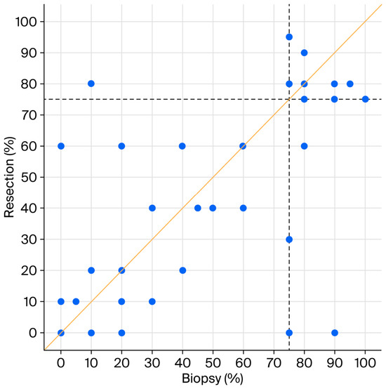
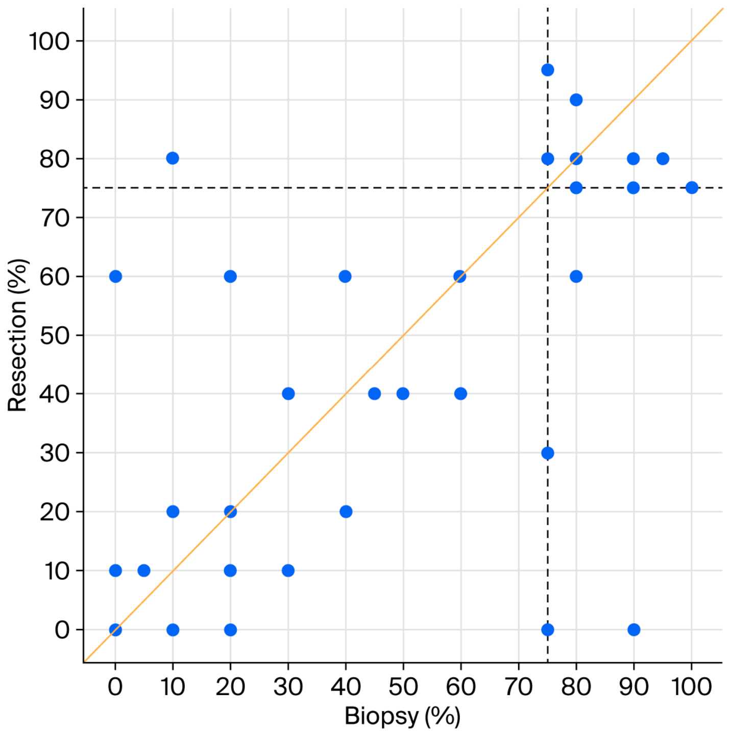
When the percentage of tumor cells showing moderate-to-strong (2+/3+) membranous staining was analyzed as a continuous variable, a high intraclass correlation was observed between paired biopsy and surgical specimen measurements (ICC = 0.817; 95% CI, 0.722–0.881; p < 0.001). The median paired difference in CLDN18.2 staining percentages (resection minus biopsy) was 0 percentage points (IQR, −10 to 0 percentage points), indicating no overall median shift despite individual inter-specimen differences. The relationship between staining percentages in paired biopsies and surgical resections is illustrated in Figure 3. Similarly, high intraclass correlations were observed in patients who received neoadjuvant therapy (ICC = 0.807; 95% CI, 0.695–0.881; p < 0.001) and in those who underwent upfront surgery (ICC = 0.867; 95% CI, 0.630–0.957; p < 0.001).
Figure 3.
Paired scatterplot showing the percentages of viable tumor cells exhibiting moderate-to-strong (2+/3+) membranous CLDN18.2 staining in pretreatment biopsy specimens (x-axis) and corresponding surgical resection specimens (y-axis). Each point represents one patient, although points may overlap when patients have identical staining percentages. The solid diagonal line represents the line of identity (y = x), whereas the dashed vertical and horizontal lines indicate the 75% positivity threshold. The median paired difference (resection minus biopsy) was 0 percentage points (IQR, −10 to 0 percentage points).
For the clinicopathological and survival analyses, patients were classified according to CLDN18.2 expression in pretreatment biopsy specimens. Overall, 19 patients (26.8%) were classified as CLDN18.2-positive, whereas 52 patients (73.2%) were classified as CLDN18.2-negative. The associations between pretreatment biopsy CLDN18.2 status and clinicopathological characteristics are presented in Table 5. No statistically significant associations were observed between pretreatment biopsy CLDN18.2 status and sex, primary tumor location, histological subtype, Lauren classification, histological grade, peritoneal metastasis, neoadjuvant chemotherapy, surgical procedure, CAP primary tumor regression grade, or receipt of adjuvant chemotherapy (all p > 0.05). Among the 59 patients with nonmetastatic (M0) disease, pretreatment biopsy CLDN18.2 status was not significantly associated with postoperative disease recurrence (p = 0.310). Similarly, no significant association was observed between pretreatment biopsy CLDN18.2 status and survival status in the overall cohort of 71 patients (p = 0.790).
Table 5.
Associations between pretreatment biopsy CLDN18.2 expression and clinicopathological characteristics.
To account for the distinct AJCC eighth-edition pathological stage-group definitions applicable to patients undergoing upfront surgery (pTNM) and those receiving neoadjuvant chemotherapy (ypTNM), the associations between pretreatment biopsy CLDN18.2 expression and pathological T, N, and M categories and stage groups were analyzed separately in the two treatment subgroups (Table 6). Among patients who underwent upfront surgery (n = 13), no statistically significant associations were observed between pretreatment biopsy CLDN18.2 expression and pT category (p = 0.765), pN category (p = 0.315), pM category (p = 0.497), or pTNM stage group (p = 0.262). Similarly, among patients who received neoadjuvant chemotherapy (n = 58), CLDN18.2 expression was not significantly associated with ypT category (p = 0.151), ypN category (p = 0.526), ypM category (p = 0.786), or ypTNM stage group (p = 0.999). These findings indicate that pretreatment biopsy CLDN18.2 expression was not significantly associated with pathological staging parameters in either treatment subgroup.
Table 6.
Associations between pretreatment biopsy CLDN18.2 expression and pathological TNM categories and stage, stratified by neoadjuvant chemotherapy status.
Continuous variables were compared between patients with biopsy-confirmed CLDN18.2-negative status and those with biopsy-confirmed CLDN18.2-positive status. No significant differences were observed in age (median, 58.0 years [IQR, 51.5–65.0] vs. 57.0 years [IQR, 51.0–73.0]; p = 0.927), the number of completed NAC cycles (median, 4.0 cycles in both groups; p = 0.500), or the number of completed adjuvant chemotherapy cycles (median, 4.0 cycles in both groups; p = 0.930).
The median follow-up time for the overall cohort calculated using the reverse Kaplan–Meier method was 75.6 months (95% CI, 60.0–91.2 months); the mean was 60.5 months (95% CI, 49.9–71.2 months).
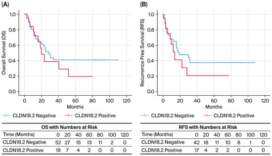
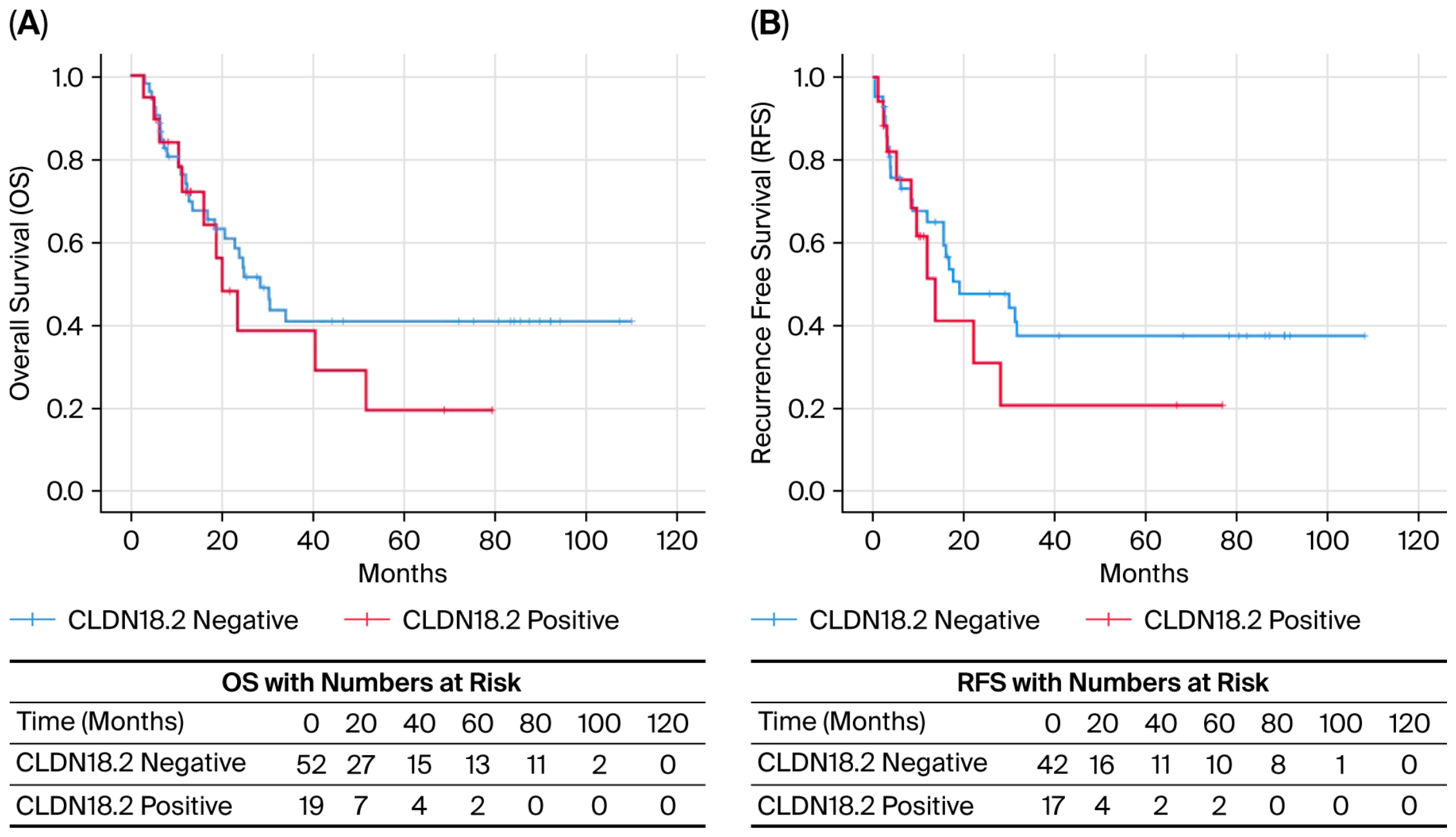
OS was evaluated in all 71 patients; 38 deaths were recorded during follow-up. Patients with CLDN18.2-positive pretreatment biopsies had a shorter median OS than those with CLDN18.2-negative biopsies (20.2 months [95% CI, 13.2–27.2] vs. 28.6 months [95% CI, 20.9–36.3], respectively). However, the difference was not statistically significant (log-rank p = 0.349).
RFS was evaluated in 59 patients with nonmetastatic (M0) disease who underwent R0 resection, following the exclusion of 12 patients with metastatic (M1) disease. The RFS cohort included 42 patients with CLDN18.2-negative pretreatment biopsies and 17 patients with CLDN18.2-positive pretreatment biopsies. During follow-up, 33 composite RFS events were recorded among the 59 M0 patients, comprising 14 first documented recurrences and 19 deaths occurring without prior documented recurrence. The remaining 26 patients were censored. The median RFS was 19.1 months in the CLDN18.2-negative group and 13.8 months in the CLDN18.2-positive group. Although the CLDN18.2-positive group showed a numerically shorter RFS, the difference was not statistically significant (log-rank p = 0.332). The corresponding Kaplan–Meier survival curves are presented in Figure 4.
Figure 4.
Kaplan–Meier survival curves stratified by pretreatment biopsy CLDN18.2 status. (A) Overall survival (OS) in the entire cohort (n = 71), with death from any cause defined as the event. (B) Recurrence-free survival (RFS) in patients with nonmetastatic disease who underwent R0 resection (n = 59), with disease recurrence or death from any cause defined as the event. Twelve patients with metastatic (M1) disease were excluded from the RFS analysis. Numbers at risk are presented below each panel.
The proportional hazards assumption was formally assessed using time-dependent covariate interaction terms, and no significant violations were observed for any of the variables included in the model (time-interaction p-values ranging from p = 0.107 to p = 0.651). Higher histological grade (HR = 1.988, 95% CI: 1.213–3.256; p = 0.006), advanced pN/ypN category (HR = 1.431, 95% CI: 1.143–1.792; p = 0.002), higher CAP primary tumor regression grade (TRG) (HR = 1.978, 95% CI: 1.055–3.707; p = 0.033) and the Lauren classification (overall p = 0.036) were significantly associated with overall survival (OS) in univariate Cox proportional hazards regression analysis. The Lauren classification showed that mixed-type tumors had significantly worse survival than intestinal-type tumors (HR = 3.971, 95% CI: 1.296–12.166; p = 0.016). In univariate analysis, OS was not significantly associated with baseline parameters including age, sex, tumor location, surgical procedure, and pretreatment biopsy CLDN18.2 status (HR = 1.399, 95% CI: 0.691–2.832; p = 0.351). To account for clinically relevant prognostic factors while maintaining model parsimony, a multivariable Cox regression analysis including pretreatment biopsy CLDN18.2 status, pathological nodal category (pN/ypN), histological grade, and Lauren classification was performed. The analysis included 71 patients among whom 38 death events occurred. CAP primary tumor regression grade (TRG) was evaluated separately in a univariable Cox regression analysis restricted to the neoadjuvant-treated subgroup (n = 58; 31 deaths) and was not included in the multivariable model.
In the multivariable analysis, a higher pathological nodal category (pN/ypN) remained significantly associated with worse overall survival (HR = 1.292; 95% CI, 1.013–1.648; p = 0.039). In contrast, histological grade (HR = 1.582; 95% CI, 0.884–2.832; p = 0.123), Lauren classification (overall p = 0.595), and pretreatment biopsy CLDN18.2 status (HR = 1.569; 95% CI, 0.750–3.284; p = 0.232) were not statistically significant in the adjusted model (Table 7).
Table 7.
Univariate and multivariable Cox regression analyses for overall survival.
4. Discussion
This study demonstrated substantial overall categorical agreement in CLDN18.2 status between pretreatment biopsies and corresponding surgical specimens. CLDN18.2 positivity was identified in 19 of 71 pretreatment biopsy specimens (26.8%) and in 15 of 71 corresponding surgical specimens (21.1%). Overall, 65 of 71 paired specimens (91.5%) showed concordant categorical CLDN18.2 status, whereas discordance was observed in the remaining six patients (8.5%). Across the entire cohort, PPA was lower than NPA (73.7% vs. 98.1%), indicating that positive categorical results were less consistently concordant across paired specimens than were negative categorical results. In addition, the percentage of tumor cells showing moderate-to-strong (2+/3+) membranous staining analyzed as a continuous variable, showed a high intraclass correlation between paired specimens (ICC = 0.817, 95% CI, 0.722–0.881; p < 0.001). These findings indicate that although CLDN18.2 expression was broadly concordant across paired specimens, overall percentage agreement alone does not fully reflect the directional asymmetry in categorical agreement.
The prevalence of CLDN18.2 expression in gastric adenocarcinoma varies considerably across studies. In the most comprehensive pooled analysis of the SPOTLIGHT and GLOW phase 3 trials, CLDN18.2 positivity was reported in 38.4% of cases [32]. In our study, the observed positivity rate of 26.8% fell within the range reported in previous studies, although it was somewhat lower than in several large cohorts. In a large cohort from the MD Anderson Cancer Center, Waters et al. reported a CLDN18.2 positivity rate of 44% using a 75% cutoff [15]. Similarly, Kwak et al. reported a positivity rate of 34.4% in a Korean cohort [16]. In contrast, studies conducted in the Turkish population have generally reported lower positivity rates. In a large Turkish cohort, Işık et al. reported CLDN18.2 positivity in 7.8% of cases using a different positivity threshold (≥40% of tumor cells with moderate-to-strong staining) and a tissue microarray-based approach [33]. Such differences may reflect geographic and ethnic heterogeneity, variation in the distribution of histological subtypes, differences in specimen type (biopsy versus resection), antibody clones, and scoring methodologies [34,35]. CLDN18.2 assessment is clinically standardized using the VENTANA CLDN18 (43-14A) assay. In the present study, we used a commercially available monoclonal antibody from Abcam with the same 43-14A clone designation. However, the shared 43-14A clone designation and the use of the same ≥75% positivity cutoff do not establish analytical equivalence or identical clinical classification between assays [36]. Because our laboratory-developed protocol was not cross-validated against the clinically validated VENTANA CLDN18 assay, the observed biopsy–resection concordance should be interpreted strictly as reflecting agreement within our laboratory-developed staining and scoring protocol. Direct comparisons with the reference assay and conclusions regarding clinical interchangeability are therefore not warranted. Cross-validation against the reference assay in an informative subset would strengthen the clinical applicability of these findings.
Among patients who received neoadjuvant therapy (n = 58), categorical CLDN18.2 status was concordant in 52 cases (89.7%), with substantial agreement between pretreatment biopsy and corresponding surgical specimens (κ = 0.705; 95% CI, 0.487–0.922; p < 0.001). This subgroup-specific finding is broadly consistent with the study by Waters et al., which reported a 74% concordance rate between matched pre and post-chemotherapy specimens [15]. All six discordant cases in our cohort occurred in patients who received neoadjuvant therapy, whereas all 13 patients who underwent upfront surgery were categorically concordant. However, the treatment groups were substantially imbalanced, and the difference in discordance rates between them was not statistically significant (10.3% vs. 0%; Fisher’s exact test, p = 0.584). Therefore, the present findings should not be interpreted as evidence that neoadjuvant treatment itself alters CLDN18.2 expression.
Of particular relevance, Nagano et al. [23] investigated CLDN18.2 expression in paired biopsy and resection specimens from 183 patients who underwent upfront gastrectomy and a separate cohort of 38 patients treated with neoadjuvant chemotherapy. In the upfront-surgery cohort, the overall categorical concordance was 86% (157/183; κ = 0.61). Among 93 tumors exhibiting moderate-to-strong CLDN18.2 staining in at least 10% of tumor cells, 81 showed heterogeneous expression patterns, with concordance decreasing to 73% (59/81) in this subgroup and to 65% (30/46) among tumors exhibiting a superficial expression pattern. In their neoadjuvant-treated cohort, biopsy–resection concordance was 82% (31/38; κ = 0.45). Notably, seven of 11 initially CLDN18.2-positive cases became negative following chemotherapy, whereas no negative-to-positive conversions were observed. Although a reduction in the percentage of CLDN18.2-positive tumor cells was suggested, the paired comparison did not reach statistical significance (p = 0.061).
In comparison, our neoadjuvant-treated subgroup demonstrated an 89.7% categorical concordance (52/58; κ = 0.705), with five positive-to-negative and one negative-to-positive discordance cases. Our findings complement those of Nagano et al. by providing additional characterization of directional agreement through positive and negative percent agreement (66.7% and 97.7%, respectively), exact binomial confidence intervals, and continuous agreement in staining percentages (ICC = 0.807; 95% CI, 0.695–0.881). However, differences in cohort composition, neoadjuvant chemotherapy regimens, tissue sampling strategies, immunohistochemical protocols, and eligibility criteria related to residual viable tumor limit direct comparisons of concordance estimates between the two studies. Furthermore, whereas Nagano et al. systematically characterized spatial patterns of intratumoral CLDN18.2 heterogeneity, our analysis was based on one representative tissue block per specimen without comprehensive multiregional mapping. Previous investigations have also described changes in CLDN18.2 expression following systemic treatment [22,37,38], and several potential biological mechanisms have been proposed [39]. Nevertheless, the present study cannot determine whether the observed discordance reflects treatment-associated alterations, pre-existing spatial heterogeneity, or sampling variation [23,40,41].
When we examined the association between CLDN18.2 status and clinicopathological characteristics, no statistically significant associations were observed with age, sex, tumor location, histological subtype, Lauren classification, histological grade, pathological stage, peritoneal metastasis, or CAP primary tumor regression grade (all p > 0.05). These findings are consistent with several previous studies. Previous studies have yielded heterogeneous findings. Waters et al. [15] and Kwak et al. [16] reported associations between CLDN18.2 expression and selected clinicopathological variables, including histological features and tumor location, although these associations were not uniform across studies. In contrast, other studies have reported that CLDN18.2 positivity is associated with intestinal-type histology, better differentiation, and lower disease stage [25,27]. Conversely, Coati et al., in a tissue microarray study of 523 cases, reported a significant association between CLDN18.2 positivity and the diffuse type according to the Lauren classification (p = 0.009) [42]. Similarly, Qi et al., in a large retrospective cohort of 536 patients with advanced gastric cancer, found that CLDN18.2 positivity was associated with the diffuse phenotype (p < 0.001) and poorer OS [43]. These discrepant findings may reflect differences in patient populations, sample sizes, disease stages, specimen types, and methodological approaches to CLDN18.2 assessment.
Peritoneal involvement was the most frequent pattern of postoperative recurrence in the nonmetastatic (M0) subgroup, occurring in 9 of the 14 patients (64.3%) with documented recurrence. The biological complexity of peritoneal dissemination in gastric cancer has been highlighted by Li et al., who identified distinct proteomic and immune microenvironmental signatures associated with postoperative peritoneal metastasis and developed an integrative predictive model incorporating proteomic, immunological, and radiomic features [44]. Their findings suggest that peritoneal metastatic progression involves multiple interacting molecular and microenvironmental processes. However, their study did not investigate paired CLDN18.2 expression, and our findings do not establish an association between CLDN18.2 status and peritoneal recurrence. Therefore, these observations should be interpreted as providing complementary biological context rather than as direct evidence linking CLDN18.2 expression to peritoneal dissemination.
In our survival analyses, patients with CLDN18.2-positive pretreatment biopsies had a numerically shorter median OS than those with CLDN18.2-negative biopsies (20.2 vs. 28.6 months); however, this difference was not statistically significant (log-rank p = 0.349). Similarly, in the RFS analysis restricted to 59 nonmetastatic patients who underwent R0 resection, the median RFS was numerically shorter in the CLDN18.2-positive group than in the CLDN18.2-negative group (13.8 vs. 19.1 months), but the difference was not statistically significant (log-rank p = 0.332). In univariable Cox regression analyses, the Lauren classification, histological grade, pathological nodal category (pN/ypN), and CAP primary tumor regression grade (TRG) were significantly associated with OS. Following adjustment for clinically relevant prognostic factors, higher pathological nodal category remained significantly associated with worse OS (HR = 1.292; 95% CI, 1.013–1.648; p = 0.039). In contrast, pretreatment biopsy CLDN18.2 positivity was not significantly associated with OS in the multivariable model (HR = 1.569; 95% CI, 0.750–3.284; p = 0.232). Importantly, the relatively wide confidence interval encompasses both a potentially lower mortality hazard and a clinically meaningful increase in it, indicating considerable uncertainty regarding the magnitude and direction of the association. Therefore, the absence of statistical significance should not be interpreted as evidence that CLDN18.2 expression lacks prognostic value. Given the limited cohort size and number of events, these findings should be considered exploratory and hypothesis-generating rather than definitive. Larger, adequately powered studies are needed to clarify the prognostic significance of pretreatment CLDN18.2 expression in gastric adenocarcinoma.
The prognostic significance of CLDN18.2 remains controversial in the literature. Arnold et al. reported that CLDN18.2 expression was not an independent prognostic factor [26]. Similarly, in a large cohort study by Waters et al., no significant association was observed between CLDN18.2 expression and survival outcomes [15]. In contrast, some meta-analyses have suggested that CLDN18.2 positivity may be associated with poorer prognosis [45]. More recently, Kuroda et al., in a large single-center cohort study and meta-analysis, demonstrated that CLDN18.2 was not an independent prognostic factor in patients with resected gastric cancer; furthermore, the meta-analysis showed no significant association with OS (HR = 0.892) [24]. No statistically significant prognostic association was demonstrated in this cohort. Nevertheless, given the relatively small sample size (n = 71), the possibility of limited statistical power to detect modest prognostic effects cannot be excluded.
The clinical significance of our findings should be interpreted in the context of the established use of CLDN18.2-directed therapy in advanced gastric cancer. Zolbetuximab combined with chemotherapy is an approved treatment for patients with CLDN18.2-positive, HER2-negative, locally advanced unresectable or metastatic gastric or gastroesophageal junction adenocarcinoma, based on evidence from the SPOTLIGHT and GLOW trials [19,20]. In contrast, the use of CLDN18.2-directed therapies in the perioperative and adjuvant settings remains investigational [9]. Beyond zolbetuximab, Tojjari et al. reviewed emerging CLDN18.2-directed therapeutic strategies, including antibody–drug conjugates, bispecific antibodies, and chimeric antigen receptor T-cell therapies, while highlighting the challenges of biomarker-based patient selection, intratumoral antigen heterogeneity, and treatment resistance [46]. These developments underscore the importance of reliable CLDN18.2 assessment across different specimen types. Nevertheless, our findings describe biopsy–resection concordance and should not be interpreted as evidence of treatment eligibility or therapeutic benefit from these emerging agents. In our neoadjuvant-treated subgroup, positive percent agreement was lower than negative percent agreement (66.7% vs. 97.7%), indicating that positive pretreatment biopsy results were less consistently concordant with matched resection specimens than negative pretreatment biopsy results. However, our study evaluated biomarker concordance rather than response to CLDN18.2-directed treatment. Therefore, these findings cannot establish whether pretreatment biopsies or postoperative resection specimens better predict clinical benefit from CLDN18.2-targeted therapies. Prospective studies incorporating paired tissue assessments and treatment-response data are needed to address this question.
Limitations of the Study
Our study has several limitations. First, the retrospective study design introduces the potential for selection bias and incomplete data. Furthermore, inclusion in the paired biopsy–resection concordance analysis required the presence of viable primary gastric carcinoma in both specimens. Consequently, patients without a residual viable primary tumor following neoadjuvant chemotherapy were excluded, this included two patients with ypT0 disease and persistent regional lymph node metastases. This eligibility criterion may introduce additional selection bias by underrepresenting patients with a marked pathological response to neoadjuvant treatment. Therefore, the observed concordance estimates may not be fully generalizable to all patients undergoing neoadjuvant chemotherapy, particularly those achieving complete primary tumor regression. The direction and magnitude of this potential bias cannot be reliably determined from the present cohort. Second, the single-center setting and relatively small sample size (n = 71), particularly the small number of patients who underwent upfront surgery (n = 13), limited our ability to determine whether biopsy–resection discordance was associated with neoadjuvant treatment. Third, CLDN18.2 expression is known to show intratumoral spatial heterogeneity. Because immunohistochemical assessment was performed on selected representative tumor blocks rather than by comprehensive multiblock mapping of the entire resection specimen, sampling-related discordance between biopsy and resection specimens cannot be excluded. Therefore, the observed discordance should not be interpreted as proof of treatment-induced alteration in CLDN18.2 expression. Although all evaluated specimens contained at least 100 viable tumor cells, the exact distribution of viable tumor cellularity was not systematically recorded and could not be quantified retrospectively. Furthermore, immunohistochemical scoring was performed by a single pathologist without reassessment by an independent second reader of discordant or borderline cases. Consequently, interobserver reproducibility could not be evaluated. These factors should be considered when interpreting the biopsy–resection concordance results, particularly for cases with staining percentages close to the predefined positivity threshold. Fourth, the involved regional lymph nodes were not systematically evaluated as a separate matched compartment. Therefore, the present study cannot determine whether CLDN18.2 expression is consistently maintained between primary gastric tumors and corresponding nodal metastases. Fifth, the heterogeneity of neoadjuvant treatment regimens prevented a sufficiently powered assessment of the effects of each regimens. Sixth, heterogeneity in adjuvant treatment protocols may have introduced additional variability into the survival analyses. Seventh, CLDN18.2 immunohistochemistry was performed using a laboratory-developed protocol with the Abcam 43-14A antibody, which was not analytically cross-validated against the reference VENTANA CLDN18 assay; therefore, direct analytical equivalence with the clinically validated assay cannot be assumed. Finally, advanced molecular analyses, including gene expression profiling and epigenetic profiling, were not performed; therefore, potential biological mechanisms underlying the observed discordance could not be evaluated.
5. Conclusions
This study demonstrates substantial categorical concordance and a high intraclass correlation of CLDN18.2 expression between pretreatment biopsies and corresponding surgical specimens in gastric adenocarcinoma. Across the entire cohort, PPA (73.7%) was lower than NPA (98.1%), and this asymmetry was more pronounced among patients who received neoadjuvant therapy (66.7% vs. 97.7%). Although all six categorically discordant cases occurred in the neoadjuvant group, the small, unbalanced upfront-surgery comparison group, and the potential influence of intratumoral spatial heterogeneity and sampling variation preclude attributing this discordance specifically to neoadjuvant therapy. No statistically significant associations were identified between pretreatment biopsy CLDN18.2 status and clinicopathological characteristics or survival outcomes in this cohort. However, the exploratory nature of the survival analyses, the limited sample size, and the relatively wide confidence interval for the adjusted CLDN18.2 hazard ratio preclude definitive conclusions regarding its prognostic significance. Therefore, a clinically meaningful prognostic association cannot be excluded. Larger prospective studies incorporating systematic multiregional sampling are needed to distinguish true temporal changes in CLDN18.2 expression from spatial heterogeneity and sampling-related discordance.
Supplementary Materials
The following supporting information can be downloaded at https://www.mdpi.com/article/10.3390/cimb48111134/s1.
Author Contributions
Project administration, conceptualization, methodology, validation, writing—original draft preparation, T.T.; writing—review and editing, resources, investigation, T.T., K.E.A. and A.A.; data curation, T.T., U.T. and C.K.P.; supervision, writing—review and editing C.K.P., T.K. and U.T.; validation, software, visualization, formal analysis, Y.K.A. All authors have read and agreed to the published version of the manuscript.
Funding
Onkolojik Bakış Derneği (Oncological Perspective) Association provided support for the procurement of the CLDN18.2 antibody used in this study.
Institutional Review Board Statement
Approved by the Çukurova University Faculty of Medicine Non-Interventional Clinical Research Ethics Committee (Decision No. 70, dated 10 July 2026). Conducted per the Declaration of Helsinki.
Informed Consent Statement
Patient consent was waived due to the retrospective nature of the study by the Çukurova University Faculty of Medicine Non-Interventional Clinical Research Ethics Committee. The study involved analysis of existing electronic medical records collected between May 2016 and May 2026, and all data were accessed and analyzed in an anonymized form to ensure patient confidentiality.
Data Availability Statement
Data supporting the findings of this study are available from the corresponding author upon reasonable request. The data are not publicly available due to ethical and privacy restrictions protecting patient confidentiality.
Acknowledgments
During the preparation of this manuscript, the authors used OpenAI ChatGPT (GPT-6) and PoolText (JournalXpert, web-based platform; version number not specified) to improve the manuscript’s English and grammar. The authors reviewed and edited all AI-assisted content and accept full responsibility for the final manuscript.
Conflicts of Interest
The authors declare no conflicts of interest.
Abbreviations
The following abbreviations are used in this manuscript:
| CLDN18.2 | Claudin 18.2 |
| OS | overall survival |
| RFS | recurrence-free survival |
| PPA | positive percent agreement |
| NPA | negative percent agreement |
| ICC | intraclass correlation coefficient |
| GEJ | gastroesophageal junction |
| IHC | immunohistochemical |
| NAC | neoadjuvant chemotherapy |
| AJCC | the American Joint Committee on Cancer |
References
- Sung, H.; Ferlay, J.; Siegel, R.L.; Laversanne, M.; Soerjomataram, I.; Jemal, A.; Bray, F. Global Cancer Statistics 2020: GLOBOCAN estimates of incidence and mortality worldwide for 36 cancers in 185 countries. CA Cancer J. Clin. 2021, 71, 209–249. [Google Scholar] [CrossRef] [Scilit] [PubMed]
- Al-Batran, S.E.; Homann, N.; Pauligk, C.; Goetze, T.O.; Meiler, J.; Kasper, S.; Kopp, H.G.; Mayer, F.; Haag, G.M.; Luley, K.; et al. Perioperative chemotherapy with fluorouracil plus leucovorin, oxaliplatin, and docetaxel versus fluorouracil or capecitabine plus cisplatin and epirubicin for locally advanced, resectable gastric or gastro-oesophageal junction adenocarcinoma (FLOT4): A ran-domised, phase 2/3 trial. Lancet 2019, 393, 1948–1957. [Google Scholar] [CrossRef] [Scilit] [PubMed]
- Sato, Y.; Okamoto, K.; Kida, Y.; Mitsui, Y.; Kawano, Y.; Sogabe, M.; Miyamoto, H.; Takayama, T. Overview of chemotherapy for gastric cancer. J. Clin. Med. 2023, 12, 1336. [Google Scholar] [CrossRef] [Scilit] [PubMed]
- Koerner, A.; Moy, R.H.; Ryeom, S.; Yoon, S.S. The present and future of neoadjuvant and adjuvant therapy for locally advanced gastric cancer. Cancers 2023, 15, 4114. [Google Scholar] [CrossRef] [Scilit] [PubMed]
- Zhou, Y.; Xu, L.; Chen, G.; Lin, D. Prediction of neoadjuvant therapy efficacy in gastric cancer: The interplay between biomarkers and radiomics and its potential for clinical translation. Front. Oncol. 2026, 15, 1642398. [Google Scholar] [CrossRef] [Scilit] [PubMed]
- Oshima, T.; Yoshikawa, T.; Miyagi, Y.; Morita, S.; Yamamoto, M.; Tanabe, K.; Nishikawa, K.; Ito, Y.; Matsui, T.; Kimura, Y.; et al. Biomarker analysis to predict the pathological response to neoadjuvant chemotherapy in locally advanced gastric cancer: An exploratory biomarker study of COMPASS, a randomized phase II trial. Oncotarget 2020, 11, 2906–2918. [Google Scholar] [CrossRef] [Scilit] [PubMed]
- Businello, G.; Angerilli, V.; Lonardi, S.; Bergamo, F.; Valmasoni, M.; Farinati, F.; Savarino, E.; Spolverato, G.; Fassan, M. Current molecular biomarkers evaluation in gastric/gastroesophageal junction adenocarcinoma. Updat. Surg. 2023, 75, 291–303. [Google Scholar] [CrossRef] [Scilit] [PubMed]
- Röcken, C. Predictive biomarkers in gastric cancer. J. Cancer Res. Clin. Oncol. 2023, 149, 467–481. [Google Scholar] [CrossRef] [Scilit] [PubMed]
- Yamamoto, K.; Nakayama, I.; Shitara, K. Targeting claudin 18.2 in gastric cancer: A review of emerging biologic agents. Expert Opin. Biol. Ther. 2026, 26, 65–80. [Google Scholar] [CrossRef] [Scilit] [PubMed]
- Grizzi, G.; Venetis, K.; Denaro, N.; Bonomi, M.; Celotti, A.; Pagkali, A.; Hahne, J.C.; Tomasello, G.; Petrelli, F.; Fusco, N.; et al. Anti-claudin treatments in gastroesophageal adenocarcinoma: Mainstream and upcoming strategies. J. Clin. Med. 2023, 12, 2973. [Google Scholar] [CrossRef] [Scilit] [PubMed]
- Chen, J.; Xu, Z.; Hu, C.; Zhang, S.; Zi, M.; Yuan, L.; Cheng, X. Targeting CLDN18.2 in cancers of the gastrointestinal tract: New drugs and new indications. Front. Oncol. 2023, 13, 1132319. [Google Scholar] [CrossRef] [Scilit] [PubMed]
- Wang, D.W.; Zhang, W.H.; Danil, G.; Yang, K.; Hu, J.K. The role and mechanism of claudins in cancer. Front. Oncol. 2022, 12, 1051497. [Google Scholar] [CrossRef] [Scilit] [PubMed]
- Sahin, U.; Koslowski, M.; Dhaene, K.; Usener, D.; Brandenburg, G.; Seitz, G.; Huber, C.; Türeci, Ö. Claudin-18 splice variant 2 is a pan-cancer target suitable for therapeutic antibody development. Clin. Cancer Res. 2008, 14, 7624–7634. [Google Scholar] [CrossRef] [Scilit] [PubMed]
- Cao, W.; Xing, H.; Li, Y.; Tian, W.; Song, Y.; Jiang, Z.; Yu, J. Claudin18.2 is a novel molecular biomarker for tumor-targeted immunotherapy. Biomark. Res. 2022, 10, 38. [Google Scholar] [CrossRef] [Scilit] [PubMed]
- Waters, R.; Sewastjanow-Silva, M.; Yamashita, K.; Abdelhakeem, A.; Iwata, K.K.; Moran, D.; Elsouda, D.; Guerrero, A.; Pizzi, M.; Vicentini, E.R.; et al. Retrospective study of Claudin 18 isoform 2 prevalence and prognostic association in gastric and gastroesophageal junction adenocarcinoma. JCO Precis. Oncol. 2024, 8, e2300543. [Google Scholar] [CrossRef] [Scilit] [PubMed]
- Kwak, Y.; Kim, T.Y.; Nam, S.K.; Hwang, H.J.; Han, D.; Oh, H.J.; Kong, S.H.; Park, D.J.; Oh, D.Y.; Lee, H.J.; et al. Clinicopathologic and molecular characterization of stages II-IV gastric cancer with Claudin 18.2 expression. Oncologist 2025, 30, oyae238. [Google Scholar] [CrossRef] [Scilit] [PubMed]
- Liu, B.L.; Cleary, J.M.; Shi, J.; Hornick, J.L.; Zhao, L. Claudin 18.2 and other therapeutic biomarkers in gastric and gastroesophageal junction adenocarcinomas. Am. J. Surg. Pathol. 2025, 49, 1233–1244. [Google Scholar] [CrossRef] [Scilit] [PubMed]
- Fassan, M.; Kuwata, T.; Matkowskyj, K.A.; Röcken, C.; Rüschoff, J. Claudin-18.2 immunohistochemical evaluation in gastric and gastroesophageal junction adenocarcinomas to direct targeted therapy: A practical approach. Mod. Pathol. 2024, 37, 100589. [Google Scholar] [CrossRef] [Scilit] [PubMed]
- Shitara, K.; Lordick, F.; Bang, Y.J.; Enzinger, P.; Ilson, D.; Shah, M.A.; Van Cutsem, E.; Xu, R.H.; Aprile, G.; Xu, J.; et al. Zolbetuximab plus mFOLFOX6 in patients with CLDN18.2-positive, HER2-negative, untreated, locally advanced unresectable or metastatic gastric or gastro-oesophageal junction adenocarcinoma (SPOTLIGHT): A multicentre, randomised, double-blind, phase 3 trial. Lancet 2023, 401, 1655–1668, Correction in Lancet 2023, 402, 290. https://doi.org/10.1016/S0140-6736(23)01481-2. Correction in Lancet 2023, 403, 30. https://doi.org/10.1016/S0140-6736(23)02762-9. [Google Scholar] [CrossRef] [Scilit] [PubMed]
- Shah, M.A.; Shitara, K.; Ajani, J.A.; Bang, Y.J.; Enzinger, P.; Ilson, D.; Lordick, F.; Van Cutsem, E.; Gallego Plazas, J.; Huang, J.; et al. Zolbetuximab plus CAPOX in CLDN18.2-positive gastric or gastroesophageal junction adenocarcinoma: The randomized, phase 3 GLOW trial. Nat. Med. 2023, 29, 2133–2141. [Google Scholar] [CrossRef] [Scilit] [PubMed]
- Dottermusch, M.; Krüger, S.; Behrens, H.M.; Halske, C.; Röcken, C. Expression of the potential therapeutic target claudin-18.2 is frequently decreased in gastric cancer: Results from a large Caucasian cohort study. Virchows Arch. 2019, 475, 563–571. [Google Scholar] [CrossRef] [Scilit] [PubMed]
- Tsutsumi, C.; Ohuchida, K.; Yamada, Y.; Shimada, Y.; Imamura, M.; Son, K.; Mochida, Y.; Katayama, N.; Iwamoto, C.; Torata, N.; et al. Claudin18.2-positive gastric cancer-specific changes in neoadjuvant chemotherapy-driven immunosuppressive tumor microenvironment. Br. J. Cancer 2025, 132, 793–804. [Google Scholar] [CrossRef] [Scilit] [PubMed]
- Nagano, S.; Kurokawa, Y.; Hagi, T.; Motoyama, Y.; Saito, T.; Takahashi, T.; Momose, K.; Yamashita, K.; Tanaka, K.; Makino, T.; et al. Intratumoral heterogeneity and chemotherapy-induced alteration of CLDN18.2 expression in resectable gastric cancer. Int. J. Clin. Oncol. 2026, 31, 621–629. [Google Scholar] [CrossRef] [Scilit] [PubMed]
- Kuroda, K.; Miki, Y.; Yoshii, M.; Matsuoka, K.; Seki, Y.; Kasashima, H.; Tamura, T.; Shibutani, M.; Toyokawa, T.; Yashiro, M.; et al. Prognostic significance of Claudin18.2 expression in gastric cancer: A large single-center cohort and meta-analysis. J. Clin. Oncol. 2026, 44, 449. [Google Scholar] [CrossRef] [Scilit]
- Ungureanu, B.S.; Lungulescu, C.V.; Pirici, D.; Turcu-Stiolica, A.; Gheonea, D.I.; Sacerdotianu, V.M.; Liliac, I.M.; Moraru, E.; Bende, F.; Saftoiu, A. Clinicopathologic relevance of Claudin 18.2 expression in gastric cancer: A meta-analysis. Front. Oncol. 2021, 11, 643872. [Google Scholar] [CrossRef] [Scilit] [PubMed]
- Arnold, A.; Daum, S.; von Winterfeld, M.; Berg, E.; Hummel, M.; Rau, B.; Stein, U.; Treese, C. Prognostic impact of Claudin 18.2 in gastric and esophageal adenocarcinomas. Clin. Transl. Oncol. 2020, 22, 2357–2363. [Google Scholar] [CrossRef] [Scilit] [PubMed]
- Lu, Y.; Wu, T.; Sheng, Y.; Dai, Y.; Xia, B.; Xue, Y. Correlation between Claudin-18 expression and clinicopathological features and prognosis in patients with gastric cancer. J. Gastrointest. Oncol. 2020, 11, 1253–1260. [Google Scholar] [CrossRef] [Scilit] [PubMed]
- WHO Classification of Tumours Editorial Board. Digestive System Tumours. In WHO Classification of Tumours Series, 5th ed.; International Agency for Research on Cancer: Lyon, France, 2019; Volume 1, pp. 1–435. [Google Scholar]
- Lauren, P. The two histological main types of gastric carcinoma: Diffuse and so-called intestinal-type carcinoma: An attempt at a histo-clinical classification. Acta Pathol. Microbiol. Scand. 1965, 64, 31–49. [Google Scholar] [CrossRef] [Scilit] [PubMed]
- Amin, M.B.; Edge, S.B.; Greene, F.L.; Byrd, D.R.; Brookland, R.K.; Washington, M.K.; Gershenwald, J.E.; Compton, C.C.; Hess, K.R.; Sullivan, D.C.; et al. AJCC Cancer Staging Manual, 8th ed.; Springer: Cham, Switzerland, 2017. [Google Scholar]
- College of American Pathologists. Protocol for the Examination of Specimens from Patients with Carcinoma of the Stomach, Version 4.4.0.0; College of American Pathologists: Northfield, IL, USA, 2023; Available online: https://www.cap.org/wp-content/uploads/2026/07/Stomach_4.4.0.0.REL_CAPCP-1.pdf?download=true (accessed on 14 October 2026).
- Shitara, K.; Xu, R.H.; Ajani, J.A.; Moran, D.; Guerrero, A.; Li, R.; Pavese, J.; Matsangou, M.; Bhattacharya, P.; Ueno, Y.; et al. Global prevalence of claudin 18 isoform 2 in tumors of patients with locally advanced unresectable or metastatic gastric or gastroesophageal junction adenocarcinoma. Gastric Cancer 2024, 27, 1058–1068. [Google Scholar] [CrossRef] [Scilit] [PubMed]
- Işık, A.; Güner, G.; Zeyneloğlu, C.; Canlı, S.D.; Taştan, H.; Akyol, A. Claudin 18.2 expression in gastric adenocarcinomas in a large Turkish cohort. Abant Med. J. 2023, 12, 122–130. [Google Scholar] [CrossRef] [Scilit]
- Wallam, S.; Park, J.; Wu, L.W.; Moy, R.H. Retrospective analysis of Claudin18.2 expression in ethnically diverse patients with gastroesophageal adenocarcinoma. J. Gastrointest. Oncol. 2025, 16, 1420–1433. [Google Scholar] [CrossRef] [Scilit] [PubMed]
- Martinez Ciarpaglini, C.; Ortega, M.; Pérez-Buira, S.; Bolea, A.; Guerra, B.C.; Bellido, C.H.; Piñero, P.T.; Naranjo-Hans, D.; Palomar, B.; Quiceno, H.; et al. CLDN18.2 expression in gastroesophageal adenocarcinoma: Prevalence, heterogeneity, and prognostic implications in Spanish patients. Virchows Arch. 2025, 487, 1337–1346. [Google Scholar] [CrossRef] [Scilit] [PubMed]
- Jasani, B.; Taniere, P.; Schildhaus, H.U.; Blighe, K.; Parry, S.; Wilkinson, D.; Atkey, N.; Clare-Antony, S.; McCabe, C.; Quinn, C.; et al. Global ring study to investigate the comparability of total assay performance of commercial Claudin 18 antibodies for evaluation in gastric cancer. Lab. Investig. 2024, 104, 100284. [Google Scholar] [CrossRef] [Scilit] [PubMed]
- Lim, S.H.; Kuwata, T.; An, M.; Hong, J.Y.; Kim, S.T.; Matsubara, Y.; Shitara, K.; Lee, J. Dynamic modulation of claudin18.2 expression and remodeling of the tumor microenvironment in gastric cancer during chemo-immunotherapy. J. Immunother. Cancer 2025, 13, e012683. [Google Scholar] [CrossRef] [Scilit] [PubMed]
- Rha, S.Y.; Kwon, W.S.; Lee, C.K.; Jung, M.; Shin, S.J.; Kim, H.; Park, S.; Yoo, J.H.; Kim, H. Co-expressing pattern of multiple biomakers and dynamic change of Claudin18.2 expression after systemic chemotherapy in advanced gastric cancer. J. Clin. Oncol. 2025, 43, 4037. [Google Scholar] [CrossRef] [Scilit]
- Wu, H.; Li, M.; Wang, G.; Liao, P.; Zhang, P.; Yang, L.; Li, Y.; Liu, T.; He, W. Clinicopathological and molecular mechanisms of CLDN18.2 in gastric cancer aggressiveness: A high-risk population study with multi-omics profiling. J. Pathol. Transl. Med. 2026, 60, 47–57. [Google Scholar] [CrossRef] [Scilit] [PubMed]
- Pak, S.; Simon, A.G.; Lyu, S.I.; Tolkach, Y.; Alakus, H.; Zander, T.; Buettner, R.; Bruns, C.J.; Schroeder, W.; Quaas, A. The ex-pression of the tight junction protein and therapeutical target Claudin 18.2 is heterogeneously distributed within esophageal and gastric adenocarcinoma. Sci. Rep. 2025, 15, 28958. [Google Scholar] [CrossRef] [Scilit] [PubMed]
- Choi, E.; Shin, J.; Ryu, M.H.; Kim, H.D.; Park, Y.S. Heterogeneity of claudin 18.2 expression in metastatic gastric cancer. Sci. Rep. 2024, 14, 17564. [Google Scholar] [CrossRef] [Scilit] [PubMed]
- Coati, I.; Lotz, G.; Fanelli, G.N.; Brignola, S.; Lanza, C.; Cappellesso, R.; Pellino, A.; Pucciarelli, S.; Spolverato, G.; Guzzardo, V.; et al. Claudin-18 expression in oesophagogastric adenocarcinomas: A tissue microarray study of 523 molecularly profiled cases. Br. J. Cancer 2019, 121, 257–263. [Google Scholar] [CrossRef] [Scilit] [PubMed]
- Qi, C.; Chong, X.; Zhou, T.; Ma, M.; Gong, J.; Zhang, M.; Li, J.; Xiao, J.; Peng, X.; Liu, Z.; et al. Clinicopathological significance and immunotherapeutic outcome of claudin 18.2 expression in advanced gastric cancer: A retrospective study. Chin. J. Cancer Res. 2024, 36, 78–89. [Google Scholar] [CrossRef] [Scilit] [PubMed]
- Li, X.; Lu, J.; Chen, F.; Yuan, J.; Zha, Y.; Li, Y.; Yan, J.; Li, Q.; Yuan, J.; Tong, Q. Comprehensive proteomic analysis and multidimensional model construction of peritoneal metastasis in gastric cancer. Cancer Lett. 2025, 614, 217509. [Google Scholar] [CrossRef] [Scilit] [PubMed]
- De Moraes, F.C.A.; Sobreira, L.E.R.; Souza, M.E.C.; Rodriguez Burbano, R.M. The role of CLDN18.2 in gastric cancer prognosis: A systematic review and meta-analysis. Biomarkers 2024, 29, 528–538. [Google Scholar] [CrossRef] [Scilit] [PubMed]
- Tojjari, A.; Alami Idrissi, Y.; Saeed, A. Emerging targets in gastric and pancreatic cancer: Focus on claudin 18.2. Cancer Lett. 2025, 611, 217362. [Google Scholar] [CrossRef] [Scilit] [PubMed]

Disclaimer/Publisher’s Note: The statements, opinions and data contained in all publications are solely those of the individual author(s) and contributor(s) and not of MDPI and/or the editor(s). MDPI and/or the editor(s) disclaim responsibility for any injury to people or property resulting from any ideas, methods, instructions or products referred to in the content. |
© 2026 by the authors. Licensee MDPI, Basel, Switzerland. This article is an open access article distributed under the terms and conditions of the Creative Commons Attribution (CC BY) license.