


Abstract
Background/Objectives: Glycogen synthase kinase-3β (GSK-3β) serves as a crucial molecular target for Alzheimer’s disease and other neurological disorders. Positron emission tomography (PET) enables non-invasive quantification of GSK-3β in vivo, offering value for early diagnosis and mechanistic studies. However, despite two decades of effort, no GSK-3β PET tracer has yet entered clinical trials. Methods: This review systematically summarizes the radiochemical synthesis and preclinical evaluation of major GSK-3β tracer classes, comparing synthetic accessibility, target affinity, brain uptake, P-gp efflux, metabolic stability, radiometabolite profiles, and blocking experiments. The literature was systematically searched in PubMed and Web of Science databases (2005–2026). Several key obstacles impeding clinical progression of GSK-3β PET radiotracers are identified. Results: First, P-gp efflux markedly reduces brain tracer accumulation, rendering even sub-nanomolar high-affinity ligands ineffective for brain imaging. Second, interfering factors such as brain-penetrant radiometabolites of [11C]OCM-44 and the extremely low plasma free fraction of [18F]OCM-50 greatly impair the quantitative accuracy of tracer uptake. Third, the paradoxical increase in brain uptake of isonicotinamide tracers upon blocking remains an unresolved confounding factor. Moreover, rodent data fail to reliably predict tracer performance in higher species, and to date, no tracer has been directly validated against GSK-3β expression in patient brain tissue. Conclusions: This review proposes a tiered validation framework comprising four levels: assessment of P-gp efflux, metabolite-corrected arterial input function, low-affinity enantiomer controls, and human brain autoradiography. The framework provides a rigorous basis for screening candidates and a systematic reference for developing next-generation GSK-3β PET tracers.
1. Introduction
Glycogen synthase kinase-3β (GSK-3β) is an intracellular serine/threonine kinase highly enriched in the mammalian central nervous system (CNS) [1,2]. Unlike most kinases, GSK-3β is constitutively active under resting conditions, and its activity is tightly regulated through complex phosphorylation events. Phosphorylation at serine 9 (Ser9) inhibits its activity, which is primarily mediated by upstream signaling pathways such as PI3K/Akt and Wnt, whereas phosphorylation at tyrosine 216 (Tyr216) promotes its activation [3,4,5]. Under physiological conditions, GSK-3β regulates neurogenesis, axonal growth, synaptic plasticity, and memory formation by phosphorylating diverse substrate proteins [6,7,8,9]. Distinguished from cell surface receptors, this intracellular kinase acts on its substrates across multiple subcellular compartments.
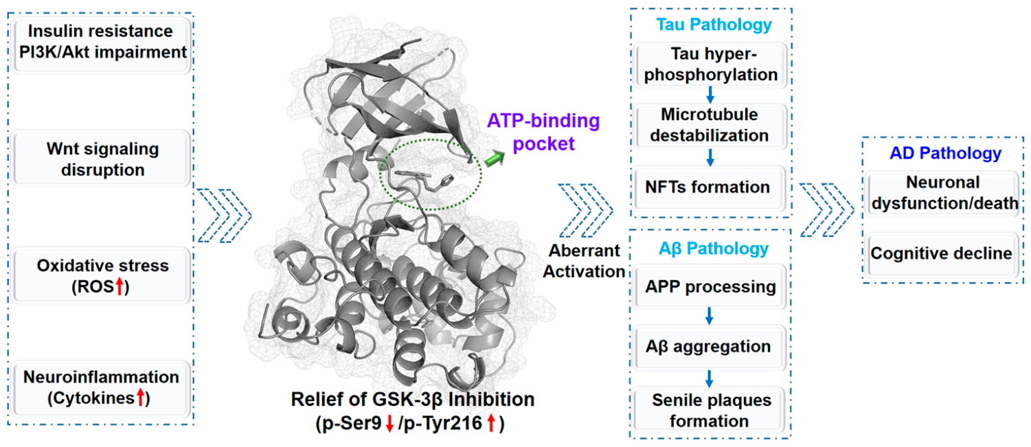
In Alzheimer’s disease (AD), GSK-3β regulatory pathways are disrupted, resulting in aberrant hyperactivation (Figure 1). Pathological triggers such as insulin resistance-induced PI3K/Akt dysfunction, impaired Wnt signaling, oxidative stress, and neuroinflammation collectively relieve GSK-3β inhibition [10,11]. This excessive activity drives both core pathological hallmarks of AD. As a primary tau kinase, GSK-3β induces tau hyperphosphorylation, which disrupts microtubules and ultimately leads to neurofibrillary tangle (NFT) formation [12,13,14]. It also modulates amyloid precursor protein (APP) processing to promote amyloid-β (Aβ) production and aggregation, facilitating senile plaque deposition [12]. Notably, Aβ oligomers further activate GSK-3β, creating a pathogenic positive feedback loop that accelerates disease progression [15,16] (Figure 1).
Therapeutic studies have further confirmed GSK-3β as a key mediator of AD pathogenesis. In 3×Tg-AD mice, increased inhibitory phosphorylation at GSK-3β Ser9 (pSer9) reduced tau hyperphosphorylation, suppressed neuronal apoptosis, and improved cognitive performance [17,18]. GSK-3β therefore functions as a central hub in the AD signaling network, integrating multiple upstream pathological stimuli and propagating signals that drive both core AD pathologies. Given this unique role, GSK-3β represents an attractive target for therapeutic development and molecular imaging.
Positron emission tomography (PET) enables in vivo quantitative assessment of target protein expression and activity using specific radiotracers [19,20,21]. Thus, GSK-3β is both a biomarker of AD pathological burden and disease progression, and a key signaling node that integrates upstream pathological signals to drive downstream core pathologies. Non-invasive, in vivo quantitative imaging of cerebral GSK-3β therefore holds significant value.
This narrative review was compiled through systematic searches of PubMed and Web of Science (2005–2026) using combinations of keywords including “GSK-3β,” “PET,” “radiotracer,” “glycogen synthase kinase-3,” “imaging probe,” and related terms. Reference lists of retrieved articles were manually screened. Conference abstracts without full publications were excluded; selected in vitro-only studies with relevant mechanistic insights were included where appropriate.
Figure 1.
GSK-3β: a key regulator of AD pathology. GSK-3β integrates upstream signals to drive AD pathologies; its inhibition rescues deficits, making it a key target for AD intervention and imaging. Structural representation of GSK-3β (PDB ID: 5K5N [22], from RCSB PDB database) with the ATP-binding pocket highlighted in the structure (rendered with PyMOL [23]). Red arrows: increase/decrease in the corresponding indicator. Green arrow: ATP-binding pocket. Blue arrows: activation/promotion processes.
2. Biological Basis of GSK-3β
2.1. Structural Features of GSK-3β
The GSK-3 family comprises two highly homologous isoforms, GSK-3α (51 kDa) and GSK-3β (47 kDa), encoded by separate genes. Their catalytic domains share up to 98% sequence identity, with differences confined mainly to the N- and C-terminus regions. GSK-3β is the predominant isoform in most brain regions, and its expression increases with age [24,25,26]. The substrate-binding domain of GSK-3β shows low conservation among kinases. Its surface hydrophobic groove serves as a key region for substrate recognition, laying a structural foundation for the development of substrate-competitive inhibitors [27]. Multiple druggable pockets are distributed at the surface of GSK-3β, including the ATP-binding site, substrate-binding site, axin/FRAT-binding site, and potential allosteric sites [28,29,30]. Notably, within the ATP-binding pocket, Asp133 of GSK-3β and Glu196 of GSK-3α represent the only non-conserved residues between the two isoforms. This “Asp-Glu switch” provides a structural foundation for the design of isoform-selective inhibitors [31,32].
Based on the above structural features, researchers have synthesized a variety of small-molecule GSK-3β inhibitors and resolved their co-crystal structures bound to this kinase. The co-crystal structure of PF-367 complexed with GSK-3β (PDB: 5K5N) confirms that this compound acts as a typical type I kinase inhibitor occupying the ATP-binding pocket [22]. Co-crystal structures of imidazo [1,2-b]pyridazine-derived inhibitors also offer valuable structural references for drug optimization [33].
In addition, covalent GSK-3β inhibitors have attracted extensive research attention [34,35,36,37]. Tideglusib, an irreversible GSK-3β inhibitor, has entered Phase II clinical trials, yet its inhibitory efficacy is not fully mediated by Cys199 [33]. By contrast, thiazolidine-2-thione derivatives and aza-indolyl maleimide-based inhibitors irreversibly target Cys199 with nanomolar potency [35,36]. Similarly, incorporating a halomethylketone group converts reversible GSK-3 inhibitors into irreversible analogs, also with nanomolar IC50 values [37].
2.2. GSK-3β and Diseases
Aberrant activation of GSK-3β is closely correlated with a broad range of human diseases, including neurological disorders, psychiatric illnesses, metabolic disorders, cancers, and inflammatory diseases.
2.2.1. Alzheimer’s Disease
AD is the most extensively studied GSK-3β-associated neurodegenerative disorder, with GSK-3β playing a central role in its pathogenesis. In AD brains, it remains excessively activated and contributes to tau and Aβ toxicity, oxidative stress, neuroinflammation, and synaptic dysfunction [38,39,40]. GSK-3β hyperphosphorylates tau at multiple sites, most notably Ser396, Ser199, and Ser404, reducing tau’s microtubule affinity and promoting neurofibrillary tangle formation [41,42,43]. Hyperactivated GSK-3β colocalizes with tangles in post-mortem AD brain tissue [42]. It also accelerates Aβ aggregation, and pharmacological GSK-3β inhibition alleviated Aβ-induced neurotoxicity and cognitive deficits in transgenic AD mice [44]. Excessive GSK-3β activity further promotes neuroinflammation, neuronal injury, and apoptosis. In induced pluripotent stem cell-derived neurons from familial and sporadic AD patients, elevated Aβ1–40 levels, increased active GSK-3β that lack Ser9 phosphorylation, and higher phospho-tau/total tau ratios were strongly correlated [45,46].
2.2.2. Parkinson’s Disease
Beyond AD, GSK-3β dysregulation is also tightly linked to the pathogenesis of Parkinson’s disease. Substantial evidence reveals that GSK-3β hyperactivation elevates the expression of α-synuclein, the main component of Lewy bodies, a typical pathological hallmark of PD [47,48]. In PD animal models, GSK-3β inhibition reduced α-synuclein aggregation and neurotoxicity, indicating that GSK-3β is a promising therapeutic target for PD. Additionally, GSK-3β participates in PD-associated neuroinflammation and mitochondrial dysfunction, which further aggravates dopaminergic neuron loss [49,50].
2.2.3. Huntington’s Disease
Similarly, aberrant GSK-3β activity has been implicated in Huntington’s disease (HD) [51,52]. HD is a neurodegenerative disorder caused by Huntingtin (HTT) gene mutation, characterized by progressive neuronal loss in the striatum and cerebral cortex. Studies verified increased GSK-3β activity in HD brain tissue, which correlated with mutant Huntingtin aggregation and cytotoxicity. Suppression of GSK-3β reduced neuronal apoptosis and mitochondrial dysfunction that is triggered by mutant Huntingtin protein [53].
2.2.4. Tauopathies
In addition to its role in specific neurodegenerative diseases, GSK-3β-mediated tau hyperphosphorylation serves as a shared pathological mechanism across various tauopathies [54]. Tauopathies are a group of neurodegenerative diseases defined by abnormal tau protein aggregation, including progressive supranuclear palsy (PSP), corticobasal degeneration (CBD), Pick’s disease (PiD), argyrophilic grain disease (AGD), and frontotemporal dementia with parkinsonism linked to chromosome 17 (FTDP-17) [54]. In these diseases, abnormal GSK-3β activation causes tau hyperphosphorylation and subsequent aggregation into neurofibrillary tangles in specific brain regions, ultimately leading to neuronal dysfunction and death [55].
2.2.5. Bipolar Disorder and Depression
GSK-3β dysregulation extends beyond neurodegenerative conditions to bipolar disorder and depression. Disordered GSK-3β signaling pathways underlie the pathogenesis of bipolar disorder (BD) [56]. Lithium, a classic mood stabilizer, exerts therapeutic effects via inhibiting GSK-3β. Its mechanisms include direct competitive binding against magnesium ions and indirect promotion of inhibitory Ser9 phosphorylation. Studies have shown that low-dose lithium regulates GSK-3β activity in a region-specific manner in the brain. Such regional differences may explain the clinical heterogeneity among BD patients [57]. Moreover, disrupted sleep–wake rhythm is a core endophenotype of depression and bipolar disorder. GSK-3β is critical for circadian rhythm and sleep–wake cycle regulation, and its dysfunction contributes to sleep disturbances in these psychiatric disorders [58].
2.2.6. Type 2 Diabetes Mellitus
The pathological roles of GSK-3β are not restricted to the central nervous system. GSK-3β plays a core regulatory role in glucose metabolism. It phosphorylates and inactivates glycogen synthase, thereby inhibiting glycogen synthesis and inducing hyperglycemia [53,59,60,61]. Both expression level and enzymatic activity of GSK-3β are significantly upregulated in type 2 diabetic patients. GSK-3β inhibition mimics insulin effects by boosting glycogen synthesis and relieving insulin resistance, making GSK-3β a valuable therapeutic target for type 2 diabetes [61]. Derivatives of the marine natural product meridianin C act as potent GSK-3β inhibitors with improved oral bioavailability, offering novel candidate drugs for diabetes treatment [60].
2.2.7. Cancer
In addition to its metabolic and neurological roles, GSK-3β also participates in tumorigenesis. GSK-3β facilitates tumor progression in multiple malignancies, such as colorectal cancer, pancreatic cancer, prostate cancer, glioblastoma, and leukemia [62,63,64]. It drives tumor initiation and progression by promoting proliferation, survival, migration, invasion, and chemoresistance, and serves as a cancer cell marker via aberrant nuclear accumulation in certain tumors. Several clinically investigated GSK-3β inhibitors, including Tideglusib [64] and 9-ING-41 [65], are being developed for the treatment of both neurological diseases and cancers.
2.2.8. Inflammatory Diseases
In addition, GSK-3β functions as a critical regulator of immune and inflammatory responses, acting via NF-κB to promote proinflammatory cytokine production while suppressing IL-10 [66,67]. The inhibitor TFGF-18 alleviated neuroinflammation in LPS-treated microglia by suppressing NF-κB and related inflammatory mediators [66]. Dual inhibition of QC and GSK-3β blocks pE-Aβ production and tau hyperphosphorylation, providing a promising strategy for AD treatment [67].
In general, aberrant GSK-3β activity is linked to neurological, psychiatric, metabolic, neoplastic, and inflammatory diseases. As a key cellular signaling hub, GSK-3β converges major pathways including PI3K/Akt, Wnt/β-catenin and NF-κB. It responds to diverse stimuli and modulates cell survival, metabolism, proliferation, and inflammation. Dysregulated GSK-3β triggers distinct tissue-specific pathologies: tau hyperphosphorylation and synaptic damage in neurons, insulin resistance in pancreatic β-cells, elevated proinflammatory cytokines in immune cells, and enhanced proliferation and migration in tumor cells.
This central role of GSK-3β presents both therapeutic opportunities and potential liabilities. Systemic inhibition may induce off-target effects on multiple pathways, while selective targeting can concurrently intervene in various diseases and pathological processes. Strategies including dual-target inhibitors and tissue-specific delivery are designed to enhance efficacy and reduce systemic toxicity. Further clinical translation of GSK-3β modulators requires fuller insight into its universal roles in diseases and the development of highly selective molecular tools.
2.3. GSK-3β PET Imaging
Given GSK-3β’s key role in various diseases, its targeted PET tracers show broad application potential. PET imaging enables non-invasive, quantitative detection of both GSK-3β expression and enzymatic activity, supporting early diagnosis and therapeutic monitoring in AD, diabetes, inflammation, and cancer [30,35,60,66]. Specific binding to pathological regions in AD transgenic mice further underscores the diagnostic value of these tracers [68]. GSK-3β thus represents an ideal target for molecular neuroimaging. To date, GSK-3β inhibitors have advanced considerably, offering diverse scaffolds and SAR data for tracer design [69,70,71,72,73,74,75,76,77]. However, PET tracer development still faces major obstacles, including limited brain uptake, P-glycoprotein (P-gp) efflux, poor metabolic stability, and difficulties in confirming in vivo target specificity, and the intrinsic challenge of targeting an intracellular kinase, which demands that tracers cross both the cell membrane and the blood–brain barrier.
This review systematically surveys reported GSK-3β PET tracers by chemical scaffold, covering their design strategies, radiochemical syntheses, preclinical evaluation profiles, and key limitations. On this basis, this review discusses key challenges and proposes strategies, including multi-parameter optimization, tiered validation, and structure-guided design. These approaches facilitate rational development of high-performance tracers for AD mechanistic research and clinical use. Tracer scaffold classification, rational design, and preclinical evaluation are presented in Figure 2.
Figure 2.
Rational design and preclinical evaluation of GSK-3β PET tracers. The radar schematic displays five preclinical indices: brain penetration, regional selectivity, target specificity, metabolic stability and disease relevance. Iterative optimization across these aspects helps obtain tracers with optimal in vivo profiles.
3. Classification of GSK-3β PET Tracers
Over the past two decades, GSK-3β PET tracers have been rapidly developed. Small-molecule inhibitors labeled with 11C or 18F have yielded tracers spanning 11 scaffold classes (Table 1). Research emphasis has shifted toward brain uptake, metabolic stability, and target specificity, with evaluation strategies expanding to transgenic models and nonhuman primates. Table 1 presents the key properties of representative tracers, providing a basis for structure–activity relationship analysis.
Table 1.
GSK-3β-targeted PET radiotracers.
Table 1 summarizes the variations in affinity, brain uptake, and translational applicability of GSK-3β tracers across different scaffolds. The majority of these tracers exert nanomolar inhibitory activity. PF-367 (IC50 = 2.10 nM), A1070722 (Ki = 0.60 nM), and the 10a-d analogs (IC50 = 1.30–5.80 nM) stand out with potent in vitro potency. Large discrepancies in brain uptake were noted: [11C]SB-216763 showed high brain uptake in rats and nonhuman primates (SUV = 1.90–2.50), in contrast to the limited brain penetration of [11C]AR-A014418 and [11C]PyrATP-1 [99]. [18F]10a–d showed heterogeneous regional distribution in rodents, and [11C]A1070722 in nonhuman primates mirrored the native GSK-3β distribution pattern. While early work relied on normal rodents, transgenic models and nonhuman primates are now widely adopted. [18F]F-CNPI has been validated in Thy1-GSK-3β transgenic mice, and [11C]OCM-44 as well as [18F]OCM-50 have undergone full pharmacokinetic characterization and blocking assays in nonhuman primates. Subsequent chapters summarize each tracer’s design, synthesis, radiolabeling, preclinical results, and limitations.
3.1. Ureas
[11C]AR-A014418, the first GSK-3β PET tracer, was synthesized via the [11C]methylation of a methoxy precursor [78] (Figure 3). [11C]AR-A014418 was synthesized by O-[11C]methylation of a deprotected hydroxyl precursor, which was prepared from an acyl chloride intermediate via Curtius rearrangement to an isocyanate, followed by coupling with 5-nitrothiazol-2-amine [78]. The carbonyl-labeled analog was prepared by [11C]CO2 fixation [79]. Despite a favorable LogP of 2.44, this tracer showed poor brain uptake (0.08%ID/g) in rats [76], and subsequent re-evaluation determined its Ki against GSK-3β to be 770 nM, indicating weak binding affinity [79]. The co-crystal structure of AR-A014418 bound to GSK-3β (Figure 3c) shows the ligand occupying the ATP-binding pocket, with its thiazole-urea core forming hydrogen bonds with Val135 and the phenyl ring engaging in σ-π interactions with Ile62 [100]. Given its submicromolar affinity and poor brain penetration, this tracer is included here only as a historical starting point.
Figure 3.
The synthesis of [11C]AR-A014418 (a) and [11C-carbonyl]AR-A014418 (b), together with AR-A014418 bound to the ATP-binding site of GSK-3β (PDB ID: 1Q5K [79]; interaction diagram generated using PyMOL [23] and PLIP [99]) (c). Route (a): (1) Trimethylsilyl chloride, Et3N, toluene, reflux; (2) Thionyl chloride, CH2Cl2; (3) Trimethylsilyl azide, 1,4-dioxane, r.t.; (4) reflux; (5) 5-nitrothiazol-2-amine, reflux; (6) HCl; (7) 11CH3I, Tetrabutylammonium hydroxide, DMF. Route (b): (1) BEMP, [11C]CO2, r.t; (2) POCl3, r.t; (3) 5-nitrothiazol-2-amine, r.t. [78,79].
3.2. Pyrazines
Additionally, [11C]PyrATP-1 was developed as a 11C-labeled pyrazine-based GSK-3β inhibitor (Ki = 4.9 nM) [80] (Figure 4). The piperazine precursor was prepared from a bromoarene via boronation, Suzuki coupling with a 2-aminopyrazine derivative, and Boc deprotection. N-[11C]methylation with [11C]CH3OTf under basic conditions afforded [11C]PyrATP-1 in a non-decay-corrected radiochemical yield (RCY) of 1–2%. Ex vivo autoradiography demonstrated specific binding in GSK-3β-rich rat brain regions, whereas in vivo PET imaging showed negligible brain uptake in rats and NHPs. Pretreatment with cyclosporine A elevated brain uptake (peak SUV ≈ 0.65), identifying [11C]PyrATP-1 as a P-gp substrate and highlighting the importance of efflux transporter screening in GSK-3β PET tracer development [80].
Figure 4.
The synthesis of [11C]-PyrATP-1. (a) tert-butyl piperazine-1-carboxylate, Benzene; (b) Triisopropyl borate, THF, −78 °C; then n-Butyllithium, −78 °C; H2O; (c) 3-amino-6-bromo-N-(pyridin-3-yl)pyrazine-2-carboxamide, Pd(dppf)Cl2, Na2CO3, Toluene/Ethanol; (d) Trifluoroacetic acid, CH2Cl2; then Na2CO3; (e) [11C]CH3OTf, DMF [80].
3.3. Maleimides
In a further advancement, [11C]SB-216763 became the first GSK-3β PET tracer to cross the blood–brain barrier [81] (Figure 5). Radiosynthesis proceeded via cyclization of a dimethoxylated arylamine, followed by N-[11C]methylation with [11C]CH3I and deprotection, yielding [11C]SB-216763 with a non-decay-corrected RCY of 1% (based on [11C]CO2). Docking studies revealed key hydrogen bonds with the hinge region and π-cation interactions with Arg141 [101]. In vivo evaluation demonstrated high initial brain uptake (SUV = 2.50 at 3 min in rats; 1.90 at 5 min in NHPs), with higher uptake in GSK-3β-rich regions, while blocking data remain unreported [81].
Figure 5.
The synthesis of [11C]-SB-216763. (a) (2,4-dimethoxyphenyl)methanamine, AcOH, 140 °C; (b) NaH, [11C]CH3I, THF, 45 °C; (c) 50% CH3SO3H, THF, 110 °C [81].
From the same scaffold, [18F]10a was subsequently developed [82] (Figure 6). The precursor was prepared from an indole derivative through activation, N-ethylation, hydroxyethylation, cyclization, OTHP deprotection, and tosylation, followed by [18F]fluorination to afford [18F]10a in 36% radiochemical yield. PET imaging in rats showed peak brain uptake >0.4%ID/cc, while blocking studies showed no significant reduction [82].
Figure 6.
The synthesis of [18F]10a. (a) Oxalyl chloride, Et2O, 0 °C; (b) NaOMe, MeOH, −78 °C; (c) Bromoethane, NaH, DMF, 0 °C; (a1) MeOH, H2SO4, reflux; (b1) NH3(aq), rt; (c1) 2-(2-bromoethoxy)tetrahydro-2H-pyran, K2CO3, NaI, DMF, 60 °C; (d) Potassium tert-butoxide, THF, 0 °C; then HCl, r.t.; (e) p-Toluenesulfonyl chloride, Pyridine, 0 °C; (f) [18F]KF/K222, CH3CN, 90 °C [82].
More recently, another maleimide-based GSK-3 tracer, [11C]2, was reported [83] (Figure 7). Its precursor was prepared via cyclization of acetoacetamide with methyl 3-indoleglyoxylate using potassium tert-butoxide, followed by [11C]methylation with [11C]CH3I to afford [11C]2 in 31 ± 4% decay-corrected RCY [83]. [11C]2 exhibited good brain uptake (2.93%ID/g) in normal mice and partially blockable binding, suggesting specific GSK-3 interaction. In 3×Tg-AD transgenic mice, amygdala uptake at 14 months was significantly elevated, consistent with increased GSK-3β expression in late-stage AD [83].
Figure 7.
The synthesis of [11C]2. (a) 3-oxobutanamide, Potassium tert-butoxide, THF, −60 °C; (b) [11C]CH3I, Cs2CO3, DMF, 80 °C [83].
3.4. Oxadiazoles
As an alternative strategy, [11C]Oxadiazole-1, -2, -3 were constructed based on the 1,3,4-oxadiazole scaffold. [84,102] Stepwise oxidation of the sulfur atom (thioether → sulfoxide → sulfone) was applied to modulate their physicochemical and biological properties [84] (Figure 8). The thiophenol precursor was prepared from a benzofuran methyl ester via hydrazinolysis, cyclization, Suzuki coupling, and subsequent functional group interconversions. [11C]Methylation gave [11C]Oxadiazole-1 (52 ± 5% RCY), which was further oxidized with Oxone® to yield [11C]Oxadiazole-2 and [11C]Oxadiazole-3.
Figure 8.
The synthesis of [11C]Oxadiazole-1, -2, and -3. (a) Hydrazine hydrate, MeOH, reflux; (b) Triethyl orthoacetate, 120 °C; (c) (4-(methylthio)phenyl)boronic acid, Pd(PPh3)4, Na2CO3, DME-H2O, reflux; (d) m-CPBA, CH2Cl2, 0 °C; (e) Trifluoroacetic anhydride, 2,6-lutidine, CH2Cl2, r.t.; (f) 1 M HCl, CH3OH, r.t.; (g) butyryl chloride, Et3N, CH2Cl2, r.t.; (h) 1 M NaOH, CH3OH, r.t.; (i) [11C]CH3I, NaOH, CH3OH, r.t. to give [11C]Oxadiazole-2; (j) CH3OH-H2O, 50 °C to give [11C]Oxadiazole-1; (k) CH3OH-H2O, 80 °C to give [11C]Oxadiazole-3 [84].
This unified approach allowed direct evaluation of how sulfur oxidation states affect brain uptake and clearance. The three tracers displayed nanomolar GSK-3β affinity (IC50 = 35–66 nM, Oxadiazole-1-3). In mice subjected to acute cold-water stress, [11C]Oxadiazole-1 and -3 showed roughly twofold higher brain uptake and area under the curve (AUC) with slower clearance versus controls, while [11C]Oxadiazole-2 showed no obvious changes, confirming that minor structural alterations can profoundly impact in vivo behavior [84].
3.5. Isonicotinamides
Among the most investigated scaffolds, the isonicotinamide chemotype has evolved from early P-gp substrates to tracers validated in transgenic models. The early candidate [11C]CMP was synthesized via O-[11C]methylation of a phenolic precursor in 25 ± 5% decay-corrected RCY [85] (Figure 9). With IC50 = 3.4 nM (CMP) and LogP = 1.1, [11C]CMP showed high affinity but low brain uptake in rats. Cyclosporine A boosted its brain accumulation approximately 3-fold, confirming it as a P-gp substrate [85].
Figure 9.
The synthesis of [11C]CMP. (a) 3-aminopyridin-4-ol, TPTU, DIPEA, CH3CN, r.t. (b) [11C]CH3I, NaOH, DMF [85].
Further optimization led to the [18F]10a-d series, bearing fluoroalkoxy chains of increasing length (n = 1-4) [86] (Figure 10). The precursors were prepared via condensation, Suzuki coupling, and tosylation, followed by [18F]fluorination in 26–40% decay-corrected RCY. All four tracers showed potent GSK-3β affinity (IC50 = 1.30–5.80 nM, 10a-d). In vivo, the longer-chain derivatives [18F]10b-d displayed improved metabolic stability and heterogeneous regional distribution, with higher uptake in GSK-3β-rich regions such as the amygdala, cerebellum, and hippocampus [86].
Figure 10.
The synthesis of [18F]10a-d. (a) 4-iodopyridin-3-amine, EDCI, DMAP, DMF, r.t.; (b) Tetrakis(triphenylphosphine)palladium, K2CO3, dioxane, 130 °C; then HCl, r.t.; (c) p-Toluenesulfonyl chloride, DMAP, Et3N, CH2Cl2; (d) [18F]KF, K222, MeCN, 100 °C [86].
Subsequently, Stein et al. developed and compared two closely related isonicotinamide tracers, [18F]F-CNBI and [18F]F-CNPIFE [87] (Figure 11). [18F]F-CNBI was synthesized via Suzuki coupling with 4-nitropyridineboronic acid followed by direct aromatic [18F]fluorination, while [18F]F-CNPIFE was prepared using 4-(2-hydroxyethoxy)phenylboronic acid, mesylation, and radiofluorination. In vitro assays confirmed that [19F]F-CNPIFE was approximately 5-fold more potent than [19F]F-CNBI against both GSK-3α and GSK-3β (IC50 ≈ 19.40 nM vs 105.30 nM for both isoforms) and exhibited favorable PAMPA permeability similar to propranolol. Its 18F-labeled counterpart achieved 9.5-fold higher brain uptake in FVB/NJ mice at 30 min compared with [18F]F-CNBI. In contrast, blocking studies showed paradoxical results: co-injection of unlabeled ligand increased [18F]F-CNBI uptake by 2.5–3.0-fold, while [18F]F-CNPIFE showed only a mild increase (SUV 0.76 to 0.88) [87]. The authors attributed this to reduced peripheral organ retention [87,103].
Figure 11.
The synthesis of [18F]F-CNBI and [18F]F-CNPIFE. (a) 4-Nitro pyridinylboronic acid, Tetrakis(triphenylphosphine)palladium, K2CO3, THF-H2O, 80 °C; (b) K222/K2CO3, 18F−, 18-Crown-6, DMSO, 165 °C; (a1) (4-(2-hydroxyethoxy)phenyl)boronic acid, Tetrakis(triphenyl-phosphine)palladium, K2CO3, THF-H2O, 80 °C; (b1) methanesulfonic anhydride, THF; (c) K222/ K2CO3, 18F−, DMSO, 125 °C [87].
To address metabolic instability observed in earlier isonicotinamide tracers, [18F]28 ([18F]GSK3-2209) was designed with a C(sp2)–F motif [88] (Figure 12). The radiolabeling precursor was obtained by direct condensation of 2-(cyclopropanecarboxamido)isonicotinic acid with an arylboronic ester-substituted amine. Copper-mediated radiofluorination afforded [18F]28 in 32% decay-corrected RCY. In vitro, [18F]28 showed good rat plasma stability (>90% intact at 60 min) but faster metabolism in mouse than human liver microsomes (54% vs 89% intact). PET imaging displayed homogeneous brain distribution with approximately 32% blockable uptake [88].
Figure 12.
The synthesis of [18F]28 ([18F]GSK3-2209). (a) 2-(cyclopropanecarboxamido)isonicotinic acid, 2-Chloro-1-methylpyridinium iodide, Et3N, CH2Cl2; (b) [18F]TEAF, Cu(OTf)2(pyridine)4, DMAc/n-BuOH, 90 °C [88].
Further extending this chemotype, [18F]2 and [18F]6 were subsequently compared [89] (Figure 13). [18F]2, identical to [18F]28 and [18F]F-CNPI, was prepared via copper-mediated radiofluorination [88], while [18F]6 was synthesized via functional group interconversion and isotopic exchange in 26% decay-corrected RCY [88]. Both tracers displayed nanomolar GSK-3β affinity (2: IC50 = 24.29 nM; 6: IC50 = 43.99 nM). [18F]6 showed inferior BBB permeability and serum stability and was not further evaluated. By contrast, [18F]2 exhibited moderate brain uptake in mice (SUV = 0.92 at 5 min), but self-blocking paradoxically increased brain uptake (1.5–2-fold) while reducing liver retention, suggesting peripheral target saturation rather than specific cerebral binding [89].
Figure 13.
The synthesis of [18F]6. (a) 1-(fluorosulfonyl)-2,3-dimethyl-1H-imidazol-3-ium trifluoromethanesulfonate, Et3N, CH3CN, r.t.; (b) K222, 18F-, K2CO3, CH3CN, r.t. [89].
Recently, [18F]F-CNPI (structurally identical to [18F]2 and [18F]28) became the first GSK-3β PET tracer validated in a GSK-3β-overexpressing transgenic mouse model [90]. This Thy1-driven GSK-3β (S9A) transgenic line exhibited 2.1–5.3-fold higher cerebral GSK-3β expression in late-stage (7.5-month-old) mice compared with wild-type controls. Brain uptake of [18F]F-CNPI exceeded that of [18F]F-CNBI across all groups and was highest in late-stage transgenic mice, with this difference further amplified by the P-gp inhibitor tariquidar. This dependence on efflux pump inhibition remains a key translational hurdle [90].
Overall, the development of isonicotinamide-based tracers follows a clear progression from early P-gp substrates ([11C]CMP) to structurally optimized mid-stage probes ([18F]10b-d, [18F]28) and recent candidates ([18F]2, [18F]F-CNPIFE, [18F]F-CNPI). Nevertheless, paradoxical blocking increases persist, and P-gp dependence remains a translational limitation for several members of this class [90].
3.6. Oxazole-4-Carboxamides
The oxazole-4-carboxamide scaffold has been extensively investigated (Figure 14). [11C]PF-367 was synthesized by O-[11C]methylation of a phenolic precursor, which was prepared from a benzoic acid derivative via cyclization with ethyl isocyanoacetate, hydrolysis, amide condensation with 5-fluoropyridin-3-amine, and BBr3-mediated demethylation [22].
Figure 14.
The synthesis of [11C]PF367, [11C]PF618, [11C]OCM-37, and [11C]OCM-44. (a) (1) Phosgene, cat. DMF, CH2Cl2, r.t.; (2) ethyl isocyanoacetate, Potassium tert-butoxide, THF, 0 °C; (3) NaOH, THF/H2O, r.t.; (4) R2-NH2, HATU, DIPEA, DMF, r.t.; (5) Boron tribromide, CH2Cl2, −78 °C; (6) [11C]CH3OTf, TBAOH, DMF, r.t. [22]. Co-crystals of PF-04802367 and GSK3β (PDB ID: 5K5N [22]) reveal PF-04802367 bound within the ATP pocket (b). The interaction diagram was generated using PyMOL [23] and PLIP [99].
The co-crystal structure of PF-367 with GSK-3β (PDB: 5K5N) shows key binding interactions (Figure 14b) [22]. The oxazole and amide nitrogen form hydrogen bonds with Val135, while a C-H···O interaction with Asp133 enables isoform selectivity via the Asp-Glu switch [104], and a cation-π interaction occurs between the triazole and Arg141 [22]. Microscale thermophoresis (MST) analysis confirmed phosphorylation-independent binding [91,105,106,107]. In NHPs, [11C]PF-367 showed clear brain uptake (SUV = 1.00) with homogeneous distribution and specific binding confirmed by blocking studies [22].
As a close structural analog of [11C]PF-367, [11C]OCM-44 was synthesized via a similar route [91] (Figure 14). Knight et al. subsequently used [3H]PF-367 and [11C]OCM-44 to examine sex differences in GSK-3 expression [108]. In AD brains, GSK-3 density decreased in females but increased in males; in P301L mice, male transgenics showed elevated Bmax with no difference in females, and in vivo PET results were consistent with these findings. This study highlights sex as an important variable in GSK-3-targeted diagnostics and clinical trials [108].
Following this, Mossine et al. further evaluated [11C]OCM-44 in NHPs with full kinetic modeling and blocking studies [92]. PET imaging yielded Vτ values of 1.4–2.9 mL/cm3, consistent with human brain GSK-3 distribution, and PF-367 blocking achieved >80% target occupancy. However, Vτ increased over time, and plasma radiometabolites suggested brain-penetrant metabolites [92].
In parallel, two 18F-labeled analogs, [18F]OCM-49 and [18F]OCM-50, were developed using distinct radiolabeling strategies (Figure 15) [93]. [18F]OCM-49 was obtained in only 0.1% non-decay-corrected RCY via Cu(OTf)2-catalyzed radiofluorination. In contrast, [18F]OCM-50, obtained in 6.6% yield via [18F]KF, became the first 18F-labeled GSK-3β tracer to undergo full kinetic validation in NHPs [92]. OCM-50 inhibited GSK-3β by over 93% at 1 μM. In SD rats, peak brain uptake of [18F]OCM-50 reached an SUV of 2.0, double that of [11C]PF-367 [93]. In rhesus monkeys, it exhibited a peak brain SUV of 1–2, a plasma free fraction (fp) of 0.6%, and a stable Vτ of 0.39–0.87 mL/cm3, with target occupancy exceeding 80% in blocking studies, though the low fp and potential off-target binding remain concerns [92].
Figure 15.
The synthesis of [18F]OCM-49 and [18F]OCM-50. (a) Cu(OTf)2, [18F]CsF, pyridine, DMF, 120 °C; (a1) [18F]KF/K222, CH3CN, 120 °C [93].
In brief, the oxazole-4-carboxamide scaffold has yielded the most extensively validated GSK-3β PET tracers. [11C]PF-367 enables phosphorylation-independent total GSK-3β imaging, while [11C]OCM-44 and [18F]OCM-50 have undergone full kinetic validation in NHPs with high target occupancy and improved brain uptake, though brain-penetrant metabolites, low fp, and potential off-target binding remain limitations.
3.7. Thiadiazolidinediones
Tideglusib, a thiadiazolidinedione-based irreversible GSK-3β inhibitor, advanced to clinical trials for AD [109,110,111]. [11C]Tideglusib was synthesized via [11C]CO2 fixation and cyclization in 2% non-decay-corrected RCY [94] (Figure 16). Rat PET imaging showed a peak brain uptake of SUV ≈ 0.70 followed by washout, a pattern inconsistent with the expected retention of an irreversible inhibitor, possibly attributable to low specific activity, rapid metabolism, or in vitro–in vivo differences.
Figure 16.
The synthesis of [11C]Tideglusib. (a) [11C]CO2, BEMP, CH3CN; (b) Phosphoryl chloride, THF/CH3CN; (c) (isothiocyanatomethyl)benzene, N-Chlorosuccinimide / Phosphoryl chloride; then H2O/Air [94].
3.8. Quinoline-Ureas
Subsequently, [11C]A1070722, a quinoline-urea-based GSK-3 tracer (Ki = 0.6 nM, A1070722 ), was synthesized via O-[11C]methylation of its BBr3-demethylated precursor in 40 ± 5% radiochemical yield [95] (Figure 17). In vervet monkeys, it displayed heterogeneous uptake consistent with endogenous GSK-3 distribution, highest in the frontal cortex and lowest in the caudate, putamen, and thalamus. Uptake failed to reach equilibrium within 90–120 min, and the Vτ was low, underscoring the need for 18F-labeled tracers.
Figure 17.
The synthesis of [11C]A1070722. (a) 6-(trifluoromethyl)pyridin-2-amine, triphosgene, CH2Cl2; (b) Boron tribromide, CH2Cl2; (c) NaOH, [11C]CH3I, DMF [95].
3.9. Thiazolylacylaminopyridines
In another approach, [18F]8, a TAAP-based tracer, relies on an intramolecular hydrogen bond to stabilize its bioactive conformation [96] (Figure 18). The tosylate precursor was prepared via sequential hydroxyethylation and TBAF-mediated deprotection. Optimized [18F]fluorination with tetrabutylammonium bicarbonate improved the decay-corrected RCY from <5% to 22.3%. The tracer showed moderate lipophilicity (LogD7.4 = 2.24 ± 0.20), high metabolic stability (more than 95% intact in plasma at 4 h), and negligible in vivo defluorination. Brain uptake in KM mice was modest (1.68%ID/g at 2 min), presumably restricted by its high topological polar surface area (tPSA) and large number of rotatable bonds [96].
Figure 18.
The synthesis of [18F]8. (a) 2-bromoethanol, Potassium tert-butoxide, DMF, 120 °C; (b) LiOH, MeOH/THF, 40 °C; (c) imidazole, TBSCl, DMF/THF, 40 °C; (d) CH3NH2·HCl, HATU, DIPEA, DMF, r.t.; (e) Tetrabutylammonium fluoride, THF, 40 °C; (f) p-Toluenesulfonyl chloride, Et3N, CH2Cl2, r.t.; (g) K222/K2CO3, 18F−, MeCN, 100 °C [96].
3.10. Imidazolyl Pyrimidines
In 2025, Li et al. reported five 11C-labeled imidazolyl pyrimidine-based tracers, [11C]13a-e [97], which were derived from the parent scaffold [112]. (Figure 19). The precursors were constructed via Buchwald–Hartwig coupling, and radiosynthesis was achieved through palladium-catalyzed [11C]CO carbonylation via three distinct routes, yielding decay-corrected RCY of 1.6–13.8% and radiochemical purity >99%. All five compounds showed high GSK-3β affinity (Ki = 2.49–4.95 nM) and favorable CNS penetration parameters. Autoradiography with [3H]13e on rat brain sections revealed a heterogeneous and blockable binding pattern, while NHP PET imaging showed low brain uptake and rapid clearance for all five tracers. [11C]13e was identified as a P-gp substrate, with brain uptake markedly increased by Tariquidar pretreatment [97].
Figure 19.
The synthesis of [11C]13a-e. (a) Tris(dibenzylideneacetone)dipalladium(0), xantphos, Cs2CO3, 1,4-dioxane, 90–100 °C; (b) [11C]CO, xantphos, [(Cinnamyl)PdCl]2/ Tetrakis(triphenyl-phosphine)palladium, piperidine/diethylamine; (for [11C]13a and [11C]13b); (c) [11C]CO, xantphos, [(Cinnamyl)PdCl]2, Diethylamine/(2-methoxy)methylamine; (for [11C]13c and [11C]13d); (d) [11C]CO, Tetrakis(triphenylphosphine)palladium, CH3I, THF, 100°C (for [11C]13e) [97].
3.11. Benzofuran-Indolylmaleimides
[125I]5 and [125I]6 are the first potential SPECT imaging tracers based on the benzofuran-indolylmaleimide scaffold [98] (Figure 20). Synthesized via Stille coupling and iododestannylation, they showed initial brain uptake of 1.79 and 1.76%ID/g in ddY mice with favorable clearance and deiodination stability. This work expanded imaging modalities for GSK-3β and provided new lead structures for SPECT development.
Figure 20.
The synthesis of [125I]5 and [125I]6. (a) Hexabutylditin, Tetrakis(triphenyl-phosphine)palladium, Et3N, 1,4-Dioxane, reflux; (b) [125I]NaI, HCl, H2O2, r.t. [98].
The above survey of 11 scaffolds reveals recurrent obstacles that transcend individual chemotypes. In the following section, we systematically analyze these cross-scaffold challenges and propose a unified framework for future optimization.
4. Comparative Analysis of Tracers Across Scaffolds
4.1. Evolution of Design Strategies
Over the past two decades, the development of GSK-3β PET tracers has undergone a clear paradigm shift. The early phase (2005–2014) focused on simple 11C-methylation of known inhibitor scaffolds, generally using high affinity (Ki < 10 nM) and moderate lipophilicity (LogP 2-3) as core screening criteria. However, the failures of AR-A014418 (Ki = 770 nM, brain uptake 0.08%ID/g) [78] and PyrATP-1 [80] (Ki = 4.9 nM but a P-gp substrate) demonstrated that affinity and LogP alone cannot ensure effective brain penetration.
The second phase (2015–2020) introduced P-gp efflux screening and metabolic stability assessment. [11C]SB-216763 [81] achieved high brain uptake (SUV ~2.50 in rats), but non-specific binding and lack of blocking data limited its utility. [11C]PF-367 [22] and the subsequent OCM series optimized isoform selectivity and brain exposure through structure-guided design, and performed full kinetic modeling in nonhuman primates (NHPs). This phase established P-gp avoidance and metabolite analysis as critical evaluation metrics.
The current third phase (2021-present) has revealed more insidious failure modes. Isonicotinamide-based tracers ([18F]2 [88,89], [18F]F-CNPIFE [87]) showed paradoxical increases in brain uptake upon blocking, indicating that peripheral target saturation can alter the plasma free fraction and produce false-positive signals. [11C]OCM-44 [91,92] was found to generate brain-penetrant radiometabolites, severely compromising quantification. These findings indicate that brain uptake and blocking studies alone are insufficient to validate target engagement; arterial input functions, metabolite correction, and low-affinity controls are essential.
4.2. Systematic Attribution of Key Limitations
4.2.1. P-gp Efflux
Among the 11 scaffold classes, at least five (pyrazines, early isonicotinamides, imidazolyl pyrimidines, some isonicotinamides, and quinoline-ureas) are clearly or highly suspected P-gp substrates. Typical evidence includes a 2- to 5-fold increase in brain uptake after cyclosporine A or tariquidar pretreatment. Importantly, P-gp recognition does not correlate simply with affinity or LogP. For example, [11C]CMP [85] (IC50 = 3.40 nM (CMP), LogP = 1.10) exhibits pronounced efflux, illustrating that even high-affinity tracers with moderate lipophilicity can be P-gp substrates. Structural analysis suggests that multiple hydrogen-bond donors and a large topological polar surface area (tPSA) are common risk factors for P-gp recognition [96]; the modest brain uptake of [18F]8 further illustrates this point, as its high tPSA and numerous rotatable bonds both contribute to its limited brain penetration, indicating that multiple physicochemical parameters beyond affinity and lipophilicity influence brain uptake.
4.2.2. Brain-Penetrant Metabolites
In NHPs, the Vτ of [11C]OCM-44 increased over time, and plasma metabolite analysis revealed the presence of less polar radioactive species capable of crossing the blood–brain barrier. This leads to systematic overestimation of target density, an issue that cannot be corrected by reference tissue models [91,92]. By contrast, [18F]OCM-50 generates mainly more polar fragments and no detectable brain-penetrant metabolites. However, its extremely low fp (0.6%) limits the amount of free tracer available for brain entry, resulting in low Vτ (0.39–0.87 mL/cm3) and suboptimal signal-to-noise ratios in low-expression regions [92,93]. These contrasting observations highlight a broader principle: rapid peripheral metabolism is not always detrimental. Indeed, rapid clearance of the parent compound can facilitate reversible binding kinetics and enable reliable quantification using equilibrium models, provided that (i) radiometabolites remain polar and do not cross the BBB, and (ii) the metabolic clearance rate is consistent across subjects. The key issue is not metabolic stability per se, but rather the absence of brain-penetrant radiometabolites [113,114].
4.2.3. Paradoxical Blocking Increases
The isonicotinamide tracers [18F]2 [88,89], [18F]F-CNPIFE [87] and [18F]F-CNBI [87] all show a 1.5- to 3.0-fold increase in brain uptake after injection of the non-radioactive blocking agent. This counter-intuitive phenomenon can be explained as follows: a high-affinity blocker rapidly occupies GSK-3β in peripheral tissues (liver, muscle, etc.), reducing peripheral clearance of the tracer and thereby increasing the AUC of free tracer in plasma, which passively increases brain uptake. This artifact can be identified by co-injection of a low-affinity enantiomer (which should not alter plasma pharmacokinetics) or by direct measurement of plasma free fraction and parent compound AUC. No GSK-3β tracer has systematically excluded this possibility to date. Beyond these paradoxical increases, several tracers also lacked any reported blocking data. For example, the first brain-penetrant maleimide [11C]SB-216763 showed high initial brain uptake but had no reported blocking or specificity data [81], while [18F]10a from the same scaffold exhibited no significant reduction in blocking studies, suggesting predominantly non-specific binding [82]. These examples further underscore the importance of systematic blocking studies, including appropriate negative controls, for validating target specificity.
4.3. Multi-Dimensional Comparison of Representative Tracers
Based on the above analysis, representative tracers are compared from the perspective of readiness for clinical translation (Table 2):
Table 2.
Comparison of representative GSK-3β PET tracers with respect to key preclinical and translational parameters.
Overall, [18F]OCM-50 [92,93] is the closest candidate to clinical application, but its low fp may limit signal in low-expression brain regions (e.g., white matter). [11C]OCM-44 [91,92] is well-validated in NHP quantification with demonstrated target occupancy, yet its brain-penetrant radiometabolites pose a major challenge for reliable quantification. [11C]PF-367 [22] is well validated but limited by the 20 min half-life of 11C. [18F]F-CNPI [90] first needs to resolve the origin of its paradoxical blocking behavior.
An additional consideration that has been largely overlooked in GSK-3β tracer development is the relationship between target density (Bmax) and tracer affinity, which in vivo determines the binding potential (BPND = fND × Bmax / KD) [115,116]. Direct saturation binding studies have provided quantitative Bmax estimates: 960 fmol/mg in mouse brain (Kd = 13.02 nM) [91], 64.50–83.40 fmol/mg in rat hippocampus [97], and 349–1104 fmol/mg in human cortex [108]. These values consistently indicate a modest target density, of which GSK-3β is estimated to account for the majority (two- to four-fold higher density than GSK-3α) [91]. Combined with the best reported low-nanomolar tracer affinities (e.g., Kd = 1.09–6.46 nM in rat hippocampus [97]) and low tissue free fractions (e.g., fp = 0.6% for [18F]OCM-50 [92]), these Bmax and Kd values suggest a modest in vivo binding potential, which is well below the BPND values (≥ 5–10) generally considered necessary for reliable quantitative PET imaging [117]. These observations indicate that limited target density is a demonstrated constraint in regions with direct Bmax measurements, but remains a hypothesis for regions and isoforms where such data are still lacking.
Across the representative tracers surveyed here, a consistent trend emerges: tracers that fail to address P-gp efflux or brain-penetrant metabolite formation have consistently stalled at the preclinical stage, whereas those with favorable profiles in these respects have advanced to NHP quantification. This empirical pattern underscores the importance of the early characterization of efflux and metabolic profile in future tracer development efforts.
It should be noted that many preclinical studies discussed in this review involved relatively small sample sizes. For example, NHP PET studies of [11C]OCM-44 and [18F]OCM-50 included only two animals per radiotracer [92], with initial [18F]OCM-50 NHP imaging performed in a single monkey [93]. Human post-mortem tissue studies using [3H]PF-367 and [11C]OCM-44 analyzed as few as 2–3 samples per group [108]. While these studies provide valuable proof-of-concept data, the limited sample sizes reduce statistical power and may affect generalizability. Larger cohorts are needed to validate these preliminary findings and support clinical translation.
4.4. Interspecies Differences in BBB Transport, Metabolism, and Target Expression
The clinical translation of GSK-3β PET tracers is significantly complicated by interspecies differences in BBB transport, metabolic profiles, and target expression. A critical evaluation of these discrepancies is essential to bridge the gap between preclinical data and human outcomes.
Firstly, regarding BBB transport and efflux mechanisms, while P-gp plays a dominant role in limiting brain penetration across species, its substrate specificity and expression levels can vary. The oxazole-4-carboxamide scaffold, exemplified by [18F]OCM-50, demonstrated higher brain uptake in rhesus monkeys than in rats, highlighting the necessity of NHP studies as a predictive model for human BBB penetration [92,93]. Conversely, tracers like [11C]PyrATP-1 exhibited species-independent P-gp substrate characteristics, as P-gp efflux inhibition increased brain uptake in both rats and NHPs [99]. These findings underscore that rodent data alone are insufficient to predict human brain exposure; validation in NHPs is crucial for assessing the translational potential of BBB penetration.
Secondly, significant metabolic differences exist, particularly in the liver. Many tracers show favorable metabolic stability in rodent plasma but undergo rapid degradation in human liver microsomes (HLM), or vice versa. For example, [18F]28 ([18F]GSK3-2209) exhibited high stability in rat plasma (> 90% intact) but demonstrated faster metabolism in mouse liver microsomes compared to HLM (54% vs. 89% intact, respectively) [88]. Such discrepancies can lead to the generation of brain-penetrant radiometabolites that confound quantitative accuracy in humans, even if not observed in rodent studies. Therefore, parallel metabolic screening in both rodent and human systems is mandatory during the optimization phase.
Lastly, GSK-3β expression and regulation differ across species and brain regions [95]. While the overall distribution pattern of GSK-3β is conserved, its density and activity, such as phosphorylation states, can vary. For instance, sex differences in GSK-3β expression observed in Alzheimer’s disease (AD) models, where male transgenics showed elevated Bmax while females did not, highlight the complexity of translating findings from animal models that may not fully recapitulate the hormonal and genetic heterogeneity of human populations [108]. Furthermore, the correlation between tracer binding and disease pathology, such as tau tangles, warrants validation in human post-mortem tissue, as demonstrated in recent studies using human AD brain homogenates [108].
In summary, a tiered translational strategy progressing from rodent screening to NHP validation, coupled with cross-species metabolic profiling and autoradiography on human brain tissue, is essential to minimize the translational pitfalls posed by interspecies differences.
5. Conclusions
5.1. Evolution of GSK-3β PET Tracer Development
Over the past two decades, GSK-3β PET tracers have evolved from none to over 20 radiolabelled compounds spanning 11 scaffold classes. Major advances include the first brain-penetrant tracer ([11C]SB-216763) [81], the first validated target occupancy ([11C]PF-367), the first full kinetic modeling in NHPs ([11C]OCM-44 [91,92] and [18F]OCM-50) [92,93], and the first demonstration of signal differences in a transgenic overexpression mouse ([18F]F-CNPI) [90]. Nevertheless, no GSK-3β tracer has yet entered human clinical trials. The root cause appears to be not insufficient affinity or brain uptake, but rather the lack of definitive target-specificity evidence, particularly data directly comparing tracer binding with autoradiography or immunohistochemistry on the same human brain tissue sections.
5.2. A Proposed Validation Framework for Candidate Tracers
To facilitate tracer development, a four-tier validation framework is proposed for new GSK-3β PET tracers. Level 1 (chemistry and primary screening) covers in vitro affinity, selectivity over GSK-3α, lipophilicity, passive permeability, and P-gp efflux ratio; the recommended thresholds are derived from empirical data on successful CNS PET tracers and should be viewed as guidelines rather than absolute cut-offs. Level 2 (rodent in vivo screening) evaluates brain uptake, regional distribution, and response to P-gp inhibition, helping to identify candidates with adequate brain penetration before more resource-intensive experiments. Level 3 (target specificity confirmation) verifies target engagement by comparing tracer uptake in GSK-3β-overexpressing transgenic mice versus wild-type or knockout controls, using a structurally similar but inactive analog as a negative control to confirm signal specificity. Level 4 (NHP full quantification) requires full quantitative PET imaging in NHPs, including arterial input function, metabolite-corrected plasma curve, multi-dose blocking for target occupancy, free-fraction-corrected Vτ, and assessment of Vτ stability over time. Tracers that meet the Level 4 criteria would be candidates for subsequent clinical trials.
Applying the above four-tier framework to the most extensively characterized tracers yields a clear translational hierarchy. [18F]OCM-50 stands as the most practical tracer, having met Level 4 criteria, despite its low fp limiting signal in low-expression regions [92,93]. [11C]OCM-44 also satisfies Level 4, but it is hindered by brain-penetrant metabolites and a short half-life [91,92]. [18F]F-CNPI remains at Level 3, awaiting resolution of its P-gp liability [90], while [11C]PF-367 is constrained by rapid metabolism and short half-life [22]. Thus, the overall ranking is: [18F]OCM-50 > [11C]OCM-44 > [18F]F-CNPI > [11C]PF-367.
5.3. Future Innovation Directions
5.3.1. Covalent Inhibitors as PET Tracers
All reported GSK-3β tracers to date are reversible ATP-competitive inhibitors. Covalent inhibitors have recently shown promise as PET probes for other targets, such as KRAS G12C [118], EGFR [119], and ST14 protease [120], suggesting that this strategy could be extended to GSK-3β. For GSK-3β, Cys199 has been identified as a potential binding site for covalent modification by electrophilic warheads, and this approach has been validated across multiple chemotypes, including halomethylketones [37], benzimidazole-based ureas [121], and 1,2,4-thiadiazole-3,5-dione derivatives [122]. Covalent binding offers the potential for higher specificity and sustained target engagement. Future efforts could explore 18F-labeled covalent inhibitors designed for irreversible binding, with careful attention to kinetic parameters that influence imaging performance.
5.3.2. Allosteric and Conformation-Selective Probes
GSK-3β activity is regulated by phosphorylation at Ser9 (inhibitory) and Tyr216 (activating). All current ATP-competitive tracers cannot distinguish between these conformational states, as [3H]PF-367 binds GSK-3β irrespective of its activation state [91]. One strategy is to develop tracers that recognize phosphorylation-specific epitopes via antibody fragments, affibodies, or conformationally sensitive small molecules. Another is to target allosteric sites, which are less conserved and offer higher selectivity, as validated in multiple CNS targets including mGluR1 ([18F]FITM) [123,124], M4 ([11C]MK-6884, already used in AD patients) [125,126], and EAAT2 ([18F]SF-2) [127]. For GSK-3β, known allosteric modulators including TDZD [128], L803-mms [129], and VP0.7 [130] provide chemical starting points. Allosteric probes can report on conformational states, as shown by [11C]MK-6884, whose binding is modulated by orthosteric agonists [126]. Despite challenges such as small flexible binding sites and induced-fit mechanisms, probes capable of distinguishing GSK-3β conformations would provide functional information beyond ATP-competitive tracers, making this a promising direction for next-generation PET imaging.
5.3.3. Dual-Modality Probes and Theranostics
In preclinical models, PET/near-infrared fluorescence (NIRF) dual-modality probes have been developed for HER2 [131,132] and ENT1 [133], demonstrating the feasibility of simultaneous whole-body PET imaging and intraoperative fluorescence-guided navigation. This strategy could accelerate translation from rodents to NHPs. Furthermore, because GSK-3β hyperactivation is implicated in multiple diseases, a highly selective tracer could share the same scaffold as a therapeutic inhibitor, forming a theranostic pair that enables patient selection and treatment monitoring in a closed loop.
5.3.4. Validation in Human Brain Tissue
To date, no GSK-3β tracer has been directly compared with phosphorylated tau or GSK-3β immunohistochemistry on human brain tissue sections. Such validation is critical because animal models do not fully recapitulate the regional distribution and post-translational modification status of GSK-3β in the human brain. Before an IND application, it would be valuable to perform autoradiography on post-mortem brain tissue from AD patients and age-matched controls, including hippocampus, cortex, and cerebellum. The signal should be blockable by excess non-radioactive inhibitor and should correlate regionally with GSK-3β expression density. This would provide the strongest available evidence for target specificity in the human context.
5.3.5. Computational Tools for Tracer Design
Beyond traditional medicinal chemistry, computational tools, including molecular docking, molecular dynamics simulations, Artificial Intelligence (AI)—assisted ligand design, and machine learning-assisted property prediction, offer complementary approaches to accelerate GSK-3β PET tracer optimization [134,135,136,137,138]. These methods can help predict blood–brain barrier penetration, P-gp recognition, and metabolic liabilities prior to synthesis, thereby reducing the number of compounds requiring experimental evaluation. However, they cannot replace experimental validation, including target engagement, metabolic profiling, and in vivo assessment, and should be regarded as tools to support design and data analysis, rather than definitive predictive methods.
5.3.6. Integrating Targeted Protein Degradation with PET Imaging
All reported GSK-3β PET tracers are occupancy-based probes that report target expression levels. An alternative approach is to combine PET imaging with targeted protein degradation for non-invasive visualization of target degradation dynamics. This strategy has been validated in PD-L1 (68Ga-PET with degradation) [139,140] and Tau (QC-01-175, derived from PET tracer 18F-T807) [141]. GSK-3β degraders already exist, including PT-65 (DC50 = 34.20 nM in SH-SY5Y cells) [142], brain-penetrant KH1 (DC50 in the region of 0.10–1.00 nM in mouse embryonic fibroblasts) [143], and dual GSK3β/CDK5 degraders (DC50 = 42 nM in PANC-1 cells) [144]. Brain delivery can be addressed via PROTAC–antibody conjugates or carrier-mediated transport [145]. The convergence of PROTAC technology, PET imaging, and brain delivery offers a promising avenue for next-generation GSK-3β probes capable of simultaneous visualization and modulation of target proteins in the living brain.
In conclusion, the development of GSK-3β PET tracers has progressed significantly over the past two decades, with multiple scaffolds validated in preclinical models and NHP studies. Nevertheless, no tracer has yet demonstrated definitive target-specificity evidence in human brain tissue, and key challenges, including P-gp efflux, brain-penetrant radiometabolites, and low plasma free fraction, remain to be addressed. Future efforts, guided by a more rigorous validation framework and innovative chemical strategies, should prioritize validation in human brain tissue, optimization of pharmacokinetic properties, and exploration of emerging approaches including covalent probes, allosteric and conformation-selective ligands, dual-modality theranostic platforms, and protein degradation-based strategies, among other factors. Continued progress in these directions holds promise for ultimately enabling the clinical translation of GSK-3β PET tracers in Alzheimer’s disease and other tauopathies, thereby providing a powerful tool for early diagnosis and targeted therapy monitoring.
Author Contributions
Conceptualization, H.C. and Z.W.; methodology, H.C., Z.W. and X.J.; investigation, H.C., L.Z., B.J., X.S., L.G., R.N. and B.L.; data curation, H.C., L.Z., B.J., X.S., L.G., R.N. and B.L.; validation, H.C., L.Z., B.J., X.S., L.G., R.N. and B.L.; formal analysis, H.C., Z.W., X.J., L.Z., B.J., X.S., L.G., R.N. and B.L.; writing—original draft preparation, H.C.; writing—review and editing, Z.W., X.J., L.Z., B.J., X.S., L.G., R.N. and B.L.; visualization, H.C., L.Z., B.J., X.S. and B.L.; supervision, Z.W. and X.J.; funding acquisition, H.C., Z.W. and X.J. All authors have read and agreed to the published version of the manuscript.
Funding
This research was supported by Beijing Natural Science Foundation (7242265), Clinical Research Funding Program of Beijing Association of Holistic Integrative Medicine (ZHKY-2025-1869(C004)), Guizhou Provincial Basic Research Program (Natural Science) (Qiankehe Basic-MS [2026] 792), Basic Research Project of China National Nuclear Corporation (CNNC-JCKY-202605), and CNNC Healthcare Nuclear Medicine Technology Innovation Program Research Project (ZHYLZD2025013).
Institutional Review Board Statement
Not applicable.
Informed Consent Statement
Not applicable.
Data Availability Statement
No new data were created or analyzed in this study. Data sharing is not applicable to this article.
Acknowledgments
During the preparation of this manuscript, the authors used DeepSeek and QWEN 3.7 for literature categorization and organization, reference verification, and language polishing. The authors have reviewed and edited the output and take full responsibility for the content of this publication. We thank the Core Facility Center of Capital Medical University for its support.
Conflicts of Interest
The authors declare no conflicts of interest.
Abbreviations
The following abbreviations are used in this manuscript:
| %ID/g | Percentage of injected dose per gram tissue |
| 3×Tg-AD | Triple Transgenic Alzheimer’s Disease (mouse model) |
| AD | Alzheimer’s disease |
| ADME | Absorption, distribution, metabolism, and excretion |
| AGD | Argyrophilic grain disease |
| Akt | Protein Kinase B (PKB) |
| AI | Artificial intelligence |
| APP | Amyloid precursor protein |
| ATP | Adenosine triphosphate |
| AUC | Area under the curve |
| Aβ | Amyloid-β |
| BBB | Blood–brain barrier |
| BD | Bipolar disorder |
| BEMP | 2-tert-Butylimino-2-diethylamino-1,3-dimethylperhydro-1,3,2-diazaphosphorine |
| Bmax | Maximum binding capacity |
| BPND | Binding potential (non-displaceable) |
| CBD | Corticobasal degeneration |
| CH2Cl2 | Dichloromethane |
| CNS | Central nervous system |
| DC50 | Half maximal degradation concentration |
| DMAc | N,N-Dimethylacetamide |
| DMAP | 4-Dimethylaminopyridine |
| DIPEA | N,N-Diisopropylethylamine |
| EDCI | 1-Ethyl-3-(3-dimethylaminopropyl)carbodiimide |
| EGFR | Epidermal growth factor receptor |
| ENT1 | Equilibrative nucleoside transporter 1 |
| Et3N | Triethylamine |
| GSK-3β | Glycogen synthase kinase-3β |
| HATU | O-(7-azabenzotriazol-1-yl)-N,N,N′,N′-tetramethyluronium hexafluorophosphate |
| HER2 | Human epidermal growth factor receptor 2 |
| HD | Huntington’s disease |
| HLM | Human liver microsomes |
| HTS | High-throughput screening |
| HTT | Huntingtin |
| IC50 | Half maximal inhibitory concentration |
| IND | Investigational New Drug (application) |
| K222 | 4,7,13,16,21,24-Hexaoxa-1,10-diazabicyclo [8.8.8]hexacosane |
| KRAS-G12C | Kirsten rat sarcoma viral oncogene homolog (KRAS) G12C mutation |
| Kd | Dissociation constant |
| Ki | Inhibition constant |
| LogD7.4 | Distribution coefficient at pH 7.4 |
| LogP | Partition coefficient |
| LPS | Lipopolysaccharide |
| m-CPBA | meta-Chloroperoxybenzoic Acid |
| MDCK | Madin–Darby canine kidney |
| MDR1 | Multidrug Resistance Protein 1 |
| MST | Microscale thermophoresis |
| NFT | Neurofibrillary tangle |
| NHP | Nonhuman primate |
| NIRF | Near-infrared fluorescence |
| NRB | Number of rotatable bonds |
| P-gp | P-glycoprotein |
| PAMPA | Parallel artificial membrane permeability assay |
| PD | Parkinson’s disease |
| PET | Positron emission tomography |
| PiD | Pick’s disease |
| PI3K | Phosphoinositide 3-Kinase |
| pSer9 | Phosphorylated Serine 9 |
| PSP | Progressive supranuclear palsy |
| pY216 | Phosphorylated Tyrosine 216 |
| QC | Glutaminyl cyclase |
| RCY | Radiochemical yield |
| r.t. | room temperature |
| SAR | Structure–activity relationship |
| SD | Sprague–Dawley |
| SPECT | Single-photon emission computed tomography |
| ST14 | Suppression of tumorigenicity 14 |
| SUV | Standardized uptake value |
| TAAP | Thiazolylacylaminopyridine |
| TBAF | Tetrabutylammonium Fluoride |
| TBAOH | Tetrabutylammonium Hydroxide |
| TBSCl | tert-Butyldimethylsilyl Chloride |
| TEAF | Tetraethylammonium Fluoride |
| tPSA | Topological polar surface area |
| TPTU | O-[2-oxo-1(2H)-pyridyl]-N,N,N′,N′-tetramethyluronium tetrafluoroborate |
| Vτ | Total distribution volume |
| Xantphos | 4,5-Bis(diphenylphosphino)-9,9-dimethylxanthene |
References
- Gianferrara, T.; Cescon, E.; Grieco, I.; Spalluto, G.; Federico, S. Glycogen Synthase Kinase 3β Involvement in Neuroinflammation and Neurodegenerative Diseases. Curr. Med. Chem. 2022, 29, 4631–4697. [Google Scholar] [CrossRef] [Scilit] [PubMed]
- Phukan, S.; Babu, V.S.; Kannoji, A.; Hariharan, R.; Balaji, V.N. GSK3β: Role in therapeutic landscape and development of modulators. Br. J. Pharmacol. 2010, 160, 1–19. [Google Scholar] [CrossRef] [Scilit] [PubMed]
- Krishnankutty, A.; Kimura, T.; Saito, T.; Aoyagi, K.; Asada, A.; Takahashi, S.I.; Ando, K.; Ohara-Imaizumi, M.; Ishiguro, K.; Hisanaga, S.I. In vivo regulation of glycogen synthase kinase 3β activity in neurons and brains. Sci. Rep. 2017, 7, 8602. [Google Scholar] [CrossRef] [Scilit] [PubMed]
- Marosi, M.; Arman, P.; Aceto, G.; D’Ascenzo, M.; Laezza, F. Glycogen Synthase Kinase 3: Ion Channels, Plasticity, and Diseases. Int. J. Mol. Sci. 2022, 23, 4413. [Google Scholar] [CrossRef] [Scilit] [PubMed]
- Noel, A.; Barrier, L.; Ingrand, S. The Tyr216 phosphorylated form of GSK3β contributes to tau phosphorylation at PHF-1 epitope in response to Aβ in the nucleus of SH-SY5Y cells. Life Sci. 2016, 158, 14–21. [Google Scholar] [CrossRef] [Scilit] [PubMed]
- Hanger, D.P.; Noble, W. Functional implications of glycogen synthase kinase-3-mediated tau phosphorylation. Int. J. Alzheimers Dis. 2011, 2011, 352805. [Google Scholar] [CrossRef] [Scilit] [PubMed]
- Wang, J.Z.; Grundke-Iqbal, I.; Iqbal, K. Kinases and phosphatases and tau sites involved in Alzheimer neurofibrillary degeneration. Eur. J. Neurosci. 2007, 25, 59–68. [Google Scholar] [CrossRef] [Scilit] [PubMed]
- Chakraborty, P.; Ibáñez de Opakua, A.; Purslow, J.A.; Fromm, S.A.; Chatterjee, D.; Zachrdla, M.; Zhuang, S.; Puri, S.; Wolozin, B.; Zweckstetter, M. GSK3β phosphorylation catalyzes the aggregation of tau into Alzheimer’s disease-like filaments. Proc. Natl. Acad. Sci. USA 2024, 121, e2414176121. [Google Scholar] [CrossRef] [Scilit] [PubMed]
- Sun, S.; Chen, S.; He, J.; Yang, B.; Zhang, Y.; Li, N.; Wang, H.; Tam, K.Y.; Ying, Z. Pharmacological interventions on GSK3β phosphorylation-mediated tau aggregation by modulating phase separation of tau proline-rich domain. Biomed. Pharmacother. 2025, 189, 118290. [Google Scholar] [CrossRef] [Scilit] [PubMed]
- Rekha, A.; Afzal, M.; Babu, M.A.; Menon, S.V.; Nathiya, D.; Supriya, S.; Mishra, S.B.; Gupta, S.; Goyal, K.; Rana, M.; et al. GSK-3β dysregulation in aging: Implications for tau pathology and Alzheimer’s disease progression. Mol. Cell Neurosci. 2025, 133, 104005. [Google Scholar] [CrossRef] [Scilit] [PubMed]
- Marchetti, B. Wnt/β-catenin signaling pathway governs a full program for dopaminergic neuron survival, neurorescue and regeneration in the MPTP mouse model of Parkinson’s disease. Int. J. Mol. Sci. 2018, 19, 3743. [Google Scholar] [CrossRef] [Scilit] [PubMed]
- Aplin, A.E.; Gibb, G.M.; Jacobsen, J.S.; Gallo, J.M.; Anderton, B.H. In vitro phosphorylation of the cytoplasmic domain of the amyloid precursor protein by glycogen synthase kinase-3β. J. Neurochem. 1996, 67, 699–707. [Google Scholar] [CrossRef] [Scilit] [PubMed]
- Amaral, A.C.; Perez-Nievas, B.G.; Siao Tick Chong, M.; Gonzalez-Martinez, A.; Argente-Escrig, H.; Rubio-Guerra, S.; Commins, C.; Muftu, S.; Eftekharzadeh, B.; Hudry, E.; et al. Isoform-selective decrease of glycogen synthase kinase-3-beta (GSK-3β) reduces synaptic tau phosphorylation, transcellular spreading, and aggregation. iScience 2021, 24, 102058. [Google Scholar] [CrossRef] [Scilit] [PubMed]
- Toral-Rios, D.; Pichardo-Rojas, P.S.; Alonso-Vanegas, M.; Campos-Peña, V. GSK3β and tau protein in Alzheimer’s disease and epilepsy. Front. Cell Neurosci. 2020, 14, 19. [Google Scholar] [CrossRef] [Scilit] [PubMed]
- Reddy, P.H. Amyloid beta-induced glycogen synthase kinase 3β phosphorylated VDAC1 in Alzheimer’s disease: Implications for synaptic dysfunction and neuronal damage. Biochim. Biophys. Acta 2013, 1832, 1913–1921. [Google Scholar] [CrossRef] [Scilit] [PubMed]
- Mohamed Asik, R.; Suganthy, N.; Aarifa, M.A.; Kumar, A.; Szigeti, K.; Mathe, D.; Gulyás, B.; Archunan, G.; Padmanabhan, P. Alzheimer’s disease: A molecular view of β-amyloid induced morbific events. Biomedicines 2021, 9, 1126. [Google Scholar] [CrossRef] [Scilit] [PubMed]
- Li, S.; Zhao, X.; Lazarovici, P.; Zheng, W. Artemether Activation of AMPK/GSK3β(ser9)/Nrf2 Signaling Confers Neuroprotection towards β-Amyloid-Induced Neurotoxicity in 3×Tg Alzheimer’s Mouse Model. Oxid. Med. Cell Longev. 2019, 2019, 1862437. [Google Scholar] [CrossRef] [Scilit] [PubMed]
- Sawmiller, D.; Habib, A.; Li, S.; Darlington, D.; Hou, H.; Tian, J.; Shytle, R.D.; Smith, A.; Giunta, B.; Mori, T.; et al. Diosmin reduces cerebral Aβ levels, tau hyperphosphorylation, neuroinflammation, and cognitive impairment in the 3×Tg-AD mice. J. Neuroimmunol. 2016, 299, 98–106. [Google Scholar] [CrossRef] [Scilit] [PubMed]
- Niu, J.; Zhong, Y.; Jin, C.; Cen, P.; Wang, J.; Cui, C.; Xue, L.; Cui, X.; Tian, M.; Zhang, H. Positron Emission Tomography Imaging of Synaptic Dysfunction in Parkinson’s Disease. Neurosci. Bull. 2024, 40, 743–758. [Google Scholar] [CrossRef] [Scilit] [PubMed]
- Rong, J.; Haider, A.; Jeppesen, T.E.; Josephson, L.; Liang, S.H. Radiochemistry for positron emission tomography. Nat. Commun. 2023, 14, 3257. [Google Scholar] [CrossRef] [Scilit] [PubMed]
- Wang, J.; Li, Y.; El Fakhri, G. Advances and Insights in Positron Emission Tomography Tracers for Metabotropic Glutamate Receptor 4 Imaging. J. Med. Chem. 2024, 67, 10517–10529. [Google Scholar] [CrossRef] [Scilit] [PubMed]
- Liang, S.H.; Chen, J.M.; Normandin, M.D.; Chang, J.S.; Chang, G.C.; Taylor, C.K.; Trapa, P.; Plummer, M.S.; Para, K.S.; Conn, E.L.; et al. Discovery of a highly selective glycogen synthase kinase-3 inhibitor (PF-04802367) that modulates tau phosphorylation in the brain: Translation for PET neuroimaging. Angew. Chem. Int. Ed. Engl. 2016, 55, 9601–9605. [Google Scholar] [CrossRef] [Scilit] [PubMed]
- Schrödinger, L.L.C. The PyMOL Molecular Graphics System, Version 2.4.1; Schrödinger, LLC: New York, NY, USA, 2020. [Google Scholar]
- Woodgett, J.R. Molecular cloning and expression of glycogen synthase kinase-3/factor A. EMBO J. 1990, 9, 2431–2438. [Google Scholar] [CrossRef] [Scilit] [PubMed]
- Llorens-Martín, M.; Jurado, J.; Hernández, F.; Avila, J. GSK-3β, a pivotal kinase in Alzheimer disease. Front. Mol. Neurosci. 2014, 7, 46. [Google Scholar] [CrossRef] [Scilit] [PubMed]
- Lauretti, E.; Dincer, O.; Praticò, D. Glycogen synthase kinase-3 signaling in Alzheimer’s disease. Biochim. Biophys. Acta Mol. Cell Res. 2020, 1867, 118664. [Google Scholar] [CrossRef] [Scilit] [PubMed]
- Liang, Z.; Li, Q.X. Discovery of selective, substrate-competitive, and passive membrane permeable glycogen synthase kinase-3β inhibitors: Synthesis, biological evaluation, and molecular modeling of new C-glycosylflavones. ACS Chem. Neurosci. 2018, 9, 1166–1183. [Google Scholar] [CrossRef] [Scilit] [PubMed]
- Silva, G.M.; Borges, R.S.; Santos, K.L.B.; Federico, L.B.; Francischini, I.A.G.; Gomes, S.Q.; Barcelos, M.P.; Silva, R.C.; Santos, C.B.R.; Silva, C.H.T.P.; et al. Revisiting the Proposition of Binding Pockets and Bioactive Poses for GSK-3β Allosteric Modulators Addressed to Neurodegenerative Diseases. Int. J. Mol. Sci. 2021, 22, 8252. [Google Scholar] [CrossRef] [Scilit] [PubMed]
- Palomo, V.; Perez, D.I.; Roca, C.; Anderson, C.; Rodríguez-Muela, N.; Perez, C.; Morales-Garcia, J.A.; Reyes, J.A.; Campillo, N.E.; Perez-Castillo, A.M.; et al. Subtly Modulating Glycogen Synthase Kinase 3β: Allosteric Inhibitor Development and Their Potential for the Treatment of Chronic Diseases. J. Med. Chem. 2017, 60, 4983–5001. [Google Scholar] [CrossRef] [Scilit] [PubMed]
- Dajani, R.; Fraser, E.; Roe, S.M.; Yeo, M.; Good, V.M.; Thompson, V.; Dale, T.C.; Pearl, L.H. Structural basis for recruitment of glycogen synthase kinase 3β to the axin-APC scaffold complex. EMBO J. 2003, 22, 494–501. [Google Scholar] [CrossRef] [Scilit] [PubMed]
- Wagner, F.F.; Benajiba, L.; Campbell, A.J.; Weïwer, M.; Sacher, J.R.; Gale, J.P.; Ross, L.; Puissant, A.; Alexe, G.; Robers, M.B.; et al. Exploiting an Asp-Glu "switch" in glycogen synthase kinase 3 to design paralog-selective inhibitors for use in acute myeloid leukemia. Sci. Transl. Med. 2018, 10, eaam8460. [Google Scholar] [CrossRef] [Scilit] [PubMed]
- Amaral, B.; Capacci, A.; Anderson, T.; Tezer, C.; Bajrami, B.; Lulla, M.; Lucas, B.; Chodaparambil, J.V.; Marcotte, D.; Kumar, P.R.; et al. Elucidation of the GSK3α structure informs the design of novel, paralog-selective inhibitors. ACS Chem. Neurosci. 2023, 14, 1080–1094. [Google Scholar] [CrossRef] [Scilit] [PubMed]
- Hartz, R.A.; Ahuja, V.T.; Sivaprakasam, P.; Xiao, H.; Krause, C.M.; Clarke, W.J.; Kish, K.; Lewis, H.; Szapiel, N.; Ravirala, R.; et al. Design, Structure-Activity Relationships, and in Vivo Evaluation of Potent and Brain-Penetrant Imidazo[1,2-b]pyridazines as Glycogen Synthase Kinase-3β (GSK-3β) Inhibitors. J. Med. Chem. 2023, 66, 4231–4252. [Google Scholar] [CrossRef] [Scilit] [PubMed]
- Domínguez, J.M.; Fuertes, A.; Orozco, L.; del Monte-Millán, M.; Delgado, E.; Medina, M. Evidence for irreversible inhibition of glycogen synthase kinase-3β by Tideglusib. J. Biol. Chem. 2012, 287, 893–904. [Google Scholar] [CrossRef] [Scilit] [PubMed]
- Ghazanfari, D.; Noori, M.S.; Bergmeier, S.C.; Hines, J.V.; McCall, K.D.; Goetz, D.J. A novel GSK-3 inhibitor binds to GSK-3β via a reversible, time and Cys-199-dependent mechanism. Bioorg. Med. Chem. 2021, 40, 116179. [Google Scholar] [CrossRef] [Scilit] [PubMed]
- Yang, Z.; Liu, H.; Pan, B.; He, F.; Pan, Z. Design and synthesis of (aza)indolyl maleimide-based covalent inhibitors of glycogen synthase kinase 3β. Org. Biomol. Chem. 2018, 16, 4127–4140. [Google Scholar] [CrossRef] [Scilit] [PubMed]
- Perez, D.I.; Palomo, V.; Pérez, C.; Gil, C.; Dans, P.D.; Luque, F.J.; Conde, S.; Martínez, A. Switching Reversibility to Irreversibility in Glycogen Synthase Kinase 3 Inhibitors: Clues for Specific Design of New Compounds. J. Med. Chem. 2011, 54, 4042–4056. [Google Scholar] [CrossRef] [Scilit] [PubMed]
- Liu, F.; Iqbal, K.; Grundke-Iqbal, I.; Gong, C.X. Involvement of aberrant glycosylation in phosphorylation of tau by cdk5 and GSK-3β. FEBS Lett. 2002, 530, 209–214. [Google Scholar] [CrossRef] [Scilit] [PubMed]
- Guardigni, M.; Pruccoli, L.; Santini, A.; De Simone, A.; Bersani, M.; Spyrakis, F.; Frabetti, F.; Uliassi, E.; Andrisano, V.; Pagliarani, B.; et al. PROTAC-induced glycogen synthase kinase 3β degradation as a potential therapeutic strategy for Alzheimer’s disease. ACS Chem. Neurosci. 2023, 14, 1963–1970. [Google Scholar] [CrossRef] [Scilit] [PubMed]
- Hartz, R.A.; Ahuja, V.T.; Luo, G.; Chen, L.; Sivaprakasam, P.; Xiao, H.; Krause, C.M.; Clarke, W.J.; Xu, S.; Tokarski, J.S.; et al. Discovery of 2-(anilino)pyrimidine-4-carboxamides as highly potent, selective, and orally active glycogen synthase kinase-3 (GSK-3) inhibitors. J. Med. Chem. 2023, 66, 7534–7552. [Google Scholar] [CrossRef] [Scilit] [PubMed]
- Luo, Z.; Li, S.; Zhang, Y.; Yin, F.; Luo, H.; Chen, X.; Cui, N.; Wan, S.; Li, X.; Kong, L.; et al. Oxazole-4-carboxamide/butylated hydroxytoluene hybrids with GSK-3β inhibitory and neuroprotective activities against Alzheimer’s disease. Eur. J. Med. Chem. 2023, 256, 115415. [Google Scholar] [CrossRef] [Scilit] [PubMed]
- Liu, X.; Yu, J.; Luo, Y.; Dong, H. Novel hybrid virtual screening protocol based on pharmacophore and molecular docking for discovery of GSK-3β inhibitors. Chem. Biol. Drug Des. 2023, 101, 326–339. [Google Scholar] [CrossRef] [Scilit] [PubMed]
- Pei, J.J.; Braak, E.; Braak, H.; Grundke-Iqbal, I.; Iqbal, K.; Winblad, B.; Cowburn, R.F. Distribution of active glycogen synthase kinase 3beta (GSK-3β) in brains staged for Alzheimer disease neurofibrillary changes. J. Neuropathol. Exp. Neurol. 1999, 58, 1010–1019. [Google Scholar] [CrossRef] [Scilit] [PubMed]
- de Cristóbal, J.; Garcia-Garcia, L.; Delgado, M.; Pozo, M.A.; Medina, M. A longitudinal FDG-PET study of transgenic mice overexpressing GSK-3β in the brain. Curr. Alzheimer Res. 2014, 11, 175–181. [Google Scholar] [CrossRef] [Scilit] [PubMed]
- Balboni, B.; Masi, M.; Rocchia, W.; Girotto, S.; Cavalli, A. GSK-3β allosteric inhibition: A dead end or a new pharmacological frontier? Int. J. Mol. Sci. 2023, 24, 7541. [Google Scholar] [CrossRef] [Scilit] [PubMed]
- Israel, M.A.; Yuan, S.H.; Bardy, C.; Reyna, S.M.; Mu, Y.; Herrera, C.; Hefferan, M.P.; Van Gorp, S.; Nazor, K.L.; Boscolo, F.S.; et al. Probing sporadic and familial Alzheimer’s disease using induced pluripotent stem cells. Nature 2012, 482, 216–220. [Google Scholar] [CrossRef] [Scilit] [PubMed]
- Khandelwal, P.J.; Dumanis, S.B.; Feng, L.R.; Maguire-Zeiss, K.; Rebeck, G.; Lashuel, H.A.; Moussa, C.E. Parkinson-related parkin reduces α-Synuclein phosphorylation in a gene transfer model. Mol. Neurodegener. 2010, 5, 47. [Google Scholar] [CrossRef] [Scilit] [PubMed]
- Credle, J.J.; George, J.L.; Wills, J.; Duka, V.; Shah, K.; Lee, Y.C.; Rodriguez, O.; Simkins, T.; Winter, M.; Moechars, D.; et al. GSK-3β dysregulation contributes to parkinson’s-like pathophysiology with associated region-specific phosphorylation and accumulation of tau and α-synuclein. Cell Death Differ. 2015, 22, 838–851. [Google Scholar] [CrossRef] [Scilit] [PubMed]
- Golpich, M.; Amini, E.; Hemmati, F.; Ibrahim, N.M.; Rahmani, B.; Mohamed, Z.; Raymond, A.A.; Dargahi, L.; Ghasemi, R.; Ahmadiani, A. Glycogen synthase kinase-3 beta (GSK-3β) signaling: Implications for Parkinson’s disease. Pharmacol. Res. 2015, 97, 16–26. [Google Scholar] [CrossRef] [Scilit] [PubMed]
- Hu, S.; Hu, M.; Liu, J.; Zhang, B.; Zhang, Z.; Zhou, F.H.; Wang, L.; Dong, J. Phosphorylation of Tau and α-Synuclein Induced Neurodegeneration in MPTP Mouse Model of Parkinson’s Disease. Neuropsychiatr. Dis. Treat. 2020, 16, 651–663. [Google Scholar] [CrossRef] [Scilit] [PubMed]
- Yu, H.; Xiong, M.; Zhang, Z. The role of glycogen synthase kinase 3 beta in neurodegenerative diseases. Front. Mol. Neurosci. 2023, 16, 1209703. [Google Scholar] [CrossRef] [Scilit] [PubMed]
- Krzystek, T.J.; Rathnayake, R.; Zeng, J.; Huang, J.; Iacobucci, G.; Yu, M.C.; Gunawardena, S. Opposing roles for GSK3β and ERK1-dependent phosphorylation of huntingtin during neuronal dysfunction and cell death in Huntington’s disease. Cell Death Dis. 2025, 16, 328. [Google Scholar] [CrossRef] [Scilit] [PubMed]
- Alhassan, H.H.; Janiyani, K.; Surti, M.; Adnan, M.; Patel, M. The dual role of glycogen synthase kinase-3 beta (GSK3β) in neurodegenerative pathologies: Interplay between autophagy and disease progression. Front. Pharmacol. 2025, 16, 1693805. [Google Scholar] [CrossRef] [Scilit] [PubMed]
- Ferrer, I.; Barrachina, M.; Puig, B. Glycogen synthase kinase-3 is associated with neuronal and glial hyperphosphorylated tau deposits in Alzheimer’s disease, Pick’s disease, progressive supranuclear palsy and corticobasal degeneration. Acta Neuropathol. 2002, 104, 583–591. [Google Scholar] [CrossRef] [Scilit] [PubMed]
- Ferrer, I.; Gomez-Isla, T.; Puig, B.; Freixes, M.; Ribé, E.; Dalfó, E.; Avila, J. Current advances on different kinases involved in tau phosphorylation, and implications in Alzheimer’s disease and tauopathies. Curr. Alzheimer Res. 2005, 2, 3–18. [Google Scholar] [CrossRef] [Scilit] [PubMed]
- Benedetti, F.; Serretti, A.; Colombo, C.; Lorenzi, C.; Tubazio, V.; Smeraldi, E. A glycogen synthase kinase 3-β promoter gene single nucleotide polymorphism is associated with age at onset and response to total sleep deprivation in bipolar depression. Neurosci. Lett. 2004, 368, 123–126. [Google Scholar] [CrossRef] [Scilit] [PubMed]
- Fenech, R.K.; Hamstra, S.I.; Finch, M.S.; Ryan, C.R.; Marko, D.M.; Roy, B.D.; Fajardo, V.A.; MacPherson, R.E.K. Low-dose lithium supplementation influences GSK3β activity in a brain region specific manner in C57BL6 male mice. J. Alzheimers Dis. 2023, 91, 615–626. [Google Scholar] [CrossRef] [Scilit] [PubMed]
- Ahnaou, A.; Drinkenburg, W.H.I.M. Disruption of glycogen synthase kinase-3β activity leads to abnormalities in physiological measures in mice. Behav. Brain Res. 2011, 221, 246–252. [Google Scholar] [CrossRef] [Scilit] [PubMed]
- Frame, S.; Cohen, P. GSK3 takes centre stage more than 20 years after its discovery. Biochem J. 2001, 359, 1–16. [Google Scholar] [CrossRef]
- Han, S.; Zhou, W.; Zhuang, C.; Chen, F. Structure-based design of marine-derived meridianin C derivatives as glycogen synthase kinase 3β inhibitors with improved oral bioavailability: From aminopyrimidyl-indoles to the sulfonyl analogues. Bioorg. Chem. 2022, 119, 105537. [Google Scholar] [CrossRef] [Scilit] [PubMed]
- Wang, F.; Li, Y.; Bai, L.; Xiao, W.; Zhong, L.; Shi, J. Advances in targeting glycogen synthase kinase 3β for diabetes therapy. Bioorg. Chem. 2025, 166, 109051. [Google Scholar] [CrossRef] [Scilit] [PubMed]
- Sharma, K.; Kesharwani, P. Design, synthesis, in vitro antiproliferative evaluation and GSK-3β kinase inhibition of a new series of pyrimidin-4-one based amide conjugates. Bioorg. Chem. 2022, 119, 105512. [Google Scholar] [CrossRef] [Scilit] [PubMed]
- Mancinelli, R.; Carpino, G.; Petrungaro, S.; Mammola, C.L.; Tomaipitinca, L.; Filippini, A.; Facchiano, A.; Ziparo, E.; Giampietri, C. Multifaceted Roles of GSK-3 in Cancer and Autophagy-Related Diseases. Oxid. Med. Cell Longev. 2017, 2017, 4629495. [Google Scholar] [CrossRef] [Scilit] [PubMed]
- Noscira, S.A. ClinicalTrials.gov. NCT01350362. Efficacy, Safety and Tolerability of Tideglusib to Treat Mild-to-Moderate Alzheimer’s Disease Patients (ARGO). 2012. Available online: https://clinicaltrials.gov/study/NCT01350362 (accessed on 9 September 2026).
- Actuate Therapeutics Inc. ClinicalTrials.gov. NCT03678883; 9-ING-41 in Patients with Advanced Cancers. 2026. Available online: https://clinicaltrials.gov/study/NCT03678883 (accessed on 9 September 2026).
- Xu, M.; Lantz, M.J.; Nichols, R.A.; Li, Q.X. Anti-neuroinflammatory effects of a semi-synthetic isoorientin-based glycogen synthase kinase-3β inhibitor in lipopolysaccharide-activated microglial cells. ACS Chem. Neurosci. 2022, 13, 43–52. [Google Scholar] [CrossRef] [Scilit] [PubMed]
- Xie, Y.; Chen, C.; Lin, S.; Yu, X.; Ye, S.; Chen, X.; Ouyang, N.; Xiong, W.; Li, C.; Xu, C.; et al. Design, synthesis and anti-AD effects of dual inhibitor targeting glutaminyl cyclase/GSK-3β. Eur. J. Med. Chem. 2023, 248, 115089. [Google Scholar] [CrossRef] [Scilit] [PubMed]
- Kethamreddy, M.; Gundam, S.R.; Rivera, A.; Bansal, A.; Lim, D.Y.; Giri, S.; Larson, N.; Lowe, V.; Baker, D.; Pandey, M. Effect of P-gp inhibitor on uptake of GSK3 targeting PET tracer [18F]F-CNPI in normal and transgenic FVB/NJ mouse models. J. Nucl. Med. 2025, 66, 252045. [Google Scholar]
- Ganai, S.A.; Mohan, S.; Padder, S.A. Exploring novel and potent glycogen synthase kinase-3β inhibitors through systematic drug designing approach. Sci. Rep. 2025, 15, 4118. [Google Scholar] [CrossRef] [Scilit] [PubMed]
- De Simone, A.; Tumiatti, V.; Andrisano, V.; Milelli, A. Glycogen synthase kinase 3β: A new gold rush in anti-Alzheimer’s disease multitarget drug discovery? J. Med. Chem. 2021, 64, 26–41. [Google Scholar] [CrossRef] [Scilit] [PubMed]
- Saha, S.; Pal, D.; Nimse, S.B. Recent Advances in the Discovery of GSK-3 Inhibitors from Synthetic Origin in the Treatment of Neurological Disorders. Curr. Drug Targets 2021, 22, 1437–1462. [Google Scholar] [CrossRef] [Scilit] [PubMed]
- Chauhan, N.; Paliwal, S.; Jain, S.; Verma, K.; Paliwal, S.; Sharma, S. GSK-3β and its Inhibitors in Alzheimer‘s Disease: A Recent Update. Mini Rev. Med. Chem. 2022, 22, 2881–2895. [Google Scholar] [CrossRef] [Scilit] [PubMed]
- Wei, J.; Wang, J.; Zhang, J.; Yang, J.; Wang, G.; Wang, Y. Development of inhibitors targeting glycogen synthase kinase-3β for human diseases: Strategies to improve selectivity. Eur. J. Med. Chem. 2022, 236, 114301. [Google Scholar] [CrossRef] [Scilit] [PubMed]
- Shri, S.R.; Manandhar, S.; Nayak, Y.; Pai, K.S.R. Role of GSK-3β Inhibitors: New Promises and Opportunities for Alzheimer’s Disease. Adv. Pharm. Bull. 2023, 13, 688–700. [Google Scholar] [CrossRef] [Scilit] [PubMed]
- Mandlik, D.S.; Mandlik, S.K.; Arulmozhi, S. Therapeutic implications of glycogen synthase kinase-3β in Alzheimer’s disease: A novel therapeutic target. Int. J. Neurosci. 2024, 134, 603–619. [Google Scholar] [CrossRef] [Scilit] [PubMed]
- Cheng, Z.; Han, T.; Yao, J.; Wang, K.; Dong, X.; Yu, F.; Huang, H.; Han, M.; Liao, Q.; He, S.; et al. Targeting glycogen synthase kinase-3β for Alzheimer‘s disease: Recent advances and future prospects. Eur. J. Med. Chem. 2024, 265, 116065. [Google Scholar] [CrossRef] [Scilit] [PubMed]
- Rajamani, G.; Naqvi, S.; Sharma, A. Structure-activity relationship of GSK-3β inhibitors: Insight into drug design for Alzheimer’s disease. RSC Med. Chem. 2026, 17, 15–51. [Google Scholar] [CrossRef] [Scilit] [PubMed]
- Vasdev, N.; Garcia, A.; Stableford, W.T.; Young, A.B.; Meyer, J.H.; Houle, S.; Wilson, A.A. Synthesis and ex vivo evaluation of carbon-11 labelled N-(4-methoxybenzyl)-N‘-(5-nitro-1,3-thiazol-2-yl)urea ([11C]AR-A014418). Bioorg. Med. Chem. Lett. 2005, 15, 5270–5273. [Google Scholar] [CrossRef] [Scilit] [PubMed]
- Hicks, J.W.; Wilson, A.A.; Rubie, E.A.; Woodgett, J.R.; Houle, S.; Vasdev, N. Towards the preparation of radiolabeled 1-aryl-3-benzyl ureas: Radiosynthesis of [11C-carbonyl] AR-A014418 by [11C]CO2 fixation. Bioorg. Med. Chem. Lett. 2012, 22, 2099–2101. [Google Scholar] [CrossRef] [Scilit] [PubMed]
- Cole, E.L.; Shao, X.; Sherman, P.; Quesada, C.; Fawaz, M.V.; Desmond, T.J.; Scott, P.J.H. Synthesis and evaluation of [11C]PyrATP-1, a novel radiotracer for PET imaging of glycogen synthase kinase-3β (GSK-3β). Nucl. Med. Biol. 2014, 41, 507–512. [Google Scholar] [CrossRef] [Scilit] [PubMed]
- Li, L.; Shao, X.; Cole, E.L.; Ohnmacht, S.A.; Ferrari, V.; Hong, Y.T.; Williamson, D.J.; Fryer, T.D.; Quesada, C.A.; Sherman, P.; et al. Synthesis and Initial in Vivo Studies with [11C]SB-216763: The First Radiolabeled Brain Penetrative Inhibitor of GSK-3. ACS Med. Chem. Lett. 2015, 6, 548–552. [Google Scholar] [CrossRef] [Scilit] [PubMed]
- Hu, K.; Patnaik, D.; Collier, T.L.; Lee, K.N.; Gao, H.; Swoyer, M.R.; Rotstein, B.H.; Krishnan, H.S.; Liang, S.H.; Wang, J.; et al. Development of [18F]Maleimide-Based Glycogen Synthase Kinase-3β Ligands for Positron Emission Tomography Imaging. ACS Med. Chem. Lett. 2017, 8, 287–292. [Google Scholar] [CrossRef] [Scilit] [PubMed]
- Giglio, J.; Fernandez, S.; Martinez, A.; Zeni, M.; Reyes, L.; Rey, A.; Cerecetto, H. Glycogen Synthase Kinase-3 Maleimide Inhibitors as Potential PET-Tracers for Imaging Alzheimer‘s Disease: 11C-Synthesis and In Vivo Proof of Concept. J. Med. Chem. 2022, 65, 1342–1351. [Google Scholar] [CrossRef] [Scilit] [PubMed]
- Kumata, K.; Yui, J.; Xie, L.; Zhang, Y.; Nengaki, N.; Fujinaga, M.; Yamasaki, T.; Shimoda, Y.; Zhang, M.R. Radiosynthesis and preliminary PET evaluation of glycogen synthase kinase 3β (GSK-3β) inhibitors containing [11C]methylsulfanyl, [11C]methylsulfinyl or [11C]methylsulfonyl groups. Bioorg. Med. Chem. Lett. 2015, 25, 3230–3233. [Google Scholar] [CrossRef] [Scilit] [PubMed]
- Prabhakaran, J.; Molotkov, A.; Mintz, A.; Mann, J.J.; Kumar, J.S. Radiosynthesis and evaluation of [11C]CMP, a high affinity GSK3 ligand. Bioorg. Med. Chem. Lett. 2019, 29, 778–781. [Google Scholar] [CrossRef] [Scilit] [PubMed]
- Zhong, Y.; Yang, S.; Cui, J.; Wang, J.; Li, L.; Chen, Y.; Chen, J.; Feng, P.; Huang, S.; Li, H.; et al. Novel 18F Labeled Isonicotinamide-Based Radioligands for Positron Emission Tomography Imaging of Glycogen Synthase Kinase-3β. Mol. Pharm. 2021, 18, 1277–1284. [Google Scholar] [CrossRef] [Scilit] [PubMed]
- Stein, H.M.; Gundam, S.R.; Bansal, A.; Nelson, N.R.; Curran, G.L.; DeGrado, T.R.; Frye, M.A.; Port, J.D.; Lowe, V.J.; Murray, M.E.; et al. Comparison of [18F]F-CNBI and [18F]F-CNPIFE as Positron Emission Tomography Tracers for Noninvasive Imaging of Glycogen Synthase Kinase-3 in Normal Mice. Eur. J. Org. Chem. 2023, 26, e202201031. [Google Scholar] [CrossRef] [Scilit]
- Xiao, Z.; Li, Y.; Haider, A.; Pfister, S.K.; Rong, J.; Chen, J.; Zhao, C.; Zhou, X.; Song, Z.; Gao, Y.; et al. Radiosynthesis and evaluation of a novel 18F-labeled tracer for PET imaging of glycogen synthase kinase 3. Am. J. Nucl. Med. Mol. Imaging 2024, 14, 327–336. [Google Scholar] [CrossRef] [Scilit] [PubMed]
- Gundam, S.R.; Bansal, A.; Kethamreddy, M.; Ghatamaneni, S.; Lowe, V.J.; Murray, M.E.; Pandey, M.K. Synthesis and preliminary evaluation of novel PET probes for GSK-3 imaging. Sci. Rep. 2024, 14, 15960. [Google Scholar] [CrossRef] [Scilit] [PubMed]
- Kethamreddy, M.; Gundam, S.R.; Rivera, A.G.; Bansal, A.; Lim, D.Y.; Giri, S.; Larson, N.B.; Wootla, B.; Lowe, V.J.; Baker, D.J.; et al. Evaluation of [18F]F-CNBI and [18F]F-CNPI PET Probes in GSK-3β Transgenic Mice. ACS Chem. Neurosci. 2025, 16, 3340–3353. [Google Scholar] [CrossRef] [Scilit] [PubMed]
- Bernard-Gauthier, V.; Mossine, A.V.; Knight, A.; Patnaik, D.; Zhao, W.N.; Cheng, C.; Krishnan, H.S.; Xuan, L.L.; Chindavong, P.S.; Reis, S.A.; et al. Structural Basis for Achieving GSK-3β Inhibition with High Potency, Selectivity, and Brain Exposure for Positron Emission Tomography Imaging and Drug Discovery. J. Med. Chem. 2019, 62, 9600–9617. [Google Scholar] [CrossRef] [Scilit] [PubMed]
- Smart, K.; Zheng, M.Q.; Holden, D.; Felchner, Z.; Zhang, L.; Han, Y.; Ropchan, J.; Carson, R.E.; Vasdev, N.; Huang, Y. In Vivo Imaging and Kinetic Modeling of Novel Glycogen Synthase Kinase-3 Radiotracers [11C]OCM-44 and [18F]OCM-50 in Non-Human Primates. Pharmaceuticals 2023, 16, 194. [Google Scholar] [CrossRef] [Scilit] [PubMed]
- Varlow, C.; Mossine, A.V.; Bernard-Gauthier, V.; Scott, P.J.H.; Vasdev, N. Radiofluorination of oxazole-carboxamides for preclinical PET neuroimaging of GSK-3. J. Fluor Chem. 2021, 245, 109760. [Google Scholar] [CrossRef] [Scilit] [PubMed]
- Mossine, A.V.; Brooks, A.F.; Jackson, I.M.; Quesada, C.A.; Sherman, P.; Cole, E.L.; Donnelly, D.J.; Scott, P.J.H.; Shao, X. Synthesis of Diverse 11C-Labeled PET Radiotracers via Direct Incorporation of [11C]CO2. Bioconjugate Chem. 2016, 27, 1382–1389. [Google Scholar] [CrossRef] [Scilit] [PubMed]
- Prabhakaran, J.; Zanderigo, F.; Solingapuram Sai, K.K.; Rubin-Falcone, H.; Jorgensen, M.J.; Kaplan, J.R.; Mintz, A.; Mann, J.J.; Kumar, J.S.D. Radiosynthesis and in Vivo Evaluation of [11C]A1070722, a High Affinity GSK-3 PET Tracer in Primate Brain. ACS Chem. Neurosci. 2017, 8, 1697–1703. [Google Scholar] [CrossRef] [Scilit] [PubMed]
- Jia, J.; Yi, L.; Xia, Z.; Yang, M.; Qiu, D.; Zhao, Z.; Peng, Z. Development of [18F]Thiazolylacylaminopyridine-Based Glycogen synthase Kinase-3β ligands for positron emission tomography imaging. Bioorg. Med. Chem. Lett. 2023, 88, 129263. [Google Scholar] [CrossRef] [Scilit] [PubMed]
- Li, Y.; Dahl, K.; Elmore, C.S.; Sandell, J.; Takano, A.; Halldin, C.; Farde, L.; Ahlgren, C.; Cochrane, A.; Rong, J.; et al. Radiosynthesis and Evaluation of 11C-Labeled Imidazolyl Pyrimidine Derivatives for Positron Emission Tomography Imaging of Glycogen Synthase Kinase 3. ACS Pharmacol. Transl. Sci. 2025, 8, 1986–1995. [Google Scholar] [CrossRef] [Scilit] [PubMed]
- Ono, M.; Kitada, A.; Watanabe, H.; Miyazaki, A.; Kimura, H.; Saji, H. Synthesis and preliminary characterization of radioiodinated benzofuran-3-yl-(indol-3-yl)maleimide derivatives as potential SPECT imaging tracers for the detection of glycogen synthase kinase-3β (GSK-3β) in the brain. J. Label. Comp. Radiopharm. 2016, 59, 317–321. [Google Scholar] [CrossRef] [Scilit] [PubMed]
- Schake, P.; Bolz, S.N.; Linnemann, K.; Schroeder, M. PLIP 2025: Introducing protein-protein interactions to the protein-ligand interaction profiler. Nucleic Acids Res. 2025, 53, W463–W465. [Google Scholar] [CrossRef] [Scilit] [PubMed]
- Bhat, R.; Xue, Y.; Berg, S.; Hellberg, S.; Ormo, M.; Nilsson, Y.; Radesater, A.C.; Jerning, E.; Markgren, P.O.; Borgegard, T.; et al. Structural insights and biological effects of glycogen synthase kinase 3-specific inhibitor AR-A014418. J. Biol. Chem. 2003, 278, 45937–45945. [Google Scholar] [CrossRef] [Scilit] [PubMed]
- Prajapat, M.; Sarma, P.; Shekhar, N.; Kaur, H.; Singh, S.; Kumar, S.; Kaur, H.; Mahendiratta, S.; Sharma, A.R.; Kaur, S.; et al. In silico docking and comparative ADMET profile of different glycogen synthase kinase 3 beta inhibitors as the potential leads for the development of anti-Alzheimer drug therapy. J. Adv. Pharm. Technol. Res. 2020, 11, 194–201. [Google Scholar] [CrossRef] [Scilit] [PubMed]
- Saitoh, M.; Kunitomo, J.; Kimura, E.; Iwashita, H.; Uno, Y.; Onishi, T.; Uchiyama, N.; Kawamoto, T.; Tanaka, T.; Mol, C.D.; et al. 2-{3-[4-(Alkylsulfinyl)phenyl]-1-benzofuran-5-yl}-5-methyl-1,3,4-oxadiazole Derivatives as Novel Inhibitors of Glycogen Synthase Kinase-3β with Good Brain Permeability. J. Med. Chem. 2009, 52, 6270–6286. [Google Scholar] [CrossRef] [Scilit] [PubMed]
- Stein, H.; Gundam, S.; Bansal, A.; Nelson, N.; Murray, M.; DeGrado, T.; Lowe, V.; Pandey, M. Synthesis, preliminary evaluation, and comparison of 18F-CNBI and 18F-CNPIFE as PET tracers for glycogen synthase kinase-3 imaging in normal mice. J. Nucl. Med. 2022, 63, 2954. [Google Scholar]
- Wang, D.; Chen, X.; Feng, J.; Jing, X.A.; Tang, J.; Chadarevian, J.P.; Park, H.; Lee, M.; Feng, F.; Zhang, C.; et al. GSK3α negatively regulates GSK3β by decreasing its protein levels and enzymatic activity in mouse embryonic stem cells. Stem Cell Rep. 2025, 20, 102512. [Google Scholar] [CrossRef] [Scilit] [PubMed]
- Dajani, R.; Fraser, E.; Roe, S.M.; Young, N.; Good, V.; Dale, T.C.; Pearl, L.H. Crystal structure of glycogen synthase kinase 3 beta: Structural basis for phosphate-primed substrate specificity and autoinhibition. Cell 2001, 105, 721–732. [Google Scholar] [PubMed]
- Ilouz, R.; Pietrokovski, S.; Eisenstein, M.; Eldar-Finkelman, H. New insights into the autoinhibition mechanism of glycogen synthase kinase-3beta. J. Mol. Biol. 2008, 383, 999–1007. [Google Scholar] [CrossRef] [Scilit] [PubMed]
- Stamos, J.L.; Chu, M.L.; Enos, M.D.; Shah, N.; Weis, W.I. Structural basis of GSK-3 inhibition by N-terminal phosphorylation and by the Wnt receptor LRP6. eLife 2014, 18, No. e01998. [Google Scholar] [CrossRef] [Scilit] [PubMed]
- Knight, A.C.; Varlow, C.; Tong, J.; Vasdev, N. In Vitro and In Vivo Evaluation of GSK-3 Radioligands in Alzheimer‘s Disease: Preliminary Evidence of Sex Differences. ACS Pharmacol. Transl. Sci. 2021, 4, 1287–1294. [Google Scholar] [CrossRef] [Scilit] [PubMed]
- Lovestone, S.; Boada, M.; Dubois, B.; Hüll, M.; Rinne, J.O.; Huppertz, H.J.; Calero, M.; Andrés, M.V.; Gómez-Carrillo, B.; León, T.; et al. A phase II trial of Tideglusib in Alzheimer’s disease. J. Alzheimers Dis. 2015, 45, 75–88. [Google Scholar] [CrossRef] [Scilit] [PubMed]
- del Ser, T.; Steinwachs, K.C.; Gertz, H.J.; Andres, M.V.; Gomez-Carrillo, B.; Medina, M.; Vericat, J.A.; Redondo, P.; Fleet, D.; Leon, T. Treatment of Alzheimer‘s disease with the GSK-3 inhibitor Tideglusib: A pilot study. J. Alzheimers Dis. 2013, 33, 205–215. [Google Scholar] [CrossRef] [Scilit] [PubMed]
- Noscira, S.A. NCT00948259; Safety Study of a Glycogen Synthase Kinase 3 (GSK3) Inhibitor in Patients with Alzheimer’s Disease. ClinicalTrials.gov: Madrid, Spain, 2009. Available online: https://inclinicaltrials.com/alzheimer-s-disease/NCT00948259/details/ (accessed on 9 September 2026).
- Andersson, L.; Arzel, E.; Berg, S.; Burrows, J.; Hellberg, S.; Huerta, F.; Pedersen, T.; Rein, T.; Rotticci, D.; Staaf, K. New pyrimidine derivatives and their use in therapy as well as the use of pyrimidine derivatives in the manufacture of a medicament for prevention and/or treatment of Alzheimer’s disease. WO2007040440A1, 12 April 2007. [Google Scholar]
- Zoghbi, S.S.; Shetty, H.U.; Ichise, M.; Fujita, M.; Imaizumi, M.; Liow, J.S.; Shah, J.; Musachio, J.L.; Pike, V.W.; Innis, R.B. PET imaging of the dopamine transporter with 18F-FECNT: A polar radiometabolite confounds brain radioligand measurements. J. Nucl. Med. 2006, 47, 520–527. [Google Scholar] [PubMed]
- Laruelle, M. Imaging synaptic neurotransmission with in vivo binding competition techniques: A critical review. J. Cereb. Blood Flow Metab. 2000, 20, 423–451. [Google Scholar] [CrossRef] [Scilit] [PubMed]
- Innis, R.B.; Cunningham, V.J.; Delforge, J.; Fujita, M.; Gjedde, A.; Gunn, R.N.; Holden, J.; Houle, S.; Huang, S.C.; Ichise, M.; et al. Consensus nomenclature for in vivo imaging of reversibly binding radioligands. J. Cereb. Blood Flow Metab. 2007, 27, 1533–1539. [Google Scholar] [CrossRef] [Scilit] [PubMed]
- Guo, Q.; Brady, M.; Gunn, R.N. A biomathematical modeling approach to central nervous system radioligand discovery and development. J. Nucl. Med. 2009, 50, 1715–1723. [Google Scholar] [CrossRef] [Scilit] [PubMed]
- Van de Bittner, G.C.; Ricq, E.L.; Hooker, J.M. A philosophy for CNS radiotracer design. Acc. Chem. Res. 2014, 47, 3127–3134. [Google Scholar] [CrossRef] [Scilit] [PubMed]
- Zhang, Z.; Wang, X.; Ye, J.; Liu, H.; Fang, J.; Zhang, M.; Li, Y.; Huang, J.; Zhang, D.; Wang, J.; et al. Development and preclinical evaluation of radiolabeled covalent G12C-specific inhibitors for direct imaging of the oncogenic KRAS mutant. Mol. Pharm. 2021, 18, 3509–3518. [Google Scholar] [CrossRef] [Scilit] [PubMed]
- Cheng, H.; Bai, L.; Zhang, X.; Chen, W.; He, S.; Liu, Y.; Wang, J.; Song, S. 68Ga labeled Olmutinib: Design, synthesis, and evaluation of a novel PET EGFR probe. Bioorg. Chem. 2024, 153, 107987. [Google Scholar] [CrossRef] [Scilit] [PubMed]
- Peng, T.; Huang, G.; Zhao, H.; Liu, J. Development of an irreversible peptidomimetic radioligand for PET imaging of ST14 protease. Bioconjugate Chem. 2025, 36, 116–126. [Google Scholar] [CrossRef] [Scilit] [PubMed]
- Venter, J.; Perez, C.; van Otterlo, W.A.L.; Martínez, A.; Blackie, M.A.L. 1-Aryl-3-(4-methoxybenzyl)ureas as potentially irreversible glycogen synthase kinase 3 inhibitors: Synthesis and biological evaluation. Bioorg. Med. Chem. Lett. 2019, 29, 1597–1600. [Google Scholar] [CrossRef] [Scilit] [PubMed]
- Xu, J.; Yang, Z.; Zhao, F.; Pan, C.; Yang, J.; Zhang, Z.; Dong, L.; Dong, Y. Design, synthesis, and biological evaluation of novel GSK-3β covalent inhibitors for cancer treatment. Bioorg. Chem. 2025, 164, 108856. [Google Scholar] [CrossRef] [Scilit] [PubMed]
- Yamasaki, T.; Fujinaga, M.; Maeda, J.; Kawamura, K.; Yui, J.; Hatori, A.; Yoshida, Y.; Nagai, Y.; Tokunaga, M.; Higuchi, M.; et al. Imaging for metabotropic glutamate receptor subtype 1 in rat and monkey brains using PET with [18F]FITM. Eur. J. Nucl. Med. Mol. Imaging 2012, 39, 632–641. [Google Scholar] [CrossRef] [Scilit] [PubMed]
- Yamasaki, T.; Fujinaga, M.; Kawamura, K.; Yui, J.; Hatori, A.; Ohya, T.; Xie, L.; Wakizaka, H.; Yoshida, Y.; Fukumura, T.; et al. In vivo measurement of the affinity and density of metabotropic glutamate receptor subtype 1 in rat brain using 18F-FITM in small-animal PET. J. Nucl. Med. 2012, 53, 1601–1607. [Google Scholar] [CrossRef] [Scilit] [PubMed]
- Tong, L.; Li, W.; Lo, M.M.-C.; Gao, X.; Wai, J.M.-C.; Rudd, M.; Tellers, D.; Joshi, A.; Zeng, Z.; Miller, P.; et al. Discovery of [11C]MK-6884: A positron emission tomography (PET) imaging agent for the study of M4 muscarinic receptor positive allosteric modulators (PAMs) in neurodegenerative diseases. J. Med. Chem. 2020, 63, 2411–2425. [Google Scholar] [CrossRef] [Scilit] [PubMed]
- Li, W.; Wang, Y.; Lohith, T.G.; Zeng, Z.; Tong, L.; Mazzola, R.; Riffel, K.; Miller, P.; Purcell, M.; Holahan, M.; et al. The PET tracer [11C]MK-6884 quantifies M4 muscarinic receptor in rhesus monkeys and patients with Alzheimer’s disease. Sci. Transl. Med. 2022, 14, eabg3684. [Google Scholar] [CrossRef] [Scilit] [PubMed]
- Song, P.; Gong, K.; Xing, L.; Kong, J.; Jiang, D.; Wang, Z.; Huang, Q.; Guan, Y.; Xie, F.; Han, J.; et al. Development of radiolabeling 2-oxo-2-(4-phenylpiperazin-1-yl)acetamide derivatives as potential molecular probes for excitatory amino acid transporter 2. Eur. J. Med. Chem. 2025, 298, 117981. [Google Scholar] [CrossRef] [Scilit] [PubMed]
- Martínez, A.; Alonso, M.; Castro, A.; Pérez, C.; Moreno, F.J. First non-ATP competitive glycogen synthase kinase 3β (GSK-3β) inhibitors: Thiadiazolidinones (TDZD) as potential drugs for the treatment of Alzheimer’s disease. J. Med. Chem. 2002, 45, 1292–1299. [Google Scholar] [CrossRef] [Scilit] [PubMed]
- Kaidanovich-Beilin, O.; Milman, A.; Weizman, A.; Pick, C.G.; Eldar-Finkelman, H. Rapid antidepressive-like activity of specific glycogen synthase kinase-3 inhibitor and its effect on β-catenin in mouse hippocampus. Biol. Psychiatry 2004, 55, 781–784. [Google Scholar] [CrossRef] [Scilit] [PubMed]
- Rico, A.; Guembelzu, G.; Palomo, V.; Martínez, A.; Aiastui, A.; Casas-Fraile, L.; Valls, A.; López de Munain, A.; Sáenz, A. Allosteric modulation of GSK-3β as a new therapeutic approach in limb girdle muscular dystrophy R1 calpain 3-related. Int. J. Mol. Sci. 2021, 22, 7367. [Google Scholar] [CrossRef] [Scilit] [PubMed]
- Adumeau, P.; Raavé, R.; Boswinkel, M.; Heskamp, S.; Wessels, H.J.C.T.; van Gool, A.J.; Moreau, M.; Bernhard, C.; Da Costa, L.; Goncalves, V.; et al. Site-specific, platform-based conjugation strategy for the synthesis of dual-labeled immunoconjugates for bimodal PET/NIRF imaging of HER2-positive tumors. Bioconjugate Chem. 2022, 33, 530–540. [Google Scholar] [CrossRef] [Scilit] [PubMed]
- Gong, J.; Li, Y.; Di, H.; Li, J.; Dong, J.; He, C.; Cao, P.; Cai, H.; He, J.; Wang, Y. Combined PET and near-infrared fluorescence probe based on lapatinib targeting HER2 for in vivo tumor imaging. Bioorg. Chem. 2025, 161, 108550. [Google Scholar] [CrossRef] [Scilit] [PubMed]
- Bian, L.; Li, P.; Chen, Y.; He, S.; Zhang, J.; Xu, X.; Li, J.; Song, S. Uridine-based PET/NIRF dual-modality imaging for precision tumor diagnosis and surgery. Eur. J. Nucl. Med. Mol. Imaging 2026, 53, 1765–1781. [Google Scholar] [CrossRef] [Scilit] [PubMed]
- Desai, P.V.; Sawada, G.A.; Watson, I.A.; Raub, T.J. Integration of in silico and in vitro tools for scaffold optimization during drug discovery: Predicting P-glycoprotein efflux. Mol. Pharm. 2013, 10, 1249–1261. [Google Scholar] [CrossRef] [Scilit] [PubMed]
- Xue, Y.; Yap, C.W.; Sun, L.Z.; Cao, Z.W.; Wang, J.F.; Chen, Y.Z. Prediction of P-glycoprotein substrates by a support vector machine approach. J. Chem. Inf. Comput Sci. 2004, 44, 1497–1505. [Google Scholar] [CrossRef] [Scilit] [PubMed]
- Han, H.; Shaker, B.; Lee, J.H.; Choi, S.; Yoon, S.; Singh, M.; Basith, S.; Cui, M.; Ahn, S.; An, J.; et al. Employing automated machine learning (AutoML) methods to facilitate the in silico ADMET properties prediction. J. Chem. Inf. Model. 2025, 65, 3215–3225. [Google Scholar] [CrossRef] [Scilit] [PubMed]
- Pandya, V.; Rao, P.; Prajapati, J.; Rawal, R.M.; Goswami, D. Pinpointing top inhibitors for GSK3β from pool of indirubin derivatives using rigorous computational workflow and their validation using molecular dynamics (MD) simulations. Sci. Rep. 2024, 14, 49. [Google Scholar] [CrossRef] [Scilit] [PubMed]
- Hu, Y.; Unwalla, R.; Denny, R.A.; Bikker, J.; Di, L.; Humblet, C. Development of QSAR models for microsomal stability: Identification of good and bad structural features for rat, human and mouse microsomal stability. J. Comput Aided Mol. Des. 2010, 24, 23–35. [Google Scholar] [CrossRef] [Scilit] [PubMed]
- Zhang, Q.; Liu, Q.; Liu, J.; Zhang, T.; Wang, H.; Shu, L.; Wang, Y.; Yang, T.; Zhuang, R.; Hong, L.; et al. Integrating PD-L1-targeted radioligand with protein degradation for precision tumor theranostics. J. Control Release 2026, 397, 115145. [Google Scholar] [CrossRef] [Scilit] [PubMed]
- Du, J.; Han, S.; Zhou, H.; Wang, J.; Wang, F.; Zhao, M.; Song, R.; Li, K.; Zhu, H.; Zhang, W.; et al. Targeted protein degradation combined with PET imaging reveals the role of host PD-L1 in determining anti-PD-1 therapy efficacy. Eur. J. Nucl. Med. Mol. Imaging 2024, 51, 3559–3571. [Google Scholar] [CrossRef] [Scilit] [PubMed]
- Silva, M.C.; Ferguson, F.M.; Cai, Q.; Donovan, K.A.; Nandi, G.; Patnaik, D.; Zhang, T.; Huang, H.T.; Lucente, D.E.; Dickerson, B.C.; et al. Targeted degradation of aberrant tau in frontotemporal dementia patient-derived neuronal cell models. Elife 2019, 8, e45457. [Google Scholar] [CrossRef] [Scilit] [PubMed]
- Wang, Y.; Jiang, X.; Feng, F.; Liu, W.; Sun, H. Discovery of PT-65 as a highly potent and selective Proteolysis-targeting chimera degrader of GSK3 for treating Alzheimer‘s disease. Eur. J. Med. Chem. 2021, 226, 113889. [Google Scholar] [CrossRef] [Scilit] [PubMed]
- Holmqvist, A.; Kocaturk, N.M.; Duncan, C.; Riley, J.; Baginski, S.; Marsh, G.; Cresser-Brown, J.; Maple, H.; Juvonen, K.; Sathe, G.; et al. Discovery of a CNS active GSK3 degrader using orthogonally reactive linker screening. Nat. Commun. 2025, 16, 8857. [Google Scholar] [CrossRef] [Scilit] [PubMed]
- Neerasa, J.; Kim, B.; Chung, H. Novel dual-targeting PROTAC degraders of GSK-3β and CDK5: A promising approach for pancreatic cancer treatment. Bioorg. Med. Chem. 2025, 120, 118085. [Google Scholar] [CrossRef] [Scilit] [PubMed]
- Tashima, T. Proteolysis-Targeting Chimera (PROTAC) Delivery into the Brain across the Blood-Brain Barrier. Antibodies 2023, 12, 43. [Google Scholar] [CrossRef] [Scilit] [PubMed]
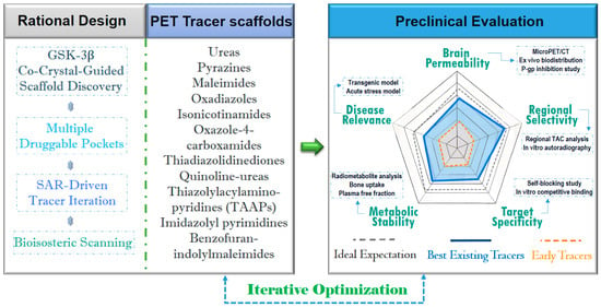
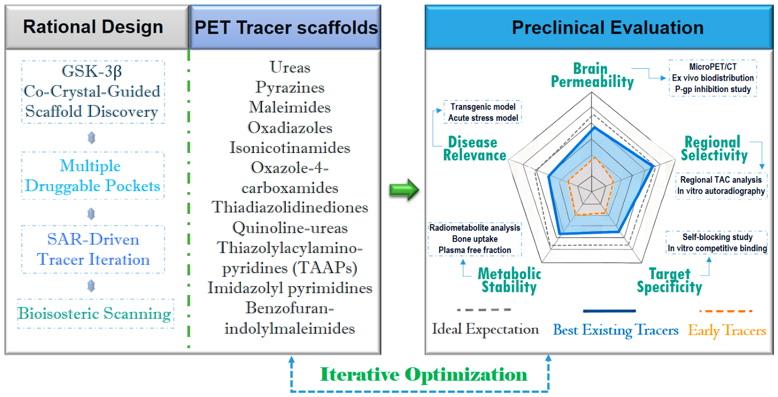
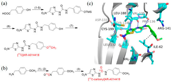
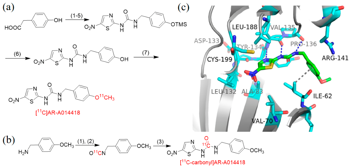
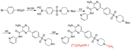
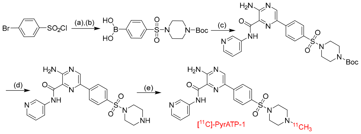
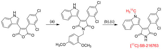
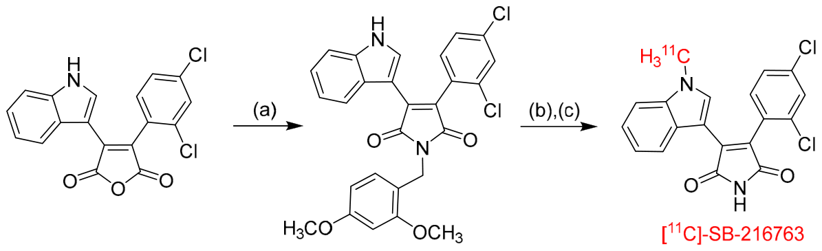
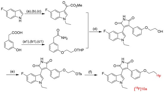
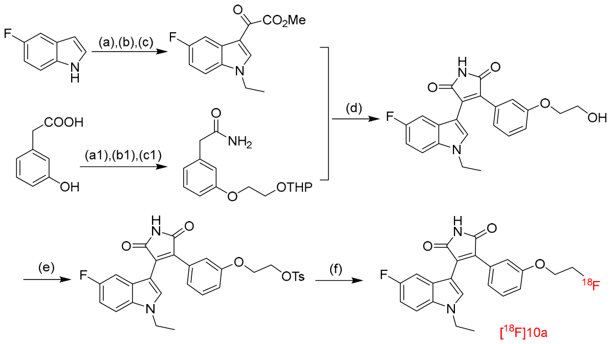
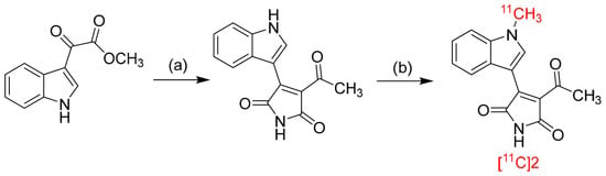
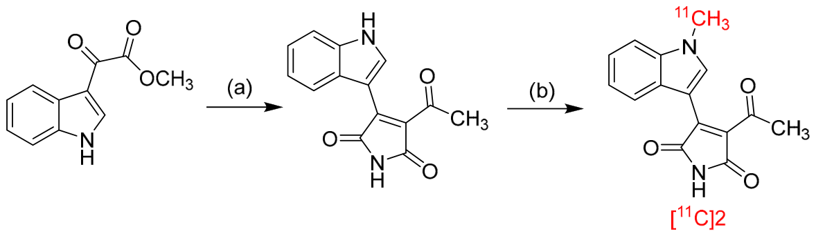
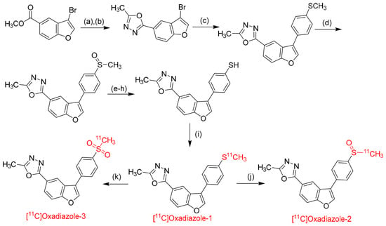
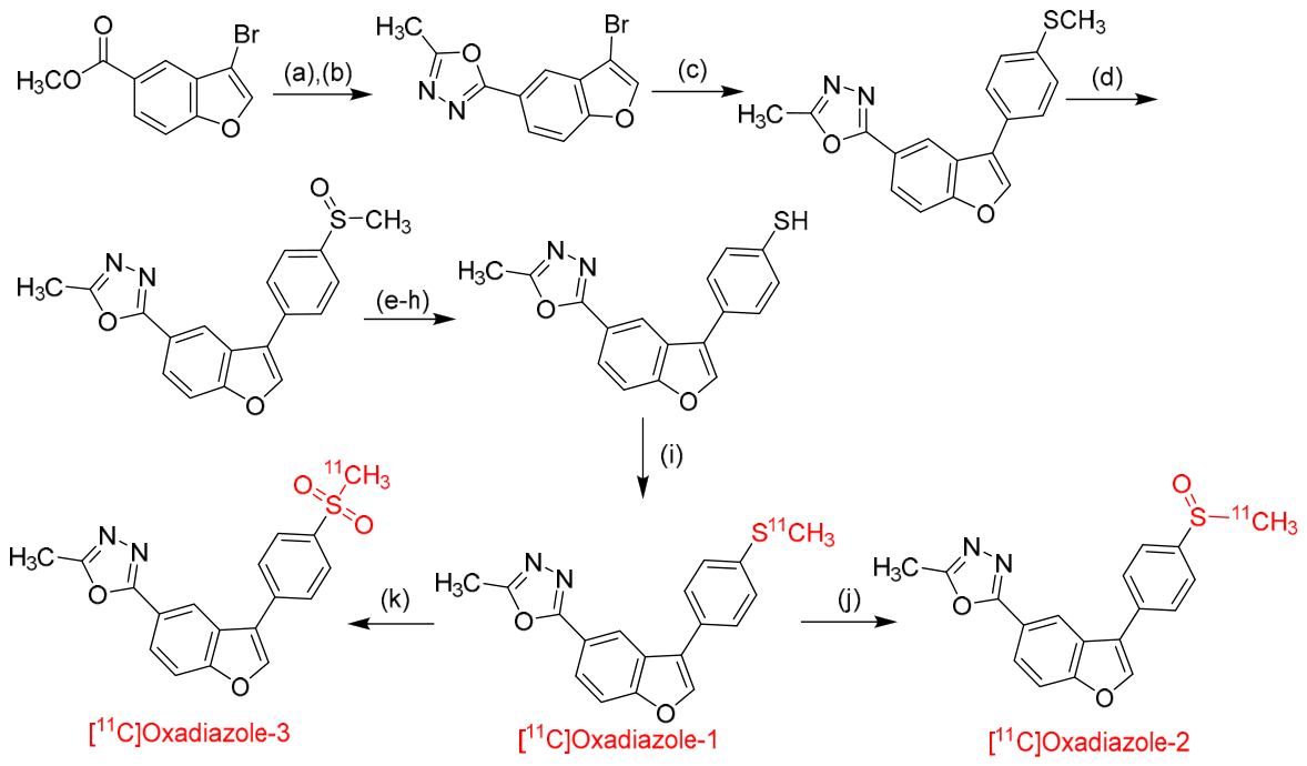


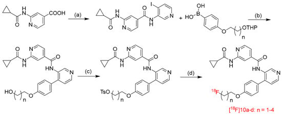
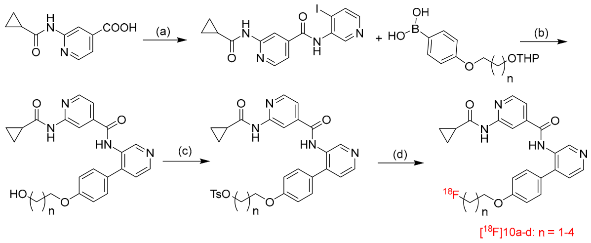
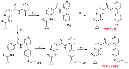
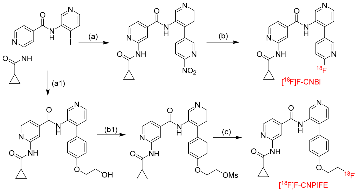
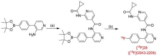
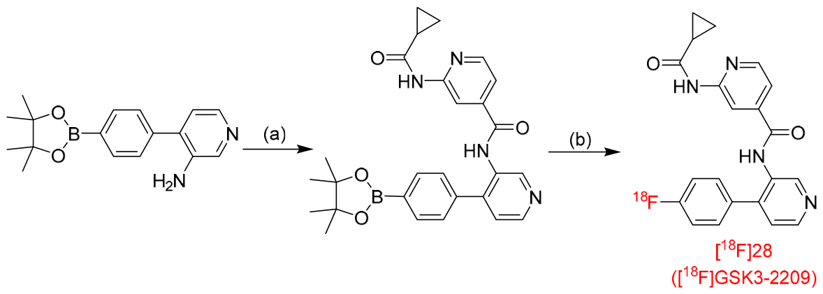
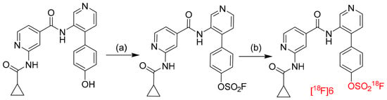
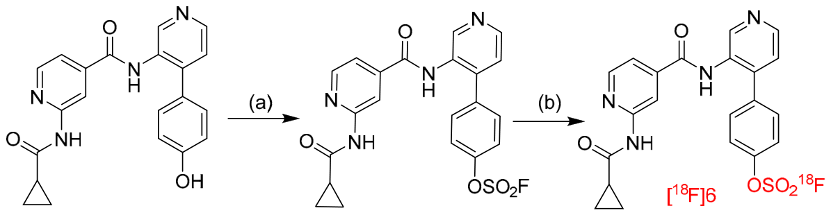
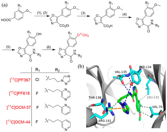
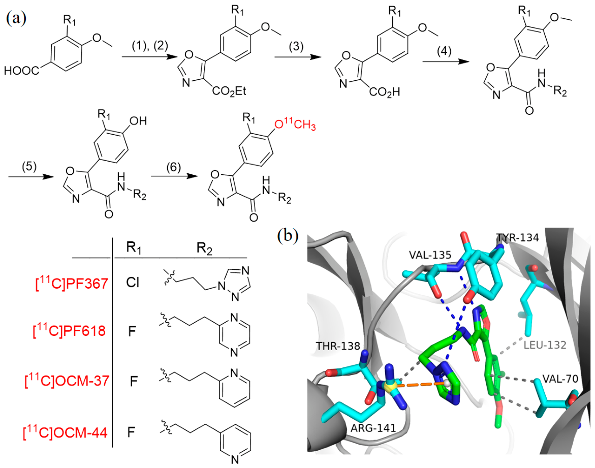
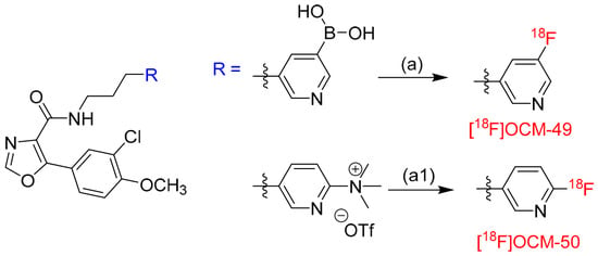
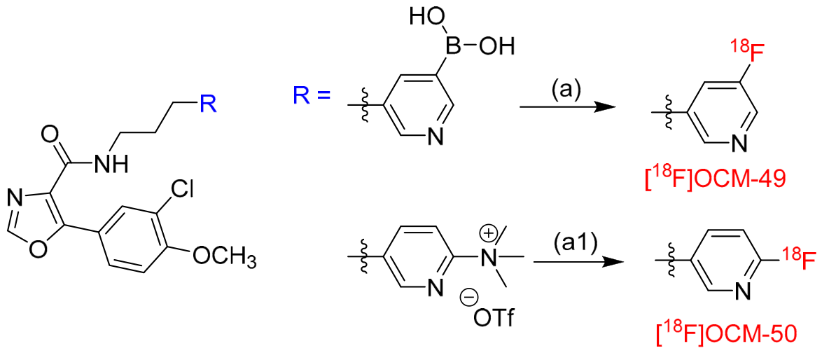
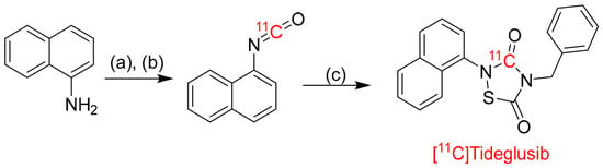
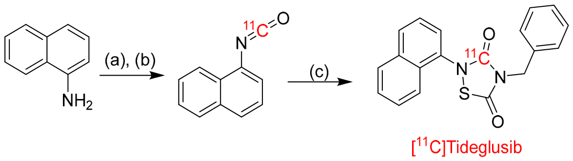


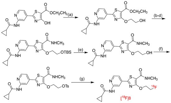
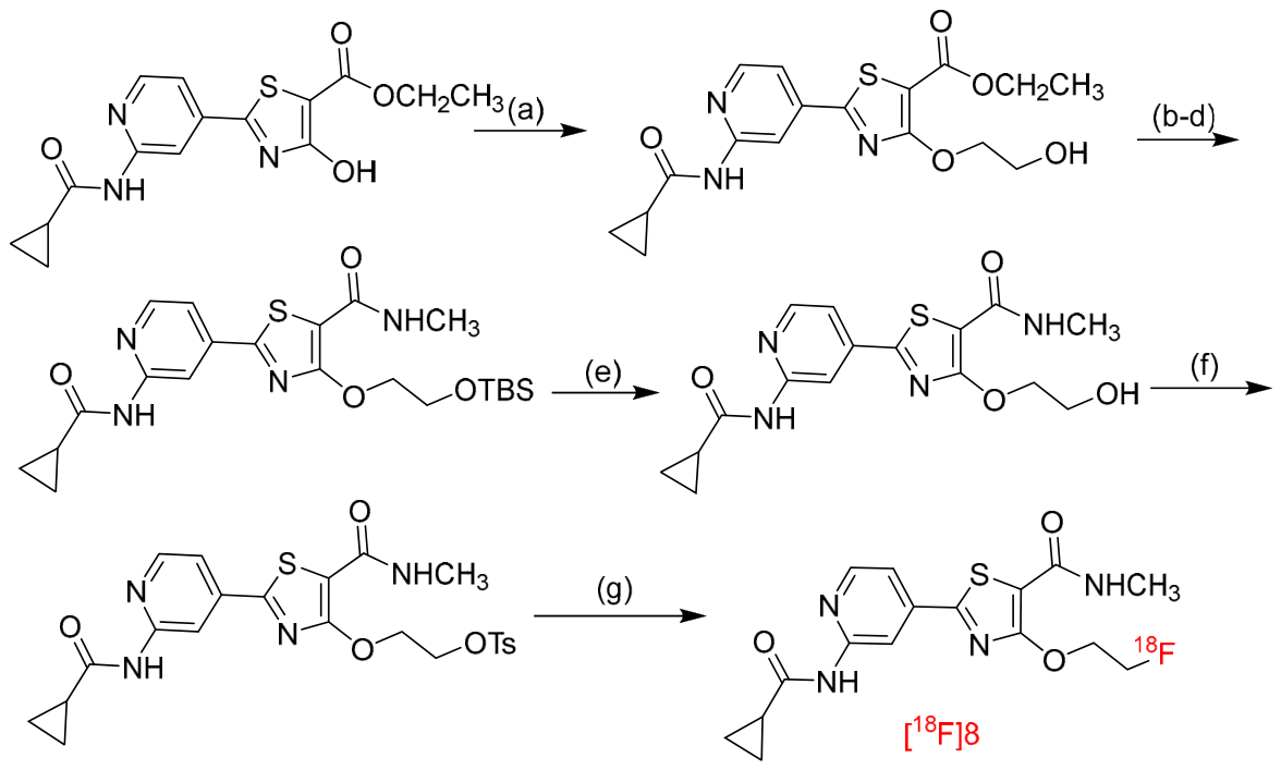
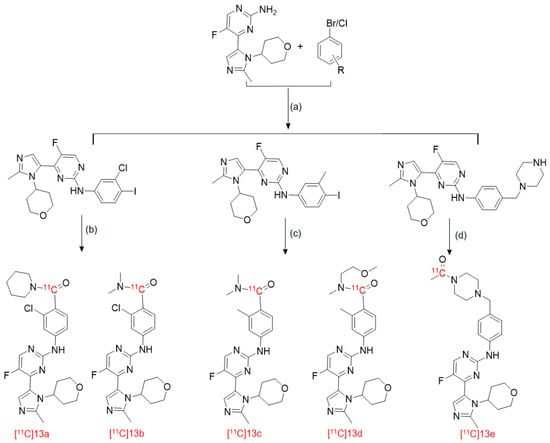
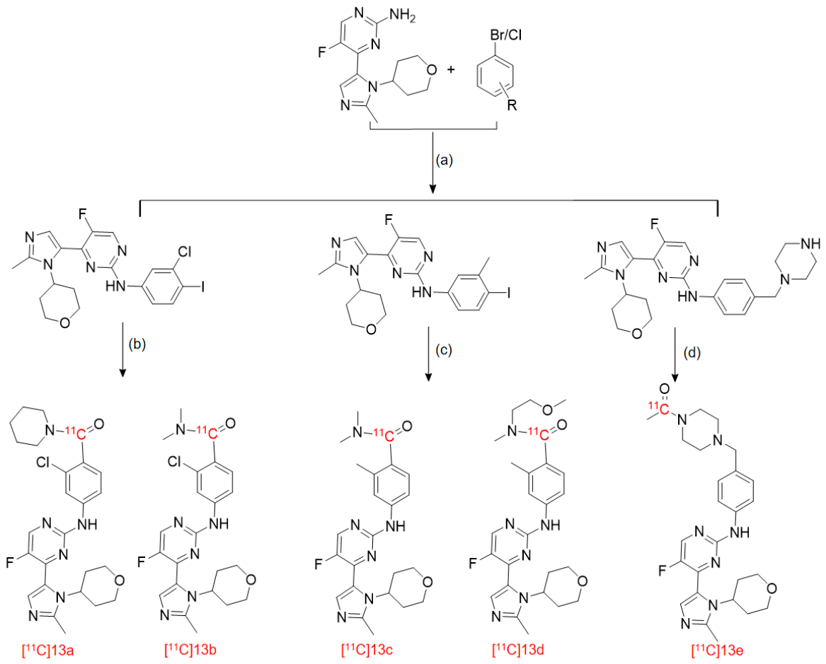



Disclaimer/Publisher’s Note: The statements, opinions and data contained in all publications are solely those of the individual author(s) and contributor(s) and not of MDPI and/or the editor(s). MDPI and/or the editor(s) disclaim responsibility for any injury to people or property resulting from any ideas, methods, instructions or products referred to in the content. |
© 2026 by the authors. Licensee MDPI, Basel, Switzerland. This article is an open access article distributed under the terms and conditions of the Creative Commons Attribution (CC BY) license.