


Abstract
Metabolic diseases are jointly driven by insulin resistance, chronic inflammation, lipotoxicity, and disrupted organelle homeostasis arising from sustained nutrient overload. These disorders substantially increase the risk of cardiovascular, renal, and other multi-organ complications and have become a major global public health burden. Mitochondria are central organelles that integrate energy metabolism with stress signaling. They participate in fatty acid β-oxidation, the tricarboxylic acid cycle, and oxidative phosphorylation, while also regulating reactive oxygen species generation, mitochondrial DNA-related inflammatory signaling, calcium homeostasis, and cell death. Under chronic metabolic stress, the mitochondrial quality control (MQC) system shifts from adaptive repair toward decompensation, characterized by impaired mitochondrial biogenesis, abnormal mitochondrial dynamics, defective mitophagy, disrupted proteostasis and mitochondrial unfolded protein response, increased oxidative stress, and impaired metabolic reprogramming. Natural small molecules possess structural diversity and multi-target, multi-pathway regulatory properties. They can modulate multiple MQC processes and improve mitochondrial function and metabolic phenotypes in preclinical models. Novel formulations, structural optimization, and mitochondria-targeted delivery can further improve their solubility, bioavailability, tissue exposure, and subcellular localization, thereby enhancing therapeutic efficacy and translational potential. This review systematically summarizes the mechanisms of MQC dysregulation in metabolic diseases, the evidence supporting natural small-molecule interventions, and strategies for formulation and delivery optimization. Future studies should strengthen causal validation of MQC, quantify intramitochondrial drug exposure, and incorporate clinically relevant endpoints to facilitate the translation of natural small-molecule MQC modulators.
1. Introduction
Chronic nutrient overload progressively overwhelms the capacity of the body to accommodate excess energy, disrupting the coordinated balance among adipose tissue storage, hepatic metabolism, and skeletal muscle substrate utilization [1]. This maladaptation promotes the development of obesity, type 2 diabetes (T2D), metabolic dysfunction-associated steatotic liver disease (MASLD), and metabolic dysfunction-associated steatohepatitis (MASH) [2,3,4]. As these diseases progress, insulin resistance, ectopic lipid deposition, chronic low-grade inflammation, and oxidative stress reinforce one another, ultimately contributing to sustained injury in the cardiovascular, renal, and nervous systems [5]. Although current lifestyle and pharmacological interventions can improve body weight, glycemic control, lipid profiles, and selected inflammatory markers, their ability to reverse fundamental metabolic dysregulation, organelle damage, and progressive organ dysfunction remains limited [6,7]. Elucidating how nutrient overload evolves from early compensatory adaptation into cellular injury and organ dysfunction is therefore essential for identifying shared pathological targets and developing more effective therapeutic strategies.
Mitochondria are central organelles that coordinate cellular energy metabolism and stress responses, with essential roles in glucose and lipid oxidation, oxidative phosphorylation, and redox homeostasis [8,9]. Under sustained nutrient overload and metabolic stress, mitochondrial respiratory efficiency progressively declines, accompanied by reactive oxygen species (ROS) accumulation, inflammatory signaling, and cellular injury [10]. To limit this damage while maintaining energy supply, cells must continuously identify, repair, and eliminate dysfunctional mitochondria and replenish the mitochondrial pool with newly generated functional organelles [11]. Mitochondrial quality control (MQC) is the dynamic regulatory network that coordinates mitochondrial biogenesis, remodeling, repair, and clearance to preserve mitochondrial integrity, proteostasis, genome stability, and metabolic function. During early metabolic stress, cells enhance mitochondrial biogenesis, adjust mitochondrial dynamics, and activate mitophagy and protein stress responses to repair or remove damaged mitochondria [12,13,14,15]. With persistent nutrient overload, however, mitochondrial repair and clearance mechanisms gradually become insufficient, leading to the accumulation of dysfunctional mitochondria, increased mitochondrial ROS (mtROS), impaired oxidative phosphorylation, and amplified metabolic inflammation. MQC dysfunction therefore represents a critical step linking nutrient overload to metabolic injury [16,17].
Natural small molecules have considerable potential for modulating MQC in metabolic diseases [18]. Their structural diversity and multitarget properties enable them to simultaneously influence energy sensing, oxidative stress, inflammatory signaling, autophagy, and lipid metabolism [19]. Studies have shown that several natural small molecules improve mitochondrial function and modulate MQC-related endpoints in preclinical models of obesity, T2D, MASLD, MASH, and associated complications [20,21,22,23]. However, their application is often limited by poor solubility, insufficient stability, low absorption, and inadequate tissue exposure in vivo, making it difficult to achieve therapeutically effective concentrations in diseased tissues and mitochondria [24]. Novel formulations, delivery systems, and structural optimization can improve systemic exposure, tissue distribution, and mitochondrial localization, thereby facilitating the translation of mitochondrial protective activity observed in vitro into therapeutic efficacy in vivo [25,26,27]. Integrating mechanistic studies with formulation and delivery optimization is therefore an important strategy for developing MQC-targeted therapies for metabolic diseases.
Accordingly, this review uses MQC as its central framework to summarize the mechanisms of MQC dysregulation in obesity, T2D, and MASLD/MASH, with particular emphasis on evidence that natural small molecules modulate mitochondrial biogenesis, dynamics, mitophagy, proteostasis, and redox homeostasis. We further review novel formulations, structural optimization approaches, and mitochondria-targeted delivery strategies and discuss the major challenges associated with mechanistic validation, drug exposure, safety, and clinical translation. This review aims to provide a framework for the discovery and development of natural small-molecule MQC modulators.
2. Mitochondrial Dysfunction and Quality Control Imbalance in Metabolic Diseases
2.1. Core Components of MQC and Their Roles in Metabolic Adaptation
MQC is a dynamic regulatory system that maintains mitochondrial structural and functional homeostasis (Figure 1). By coordinating mitochondrial biogenesis, fission and fusion, mitophagy, proteostasis, and mitochondrial DNA (mtDNA) maintenance, MQC facilitates the removal of damaged mitochondria and replenishment of the functional mitochondrial pool, thereby preserving energy metabolism and redox balance [28,29,30]. When MQC is disrupted, damaged mitochondria progressively accumulate, leading to insufficient ATP production, elevated mtROS, inflammatory activation, and cellular dysfunction, ultimately contributing to the progression of metabolic diseases [12,31,32].
Figure 1.
Major components of mitochondrial quality control. Mitochondrial quality control maintains mitochondrial homeostasis by coordinating mitochondrial biogenesis, fusion and fission, mitophagy, proteostasis and the mitochondrial UPRmt, mtDNA maintenance, and interorganelle communication. Together, these processes replenish functional mitochondria, segregate and eliminate damaged components, maintain protein and genome integrity, regulate metabolic exchange among organelles, and limit mtDNA-mediated inflammatory signaling.
2.1.1. Mitochondrial Biogenesis
Mitochondrial biogenesis is essential for replenishing functional mitochondria and maintaining mitochondrial abundance and metabolic capacity [33]. During energy deprivation, exercise, or oxidative stress, an increased AMP/ATP ratio activates AMP-activated protein kinase (AMPK) and promotes the activity of the NAD+-dependent deacetylase sirtuin 1 (SIRT1) [34,35]. AMPK-mediated phosphorylation and SIRT1-mediated deacetylation cooperatively enhance the transcriptional coactivator activity of the peroxisome proliferator-activated receptor gamma coactivator 1-alpha (PGC-1α). PGC-1α subsequently coactivates nuclear respiratory factor 1 (NRF1) and GA-binding protein (GABP, also known as nuclear respiratory factor 2), thereby inducing the expression of mitochondrial transcription factor A (TFAM) and other nuclear-encoded mitochondrial proteins [36,37,38]. Following its import into mitochondria, TFAM regulates mtDNA replication, transcription, and stability [39,40]. This process coordinates the expression of nuclear- and mitochondrial-encoded proteins, facilitates respiratory-chain complex assembly, and restores oxidative phosphorylation capacity. The AMPK/SIRT1/PGC-1α/NRF1/2/TFAM axis therefore constitutes a central regulatory pathway for mitochondrial biogenesis [41,42].
2.1.2. Mitochondrial Dynamics
Mitochondria continuously undergo fusion and fission to maintain the structural plasticity of the mitochondrial network and adapt to changes in energy demand, damage segregation, and cell division [41,43,44]. Outer mitochondrial membrane fusion is primarily mediated by mitofusin 1 (MFN1) and mitofusin 2 (MFN2), whereas inner membrane fusion depends on optic atrophy 1 (OPA1). Fusion enables adjacent mitochondria to exchange proteins, lipids, and mtDNA, thereby alleviating localized damage through functional complementation and preserving mitochondrial membrane potential and respiratory activity [45,46,47].
Mitochondrial fission is mainly mediated by dynamin-related protein 1 (DRP1). In response to metabolic stress or mitochondrial injury, cytosolic DRP1 is recruited to the outer mitochondrial membrane, where it interacts with receptors including Mitochondrial fission 1 protein (FIS1), Mitochondrial fission factor, and mitochondrial dynamics proteins. DRP1 then assembles into spiral complexes that constrict the mitochondrial membrane and complete fission [48]. Moderate fission segregates depolarized or structurally damaged mitochondrial fragments from the healthy network, facilitating their subsequent removal by mitophagy. Sustained DRP1 hyperactivation, however, causes excessive mitochondrial fragmentation, membrane depolarization, mtROS accumulation, and cell death [49,50]. The balance between mitochondrial fusion and fission therefore determines mitochondrial network integrity, damage segregation efficiency, and metabolic adaptability.
2.1.3. Mitophagy
Mitophagy is the principal MQC mechanism responsible for eliminating irreversibly damaged mitochondria [51]. Under normal mitochondrial membrane potential, PTEN-induced kinase 1 (PINK1) is imported into the inner mitochondrial membrane, where it is continuously cleaved and degraded by mitochondrial proteases [52]. When the membrane potential declines, PINK1 import is impaired, resulting in its accumulation and autophosphorylation on the outer mitochondrial membrane. Activated PINK1 subsequently recruits and activates Parkin, which promotes the ubiquitination of outer mitochondrial membrane proteins. These ubiquitin chains are recognized by autophagy receptors such as OPTN, NDP52, and p62. Through their interaction with microtubule-associated protein 1 light chain 3 (LC3), these receptors promote the engulfment of damaged mitochondria by autophagosomes, which subsequently fuse with lysosomes to complete mitochondrial degradation [53,54,55].
In addition to the PINK1/Parkin pathway, mitochondrial outer membrane receptors can directly recruit autophagosomes through their LC3-interacting regions and mediate mitochondrial clearance under hypoxia, inflammation, and metabolic overload. Following outer mitochondrial membrane rupture, prohibitin 2 (PHB2) can function as an inner membrane mitophagy receptor by binding to LC3. Autophagy and beclin 1 regulator 1 (AMBRA1) also contribute to damage clearance by promoting autophagosome formation and mitochondrial recognition. After mitophagy, mitochondrial components are recycled, while coordinated mitochondrial biogenesis restores mitochondrial abundance and function [56,57,58,59].
Effective mitochondrial clearance requires the coordinated progression of damage recognition, autophagosome engulfment, and lysosomal degradation [60]. Changes in the expression of PINK1, Parkin, or LC3 reflect only individual stages of this process. Mitophagy should therefore be evaluated by assessing mitophagic flux, mitochondrial colocalization with autophagosomes or lysosomes, and the recovery of membrane potential, ATP production, and respiratory function [61].
2.1.4. Mitochondrial Proteostasis and the Mitochondrial Unfolded Protein Response
Mitochondrial proteostasis depends on the coordinated import, folding, and degradation of proteins required for respiratory-chain assembly and metabolic enzyme activity [62]. When oxidative stress, impaired protein import, or respiratory-chain injury causes misfolded proteins to accumulate within mitochondria, cells activate the mitochondrial unfolded protein response (UPRmt) to alleviate proteotoxic stress and restore mitochondrial function [63,64].
During this response, molecular chaperones such as heat shock protein (HSP) 60 promote the refolding of abnormal proteins, whereas proteases including lon protease 1 and caseinolytic peptidase P (CLPP) remove proteins that cannot be repaired [65,66]. As the stress signal is transmitted to the nucleus, activating transcription factor 5 (ATF5) induces the expression of molecular chaperones, proteases, and antioxidant genes, while transcription factors such as C/EBP homologous protein (CHOP) also contribute to stress adaptation [67]. Moderate UPRmt activation restores proteostasis and metabolic function. Persistent protein-folding stress that exceeds mitochondrial repair capacity may instead aggravate inflammation, metabolic dysfunction, and cell death [63].
2.1.5. mtDNA Maintenance and Mitochondrial Interactions with Other Organelles
The integrity of mtDNA is essential for maintaining respiratory-chain function and oxidative phosphorylation [68]. Rather than being associated with conventional histone-based chromatin, mtDNA is compacted by TFAM and other proteins into mitochondrial nucleoids. Nevertheless, its localization within the redox-active mitochondrial environment and its dependence on specialized replication, repair, and turnover mechanisms make it susceptible to oxidative lesions and replication-associated errors [69,70]. Limited damage can be managed through DNA repair, selective degradation or turnover of damaged genomes, nucleoid remodeling, and compensatory replication. Persistent mtDNA damage or mutations impair mitochondrial gene expression and respiratory-chain assembly, ultimately compromising oxidative phosphorylation. Once released into the cytosol or extracellular space, mtDNA can function as a damage-associated molecular pattern. Cytosolic mtDNA primarily activates the cyclic GMP-AMP synthase–stimulator of interferon genes (cGAS–STING) pathway, whereas extracellular or endosomal mtDNA can engage toll-like receptor 9 (TLR9), and oxidized mtDNA can facilitate NLR family pyrin domain-containing 3 (NLRP3) inflammasome activation [71,72,73].
Mitochondria also form contact sites with the endoplasmic reticulum, lipid droplets, and lysosomes to coordinate substrate transport, energy metabolism, and damage clearance [74]. Mitochondria-associated endoplasmic reticulum membranes participate in lipid transport, Ca2+ exchange, mitochondrial fission, and autophagosome formation. Mitochondria-lipid droplet contacts facilitate the transfer of released fatty acids to mitochondrial β-oxidation, thereby limiting lipotoxicity [75,76]. Mitochondria–lysosome interactions contribute to damage recognition, mitochondrial network remodeling, and autophagic degradation [77]. Thus, mtDNA maintenance and interorganelle communication jointly connect mitochondrial structural integrity with metabolic adaptation and inflammatory responses.
2.2. MQC Dysregulation in Obesity, Insulin Resistance, and T2D
Obesity-associated MQC dysfunction exhibits marked tissue specificity and stage dependence [78]. During the early stages of energy surplus, adipocytes maintain metabolic adaptation by enhancing lipid storage, substrate oxidation, and mitochondrial biogenesis [79]. As adipocyte hypertrophy progresses, local hypoxia, endoplasmic reticulum stress, and inflammatory responses intensify, accompanied by impaired mitochondrial biogenesis, reduced oxidative phosphorylation efficiency, disrupted mitochondrial dynamics, and defective mitophagy. In brown and beige adipose tissues, reductions in mitochondrial abundance, uncoupling protein 1 (UCP1) dependent thermogenesis, and fatty acid oxidation further decrease energy expenditure [80,81]. mtROS and mtDNA released from damaged mitochondria activate the NLRP3 inflammasome and cGAS-STING pathway, promoting macrophage infiltration and lipolysis [72,73]. The resulting fatty acid flux to the liver and skeletal muscle contributes to systemic insulin resistance.
MQC abnormalities in skeletal muscle are major contributors to insulin resistance [82]. A mismatch between mitochondrial oxidative capacity and fatty acid supply in skeletal muscle leads to the accumulation of lipid intermediates, including diacylglycerols, ceramides, and acylcarnitines, which interfere with IRS/PI3K/AKT signaling and glucose transporter type 4 (GLUT4) translocation [83,84]. Impaired mitochondrial biogenesis, excessive fission, and defective mitophagy further increase mtROS generation and disrupt insulin signaling [85].
Pancreatic β cells are highly dependent on mitochondrial oxidative metabolism to generate ATP, trigger Ca2+ influx, and stimulate insulin secretion. Chronic glucotoxicity and lipotoxicity induce mitochondrial depolarization, mtROS accumulation, mtDNA damage, DRP1-mediated excessive fission, and defective mitophagy [86,87]. These alterations reduce ATP production and impair glucose sensing. Persistent stress further promotes β-cell apoptosis, senescence, and functional failure [88]. The progression from obesity to T2D can therefore be viewed as the progressive coupling of MQC dysfunction across adipose tissue, skeletal muscle, the liver, and pancreatic β-cells, ultimately forming a multi-organ pathological network linking lipotoxicity, inflammation, insulin resistance, and β-cell failure.
2.3. Mitochondrial Injury and Metabolic Inflammation in MASLD/MASH
The liver is a central regulator of lipid and glucose metabolism. During early MASLD, hepatocytes increase fatty acid β-oxidation, tricarboxylic acid cycle activity, and oxidative phosphorylation to accommodate substrate overload [89,90]. As fatty acid influx persists, mitochondrial oxidative capacity progressively becomes overwhelmed, resulting in electron leakage, elevated mtROS, NAD+/NADH imbalance, reduced ATP-production efficiency, and lipid peroxidation. These alterations promote the transition from hepatic steatosis to hepatocellular injury [91].
During disease progression, impaired mitochondrial biogenesis, DRP1-mediated excessive fission, defective MFN1/2- and OPA1-dependent fusion, and disruption of PINK1/Parkin-mediated mitophagy collectively promote the accumulation of dysfunctional mitochondria [92,93]. Concurrent lysosomal dysfunction, disrupted mitochondrial proteostasis, mtDNA damage, and abnormalities in mitochondria-associated endoplasmic reticulum membranes further impair Ca2+ homeostasis, lipid transport, and respiratory-chain function [94]. These changes establish a self-reinforcing cycle involving lipid overload, mitochondrial injury, and oxidative stress.
Damaged mitochondria also activate cGAS-STING, TLR9, NF-κB, and the NLRP3 inflammasome through mtROS, oxidized mtDNA, cardiolipin, and lipid peroxidation products, thereby promoting inflammatory mediator release and Kupffer cell activation [95,96,97]. Hepatocyte apoptosis, pyroptosis, and ferroptosis further enhance immune-cell recruitment and stimulate hepatic stellate cell activation and extracellular matrix deposition, driving the progression of MASH and liver fibrosis [98,99]. Therapeutic strategies for MASLD/MASH should therefore restore coordination among mitochondrial biogenesis, dynamics, damage clearance, and energy metabolism to disrupt the mutually reinforcing cycle of lipid accumulation, metabolic inflammation, and fibrosis (Figure 2) [100].
Figure 2.
Tissue-specific mitochondrial quality control dysregulation during systemic metabolic stress. High-fat diet, hyperglycemia, excess free fatty acids, and chronic inflammation impair mitochondrial biogenesis, dynamics, mitophagy, and oxidative metabolism in adipose tissue, skeletal muscle, the liver, and pancreatic β-cells. These alterations promote adipocyte hypertrophy and lipolysis, insulin resistance, hepatic steatosis, and β-cell dysfunction, thereby reinforcing multi-organ metabolic deterioration. ↑ indicates an increase, and ↓ indicates a decrease.
3. Natural Small Molecules Targeting MQC in Metabolic Diseases
Natural small molecules are structurally diverse and often act on multiple targets and pathways, enabling them to attenuate metabolic injury by modulating several MQC-related processes, including mitochondrial biogenesis, dynamics, damage clearance, redox homeostasis, and interorganelle communication [19,101]. Current evidence can be broadly classified into three categories: mitochondrial biogenesis and metabolic adaptation, mitochondrial dynamics and mitophagy, and stress defense and homeostatic maintenance. Given the heterogeneity of disease models, experimental endpoints, and causal validation across studies, the effects of natural small molecules on MQC should be evaluated using integrated functional evidence, including mitochondrial respiration, membrane potential, morphology, and mitophagic flux [18,102].
3.1. Promotion of Mitochondrial Biogenesis and Metabolic Adaptation
The AMPK/SIRT1/PGC-1α axis is a major pathway through which natural small molecules promote mitochondrial biogenesis and metabolic adaptation [34]. AMPK enhances PGC-1α activity through direct phosphorylation and SIRT1-dependent deacetylation [103]. Activated PGC-1α subsequently induces mitochondrial biogenesis-related factors, including NRF1, GABP, and TFAM, thereby promoting mtDNA replication, respiratory chain assembly, and oxidative metabolism [104].
Luteolin activates AMPK/PGC-1α signaling in Western diet-fed mice, enhances mitochondrial biogenesis and mitochondrial enzyme activity, and reduces hepatic lipid accumulation [105]. In a high-fat diet-induced model of skeletal muscle insulin resistance, astaxanthin activates AMPK, promotes mitochondrial remodeling and biogenesis, and improves oxidative metabolism, glucose tolerance, and exercise capacity [106]. Melatonin similarly enhances mitochondrial biogenesis in skeletal muscle and the liver through pathways involving CaMKII, AMPK, and PGC-1α, thereby improving glucose and lipid metabolism in obese or diabetic animals [107,108]. Fucoxanthin binds to and inhibits PKM1, reducing glycolytic flux in skeletal muscle while enhancing PGC-1α/TFAM-mediated mitochondrial biogenesis and PPARα/CPT1-dependent fatty acid β-oxidation. These effects reduce lipid accumulation and improve insulin resistance [109]. In human studies, short-term resveratrol supplementation produces calorie restriction-like metabolic effects and modulates AMPK, SIRT1, and PGC-1α-associated energy metabolism [110].
Saponins and quinones have shown particularly consistent effects on mitochondrial remodeling in adipose tissue. Ginsenoside F2 suppresses adipogenesis and promotes mitochondrial biogenesis through AMPK activation [111]. Ginsenoside Rg3 restores inflammation-impaired thermogenesis in brown and beige adipose tissues, accompanied by increased UCP1 expression, oxygen consumption, and fatty acid oxidation [112,113]. Tanshinone I activates AMPK, enhances fatty acid oxidation and mitochondrial biogenesis, and promotes brown adipose tissue activation and white adipose tissue browning, thereby attenuating high-fat diet-induced obesity [114]. Tanshinone IIA and its derivative TAN20 increase mitochondrial abundance and thermogenic activity through the AMPK/PGC-1α/UCP1 axis, improving obesity and insulin resistance in a UCP1-dependent manner [115,116].
In addition to promoting mitochondrial biogenesis, enhancing mitochondrial substrate oxidation is important for alleviating lipotoxicity. Baicalin directly targets hepatic CPT1A and facilitates the transport of long-chain fatty acids into mitochondria for β-oxidation, thereby ameliorating diet-induced obesity and hepatic steatosis [117]. Alnustone binds to the Ca2+ binding sites of calmodulin, increases cytosolic and mitochondrial Ca2+ levels, and enhances mitochondrial fatty acid β-oxidation. Consequently, it reduces hepatic steatosis and insulin resistance in MASLD mice and ameliorates MASH-associated liver fibrosis [118]. Berberine restores mitochondrial energy homeostasis through PGC-1α in diabetic nephropathy models, further suggesting that natural small molecules can protect metabolic organs by regulating mitochondrial biogenesis and substrate utilization [119].
3.2. Regulation of Mitochondrial Dynamics and Mitophagy
Mitochondrial fusion and fission maintain network integrity and facilitate the segregation of damaged mitochondrial components [43]. Persistent exposure to high glucose, fatty acids, or inflammatory stimuli can induce excessive DRP1 activation, resulting in mitochondrial fragmentation, membrane depolarization, mtROS accumulation, and cell death. Xu et al. [120] found that epigallocatechin gallate inhibited DRP1-associated mitochondrial apoptotic signaling and alleviated high-glucose-induced β-cell dysfunction, suggesting that it protects β cells by modulating the interplay between mitochondrial dynamics and cell death. In diabetic mice and high-glucose-treated endothelial progenitor cells, astragaloside IV decreases DRP1 expression while increasing MFN1, MFN2, and OPA1 expression. It also attenuates mtROS accumulation and mitochondrial dysfunction through the GSK-3β/Nrf2 axis [121]. Coenzyme Q10 preserves mitochondrial dynamics, promotes white adipose tissue browning, and improves obesity-related metabolic abnormalities in high-fat diet-fed animals [122].
Mitophagy is responsible for the selective removal of irreversibly damaged mitochondria and represents an important component of MQC targeted by natural small molecules. Quercetin activates AMPK-dependent hepatic mitophagy, reducing lipid accumulation and mitochondrial injury and thereby improving NAFLD-related phenotypes [21]. Quercetin can also promote fatty acid β-oxidation by stimulating lipophagy; however, because lipophagy primarily reflects lipid droplet mobilization, it should be evaluated separately from mitophagy [123]. In palmitate-treated HepG2 cells, hesperetin restores mitochondrial membrane potential, ATP production, and fatty acid oxidation while enhancing PINK1/Parkin-dependent mitophagy and suppressing NLRP3 inflammasome activation and cell death [124].
Lycopene downregulates DRP1, increases MFN2 and OPA1 expression, and activates PINK1/Parkin-mediated mitophagy. These effects restore mitochondrial membrane potential and ATP production, reduce oxidative stress, hepatocyte apoptosis, and hepatic lipid accumulation, and ultimately ameliorate MASLD and preserve liver function [125]. Myricetin stabilizes PINK1 and activates PINK1/Parkin-dependent mitophagy, thereby restoring mitochondrial function and fatty acid β-oxidation. It consequently reduces hepatic steatosis, inflammation, fibrosis, and insulin resistance in MASLD/MASH models [126].
Kaempferol improves mitochondrial homeostasis in diabetic nephropathy by coordinating mitochondrial dynamics and mitophagy. In diabetic nephropathy rats and advanced glycation end product-treated HK-2 cells, kaempferol increases MFN1 and OPA1 expression, decreases DRP1 and FIS1 expression, and enhances PINK1/Parkin-mediated mitophagy. These changes restore mitochondrial membrane potential and ATP production while reducing oxidative stress, inflammation, and apoptosis [127]. Diosgenin restores autophagy through the CaMKK2/AMPK/mTOR pathway and activates PINK1/MFN2/Parkin-mediated mitophagy. It also suppresses excessive mitochondrial fission and promotes fusion by downregulating DRP1 and phosphorylated DRP1 while increasing MFN1, MFN2, and OPA1 expression, thereby alleviating renal injury in type 2 diabetic nephropathy [128].
3.3. Improvement of Mitochondrial Redox Homeostasis, Interorganelle Communication, and Cell Death Signaling
Metabolic overload increases electron leakage from the respiratory chain and mtROS production, leading to oxidative damage to proteins, membrane lipids, and mtDNA [129]. Natural small molecules frequently alleviate these effects by regulating SIRT3, Nrf2, and mitochondrial antioxidant systems. Nevertheless, general antioxidant effects should be distinguished from direct MQC modulation. A more convincing MQC effect requires that decreased mtROS be accompanied by improvements in mitochondrial respiration, membrane potential, dynamics, mitophagy, or damage clearance [29].
Curcumin is one of the most extensively studied polyphenols in this field. It alleviates hepatic steatosis by improving mitochondrial function through SIRT3 in overfed rats and steatotic cells [20]. Kuo et al. further demonstrated that curcumin reduced hepatic NF-κB activity, thiobarbituric acid-reactive substances, and inflammatory cytokine expression while increasing glutathione levels in ob/ob mice. Curcumin also restored mtDNA copy number, NRF1 and TFAM expression, mitochondrial complex I activity, and ATP production and reduced hepatic lipid accumulation [130]. These findings indicate that curcumin alleviates obesity-associated hepatic steatosis by improving redox homeostasis, promoting mitochondrial biogenesis, and restoring respiratory chain function.
In NASH mice and palmitate-treated AML12 cells, nobiletin improves mitochondrial complex I activity, ATP production, mitochondrial Ca2+ uptake, and ROS homeostasis while reducing hepatocyte death, inflammation, and fibrosis [22]. By linking respiratory chain function and Ca2+ homeostasis to disease phenotypes, this study identifies mitochondrial Ca2+ regulation as a potential mechanism through which natural small molecules may ameliorate MASH. Matrine alleviates lipotoxicity-induced endoplasmic reticulum stress and mitochondrial injury by regulating SERCA-mediated Ca2+ homeostasis. In animal and cellular models, it suppresses PERK-, IRE1-, and ATF6-related signaling, restores mitochondrial membrane potential, and reduces ROS production, apoptosis, and hepatic lipid accumulation [131].
mtROS accumulation and mtDNA damage also connect mitochondrial dysfunction with metabolic inflammation. Nrf2 activation enhances antioxidant and detoxification systems and reduces lipid peroxidation and mitochondrial injury [132]. Restoration of mitochondrial function may further suppress inflammatory pathways. Berberine modulates AMPK, Nrf2, NF-κB, and NLRP3-related metabolic and inflammatory signaling while improving glucose metabolism and insulin sensitivity [133,134].
Mitochondrial proteostasis and the UPRmt are important components of MQC. These processes regulate the folding, import, degradation, and assembly of respiratory chain subunits, whereas their disruption impairs electron transport and aggravates oxidative injury [135,136]. Nicotinamide riboside induces a SIRT1/SIRT3-dependent UPRmt and increases CLPP and HSP10 expression, thereby promoting respiratory chain assembly, fatty acid β-oxidation, and energy metabolism. These effects prevent and reverse high-fat, high-sucrose diet-induced hepatic steatosis [137].
The iridoid glycoside agnuside directly binds to and stabilizes the complex I assembly factor NDUFAF6, reducing its ubiquitination and degradation. Agnuside consequently promotes the assembly of complex I and respiratory chain supercomplexes, enhances mitochondrial respiration, ATP production, and adipose tissue thermogenesis, and improves high-fat diet-induced obesity and insulin resistance [138]. However, evidence that natural small molecules directly regulate core UPRmt components in metabolic diseases remains limited. Most studies have instead focused on endoplasmic reticulum stress, JNK/CHOP signaling, and mitochondrial apoptotic pathways. For example, dihydroartemisinin alleviates metabolic injury by regulating SERCA-dependent Ca2+ homeostasis and endoplasmic reticulum stress-associated mitochondrial death signaling, respectively [139,140].
Overall, natural small molecules can restore MQC through multiple mechanisms, including the promotion of mitochondrial biogenesis, coordination of mitochondrial dynamics and mitophagy, improvement of redox homeostasis, and maintenance of mitochondrial proteostasis (Table 1). Future studies should incorporate mitochondrial respiratory measurements, mitophagic flux assays, target-specific interventions, and genetic validation to establish their direct effects on the MQC network and clarify their translational potential.
Table 1.
Natural small molecules modulating MQC in metabolic diseases.
4. Formulation, Structural Optimization, and Mitochondria-Targeted Delivery
Although many natural small molecules can improve mitochondrial function, their therapeutic development is often limited by poor water solubility, insufficient stability, low gastrointestinal absorption, and rapid metabolism. These unfavorable drug-like properties may prevent effective concentrations from being achieved in target tissues and mitochondria. When the concentrations required for in vitro activity substantially exceed the achievable free-drug exposure in vivo, the associated MQC effects are unlikely to translate into consistent therapeutic benefits [19]. Therefore, formulation development, structural optimization, and targeted delivery are essential for translating natural small molecules into viable therapeutics.
Current strategies can be organized into three sequential levels. First, formulation approaches, including liposomes, nanoemulsions, and polymeric nanoparticles, are used to improve aqueous solubility, chemical and metabolic stability, gastrointestinal absorption, and systemic exposure [141,142]. Second, tissue-directed delivery systems, such as liver-targeted liposomes, locally administered hydrogels, and inflammation-responsive carriers, aim to increase drug retention and exposure at diseased sites while limiting off-target distribution [143,144]. Third, subcellular targeting strategies employ membrane-potential-driven lipophilic cations, particularly triphenylphosphonium (TPP+), mitochondria-penetrating peptides, fusogenic MITO-Porter systems, and mitochondria-targeted or stimuli-activated prodrugs to enhance mitochondrial accumulation and/or organelle-selective drug release [145]. Together, these approaches establish a stepwise optimization framework that progresses from improving drug-like properties to enhancing tissue exposure and ultimately achieving greater mitochondrial delivery specificity (Table 2).
4.1. Nanocarriers and Tissue-Targeted Delivery
Liposomes, polymeric nanoparticles, and other nanocarriers can improve the dispersibility and stability of hydrophobic natural small molecules, prolong systemic circulation, and increase drug exposure in metabolically active organs such as the liver. The high-phosphatidylcholine curcumin liposome hPLipo/Cur, composed of DSPC, cholesterol, and DSPE-PEG2000, reduced hepatic lipid accumulation and macrophage infiltration more effectively than free curcumin in experimental steatohepatitis. Mechanistically, hPLipo/Cur inhibited NRF2 degradation and promoted its nuclear translocation, thereby reducing mtROS production, lipid peroxidation, and ferroptosis. These findings suggest that enhanced solubility and tissue delivery can amplify the effects of curcumin on mitochondrial oxidative stress [146].
A galactose-modified oxidized starch–lysozyme nanocarrier enhances resveratrol uptake by targeting the asialoglycoprotein receptor expressed on hepatocytes. In a high-fat diet-induced fatty liver disease model, this formulation reduced hepatic lipid accumulation and insulin resistance and modulated AMPK/SIRT1 related metabolic signaling. Compared with free resveratrol, enhanced liver-selective uptake enabled the formulation to improve hepatocellular energy metabolism and lipid homeostasis more effectively [147].
Similarly, galactose-modified PLGA-PEG nanoparticles improved the aqueous solubility, sustained release, and hepatocyte uptake of epicatechin. In high-fat diet-induced MASLD mice, this formulation reduced hepatic lipid accumulation, improved insulin sensitivity, activated AMPK, inhibited mTORC1, and restored autophagic activity. These effects were accompanied by increased mitochondrial membrane potential, reduced mtROS production, and restored expression of mitochondrial biogenesis-related proteins [148].
Adipose tissue-targeted nanocarriers may enhance mitochondrial thermogenesis. Tian et al. developed AE@PEP-Lip, an SS-31-functionalized cationic liposome co-delivering allicin and empagliflozin. The formulation accumulated in white adipose tissue and mitochondria, reduced mtROS and mitochondrial fragmentation, and enhanced mitochondrial respiration and thermogenesis. Allicin also activated AMPK signaling and increased uncoupling protein expression. AE@PEP-Lip promoted white adipose tissue browning in obese mice and reduced localized fat deposition in a porcine model, demonstrating the potential of combined tissue- and mitochondria-targeted delivery to improve metabolic efficacy [149].
4.2. Mitochondria-Targeted Delivery Strategies
Mitochondria-targeted delivery aims to increase the effective concentrations of natural small molecules within the mitochondrial matrix or near the inner or outer mitochondrial membrane, enabling direct modulation of the respiratory chain, redox systems, and MQC-related proteins [150]. TPP conjugation is one of the most widely used mitochondrial targeting strategies. Driven by the negative potential across the inner mitochondrial membrane, TPP-containing compounds preferentially accumulate within mitochondria. TPP is therefore frequently conjugated to natural small molecules, lipids, polymers, or prodrugs [145].
Che et al. conjugated galactose to whey protein isolate through the Maillard reaction to target hepatocyte asialoglycoprotein receptors and further introduced TPP to construct the AST@TPP-WPI-Gal astaxanthin nanocarrier. This formulation accumulated within mitochondria in steatotic HepG2 cells and enhanced the antioxidant and antilipogenic effects of astaxanthin. In high-fat diet-induced NAFLD mice, it improved serum lipid profiles and liver function and reduced hepatic lipid accumulation by approximately 40% more than free astaxanthin [151].
Mitochondria-targeted micelles can also increase the subcellular exposure of curcumin. Zhang et al. [152] developed self-assembled Cur-CTPP-PEG-PCL micelles with an encapsulation efficiency of 92.88%. These micelles exhibited efficient endosomal escape and mitochondrial colocalization. Compared with free curcumin and non-targeted micelles, Cur-CTPP-PEG-PCL prolonged the in vivo retention of curcumin and increased its bioavailability. In mice with CCl4-induced liver fibrosis, the formulation produced greater reductions in liver enzyme levels and collagen deposition.
The efficiency of mitochondria-targeted delivery depends on mitochondrial membrane potential, intracellular trafficking, and carrier safety. The uptake of lipophilic cations such as TPP relies on mitochondrial membrane potential. Therefore, severe mitochondrial damage or depolarization may reduce carrier accumulation. Excessive cationic accumulation may also disrupt the respiratory chain, membrane potential, and membrane permeability [153]. Moreover, nanocarriers may become trapped within endosomes or lysosomes after cellular uptake, limiting their transport to mitochondria. Mitochondrial exposure should therefore be confirmed using subcellular fractionation followed by quantitative drug analysis, confocal colocalization, mass spectrometry imaging, or radiotracer techniques [154]. These measurements should be integrated with assessments of mitochondrial respiration, membrane potential, and MQC-related functions to determine the biological effects of the delivery system.
4.3. Structural Optimization and Translational Evaluation
Structural optimization can alter the solubility, metabolic stability, membrane permeability, and subcellular distribution of natural small molecules through scaffold functionalization, linker modification, and conjugation with targeting moieties [19]. MitoQ, AntiOxCIN4, Mito-Esc, and SkQ1 are representative stable mitochondria-targeted conjugates.
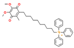
MitoQ has the most extensive evidence among these structurally optimized compounds. It consists of a ubiquinone antioxidant moiety linked to TPP through a ten-carbon alkyl chain, enabling its accumulation near the inner mitochondrial membrane and participation in redox cycling [155]. In ATM+/−/ApoE−/− mice and high-fat diet-fed mice, MitoQ attenuated hyperglycemia, hyperlipidemia, hepatic steatosis, and mitochondrial oxidative injury and improved body weight- and liver function-related outcomes [156]. A 2024 human mechanistic study using lipid infusion combined with a hyperinsulinemic-euglycemic clamp further demonstrated that MitoQ reduced mitochondrial oxidative stress in skeletal muscle and enhanced insulin-stimulated GLUT4 translocation and glucose uptake [157].
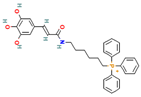
AntiOxCIN4 consists of a dietary caffeic acid-derived antioxidant scaffold linked to TPP through a six-carbon spacer. Structure–activity studies have shown that AntiOxCIN4 exhibits greater mitochondrial targeting and antioxidant activity than its non-targeted parent compound [158]. In mice fed a high-fat, high-sucrose Western diet for 16 weeks, AntiOxCIN4 reduced hepatic lipid accumulation and markers of hepatocellular injury, enhanced fatty acid oxidation and oxidative phosphorylation, and strengthened antioxidant defenses through the PGC-1α/SIRT3 axis. It also increased lysosomal proteolytic capacity and restored impaired autophagic flux, thereby linking improved energy metabolism and antioxidant defense with cellular quality control [159,160].
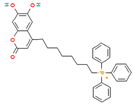
Mito-Esc consists of the natural coumarin esculetin conjugated to TPP through an eight-carbon linker. Early studies demonstrated that Mito-Esc improved endothelial mitochondrial respiration and biogenesis through AMPK/eNOS/SIRT3 signaling and attenuated atherosclerosis in ApoE−/− mice [161]. Subsequent studies in high-fat diet-induced NAFLD/MASH models showed that Mito-Esc reduced hepatic lipid accumulation, inflammation, and fibrosis through the AMPK/SIRT1 axis [162]. In db/db mice, it also suppressed hepatic gluconeogenesis and improved IRS/AKT signaling, insulin resistance, and hyperglycemia-associated atherosclerosis [163]. The consistent validation of Mito-Esc across fatty liver disease, T2D, and cardiovascular complication models strengthens its potential as a mitochondria-targeted candidate for metabolic diseases.
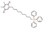
SkQ1 consists of plastoquinone conjugated to TPP through a ten-carbon linker. In alloxan-induced experimental diabetes, preventive administration of SkQ1 reduced blood glucose levels and modulated mRNA and miRNA expression associated with oxidative metabolism [164]. In genetically diabetic db/db mice, SkQ1 did not significantly alter body weight or hyperglycemia but reduced oxidative stress in wounds and improved angiogenesis and skin repair [165]. These findings suggest that the effects of SkQ1 may differ between primary metabolic phenotypes and diabetic complications. Future studies should therefore distinguish its glucose-lowering, mitochondrial antioxidant, and organ-protective effects.
Table 2.
Delivery and structural optimization strategies for natural small molecules targeting MQC in metabolic diseases.


5. Clinical Evidence and Current Clinical-Trial Landscape
Human evidence remains substantially less mature than the cellular and animal literature summarized above. Selected trials have evaluated resveratrol, curcuminoids, astaxanthin, nicotinamide riboside, urolithin A, and MitoQ in obesity, insulin resistance, or fatty liver disease (Table 3). Most studies were small and short in duration and primarily assessed metabolic or imaging-based surrogate outcomes. Direct mitochondrial measurements were uncommon: resveratrol improved skeletal-muscle mitochondrial respiration in obese men [110], whereas nicotinamide riboside did not improve insulin sensitivity or skeletal-muscle mitochondrial respiration, content, or morphology [166,167].
Table 3.
Selected completed and ongoing clinical studies relevant to metabolic disease.
Among interventions with more direct mechanistic relevance, urolithin A produced a mitochondrial molecular signature in older adults [168], and MitoQ reduced mitochondrial oxidative burden and attenuated lipid-induced skeletal-muscle insulin resistance in an acute human study [157]. However, these findings do not establish long-term efficacy in metabolic disease. An ongoing trial is evaluating urolithin A during dietary weight loss in adults with obesity (NCT07377136), including mitochondrial and metabolic biomarkers. No metabolic-disease clinical trials were identified for AntiOxCIN4, Mito-Esc, or SkQ1; their metabolic evidence therefore remains preclinical. Overall, improvements in metabolic surrogates should not be interpreted as proof of MQC engagement unless tissue exposure and mitochondrial endpoints are assessed directly.
6. Translational Challenges and Future Perspectives
6.1. Limitations of Preclinical Evidence and Causal Validation
The use of natural small molecules to treat metabolic diseases through MQC modulation remains at the stage of mechanistic validation and translational development. Current evidence is derived primarily from cellular and animal studies, which commonly assess ROS production, ATP levels, mitochondrial membrane potential, MQC-related protein expression, and histopathological changes [18,171]. The translational relevance of some preclinical studies is limited by clinically unattainable exposure levels, preventive treatment protocols, and reliance on a single sex or a single disease model. Changes in the expression of PGC-1α, PINK1, Parkin, DRP1, or MFN proteins are commonly interpreted as evidence of MQC modulation. Although these measurements alone cannot establish flux, target engagement, or causal necessity, future studies should combine pharmacological interventions with genetic loss-of-function approaches, longitudinal reporters of mitophagy or mitochondrial dynamics, respiratory flux measurements, and tissue-specific validation to improve the reliability of the mechanistic evidence.
6.2. Clinical Validation of MQC Modulation and Trial Design
In clinical studies, improvements in glycemia, lipid profiles, body weight, liver enzymes, and hepatic fat content indicate metabolic benefits but do not demonstrate effective MQC modulation in humans without corresponding mitochondrial and mechanistic measurements. Mitochondrial function in peripheral blood mononuclear cells, circulating mtDNA, oxidative stress and inflammatory biomarkers, metabolomic and lipidomic profiles, and imaging-based tissue changes may therefore provide important links between clinical phenotypes and mitochondrial mechanisms [95,168,172]. Clinically meaningful outcomes may include insulin sensitivity assessed by the hyperinsulinemic-euglycemic clamp, hepatic fat content quantified using magnetic resonance imaging proton density fat fraction (MRI-PDFF), histological activity or fibrosis in MASH, physical function, and validated organ-specific outcome measures. Most existing human studies are limited by small sample sizes and short intervention periods and are powered primarily to detect changes in surrogate metabolic outcomes. Clinical trials targeting MQC should therefore prespecify at least one mechanistic endpoint and one clinically meaningful endpoint.
6.3. Drug Exposure, Formulation Standardization, and Emerging Technologies
Exposure in vivo is a major determinant of whether mechanisms identified in vitro can be translated into consistent therapeutic effects. Gastrointestinal absorption, gut microbial metabolism, hepatic first-pass metabolism, and tissue distribution substantially influence the actual exposure to active compounds and their metabolites [173,174,175]. Mitochondria-targeted formulations present additional challenges, including membrane potential-dependent accumulation, cation-associated toxicity, inefficient intracellular trafficking, carrier accumulation, and immune responses. Because obesity, T2D, and MASLD/MASH generally require long-term treatment, pharmacokinetics, drug concentrations in target tissues and mitochondria, repeated-dose safety, and drug interactions should be evaluated alongside therapeutic efficacy. Network pharmacology, artificial intelligence, and multi-omics technologies provide valuable tools for characterizing the multitarget effects of natural small molecules [176]. Molecular docking, transcriptomics, proteomics, metabolomics, lipidomics, and single-cell and spatial omics can facilitate target discovery, identify responsive cell populations, and reconstruct regulatory networks [177,178]. However, computational predictions and correlative molecular changes primarily generate mechanistic hypotheses and cannot replace causal validation.
Mitochondrial respiration, ATP production, and membrane potential can be used to assess bioenergetic function, whereas mitochondrial morphology, dynamics, and mitophagic flux can reveal network remodeling and damage clearance. Measurements of mtROS, mtDNA damage, the UPRmt, and lysosomal function can further characterize stress adaptation and quality control capacity. Integrating these endpoints with metabolic flux analysis, pharmacokinetics, tissue distribution, and intramitochondrial drug exposure within the same experimental design would clarify whether formulation optimization or structural modification genuinely enhances MQC modulation [179,180,181]. Clinical studies should likewise incorporate reproducible and accessible mechanistic endpoints to establish associations between mitochondrial functional changes, imaging findings, metabolic outcomes, and clinical benefits.
7. Conclusions
Mitochondrial quality control dysregulation provides an integrative framework linking nutrient overload, oxidative stress, organelle damage, chronic inflammation, and metabolic reprogramming. Obesity, T2D, and MASLD/MASH are commonly associated with impaired mitochondrial biogenesis, imbalanced fusion and fission, defective mitophagy, insufficient UPRmt adaptation, and increased mtROS- and mtDNA-mediated inflammatory signaling [8,12]. MQC should therefore be viewed as a dynamic network connecting cellular energy homeostasis with metabolic function.
Natural small molecules can simultaneously modulate metabolic signaling, mitochondrial respiration, autophagy, and antioxidant defenses, supporting their use as both pharmacological probes and potential lead compounds. However, current evidence is derived predominantly from cellular and animal studies, and relatively few studies have demonstrated concurrent improvements in mitochondrial function, MQC-related fluxes, and metabolic phenotypes. Poor aqueous solubility, rapid metabolism, and insufficient target-tissue exposure further limit their in vivo efficacy. Liposomes, nanoparticles, polymeric micelles, TPP conjugation, and stimuli-responsive prodrugs can improve pharmacokinetics and mitochondrial delivery but may also introduce risks such as carrier accumulation, immune responses, and mitochondrial toxicity [171,182]. Future studies should therefore integrate pharmacokinetics, tissue and mitochondrial exposure, MQC function, metabolic outcomes, and long-term safety, supported by validation across multiple models and clinically relevant mitochondrial biomarkers.
Overall, natural small molecules targeting MQC for metabolic diseases are at a critical transition from mechanistic discovery to therapeutic translation. Their development into clinically valuable drug candidates will require quantitative confirmation of mitochondrial exposure, causal evidence of MQC modulation, reproducible metabolic benefits across models, and validated long-term safety. Establishing a verifiable, quantifiable, and translational research framework centered on MQC will facilitate the development of natural small molecules for precise mitochondrial intervention and metabolic disease treatment.
Author Contributions
Conceptualization, C.Z. and Y.G.; writing—original draft preparation, C.Z. and Y.G.; writing—review and editing, T.Z. and Q.Q.; visualization, L.S.; supervision, T.Z.; funding acquisition, L.S. and Q.Q. All authors have read and agreed to the published version of the manuscript.
Funding
This work was supported by the National Natural Science Foundation of China (No. 82608371), the Fundamental Research Funds for the Central Universities (No. 2026-JYB-XJSJJ009), and the Fundamental Research Funds for the Central Universities of Beijing University of Chinese Medicine (No. 2025-JYB-JBGS-025).
Institutional Review Board Statement
Not applicable.
Informed Consent Statement
Not applicable.
Data Availability Statement
No new data were created or analyzed in this study. Data sharing is not applicable.
Acknowledgments
The authors gratefully acknowledge their colleagues at Beijing University of Chinese Medicine for their valuable discussions and constructive advice during the preparation of this manuscript. During the preparation of this manuscript, the authors used OpenAI image generation tools (gpt-image-2, version 2026-04-21) to assist in generating selected graphical elements used in the figures. The authors have reviewed and edited the output and take full responsibility for the content of this publication.
Conflicts of Interest
The authors declare no conflicts of interest.
Abbreviations
MQC, mitochondrial quality control; T2D, type 2 diabetes; MASLD, metabolic dysfunction-associated steatotic liver disease; MASH, metabolic dysfunction-associated steatohepatitis; ROS, reactive oxygen species; mtROS, mitochondrial ROS; mtDNA, mitochondrial DNA; AMPK, AMP-activated protein kinase; SIRT1, NAD+-dependent deacetylase sirtuin; PGC-1α, peroxisome proliferator-activated receptor gamma coactivator 1-alpha; NRF1, nuclear respiratory factor 1; GABP, GA-binding protein; TFAM, mitochondrial transcription factor A; MFN1, mitofusin 1; MFN2, mitofusin 2; OPA1, optic atrophy 1; DRP1, dynamin-related protein 1; FIS1, mitochondrial fission 1 protein; PINK1, PTEN-induced kinase 1; LC3, microtubule-associated protein 1 light chain 3; PHB2, prohibitin 2; AMBRA1, autophagy and beclin 1 regulator 1; UPRmt, mitochondrial unfolded protein response; HSP, heat shock protein; CLPP, caseinolytic peptidase P; ATF5, activating transcription factor 5; CHOP, C/EBP homologous protein; cGAS–STING, cyclic GMP-AMP synthase-stimulator of interferon genes; TLR9, toll-like receptor 9; NLRP3, NLR family pyrin domain-containing 3; UCP1, uncoupling protein 1; GLUT4, glucose transporter type 4; TPP+, triphenylphosphonium.
References
- Qiu, H.; Schlegel, V. Impact of nutrient overload on metabolic homeostasis. Nutr. Rev. 2018, 76, 693–707. [Google Scholar] [CrossRef] [Scilit] [PubMed]
- Tinajero, M.G.; Malik, V.S. An update on the epidemiology of type 2 diabetes: A global perspective. Endocrinol. Metab. Clin. 2021, 50, 337–355. [Google Scholar] [CrossRef] [Scilit] [PubMed]
- Chan, W.; Chuah, K.; Rajaram, R.B.; Lim, L.; Ratnasingam, J.; Vethakkan, S.R. Metabolic dysfunction-associated steatotic liver disease (masld): A state-of-the-art review. J. Obes. Metab. Syndr. 2023, 32, 197–213. [Google Scholar] [CrossRef] [Scilit] [PubMed]
- Do, A.; Zahrawi, F.; Mehal, W.Z. Therapeutic landscape of metabolic dysfunction-associated steatohepatitis (mash). Nat. Rev. Drug Discov. 2025, 24, 171–189. [Google Scholar] [CrossRef] [Scilit] [PubMed]
- Sandireddy, R.; Sakthivel, S.; Gupta, P.; Behari, J.; Tripathi, M.; Singh, B.K. Systemic impacts of metabolic dysfunction-associated steatotic liver disease (masld) and metabolic dysfunction-associated steatohepatitis (mash) on heart, muscle, and kidney related diseases. Front. Cell Dev. Biol. 2024, 12, 1433857. [Google Scholar] [CrossRef] [Scilit] [PubMed]
- Hinte, L.C.; Castellano-Castillo, D.; Ghosh, A.; Melrose, K.; Gasser, E.; Noé, F.; Massier, L.; Dong, H.; Sun, W.; Hoffmann, A.; et al. Adipose tissue retains an epigenetic memory of obesity after weight loss. Nature 2024, 636, 457–465. [Google Scholar] [CrossRef] [Scilit] [PubMed]
- Rahimi, G.R.M.; Yousefabadi, H.A.; Niyazi, A.; Rahimi, N.M.; Alikhajeh, Y. Effects of lifestyle intervention on inflammatory markers and waist circumference in overweight/obese adults with metabolic syndrome: A systematic review and meta-analysis of randomized controlled trials. Biol. Res. Nurs. 2022, 24, 94–105. [Google Scholar] [CrossRef] [Scilit] [PubMed]
- Suomalainen, A.; Nunnari, J. Mitochondria at the crossroads of health and disease. Cell 2024, 187, 2601–2627. [Google Scholar] [CrossRef] [Scilit] [PubMed]
- Granath-Panelo, M.; Kajimura, S. Mitochondrial heterogeneity and adaptations to cellular needs. Nat. Cell Biol. 2024, 26, 674–686. [Google Scholar] [CrossRef] [Scilit] [PubMed]
- Murphy, M.P.; O’Neill, L.A.J. A break in mitochondrial endosymbiosis as a basis for inflammatory diseases. Nature 2024, 626, 271–279. [Google Scholar] [CrossRef] [Scilit] [PubMed]
- Liu, Y.J.; Sulc, J.; Auwerx, J. Mitochondrial genetics, signalling and stress responses. Nat. Cell Biol. 2025, 27, 393–407. [Google Scholar] [CrossRef] [Scilit] [PubMed]
- Liu, B.; Xu, C.; Liu, Y.; Lu, Z.; Fu, T.; Li, G.; Deng, Y.; Luo, G.; Ding, S.; Li, N.; et al. Mitochondrial quality control in human health and disease. Mil. Med. Res. Med. Res. 2024, 11, 32. [Google Scholar] [CrossRef] [Scilit] [PubMed]
- Tábara, L.; Segawa, M.; Prudent, J. Molecular mechanisms of mitochondrial dynamics. Nat. Rev. Mol. Cell Biol. 2025, 26, 123–146. [Google Scholar] [CrossRef] [Scilit] [PubMed]
- Wu, L.; Wang, L.; Du, Y.; Zhang, Y.; Ren, J. Mitochondrial quality control mechanisms as therapeutic targets in doxorubicin-induced cardiotoxicity. Trends Pharmacol. Sci. 2023, 44, 34–49. [Google Scholar] [CrossRef] [Scilit] [PubMed]
- Hong, W.; Huang, H.; Zeng, X.; Duan, C. Targeting mitochondrial quality control: New therapeutic strategies for major diseases. Mil. Med. Res. Med. Res. 2024, 11, 59. [Google Scholar] [CrossRef] [Scilit] [PubMed]
- Veluthakal, R.; Esparza, D.; Hoolachan, J.M.; Balakrishnan, R.; Ahn, M.; Oh, E.; Jayasena, C.S.; Thurmond, D.C. Mitochondrial dysfunction, oxidative stress, and inter-organ miscommunications in t2d progression. Int. J. Mol. Sci. 2024, 25, 1504. [Google Scholar] [CrossRef] [Scilit] [PubMed]
- Luo, W.; Yu, M.; Zheng, C.; Li, X.; Ma, X.; Zeng, G.; Zhang, J.; Ji, X.; Sun, L. Mitochondrial dysfunction in mash: Focusing on chronic inflammation and intercellular communication. Biochem. Biophys. Res. Commun. Biochem. Res. 2026, 804, 153356. [Google Scholar] [CrossRef] [Scilit] [PubMed]
- Lin, Q.; Jia, L.; Lei, M.; Gao, D.; Zhang, N.; Sha, L.; Liu, X.; Liu, Y. Natural products as pharmacological modulators of mitochondrial dysfunctions for the treatment of diabetes and its complications: An update since 2010. Pharmacol. Res. 2024, 200, 107054. [Google Scholar] [CrossRef] [Scilit] [PubMed]
- Atanasov, A.G.; Zotchev, S.B.; Dirsch, V.M.; International, N.P.S.T.; Supuran, C.T. Natural products in drug discovery: Advances and opportunities. Nat. Rev. Drug Discov. 2021, 20, 200–216. [Google Scholar] [CrossRef] [Scilit] [PubMed]
- Du, S.; Zhu, X.; Zhou, N.; Zheng, W.; Zhou, W.; Li, X. Curcumin alleviates hepatic steatosis by improving mitochondrial function in postnatal overfed rats and fatty l02 cells through the sirt3 pathway. Food Funct. 2022, 13, 2155–2171. [Google Scholar] [CrossRef] [Scilit] [PubMed]
- Cao, P.; Wang, Y.; Zhang, C.; Sullivan, M.A.; Chen, W.; Jing, X.; Yu, H.; Li, F.; Wang, Q.; Zhou, Z.; et al. Quercetin ameliorates nonalcoholic fatty liver disease (nafld) via the promotion of ampk-mediated hepatic mitophagy. J. Nutr. Biochem. 2023, 120, 109414. [Google Scholar] [CrossRef] [Scilit] [PubMed]
- Li, S.; Li, X.; Chen, F.; Liu, M.; Ning, L.; Yan, Y.; Zhang, S.; Huang, S.; Tu, C. Nobiletin mitigates hepatocytes death, liver inflammation, and fibrosis in a murine model of nash through modulating hepatic oxidative stress and mitochondrial dysfunction. J. Nutr. Biochem. 2022, 100, 108888. [Google Scholar] [CrossRef] [Scilit] [PubMed]
- Yu, L.; Dong, X.; Xue, X.; Xu, S.; Zhang, X.; Xu, Y.; Wang, Z.; Wang, Y.; Gao, H.; Liang, Y.; et al. Melatonin attenuates diabetic cardiomyopathy and reduces myocardial vulnerability to ischemia-reperfusion injury by improving mitochondrial quality control: Role of sirt6. J. Pineal Res. 2021, 70, e12698. [Google Scholar] [CrossRef] [Scilit] [PubMed]
- Qiu, C.; Zhang, J.Z.; Wu, B.; Xu, C.C.; Pang, H.H.; Tu, Q.C.; Lu, Y.Q.; Guo, Q.Y.; Xia, F.; Wang, J.G. Advanced application of nanotechnology in active constituents of traditional chinese medicines. J. Nanobiotechnol. 2023, 21, 456. [Google Scholar] [CrossRef] [Scilit] [PubMed]
- Wang, S.; Dong, G.; Sheng, C. Structural simplification of natural products. Chem. Rev. 2019, 119, 4180–4220. [Google Scholar] [CrossRef] [Scilit] [PubMed]
- Huang, L.; Huang, X.; Yang, X.; Hu, J.; Zhu, Y.; Yan, P.; Xie, Y. Novel nano-drug delivery system for natural products and their application. Pharmacol. Res. 2024, 201, 107100. [Google Scholar] [CrossRef] [Scilit] [PubMed]
- Kulkarni, C.A.; Fink, B.D.; Gibbs, B.E.; Chheda, P.R.; Wu, M.; Sivitz, W.I.; Kerns, R.J. A novel triphenylphosphonium carrier to target mitochondria without uncoupling oxidative phosphorylation. J. Med. Chem. 2021, 64, 662–676. [Google Scholar] [CrossRef] [Scilit] [PubMed]
- Ng, M.Y.W.; Wai, T.; Simonsen, A. Quality control of the mitochondrion. Dev. Cell 2021, 56, 881–905. [Google Scholar] [CrossRef] [Scilit] [PubMed]
- Pickles, S.; Vigié, P.; Youle, R.J. Mitophagy and quality control mechanisms in mitochondrial maintenance. Curr. Biol. 2018, 28, R170–R185. [Google Scholar] [CrossRef] [Scilit] [PubMed]
- Zhao, D.; Yan, Z.; Yu, Y.; Zhang, Y.; Luan, Z. From mechanisms to therapies: Exploiting epigenetic and post-translational modifications of mitochondrial quality control in diabetic kidney disease. Front. Endocrinol. 2026, 17, 1821507. [Google Scholar] [CrossRef] [Scilit] [PubMed]
- Baek, J.; Lee, Y.H.; Jeong, H.Y.; Lee, S. Mitochondrial quality control and its emerging role in the pathogenesis of diabetic kidney disease. Kidney Res. Clin. Pract. 2023, 42, 560–564. [Google Scholar] [CrossRef] [Scilit] [PubMed]
- Zong, Y.; Li, H.; Liao, P.; Chen, L.; Pan, Y.; Zheng, Y.; Zhang, C.; Liu, D.; Zheng, M.; Gao, J. Mitochondrial dysfunction: Mechanisms and advances in therapy. Signal Transduct. Tar. 2024, 9, 124. [Google Scholar] [CrossRef] [Scilit] [PubMed]
- Popov, L. Mitochondrial biogenesis: An update. J. Cell Mol. Med. 2020, 24, 4892–4899. [Google Scholar] [CrossRef] [Scilit] [PubMed]
- Herzig, S.; Shaw, R.J. Ampk: Guardian of metabolism and mitochondrial homeostasis. Nat. Rev. Mol. Cell Biol. 2018, 19, 121–135. [Google Scholar] [CrossRef] [Scilit] [PubMed]
- Tang, B.L. Sirt1 and the mitochondria. Mol. Cells 2016, 39, 87–95. [Google Scholar] [CrossRef] [Scilit] [PubMed]
- Wang, S.; Long, H.; Hou, L.; Feng, B.; Ma, Z.; Wu, Y.; Zeng, Y.; Cai, J.; Zhang, D.; Zhao, G. The mitophagy pathway and its implications in human diseases. Signal Transduct. Tar. 2023, 8, 304. [Google Scholar] [CrossRef] [Scilit] [PubMed]
- Qian, L.; Zhu, Y.; Deng, C.; Liang, Z.; Chen, J.; Chen, Y.; Wang, X.; Liu, Y.; Tian, Y.; Yang, Y. Peroxisome proliferator-activated receptor gamma coactivator-1 (pgc-1) family in physiological and pathophysiological process and diseases. Signal Transduct. Tar. 2024, 9, 50. [Google Scholar] [CrossRef] [Scilit] [PubMed]
- Covarrubias, A.J.; Perrone, R.; Grozio, A.; Verdin, E. Nad(+) metabolism and its roles in cellular processes during ageing. Nat. Rev. Mol. Cell Biol. 2021, 22, 119–141. [Google Scholar] [CrossRef] [Scilit] [PubMed]
- Kang, I.; Chu, C.T.; Kaufman, B.A. The mitochondrial transcription factor tfam in neurodegeneration: Emerging evidence and mechanisms. FEBS Lett. 2018, 592, 793–811. [Google Scholar] [CrossRef] [Scilit] [PubMed]
- Brüser, C.; Keller-Findeisen, J.; Jakobs, S. The tfam-to-mtdna ratio defines inner-cellular nucleoid populations with distinct activity levels. Cell Rep. 2021, 37, 110000. [Google Scholar] [CrossRef] [Scilit] [PubMed]
- Giacomello, M.; Pyakurel, A.; Glytsou, C.; Scorrano, L. The cell biology of mitochondrial membrane dynamics. Nat. Rev. Mol. Cell Biol. 2020, 21, 204–224. [Google Scholar] [CrossRef] [Scilit] [PubMed]
- Ashcroft, S.P.; Stocks, B.; Egan, B.; Zierath, J.R. Exercise induces tissue-specific adaptations to enhance cardiometabolic health. Cell Metab. 2024, 36, 278–300. [Google Scholar] [CrossRef] [Scilit] [PubMed]
- Chen, W.; Zhao, H.; Li, Y. Mitochondrial dynamics in health and disease: Mechanisms and potential targets. Signal Transduct. Target. Ther. 2023, 8, 333. [Google Scholar] [CrossRef] [Scilit] [PubMed]
- Quintana-Cabrera, R.; Scorrano, L. Determinants and outcomes of mitochondrial dynamics. Mol. Cell 2023, 83, 857–876. [Google Scholar] [CrossRef] [Scilit] [PubMed]
- Gao, S.; Hu, J. Mitochondrial fusion: The machineries in and out. Trends Cell Biol. 2021, 31, 62–74. [Google Scholar] [CrossRef] [Scilit] [PubMed]
- Alghamdi, A. A detailed review of pharmacology of mfn1 (mitofusion-1)-mediated mitochondrial dynamics: Implications for cellular health and diseases. Saudi Pharm. J. 2024, 32, 102012. [Google Scholar] [CrossRef] [Scilit] [PubMed]
- Zanfardino, P.; Amati, A.; Perrone, M.; Petruzzella, V. The balance of mfn2 and opa1 in mitochondrial dynamics, cellular homeostasis, and disease. Biomolecules 2025, 15, 433. [Google Scholar] [CrossRef] [Scilit] [PubMed]
- Kalia, R.; Wang, R.Y.; Yusuf, A.; Thomas, P.V.; Agard, D.A.; Shaw, J.M.; Frost, A. Structural basis of mitochondrial receptor binding and constriction by drp1. Nature 2018, 558, 401–405. [Google Scholar] [CrossRef] [Scilit] [PubMed]
- Dubey, N.; Goyal, A.; Parakh, N.; Narang, R.; Arava, S.K.; Kumar, A.; Yadav, M.; Yadav, H.N. Balancing act: Drp1 inhibition and mitochondrial homeostasis in cardiovascular diseases. Vasc. Pharmacol. 2025, 160, 107531. [Google Scholar] [CrossRef] [Scilit] [PubMed]
- Tilokani, L.; Nagashima, S.; Paupe, V.; Prudent, J. Mitochondrial dynamics: Overview of molecular mechanisms. Essays Biochem. 2018, 62, 341–360. [Google Scholar] [CrossRef] [Scilit] [PubMed]
- Onishi, M.; Yamano, K.; Sato, M.; Matsuda, N.; Okamoto, K. Molecular mechanisms and physiological functions of mitophagy. Embo J. 2021, 40, EMBJ2020104705. [Google Scholar] [CrossRef] [Scilit] [PubMed]
- Narendra, D.P.; Youle, R.J. The role of pink1–parkin in mitochondrial quality control. Nat. Cell Biol. 2024, 26, 1639–1651. [Google Scholar] [CrossRef] [Scilit] [PubMed]
- Harper, J.W.; Ordureau, A.; Heo, J. Building and decoding ubiquitin chains for mitophagy. Nat. Rev. Mol. Cell Biol. 2018, 19, 93–108. [Google Scholar] [CrossRef] [Scilit] [PubMed]
- D’Arcy, M.S. Mitophagy in health and disease. Molecular mechanisms, regulatory pathways, and therapeutic implications. Apoptosis Int. J. Program. Cell Death 2024, 29, 1415–1428. [Google Scholar] [CrossRef] [Scilit] [PubMed]
- Killackey, S.A.; Philpott, D.J.; Girardin, S.E. Mitophagy pathways in health and disease. J. Cell Biol. 2020, 219, e202004029. [Google Scholar] [CrossRef] [Scilit] [PubMed]
- Di Rita, A.; Peschiaroli, A.; Acunzo, P.D.; Strobbe, D.; Hu, Z.; Gruber, J.; Nygaard, M.; Lambrughi, M.; Melino, G.; Papaleo, E.; et al. Huwe1 e3 ligase promotes pink1/parkin-independent mitophagy by regulating ambra1 activation via ikkα. Nat. Commun. 2018, 9, 3755. [Google Scholar] [CrossRef] [Scilit] [PubMed]
- Yan, C.; Gong, L.; Chen, L.; Xu, M.; Abou-Hamdan, H.; Tang, M.; Désaubry, L.; Song, Z. Phb2 (prohibitin 2) promotes pink1-prkn/parkin-dependent mitophagy by the parl-pgam5-pink1 axis. Autophagy 2020, 16, 419–434. [Google Scholar] [CrossRef] [Scilit] [PubMed]
- Wei, Y.; Chiang, W.; Sumpter, R.; Mishra, P.; Levine, B. Prohibitin 2 is an inner mitochondrial membrane mitophagy receptor. Cell 2017, 168, 224–238. [Google Scholar] [CrossRef] [Scilit] [PubMed]
- McWilliams, T.G.; Prescott, A.R.; Montava-Garriga, L.; Ball, G.; Singh, F.; Barini, E.; Muqit, M.M.K.; Brooks, S.P.; Ganley, I.G. Basal mitophagy occurs independently of pink1 in mouse tissues of high metabolic demand. Cell Metab. 2018, 27, 439–449. [Google Scholar] [CrossRef] [Scilit] [PubMed]
- Onishi, M.; Okamoto, K. Mitochondrial clearance: Mechanisms and roles in cellular fitness. FEBS Lett. 2021, 595, 1239–1263. [Google Scholar] [CrossRef] [Scilit] [PubMed]
- Katayama, H.; Hama, H.; Nagasawa, K.; Kurokawa, H.; Sugiyama, M.; Ando, R.; Funata, M.; Yoshida, N.; Homma, M.; Nishimura, T.; et al. Visualizing and modulating mitophagy for therapeutic studies of neurodegeneration. Cell 2020, 181, 1176–1187. [Google Scholar] [CrossRef] [Scilit] [PubMed]
- Pfanner, N.; Warscheid, B.; Wiedemann, N. Mitochondrial proteins: From biogenesis to functional networks. Nat. Rev. Mol. Cell Biol. 2019, 20, 267–284. [Google Scholar] [CrossRef] [Scilit] [PubMed]
- Shpilka, T.; Haynes, C.M. The mitochondrial upr: Mechanisms, physiological functions and implications in ageing. Nat. Rev. Mol. Cell Biol. 2018, 19, 109–120. [Google Scholar] [CrossRef] [Scilit] [PubMed]
- Münch, C. The different axes of the mammalian mitochondrial unfolded protein response. BMC Biol. 2018, 16, 81. [Google Scholar] [CrossRef] [Scilit] [PubMed]
- Fan, F.; Duan, Y.; Yang, F.; Trexler, C.; Wang, H.; Huang, L.; Li, Y.; Tang, H.; Wang, G.; Fang, X.; et al. Deletion of heat shock protein 60 in adult mouse cardiomyocytes perturbs mitochondrial protein homeostasis and causes heart failure. Cell Death Differ. 2020, 27, 587–600. [Google Scholar] [CrossRef] [Scilit] [PubMed]
- Li, J.; Deng, Y.; Gasser, M.; Zhu, J.; Walker, E.M.; Sidarala, V.; Reck, E.C.; Hubers, D.L.; Pasmooij, M.B.; Shin, C.; et al. Lonp1 regulation of mitochondrial protein folding provides insight into beta cell failure in type 2 diabetes. Nat. Metab. 2025, 7, 1570–1592. [Google Scholar] [CrossRef] [Scilit] [PubMed]
- Fiorese, C.J.; Schulz, A.M.; Lin, Y.; Rosin, N.; Pellegrino, M.W.; Haynes, C.M. The transcription factor atf5 mediates a mammalian mitochondrial upr. Curr. Biol. 2016, 26, 2037–2043. [Google Scholar] [CrossRef] [Scilit] [PubMed]
- Falkenberg, M.; Larsson, N.; Gustafsson, C.M. Replication and transcription of human mitochondrial dna. Annu. Rev. Biochem. 2024, 93, 47–77. [Google Scholar] [CrossRef] [Scilit] [PubMed]
- Fontana, G.A.; Gahlon, H.L. Mechanisms of replication and repair in mitochondrial dna deletion formation. Nucleic Acids Res. 2020, 48, 11244–11258. [Google Scholar] [CrossRef] [Scilit] [PubMed]
- Newman, L.E.; Shadel, G.S. Mitochondrial dna release in innate immune signaling. Annu. Rev. Biochem. 2023, 92, 299–332. [Google Scholar] [CrossRef] [Scilit] [PubMed]
- Hu, M.; Shu, H. Mitochondrial dna-triggered innate immune response: Mechanisms and diseases. Cell Mol. Immunol. 2023, 20, 1403–1412. [Google Scholar] [CrossRef] [Scilit] [PubMed]
- Zhong, Z.; Liang, S.; Sanchez-Lopez, E.; He, F.; Shalapour, S.; Lin, X.; Wong, J.; Ding, S.; Seki, E.; Schnabl, B.; et al. New mitochondrial dna synthesis enables nlrp3 inflammasome activation. Nature 2018, 560, 198–203. [Google Scholar] [CrossRef] [Scilit] [PubMed]
- Bai, J.; Cervantes, C.; Liu, J.; He, S.; Zhou, H.; Zhang, B.; Cai, H.; Yin, D.; Hu, D.; Li, Z.; et al. Dsba-l prevents obesity-induced inflammation and insulin resistance by suppressing the mtdna release-activated cgas-cgamp-sting pathway. Proc. Natl. Acad. Sci. USA 2017, 114, 12196–12201. [Google Scholar] [CrossRef] [Scilit] [PubMed]
- Wu, H.; Carvalho, P.; Voeltz, G.K. Here, there, and everywhere: The importance of er membrane contact sites. Science 2018, 361, eaan5835. [Google Scholar] [CrossRef] [Scilit] [PubMed]
- Rieusset, J. The role of endoplasmic reticulum-mitochondria contact sites in the control of glucose homeostasis: An update. Cell Death Dis. 2018, 9, 388. [Google Scholar] [CrossRef] [Scilit] [PubMed]
- Benador, I.Y.; Veliova, M.; Mahdaviani, K.; Petcherski, A.; Wikstrom, J.D.; Assali, E.A.; Acín-Pérez, R.; Shum, M.L.; Oliveira, M.F.; Cinti, S.; et al. Mitochondria bound to lipid droplets have unique bioenergetics, composition, and dynamics that support lipid droplet expansion. Cell Metab. 2018, 27, 869–885. [Google Scholar] [CrossRef] [Scilit] [PubMed]
- Wong, Y.C.; Ysselstein, D.; Krainc, D. Mitochondria–lysosome contacts regulate mitochondrial fission via rab7 gtp hydrolysis. Nature 2018, 554, 382–386. [Google Scholar] [CrossRef] [Scilit] [PubMed]
- Xia, W.; Veeragandham, P.; Cao, Y.; Xu, Y.; Rhyne, T.E.; Qian, J.; Hung, C.; Zhao, P.; Jones, Y.; Gao, H.; et al. Obesity causes mitochondrial fragmentation and dysfunction in white adipocytes due to rala activation. Nat. Metab. 2024, 6, 273–289. [Google Scholar] [CrossRef] [Scilit] [PubMed]
- Chouchani, E.T.; Kajimura, S. Metabolic adaptation and maladaptation in adipose tissue. Nat. Metab. 2019, 1, 189–200. [Google Scholar] [CrossRef] [Scilit] [PubMed]
- Chouchani, E.T.; Kazak, L.; Spiegelman, B.M. New advances in adaptive thermogenesis: Ucp1 and beyond. Cell Metab. 2019, 29, 27–37. [Google Scholar] [CrossRef] [Scilit] [PubMed]
- Becher, T.; Palanisamy, S.; Kramer, D.J.; Eljalby, M.; Marx, S.J.; Wibmer, A.G.; Butler, S.D.; Jiang, C.S.; Vaughan, R.; Sch Der, H.; et al. Brown adipose tissue is associated with cardiometabolic health. Nat. Med. 2021, 27, 58–65. [Google Scholar] [CrossRef] [Scilit] [PubMed]
- Sligar, J.; DeBruin, D.A.; Saner, N.J.; Philp, A.M.; Philp, A. The importance of mitochondrial quality control for maintaining skeletal muscle function across health span. Am. J. Physiol.-Cell Physiol. 2022, 322, C461–C467. [Google Scholar] [CrossRef] [Scilit] [PubMed]
- Tang, W.; Liu, H.; Li, X.; Deng, S.; Gao, C. Influence and treatment of insulin receptor substrate/pi3k/akt-mediated insulin resistance in diabetes mellitus (review). Mol. Med. Rep. 2025, 33, 63. [Google Scholar] [CrossRef] [Scilit] [PubMed]
- Richter, E.A.; Hargreaves, M. Exercise, glut4, and skeletal muscle glucose uptake. Physiol. Rev. 2013, 93, 993–1017. [Google Scholar] [CrossRef] [Scilit] [PubMed]
- Haythorne, E.; Rohm, M.; van de Bunt, M.; Brereton, M.F.; Tarasov, A.I.; Blacker, T.S.; Sachse, G.; Silva Dos Santos, M.; Terron Exposito, R.; Davis, S.; et al. Diabetes causes marked inhibition of mitochondrial metabolism in pancreatic β-cells. Nat. Commun. 2019, 10, 2474. [Google Scholar] [CrossRef] [Scilit] [PubMed]
- Chen, Y.; Liu, X.; Liu, Y.; Li, Y.; Li, D.; Mei, Z.; Deng, Y. Mitochondrial quality control in diabetes mellitus and complications: Molecular mechanisms and therapeutic strategies. Cell Death Dis. 2025, 16, 652. [Google Scholar] [CrossRef] [Scilit] [PubMed]
- Rutter, G.A.; Sidarala, V.; Kaufman, B.A.; Soleimanpour, S.A. Mitochondrial metabolism and dynamics in pancreatic beta cell glucose sensing. Biochem. J. 2023, 480, 773–789. [Google Scholar] [CrossRef] [Scilit] [PubMed]
- Ter Horst, K.W.; Gilijamse, P.W.; Versteeg, R.I.; Ackermans, M.T.; Nederveen, A.J.; la Fleur, S.E.; Romijn, J.A.; Nieuwdorp, M.; Zhang, D.; Samuel, V.T.; et al. Hepatic diacylglycerol-associated protein kinase cε translocation links hepatic steatosis to hepatic insulin resistance in humans. Cell Rep. 2017, 19, 1997–2004. [Google Scholar] [CrossRef] [Scilit] [PubMed]
- Loomba, R.; Friedman, S.L.; Shulman, G.I. Mechanisms and disease consequences of nonalcoholic fatty liver disease. Cell 2021, 184, 2537–2564. [Google Scholar] [CrossRef] [Scilit] [PubMed]
- Friedman, S.L.; Neuschwander-Tetri, B.A.; Rinella, M.; Sanyal, A.J. Mechanisms of nafld development and therapeutic strategies. Nat. Med. 2018, 24, 908–922. [Google Scholar] [CrossRef] [Scilit] [PubMed]
- Fromenty, B.; Roden, M. Mitochondrial alterations in fatty liver diseases. J. Hepatol. 2023, 78, 415–429. [Google Scholar] [CrossRef] [Scilit] [PubMed]
- Karkucinska Wieckowska, A.; Simoes, I.C.M.; Kalinowski, P.; Lebiedzinska Arciszewska, M.; Zieniewicz, K.; Milkiewicz, P.; Górska Ponikowska, M.; Pinton, P.; Malik, A.N.; Krawczyk, M.; et al. Mitochondria, oxidative stress and nonalcoholic fatty liver disease: A complex relationship. Eur. J. Clin. Investig. 2022, 52, e13622. [Google Scholar] [CrossRef] [Scilit] [PubMed]
- Mansouri, A.; Gattolliat, C.; Asselah, T. Mitochondrial dysfunction and signaling in chronic liver diseases. Gastroenterology 2018, 155, 629–647. [Google Scholar] [CrossRef] [Scilit] [PubMed]
- Longo, M.; Meroni, M.; Paolini, E.; Macchi, C.; Dongiovanni, P. Mitochondrial dynamics and nonalcoholic fatty liver disease (nafld): New perspectives for a fairy-tale ending? Metab. Clin. Exp. 2021, 117, 154708. [Google Scholar] [CrossRef] [Scilit] [PubMed]
- Garcia-Martinez, I.; Santoro, N.; Chen, Y.; Hoque, R.; Ouyang, X.; Caprio, S.; Shlomchik, M.J.; Coffman, R.L.; Candia, A.; Mehal, W.Z. Hepatocyte mitochondrial dna drives nonalcoholic steatohepatitis by activation of tlr9. J. Clin. Investig. 2016, 126, 859–864. [Google Scholar] [CrossRef] [Scilit] [PubMed]
- Luo, X.; Li, H.; Ma, L.; Zhou, J.; Guo, X.; Woo, S.; Pei, Y.; Knight, L.R.; Deveau, M.; Chen, Y.; et al. Expression of sting is increased in liver tissues from patients with nafld and promotes macrophage-mediated hepatic inflammation and fibrosis in mice. Gastroenterology 2018, 155, 1971–1984. [Google Scholar] [CrossRef] [Scilit] [PubMed]
- Mridha, A.R.; Wree, A.; Robertson, A.A.B.; Yeh, M.M.; Johnson, C.D.; Van Rooyen, D.M.; Haczeyni, F.; Teoh, N.C.H.; Savard, C.; Ioannou, G.N.; et al. Nlrp3 inflammasome blockade reduces liver inflammation and fibrosis in experimental nash in mice. J. Hepatol. 2017, 66, 1037–1046. [Google Scholar] [CrossRef] [Scilit] [PubMed]
- Krenkel, O.; Tacke, F. Liver macrophages in tissue homeostasis and disease. Nat. Rev. Immunol. 2017, 17, 306–321. [Google Scholar] [CrossRef] [Scilit] [PubMed]
- Gaul, S.; Leszczynska, A.; Alegre, F.; Kaufmann, B.; Johnson, C.D.; Adams, L.A.; Wree, A.; Damm, G.; Seehofer, D.; Calvente, C.J.; et al. Hepatocyte pyroptosis and release of inflammasome particles induce stellate cell activation and liver fibrosis. J. Hepatol. 2021, 74, 156–167. [Google Scholar] [CrossRef] [Scilit] [PubMed]
- Wang, D.; Miao, J.; Zhang, L.; Zhang, L. Research advances in the diagnosis and treatment of masld/mash. Ann. Med. 2025, 57, 2445780. [Google Scholar] [CrossRef] [Scilit] [PubMed]
- Chen, Q.; Ruan, D.; Shi, J.; Du, D.; Bian, C. The multifaceted roles of natural products in mitochondrial dysfunction. Front. Pharmacol. 2023, 14, 1093038. [Google Scholar] [CrossRef] [Scilit] [PubMed]
- Su, Z.; Guo, Y.; Huang, X.; Feng, B.; Tang, L.; Zheng, G.; Zhu, Y. Phytochemicals: Targeting mitophagy to treat metabolic disorders. Front. Cell Dev. Biol. 2021, 9, 686820. [Google Scholar] [CrossRef] [Scilit] [PubMed]
- Jäger, S.; Handschin, C.; St Pierre, J.; Spiegelman, B.M. AMP-activated protein kinase (ampk) action in skeletal muscle via direct phosphorylation of pgc-1α. Proc. Natl. Acad. Sci. USA 2007, 104, 12017–12022. [Google Scholar] [CrossRef] [Scilit] [PubMed]
- Halling, J.F.; Pilegaard, H. Pgc-1α-mediated regulation of mitochondrial function and physiological implications. Appl. Physiol. Nutr. Metab. 2020, 45, 927–936. [Google Scholar] [CrossRef] [Scilit] [PubMed]
- Wang, T.; Xu, Q.; Cao, Y.; Zhang, C.; Chen, S.; Zhang, Y.; Liang, T. Luteolin ameliorates hepatic steatosis and enhances mitochondrial biogenesis via ampk/pgc-1α pathway in western diet-fed mice. J. Nutr. Sci. Vitaminol. 2023, 69, 259–267. [Google Scholar] [CrossRef] [Scilit] [PubMed]
- Nishida, Y.; Nawaz, A.; Kado, T.; Takikawa, A.; Igarashi, Y.; Onogi, Y.; Wada, T.; Sasaoka, T.; Yamamoto, S.; Sasahara, M.; et al. Astaxanthin stimulates mitochondrial biogenesis in insulin resistant muscle via activation of ampk pathway. J. Cachexia Sarcopenia Muscle 2020, 11, 241–258. [Google Scholar] [CrossRef] [Scilit] [PubMed]
- Salagre, D.; Navarro-Alarcón, M.; Villalón-Mir, M.; Alcázar-Navarrete, B.; Gómez-Moreno, G.; Tamimi, F.; Agil, A. Chronic melatonin treatment improves obesity by inducing uncoupling of skeletal muscle serca-sln mediated by camkii/ampk/pgc1α pathway and mitochondrial biogenesis in female and male zücker diabetic fatty rats. Biomed. Pharmacother. 2024, 172, 116314. [Google Scholar] [CrossRef] [Scilit] [PubMed]
- de Luxán-Delgado, B.; Potes, Y.; Rubio-González, A.; Solano, J.J.; Boga, J.A.; Antuña, E.; Cachán-Vega, C.; Bermejo-Millo, J.C.; Menéndez-Coto, N.; García-González, C.; et al. Melatonin alleviates liver mitochondrial dysfunction in leptin-deficient mice. Int. J. Mol. Sci. 2024, 25, 8677. [Google Scholar] [CrossRef] [Scilit] [PubMed]
- Wu, C.; Zhang, C.; Li, F.; Yan, Y.; Wu, Y.; Li, B.; Tong, H.; Lang, J. Fucoxanthin mitigates high-fat-induced lipid deposition and insulin resistance in skeletal muscle through inhibiting pkm1 activity. J. Agric. Food Chem. 2024, 72, 18013–18026. [Google Scholar] [CrossRef] [Scilit] [PubMed]
- Timmers, S.; Konings, E.; Bilet, L.; Houtkooper, R.H.; van de Weijer, T.; Goossens, G.H.; Hoeks, J.; van der Krieken, S.; Ryu, D.; Kersten, S.; et al. Calorie restriction-like effects of 30 days of resveratrol supplementation on energy metabolism and metabolic profile in obese humans. Cell Metab. 2011, 14, 612–622. [Google Scholar] [CrossRef] [Scilit] [PubMed]
- Zhou, J.; Zhang, J.; Li, J.; Guan, Y.; Shen, T.; Li, F.; Li, X.; Yang, X.; Hu, W. Ginsenoside f2 suppresses adipogenesis in 3t3-l1 cells and obesity in micevia the ampk pathway. J. Agric. Food Chem. 2021, 69, 9299–9312. [Google Scholar] [CrossRef] [Scilit] [PubMed]
- Feng, F.; Ko, H.; Truong, T.M.T.; Song, W.; Ko, E.; Kang, I. Ginsenoside rg3, enriched in red ginseng extract, improves lipopolysaccharides-induced suppression of brown and beige adipose thermogenesis with mitochondrial activation. Sci. Rep. 2024, 14, 9157. [Google Scholar] [CrossRef] [Scilit] [PubMed]
- Kim, K.; Nam, K.H.; Yi, S.A.; Park, J.W.; Han, J.; Lee, J. Ginsenoside rg3 induces browning of 3t3-l1 adipocytes by activating ampk signaling. Nutrients 2020, 12, 427. [Google Scholar] [CrossRef] [Scilit] [PubMed]
- Jung, D.Y.; Suh, N.; Jung, M.H. Tanshinone 1 prevents high fat diet-induced obesity through activation of brown adipocytes and induction of browning in white adipocytes. Life Sci. 2022, 298, 120488. [Google Scholar] [CrossRef] [Scilit] [PubMed]
- Fang, Z.; Lin, R.; Yuan, B.; Liu, Y.; Zhang, H. Tanshinone iia inhibits atherosclerotic plaque formation by down-regulating mmp-2 and mmp-9 expression in rabbits fed a high-fat diet. Life Sci. 2007, 81, 1339–1345. [Google Scholar] [CrossRef] [Scilit] [PubMed]
- Ma, L.; Zhao, Z.; Guo, X.; Li, J.; Xu, L.; Mei, W.; Dong, G.; Zhong, Z.; Yang, Z. Tanshinone iia and its derivative activate thermogenesis in adipocytes and induce “beiging” of white adipose tissue. Mol. Cell Endocrinol. 2022, 544, 111557. [Google Scholar] [CrossRef] [Scilit] [PubMed]
- Dai, J.; Liang, K.; Zhao, S.; Jia, W.; Liu, Y.; Wu, H.; Lv, J.; Cao, C.; Chen, T.; Zhuang, S.; et al. Chemoproteomics reveals baicalin activates hepatic cpt1 to ameliorate diet-induced obesity and hepatic steatosis. Proc. Natl. Acad. Sci. USA 2018, 115, E5896–E5905. [Google Scholar] [CrossRef] [Scilit] [PubMed]
- Hu, S.; Liang, X.; Qin, Y.; Li, Y.; Liu, Y.; Liu, C.; Lin, Z.; Geng, C.; Xu, Y.; Wei, D.; et al. Alnustone ameliorates metabolic dysfunction-associated steatotic liver disease by facilitating mitochondrial fatty acid β-oxidation via targeting calmodulin. Adv. Sci. 2025, 12, e11984. [Google Scholar] [CrossRef] [Scilit] [PubMed]
- Qin, X.; Jiang, M.; Zhao, Y.; Gong, J.; Su, H.; Yuan, F.; Fang, K.; Yuan, X.; Yu, X.; Dong, H.; et al. Berberine protects against diabetic kidney disease via promoting pgc-1α-regulated mitochondrial energy homeostasis. Br. J. Pharmacol. 2020, 177, 3646–3661. [Google Scholar] [CrossRef] [Scilit] [PubMed]
- Jia, X.; Mao, D.; Guo, J.; Ke, J.; Zhu, Y.; Zhao, X.; Luo, Z.; Liu, X.; Tang, R.; Hou, R.; et al. Epigallocatechin gallate attenuated high glucose-induced pancreatic beta cell dysfunction by modulating drp1-mediated mitochondrial apoptosis pathways. Sci. Rep. 2024, 14, 16809. [Google Scholar] [CrossRef] [Scilit] [PubMed]
- Zou, X.; Liu, X.; Qu, W.; Zhang, X.; Zou, Y.; Lin, X.; Hu, W.; Gao, R.; He, Y.; Zhou, S.; et al. Astragaloside iv relieves mitochondrial oxidative stress damage and dysfunction in diabetic mice endothelial progenitor cells by regulating the gsk-3β/nrf2 axis. Appl. Biochem. Biotech. 2025, 197, 3964–3981. [Google Scholar] [CrossRef] [Scilit] [PubMed]
- Pan, T.; Chen, S.; Kung, C.; Chen, H.; Cheng, P.; Shen, H.; Shyu, I.; Lee, Y. Coenzyme q10 ameliorates obesity by promoting white adipose tissue browning and preserving mitochondrial dynamics in ovariectomized rats fed a high-fat diet. J. Nutr. Biochem. 2026, 149, 110187. [Google Scholar] [CrossRef] [Scilit] [PubMed]
- Fukaya, M.; Sato, Y.; Kondo, S.; Adachi, S.; Yoshizawa, F.; Sato, Y. Quercetin enhances fatty acid β-oxidation by inducing lipophagy in aml12 hepatocytes. Heliyon 2021, 7, e07324. [Google Scholar] [CrossRef] [Scilit] [PubMed]
- Li, W.; Cai, Z.; Schindler, F.; Afjehi-Sadat, L.; Montsch, B.; Heffeter, P.; Heiss, E.H.; Weckwerth, W. Elevated pink1/parkin-dependent mitophagy and boosted mitochondrial function mediate protection of hepg2 cells from excess palmitic acid by hesperetin. J. Agric. Food Chem. 2024, 72, 13039–13053. [Google Scholar] [CrossRef] [Scilit] [PubMed]
- Xu, Z.; Wu, X.; Ye, L.; Li, Z.; Zhao, J.; Zhang, Z.; Sun, Y. Lycopene ameliorates metabolic dysfunction-associated steatotic liver disease via pink1/parkin-mediated mitophagy activation and apoptosis attenuation. Antioxidants 2026, 15, 648. [Google Scholar] [CrossRef] [Scilit] [PubMed]
- Xu, W.; Nie, L.; Cao, J.; Shan, C.; Cao, Y.; Yu, J.; Tuo, P.; Yu, J.; Fujii, H.; Thuy, L.T.T.; et al. Myricetin stabilizes pink1 to activate mitophagy and ameliorate metabolic dysfunction-associated steatotic liver disease. Phytomed. Int. J. Phytother. Phytopharm. 2026, 155, 158009. [Google Scholar] [CrossRef] [Scilit] [PubMed]
- Xia, C.; Zhang, J.; Chen, H.; Jiang, W.; Zhou, S.; Zheng, H.; Sun, W. Kaempferol improves mitochondrial homeostasis via mitochondrial dynamics and mitophagy in diabetic kidney disease. Int. Immunopharmacol. 2025, 162, 115121. [Google Scholar] [CrossRef] [Scilit] [PubMed]
- Zhong, Y.; Jin, R.; Luo, R.; Liu, J.; Ren, L.; Zhang, Y.; Shan, Z.; Peng, X. Diosgenin targets camkk2 to alleviate type ii diabetic nephropathy through improving autophagy, mitophagy and mitochondrial dynamics. Nutrients 2023, 15, 3554. [Google Scholar] [CrossRef] [Scilit] [PubMed]
- Forman, H.J.; Zhang, H. Targeting oxidative stress in disease: Promise and limitations of antioxidant therapy. Nat. Rev. Drug Discov. 2021, 20, 689–709. [Google Scholar] [CrossRef] [Scilit] [PubMed]
- Kuo, J.; Chang, H.; Tsai, T.; Lee, T. Positive effect of curcumin on inflammation and mitochondrial dysfunction in obese mice with liver steatosis. Int. J. Mol. Med. 2012, 30, 673–679. [Google Scholar] [CrossRef] [Scilit] [PubMed]
- Gao, X.; Guo, S.; Zhang, S.; Liu, A.; Shi, L.; Zhang, Y. Matrine attenuates endoplasmic reticulum stress and mitochondrion dysfunction in nonalcoholic fatty liver disease by regulating serca pathway. J. Transl. Med. 2018, 16, 319. [Google Scholar] [CrossRef] [Scilit] [PubMed]
- Saha, S.; Buttari, B.; Panieri, E.; Profumo, E.; Saso, L. An overview of nrf2 signaling pathway and its role in inflammation. Molecules 2020, 25, 5474. [Google Scholar] [CrossRef] [Scilit] [PubMed]
- Zhou, H.; Feng, L.; Xu, F.; Sun, Y.; Ma, Y.; Zhang, X.; Liu, H.; Xu, G.; Wu, X.; Shen, Y.; et al. Berberine inhibits palmitate-induced nlrp3 inflammasome activation by triggering autophagy in macrophages: A new mechanism linking berberine to insulin resistance improvement. Biomed. Pharmacother. 2017, 89, 864–874. [Google Scholar] [CrossRef] [Scilit] [PubMed]
- Deng, Y.; Tang, K.; Chen, R.; Nie, H.; Liang, S.; Zhang, J.; Zhang, Y.; Yang, Q. Berberine attenuates hepatic oxidative stress in rats with non-alcoholic fatty liver disease via the nrf2/are signalling pathway. Exp. Ther. Med. 2019, 17, 2091–2098. [Google Scholar] [CrossRef] [Scilit] [PubMed]
- Naresh, N.U.; Haynes, C.M. Signaling and regulation of the mitochondrial unfolded protein response. Cold Spring Harb. Perspect. Biol. 2019, 11, a033944. [Google Scholar] [CrossRef] [Scilit] [PubMed]
- Lu, B.; Guo, S. Mechanisms linking mitochondrial dysfunction and proteostasis failure. Trends Cell Biol. 2020, 30, 317–328. [Google Scholar] [CrossRef] [Scilit] [PubMed]
- Gariani, K.; Menzies, K.J.; Ryu, D.; Wegner, C.J.; Wang, X.; Ropelle, E.R.; Moullan, N.; Zhang, H.; Perino, A.; Lemos, V.; et al. Eliciting the mitochondrial unfolded protein response by nicotinamide adenine dinucleotide repletion reverses fatty liver disease in mice. Hepatology 2016, 63, 1190–1204. [Google Scholar] [CrossRef] [Scilit] [PubMed]
- Zhao, Q.; Xie, L.; Wang, Q.; Dai, S.; Li, B.; Zhang, Y.; Sun, Z.; Gao, Y. Agnuside stabilizes the complex i assembly factor ndufaf6 to reinforce mitochondrial efficiency and thermogenic responsiveness. Adv. Sci. 2026, e16501. [Google Scholar] [CrossRef] [Scilit] [PubMed]
- Li, T.; Chen, Y.; Tan, P.; Shi, H.; Huang, Z.; Cai, T.; Cheng, Y.; Du, Y.; Fu, W. Dihydroartemisinin alleviates steatosis and inflammation in nonalcoholic steatohepatitis by decreasing endoplasmic reticulum stress and oxidative stress. Bioorg. Chem. 2022, 122, 105737. [Google Scholar] [CrossRef] [Scilit] [PubMed]
- Chen, X.; Bian, M.; Zhang, C.; Kai, J.; Yao, Z.; Jin, H.; Lu, C.; Shao, J.; Chen, A.; Zhang, F.; et al. Dihydroartemisinin inhibits er stress-mediated mitochondrial pathway to attenuate hepatocyte lipoapoptosis via blocking the activation of the pi3k/akt pathway. Biomed. Pharmacother. 2018, 97, 975–984. [Google Scholar] [CrossRef] [Scilit] [PubMed]
- Mitchell, M.J.; Billingsley, M.M.; Haley, R.M.; Wechsler, M.E.; Peppas, N.A.; Langer, R. Engineering precision nanoparticles for drug delivery. Nat. Rev. Drug Discov. 2021, 20, 101–124. [Google Scholar] [CrossRef] [Scilit] [PubMed]
- Liu, C.; Wang, P.; Gu, R.; Zhao, K.; Gao, Y.; Tang, B.; Shi, M.; Li, Z. Nanodelivery of bioactive natural products: A targeted therapeutic breakthrough for atherosclerosis. Pharmaceutics 2025, 17, 1102. [Google Scholar] [CrossRef] [Scilit] [PubMed]
- Meng, X.; Zhu, G.; Yang, Y.; Sun, T. Targeted delivery strategies: The interactions and applications of nanoparticles in liver diseases. Biomed. Pharmacother. 2024, 175, 116702. [Google Scholar] [CrossRef] [Scilit] [PubMed]
- Li, J.; Mooney, D.J. Designing hydrogels for controlled drug delivery. Nat. Rev. Mater. 2016, 1, 16071. [Google Scholar] [CrossRef] [Scilit] [PubMed]
- Zielonka, J.; Joseph, J.; Sikora, A.; Hardy, M.; Ouari, O.; Vasquez-Vivar, J.; Cheng, G.; Lopez, M.; Kalyanaraman, B. Mitochondria-targeted triphenylphosphonium-based compounds: Syntheses, mechanisms of action, and therapeutic and diagnostic applications. Chem. Rev. 2017, 117, 10043–10120. [Google Scholar] [CrossRef] [Scilit] [PubMed]
- Wang, X.; Fang, R.; Zhao, T.; Ding, L.; Cai, M.; Zhang, D.; Xu, N.; Yang, S.; Zhao, S.; Zhang, H.; et al. Curcumin-loaded liposomes (hplipo/cur) with liver-targeting properties for efficient nafld treatment by alleviating mitochondrial ros-mediated ferroptosis via nrf2 pathway. Mol. Pharm. 2026, 23, 2398–2411. [Google Scholar] [CrossRef] [Scilit] [PubMed]
- Teng, W.; Zhao, L.; Yang, S.; Zhang, C.; Liu, M.; Luo, J.; Jin, J.; Zhang, M.; Bao, C.; Li, D.; et al. The hepatic-targeted, resveratrol loaded nanoparticles for relief of high fat diet-induced nonalcoholic fatty liver disease. J. Control Release 2019, 307, 139–149. [Google Scholar] [CrossRef] [Scilit] [PubMed]
- Han, D.; Lee, H.; Lee, S.; Lee, K.; Lee, N.; Jin, T.; Lee, J.; Kim, J.; Kim, D.K.; Shin, J.; et al. Hepatocyte-targeted epicatechin nanoparticles promote autophagy and enhance mitochondrial function in metabolic dysfunction-associated steatotic liver disease. Int. J. Nanomed. 2025, 20, 14911–14933. [Google Scholar] [CrossRef] [Scilit] [PubMed]
- Tian, S.; Xie, H.; Zhong, Q.; Sun, C.; Yuan, P.; Xu, F.; Lu, L.; Cao, J.; Li, M.; Li, H. Mitochondria-targeted liposomes boost thermogenesis for adipose tissue regulation. Adv. Healthc. Mater. 2026, 15, e05925. [Google Scholar] [CrossRef] [Scilit] [PubMed]
- Khan, T.; Waseem, R.; Zehra, Z.; Aiman, A.; Bhardwaj, P.; Ansari, J.; Hassan, M.I.; Islam, A. Mitochondrial dysfunction: Pathophysiology and mitochondria-targeted drug delivery approaches. Pharmaceutics 2022, 14, 2657. [Google Scholar] [CrossRef] [Scilit] [PubMed]
- Che, T.; Song, Y.; Su, W.; Xing, S.; Wang, H.; Tan, M. Hepatic parenchymal cell and mitochondrial-targeted astaxanthin nanocarriers for relief of high fat diet-induced nonalcoholic fatty liver disease. Food Funct. 2023, 14, 2908–2920. [Google Scholar] [CrossRef] [Scilit] [PubMed]
- Zhang, L.; Pan, X.; Xu, L.; Zhang, L.; Huang, H. Mitochondria-targeted curcumin loaded ctpp–peg–pcl self-assembled micelles for improving liver fibrosis therapy. RSC Adv. 2021, 11, 5348–5360. [Google Scholar] [CrossRef] [Scilit] [PubMed]
- Pegoraro, C.; Domingo-Ortí, I.; Conejos-Sánchez, I.; Vicent, M.J. Unlocking the mitochondria for nanomedicine-based treatments: Overcoming biological barriers, improving designs, and selecting verification techniques. Adv. Drug Deliv. Rev. 2024, 207, 115195. [Google Scholar] [CrossRef] [Scilit] [PubMed]
- Smith, S.A.; Selby, L.I.; Johnston, A.P.R.; Such, G.K. The endosomal escape of nanoparticles: Toward more efficient cellular delivery. Bioconjugate Chem. 2019, 30, 263–272. [Google Scholar] [CrossRef] [Scilit] [PubMed]
- James, A.M.; Sharpley, M.S.; Manas, A.B.; Frerman, F.E.; Hirst, J.; Smith, R.A.J.; Murphy, M.P. Interaction of the mitochondria-targeted antioxidant mitoq with phospholipid bilayers and ubiquinone oxidoreductases. J. Biol. Chem. 2007, 282, 14708–14718. [Google Scholar] [CrossRef] [Scilit] [PubMed]
- Mercer, J.R.; Yu, E.; Figg, N.; Cheng, K.; Prime, T.A.; Griffin, J.L.; Masoodi, M.; Vidal-Puig, A.; Murphy, M.P.; Bennett, M.R. The mitochondria-targeted antioxidant mitoq decreases features of the metabolic syndrome in atm+/−/apoe−/− mice. Free Radic. Biol. Med. 2012, 52, 841–849. [Google Scholar] [CrossRef] [Scilit] [PubMed]
- Fiorenza, M.; Onslev, J.; Henríquez-Olguín, C.; Persson, K.W.; Hesselager, S.A.; Jensen, T.E.; Wojtaszewski, J.R.F.P.; Hostrup, M.; Bangsbo, J. Reducing the mitochondrial oxidative burden alleviates lipid-induced muscle insulin resistance in humans. Sci. Adv. 2024, 10, eadq4461. [Google Scholar] [CrossRef] [Scilit] [PubMed]
- Teixeira, J.; Cagide, F.; Benfeito, S.; Soares, P.; Garrido, J.; Baldeiras, I.; Ribeiro, J.A.; Pereira, C.M.; Silva, A.F.; Andrade, P.B.; et al. Development of a mitochondriotropic antioxidant based on caffeic acid: Proof of concept on cellular and mitochondrial oxidative stress models. J. Med. Chem. 2017, 60, 7084–7098. [Google Scholar] [CrossRef] [Scilit] [PubMed]
- Amorim, R.; Cagide, F.; Tavares, L.C.; Sim Es, R.F.; Soares, P.; Benfeito, S.; Baldeiras, I.; Jones, J.G.; Borges, F.; Oliveira, P.J.; et al. Mitochondriotropic antioxidant based on caffeic acid antioxcin(4) activates nrf2-dependent antioxidant defenses and quality control mechanisms to antagonize oxidative stress-induced cell damage. Free Radic. Biol. Med. 2022, 179, 119–132. [Google Scholar] [CrossRef] [Scilit] [PubMed]
- Amorim, R.; Simões, I.C.M.; Teixeira, J.; Cagide, F.; Potes, Y.; Soares, P.; Carvalho, A.; Tavares, L.C.; Benfeito, S.; Pereira, S.P.; et al. Mitochondria-targeted anti-oxidant antioxcin4 improved liver steatosis in western diet-fed mice by preventing lipid accumulation due to upregulation of fatty acid oxidation, quality control mechanism and antioxidant defense systems. Redox Biol. 2022, 55, 102400. [Google Scholar] [CrossRef] [Scilit] [PubMed]
- Karnewar, S.; Vasamsetti, S.B.; Gopoju, R.; Kanugula, A.K.; Ganji, S.K.; Prabhakar, S.; Rangaraj, N.; Tupperwar, N.; Kumar, J.M.; Kotamraju, S. Mitochondria-targeted esculetin alleviates mitochondrial dysfunction by ampk-mediated nitric oxide and sirt3 regulation in endothelial cells: Potential implications in atherosclerosis. Sci. Rep. 2016, 6, 24108. [Google Scholar] [CrossRef] [Scilit] [PubMed]
- Singuru, G.; Pulipaka, S.; Shaikh, A.; Balaji Andugulapati, S.; Thennati, R.; Kotamraju, S. Therapeutic efficacy of mitochondria-targeted esculetin in the improvement of nafld-nash via modulating ampk-sirt1 axis. Int. Immunopharmacol. 2023, 124, 111070. [Google Scholar] [CrossRef] [Scilit] [PubMed]
- Singuru, G.; Pulipaka, S.; Shaikh, A.; Sahoo, S.; Jangam, A.; Thennati, R.; Kotamraju, S. Mitochondria targeted esculetin administration improves insulin resistance and hyperglycemia-induced atherosclerosis in db/db mice. J. Mol. Med. 2024, 102, 927–945. [Google Scholar] [CrossRef] [Scilit] [PubMed]
- Dvoretskaya, Y.; Glanz, V.; Gryaznova, M.; Syromyatnikov, M.; Popov, V. Mitochondrial antioxidant skq1 has a beneficial effect in experimental diabetes as based on the analysis of expression of micrornas and mrnas for the oxidative metabolism regulators. Antioxidants 2021, 10, 1749. [Google Scholar] [CrossRef] [Scilit] [PubMed]
- Demyanenko, I.A.; Zakharova, V.V.; Ilyinskaya, O.P.; Vasilieva, T.V.; Fedorov, A.V.; Manskikh, V.N.; Zinovkin, R.A.; Pletjushkina, O.Y.; Chernyak, B.V.; Skulachev, V.P.; et al. Mitochondria-targeted antioxidant skq1 improves dermal wound healing in genetically diabetic mice. Oxid. Med. Cell Longev. 2017, 2017, 6408278. [Google Scholar] [CrossRef] [Scilit] [PubMed]
- Dollerup, O.L.; Christensen, B.; Svart, M.; Schmidt, M.S.; Sulek, K.; Ringgaard, S.; Stødkilde-Jørgensen, H.; Møller, N.; Brenner, C.; Treebak, J.T.; et al. A randomized placebo-controlled clinical trial of nicotinamide riboside in obese men: Safety, insulin-sensitivity, and lipid-mobilizing effects. Am. J. Clin. Nutr. 2018, 108, 343–353. [Google Scholar] [CrossRef] [Scilit] [PubMed]
- Dollerup, O.L.; Chubanava, S.; Agerholm, M.; Søndergård, S.D.; Altıntaş, A.; Møller, A.B.; Høyer, K.F.; Ringgaard, S.; Stødkilde-Jørgensen, H.; Lavery, G.G.; et al. Nicotinamide riboside does not alter mitochondrial respiration, content or morphology in skeletal muscle from obese and insulin-resistant men. J. Physiol. 2020, 598, 731–754. [Google Scholar] [CrossRef] [Scilit] [PubMed]
- Andreux, P.A.; Blanco-Bose, W.; Ryu, D.; Burdet, F.; Ibberson, M.; Aebischer, P.; Auwerx, J.; Singh, A.; Rinsch, C. The mitophagy activator urolithin a is safe and induces a molecular signature of improved mitochondrial and cellular health in humans. Nat. Metab. 2019, 1, 595–603. [Google Scholar] [CrossRef] [Scilit] [PubMed]
- Saberi-Karimian, M.; Keshvari, M.; Ghayour-Mobarhan, M.; Salehizadeh, L.; Rahmani, S.; Behnam, B.; Jamialahmadi, T.; Asgary, S.; Sahebkar, A. Effects of curcuminoids on inflammatory status in patients with non-alcoholic fatty liver disease: A randomized controlled trial. Complement. Ther. Med. 2020, 49, 102322. [Google Scholar] [CrossRef] [Scilit] [PubMed]
- Ciaraldi, T.P.; Boeder, S.C.; Mudaliar, S.R.; Giovannetti, E.R.; Henry, R.R.; Pettus, J.H. Astaxanthin, a natural antioxidant, lowers cholesterol and markers of cardiovascular risk in individuals with prediabetes and dyslipidaemia. Diabetes Obes. Metab. 2023, 25, 1985–1994. [Google Scholar] [CrossRef] [Scilit] [PubMed]
- Luo, Y.; Zeng, Y.; Peng, J.; Zhang, K.; Wang, L.; Feng, T.; Nhamdriel, T.; Fan, G. Phytochemicals for the treatment of metabolic diseases: Evidence from clinical studies. Biomed. Pharmacother. 2023, 165, 115274. [Google Scholar] [CrossRef] [Scilit] [PubMed]
- Ajaz, S.; McPhail, M.J.; Gnudi, L.; Trovato, F.M.; Mujib, S.; Napoli, S.; Carey, I.; Agarwal, K. Mitochondrial dysfunction as a mechanistic biomarker in patients with non-alcoholic fatty liver disease (nafld). Mitochondrion 2021, 57, 119–130. [Google Scholar] [CrossRef] [Scilit] [PubMed]
- Zhang, F.; He, F.; Li, L.; Guo, L.; Zhang, B.; Yu, S.; Zhao, W. Bioavailability based on the gut microbiota: A new perspective. Microbiol. Mol. Biol. Rev. 2020, 84, e00019–e00072. [Google Scholar] [CrossRef] [Scilit] [PubMed]
- Zhang, D.; Hop, C.E.C.A.; Patilea-Vrana, G.; Gampa, G.; Seneviratne, H.K.; Unadkat, J.D.; Kenny, J.R.; Nagapudi, K.; Di, L.; Zhou, L.; et al. Drug concentration asymmetry in tissues and plasma for small molecule-related therapeutic modalities. Drug Metab. Dispos. Biol. Fate Chem. 2019, 47, 1122–1135. [Google Scholar] [PubMed]
- Zhai, S.Y.; Li, L.R.; Jia, J.H.; Cao, Y.X.; Li, M.Y.; Zheng, Y.F.; Sun, W.L. Food and medicine homology components ameliorate metabolic diseases via dual pathways: Blood absorption and nonabsorptive pathways. Food Med. Homol. 2026, 2, 9420080. [Google Scholar] [CrossRef] [Scilit]
- Wang, Y.H.; Peng, W.Y.; Li, C.F.; Wu, Y.L.; Sheng, J.; Zi, C.T.; Wu, X.Y. The improvement effect of ellagic acid and urolithins on metabolic diseases: Pharmacology and mechanism. Food Med. Homol. 2025, 3, 9420058. [Google Scholar] [CrossRef] [Scilit]
- Mullowney, M.W.; Duncan, K.R.; Elsayed, S.S.; Garg, N.; van der Hooft, J.J.J.; Martin, N.I.; Meijer, D.; Terlouw, B.R.; Biermann, F.; Blin, K.; et al. Artificial intelligence for natural product drug discovery. Nat. Rev. Drug Discov. 2023, 22, 895–916. [Google Scholar] [CrossRef] [Scilit] [PubMed]
- Wang, X.; Wang, Z.; Zheng, J.; Li, S. TCM network pharmacology: A new trend towards combining computational, experimental and clinical approaches. Chin. J. Nat. Med. 2021, 19, 1–11. [Google Scholar] [CrossRef] [Scilit] [PubMed]
- McWilliams, T.G.; Prescott, A.R.; Allen, G.F.G.; Tamjar, J.; Munson, M.J.; Thomson, C.; Muqit, M.M.K.; Ganley, I.G. Mito-qc illuminates mitophagy and mitochondrial architecture in vivo. J. Cell Biol. 2016, 214, 333–345. [Google Scholar] [CrossRef] [Scilit] [PubMed]
- Tovar-Ferrero, O.; Rubio, J.; Zorzano, A.; Martínez-Corrales, G.; Liesa, M. Measuring mitochondrial membrane potential. EMBO J. 2025, 44, 7334–7345. [Google Scholar] [CrossRef] [Scilit] [PubMed]
- Mookerjee, S.A.; Gerencser, A.A.; Nicholls, D.G.; Brand, M.D. Quantifying intracellular rates of glycolytic and oxidative atp production and consumption using extracellular flux measurements. J. Biol. Chem. 2017, 292, 7189–7207. [Google Scholar] [CrossRef] [Scilit] [PubMed]
- Gropman, A.L.; Uittenbogaard, M.N.; Chiaramello, A.E. Challenges and opportunities to bridge translational to clinical research for personalized mitochondrial medicine. Neurother. J. Am. Soc. Exp. Neurother. 2024, 21, e00311. [Google Scholar] [CrossRef] [Scilit] [PubMed]
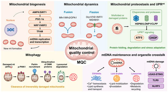
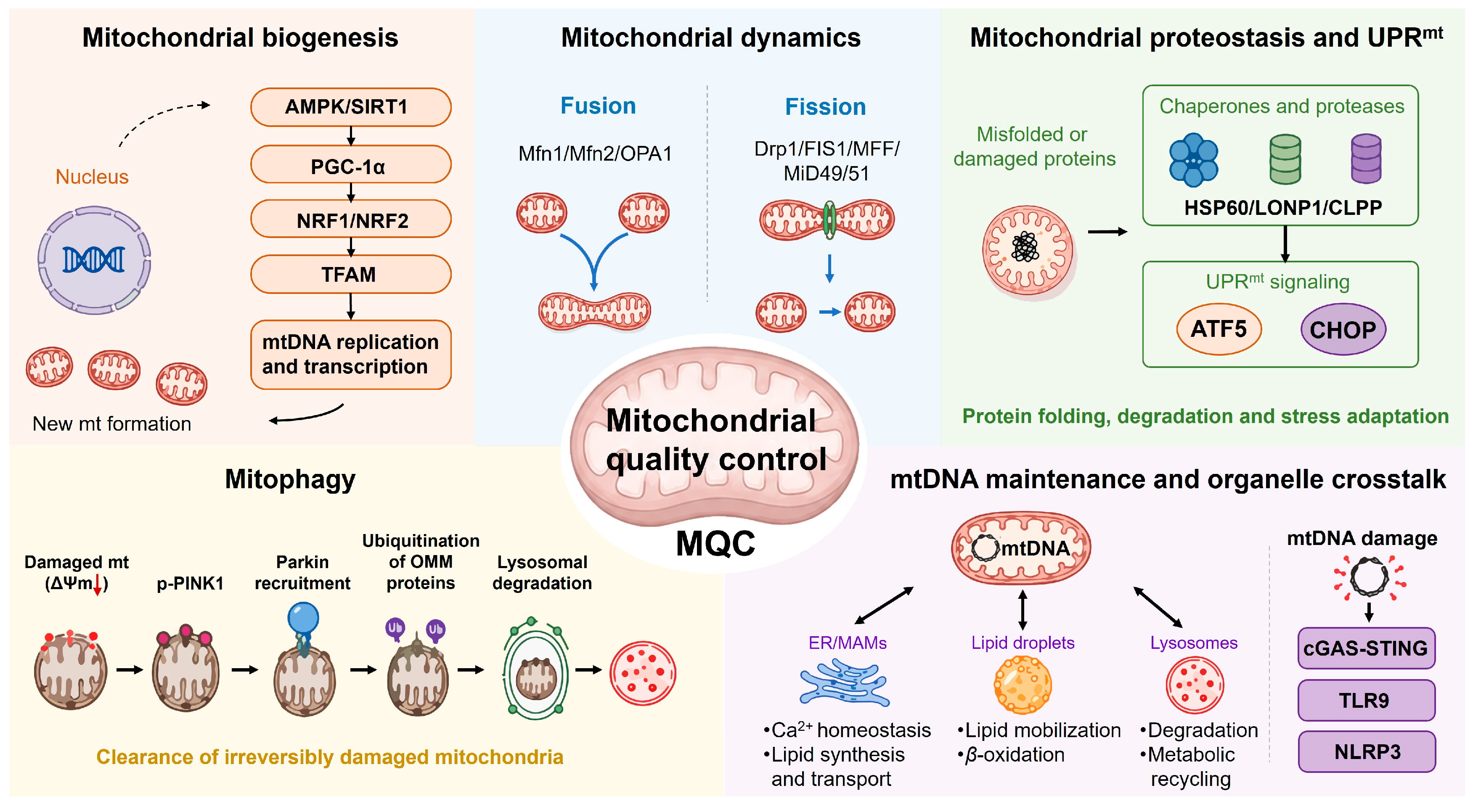
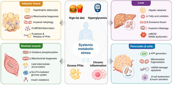
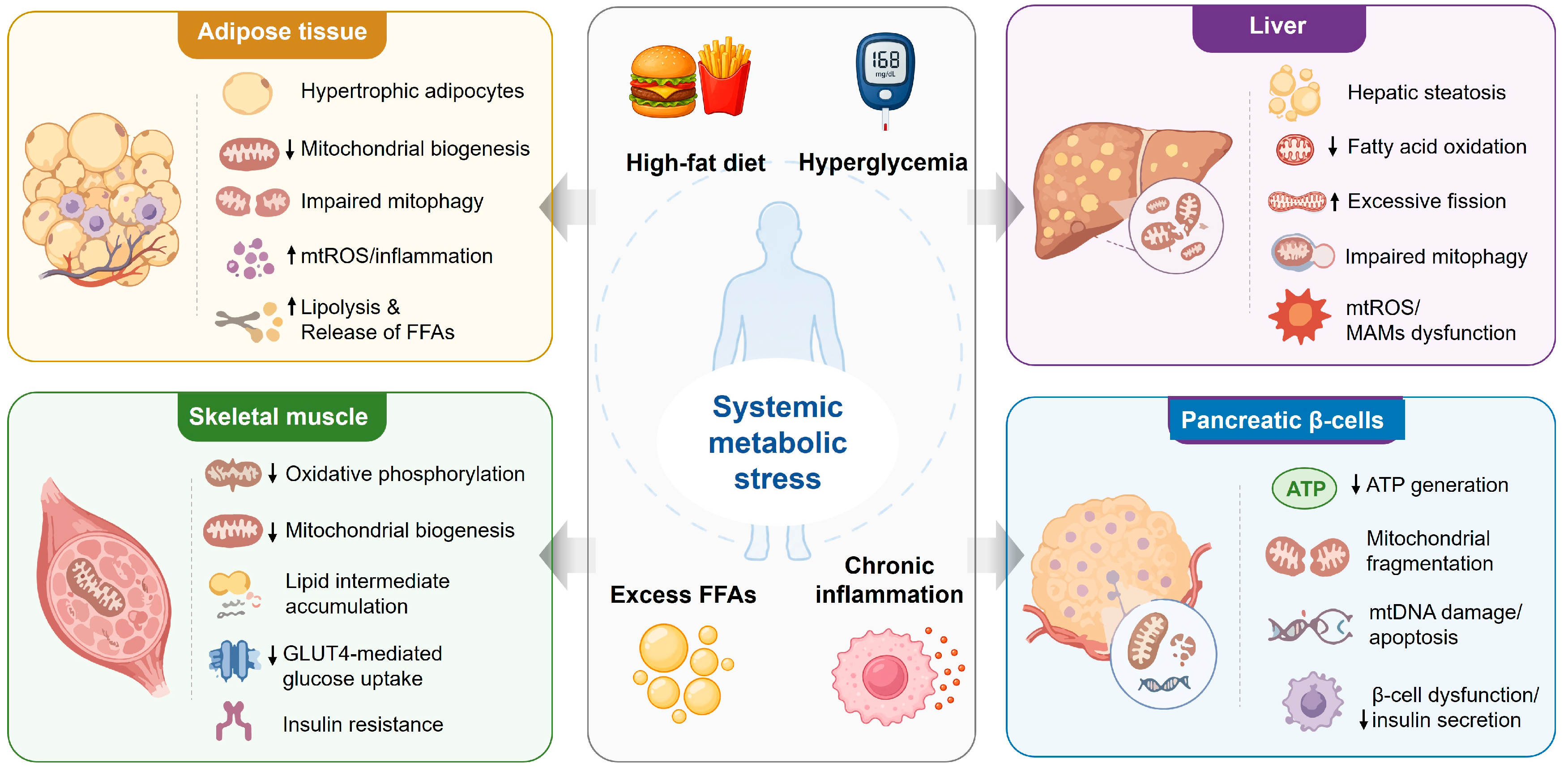

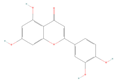
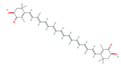
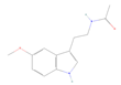
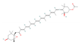
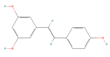

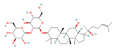
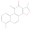
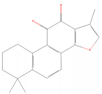


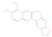


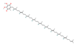
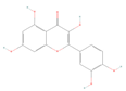
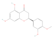

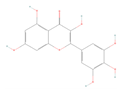
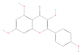
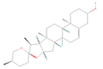
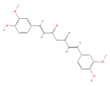
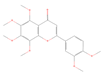
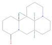
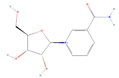
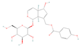
Disclaimer/Publisher’s Note: The statements, opinions and data contained in all publications are solely those of the individual author(s) and contributor(s) and not of MDPI and/or the editor(s). MDPI and/or the editor(s) disclaim responsibility for any injury to people or property resulting from any ideas, methods, instructions or products referred to in the content. |
© 2026 by the authors. Licensee MDPI, Basel, Switzerland. This article is an open access article distributed under the terms and conditions of the Creative Commons Attribution (CC BY) license.