


Abstract
Background/Objectives: Corneal endothelial cells (CECs) maintain corneal transparency through barrier and pump functions but have minimal proliferative activity in the adult human eye. After injury, repair relies mainly on migration, spreading, and enlargement of surviving cells. Functional recovery may nevertheless fail because of endothelial-to-mesenchymal transition, oxidative stress, mitochondrial dysfunction, endoplasmic reticulum (ER) stress, and apoptosis. This review summarizes the mechanisms of corneal endothelial injury repair, with emphasis on pharmacologically relevant signaling pathways, post-translational modifications (PTMs), and organelle homeostasis. Methods: This narrative review synthesizes experimental and clinical evidence on corneal endothelial injury repair, with emphasis on cellular signaling, PTMs, organelle homeostasis, and emerging therapeutic strategies. Relevant literature was identified through PubMed searches using combinations of terms related to corneal endothelial cells, endothelial repair, wound healing, Fuchs endothelial corneal dystrophy, signaling pathways, PTMs, organelle homeostasis, cell therapy, and gene- or RNA-based therapy; reference lists of key publications were also screened. Literature was selected according to relevance to the mechanistic and translational scope of the review, with attention to evidence from adult human tissue, cultured human CECs, animal models, disease-specific models, and clinical studies. Results: RhoA/ROCK, PI3K/Akt, Wnt/β-catenin, FGF, and TGF-β pathways regulate migration, survival, proliferation, and phenotypic stability. Phosphorylation-dependent cytoskeletal regulation and the SENP1–PAX6 SUMOylation axis have relatively direct links to repair, whereas microtubule modifications and methylation remain less well defined. Mitochondrial bioenergetics, redox balance, and ER proteostasis are important for CEC survival and functional maintenance, while the roles of Golgi trafficking and primary cilia require further study. Therapeutic strategies include endothelial keratoplasty, ROCK inhibitor-assisted Descemet stripping only (DSO), cultured CEC transplantation, stem-cell-derived endothelial cell therapy, pharmacological interventions, and gene- and RNA-based therapies. Conclusions: Current evidence supports matching treatment to residual endothelial reserve, disease stage, and the dominant disease mechanism. Established endothelial replacement remains central for advanced failure, whereas ROCK inhibitor-assisted DSO provides a donor-sparing option in selected eyes with sufficient residual endothelial reserve. Cell-based therapies have reached clinical or regulatory application, while most PTM-, organelle-, and gene/RNA-directed mechanisms remain at preclinical or early translational stages.
1. Introduction
The corneal endothelium maintains stromal deturgescence through coordinated barrier and pump functions and is therefore essential for corneal transparency [1,2]. Adult human CECs show minimal proliferative activity in vivo [3]. Following cell loss, endothelial repair occurs predominantly through migration, spreading, and enlargement of surviving cells rather than substantial cell proliferation [4]. When cell loss exceeds the compensatory capacity of the residual endothelium, these responses are no longer sufficient to maintain fluid transport, resulting in stromal edema and loss of corneal transparency [2,5]. Endothelial reserve may be reduced by inherited dystrophies or by acquired injury, including surgical and traumatic insults [5,6].
Successful repair therefore requires more than restoration of surface coverage. Redistribution of surviving cells must re-establish a stable functional monolayer, whereas endothelial-to-mesenchymal transition (EndMT), loss of endothelial phenotype, and fibrotic extracellular-matrix deposition can divert the repair response toward dysfunction [7,8]. Oxidative stress, mitochondrial dysfunction, and persistent ER stress can further compromise CEC survival and promote apoptosis [9,10].
Although previous reviews have summarized corneal endothelial biology, regenerative capacity, and emerging therapeutic approaches, the mechanistic relationships among migration, cytoskeletal regulation, post-translational modifications, organelle homeostasis, and therapeutic intervention remain incompletely integrated. Moreover, evidence obtained from cultured CECs, animal models, chronic FECD, and adult human corneal tissue does not necessarily have equivalent relevance to acute endothelial repair. In particular, morphological wound closure may not correspond to restoration of endothelial pump–barrier function. Therefore, this review integrates the molecular mechanisms of corneal endothelial repair with the experimental context, functional endpoints, and translational maturity of the available evidence, while highlighting mechanisms that remain insufficiently validated in adult human endothelium. This article is a narrative review; literature was identified through database searches and reference-list screening, with study selection guided by relevance to the mechanistic and translational scope rather than by a predefined systematic-review protocol.
Literature identification and scope. This article is a narrative review. Relevant literature was identified through database searches using combinations of keywords related to corneal endothelial cells, endothelial injury and repair, wound healing, Fuchs endothelial corneal dystrophy, cellular signaling, post-translational modifications, organelle homeostasis, cell-based therapy, and gene- or RNA-based therapy. Reference lists of key publications were additionally screened. Literature was selected according to relevance to the mechanistic and translational scope of the review rather than according to a predefined systematic-review protocol. Particular attention was given to distinguishing evidence obtained from adult human tissue, primary cultured human CECs, animal models, disease-specific experimental systems, and clinical studies, as well as to distinguishing morphological wound closure from restoration of endothelial function.
2. Cellular and Signaling Basis of Corneal Endothelial Injury Repair
Adult human CEC repair is constrained by limited proliferative activity and therefore depends mainly on redistribution and survival of the remaining endothelium. The outcome is determined by whether migration and spreading restore a stable pump–barrier monolayer or are accompanied by persistent EndMT, stress, or cell loss (Figure 1).
Figure 1.
Cellular trajectories and outcomes of corneal endothelial injury repair. The mature corneal endothelium is a largely quiescent monolayer that maintains barrier and pump function. After injury, surviving CECs respond mainly through migration, spreading, cytoskeletal remodeling, and survival signaling, whereas proliferation remains limited and context-dependent. Successful repair restores endothelial organization and function, whereas persistent stress can promote EndMT, organelle dysfunction, apoptosis, and abnormal extracellular-matrix deposition, leading to corneal edema and decompensation. Peripheral endothelial cells may also contribute to central repopulation after focal cell loss. Created in BioRender. Au, Z. (2026) https://BioRender.com/uzqagnk.
2.1. Proliferative Quiescence and Migration-Dominated Repair
Adult human CECs are largely non-proliferative in vivo and are maintained predominantly in the G1 phase of the cell cycle. Contact inhibition, increased expression of the cyclin-dependent kinase inhibitor p27Kip1, and TGF-β2 in the aqueous humor contribute to this arrest [3]. Despite their limited proliferative activity in vivo, CECs can re-enter the cell cycle and undergo limited expansion under appropriate ex vivo conditions [11,12].
Following endothelial injury, cells at the wound margin migrate over exposed Descemet’s membrane, spread, and enlarge to restore monolayer coverage [4]. Because cell loss is compensated mainly by redistribution rather than proliferation, repaired regions may contain larger and less uniformly shaped cells than intact endothelium [4]. Efficient closure requires coordinated changes in polarity, actin–myosin organization, adhesion, microtubules, and cell–cell contacts. Surface coverage should not be equated with functional recovery, because re-covering Descemet’s membrane does not establish restoration of junctional organization, ion transport, or pump function [2,4].
2.2. EndMT and Maladaptive Repair
Corneal endothelial injury can trigger EndMT. CECs lose their characteristic endothelial morphology and junctional phenotype, acquire spindle-shaped or fibroblast-like features, express mesenchymal markers such as α-smooth muscle actin (α-SMA), and increase extracellular-matrix production [8,13].
Persistent EndMT can interfere with restoration of a functional monolayer and contribute to retrocorneal fibrous membrane formation. TGF-β/Smad signaling is a major driver of this response. In experimental endothelial injury, inhibition of Smad signaling reduces phospho-Smad2, α-SMA, and collagen I expression and suppresses retrocorneal fibrous membrane formation without preventing closure of the endothelial defect [7]. SIRT1 activation can also attenuate injury- and TGF-β-induced EndMT through inhibition of Smad2/3 signaling [14].
Epigenetic dysregulation may reinforce a profibrotic endothelial state in Fuchs endothelial corneal dystrophy (FECD). Hypermethylation of the miR-199B promoter is associated with reduced miR-199b-5p expression; because miR-199b-5p suppresses SNAI1 and ZEB1, its loss may favor extracellular-matrix accumulation and mesenchymal features [15]. A direct role for this mechanism in acute wound repair has not been established.
2.3. Cytoskeletal and Survival Signaling: RhoA/ROCK and PI3K/Akt
RhoA/ROCK signaling is one of the best-characterized regulators of corneal endothelial wound healing. RhoA activates ROCK and thereby alters actomyosin contractility, adhesion, spreading, and directional migration [16,17,18]. Topical Y-27632 accelerates endothelial recovery in experimental cryoinjury [19], whereas ripasudil promotes corneal endothelial wound healing in experimental models [20]. In cultured CECs, ROCK inhibition enhances adhesion and migration and can also increase proliferation under selected conditions [17,21].
ROCK inhibition can influence cell-cycle regulators as well as cytoskeletal tension. Changes in cyclin D and p27 contribute to ROCK inhibitor-mediated cell-cycle progression during endothelial wound healing [22], indicating that the pathway can support limited proliferation when the cellular environment is permissive.
PI3K/Akt signaling contributes primarily to survival and cell-cycle progression. Ribonuclease 5 activates PI3K/Akt in human CECs, increases survival and proliferation, and accelerates wound closure in cultured cells and an experimental injury model [23]. Functional coupling with ROCK-associated cell-cycle control has also been reported [22]. Current evidence therefore supports PI3K/Akt as a survival and proliferation pathway rather than the dominant regulator of endothelial migration.
2.4. Context-Dependent Regulation of Proliferation and Phenotypic Stability
Wnt/β-catenin, FGF, and TGF-β signaling exert context-dependent effects on CEC proliferation and phenotypic stability.
Wnt/β-catenin signaling has been associated with increased proliferation of cultured human CECs. In a conditioned-medium model, increased β-catenin-dependent signaling was accompanied by greater telomerase activity and proliferation and reduced apoptosis [24]. Its contribution to physiological wound healing in the adult human cornea remains less well defined. The experimental evidence, functional endpoints, and translational limitations of these major signaling pathways are summarized in Table 1.
Table 1.
Evidence supporting major signaling pathways in corneal endothelial repair.
FGF responses depend strongly on ligand and context. FGF2 promotes CEC proliferation but can simultaneously induce fibrotic phenotypic changes; FGF2-induced SNAI1 has been linked to CDK2-dependent proliferation and ZEB1-dependent type I collagen expression [26]. In contrast, FGF10 accelerated endothelial recovery in a rabbit injury model, increased ZO-1 and Na+/K+-ATPase expression, and reduced EndMT-associated and inflammatory responses [27].
In contrast to the proliferative effects described above, TGF-β/Smad signaling is more consistently associated with EndMT and loss of endothelial phenotype [7,8]. Pathway inhibition can reduce mesenchymal conversion and facilitate expansion of cultured human CECs while preserving a more typical endothelial morphology [8,28].
2.5. Repair-Supportive Culture Conditions and Ex Vivo Expansion
Ex vivo expansion increases the number of CECs available for transplantation, but proliferation must be achieved without progressive loss of the endothelial phenotype. Established protocols commonly use serum-containing media and mitogenic supplements such as epidermal growth factor and fibroblast growth factor, although medium composition varies among laboratories [11,12]. Serum supports expansion but also contains factors, including TGF-β isoforms, that can promote EndMT under some conditions [8].
Cell-isolation and microenvironmental cues also influence expansion. Collagenase-based isolation has been reported to yield more viable CECs and preserve endothelial morphology and adhesion more effectively than EDTA-based dissociation in some culture systems [29,30], and viable single-cell recovery methods may improve subsequent expansion and delivery [31]. Lysophosphatidic acid can increase endothelial density through stromal interleukin-1β secretion [32], whereas conditioned medium from orbital adipose-derived stem cells promotes expansion and functional characteristics of cultured human CECs [33].
ROCK inhibition is frequently incorporated into CEC culture protocols because Y-27632 and related inhibitors improve attachment, spreading, migration, and cell yield under selected conditions [17,25]. TGF-β inhibition serves a different purpose by limiting mesenchymal conversion [8]. Dual-medium approaches further separate proliferation-supportive expansion from later phenotype stabilization [12,34].
These systems show that the proliferative restriction of adult CECs can be partially overcome ex vivo. The central challenge is to increase cell number without uncoupling proliferation from endothelial identity and pump–barrier function.
3. Post-Translational Regulation of Corneal Endothelial Injury Repair
PTMs regulate protein activity, stability, localization, and molecular interactions and can therefore rapidly modify cellular responses to injury. In CECs, direct evidence is strongest for phosphorylation-dependent regulation of the actomyosin cytoskeleton and for the SENP1–PAX6 SUMOylation pathway. Microtubule acetylation and detyrosination have been associated mainly with FECD-related endothelial behavior, whereas the roles of protein and histone methylation in injury repair remain poorly defined (Figure 2).
Figure 2.
Post-translational modifications and signaling pathways associated with corneal endothelial injury repair. PTMs may influence protein activity, cytoskeletal organization, and cellular responses in corneal endothelial cells, although the supporting evidence varies among pathways. Phosphorylation-dependent RhoA/ROCK–MLC and PI3K/Akt signaling have relatively direct experimental support in CECs, whereas the SENP1–PAX6 SUMOylation axis represents a mechanistically defined example supported by cellular and injury-model evidence. The specific roles of downstream Akt effectors in endothelial repair remain less certain and are therefore shown as indirect or context-dependent relationships. Protein and histone methylation are presented as having limited evidence, while microtubule acetylation/detyrosination is shown as a disease-associated observation in FECD-derived CECs rather than an established causal driver of acute endothelial repair. Solid arrows indicate relatively direct experimental evidence in CECs; dashed arrows indicate proposed, indirect, or context-dependent relationships. Created in BioRender. Au, Z. (2026) https://BioRender.com/w8udfgh.
3.1. Phosphorylation and Cytoskeletal Remodeling
Phosphorylation is a major mechanism linking injury-associated signaling to cytoskeletal behavior. RhoA/ROCK activation increases actomyosin contractility through phosphorylation of myosin light chain (MLC). In experimental corneal endothelial injury, inhibition of ROCK or MLC reduces actomyosin contraction, cell detachment, and apoptosis [35], directly linking phosphorylation-dependent actomyosin regulation to CEC survival and cytoskeletal responses after injury.
Non-muscle myosin II also contributes to directional migration. Its pharmacological inhibition increases the persistence of lamellipodial protrusions and promotes migration while preserving junctional integrity more effectively than ROCK inhibition in some experimental settings [18]. These findings indicate that modulation of actomyosin tension can alter the balance among traction, protrusion, adhesion, and junctional stability during wound closure.
Phosphorylation-dependent signaling also contributes to cell survival and cell-cycle progression. PI3K/Akt activation promotes CEC survival and proliferation [23], whereas ROCK inhibitor-mediated cell-cycle progression has been associated with regulation of cyclin D and p27 [22]. The specific Akt substrates required for corneal endothelial repair remain incompletely defined, and downstream mechanisms established in other cell types should not be assumed to operate identically in injured CECs.
3.2. SUMOylation and the SENP1–PAX6 Axis
SUMOylation is a reversible PTM in which small ubiquitin-like modifier proteins are attached to lysine residues of target proteins, thereby altering their stability, localization, interactions, or transcriptional activity [36]. The SENP1–PAX6 pathway provides one of the clearest examples of a defined PTM directly linked to corneal endothelial injury repair.
PAX6 expression increases after endothelial injury and supports junctional recovery and barrier integrity. PAX6 is modified predominantly by SUMO1 at K53 and K89, whereas the SUMO-specific protease SENP1 removes this modification. SENP1-mediated deSUMOylation increases PAX6 stability by reducing its degradation [37].
In SENP1+/− mice, increased PAX6 SUMOylation is associated with reduced PAX6 protein abundance, delayed endothelial repair, greater corneal opacity, and increased corneal thickness after injury. PAX6 overexpression partially rescues this phenotype and improves ZO-1-associated junctional recovery [37]. Thus, the modifying enzyme, substrate, modification sites, effect on protein stability, and consequences for endothelial repair have all been experimentally linked. However, SENP1/PAX6-targeted intervention has not yet been evaluated as a therapeutic strategy in human corneal endothelial disease.
3.3. Microtubule Acetylation and Detyrosination
Microtubules contribute to cell polarity, intracellular trafficking, and persistent directional migration. Acetylation and detyrosination of tubulin are PTMs commonly associated with relatively stable microtubule populations [38]. Evidence connecting these modifications to CEC behavior, however, derives mainly from studies of FECD rather than acute wound repair.
In FECD-derived CECs, overexpression of the full-length TCF4-B isoform increases migration and is associated with increased TUBB4A expression. Increased levels of acetylated and detyrosinated tubulin have also been observed in ex vivo FECD endothelial specimens compared with healthy donor tissue [39]. These findings associate altered microtubule PTMs with endothelial behavior in FECD, but they do not establish that either modification is required for acute corneal endothelial wound healing.
This mechanism should be distinguished from RNA toxicity caused by the intronic CTG18.1 repeat expansion in TCF4. The microtubule findings concern TCF4 isoform expression and TUBB4A-associated cytoskeletal changes [39], whereas repeat-associated RNA toxicity is discussed with RNA-targeted therapy in Section 5.4.
3.4. Protein and Histone Methylation: An Incompletely Defined PTM Layer
Methylation of lysine and arginine residues can regulate protein interactions, stability, localization, and signaling, whereas histone methylation can alter chromatin structure and transcriptional activity [40]. In CECs, however, specific methyltransferases, demethylases, methylated protein substrates, and injury-dependent histone methylation patterns have not been systematically characterized.
Histone methylation states, including regulatory marks such as H3K4me3 and H3K27me3, represent plausible but currently unvalidated regulatory layers in CEC injury repair. DNA methylation is mechanistically distinct from protein PTMs and is considered separately in the context of longer-term endothelial-state regulation.
3.5. Temporal Coordination and Unresolved Interactions Among PTMs
These PTMs differ both in their likely kinetics and in the strength of evidence linking them to endothelial repair. Phosphorylation can rapidly alter signaling and actomyosin contractility, whereas SUMOylation can modify the stability of regulatory proteins such as PAX6. Microtubule acetylation and detyrosination may reflect more sustained changes in cytoskeletal organization, while protein and histone methylation may provide a slower regulatory layer.
Direct interactions among these PTM systems have not been systematically mapped during corneal endothelial repair. Their temporal coordination across migration, wound closure, junctional reassembly, and recovery of endothelial function therefore remains unresolved.
4. Organelle Homeostasis and Inter-Organelle Communication During Corneal Endothelial Injury Repair
CECs depend on sustained energy production, protein quality control, and intracellular trafficking to maintain barrier and pump functions. These requirements become particularly important after injury. Mitochondria and the ER have the strongest direct links to CEC survival and dysfunction; evidence for Golgi-dependent regulation of repair is more limited, whereas primary cilia are supported mainly by developmental and experimental injury models (Figure 3).
Figure 3.
Organelle homeostasis in corneal endothelial injury repair. Mitochondrial bioenergetics, redox control, and ER proteostasis support CEC survival and functional recovery after injury. Oxidative stress impairs mitochondrial function, whereas persistent ER stress activates the unfolded protein response and can promote mitochondrial dysfunction and apoptosis; functional ER–mitochondrial coupling is supported, but a causal role for remodeling of mitochondria-associated membranes has not been established. ER-to-Golgi trafficking occurs in CECs, although broader Golgi-dependent regulation of repair-related cargo delivery, including Na+/K+-ATPase, remains uncertain. Primary cilia can reassemble after experimental endothelial injury, but the roles of the proposed regulators and their causal contribution to repair in adult human CECs remain unresolved. The developmental timeline shown is derived from mouse corneal endothelium. Solid arrows indicate experimentally supported relationships, whereas dashed arrows indicate proposed, indirect, or incompletely established relationships. Created in BioRender. Au, Z. (2026) https://BioRender.com/ivt9mpv.
4.1. Mitochondrial Bioenergetics and Redox Homeostasis
Mitochondria provide ATP required for ion transport and other energy-dependent processes that maintain endothelial function. Suppression of COL8A2 in cultured human CECs reduces viability and proliferation, impairs barrier properties, and is associated with mitochondrial abnormalities and oxidative stress [41]. Conversely, CRISPR/dCas9-mediated activation of COL8A2 increases mitochondrial membrane potential and ATP production and improves endothelial wound healing in a rat injury model; these effects are accompanied by increased YAP phosphorylation and improved transendothelial electrical resistance [42]. These findings should be interpreted in their experimental context: activation of physiological COL8A2 expression and suppression of pathogenic mutant COL8A2 address different biological situations and are not necessarily contradictory.
Oxidative injury is also closely linked to mitochondrial dysfunction. In UVA-exposed CECs, increased reactive oxygen species (ROS) is accompanied by reduced SIRT1 expression, loss of mitochondrial membrane potential, altered fission–fusion dynamics, and disturbed energy metabolism; SIRT1 activation with SRT1720 attenuates these changes and reduces apoptosis [9]. MitoQ similarly reduces mitochondrial ROS and fragmentation, preserves membrane potential, and improves viability under combined UVA and cigarette-smoke stress [43].
Nrf2 contributes to antioxidant defense in corneal tissues and FECD-associated oxidative stress [44]. In experimental CEC models, lycopene promotes p62-associated autophagic activity, Keap1 degradation, Nrf2 nuclear translocation, and endothelial survival [45]. Non-coding RNA regulation may intersect with this stress response: NEAT1 is reduced in FECD and experimental UVA injury, whereas loss of NEAT1 increases oxidative susceptibility and mitochondrial abnormalities and CRISPR-mediated NEAT1 activation attenuates endothelial damage [46].
These studies support mitochondrial membrane potential, ATP production, redox control, and mitochondrial dynamics as determinants of CEC survival and functional preservation. Much of the evidence derives from oxidative-stress and disease models rather than uncomplicated acute wound closure.
4.2. ER Stress, Unfolded Protein Response, and Proteostasis
The ER is responsible for protein folding, quality control, and early secretory processing. Accumulation of unfolded or misfolded proteins activates the unfolded protein response (UPR), which initially attempts to restore proteostasis but can trigger pro-apoptotic signaling when severe or prolonged.
In diabetic corneal endothelium, ER-stress markers increase before overt morphological abnormalities, and experimental ER stress contributes to endothelial cell loss and corneal edema. Pharmacological reduction in ER stress with 4-phenylbutyric acid (4-PBA) improves endothelial survival and recovery of barrier and pump function in experimental diabetic endothelial dysfunction [47].
Tunicamycin also activates major UPR branches in cultured human CECs, including PERK–eIF2α–CHOP, IRE1α–XBP1, and ATF6 signaling, and reduces viability. This response is accompanied by Bax activation, cytochrome c release, caspase-9 and caspase-3 activation, reduced mitochondrial membrane potential, and decreased ATP production; FECD-derived CECs also show increased basal expression of several ER-stress-associated proteins [10]. Persistent ER stress can therefore reduce the viable cell population available for repair and compromise functional recovery.
4.3. ER–Mitochondrial Crosstalk During Endothelial Stress
The ER and mitochondria communicate through calcium exchange, lipid metabolism, energy regulation, and stress signaling. Physical contact sites, commonly termed mitochondria-associated membranes (MAMs), provide one structural basis for this communication [48], but direct evidence that MAM abundance or architecture changes during corneal endothelial wound healing is lacking.
Functional coupling between ER stress and mitochondrial dysfunction is better supported. Tunicamycin-induced ER stress reduces mitochondrial membrane potential and ATP production, suppresses oxidative phosphorylation, and produces mitochondrial swelling and fragmentation together with intrinsic apoptosis [10]. In diabetic corneal endothelium, 4-PBA improves mitochondrial bioenergetic deficits together with endothelial function [47], whereas ER-stress reduction in tunicamycin-treated CECs improves survival without fully normalizing mitochondrial bioenergetics [10].
Current evidence therefore supports functional ER–mitochondrial crosstalk as a determinant of CEC survival during stress but does not establish altered MAM formation or remodeling as a causal mechanism in FECD or acute wound repair.
4.4. Golgi-Dependent Processing and Intracellular Trafficking
The Golgi apparatus processes and sorts proteins after their exit from the ER, functions relevant to recovery of membrane, junctional, and extracellular-matrix proteins. Direct studies of Golgi-specific regulation during corneal endothelial wound healing remain scarce. Although ER-to-Golgi trafficking has been demonstrated in cultured CECs, the broader contribution of Golgi remodeling or cargo delivery to acute endothelial repair remains uncertain.
In cultured CECs, procollagen I and the collagen-specific chaperone Hsp47 traffic together from the ER toward the cis-Golgi compartment, and disruption of intracellular transport alters their subcellular distribution [49]. These findings establish active ER-to-Golgi trafficking in CECs but do not demonstrate that broader Golgi remodeling controls migration, wound closure, or pump-related cargo delivery during repair.
4.5. Primary Cilia as Injury-Responsive Organelles
Primary cilia are microtubule-based sensory organelles implicated in corneal development and disease [50]. During mouse corneal endothelial development, cilia appear as the monolayer matures and subsequently shorten or disappear in adult endothelium. Following mechanical injury, they reassemble in mature CECs near the wound before pronounced cell elongation and polarization [51]. Thus, the developmental and injury-response observations discussed here derive primarily from mouse models rather than adult human endothelial tissue.
IFT88-dependent intraflagellar transport is required for normal ciliary structure. Hypomorphic IFT88 mutation or experimental knockdown produces abnormal cilia, irregular endothelial patterning, mislocalization of junction-associated proteins, and altered acetylated tubulin accumulation [51], supporting a role for ciliary integrity in endothelial organization in experimental models.
Evidence in human corneal endothelium is more limited. Primary cilia have been identified in specimens from patients with bullous keratopathy, including FECD [52], but these observations do not establish whether ciliary reassembly is beneficial, maladaptive, or secondary to stress. Interactions between primary cilia and PI3K signaling described in non-corneal systems [53] should not yet be considered established mechanisms of adult human CEC repair. The causal contribution of injury-responsive ciliogenesis therefore remains unresolved.
5. Therapeutic Strategies for Restoration of Corneal Endothelial Function
Therapeutic strategies for corneal endothelial dysfunction act at different biological levels: replacing failed tissue, mobilizing residual CECs, supplying an exogenous endothelial population, preserving stressed cells, or correcting a defined molecular defect (Figure 4).
Figure 4.
Therapeutic strategies for corneal endothelial injury repair. Therapeutic strategies address distinct biological limitations in corneal endothelial dysfunction rather than representing a sequential treatment pathway. Established surgical replacement includes penetrating keratoplasty (PK) and Descemet’s membrane endothelial keratoplasty (DMEK); donor-sparing endogenous repair includes Descemet stripping only (DSO), with ROCK inhibition used to support endothelial migration and recovery in selected settings and with efficacy dependent on sufficient residual peripheral CECs. Cell-based approaches provide exogenous CECs or endothelial substitutes, including cultured CECs and iPSC-derived endothelial-like cells. Pharmacological approaches aim to enhance endogenous repair or preserve stressed but viable cells, whereas gene- and RNA-based strategies target defined molecular abnormalities, including COL8A2-associated disease and TCF4 repeat-associated RNA toxicity. The relevance of each therapeutic approach depends on factors such as residual endothelial reserve, disease stage, reversibility of cellular dysfunction, and the presence of a defined molecular defect. Created in BioRender. Au, Z. (2026) https://BioRender.com/gy43htz.
5.1. Surgical Replacement and Donor-Sparing Repair
Corneal transplantation remains the standard treatment for advanced endothelial failure. For isolated endothelial disease, selective endothelial keratoplasty has largely replaced full-thickness penetrating keratoplasty (PK), and Descemet’s membrane endothelial keratoplasty (DMEK) has become a widely adopted procedure [54,55]. DMEK selectively replaces the recipient endothelium and Descemet’s membrane while preserving the host stroma, but it remains dependent on donor tissue and is associated with procedure-specific complications including graft detachment and challenges in eyes with glaucoma or complex anterior-segment anatomy [56,57].
DSO, also termed Descemetorhexis without endothelial keratoplasty, removes central diseased Descemet’s membrane and endothelium and relies on migration and enlargement of healthier peripheral CECs to repopulate the central cornea. Long-term studies indicate that selected patients with FECD can achieve sustained corneal clearance after DSO, although failure and subsequent endothelial keratoplasty remain possible [58].
ROCK inhibition can enhance this endogenous response. In a 1-year, phase 2, randomized, placebo-controlled multicenter trial, 65 patients with FECD received topical ripasudil four times daily (QID; n = 21), twice daily (BID; n = 22), or placebo (n = 22) for 12 weeks after DSO [59]. At week 12, mean central endothelial cell density was 531 ± 312 cells/mm2 in the QID group versus 228 ± 298 cells/mm2 in the placebo group (p = 0.0065). Corneal edema had resolved in 17/21 eyes (81.0%) receiving QID ripasudil compared with 2/22 eyes (9.1%) receiving placebo (p < 0.0001), while rescue treatment was required in 2/21 (9.5%) and 6/22 (27.3%) eyes, respectively (p = 0.0092). More than 95% of participants completed the one-year study, and adverse events were mild and did not lead to treatment discontinuation [59]. Importantly, these findings should not be generalized to advanced endothelial failure or endothelial dysfunction of other etiologies. The benefit of ROCK inhibitor-assisted DSO depends on sufficient residual peripheral endothelial capacity and therefore represents a donor-sparing strategy for selected eyes rather than a treatment for severe endothelial depletion [58,59].
5.2. Cell-Based and Tissue-Engineered Therapies
Ex vivo expansion of human CECs provides a means of treating more patients from a limited supply of donor tissue. Two principal delivery formats are being developed: intracameral injection of cultured CECs and transplantation of engineered endothelial constructs in which expanded cells are combined with a carrier or scaffold [60]. Cell injection avoids transplantation of an intact endothelial graft but requires sufficient cell attachment, survival, and redistribution across the posterior corneal surface.
Clinical proof of concept was established by Kinoshita and colleagues, who injected cultured donor-derived human CECs together with a ROCK inhibitor into the anterior chamber of 11 patients with bullous keratopathy [61]. At 24 weeks, all 11 treated eyes had a central endothelial cell density greater than 500 cells/mm2, with a mean density of 1924 cells/mm2; ten eyes had a central corneal thickness below 630 μm and nine improved by at least two lines of best-corrected visual acuity [61]. Longer-term follow-up has examined the durability of cultured CEC transplantation [62].
Neltependocel (Vyznova), an allogeneic donor-derived cultured human corneal endothelial cell suspension, received regulatory approval in Japan in March 2023 for reconstruction of the damaged corneal endothelial monolayer [63]. This approval demonstrates that ex vivo CEC expansion can be translated into a standardized therapeutic product, while also underscoring the importance of manufacturing consistency, product characterization, and post-marketing safety surveillance.
Pluripotent stem cells provide a potential donor-independent source of endothelial-like cells. Protocols have been developed to differentiate human pluripotent stem cells toward a corneal endothelial phenotype [64,65], and hiPSC-derived corneal endothelial progenitor-like cells have restored endothelial function in a rabbit model [66]. A first-in-human study evaluated an allogeneic iPSC-derived corneal endothelial cell substitute in one patient with bullous keratopathy and reported improvement in stromal edema and visual acuity during 1 year of follow-up [67]. The study provided feasibility evidence rather than definitive efficacy because of its single-patient design. A de novo in-frame deletion involving exon 22 of EP300 was identified in the transplanted cell product without a related clinical adverse event, underscoring the need for genomic surveillance and manufacturing quality control [67].
Tissue-engineered endothelial constructs combine expanded CECs with natural or synthetic carriers. Hydrogels and related biomaterials may facilitate handling, attachment, spatial organization, and local control of the repair environment [60,68], although these approaches remain less clinically mature than cell-suspension therapy. Across cell-based strategies, major requirements include sustained endothelial phenotype, reproducible manufacturing, genomic stability where relevant, efficient attachment to Descemet’s membrane, and durable pump–barrier function.
5.3. Pharmacological Modulation of Endothelial Survival and Repair
Pharmacological interventions can be broadly grouped into agents that enhance endogenous repair, agents that preserve stressed but viable CECs, and disease-specific pharmacological rescue strategies.
ROCK inhibitors currently provide the clearest example of pharmacological enhancement of endogenous endothelial repair. Experimental studies demonstrate improved wound healing after ROCK inhibition [20,69], ripasudil can improve functional properties of FECD-derived endothelial cells [70], and the DSO trial extends these observations to clinically measurable donor-free endothelial repopulation [59].
Other pharmacological approaches primarily aim to preserve residual CECs. Lycopene activates the p62–Keap1–Nrf2 antioxidant pathway [45], SIRT1 activation with SRT1720 attenuates UVA-induced oxidative stress and mitochondrial dysfunction [9], MitoQ reduces mitochondrial ROS and preserves mitochondrial function [43], and 4-PBA reduces ER stress and improves endothelial survival and function in experimental disease models [47].
The pan-caspase inhibitor emricasan reduces apoptosis and extracellular-matrix accumulation in FECD-derived cell models and improves endothelial density and morphology in a COL8A2 mutant mouse model [71]. Mutation-specific rescue has also been explored for SLC4A11-associated disease: diclofenac and nepafenac improved cell-surface trafficking of several misfolded SLC4A11 variants in a heterologous expression system, and diclofenac restored water transport for selected mutants [72]. These findings remain proof of concept rather than established clinical treatment. Most pharmacological strategies other than ROCK inhibition remain preclinical [73].
5.4. Gene- and RNA-Based Therapies
Gene- and RNA-directed therapies are particularly attractive when a defined molecular defect can be identified. Because adult CECs have limited turnover in vivo, durable molecular correction could potentially produce prolonged effects, although most approaches remain experimental. The current translational status, supporting evidence, and major limitations of the therapeutic strategies discussed in this section are summarized in Table 2.
Table 2.
Translational landscape of current and emerging therapies for corneal endothelial dysfunction.
In COL8A2-associated early-onset FECD, adenovirus-mediated CRISPR/Cas9 targeting of the COL8A2 start codon reduced mutant COL8A2 expression, prevented endothelial cell loss, and restored pump function in a mutant mouse model [74]. Gene activation rather than disruption has also been used to modify repair: CRISPR/dCas9-mediated activation of SOX2 increased proliferation of cultured human CECs and improved endothelial wound healing in a rat model [80], although SOX2 activation is not a disease-specific therapy.
TCF4 repeat-associated FECD provides a complementary RNA-targeted example. Expansion of the intronic CTG18.1 repeat is strongly associated with FECD [81,82]; repeat-containing transcripts form nuclear RNA foci, sequester muscleblind-like RNA-binding proteins, and disrupt pre-mRNA splicing [83,84]. Altered processing of extracellular-matrix transcripts, including FN1, and increased susceptibility to ultraviolet-associated ferroptotic injury have also been reported [85,86]. Antisense oligonucleotides (ASOs) directed against the expanded transcript reduce RNA foci, decrease MBNL sequestration, and partially correct abnormal splicing in patient-derived CECs [75]. This toxic RNA mechanism is distinct from the TCF4 protein-isoform-associated microtubule phenotype discussed in Section 3.3.
Viral vectors, including AAV, have been investigated for gene delivery in human corneal fibroblasts, corneal stroma, animal cornea, and other anterior-segment tissues [76,77,78,79]. These studies establish broader delivery feasibility but not optimized human CEC transduction or therapeutic restoration of endothelial function. Evidence of successful gene delivery or molecular modulation should therefore not be equated with evidence of durable recovery of endothelial pump–barrier function. Clinical translation will require efficient endothelial targeting, durable therapeutic activity, and control of vector-associated immune responses and unintended genome-editing effects [87,88].
6. Current Challenges and Future Directions
Several issues continue to limit the interpretation and clinical translation of corneal endothelial repair studies. Future work should distinguish durable functional recovery from morphological improvement, define the temporal and cellular context of repair-associated mechanisms, establish causal relationships between cellular dysfunction and repair failure, and determine how emerging therapies can be applied safely to appropriately selected patients.
6.1. Defining Functional and Durable Repair
Wound closure and endothelial cell density are useful measures of repair but do not establish restoration of normal endothelial function. Functional assessment should also include junctional integrity, barrier and pump activity, corneal thickness and transparency, and the durability of recovery [2,4]. Because migration and surface closure may precede complete junctional reassembly and pump recovery, early structural improvement should be distinguished from sustained functional restoration.
6.2. Cell-State Heterogeneity and Temporal Regulation of Repair Mechanisms
Human corneal endothelium is molecularly heterogeneous, and single-cell studies have begun to define endothelial subpopulations and FECD-associated states [46,89]. The sequence of cell-state changes after acute injury remains less clear. Time-course single-cell and spatial profiling could determine when CECs enter migratory, stress-associated, apoptotic, or mesenchymal states and whether these states resolve during successful repair.
Longer-lived epigenetic and RNA-associated changes may provide an additional layer of regulation. DNA methylation abnormalities have been identified in FECD [90], whereas increased N4-acetylcytidine (ac4C) and N-acetyltransferase 10 (NAT10) expression have been reported after mesenchymal stem-cell treatment in a mouse cryoinjury model [91]. Their causal contribution to endogenous CEC repair remains uncertain.
Temporal regulation is also important at the protein level. Phosphorylation, SUMOylation, microtubule acetylation and detyrosination, and other PTMs may operate at different stages of repair. Time-course phosphoproteomic, acetylomic, and SUMO-proteomic analyses integrated with cellular-state profiling could help distinguish molecular changes that drive repair from those that accompany persistent EndMT or cellular stress.
6.3. Establishing Causal Relationships Between Organelle Dysfunction and Repair Failure
Mitochondrial dysfunction and ER stress have relatively strong experimental links to CEC injury and survival [9,10], but causality is less well established for other organelle-associated changes. Remodeling of mitochondria-associated membranes during repair has not been demonstrated, Golgi-specific regulation of wound healing remains poorly characterized, and the contribution of primary cilia to adult human CEC repair remains unresolved.
Future studies should therefore focus on whether organelle abnormalities actively determine repair outcome or arise secondarily after endothelial dysfunction. Live-cell imaging, metabolic analysis, and organelle-specific perturbation may help establish the temporal and causal relationships between organelle homeostasis, cell survival, and functional recovery.
6.4. Safety, Durability, and Patient Selection for Emerging Therapies
Clinical translation requires consideration of treatment-specific risks in addition to efficacy. For ROCK inhibitors, the optimal dose and treatment duration for endothelial-repair indications remain incompletely defined, and larger studies with longer follow-up are needed to establish safety and durability [92]. Whether prolonged ROCK inhibition affects endothelial phenotypic stability, junctional organization, or susceptibility to EndMT also requires direct investigation.
Cell-based therapies raise different translational issues. Long-term immune compatibility, phenotypic stability, and, for pluripotent-cell-derived products, genomic stability and tumorigenic risk require continued evaluation. The genomic alteration identified in the cell product used in a first-in-human iPSC-derived corneal endothelial study illustrates the importance of genomic surveillance [67]. Broader implementation will also require reproducible GMP-compatible production, control of batch-to-batch variability, validated identity and potency assays, and practical solutions for storage, transport, scalability, and cost [93]. These manufacturing and implementation issues are recognized as major barriers to wider clinical deployment of CEC injection therapy.
Gene- and RNA-based approaches require efficient endothelial delivery and control of molecular specificity. For viral-vector and genome-editing strategies, transduction efficiency, immune responses, unintended editing events, and durability remain important considerations [87,88]. TCF4-targeted ASOs reduce repeat-associated RNA toxicity in patient-derived CECs [75], but optimal intraocular delivery, dose, treatment duration, and the need for repeated administration remain to be established.
Patient selection is closely linked to these safety and efficacy considerations. Residual endothelial reserve, reversibility of cellular dysfunction, disease stage, and the presence of an actionable molecular defect may help to determine whether endogenous repair, cell replacement, or molecular therapy is appropriate. DSO, for example, depends on sufficient peripheral endothelium [58], whereas advanced cell loss is more likely to require endothelial replacement. Molecularly defined disease may provide opportunities for targeted intervention, as illustrated experimentally by COL8A2 gene editing and TCF4-directed antisense therapy [74,75]. However, validated thresholds for assigning patients to specific treatment strategies have not yet been established.
Combination therapy is biologically plausible, but its superiority should not be assumed without direct evidence. The optimal sequence and duration of interventions targeting migration, cellular stress, or other repair mechanisms remain to be determined.
6.5. Improving Experimental Models and Translational Relevance
Translation is complicated by species differences in corneal endothelial proliferation and wound-healing behavior [94]. Findings from animal models, particularly apparent increases in proliferation, should therefore be interpreted cautiously in relation to adult human CECs.
No single model reproduces all features of the adult human corneal endothelium. Primary human CEC cultures, donor corneal tissue, ex vivo injury models, engineered endothelial constructs, and validated stem-cell-derived cells each capture different aspects of endothelial biology. Greater integration of these systems should improve assessment of translational relevance. Consistent reporting of cell source, passage, injury model, treatment dose and duration, functional endpoints, and adverse events would further improve comparison across studies. Long-term follow-up will be particularly important for cell-, gene-, and RNA-based therapies to assess phenotypic stability, genomic safety, immune responses, and durability of therapeutic benefit.
7. Conclusions
Corneal endothelial repair in adult humans depends primarily on migration, spreading, enlargement, and survival of residual CECs rather than substantial cell proliferation. Successful repair requires more than closure of the endothelial defect; restoration of junctional organization, pump–barrier function, and phenotypic stability is equally important. Among molecular mechanisms, RhoA/ROCK signaling and phosphorylation-dependent cytoskeletal regulation have relatively strong experimental support, while the SENP1–PAX6 SUMOylation axis provides a more directly defined PTM mechanism. By contrast, Wnt/β-catenin, FGF, and PI3K/Akt signaling remain more context-dependent, and microtubule PTMs, methylation, Golgi trafficking, and primary-cilium-associated mechanisms require further validation in adult human corneal endothelium.
From a therapeutic perspective, endothelial keratoplasty remains central for advanced endothelial failure, whereas DSO combined with ROCK inhibition provides clinical evidence for pharmacologically enhanced endogenous repair in carefully selected FECD eyes with sufficient residual endothelial reserve. Cultured CEC therapy has progressed to clinical and regulatory application, while iPSC-derived endothelial substitutes have reached first-in-human feasibility but remain limited by small clinical experience, phenotypic uncertainty, manufacturing requirements, and genomic safety concerns. Most molecular and pharmacological strategies beyond ROCK inhibition remain preclinical. Future studies should prioritize direct validation in adult human CECs, standardized functional endpoints, long-term durability, and patient-selection criteria. Overall, therapeutic development should focus not simply on increasing cell number or accelerating wound closure, but on restoring durable endothelial pump–barrier function while matching the intervention to residual endothelial competence and the underlying disease mechanism.
Author Contributions
Conceptualization, L.W., S.Y. and B.X.; methodology, L.W.; investigation, L.W., Z.G., L.Z., L.L. and W.X.; data curation, Z.G. and L.Z.; writing—original draft preparation, L.W.; writing—review and editing, S.Y. and B.X.; visualization, Z.G. and L.Z.; supervision, S.Y. and B.X.; project administration, L.L.; funding acquisition, S.Y. and B.X. All authors have read and agreed to the published version of the manuscript.
Funding
This research was funded by the National Natural Science Foundation of China (grant numbers 31601104 and 32400697), the Natural Science Foundation of Shandong Province (grant numbers ZR2021MC158 and ZR2025MS505), and the Qingdao Municipal Natural Science Foundation (grant number 24-4-4-zrjj-23-jch).
Institutional Review Board Statement
Not applicable.
Informed Consent Statement
Not applicable.
Data Availability Statement
No new data were created or analyzed in this study. Data sharing is not applicable to this article.
Acknowledgments
The authors thank BioRender.com for the preparation of the schematic illustrations used in this review.
Conflicts of Interest
The authors declare no conflicts of interest.
Abbreviations
The following abbreviations are used in this manuscript:
| 4-PBA | 4-phenylbutyric acid |
| AAV | Adeno-associated virus |
| ac4C | N4-acetylcytidine |
| Akt | Protein kinase B |
| α-SMA | Alpha-smooth muscle actin |
| ASO | Antisense oligonucleotide |
| ATP | Adenosine triphosphate |
| CEC | Corneal endothelial cell |
| CHED | Congenital hereditary endothelial dystrophy |
| CHOP | C/EBP homologous protein |
| COL8A2 | Collagen type VIII alpha 2 chain |
| CRISPR | Clustered regularly interspaced short palindromic repeats |
| DMEK | Descemet’s membrane endothelial keratoplasty |
| DSO | Descemet stripping only |
| ECM | Extracellular matrix |
| EDTA | Ethylenediaminetetraacetic acid |
| eIF2α | Eukaryotic translation initiation factor 2 alpha subunit |
| EndMT | Endothelial-to-mesenchymal transition |
| ER | Endoplasmic reticulum |
| ETV4 | ETS variant transcription factor 4 |
| FECD | Fuchs endothelial corneal dystrophy |
| GRP78 | Glucose-regulated protein 78 |
| GSK-3β | Glycogen synthase kinase-3β |
| HCEC | Human corneal endothelial cell |
| HDAC6 | Histone deacetylase 6 |
| H3K4me3 | Trimethylation of histone H3 at lysine 4 |
| H3K27me3 | Trimethylation of histone H3 at lysine 27 |
| IFT88 | Intraflagellar transport protein 88 |
| IL-1β | Interleukin-1β |
| iPSC | Induced pluripotent stem cell |
| JUN | Jun proto-oncogene |
| Keap1 | Kelch-like ECH-associated protein 1 |
| LIMK | LIM domain kinase |
| MAM | Mitochondria-associated endoplasmic reticulum membrane |
| MLC | Myosin light chain |
| MLCK | Myosin light chain kinase |
| MLCP | Myosin light chain phosphatase |
| mTORC1 | Mechanistic target of rapamycin complex 1 |
| Nrf2 | Nuclear factor erythroid 2-related factor 2 |
| NSAID | Non-steroidal anti-inflammatory drug |
| p62 | Sequestosome 1 (SQSTM1) |
| PAC1 | Pituitary adenylate cyclase-activating polypeptide type I receptor |
| PAX6 | Paired box protein 6 |
| PI3K | Phosphoinositide 3-kinase |
| PIP2 | Phosphatidylinositol 4,5-bisphosphate |
| PIP3 | Phosphatidylinositol (3,4,5)-trisphosphate |
| PK | Penetrating keratoplasty |
| PRAS40 | Proline-rich Akt substrate of 40 kDa |
| PTM | Post-translational modification |
| RhoA | Ras homolog family member A |
| ROCK | Rho-associated coiled-coil-containing protein kinase |
| ROS | Reactive oxygen species |
| SENP1 | SUMO-specific protease 1 |
| SIRT1 | Sirtuin 1 |
| SMAD | SMAD family protein(s) |
| SNAI1 | Snail family transcriptional repressor 1 |
| SOX2 | SRY-box transcription factor 2 |
| SUMO | Small ubiquitin-like modifier |
| TAZ | Transcriptional coactivator with PDZ-binding motif |
| TCF4 | Transcription factor 4 |
| TGF-β | Transforming growth factor-beta |
| TUBB4A | Tubulin beta 4A class IVa |
| UPR | Unfolded protein response |
| UVA | Ultraviolet A |
| YAP | Yes-associated protein |
| ZEB1 | Zinc finger E-box-binding homeobox 1 |
| ZO-1 | Zonula occludens-1 |
References
- DelMonte, D.W.; Kim, T. Anatomy and physiology of the cornea. J. Cataract. Refract. Surg. 2011, 37, 588–598. [Google Scholar] [CrossRef] [Scilit]
- Price, M.O.; Mehta, J.S.; Jurkunas, U.V.; Price, F.W., Jr. Corneal endothelial dysfunction: Evolving understanding and treatment options. Prog. Retin. Eye Res. 2021, 82, 100904. [Google Scholar] [CrossRef] [Scilit]
- Joyce, N.C. Proliferative capacity of the corneal endothelium. Prog. Retin. Eye Res. 2003, 22, 359–389. [Google Scholar] [CrossRef] [Scilit]
- Vercammen, H.; Miron, A.; Oellerich, S.; Melles, G.R.J.; Dhubhghaill, S.N.; Koppen, C.; Van Den Bogerd, B. Corneal endothelial wound healing: Understanding the regenerative capacity of the innermost layer of the cornea. Transl. Res. J. Lab. Clin. Med. 2022, 248, 111–127. [Google Scholar] [CrossRef] [Scilit]
- Jeang, L.J.; Margo, C.E.; Espana, E.M. Diseases of the corneal endothelium. Exp. Eye Res. 2021, 205, 108495. [Google Scholar] [CrossRef] [Scilit]
- Soh, Y.Q.; Kocaba, V.; Weiss, J.S.; Jurkunas, U.V.; Kinoshita, S.; Aldave, A.J.; Mehta, J.S. Corneal dystrophies. Nat. Rev. Dis. Primers 2020, 6, 46. [Google Scholar] [CrossRef] [Scilit]
- Sumioka, T.; Ikeda, K.; Okada, Y.; Yamanaka, O.; Kitano, A.; Saika, S. Inhibitory effect of blocking TGF-beta/Smad signal on injury-induced fibrosis of corneal endothelium. Mol. Vis. 2008, 14, 2272–2281. [Google Scholar]
- Aouimeur, I.; Sagnial, T.; Coulomb, L.; Maurin, C.; Thomas, J.; Forestier, P.; Ninotta, S.; Perrache, C.; Forest, F.; Gain, P.; et al. Investigating the Role of TGF-β Signaling Pathways in Human Corneal Endothelial Cell Primary Culture. Cells 2023, 12, 1624. [Google Scholar] [CrossRef] [Scilit]
- Dong, C.; Li, Z.; Wang, X.; Zou, D.; Duan, H.; Zhao, C.; Zhou, Q.; Shi, W. SRT1720 attenuates UVA-induced corneal endothelial damage via inhibition of oxidative stress and cellular apoptosis. Exp. Eye Res. 2023, 231, 109464. [Google Scholar] [CrossRef] [Scilit]
- Qureshi, S.; Lee, S.; Steidl, W.; Ritzer, L.; Parise, M.; Chaubal, A.; Kumar, V. Endoplasmic Reticulum Stress Disrupts Mitochondrial Bioenergetics, Dynamics and Causes Corneal Endothelial Cell Apoptosis. Investig. Ophthalmol. Vis. Sci. 2023, 64, 18. [Google Scholar] [CrossRef] [Scilit]
- Smeringaiova, I.; Utheim, T.P.; Jirsova, K. Ex vivo expansion and characterization of human corneal endothelium for transplantation: A review. Stem Cell Res. Ther. 2021, 12, 554. [Google Scholar] [CrossRef] [Scilit]
- Peh, G.S.; Chng, Z.; Ang, H.P.; Cheng, T.Y.; Adnan, K.; Seah, X.Y.; George, B.L.; Toh, K.P.; Tan, D.T.; Yam, G.H.; et al. Propagation of human corneal endothelial cells: A novel dual media approach. Cell Transplant. 2015, 24, 287–304. [Google Scholar] [CrossRef] [Scilit]
- Roy, O.; Leclerc, V.B.; Bourget, J.M.; Thériault, M.; Proulx, S. Understanding the process of corneal endothelial morphological change in vitro. Investig. Ophthalmol. Vis. Sci. 2015, 56, 1228–1237. [Google Scholar] [CrossRef] [Scilit]
- Yu, Y.; Guo, R.; Ling, J.; Xu, C.; Ma, M.; Dong, X.; Wu, J.; Huang, T. SIRT1 Activation Suppresses Corneal Endothelial-Mesenchymal Transition via the TGF-β/Smad2/3 Pathway. Curr. Issues Mol. Biol. 2024, 46, 13846–13859. [Google Scholar] [CrossRef] [Scilit]
- Pan, P.; Weisenberger, D.J.; Zheng, S.; Wolf, M.; Hwang, D.G.; Rose-Nussbaumer, J.R.; Jurkunas, U.V.; Chan, M.F. Aberrant DNA methylation of miRNAs in Fuchs endothelial corneal dystrophy. Sci. Rep. 2019, 9, 16385. [Google Scholar] [CrossRef] [Scilit]
- Heasman, S.J.; Ridley, A.J. Mammalian Rho GTPases: New insights into their functions from in vivo studies. Nat. Rev. Mol. Cell Biol. 2008, 9, 690–701. [Google Scholar] [CrossRef] [Scilit]
- Pipparelli, A.; Arsenijevic, Y.; Thuret, G.; Gain, P.; Nicolas, M.; Majo, F. ROCK inhibitor enhances adhesion and wound healing of human corneal endothelial cells. PLoS ONE 2013, 8, e62095. [Google Scholar] [CrossRef] [Scilit]
- Ho, W.T.; Chang, J.S.; Chou, S.F.; Hwang, W.L.; Shih, P.J.; Chang, S.W.; Yang, M.H.; Jou, T.S.; Wang, I.J. Targeting non-muscle myosin II promotes corneal endothelial migration through regulating lamellipodial dynamics. J. Mol. Med. 2019, 97, 1345–1357. [Google Scholar] [CrossRef] [Scilit]
- Miyagi, H.; Kim, S.; Li, J.; Murphy, C.J.; Thomasy, S.M. Topical Rho-Associated Kinase Inhibitor, Y27632, Accelerates Corneal Endothelial Regeneration in a Canine Cryoinjury Model. Cornea 2019, 38, 352–359. [Google Scholar] [CrossRef] [Scilit]
- Okumura, N.; Okazaki, Y.; Inoue, R.; Kakutani, K.; Nakano, S.; Kinoshita, S.; Koizumi, N. Effect of the Rho-Associated Kinase Inhibitor Eye Drop (Ripasudil) on Corneal Endothelial Wound Healing. Investig. Ophthalmol. Vis. Sci. 2016, 57, 1284–1292. [Google Scholar] [CrossRef] [Scilit]
- Kim, K.W.; Shin, Y.J.; Lee, S.C.S. Novel ROCK Inhibitors, Sovesudil and PHP-0961, Enhance Proliferation, Adhesion and Migration of Corneal Endothelial Cells. Int. J. Mol. Sci. 2022, 23, 14690. [Google Scholar] [CrossRef] [Scilit]
- Okumura, N.; Nakano, S.; Kay, E.P.; Numata, R.; Ota, A.; Sowa, Y.; Sakai, T.; Ueno, M.; Kinoshita, S.; Koizumi, N. Involvement of cyclin D and p27 in cell proliferation mediated by ROCK inhibitors Y-27632 and Y-39983 during corneal endothelium wound healing. Investig. Ophthalmol. Vis. Sci. 2014, 55, 318–329. [Google Scholar] [CrossRef] [Scilit]
- Kim, K.W.; Park, S.H.; Lee, S.J.; Kim, J.C. Ribonuclease 5 facilitates corneal endothelial wound healing via activation of PI3-kinase/Akt pathway. Sci. Rep. 2016, 6, 31162. [Google Scholar] [CrossRef] [Scilit]
- Liu, J.; Wen, Y.; Luo, W.; Liu, Y.; Sha, X. Human Amniotic Epithelial Cells Promote the Proliferation of Human Corneal Endothelial Cells by Regulating Telomerase Activity via the Wnt/β-catenin Pathway. Curr. Eye Res. 2021, 46, 159–167. [Google Scholar] [CrossRef] [Scilit]
- Meekins, L.C.; Rosado-Adames, N.; Maddala, R.; Zhao, J.J.; Rao, P.V.; Afshari, N.A. Corneal Endothelial Cell Migration and Proliferation Enhanced by Rho Kinase (ROCK) Inhibitors in In Vitro and In Vivo Models. Investig. Ophthalmol. Vis. Sci. 2016, 57, 6731–6738. [Google Scholar] [CrossRef] [Scilit]
- Lee, J.G.; Jung, E.; Heur, M. Fibroblast growth factor 2 induces proliferation and fibrosis via SNAI1-mediated activation of CDK2 and ZEB1 in corneal endothelium. J. Biol. Chem. 2018, 293, 3758–3769. [Google Scholar] [CrossRef] [Scilit]
- Wang, X.; Zhou, Q.; Zhao, C.; Duan, H.; Li, W.; Dong, C.; Gong, Y.; Li, Z.; Shi, W. Multiple roles of FGF10 in the regulation of corneal endothelial wound healing. Exp. Eye Res. 2021, 205, 108517. [Google Scholar] [CrossRef] [Scilit]
- Okumura, N.; Kay, E.P.; Nakahara, M.; Hamuro, J.; Kinoshita, S.; Koizumi, N. Inhibition of TGF-β signaling enables human corneal endothelial cell expansion in vitro for use in regenerative medicine. PLoS ONE 2013, 8, e58000. [Google Scholar] [CrossRef] [Scilit]
- Bandeira, F.; Grottone, G.T.; Covre, J.L.; Cristovam, P.C.; Loureiro, R.R.; Pinheiro, F.I.; Casaroli-Marano, R.P.; Donato, W.; Gomes JÁ, P. A Framework for Human Corneal Endothelial Cell Culture and Preliminary Wound Model Experiments with a New Cell Tracking Approach. Int. J. Mol. Sci. 2023, 24, 2982. [Google Scholar] [CrossRef] [Scilit]
- Santerre, K.; Proulx, S. Isolation efficiency of collagenase and EDTA for the culture of corneal endothelial cells. Mol. Vis. 2022, 28, 331–339. [Google Scholar]
- Ong, H.S.; Peh, G.; Neo, D.J.H.; Ang, H.P.; Adnan, K.; Nyein, C.L.; Morales-Wong, F.; Bhogal, M.; Kocaba, V.; Mehta, J.S. A Novel Approach of Harvesting Viable Single Cells from Donor Corneal Endothelium for Cell-Injection Therapy. Cells 2020, 9, 1428. [Google Scholar] [CrossRef] [Scilit]
- Hsueh, Y.J.; Meir, Y.J.; Lai, J.Y.; Chen, H.C.; Ma, D.H.; Huang, C.C.; Lu, T.T.; Cheng, C.M.; Wu, W.C. Lysophosphatidic acid improves corneal endothelial density in tissue culture by stimulating stromal secretion of interleukin-1β. J. Cell. Mol. Med. 2020, 24, 6596–6608. [Google Scholar] [CrossRef] [Scilit]
- Sun, P.; Shen, L.; Zhang, C.; Du, L.; Wu, X. Promoting the expansion and function of human corneal endothelial cells with an orbital adipose-derived stem cell-conditioned medium. Stem Cell Res. Ther. 2017, 8, 287. [Google Scholar] [CrossRef] [Scilit]
- Rinkoski, T.A.; Bahler, C.K.; Pacheco, J.M.; Khanna, M.L.; Holmes, D.M.; Chowdhury, U.R.; Baratz, K.H.; Patel, S.V.; Maguire, L.J.; Wieben, E.D.; et al. Characterization of a dual media system for culturing primary normal and Fuchs endothelial corneal dystrophy (FECD) endothelial cells. PLoS ONE 2021, 16, e0258006. [Google Scholar] [CrossRef] [Scilit]
- Okumura, N.; Fujii, K.; Kagami, T.; Makiko, N.; Kitahara, M.; Kinoshita, S.; Koizumi, N. Activation of the Rho/Rho Kinase Signaling Pathway Is Involved in Cell Death of Corneal Endothelium. Investig. Ophthalmol. Vis. Sci. 2016, 57, 6843–6851. [Google Scholar] [CrossRef] [Scilit]
- Hay, R.T. SUMO: A history of modification. Mol. Cell 2005, 18, 1–12. [Google Scholar] [CrossRef] [Scilit]
- Yu, F.; Zhang, W.; Yan, C.; Yan, D.; Zhou, M.; Chen, J.; Zhao, X.; Zhu, A.; Zhou, J.; Liu, H.; et al. PAX6, modified by SUMOylation, plays a protective role in corneal endothelial injury. Cell Death Dis. 2020, 11, 683. [Google Scholar] [CrossRef] [Scilit]
- Shvedunova, M.; Akhtar, A. Modulation of cellular processes by histone and non-histone protein acetylation. Nat. Rev. Mol. Cell Biol. 2022, 23, 329–349. [Google Scholar] [CrossRef] [Scilit]
- Yan, J.; Mehta, S.; Patel, K.; Dhupar, N.; Little, N.; Ong Tone, S. Transcription factor 4 promotes increased corneal endothelial cellular migration by altering microtubules in Fuchs endothelial corneal dystrophy. Sci. Rep. 2024, 14, 10276. [Google Scholar] [CrossRef] [Scilit]
- Biggar, K.K.; Li, S.S. Non-histone protein methylation as a regulator of cellular signalling and function. Nat. Rev. Mol. Cell Biol. 2015, 16, 5–17. [Google Scholar] [CrossRef] [Scilit]
- Hwang, J.S.; Ma, D.J.; Choi, J.; Shin, Y.J. COL8A2 Regulates the Fate of Corneal Endothelial Cells. Investig. Ophthalmol. Vis. Sci. 2020, 61, 26. [Google Scholar] [CrossRef] [Scilit]
- Ryu, Y.; Seo, J.H.; Kim, H.S.; Nam, Y.J.; Bo Noh, K.; Oh, S.H.; Hwang, J.S.; Shin, Y.J. COL8A2 activation enhances function of corneal endothelial cells through HIPPO signaling/mitochondria pathway. Matrix Biol. J. Int. Soc. Matrix Biol. 2024, 134, 119–131. [Google Scholar] [CrossRef] [Scilit]
- Bannon, S.T.; Shatz, N.; Wong, R.; Parekh, M.; Jurkunas, U.V. MitoQ relieves mitochondrial dysfunction in UVA and cigarette smoke-induced Fuchs endothelial corneal dystrophy. Exp. Eye Res. 2024, 247, 110056. [Google Scholar] [CrossRef] [Scilit]
- Lovatt, M.; Kocaba, V.; Hui Neo, D.J.; Soh, Y.Q.; Mehta, J.S. Nrf2: A unifying transcription factor in the pathogenesis of Fuchs’ endothelial corneal dystrophy. Redox Biol. 2020, 37, 101763. [Google Scholar] [CrossRef] [Scilit]
- Liu, C.; Shen, J.; Niu, G.; Khusbu, K.; Wang, Z.; Liu, X.; Bi, Y. Lycopene Protects Corneal Endothelial Cells from Oxidative Stress by Regulating the P62-Autophagy-Keap1/Nrf2 Pathway. J. Agric. Food Chem. 2025, 73, 10230–10245. [Google Scholar] [CrossRef] [Scilit]
- Wang, Q.; Dou, S.; Zhang, B.; Jiang, H.; Qi, X.; Duan, H.; Wang, X.; Dong, C.; Zhang, B.N.; Xie, L.; et al. Heterogeneity of human corneal endothelium implicates lncRNA NEAT1 in Fuchs endothelial corneal dystrophy. Mol. Ther. Nucleic Acids 2022, 27, 880–893. [Google Scholar] [CrossRef] [Scilit]
- Chen, C.; Zhang, B.; Xue, J.; Li, Z.; Dou, S.; Chen, H.; Wang, Q.; Qu, M.; Wang, H.; Zhang, Y.; et al. Pathogenic Role of Endoplasmic Reticulum Stress in Diabetic Corneal Endothelial Dysfunction. Investig. Ophthalmol. Vis. Sci. 2022, 63, 4. [Google Scholar] [CrossRef] [Scilit]
- Cohen, S.; Valm, A.M.; Lippincott-Schwartz, J. Interacting organelles. Curr. Opin. Cell Biol. 2018, 53, 84–91. [Google Scholar] [CrossRef] [Scilit]
- Ko, M.K.; Kay, E.P. Hsp47-dependent and -independent intracellular trafficking of type I collagen in corneal endothelial cells. Mol. Vis. 1999, 5, 17. [Google Scholar]
- Song, T.; Zhou, J. Primary cilia in corneal development and disease. Zool. Res. 2020, 41, 495–502. [Google Scholar] [CrossRef] [Scilit]
- Blitzer, A.L.; Panagis, L.; Gusella, G.L.; Danias, J.; Mlodzik, M.; Iomini, C. Primary cilia dynamics instruct tissue patterning and repair of corneal endothelium. Proc. Natl. Acad. Sci. USA 2011, 108, 2819–2824. [Google Scholar] [CrossRef] [Scilit]
- Deguchi, H.; Tanioka, H.; Watanabe, M.; Horiuchi, N.; Fukuoka, H.; Hieda, O.; Inatomi, T.; Kinoshita, S.; Sotozono, C. Identification and Analysis of Primary Cilia in the Corneal Endothelial Cells of Patients with Bullous Keratopathy. Curr. Eye Res. 2024, 49, 10–15. [Google Scholar] [CrossRef] [Scilit]
- Conduit, S.E.; Pearce, W.; Bhamra, A.; Bilanges, B.; Bozal-Basterra, L.; Foukas, L.C.; Cobbaut, M.; Castillo, S.D.; Danesh, M.A.; Adil, M.; et al. A class I PI3K signalling network regulates primary cilia disassembly in normal physiology and disease. Nat. Commun. 2024, 15, 7181. [Google Scholar] [CrossRef] [Scilit]
- Català, P.; Thuret, G.; Skottman, H.; Mehta, J.S.; Parekh, M.; Dhubhghaill, S.N.; Collin, R.W.J.; Nuijts, R.; Ferrari, S.; LaPointe, V.L.S.; et al. Approaches for corneal endothelium regenerative medicine. Prog. Retin. Eye Res. 2022, 87, 100987. [Google Scholar] [CrossRef] [Scilit]
- Flockerzi, E.; Turner, C.; Seitz, B.; Collaborators, G.S.G. Descemet’s membrane endothelial keratoplasty is the predominant keratoplasty procedure in Germany since 2016: A report of the DOG-section cornea and its keratoplasty registry. Br. J. Ophthalmol. 2024, 108, 646–653. [Google Scholar] [CrossRef] [Scilit]
- Patefield, A.; Meng, Y.; Airaldi, M.; Coco, G.; Vaccaro, S.; Parekh, M.; Semeraro, F.; Gadhvi, K.A.; Kaye, S.B.; Zheng, Y.; et al. Deep Learning Using Preoperative AS-OCT Predicts Graft Detachment in DMEK. Transl. Vis. Sci. Technol. 2023, 12, 14. [Google Scholar] [CrossRef] [Scilit]
- Zwingelberg, S.B.; Karabiyik, G.; Gehle, P.; von Brandenstein, M.; Eibichova, S.; Lotz, C.; Groeber-Becker, F.; Kampik, D.; Jurkunas, U.; Geerling, G.; et al. Advancements in bioengineering for descemet membrane endothelial keratoplasty (DMEK). npj Regen. Med. 2025, 10, 10. [Google Scholar] [CrossRef] [Scilit]
- Hakim, F.E.; Nagra, A.K.; Dhaliwal, D.K. Descemet Stripping Only: Long-Term Outcomes. Cornea 2024, 43, 994–998. [Google Scholar] [CrossRef] [Scilit]
- Colby, K.; Kruse, F.E.; Kinoshita, S. Descemet Stripping Only in Fuchs Endothelial Corneal Dystrophy: Results of a Randomized Clinical Trial of Topical Ripasudil and Directions for Future Innovation. Am. J. Ophthalmol. 2026, 290, 145–160. [Google Scholar] [CrossRef] [Scilit]
- Fu, L.; Perez, A.V.; Maqsood, S.; Kopsachilis, N.; Foti, R.; D’Esposito, F.; Musa, M.; Tognetto, D.; Gagliano, C.; Zeppieri, M. Artificial and Bioengineered Therapeutic Options for Corneal Endothelial Disease. Bioengineering 2025, 12, 1064. [Google Scholar] [CrossRef] [Scilit]
- Kinoshita, S.; Koizumi, N.; Ueno, M.; Okumura, N.; Imai, K.; Tanaka, H.; Yamamoto, Y.; Nakamura, T.; Inatomi, T.; Bush, J.; et al. Injection of Cultured Cells with a ROCK Inhibitor for Bullous Keratopathy. N. Engl. J. Med. 2018, 378, 995–1003. [Google Scholar] [CrossRef] [Scilit]
- Sun, P.; Shen, L.; Li, Y.B.; Du, L.Q.; Wu, X.Y. Long-term observation after transplantation of cultured human corneal endothelial cells for corneal endothelial dysfunction. Stem Cell Res. Ther. 2022, 13, 228. [Google Scholar] [CrossRef] [Scilit]
- Pharmaceuticals and Medical Devices Agency (PMDA). Review Reports: Regenerative Medical Products—Neltependocel (Vyznova). Available online: https://www.pmda.go.jp/english/review-services/reviews/approved-information/0004.html (accessed on 16 October 2026).
- Ng, X.Y.; Peh, G.S.L.; Yam, G.H.; Tay, H.G.; Mehta, J.S. Corneal Endothelial-like Cells Derived from Induced Pluripotent Stem Cells for Cell Therapy. Int. J. Mol. Sci. 2023, 24, 12433. [Google Scholar] [CrossRef] [Scilit]
- Zhao, J.J.; Afshari, N.A. Generation of Human Corneal Endothelial Cells via In Vitro Ocular Lineage Restriction of Pluripotent Stem Cells. Investig. Ophthalmol. Vis. Sci. 2016, 57, 6878–6884. [Google Scholar] [CrossRef] [Scilit]
- Hsueh, Y.J.; Chen, H.C.; Pan, Y.Y.; Hsiao, F.C.; Yang, S.J.; Liu, M.C.; Lai, W.Y.; Li, G.; Hui-Kang Ma, D.; James Meir, Y.J. The hiPSC-derived corneal endothelial progenitor-like cell recovers the rabbit model of corneal endothelial dystrophy. J. Adv. Res. 2025, 70, 355–369. [Google Scholar] [CrossRef] [Scilit]
- Hirayama, M.; Hatou, S.; Nomura, M.; Hokama, R.; Hirayama, O.I.; Inagaki, E.; Aso, K.; Sayano, T.; Dohi, H.; Hanatani, T.; et al. A first-in-human clinical study of an allogenic iPSC-derived corneal endothelial cell substitute transplantation for bullous keratopathy. Cell Rep. Med. 2025, 6, 101847. [Google Scholar] [CrossRef] [Scilit]
- Wang, X.; Zhou, W.; Xue, H.; Xue, W.; Gao, G.; Zhu, B.; Song, H.; Xu, Q.; Zhao, J.; Shen, W. Advanced Corneal Hydrogels: From Passive Replacement to Active Regeneration and Intelligent Interaction. Adv. Sci. 2026, 13, e22362. [Google Scholar] [CrossRef] [Scilit]
- Okumura, N.; Koizumi, N.; Ueno, M.; Sakamoto, Y.; Takahashi, H.; Hirata, K.; Torii, R.; Hamuro, J.; Kinoshita, S. Enhancement of corneal endothelium wound healing by Rho-associated kinase (ROCK) inhibitor eye drops. Br. J. Ophthalmol. 2011, 95, 1006–1009. [Google Scholar] [CrossRef] [Scilit]
- Schlötzer-Schrehardt, U.; Zenkel, M.; Strunz, M.; Gießl, A.; Schondorf, H.; da Silva, H.; Schmidt, G.A.; Greiner, M.A.; Okumura, N.; Koizumi, N.; et al. Potential Functional Restoration of Corneal Endothelial Cells in Fuchs Endothelial Corneal Dystrophy by ROCK Inhibitor (Ripasudil). Am. J. Ophthalmol. 2021, 224, 185–199. [Google Scholar] [CrossRef] [Scilit]
- Fujimoto, S.; Endo, M.; Tonomura, S.; Tsuji, F.; Haraguchi, H.; Hasegawa, K.; Numao, T.; Izumi, A.; Tourtas, T.; Schlötzer-Schrehardt, U.; et al. Therapeutic Potential of Emricasan, a Pan-Caspase Inhibitor, in Reducing Cell Death and Extracellular Matrix Accumulation in Fuchs Endothelial Corneal Dystrophy. Cells 2025, 14, 498. [Google Scholar] [CrossRef] [Scilit]
- Alka, K.; Casey, J.R. Ophthalmic Nonsteroidal Anti-Inflammatory Drugs as a Therapy for Corneal Dystrophies Caused by SLC4A11 Mutation. Investig. Ophthalmol. Vis. Sci. 2018, 59, 4258–4267. [Google Scholar] [CrossRef] [Scilit]
- Chan, M.F.; Pan, P.; Wolfreys, F.D. Novel Mechanisms Guide Innovative Molecular-Based Therapeutic Strategies for Fuchs Endothelial Corneal Dystrophy. Cornea 2023, 42, 929–933. [Google Scholar] [CrossRef] [Scilit]
- Uehara, H.; Zhang, X.; Pereira, F.; Narendran, S.; Choi, S.; Bhuvanagiri, S.; Liu, J.; Ravi Kumar, S.; Bohner, A.; Carroll, L.; et al. Start codon disruption with CRISPR/Cas9 prevents murine Fuchs’ endothelial corneal dystrophy. eLife 2021, 10, e55637. [Google Scholar] [CrossRef] [Scilit]
- Zarouchlioti, C.; Sanchez-Pintado, B.; Hafford Tear, N.J.; Klein, P.; Liskova, P.; Dulla, K.; Semo, M.; Vugler, A.A.; Muthusamy, K.; Dudakova, L.; et al. Antisense Therapy for a Common Corneal Dystrophy Ameliorates TCF4 Repeat Expansion-Mediated Toxicity. Am. J. Hum. Genet. 2018, 102, 528–539. [Google Scholar] [CrossRef] [Scilit]
- Mohan, R.R.; Martin, L.M.; Sinha, N.R. Novel insights into gene therapy in the cornea. Exp. Eye Res. 2021, 202, 108361. [Google Scholar] [CrossRef] [Scilit]
- Sharma, A.; Ghosh, A.; Hansen, E.T.; Newman, J.M.; Mohan, R.R. Transduction efficiency of AAV 2/6, 2/8 and 2/9 vectors for delivering genes in human corneal fibroblasts. Brain Res. Bull. 2010, 81, 273–278. [Google Scholar] [CrossRef] [Scilit]
- Sharma, A.; Tovey, J.C.; Ghosh, A.; Mohan, R.R. AAV serotype influences gene transfer in corneal stroma in vivo. Exp. Eye Res. 2010, 91, 440–448. [Google Scholar] [CrossRef] [Scilit][Green Version]
- Wang, L.; Xiao, R.; Andres-Mateos, E.; Vandenberghe, L.H. Single stranded adeno-associated virus achieves efficient gene transfer to anterior segment in the mouse eye. PLoS ONE 2017, 12, e0182473. [Google Scholar] [CrossRef] [Scilit]
- Chang, Y.K.; Hwang, J.S.; Chung, T.Y.; Shin, Y.J. SOX2 Activation Using CRISPR/dCas9 Promotes Wound Healing in Corneal Endothelial Cells. Stem Cells 2018, 36, 1851–1862. [Google Scholar] [CrossRef] [Scilit]
- Wieben, E.D.; Aleff, R.A.; Tosakulwong, N.; Butz, M.L.; Highsmith, W.E.; Edwards, A.O.; Baratz, K.H. A common trinucleotide repeat expansion within the transcription factor 4 (TCF4, E2-2) gene predicts Fuchs corneal dystrophy. PLoS ONE 2012, 7, e49083. [Google Scholar] [CrossRef] [Scilit]
- Hafford-Tear, N.J.; Tsai, Y.C.; Sadan, A.N.; Sanchez-Pintado, B.; Zarouchlioti, C.; Maher, G.J.; Liskova, P.; Tuft, S.J.; Hardcastle, A.J.; Clark, T.A.; et al. CRISPR/Cas9-targeted enrichment and long-read sequencing of the Fuchs endothelial corneal dystrophy-associated TCF4 triplet repeat. Genet. Med. Off. J. Am. Coll. Med. Genet. 2019, 21, 2092–2102. [Google Scholar] [CrossRef] [Scilit]
- Fautsch, M.P.; Wieben, E.D.; Baratz, K.H.; Bhattacharyya, N.; Sadan, A.N.; Hafford-Tear, N.J.; Tuft, S.J.; Davidson, A.E. TCF4-mediated Fuchs endothelial corneal dystrophy: Insights into a common trinucleotide repeat-associated disease. Prog. Retin. Eye Res. 2021, 81, 100883. [Google Scholar] [CrossRef] [Scilit]
- Ong Tone, S.; Kocaba, V.; Böhm, M.; Wylegala, A.; White, T.L.; Jurkunas, U.V. Fuchs endothelial corneal dystrophy: The vicious cycle of Fuchs pathogenesis. Prog. Retin. Eye Res. 2021, 80, 100863. [Google Scholar] [CrossRef] [Scilit]
- Inagaki, S.; Yuasa, T.; Tourtas, T.; Schlötzer-Schrehardt, U.; Kruse, F.; Koizumi, N.; Okumura, N. TCF4 expansion-associated loss of FN1 intron retention drives extracellular matrix accumulation in Fuchs endothelial corneal dystrophy. Exp. Eye Res. 2025, 255, 110398. [Google Scholar] [CrossRef] [Scilit]
- Saha, S.; Skeie, J.M.; Schmidt, G.A.; Eggleston, T.; Shevalye, H.; Sales, C.S.; Phruttiwanichakun, P.; Dusane, A.; Field, M.G.; Rinkoski, T.A.; et al. TCF4 trinucleotide repeat expansions and UV irradiation increase susceptibility to ferroptosis in Fuchs endothelial corneal dystrophy. Redox Biol. 2024, 77, 103348. [Google Scholar] [CrossRef] [Scilit]
- Colella, P.; Ronzitti, G.; Mingozzi, F. Emerging Issues in AAV-Mediated In Vivo Gene Therapy. Mol. Ther. Methods Clin. Dev. 2018, 8, 87–104. [Google Scholar] [CrossRef] [Scilit]
- Kantor, A.; McClements, M.E.; MacLaren, R.E. CRISPR-Cas9 DNA Base-Editing and Prime-Editing. Int. J. Mol. Sci. 2020, 21, 6240. [Google Scholar] [CrossRef] [Scilit]
- Yang, G.N.; Sun, Y.B.Y.; Roberts, P.K.; Moka, H.; Sung, M.K.; Gardner-Russell, J.; El Wazan, L.; Toussaint, B.; Kumar, S.; Machin, H.; et al. Exploring single-cell RNA sequencing as a decision-making tool in the clinical management of Fuchs’ endothelial corneal dystrophy. Prog. Retin. Eye Res. 2024, 102, 101286. [Google Scholar] [CrossRef] [Scilit]
- Khuc, E.; Bainer, R.; Wolf, M.; Clay, S.M.; Weisenberger, D.J.; Kemmer, J.; Weaver, V.M.; Hwang, D.G.; Chan, M.F. Comprehensive characterization of DNA methylation changes in Fuchs endothelial corneal dystrophy. PLoS ONE 2017, 12, e0175112. [Google Scholar] [CrossRef] [Scilit]
- Fei, X.; Cai, Y.; Lin, F.; Huang, Y.; Liu, T.; Liu, Y. Amniotic fluid mesenchymal stem cells repair mouse corneal cold injury by promoting mRNA N4-acetylcytidine modification and ETV4/JUN/CCND2 signal axis activation. Hum. Cell 2021, 34, 86–98. [Google Scholar] [CrossRef] [Scilit]
- Ghenciu, L.A.; Andrei, D.; Borza, C.; Iacob, R.; Stoicescu, E.R.; Bolintineanu, S.L.; Iacob, D.; Haţegan, O.A. Therapeutic Potential of Rho Kinase Inhibitors in Corneal Disease: A Systematic Review of Preclinical and Clinical Studies. Biomedicines 2025, 13, 1602. [Google Scholar] [CrossRef] [Scilit]
- Choi, Y.; Jung, M.Y.; Han, E.; Park, C.Y. Bioengineering Strategies for Corneal Endothelial Cell Injection Therapy: Advances, Challenges, and Clinical Translation. Bioengineering 2025, 12, 1162. [Google Scholar] [CrossRef] [Scilit]
- Park, S.; Leonard, B.C.; Raghunathan, V.K.; Kim, S.; Li, J.Y.; Mannis, M.J.; Murphy, C.J.; Thomasy, S.M. Animal models of corneal endothelial dysfunction to facilitate development of novel therapies. Ann. Transl. Med. 2021, 9, 1271. [Google Scholar] [CrossRef] [Scilit]
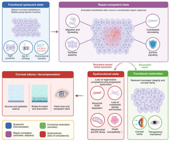
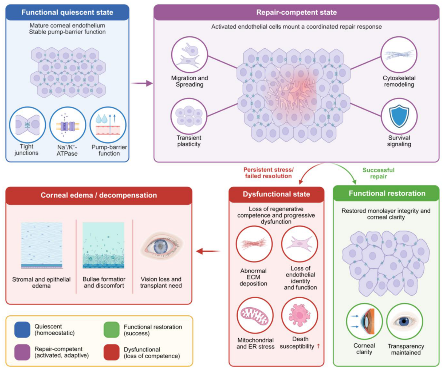
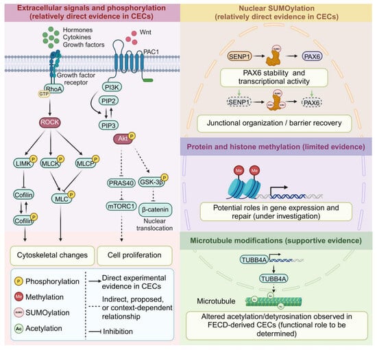
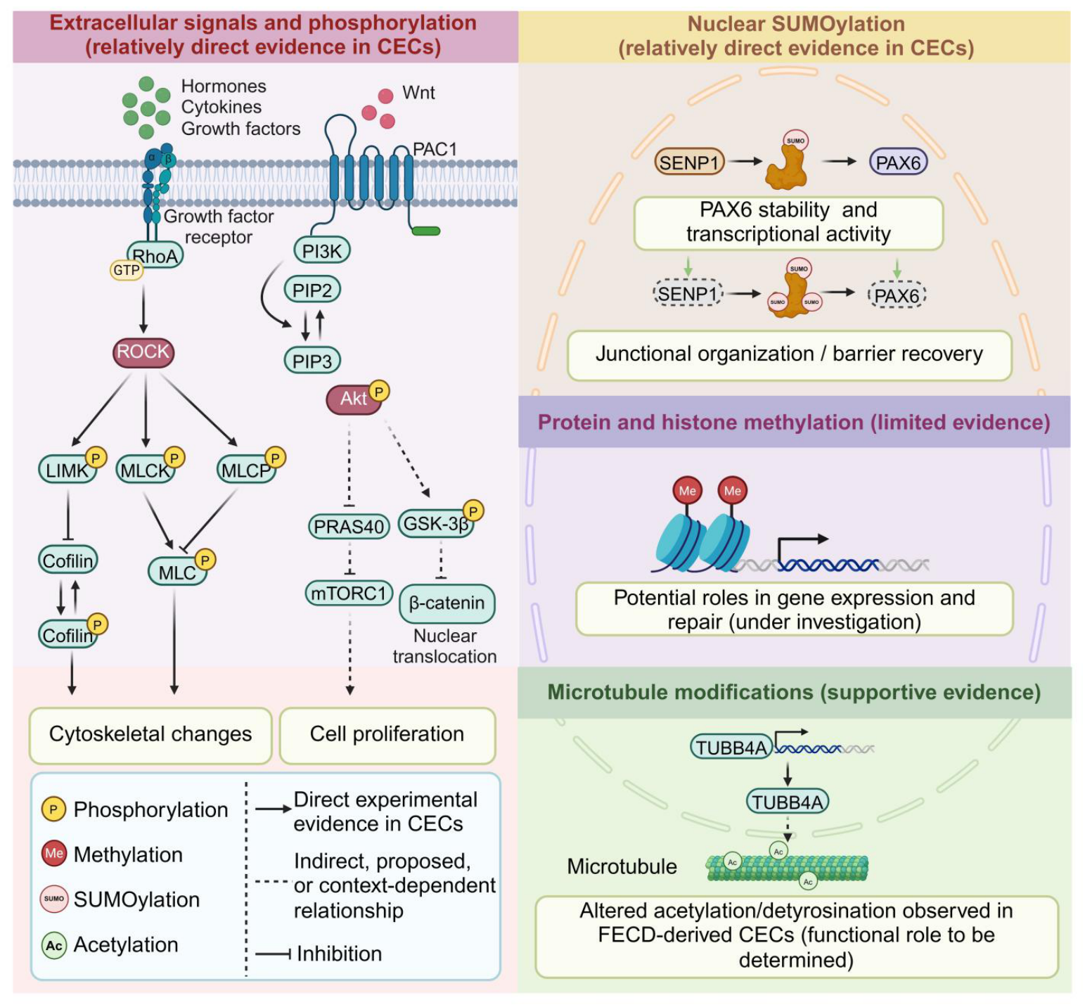
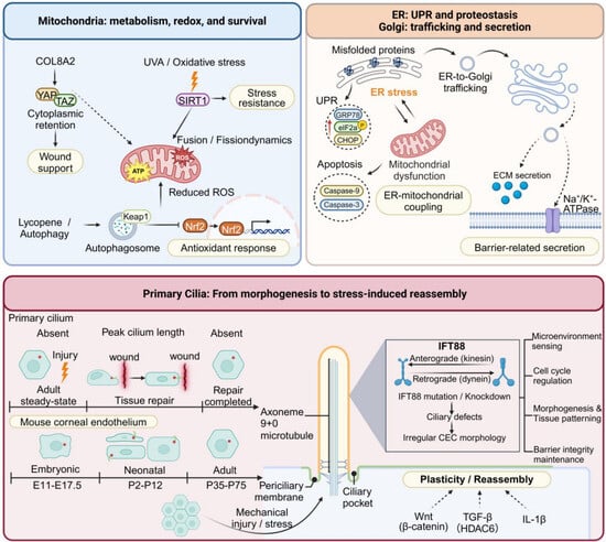
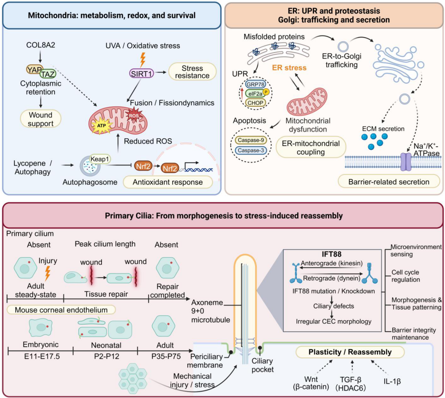
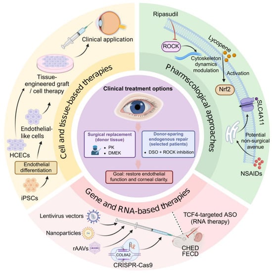
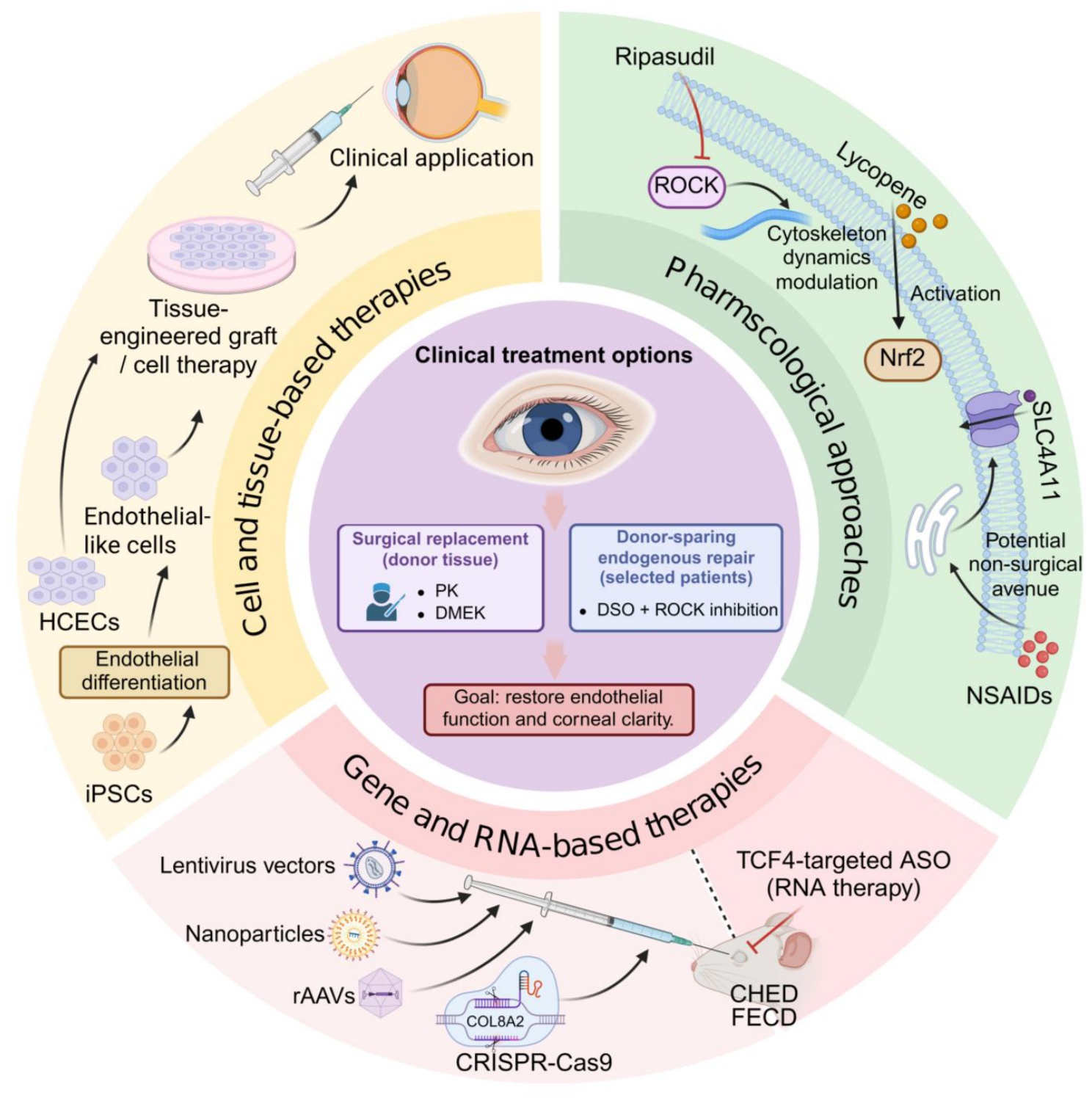

Disclaimer/Publisher’s Note: The statements, opinions and data contained in all publications are solely those of the individual author(s) and contributor(s) and not of MDPI and/or the editor(s). MDPI and/or the editor(s) disclaim responsibility for any injury to people or property resulting from any ideas, methods, instructions or products referred to in the content. |
© 2026 by the authors. Licensee MDPI, Basel, Switzerland. This article is an open access article distributed under the terms and conditions of the Creative Commons Attribution (CC BY) license.