


Abstract
Background/Objectives: During prostate cancer progression, downregulation or loss of prostate-specific membrane antigen (PSMA) expression, together with interlesional heterogeneity, may limit the efficacy of PSMA-targeted radioligand therapy. Fibroblast activation protein (FAP), expressed mainly by activated cancer-associated fibroblasts in the tumor microenvironment, represents a potential complementary target. We developed the PSMA/FAP bispecific radioligand with a biphenyl scaffold and evaluated its feasibility and therapeutic potential. Methods: The radiochemical purity and in vitro stability were evaluated by Radio-HPLC. Cellular uptake was evaluated in 22Rv1 (PSMA+), U87MG (FAP+), and PC-3 (PSMA−/FAP−) cells. SPECT/CT imaging and biodistribution studies were performed using mice bearing separate 22Rv1 (PSMA+) and U87MG (FAP+) xenografts. Therapeutic efficacy was assessed after a single administration of 30 or 60 MBq [177Lu]Lu-PSFA-1, together with an acute toxicity evaluation. Results: [177Lu]Lu-PSFA-1 showed PSMA- and FAP-associated uptake. SPECT/CT imaging and biodistribution studies showed persistent uptake in both tumor types, with higher uptake in U87MG tumors and increasing tumor-to-background ratios over time. A single administration of 30 or 60 MBq delayed tumor growth and prolonged survival, with more durable tumor control at 60 MBq. No evident acute toxicity was observed. Conclusions: [177Lu]Lu-PSFA-1 specifically targeted both PSMA- and FAP-positive tumors, with sustained tumor retention and antitumor activity. These findings support further evaluation of [177Lu]Lu-PSFA-1 as a PSMA/FAP bispecific radioligand, although its potential benefit in heterogeneous prostate cancer requires further validation.
1. Introduction
Prostate cancer (PCa) is one of the most common malignancies in men and remains a major cause of cancer-related death worldwide [1]. Despite substantial advances in androgen deprivation therapy, androgen receptor pathway inhibitors, chemotherapy, and other systemic treatments, a considerable proportion of patients eventually progress to metastatic castration-resistant prostate cancer (mCRPC), for which treatment remains challenging [2]. Among the molecular biomarkers investigated in prostate cancer, prostate-specific membrane antigen (PSMA) has emerged as an important target for molecular imaging and targeted radionuclide therapy [3,4]. In particular, [177Lu]Lu-PSMA-617 has demonstrated clear clinical benefit in patients with PSMA-positive mCRPC, establishing PSMA-targeted radioligand therapy (RLT) as an important treatment option for advanced prostate cancer [5,6]. However, the efficacy of PSMA-targeted RLT depends on sufficient and relatively homogeneous PSMA expression across tumor lesions. PSMA expression can vary substantially among patients, between metastatic sites, and over the course of disease progression and treatment [3,7,8]. In advanced prostate cancer, some lesions may exhibit low or absent PSMA expression, resulting in reduced radioligand uptake and limiting the therapeutic efficacy of PSMA-targeted RLT [7,8]. This has prompted interest in dual-targeting radioligands that combine PSMA with a complementary target.
Fibroblast activation protein (FAP), predominantly expressed by activated cancer-associated fibroblasts (CAFs) in the tumor microenvironment (TME), represents a complementary target to PSMA [9]. FAP-targeted PET has shown favorable tumor uptake across a broad range of malignancies and may improve lesion detection in prostate cancer with low or absent PSMA expression [10,11]. Dual targeting of PSMA and FAP may therefore broaden tumor targeting in heterogeneous prostate cancer, particularly in lesions with low or absent PSMA expression [12]. However, many small-molecule FAP-targeted radioligands exhibit relatively short tumor retention, which may limit their therapeutic potential. Improving tumor retention therefore remains an important goal in the development of FAP-targeted radioligand therapy [13].
FAP-based heterobivalent radiopharmaceuticals have also shown therapeutic potential. For example, the FAP/integrin αvβ3 dual-targeting agent [177Lu]Lu-DOTA-FAPI-RGD showed favorable tumor dosimetry and encouraging preliminary efficacy in preclinical and first-in-human evaluations, supporting the therapeutic potential of FAP-based heterobivalent radioligands [14]. Heterobivalent radioligands targeting PSMA and gastrin-releasing peptide receptor (GRPR) have also been investigated in prostate cancer [15], and a [177Lu]Lu-labeled PSMA/integrin αvβ3 dual-targeting radioligand produced greater tumor growth delay than the corresponding monospecific radioligands in preclinical models [16]. Our previously developed [68Ga]Ga-PSFA-01 demonstrated dual-targeting capability in preclinical and first-in-human PET studies [17]. A subsequent series of biphenyl-based PSMA/FAP bispecific ligands bearing DOTA or NOTA chelators retained high affinity for both targets and showed prolonged tumor retention in preclinical models [18].
On the basis of the well-established coordination chemistry of DOTA with 177Lu3+, DOTA-PSFA-1 was selected for 177Lu radiolabeling to extend the PSMA/FAP dual-targeting strategy from diagnostic imaging to radioligand therapy. We developed [177Lu]Lu-PSFA-1 and systematically characterized its radiopharmaceutical properties, dual-targeting activity, pharmacokinetics, biodistribution, safety, and therapeutic efficacy.
2. Results
2.1. Chemistry and Radiochemistry
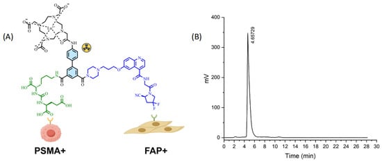
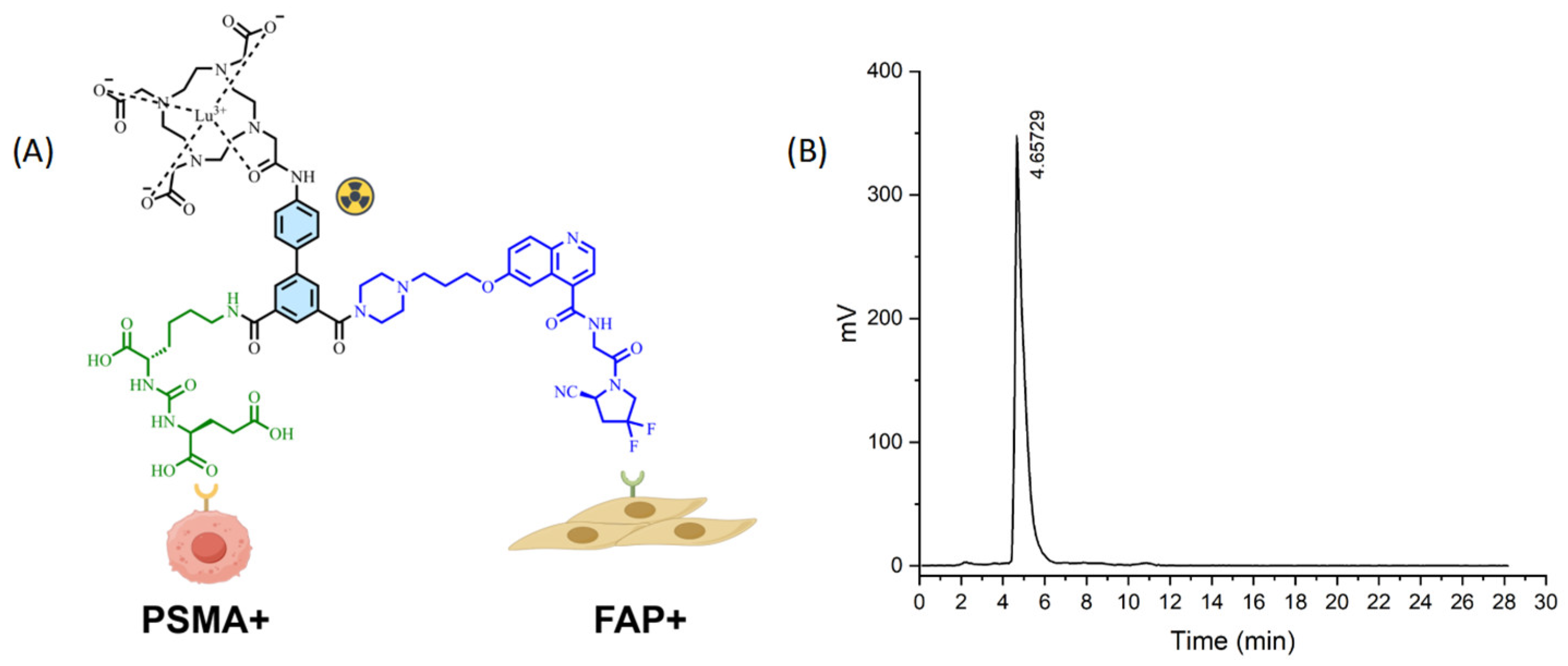
The DOTA-conjugated precursor was prepared through amide coupling reactions, including active ester conjugation and catalyst-assisted coupling. The chemical structure of [177Lu]Lu-PSFA-1 is shown in Figure 1A. After purification by HPLC, DOTA-PSFA-1 was obtained with a chemical purity of >99%. Radiolabeling with 177Lu afforded [177Lu]Lu-PSFA-1 with a radiochemical purity (RCP) of >98%, as determined by radio-HPLC, with a retention time of 4.657 min (Figure 1B). The molar activity, injected dose, non-decay-corrected RCY, and injected mass for each in vivo experiment are listed in Table S2. Detailed synthetic procedures and characterization data (HRMS) are provided in the Supplementary Materials.
Figure 1.
(A) Chemical structure of [177Lu]Lu-PSFA-1 and its functional components. (B) Radiochemical HPLC analysis of [177Lu]Lu-PSFA-1.
2.2. Lipophilicity and Stability
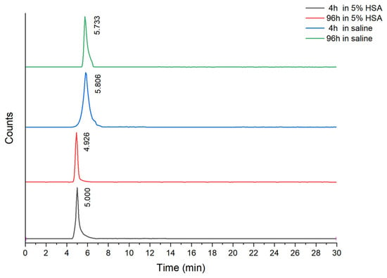
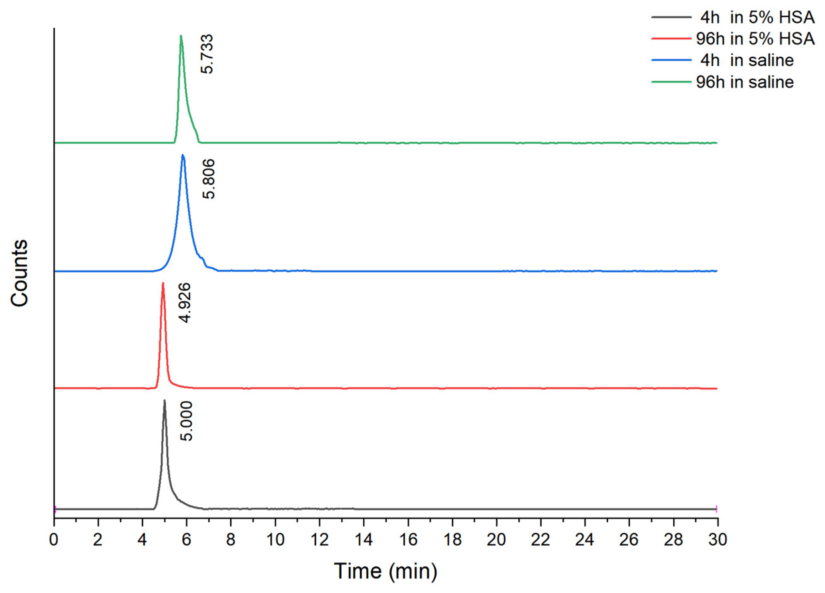
The n-octanol/PBS partition coefficient (log P) of [177Lu]Lu-PSFA-1 was −2.40 ± 0.02. The reported logD7.4 value of [177Lu]Lu-PSMA-617 was −3.29 ± 0.02 [19], whereas the log P value of [177Lu]Lu-FAPI-04 was −2.44 ± 0.10 [20]. Radio-HPLC analysis showed that [177Lu]Lu-PSFA-1 remained stable during incubation in saline and 5% human serum albumin (HSA), with RCP remaining above 98% at both 4 and 96 h (Figure 2).
Figure 2.
In vitro stability of [177Lu]Lu-PSFA-1 after incubation in saline and 5% HSA for 4 h and 96 h.
2.3. In Vitro Cell Assays
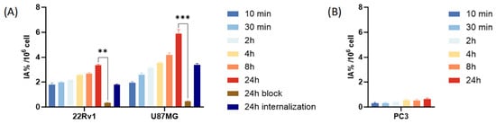
The nonradioactive reference compound [natLu]Lu-DOTA-PSFA-1 exhibited high binding affinity for both PSMA (Ki = 0.93 ± 0.08 nM) and FAP (Ki = 0.16 ± 0.04 nM). 22Rv1 (PSMA+), U87MG (FAP+), and PC-3 (PSMA−/FAP−) cells were used to evaluate the target-specific cellular uptake of [177Lu]Lu-PSFA-1. As shown in Figure 3, uptake in both 22Rv1 and U87MG cells increased over the 24 h incubation period, reaching 3.33 ± 0.09 and 5.88 ± 0.32% IA/106 cells at 24 h, respectively. At 24 h, ZJ43 reduced uptake in 22Rv1 cells to 0.32 ± 0.05% IA/106 cells, corresponding to a 90.4% reduction. Similarly, FAPI-04 reduced uptake in U87MG cells to 0.46 ± 0.04% IA/106 cells, corresponding to a 92.2% reduction. In contrast, uptake in PSMA/FAP-negative PC-3 cells remained low throughout the observation period (<0.7% IA/106 cells at 24 h). These findings support PSMA- and FAP-mediated uptake of [177Lu]Lu-PSFA-1 in 22Rv1 and U87MG cells, respectively. Western blot analysis confirmed PSMA expression in 22Rv1 xenografts and FAP expression in U87MG xenografts, with β-actin-normalized relative protein levels of approximately 0.50 and 0.36, respectively (Figure S3).
Figure 3.
In vitro studies. (A) Cellular uptake and internalization assay of [177Lu]Lu-PSFA-1 and blocking studies. Uptake was blocked with ZJ43 in 22Rv1 cells and FAPI-04 in U87MG cells. (B) Cell uptake of [177Lu]Lu-PSFA-1 in PC-3 cells at different times. Data are presented as mean ± SD (** p < 0.01; *** p < 0.001).
Furthermore, the internalization rates of [177Lu]Lu-PSFA-1 at 24 h were 54.18% in 22Rv1 cells and 57.10% in U87MG cells, indicating efficient internalization of the radioligand into target cells and providing a basis for its radiotherapeutic effects.
2.4. Micro-SPECT/CT Imaging
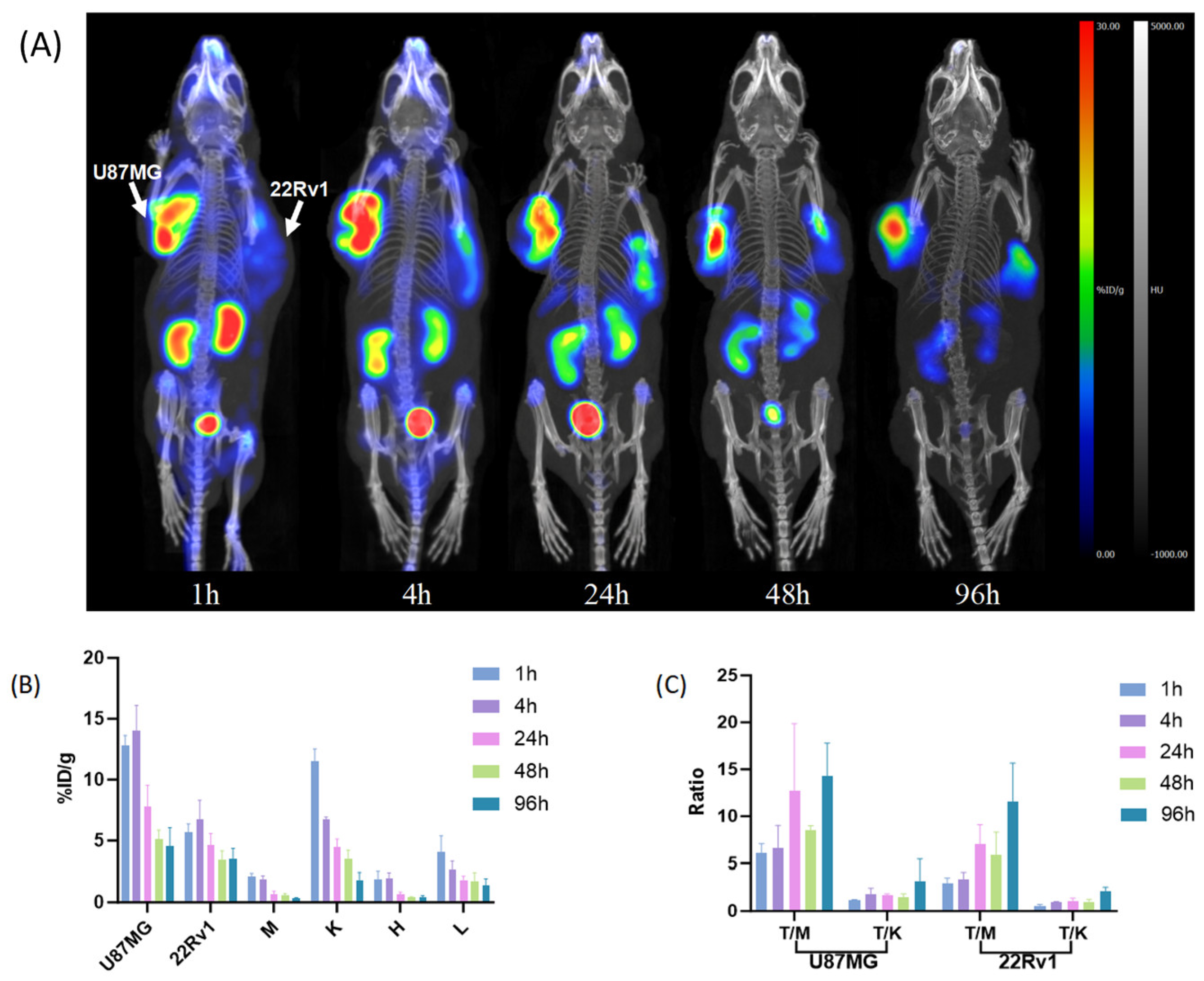
Longitudinal whole-body SPECT/CT imaging was performed at 1, 4, 24, 48, and 96 h after injection of [177Lu]Lu-PSFA-1 in mice bearing U87MG and 22Rv1 xenografts. Maximum-intensity-projection (MIP) images demonstrated clear radiotracer accumulation in both tumor types throughout the imaging period (Figure 4A). Physiological activity was also observed in the kidneys and urinary bladder. Bladder activity decreased rapidly over time, consistent with urinary excretion of the radiotracer. With progressive clearance from nontarget tissues, tumor-to-background contrast improved at later time points. Tumor uptake was relatively high at early time points, with the highest mean values observed at 4 h p.i. (13.99 ± 2.09% ID/g in U87MG tumors and 6.77 ± 1.54% ID/g in 22Rv1 tumors). Both tumor types showed sustained retention over the 96-h imaging period (Figure 4A,B).
Figure 4.
SPECT/CT images in mice bearing U87MG and 22Rv1 tumors. (A) Longitudinal maximum-intensity SPECT/CT projection of animals injected with [177Lu]Lu-PSFA-1. (B) Quantitative SPECT/CT-derived uptake in tumors and major organs (muscle, M; kidney, K; heart, H; liver, L) over time. (C) Tumor-to-muscle (T/M) and tumor-to-kidney (T/K) ratios at different time points. Data are presented as mean ± SD (n = 3 mice).
Quantitative analysis showed an overall increase in tumor-to-muscle (T/M) and tumor-to-kidney (T/K) ratios over time (Figure 4C). The highest ratios were observed at 96 h, reaching T/M = 14.30 ± 3.49 and T/K = 3.15 ± 2.33 for U87MG tumors and T/M = 11.56 ± 4.11 and T/K = 2.09 ± 0.45 for 22Rv1 tumors.
2.5. Pharmacokinetics and Biodistribution
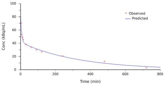
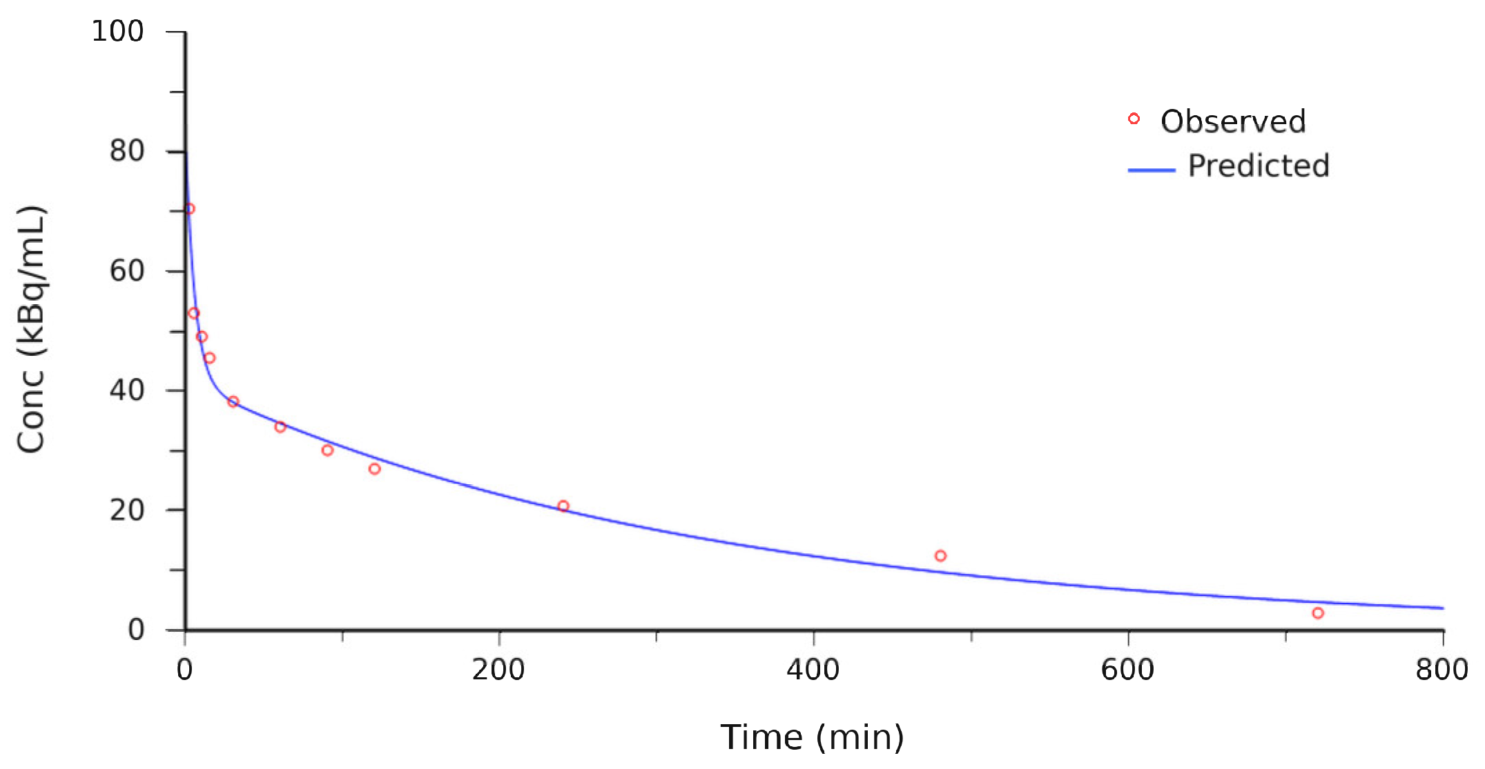
The mean blood time–activity curve of [177Lu]Lu-PSFA-1 is shown in Figure 5. Pharmacokinetic analysis using WinNonlin was best described by a two-compartment model, with a distribution half-life (t1/2α) of 4.08 min and an elimination half-life (t1/2β) of 228.78 min.
Figure 5.
Blood time–activity curves of [177Lu]Lu-PSFA-1 in BALB/c mice (n = 5).
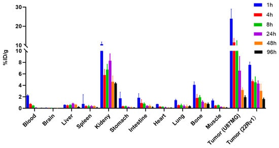
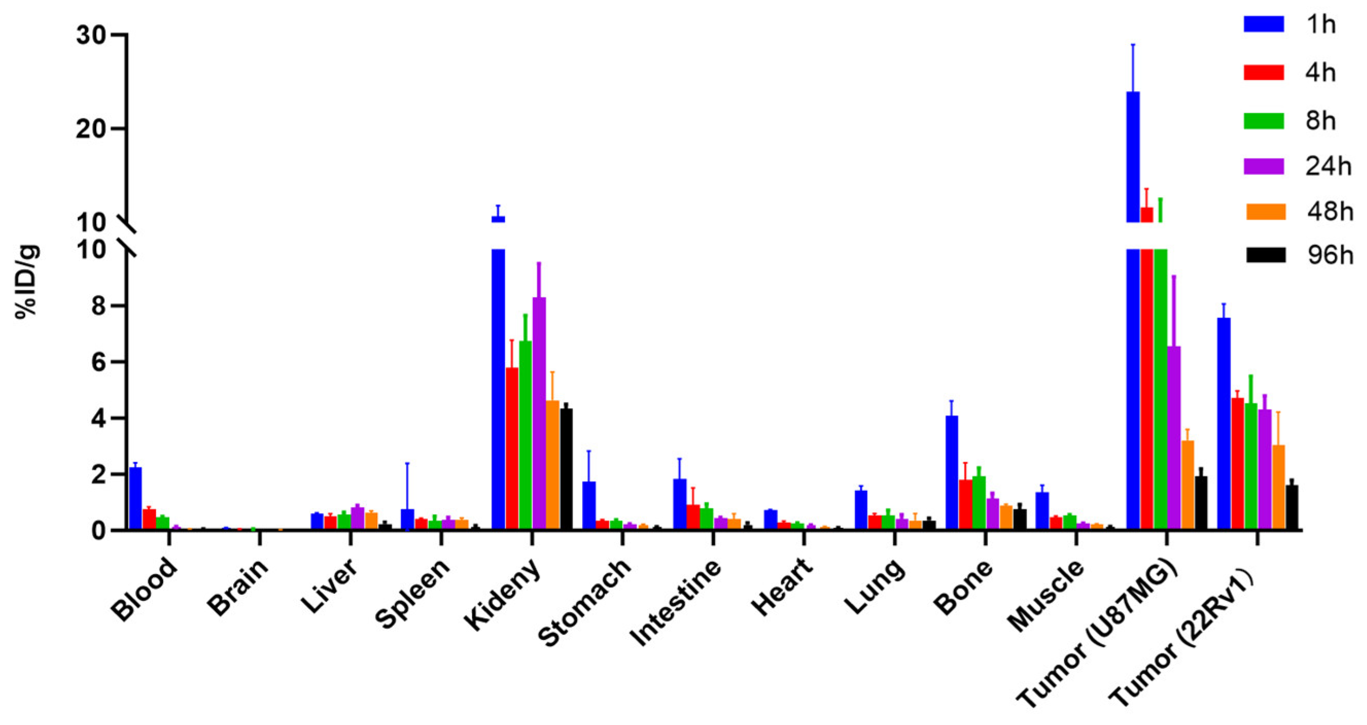
To further evaluate the pharmacokinetic properties of [177Lu]Lu-PSFA-1 in vivo, we investigated the distribution in tumor-bearing mice through biodistribution studies. As shown in Figure 6, U87MG tumor uptake decreased from 23.93 ± 5.02% ID/g at 1 h p.i. to 1.93 ± 0.27% ID/g at 96 h p.i., with persistent tumor uptake observed up to 96 h. Uptake in 22Rv1 tumors was lower than that in U87MG tumors throughout the study and decreased from 7.57 ± 0.50% ID/g at 1 h p.i. to 1.60 ± 0.19% ID/g at 96 h p.i. At 1 h p.i., radioactivity was distributed across several normal organs and subsequently decreased with time, consistent with the SPECT/CT findings. Among normal tissues, the kidneys showed the highest uptake, decreasing from 10.64 ± 1.13% ID/g at 1 h p.i. to 4.32 ± 1.64% ID/g at 96 h p.i., whereas uptake in most other normal tissues remained relatively low.
Figure 6.
Biodistribution studies of [177Lu]Lu-PSFA-1 in U87MG and 22Rv1 tumor-bearing mice at different time points after injection (n = 4 per time point). Data are expressed as mean ± SD.
2.6. Acute Toxicity Test
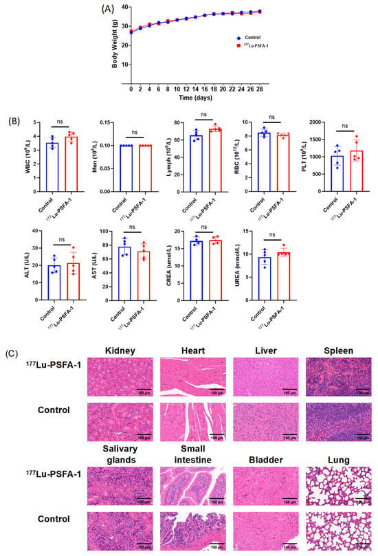
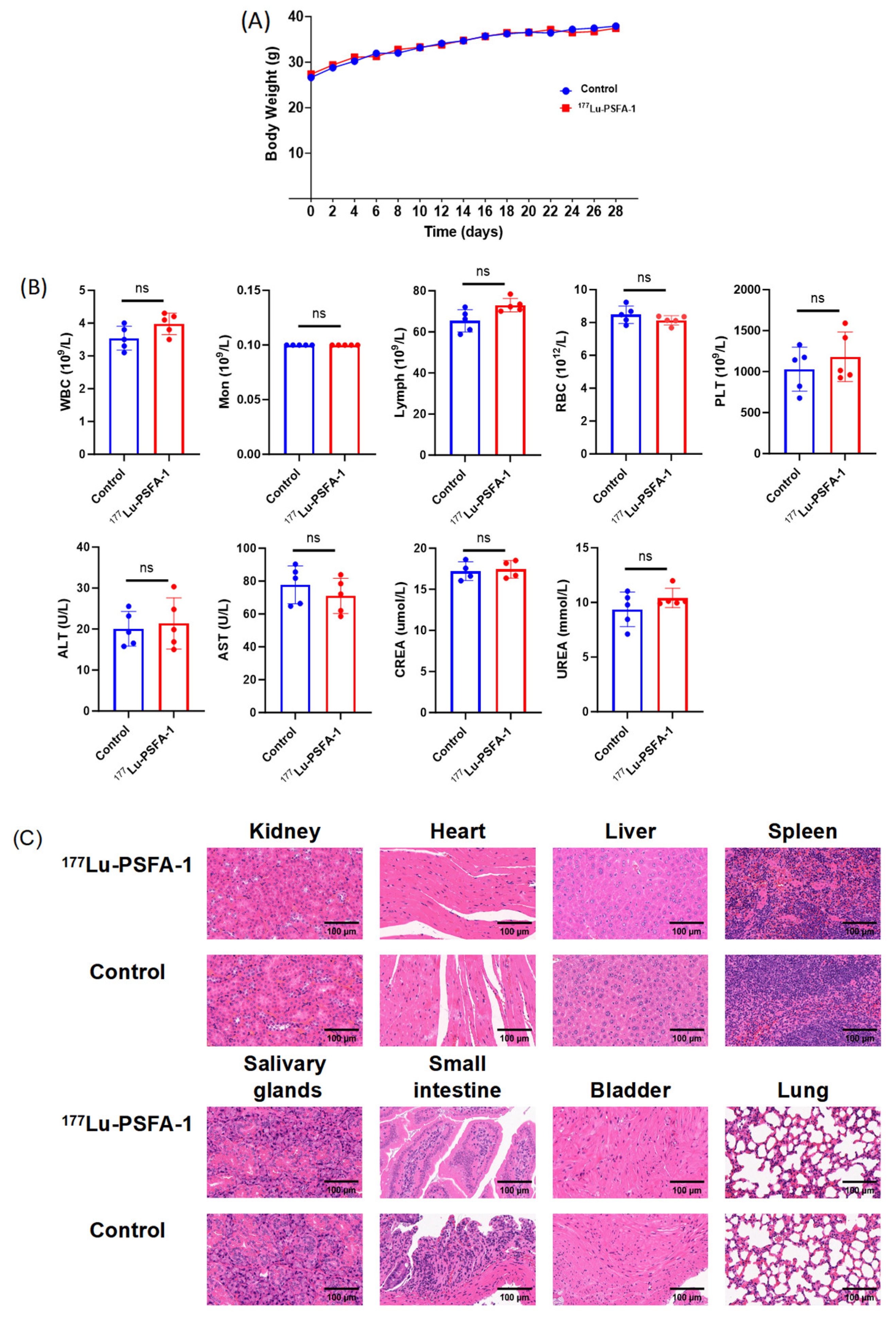
During the 28-day observation period, no obvious abnormalities in general condition, food intake, activity, mental status, or skin appearance were observed in either group. Body weight increased gradually in both groups, with no significant difference between the [177Lu]Lu-PSFA-1 and control groups (p > 0.05; Figure 7A). On day 28, hematological and serum biochemical parameters remained within the normal ranges, with no significant differences between the two groups (all p > 0.05; Figure 7B).
Figure 7.
Safety evaluation of [177Lu]Lu-PSFA-1 in mice. (A) Body weight changes during the 28-day observation period. (B) Hematological and serum biochemical parameters. Blue and red dots represent individual mice in the control and [177Lu]Lu-PSFA-1 groups, respectively. (C) Representative H&E staining of major organs. Quantitative data are presented as mean ± SD. ns, not significant. Scale bars, 100 μm.
H&E staining showed no obvious pathological changes in the major organs of mice treated with [177Lu]Lu-PSFA-1. No apparent abnormalities were observed in the kidney, heart, liver, spleen, salivary glands, small intestine, bladder, or lung compared with the control group (Figure 7C). Overall, a single intravenous administration of 74 MBq [177Lu]Lu-PSFA-1 was well tolerated, with no evident acute toxicity during the 28-day observation period.
2.7. Radioligand Therapy Study
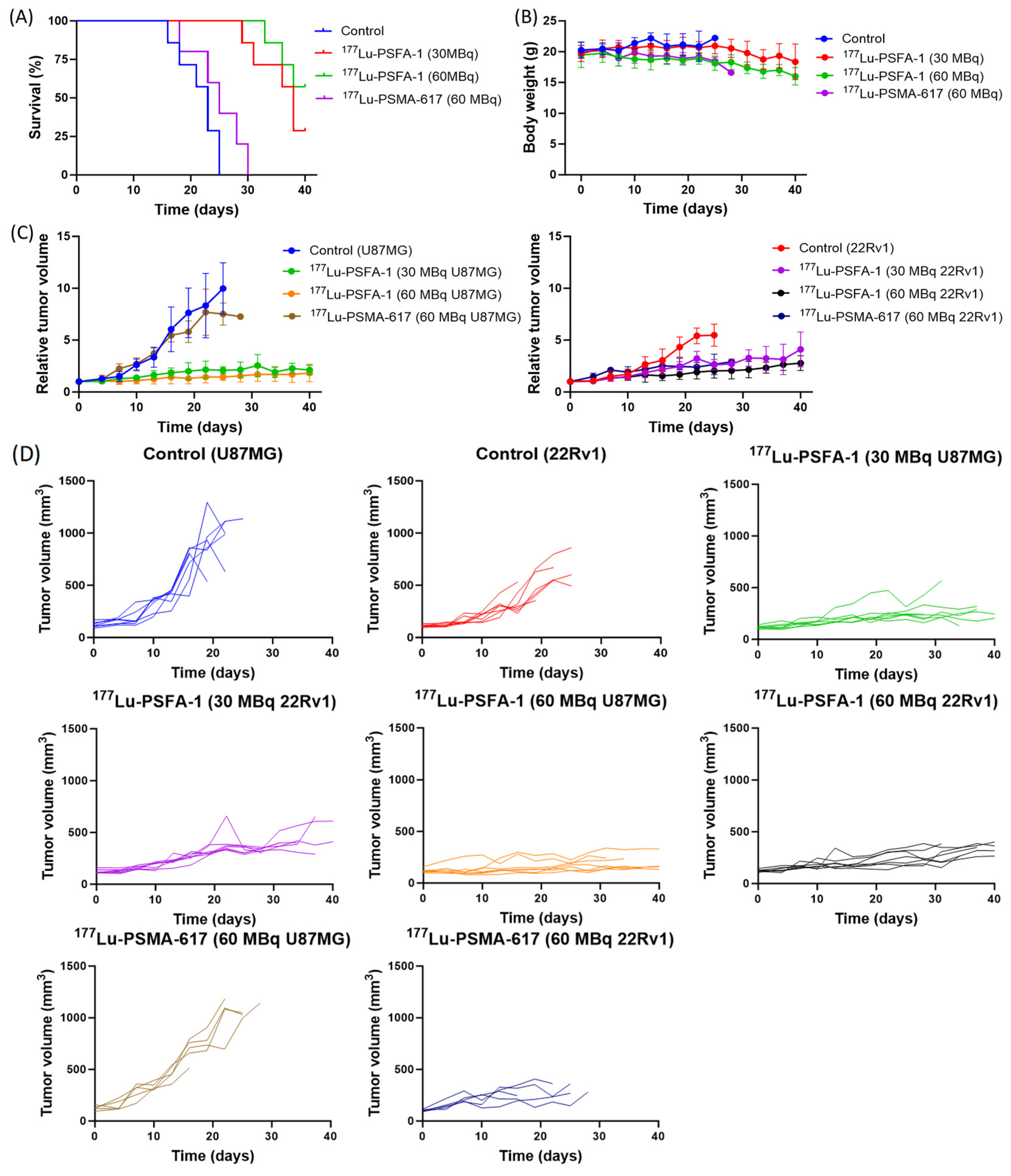
The high tumor uptake and internalization rate of [177Lu]Lu-PSFA-1 encouraged us to evaluate its therapeutic efficacy. The therapeutic efficacy of [177Lu]Lu-PSFA-1 was evaluated in mice bearing paired U87MG and 22Rv1 xenografts. Animals received a single administration of saline, 30 or 60 MBq [177Lu]Lu-PSFA-1, or 60 MBq [177Lu]Lu-PSMA-617. Both 60 MBq treatment groups received the same injected ligand mass per mouse. Animals were followed for up to 40 days or until a predefined humane endpoint was reached (Figure 8).
Figure 8.
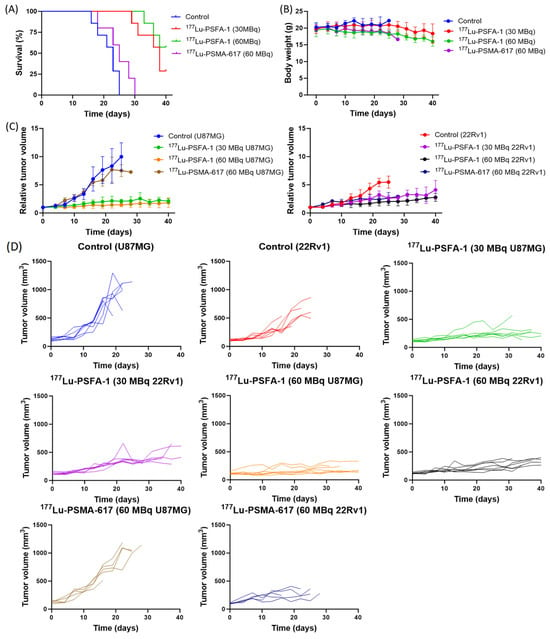
Therapeutic efficacy and tolerability of [177Lu]Lu-PSFA-1 (30 and 60 MBq) in 22Rv1 and U87MG tumor–bearing mice. (A) Survival curves. (B) Body weight changes after treatment. (C) Relative tumor volume in the different groups. (D) Individual tumor-volume trajectories (n = 7 for the control and [177Lu]Lu-PSFA-1 groups and n = 5 for the [177Lu]Lu-PSMA-617 group). Data are presented as mean ± SD.
Median survival was 23 days in the control group, 38 days in the 30 MBq [177Lu]Lu-PSFA-1 group, and 25 days in the 60 MBq [177Lu]Lu-PSMA-617 group, whereas median survival was not reached in the 60 MBq [177Lu]Lu-PSFA-1 group during the 40-day observation period (Figure 8A). At day 40, survival rates were 0% (0/7), 28.6% (2/7), 57.1% (4/7), and 0% (0/5) in the control, 30 MBq [177Lu]Lu-PSFA-1, 60 MBq [177Lu]Lu-PSFA-1, and 60 MBq [177Lu]Lu-PSMA-617 groups, respectively. Survival was significantly prolonged in both [177Lu]Lu-PSFA-1 treatment groups compared with the control group (log-rank Mantel–Cox test, p < 0.01). Moreover, the 60 MBq [177Lu]Lu-PSFA-1 group showed significantly longer survival than the [177Lu]Lu-PSMA-617 group at the same administered activity (p < 0.01). Body weights remained relatively stable during the early treatment period but gradually decreased at later time points (Figure 8B).
Both administered activities of [177Lu]Lu-PSFA-1 suppressed tumor growth compared with saline controls (Figure 8C,D). Seven days post-injection, the relative U87MG tumor volume of the [177Lu]Lu-PSFA-1 was significantly lower than that of the control group (p < 0.05). In U87MG tumors, [177Lu]Lu-PSFA-1 produced greater growth inhibition than [177Lu]Lu-PSMA-617 at 60 MBq, with significantly lower relative tumor volumes on day 13 (1.25 ± 0.48, n = 7 vs. 3.71 ± 0.54, n = 5; p < 0.0001). The corresponding value in saline-treated controls was 3.36 ± 0.96 (n = 7). In 22Rv1 tumors, no significant difference in relative tumor volume was observed between the 60 MBq [177Lu]Lu-PSFA-1 and [177Lu]Lu-PSMA-617 groups on day 10 (p > 0.05). However, [177Lu]Lu-PSFA-1 significantly inhibited tumor growth compared with saline controls (p < 0.05). Individual tumor-volume trajectories are shown in Figure 8D. Only animals remaining in the study were included in the analyses, and missing values after humane endpoints were not imputed.
3. Discussion
PSMA-targeted radioligand therapy has become an important treatment option for advanced prostate cancer [5,21]. However, the efficacy of PSMA-directed therapy is dependent on sufficient radioligand uptake within tumor lesions [7,8]. PSMA expression varies across patients and metastatic sites and may also change during disease progression and treatment. Such spatiotemporal heterogeneity in PSMA expression may result in inadequate radioligand delivery to PSMA-low or PSMA-negative lesions [3]. FAP provides a biologically distinct target that is predominantly expressed by CAFs in the TME [9]. In the present study, we extended our previously established PSMA/FAP dual-targeting from diagnostic imaging to 177Lu-based radioligand therapy. [177Lu]Lu-PSFA-1 showed uptake in both 22Rv1 and U87MG tumors and demonstrated measurable therapeutic effects.
[177Lu]Lu-PSFA-1 showed a radiochemical purity of >98% and remained stable for up to 96 h. Blood radioactivity followed a two-compartment model, characterized by rapid distribution and a slower terminal elimination phase, suggesting rapid tissue distribution followed by slower systemic clearance. This pharmacokinetic profile may facilitate early tumor delivery and subsequent tumor retention. Rapid tumor washout limits the therapeutic potential of many small-molecule FAPIs [22,23]. Prolonged tumor retention, as achieved with [177Lu]Lu-FAP-2286 and multimeric FAPI derivatives, may increase tumor absorbed dose and improve the suitability of FAP-targeted ligands for radionuclide therapy [24,25].
The in vitro findings confirmed that both targeting components of [177Lu]Lu-PSFA-1 remained functional after radiolabeling. Uptake increased with incubation time in both 22Rv1 and U87MG cells and reached its highest measured level at 24 h and was markedly reduced by the corresponding PSMA or FAP inhibitor, whereas uptake in PSMA/FAP-negative PC-3 cells remained low. Quantitative SPECT/CT analysis showed the highest mean tumor uptake at 4 h, followed by an overall decline, while tumor-to-background ratios generally increased over time. In contrast, ex vivo biodistribution showed the highest U87MG tumor uptake at 1 h. This difference may partly reflect differences in quantification methods and injected ligand masses (0.78–0.80 vs. 0.079–0.084 μg/mouse) [26]. Both methods demonstrated higher uptake in U87MG than in 22Rv1 tumors, with measurable tumor retention up to 96 h. This interpretation is consistent with our previous observation that the PSMA-targeting motif improved the retention of FAP-targeted variants [18]. Additional receptor–ligand interactions mediated by the PSMA pharmacophore may also contribute to the overall binding properties of the bispecific ligand. The higher uptake observed in FAP-positive U87MG tumors further supports FAP as a complementary target when PSMA-mediated delivery is limited. Previous studies have reported low endogenous FAP expression in cultured U87MG cells, whereas U87MG xenografts may recruit FAP-positive murine stromal fibroblasts [27]. Therefore, FAP expression detected in U87MG xenografts may originate from both tumor cells and host stromal cells [28]. However, PSMA and FAP expression were not systematically assessed in each xenograft model. Thus, the relative contributions of these targets to the differences in radioligand uptake between 22Rv1 and U87MG tumors remain uncertain. More broadly, CAFs contribute to extracellular-matrix remodeling, tumor-cell survival, immune regulation, and treatment resistance [29]. A PSMA/FAP bispecific ligand has the potential to target both PSMA-expressing malignant cells and FAP-positive stromal components [30]. For 177Lu-based therapy, targeting FAP-positive stromal cells may further extend radiation delivery to neighboring malignant cells through the β−-particle cross-fire effect [22,31].
A single administration of [177Lu]Lu-PSFA-1 delayed tumor growth and prolonged survival, with a stronger response at 60 MBq. The response was more pronounced in U87MG tumors, consistent with their higher radioligand uptake. At the same administered activity, [177Lu]Lu-PSFA-1 showed greater growth inhibition than [177Lu]Lu-PSMA-617 in U87MG tumors, whereas the difference between these radioligands in 22Rv1 tumors was not significant at the evaluated time point. These findings warrant further investigation of the bispecific strategy but do not establish a therapeutic advantage in heterogeneous prostate cancer lesions. In healthy mice, no evident toxicity was detected during the 28-day observation period after a single 74 MBq administration. However, this relatively short observation period does not exclude potential delayed renal or hematological toxicity. Longer-term safety evaluation and organ-level dosimetry, particularly for the kidneys and bone marrow, are therefore needed before clinical translation. The biodistribution profile of [177Lu]Lu-PSFA-1 also warrants consideration with respect to potential normal-organ toxicity. The kidneys showed the highest uptake among normal tissues, decreasing from 10.64 ± 1.13% ID/g at 1 h to 4.32 ± 1.64% ID/g at 96 h. Our study showed that incorporation of the biphenyl scaffold improved the renal profile of PSMA/FAP bispecific radioligands compared with earlier PSFA constructs. Nevertheless, appreciable renal retention was still observed at later time points. The kidneys should therefore be carefully evaluated in future dosimetry and repeated-cycle treatment studies.
Several limitations should be considered. First, the present study used the relatively low-PSMA 22Rv1 model but did not include a high-PSMA model. Future studies should include LNCaP or PC3-PIP tumors to determine how therapeutic performance changes across different levels of PSMA expression. Second, the separate 22Rv1 and U87MG xenografts do not reproduce the coexistence of PSMA-positive malignant cells and FAP-positive stromal cells within a single prostate cancer lesion. Models with spatially characterized tumor and stromal compartments are needed to test whether dual targeting improves radionuclide delivery under genuinely heterogeneous conditions. Third, our study evaluated only two activity levels after a single administration. Further studies should also evaluate fractionated or repeated dosing regimens [32]. Longitudinal tumor-volume analysis was also limited by the loss of animals after reaching humane endpoints, resulting in potentially informative missing observations. Therefore, statistical comparisons were restricted to early time points, whereas later tumor-growth data were interpreted descriptively. This limitation should be considered when interpreting treatment effects over the 40-day observation period. In particular, 225Ac or 161Tb may offer different radiation ranges and radiobiological properties [19]. Preclinical studies of PSMA-targeted radioligands have demonstrated higher tumor absorbed doses and enhanced antitumor efficacy with 161Tb compared with 177Lu, which has been attributed in part to the additional emission of conversion and Auger electrons [33]. Overall, the present findings support further development of [177Lu]Lu-PSFA-1 as a PSMA/FAP bispecific radioligand. By retaining PSMA-directed targeting while extending recognition to FAP-positive tumor components, this dual-targeting strategy may broaden radioligand delivery in heterogeneous tumors. Further structural optimization and validation in models that more closely recapitulate the spatial heterogeneity of prostate cancer are needed to establish its potential therapeutic advantages.
4. Materials and Methods
4.1. General Materials
All chemicals, solvents, and reagents used for synthesis and analysis were of analytical grade and were purchased from Macklin Biochemical Technology Co., Ltd. (Shanghai, China). [177Lu]LuCl3 solution was supplied by China Isotope & Radiation Corporation (Beijing, China), with radionuclidic and radiochemical purities of >99%. Radioactivity was measured using a CRC-25R dose calibrator (Capintec, Inc., Ramsey, NJ, USA) and an AMG 425-6c1 γ-counter (Hidex, Turku, Finland). Radiolabeling efficiency and radiochemical purity were assessed using radio-thin-layer chromatography (radio-TLC) and radio-high-performance liquid chromatography (radio-HPLC, Waters, Milford, MA, USA). HPLC analysis was performed using a Phenomenex Gemini C18 column (5 μm, 100 Å, 4.6 × 150 mm, Phenomenex, Torrance, CA, USA). Small-animal SPECT/CT imaging was performed using a nanoSPECT/CT preclinical scanner (Mediso Ltd., Budapest, Hungary).
4.2. Chemical Synthesis, Radiolabeling and Quality Control
The detailed scheme of the chemical synthesis protocol and characterization of DOTA-PSFA-1 are provided in the Supplementary Materials. For radiolabeling with 177LuCl3, 185–740 MBq of 177LuCl3 was mixed with 0.2 mL of 0.5 M NaOAc (pH = 5.5) and incubated with DOTA-PSFA-1 (20–100 μg) or PSMA-617 (50 μg) at 95 °C for 15 min, respectively. Radiolabeling efficiency and radiochemical purity were assessed by radio-TLC and radio-HPLC. The radiolabeled product was passed through a 0.22-μm sterile filter to obtain the final formulation for subsequent in vitro and in vivo experiments.
Radiochemical purity was determined using a Waters 515 HPLC system equipped with a γ-detector (BIOSCAN Flow-Count, NaI scintillation detector). The mobile phases consisted of water containing 0.4% phosphoric acid (solvent A) and acetonitrile (solvent B), at a flow rate of 1 mL/min. The gradient was as follows: 5% B for 3 min, followed by a linear increase from 5% to 25% B over 10 min and then 25% B for 5 min.
The molar activity was obtained as the ratio of the final radiolabeled product activity to the molar amount of DOTA-PSFA-1 precursor. The non-decay-corrected radiochemical yield (RCY) was obtained as the ratio of the final radiolabeled product activity to the initial 177Lu activity.
4.3. In Vitro Stability and Partition Coefficient
For the in vitro stability study, [177Lu]Lu-PSFA-1 was incubated in saline and 5% human serum albumin (HSA). At 4 and 96 h, 10 μL aliquots were collected and analyzed by radio-HPLC to determine radiochemical purity. For samples incubated in 5% HSA, an equal volume of acetonitrile was added to precipitate proteins. After centrifugation, an appropriate volume of the supernatant was collected for radio-HPLC analysis.
For determination of the partition coefficient, 100 μL of [177Lu]Lu-PSFA-1 (approximately 0.37 MBq) was added to a mixture of 2.9 mL PBS (0.1 M, pH 7.4) and 3.0 mL n-octanol. The mixture was vortexed for 2 min at room temperature and centrifuged at 3000 rpm for 5 min. After complete phase separation, three 500-μL aliquots were collected from each phase, and radioactivity was measured using a γ-counter (counts per minute, CPM). The partition coefficient was calculated as: log P = log (CPMoctanol/CPMwater) and reported as log P ± SD. The experiment was performed in triplicate and repeated three times.
4.4. Cell Lines and Animal Models
The human prostate cancer cell lines 22Rv1 and PC-3 and the human glioblastoma cell line U87MG were obtained from the Cell Bank of the Chinese Academy of Sciences. 22Rv1 (PSMA+) and PC-3 (PSMA−/FAP−) cells were cultured in Roswell Park Memorial Institute 1640 (RPMI-1640) medium (Gibco, New York, NY, USA) supplemented with 10% fetal bovine serum (FBS; Gibco). U87MG (FAP+) cells were cultured in Dulbecco’s modified Eagle medium (DMEM) supplemented with 10% FBS. Cells were maintained at 37 °C in a humidified atmosphere containing 5% CO2 and passaged at 80–90% confluence using trypsin–EDTA (0.25% trypsin and 0.02% EDTA; Gibco).
Male BALB/c nude mice were purchased from Charles River Laboratories (Beijing, China). Mice were approximately 3–4 weeks old and weighed 13–15 g. To establish paired xenograft models, approximately 2 × 10⁶ 22Rv1 cells and 2.5 × 10⁶ U87MG cells in 0.1 mL were inoculated subcutaneously on the right and left flanks, respectively.
4.5. Cell Binding and Internalization Studies
The binding affinities of [natLu]Lu-DOTA-PSFA-1 for PSMA and FAP were determined using enzyme inhibition assays with recombinant proteins, as previously described [18].
Cells were seeded in 24-well plates at a density of 1 × 10⁵ cells per well in 1 mL of culture medium and incubated overnight at 37 °C in 5% CO2. The culture medium was replaced 2 h before the experiment. Before addition of the radioligand, the medium was removed, and the cells were washed with PBS.
[177Lu]Lu-PSFA-1 (0.074 MBq in 100 μL) was added to each well, and the total volume was adjusted to 1 mL with the corresponding culture medium. Cells were incubated at 37 °C for 10 and 30 min and for 2, 4, 8, and 24 h. At each time point, the medium was removed, and the cells were washed twice with 0.5 mL of ice-cold PBS containing 0.2% BSA. Cells were then lysed with 0.5 mL of 0.5 M NaOH, and cell-associated radioactivity was measured using a γ-counter. Cellular uptake was expressed as % IA/106 cells. Five replicate wells were analyzed at each time point, and the experiment was independently repeated three times.
For internalization studies, 22Rv1 and U87MG cells were incubated with [177Lu]Lu-PSFA-1 (0.074 MBq in 100 μL) for 24 h. After removal of the culture medium, cells were incubated with 0.5 mL glycine-HCl buffer in PBS (50 mM, pH 2.8) for 5 min at 37 °C to remove surface-bound activity. The cells were then washed twice with 0.5 mL ice-cold PBS and lysed with 0.5 mL of 0.5 M NaOH. Radioactivity was measured using a γ-counter. For blocking studies, 22Rv1 cells were preincubated with ZJ43 (10 μg/well) and U87MG cells with FAPI-04 (10 μg/well) for 15 min before addition of [177Lu]Lu-PSFA-1. The subsequent procedure was identical to that used for the cellular uptake assay.
4.6. Pharmacokinetics
Five healthy female BALB/c mice were administered [177Lu]Lu-PSFA-1 (0.37 MBq in 100 μL) via tail-vein injection. Serial blood samples (3 μL) were collected at 2, 5, 15, 30, 60, 90, 120, 240, 360, 480, and 720 min after injection. Blood radioactivity was measured using a γ-counter and converted to radioactivity concentration (kBq/mL). The resulting blood time–activity data were analyzed using WinNonlin version 8.1 (Certara, Princeton, NJ, USA) to determine the distribution and elimination half-lives of [177Lu]Lu-PSFA-1.
4.7. Acute Toxicity Test
Healthy male ICR mice (3–4 weeks old, 20–25 g) were divided into two groups (n = 5 per group) and intravenously administered [177Lu]Lu-PSFA-1 (74 MBq in 150 μL) or saline (150 μL). Body weight and general condition were monitored for 28 days. Subsequently, the mice were euthanized for hematological and serum biochemical analyses and hematoxylin and eosin (H&E) staining of major organs.
4.8. Micro-SPECT/CT Imaging
SPECT/CT imaging studies were performed using a nanoSPECT/CT preclinical scanner (Mediso Ltd., Budapest, Hungary). A total of 3 mice bearing 22Rv1 and U87MG xenografts were included in the study. Approximately 7.4 MBq of [177Lu]Lu-PSFA-1 was injected intravenously to 22Rv1 and U87MG tumor-bearing mice. Whole-body SPECT/CT images were acquired at 1, 4, 24, 48, and 96 h p.i. under anesthesia with 1.5% isoflurane in air.
Images were reconstructed using the Tera-Tomo 3D iterative reconstruction algorithm (Mediso Ltd.), with CT-based attenuation and scatter corrections. Quantitative reconstruction was performed using the original geometry and activity calibration settings, with the acquisition start time selected as the decay reference. Reconstructed images were analyzed using InterView Fusion (version 3.11.011.0000; Mediso Ltd., Budapest, Hungary). Three-dimensional volumes of interest (VOIs) were manually delineated along the tumor boundaries and selected normal tissues on the fused SPECT/CT images. Quantitative uptake was obtained from the mean activity within each VOI and expressed as the percentage injected dose per gram of tissue (%ID/g). Tumor-to-muscle (T/M) and tumor-to-kidney (T/K) ratios were calculated from the corresponding mean %ID/g values. Data are presented as mean ± SD (n = 3).
4.9. Biodistribution Studies
Biodistribution studies were performed in 22Rv1 and U87MG tumor-bearing mice. Mice were randomly assigned to six time-point groups (n = 4 per time point). Each mouse received [177Lu]Lu-PSFA-1 (740 kBq in 0.1 mL saline) via tail-vein injection and was euthanized at 1, 4, 8, 24, 48, or 96 h p.i. Tumors and selected organs and tissues were collected, wet-weighed, and measured for radioactivity using a γ-counter. Radiotracer uptake was calculated as the percentage of injected dose per gram of tissue (%ID/g). Data are expressed as mean ± SD (n = 4 per time point).
4.10. Therapeutic Efficacy Studies
22Rv1 and U87MG tumor-bearing mice were assigned to four treatment groups and received a single intravenous administration of saline (n = 7 per group), 30 MBq [177Lu]Lu-PSFA-1 (n = 7 per group), 60 MBq [177Lu]Lu-PSFA-1 (n = 7 per group), or 60 MBq [177Lu]Lu-PSMA-617 (n = 5 per group) on day 0. Body weight and tumor dimensions were recorded on day 0 and two to three times per week thereafter for up to 40 days. Tumor volume was calculated using V = 0.5 × length × width2. Treatment efficacy was evaluated on the basis of relative tumor volume (Vt/V0) and survival. Animals were followed for a maximum of 40 days or until a predefined humane endpoint was reached. Endpoint criteria included ≥15% loss of body weight, a total tumor volume >1800 mm3, active tumor ulceration, or abnormal behavior.
4.11. Statistical Analysis
Data are presented as mean ± SD. Biodistribution data were analyzed using unpaired two-tailed Student’s t-tests where appropriate. Tumor volumes were compared using one-way ANOVA followed by Tukey’s multiple-comparisons test at selected time points. Kaplan–Meier survival analysis was performed, with log-rank tests used to evaluate statistical significance between groups. A p value of < 0.05 was considered statistically significant. Statistical analyses were performed using GraphPad Prism version 9.5.1 (GraphPad Software, San Diego, CA, USA).
5. Conclusions
In this study, [177Lu]Lu-PSFA-1 exhibited favorable radiopharmaceutical properties and specific dual-targeting capability of both PSMA and FAP. SPECT/CT imaging and biodistribution studies showed sustained tumor retention and progressive clearance from most normal tissues, although renal uptake remained notable. A single administration of [177Lu]Lu-PSFA-1 delayed tumor growth and prolonged survival in mice bearing 22Rv1 and U87MG tumors, with greater therapeutic effect at 60 MBq. Nevertheless, further structural optimization of [177Lu]Lu-PSFA-1 is warranted to enhance its uptake in 22Rv1 tumors. Overall, these findings support further development of [177Lu]Lu-PSFA-1 as a PSMA/FAP bispecific radioligand, while its potential advantage in heterogeneous prostate cancer requires further investigation.
Supplementary Materials
The following supporting information can be downloaded at: https://www.mdpi.com/article/10.3390/ph19111672/s1, Figure S1: Synthetic route of the precursors. Reagents and Conditions; Figure S2. HRMS spectrum of compound [natLu]Lu-DOTA-PSFA-1. Figure S3. Western blot analysis of PSMA and FAP expression in 22Rv1 and U87MG xenograft tissues, respectively. Table S1. Biodistribution results of [177Lu]Lu-PSFA-1 in 22Rv1 and U87MG tumor-bearing mice at 1, 4, 8, 24, 48, and 96 h p.i.; each mouse received 740 kBq. Data are expressed as %ID/g and presented as mean ± SD (n = 4). Table S2. Radiochemical yields, molar activities, and injected mass of [177Lu]Lu-PSFA-1 for in vivo studies.
Author Contributions
Writing—original draft, R.X.; writing—review and editing, Y.L.; conceptualization, X.W.; methodology, X.Z.; methodology, H.W.; project administration, X.X.; investigation, S.G.; investigation, J.X.; conceptualization, M.C.; supervision, B.X. All authors have read and agreed to the published version of the manuscript.
Funding
This research received no external funding.
Institutional Review Board Statement
The animal study protocol was approved by the Institutional Animal Care and Use Committee of Beijing Vital River Laboratory Animal Technology Co., Ltd. (Approval No. PA25123104-0, date of approval: 31 December 2025).
Informed Consent Statement
Not applicable.
Data Availability Statement
The original contributions presented in this study are included in the article/Supplementary Material. Further inquiries can be directed to the corresponding author.
Acknowledgments
The authors gratefully acknowledge the National Infrastructures for Translational Medicine, Institute of Clinical Medicine, Peking Union Medical College Hospital, Chinese Academy of Medical Sciences and Peking Union Medical College, Beijing 100730, China, for providing access to the SPECT/CT imaging facility used in this study. During the preparation of this manuscript, the authors used ChatGPT (OpenAI, GPT-5.6 Sol) for grammatical correction and formatting checks. The authors have reviewed and edited the output and take full responsibility for the content of this publication.
Conflicts of Interest
The authors declare no conflicts of interest.
Abbreviations
The following abbreviations are used in this manuscript:
| PSMA | Prostate-specific membrane antigen |
| FAP | Fibroblast activation protein |
| PCa | Prostate cancer |
| mCRPC | Metastatic castration-resistant prostate cancer |
| RLT | Radioligand therapy |
| CAFs | Cancer-associated fibroblasts |
| TME | Tumor microenvironment |
| FAPI | Fibroblast activation protein inhibitor |
| GRPR | Gastrin-releasing peptide receptor |
| PET | Positron emission tomography |
| SPECT/CT | Single-photon emission computed tomography/computed tomography |
| MIP | Maximum-intensity projection |
| RCP | Radiochemical purity |
| RCY | Radiochemical yield |
| HPLC | High-performance liquid chromatography |
| HRMS | High-resolution mass spectrometry |
| HSA | Human serum albumin |
| PBS | Phosphate-buffered saline |
| FBS | Fetal bovine serum |
| RPMI-1640 | Roswell Park Memorial Institute 1640 medium |
| DMEM | Dulbecco’s modified Eagle medium |
| BSA | Bovine serum albumin |
| EDTA | Ethylenediaminetetraacetic acid |
| VOI | Volume of interest |
| T/M | Tumor-to-muscle ratio |
| T/K | Tumor-to-kidney ratio |
| CPM | Counts per minute |
| ANOVA | Analysis of variance |
| H&E | Hematoxylin and eosin |
| WBC | White blood cell count |
| MON | Monocyte count |
| LYM | Lymphocyte count |
| RBC | Red blood cell count |
| PLT | Platelet count |
| ALT | Alanine aminotransferase |
| AST | Aspartate aminotransferase |
| CREA | Creatinine |
| UREA | Urea |
References
- Bray, F.; Laversanne, M.; Sung, H.; Ferlay, J.; Siegel, R.L.; Soerjomataram, I.; Jemal, A. Global cancer statistics 2022: GLOBOCAN estimates of incidence and mortality worldwide for 36 cancers in 185 countries. CA Cancer J. Clin. 2024, 74, 229–263. [Google Scholar] [CrossRef] [Scilit] [PubMed]
- Hatano, K.; Nonomura, N. Systemic Therapies for Metastatic Castration-Resistant Prostate Cancer: An Updated Review. World J. Mens. Health 2023, 41, 769–784. [Google Scholar] [CrossRef] [Scilit] [PubMed]
- Bakht, M.K.; Beltran, H. Biological determinants of PSMA expression, regulation and heterogeneity in prostate cancer. Nat. Rev. Urol. 2024, 22, 26–45. [Google Scholar] [CrossRef] [Scilit] [PubMed]
- Li, K.; Wang, Q.; Tang, X.; Akakuru, O.U.; Li, R.; Wang, Y.; Zhang, R.; Jiang, Z.; Yang, Z. Advances in Prostate Cancer Biomarkers and Probes. Cyborg Bionic Syst. 2024, 5, 0129. [Google Scholar] [CrossRef] [Scilit] [PubMed]
- Sartor, O.; de Bono, J.; Chi, K.N.; Fizazi, K.; Herrmann, K.; Rahbar, K.; Tagawa, S.T.; Nordquist, L.T.; Vaishampayan, N.; El-Haddad, G.; et al. Lutetium-177-PSMA-617 for Metastatic Castration-Resistant Prostate Cancer. N. Engl. J. Med. 2021, 385, 1091–1103. [Google Scholar] [CrossRef] [Scilit] [PubMed]
- Shen, Z.; Li, Z.; Li, Y.; Tang, X.; Lu, J.; Chen, L.; Cheng, Z.Z.; Liao, H.; Zhou, S. PSMA PET/CT for prostate cancer diagnosis: Current applications and future directions. J. Cancer Res. Clin. Oncol. 2025, 151, 155. [Google Scholar] [CrossRef] [Scilit] [PubMed]
- Seifert, R.; Seitzer, K.; Herrmann, K.; Kessel, K.; Schafers, M.; Kleesiek, J.; Weckesser, M.; Boegemann, M.; Rahbar, K. Analysis of PSMA expression and outcome in patients with advanced Prostate Cancer receiving 177Lu-PSMA-617 Radioligand Therapy. Theranostics 2020, 10, 7812–7820. [Google Scholar] [CrossRef] [Scilit] [PubMed]
- Kuo, P.H.; Morris, M.J.; Hesterman, J.; Kendi, A.T.; Rahbar, K.; Wei, X.X.; Fang, B.; Adra, N.; Garje, R.; Michalski, J.M.; et al. Quantitative 68Ga-PSMA-11 PET and Clinical Outcomes in Metastatic Castration-resistant Prostate Cancer Following 177Lu-PSMA-617 (VISION Trial). Radiology 2024, 312, e233460. [Google Scholar] [CrossRef] [Scilit] [PubMed]
- Luo, Y.; Fu, H.; Yu, C. Based on small molecules: Development and application of fibroblast activation protein inhibitors radiopharmaceutical in tumor precision therapy. Front. Pharmacol. 2025, 16, 1593380. [Google Scholar] [CrossRef] [Scilit] [PubMed]
- Kratochwil, C.; Flechsig, P.; Lindner, T.; Abderrahim, L.; Altmann, A.; Mier, W.; Adeberg, S.; Rathke, H.; Röhrich, M.; Winter, H.; et al. 68Ga-FAPI PET/CT: Tracer Uptake in 28 Different Kinds of Cancer. J. Nucl. Med. 2019, 60, 801–805. [Google Scholar] [CrossRef] [Scilit] [PubMed]
- Kesch, C.; Yirga, L.; Dendl, K.; Handke, A.; Darr, C.; Krafft, U.; Radtke, J.P.; Tschirdewahn, S.; Szarvas, T.; Fazli, L.; et al. High fibroblast-activation-protein expression in castration-resistant prostate cancer supports the use of FAPI-molecular theranostics. Eur. J. Nucl. Med. Mol. Imaging 2021, 49, 385–389. [Google Scholar] [CrossRef] [Scilit] [PubMed]
- Huang, R.R.; Zuo, C.; Mona, C.E.; Holzgreve, A.; Morrissey, C.; Nelson, P.S.; Brady, L.; True, L.; Sisk, A.; Czernin, J.; et al. FAP and PSMA Expression by Immunohistochemistry and PET Imaging in Castration-Resistant Prostate Cancer: A Translational Pilot Study. J. Nucl. Med. 2024, 65, 1952–1958. [Google Scholar] [CrossRef] [Scilit] [PubMed]
- Koustoulidou, S.; Hoorens, M.W.H.; Dalm, S.U.; Mahajan, S.; Debets, R.; Seimbille, Y.; de Jong, M. Cancer-Associated Fibroblasts as Players in Cancer Development and Progression and Their Role in Targeted Radionuclide Imaging and Therapy. Cancers 2021, 13, 1100. [Google Scholar] [CrossRef] [Scilit] [PubMed]
- Xiang, J.; Wen, X.; Wang, R.; Wang, J.; Peng, X.; Wang, Y.; Liang, N.; Cao, D.; Chen, X.; Zhu, Z.; et al. Safety, Biodistribution, and Dosimetry of FAP and Integrin αvβ3 Dual-Targeting Radioligand 177Lu-DOTA-FAPI-RGD: Preclinical Validation and First-in-Human Study in Patients with Advanced Adenocarcinomas. J. Nucl. Med. 2026, 67. [Google Scholar] [CrossRef] [Scilit] [PubMed]
- Escudero-Castellanos, A.; Ocampo-Garcia, B.; Ferro-Flores, G.; Santos-Cuevas, C.; Morales-Avila, E.; Luna-Gutierrez, M.; Isaac-Olive, K. Synthesis and preclinical evaluation of the 177Lu-DOTA-PSMA(inhibitor)-Lys3-bombesin heterodimer designed as a radiotheranostic probe for prostate cancer. Nucl. Med. Commun. 2019, 40, 278–286. [Google Scholar] [CrossRef] [Scilit] [PubMed]
- Qi, X.; Li, Z.; Wang, J.; Chen, Q.; Qin, Y.; Zheng, X.; Wang, D.; Huang, Q.; Wang, J.; Zhang, S.; et al. PSMA and Integrin αvβ3 Dual-Targeted Radioligands for Prostate Cancer Targeted Radionuclide Therapy. J. Med. Chem. 2026, 69, 19438–19450. [Google Scholar] [CrossRef] [Scilit] [PubMed]
- Wang, X.; Zhang, X.; Zhang, X.; Guan, L.; Gao, X.; Xu, L.; Pang, H.; Du, J.; Zhang, J.; Cui, M. Design, preclinical evaluation, and first-in-human PET study of [68Ga]Ga-PSFA-01: A PSMA/FAP heterobivalent tracer. Eur. J. Nucl. Med. Mol. Imaging 2024, 52, 1166–1176. [Google Scholar] [CrossRef] [Scilit] [PubMed]
- Wang, X.; Wang, M.; Chen, Y.; Jian, R.; Yu, H.; Zhang, X.; Zhang, J.; Yao, S.; Du, J.; Cui, M. Preclinical evaluation and first-in-human study of 68Ga- and Al18F-Labeled PSMA/FAP bispecific tracers for prostate cancer imaging. Eur. J. Nucl. Med. Mol. Imaging 2025, 53, 2489–2501. [Google Scholar] [CrossRef] [Scilit] [PubMed]
- Bera, A.; Ragland, G.; Zhang, Y.; Madel, P.G.; B’Lanton, J.; Siriki, A.; Chen, C.-T.; Szmulewitz, R.Z.; Chitneni, S.K. Radiochemistry and comparative in vitro assessment of PSMA-617 labeled with lead-212 (212Pb), actinium-225 (225Ac), and lutetium-177 (177Lu). EJNMMI Radiopharm. Chem. 2026, 11, 42. [Google Scholar] [CrossRef] [Scilit] [PubMed]
- Yang, T.; Peng, L.; Qiu, J.; He, X.; Zhang, D.; Wu, R.; Liu, J.; Zhang, X.; Zha, Z. A radiohybrid theranostics ligand labeled with fluorine-18 and lutetium-177 for fibroblast activation protein-targeted imaging and radionuclide therapy. Eur. J. Nucl. Med. Mol. Imaging 2023, 50, 2331–2341. [Google Scholar] [CrossRef] [Scilit] [PubMed]
- Morris, M.J.; Castellano, D.; Herrmann, K.; de Bono, J.S.; Shore, N.D.; Chi, K.N.; Crosby, M.; Piulats, J.M.; Fléchon, A.; Wei, X.X.; et al. 177Lu-PSMA-617 versus a change of androgen receptor pathway inhibitor therapy for taxane-naive patients with progressive metastatic castration-resistant prostate cancer (PSMAfore): A phase 3, randomised, controlled trial. Lancet 2024, 404, 1227–1239, Correction in Lancet 2024, 404, 2542. [Google Scholar] [CrossRef] [Scilit] [PubMed]
- Prive, B.M.; Boussihmad, M.A.; Timmermans, B.; van Gemert, W.A.; Peters, S.M.B.; Derks, Y.H.W.; van Lith, S.A.M.; Mehra, N.; Nagarajah, J.; Heskamp, S.; et al. Fibroblast activation protein-targeted radionuclide therapy: Background, opportunities, and challenges of first (pre)clinical studies. Eur. J. Nucl. Med. Mol. Imaging 2023, 50, 1906–1918. [Google Scholar] [CrossRef] [Scilit] [PubMed]
- Zboralski, D.; Hoehne, A.; Bredenbeck, A.; Schumann, A.; Nguyen, M.; Schneider, E.; Ungewiss, J.; Paschke, M.; Haase, C.; von Hacht, J.L.; et al. Preclinical evaluation of FAP-2286 for fibroblast activation protein targeted radionuclide imaging and therapy. Eur. J. Nucl. Med. Mol. Imaging 2022, 49, 3651–3667. [Google Scholar] [CrossRef] [Scilit] [PubMed]
- Mattiussi, S.; Herth, M.M.; Battisti, U.M. Advancing fibroblast activation protein inhibitors for targeted radioligand therapy: Strategies and innovations to increase tumor residence time. Eur. J. Med. Chem. 2026, 309, 118737. [Google Scholar] [CrossRef] [Scilit] [PubMed]
- Pang, Y.; Zhao, L.; Fang, J.; Chen, J.; Meng, L.; Sun, L.; Wu, H.; Guo, Z.; Lin, Q.; Chen, H. Development of FAPI Tetramers to Improve Tumor Uptake and Efficacy of FAPI Radioligand Therapy. J. Nucl. Med. 2023, 64, 1449–1455. [Google Scholar] [CrossRef] [Scilit] [PubMed]
- Tschan, V.J.; Borgna, F.; Schibli, R.; Muller, C. Impact of the mouse model and molar amount of injected ligand on the tissue distribution profile of PSMA radioligands. Eur. J. Nucl. Med. Mol. Imaging 2022, 49, 470–480. [Google Scholar] [CrossRef] [Scilit] [PubMed]
- van der Heide, C.D.; Campeiro, J.D.; Ruigrok, E.A.M.; van den Brink, L.; Ponnala, S.; Hillier, S.M.; Dalm, S.U. In vitro and ex vivo evaluation of preclinical models for FAP-targeted theranostics: Differences and relevance for radiotracer evaluation. EJNMMI Res. 2024, 14, 125. [Google Scholar] [CrossRef] [Scilit] [PubMed]
- Hyväkkä, A.; Seppänen, M.; Sundvall, M.; Sivonen, V.; Mattila, K.; Anttinen, M.; Ettala, O.; Högerman, M.; Kirjavainen, A.; Eskola, O.; et al. Comparison of [18F]DCFPyL and [18F]FAPI-74 PET/CT imaging in metastatic prostate cancer. Eur. J. Nucl. Med. Mol. Imaging 2026, 53, 6837–6848. [Google Scholar] [CrossRef] [Scilit] [PubMed]
- Raaijmakers, K.T.P.M.; Adema, G.J.; Bussink, J.; Ansems, M. Cancer-associated fibroblasts, tumor and radiotherapy: Interactions in the tumor micro-environment. J. Exp. Clin. Cancer Res. 2024, 43. [Google Scholar] [CrossRef] [Scilit] [PubMed]
- Helisch, A.; Kratochwil, C.; Kleist, C.; Krämer, S.; Rosales Castillo, J.J.; Dendl, K.; Rathke, H.; von Goetze, I.; Schreckenberger, M.; Jäger, D.; et al. Feasibility, Tolerability, and Preliminary Clinical Response of Fractionated Radiopharmaceutical Therapy with 213Bi-FAPI-46: Pilot Experience in Patients with End-Stage, Progressive Metastatic Tumors. J. Nucl. Med. 2024, 65, 1917–1922. [Google Scholar] [CrossRef] [Scilit] [PubMed]
- Wu, C.; Kurniawan, A.; Li, G.; Ming, Z.; Nijkamp, M.W.; van den Broek, E.; Noordzij, W.; Glaudemans, A.; Antunes, I.F.; Cornelissen, B. Context-dependent efficacy of fibroblast activation protein-targeted radionuclide therapy (FAP-TRT): The dual role of CAFs. Eur. J. Nucl. Med. Mol. Imaging 2026, 53, 7313–7326. [Google Scholar] [CrossRef] [Scilit] [PubMed]
- Timmermand, O.V.; Östholm, A.; Zedan, W.; Strand, J.; Altai, M.; Örbom, A. Fractionation gives therapeutic benefit in animal model of [177Lu]Lu-PSMA-617 therapy of prostate cancer. EJNMMI Res. 2026, 16, 47. [Google Scholar] [CrossRef] [Scilit] [PubMed]
- Tschan, V.J.; Busslinger, S.D.; Bernhardt, P.; Grundler, P.V.; Zeevaart, J.R.; Koster, U.; van der Meulen, N.P.; Schibli, R.; Muller, C. Albumin-Binding and Conventional PSMA Ligands in Combination with 161Tb: Biodistribution, Dosimetry, and Preclinical Therapy. J. Nucl. Med. 2023, 64, 1625–1631. [Google Scholar] [CrossRef] [Scilit] [PubMed]
Disclaimer/Publisher’s Note: The statements, opinions and data contained in all publications are solely those of the individual author(s) and contributor(s) and not of MDPI and/or the editor(s). MDPI and/or the editor(s) disclaim responsibility for any injury to people or property resulting from any ideas, methods, instructions or products referred to in the content. |
© 2026 by the authors. Licensee MDPI, Basel, Switzerland. This article is an open access article distributed under the terms and conditions of the Creative Commons Attribution (CC BY) license.