


Highlights
What are the main findings?
- This review provides an overview of SERS nanotags, explaining their structural design, functional components, and underlying mechanisms for Raman signal enhancement.
- This work summarizes the applications of SERS nanotags for the detection and diagnosis of human-health-related analyte detection, highlighting recent trends in assay design and application.
What are the implications of the main findings?
- By unifying the fundamental principles and current biosensing applications of SERS nanotags, this review serves as a broad overview for researchers seeking to design, apply, or further develop SERS-based nanosensors.
- The insights presented help identify key contributors, emerging trends, and future directions in SERS nanotag research, promoting collaboration and accelerating translation of these technologies into clinical and real-world use.
Abstract
Novel, accurate molecular diagnostics are driving new advances across medicine, public health, and environmental monitoring. Surface-enhanced Raman spectroscopy (SERS) nanotags are powerful platforms for ultrasensitive, multiplexed, and quantitative detection of molecular targets. This review focuses on indirect sensing strategies, where SERS nanotags act as signal transducers, resulting in enhanced and unique Raman spectra upon binding of target analytes (high specificity) and allowing for ultralow limits of detection. These indirect SERS sensors typically consist of a plasmonic core, a Raman reporter molecule, and a ligand that targets the analyte of interest. Each of these components contributes to the sensitivity, stability, and selectivity of the system. Rational design of SERS nanotags requires balancing enhancement efficiency with reproducibility, biocompatibility, and assay integration. The choice of reporter molecules, for instance, governs spectral uniqueness and enables multiplexed detection of multiple analytes within a single sample. Recent advances in artificial intelligence and machine learning are accelerating nanotag development by enabling predictive control over nanostructure geometry, composition, and optical response. SERS nanotags are increasingly being integrated into diagnostic formats, such as lateral flow assays and microfluidic devices, offering both qualitative and quantitative analysis at the point of care. This review provides an overview of key design principles, common strategies for nanostructure functionalization and stabilization, and emerging biosensing applications, serving as a practical guide for researchers seeking to design and implement SERS nanotags.
Keywords:
biosensing; nanotag; nano sensors; SERS; multiplexing; point of care; lateral flow device; immunoassay 1. Introduction
Advances in medicine are being driven by the development of new molecular-based technologies for disease diagnostics, imaging, and therapeutics [1,2]. These interrelated fields are aiding in improving the understanding of the mechanisms of disease, which leads to early and enhanced detection, as well as the discovery of new therapeutic targets. Additionally, advances in these fields, combined with artificial intelligence and machine learning approaches, are revolutionizing healthcare and the development of precision medicine [3,4,5]. Molecular diagnostics involves the monitoring of biomarkers, including proteins, nucleic acids, small molecules, and metabolites. Conventionally, biomarkers are monitored through techniques such as PCR, ELISAs, HPLC, mass spectrometry, and other lab-based testing methods. Conventional tests often require multi-step sample processing, long analysis times, and non-portable equipment. Wearable sensors, i.e., smart watches, fitness devices, continuous glucose monitors, etc., have limited selectivity and specificity, and issues with reusability and handling of large datasets [6,7,8,9,10]. There are also limited wearable chemical sensors for monitoring neurological health [11]. There is an unmet need for sensors that are highly sensitive and specific, rapid, low-cost, quantitative, and that require little to no sample preparation.
Raman spectroscopy is an inelastic light scattering technique that measures the frequency shift between incident laser photons and those scattered by a sample. These frequency shifts correspond to molecular vibrations that are highly specific to individual chemical bonds and molecular structures, enabling precise molecular fingerprinting [12]. Compared with other analytical techniques, Raman spectroscopy offers several advantages: it enables rapid, specific, and non-destructive analysis; requires minimal sample preparation; and can be implemented using portable instrumentation for on-site or in-field measurements [13]. Despite these advantages, conventional Raman spectroscopy suffers from inherently low sensitivity because Raman scattering is an intrinsically weak process; only about one in every 108 photons is Raman scattered. This limitation renders normal Raman spectroscopy unsuitable for detecting low-abundance analytes, such as trace biomarkers in complex biological matrices, where weak target signals are easily masked by fluorescence or background contributions [14]. To overcome this sensitivity barrier while retaining many of the inherent advantages of regular Raman, surface-enhanced Raman spectroscopy (SERS) has emerged as a powerful alternative.
SERS enhances Raman signals by exploiting localized surface plasmon resonance (LSPR), a phenomenon that occurs when conduction electrons in a noble metal nanostructure collectively oscillate in response to incident light [15]. This oscillation generates intense, localized electromagnetic fields near the metal surface that can amplify the Raman scattering of molecules in close proximity by several orders of magnitude. When two or more plasmonic nanostructures are close enough for their electric fields to overlap, “hotspots” can form (Figure 1), which can generate signal enhancements of up to 1010-fold [16].
Figure 1.
Schematic showing the hotspot effect generated by the localized plasmon resonance generated between two nanoparticles [17]. The arrows shows the direction of electric field, and the (+) and (−) symbols denotes the positive and negative surface charge, respectively. Reproduced from Ref. [17].
SERS-based sensing can be broadly classified into direct and indirect detection modes (Figure 2). In direct SERS, the analyte of interest is directly brought to the metal surface, producing an enhanced Raman spectrum that reflects the intrinsic molecular vibrations of the target analyte. However, many biologically relevant molecules have a low affinity for metal surfaces or exhibit overlapping vibrational signatures, complicating spectral interpretation in complex biological samples using direct sensing [14]. Consequently, indirect SERS approaches are commonly employed in biosensing applications, where signal transduction is achieved via a well-defined Raman reporter molecule rather than the analyte itself [18].
Figure 2.
Comparison of direct (left) and indirect sensing (right) for targeted (top) and untargeted (bottom) modalities [18]. Reproduced from Ref. [18].
This review will focus on the design of SERS nanotags and the application of the nanotags for indirect SERS sensing. SERS nanotags are primarily nanoparticle (NP)-based sensors used to detect analytes with high specificity. SERS-based sensors can be applied for imaging [19], precision drug delivery [20], and photothermal therapy [21]; however, this review will focus on the applications of SERS sensors for diagnostic and analyte monitoring published within the past decade. SERS nanotags appear in the literature under different names, such as “SERS tags,” “bioconjugate probes,” and “nanosensors,” [22,23,24]. In this review, the term “nanotags” will be used for clarity and consistency. A SERS nanotag is typically comprised of three essential components: (1) a plasmonic nanostructure (such as a gold or silver nanoparticle) that provides the source of electromagnetic enhancement, (2) a Raman reporter molecule that generates a strong, distinct, and quantifiable vibrational signal, and (3) an analyte-specific targeting ligand (i.e., antibody, aptamer) that enables selective binding or recognition of the target analyte. The design of an effective SERS nanotag requires careful consideration of each component to optimize signal enhancement, stability, and analytical performance.
When an SERS nanotag binds to its target, nanoparticle aggregation or clustering can occur, creating additional electromagnetic hotspots that further amplify the Raman signal. The resulting increase in reporter signal intensity correlates directly with analyte concentration, enabling quantitative detection [25]. However, for alternative sensing methods such as competitive or displacement-based assays, higher analyte concentrations can lead to a decrease in reporter signal intensity as the analyte displaces reporter molecules or inhibits nanoparticle aggregation. In both cases, quantitative analysis is achieved by constructing calibration curves that relate analyte concentration to the integrated intensity of the reporter peak, allowing unknown sample concentrations to be determined from the fitted response [25]. This quantitative capability, arising from the precise interplay of nanostructure design, reporter selection, and surface chemistry, forms the foundation for reliable SERS-based sensing.
Through their high sensitivity, molecular specificity, and versatility in functionalization, SERS nanotag assays have emerged as powerful and adaptable sensing platforms for biological detection. Their ability to convert molecular recognition events into quantifiable optical signals has positioned SERS-based sensors as a promising tool for various applications, including point-of-care diagnostics, biomedical imaging, and drug monitoring [20,26,27]. This review will focus on the design, implementation, and advances of SERS sensors used for small molecule sensing and disease diagnostics.
2. Plasmonic Nanostructure
The first component of the SERS nanotag is the plasmonic nanostructure, which provides the electromagnetic enhancement necessary to generate a strong Raman signal from the reporter molecule. Since the inception of SERS in 1977, researchers have explored a wide variety of metals and morphologies to optimize plasmonic performance. Gold (Au) and silver (Ag) are the most frequently used metals for SERS nanostructures. Among these, gold and silver have become the dominant materials because their localized surface plasmon resonance (LSPR) bands can be tuned within the visible to near-infrared (NIR) range. These are the spectral regions where Raman scattering is least affected by fluorescence background and where biological samples are relatively transparent.
2.1. Spherical Nanoparticles
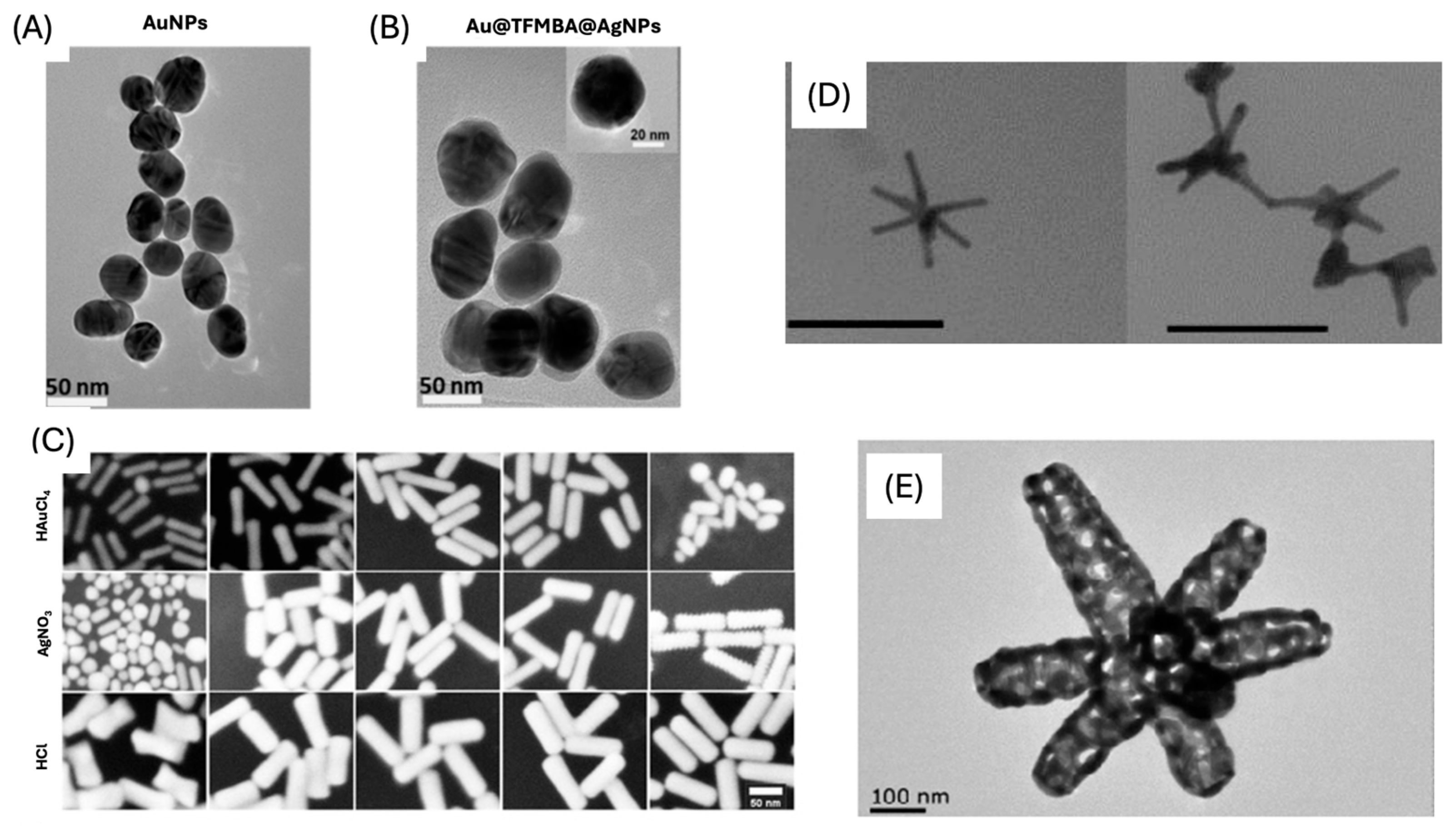
Colloidal gold nanoparticles (AuNPs) are a widely used plasmonic substrate for SERS nanotags (Figure 3A). Their popularity is due to the simple and reproducible synthesis, the chemical stability/inertness of gold, and the well-established methods available to control particle size and surface chemistry [28]. Typically, spherical AuNPs are synthesized by reducing tetrachloroaurate with trisodium citrate dihydrate, a method that allows for fine control over particle size through the stoichiometry of the reducing agent, reaction times, and temperature. Because of these favorable characteristics, AuNPs have been applied extensively in a variety of biophotonic applications, including SERS biosensing [29]. While gold is the most employed material, silver nanoparticles (AgNPs) also offer exceptionally strong plasmonic activity and can produce higher electromagnetic enhancement factors due to their sharper plasmon resonances. AgNPs also have quick, reliable, and tunable syntheses, making them preferable SERS substrates [30]. However, AgNPs are generally less chemically stable than AuNPs, tending to oxidize under ambient or biological conditions, which can alter their optical properties and limit their long-term use.
2.2. Anisotropic Nanoparticles
The chemical, optical, and electrical characteristics of NPs can change depending on their form. Beyond simple spheres, researchers have designed nanostructures with diverse morphologies, such as nanostars, nanorods, nanocubes, and nanoprisms [31]. Non-spherical NPs are considered anisotropic nanostructures (Figure 3). The properties of anisotropic NPs vary depending on the shape of the NPs, in contrast to spherical NPs, which have homogeneous qualities in all directions [31]. Among these anisotropic structures, nanostars (NSs) are a widely used substrate due to the fact that they have been shown to possess higher SERS enhancement capabilities compared to spherical NPs [32]. This increased enhancement is due to the strong electromagnetic field around the tips of the star, indicating more intense “hot spot” areas. Structures featuring sharp edges, tips, or interparticle gaps can generate stronger localized electromagnetic fields and create highly efficient SERS hotspots [33]. Another common type of anisotropic structure used is gold nanorods (AuNRs). These structures are unique because they have dual-mode LSPR maxima, arising from the transverse (perpendicular to the long axis) and longitudinal (along the long axis) plasmon modes, which are tunable through changing the aspect ratio (ratio of length to diameter) of the nanorods [34].
Figure 3.
TEM images of (A) Au NPs and (B) Au@TFMBA@Ag NPs [35]. (C) SEM images of AuNRs [36]. (D) TEM images of the AuNS [32]. (E) TEM image of a typical Au nanocage [37]. (A,B) were adapted from Ref. [35]. (C) adapted with permission from Ref. [36]. (D) adapted from Ref. [32]. (E) adapted from Ref. [37].
2.3. Core-Shell Nanoparticles
Synthesis of core-shell nanoparticles is a common element in the development of SERS nanotags. Core-shell morphologies allow for RRMs bound to the core nanoparticle to be enhanced through the exploitation of the dual plasmonic effect, which arises when the LSPR from two metals overlap and hybridize to form an intense electric field between the two metal surfaces [38]. Additionally, embedding RRMs between the core and the shell can minimize desorption from the surface and help maintain the SERS signal for nanotags over extended periods of time [39]. Another common core-shell nanoparticle geometry is the combination of a metal shell with iron oxide core nanoparticles, which allows for the magnetic properties of iron oxide to be combined with the plasmonic properties of the metal shell [37]. Analytes of interest are bound to the surface of the magnetic nanoparticles, and then magnets are used to pull the analyte-bound nanotags from solution. Through this separation and isolation of the analytes, the limit of detection (LOD) can be lowered. To further lower LODs, nanoparticles are coated in silica, which protects adsorbed RRMs and prevents their dissociation from the metal surface, stabilizing the nanoparticles and resulting in increased SERS signal [25].
2.4. Supported Nanoparticles
Supported nanoparticles form a major class of SERS substrates in which plasmonic metals are immobilized on higher-dimensional materials to create stable, high-density hotspots for sensitive molecular detection. Graphene oxide (GO) is a 2D carbon structure to which AuNPs are easily attached (GO@AuNP) and are a common example of a type of supported morphology. GO provides a stable surface that reduces nanoparticle aggregation and increases SERS enhancement due to the electronic interaction between the surface plasmons and pi-conjugated domains of GO [40]. There are many other 2D and 3D structures conjugated with NPs that have been used to increase stability, optimize electrochemical enhancement, or facilitate analyte detection, such as metal-organic frameworks (MOFs), carbon-dots, polymers, paper, etc. [41].
Here, we summarize the major classes of plasmonic nanostructures commonly employed for SERS sensing. Rational control over nanoparticle shape, size, and interparticle spacing is essential for optimizing electromagnetic enhancement and overall sensing performance. Although precise experimental tuning of these parameters remains challenging, recent progress in computational modeling, high-throughput synthesis, and data-driven analysis has enabled more predictive design of plasmonic architectures with tailored optical properties. Machine-learning approaches trained on simulated and experimental datasets are now being used to uncover structure–property relationships, optimize synthetic conditions, and guide the fabrication of nanostructures with user-defined plasmonic responses [42]. Together, these advances offer a powerful route toward intelligently tunable, application-specific SERS substrates.
3. Raman Reporter Molecules
In SERS nanotags, signals from Raman reporter molecules (RRMs) are critical for the indirect quantification of target analytes. RRMs are chosen based on several key characteristics: strong and unique Raman peaks, a large Raman scattering cross-section, chemical stability, and compatibility with nanoparticle surface chemistry. When multiple nanotags bind to a target, the proximity of their plasmonic nanostructures generates SERS “hotspots,” dramatically enhancing the Raman signal of the attached reporter molecules. The intensity of the RRM correlates directly with the amount of target present, enabling quantitative analysis through calibration curves that relate RRM intensity or peak area to analyte concentration [25]. Selecting an appropriate RRM is therefore essential, as it determines signal strength, spectral reproducibility, and assay sensitivity. RRMs used in nanotags generally fall into two major categories: dye-based and non-dye-based molecules. A full table of common RRMs along with their structures is provided in Table 1.
Table 1.
Table of common RRMs, the IUPAC names, and respective structures.
Optimal SERS enhancement occurs when RRMs form a uniform monolayer or sub-monolayer on the nanoparticle surface. This means that during nanotag fabrication, parameters influencing surface monolayer formation are particularly critical. These include reporter concentration, incubation time, solvent composition, pH, ionic strength, and the presence of competing ligands or stabilizers (e.g., citrate, CTAB, PVP), all of which affect surface coverage, molecular orientation, and packing density [43,44,45]. Reporter molecules containing thiol (–SH) and amine (–NH2) groups are often favored due to their strong binding affinity for gold and silver nanoparticles, enabling stable attachment and controlled monolayer formation [43]. Incomplete ligand exchange, multilayer formation, and desorption under physiological conditions can all introduce spectral variability and batch-to-batch inconsistencies in SERS sensors [46,47]. It is essential to optimize co-absorption strategies with the selected targeting ligand to ensure optimal signal intensity is balanced with colloidal stability and ligand accessibility [19].
3.1. Dye RRMs
Dye molecules and aromatic chromophores are commonly used as RRMs because they possess extended conjugated π-electron systems, which produce strong Raman scattering due to increased molecular polarizability. These dyes exhibit intense, well-defined vibrational modes and often are in resonance with the excitation wavelength, further enhancing the SERS signals. Dye RRMs are valued for their high sensitivity, distinct spectral features, and robust signal output, making them especially suitable for indirect detection formats in which the RRM serves as the quantifiable element of the assay. Common dye RRMs include Cyanine 5 (Cy5), malachite green (MG), Rhodamine 6G (R6G), and Nile Blue A (NBA). Dye RRMs can operate bifunctionally, as both reporter molecules and targeting ligands. For example, Li et al. employed a modified Rhodamine B structure to bind and detect Hg2+ ions in contaminated milk samples [48].
3.2. Non-Dye RRMs
Non-dye RRMs derive their Raman activity from specific chemical moieties, such as thiols, amines, nitro groups, nitriles, and aromatic rings. These structures generate strong, characteristic Raman peaks. These RRMs provide highly reproducible spectral signatures and can be chemically tailored to serve additional roles. In many designs, non-dye reporters act as bifunctional linkers: one functional group binds the nanoparticle surface, while another reacts with targeting ligands such as antibodies, peptides, or aptamers. This dual functionality simplifies fabrication and ensures a well-defined geometry between the plasmonic core, the reporter, and the biorecognition element. For example, the Porter group at the University of Utah has popularized the use of the DSNB molecule (Figure 4) as a bifunctional linker where the thiol group binds to the metal surface, the nitro group provides a strong and distinct Raman signal, and an antibody is covalently attached using the succinimide group [49].
Figure 4.
Scheme of how DSNB functions as a reporter and a linker molecule to covalently attach antibodies to gold nanoparticles. The arrows indicate the reaction pathway to covalently link an antibody to a gold nanoparticle using DSNB. [49]. Reproduced with permission from Ref. [49].
3.3. Multiplexing RRMs
Multiplex SERS employs two or more RRMs with non-overlapping vibrational signatures to enable the simultaneous detection of multiple analytes. Each RRM is incorporated into a distinct nanotag population, and their combined spectra allow for parallel identification/quantification from a single measurement. Effective multiplexing requires RRMs that provide strong, narrow, and well-separated peaks that remain distinguishable even in complex samples. Sometimes chemometric approaches such as PCA combined with partial least squares regression (PLS-R) can be used to separate overlapping features and enhance the reliability of multiplex detection. Kearns et al. developed a multiplex approach with three unique RRMs on SERS tags to detect three different pathogenic bacteria with PCA (Figure 5) [24]. Other authors also used this multiplexed RRMs technique to detect multiple analytes for cancer [50,51,52,53,54,55], cardiovascular diseases [56,57,58,59], and traumatic brain injury [60,61]. Multiplexed RRMs significantly expand the capabilities of SERS nanotags, offering higher information density, improved diagnostic throughput, and the ability to analyze multiple biomarkers or chemical targets simultaneously.
Figure 5.
(a) Multiplex SERS using three unique RRMs (PPY, MGITC, DACITC) to distinguish three species of bacteria (MRSA, E. coli, and S. typh) visually, (b) Principal component analysis for the three species, and (c) difference of AOC of the characteristic peak between control and triplex [24]. Reproduced with permission from Ref. [24].
4. Targeting Ligand
Choosing an effective target ligand while designing an SERS nanotag is another important step in the nanotag synthesis process. The type of targeting ligand (antibody, aptamer, or other) is key in determining the specificity and sensitivity of the nanotag. Other considerations include selecting the proper conjugation method, stabilization method, and assay design to match the desired targeting ligand. All these considerations must be combined and optimized to create a nanotag that is specific and effective for detecting the target analyte.
4.1. Antibody
Antibodies are widely used ligands for SERS nanotags. Antibodies are y-shaped proteins that have high affinity and specificity for antigens. Antibodies are produced by many organisms as part of their adaptive immune system against pathogens, such as viruses or bacteria. However, advances in microbiology and molecular biology have allowed scientists to generate antibodies against almost any target of interest [62]. Antibodies have distinct binding pockets that utilize non-covalent interactions to form a very specific interaction with an antigen. Due to this specificity, multiple antibodies can bind to the same antigen at different sites, allowing for an antigen to be “sandwiched” between two antibodies. For SERS nanotags, IgG-type antibodies are typically used; however, some groups have explored using antibody variants, such as single-chain variable fragments (scFv) or nanobodies, as targeting ligands [63,64].
4.2. Aptamer
Aptamers are short, single-stranded DNA or RNA sequences that fold into unique three-dimensional structures capable of binding molecular targets with high affinity and specificity. Unlike antibodies, which possess fixed tertiary structures, aptamers adopt multiple conformations depending on their local environment, and this dynamic folding enables selective recognition through a combination of hydrogen bonding, electrostatic interactions, and van der Waals forces [65]. Aptamers are relatively easy to generate using the Systematic Evolution of Ligands by Exponential Enrichment (SELEX) process, an iterative in vitro selection method in which large randomized nucleic-acid libraries are enriched toward sequences that bind a desired target. Compared with antibodies, aptamers offer several advantages, including low-cost chemical synthesis, excellent batch-to-batch reproducibility, long-term stability, and the ability to regenerate their binding activity after denaturation [65]. For SERS nanotag construction, aptamers are commonly conjugated to plasmonic nanostructures through the incorporation of thiol-modified oligonucleotides into the aptamer sequence for conjugation via gold-sulfur bonding. Other conjugation approaches are very similar to antibody conjugation strategies, including covalent binding through EDC (1-ethyl-3-(-3-dimethylaminopropyl) carbodiimide hydrochloride) and NHS (N-hydroxysuccinimide) coupling chemistry. Despite their benefits, aptamers can be limited by susceptibility to nuclease degradation and by the sensitivity of their binding performance to environmental factors that alter their tertiary structure. Aptamer design and generation have been enhanced by advances in computational approaches. For example, Quarin et al. utilized an automated computational program to systemically improve their aptamer-based catalytic sensor for SERS-based detection of SARS-CoV-2 and malaria genetic biomarkers [66].
4.3. Other Targeting Ligands
While antibody and aptamer-based nanotags are extremely common and most prevalent for the scope of this work, there are other types of targeting ligands that can be used to bind and indirectly detect target analytes. One emerging targeting ligand is the molecularly imprinted polymer (MIP), which is a highly stable synthetic polymer engineered with a specific binding pocket to mimic an antibody [67]. Another major class includes non-antibody proteins like enzymes, receptor proteins, and binding proteins that can bind selectively to cell receptors, metal ions, and other biologically relevant targets. Finally, host–guest ligands are supramolecular molecules (e.g., cyclodextrins, cucurbiturils, crown ethers) that selectively bind target analytes through size- and shape-complementary noncovalent inclusion interactions, enabling stable and reversible recognition for indirect SERS detection. These alternative targeting ligands have been mentioned here to provide a broad overview of possible ligands; however, they are not commonly applied to the indirect sensing applications detailed in this work.
4.4. Conjugation Strategies
The conjugation of targeting ligands to plasmonic nanostructures is a fundamental step in SERS nanotag fabrication, as it determines biological specificity, as well as influencing colloidal stability and signal reliability. The selected ligand must preserve both the optical enhancement properties of the plasmonic core and the functional integrity of the Raman reporter molecules. Inadequate or poorly controlled ligand conjugation can result in reduced binding efficiency, substrate aggregation, or attenuation of electromagnetic enhancement due to excessive interfacial spacing. Accordingly, multiple strategies have been developed to balance stability, control over ligand orientation, and reproducibility. The following sections summarize and compare the primary conjugation approaches used in SERS nanotag synthesis for both antibodies and aptamer-type ligands.
4.4.1. Direct Adsorption
The simplest way to conjugate a plasmonic substrate with antibodies is through direct adsorption via electrostatic interactions [68]. In direct adsorption, the positively charged region of the antibodies interacts with the negatively charged surface of a nanoparticle, which arises from the reducing agent used during nanoparticle synthesis (such as trisodium citrate) [28]. Additionally, thiol bonds between the cysteine groups within antibodies and the Au or Ag nanoparticle surface can react, thereby impacting adsorption. To aid in the absorption of antibodies to the nanoparticle surface, pH can be adjusted (Figure 6) to maintain the negative charge of the nanoparticle surface and facilitate electrostatic interactions [69].
Figure 6.
Figure showing how pH impacts antibody orientation during direct conjugation with AuNPs [69]. Reproduced with permission from Ref. [69].
4.4.2. Linker Aided Direct Adsorption
Another method for conjugation of antibodies to the surface of nanostructures is through a linker molecule. Linker molecules can be homobifunctional or heterobifunctional. Homobifunctional linkers have the same reactive moiety on each side of the molecule, while heterobifunctional linkers have different reactive moieties on each side of the molecule. DSP [3,3′-dithiobis(succinimidylproprionate)] (Figure 7), sometimes also denoted as DTSP, has been used as a homobifunctional linker to conjugate antibodies to reporter-labeled nanoparticles. The NHS ester moieties react with amine groups of lysines within antibodies to form stable amide bonds. The thiol groups adsorb to the surface of nanoparticles via Au/Ag-S bonds. Bashir et al. adsorbed DSP to mesoporous AuNPs to conjugate anti-PLAP for ovarian cancer [70]. DSP has similarly been used to conjugate different antibodies to AuNPs [71] and Au@AgNPs [35]. DTSSP [3,3′-dithiobis(sulfosuccinimidylproprionate)] (Figure 7) is a water-soluble homobifunctional linker alternative to DSP. The sulfo-succinimidyl esters react with amines in the lysine groups in antibodies to create a stable amide bond. Antibodies are first incubated with DTSSP to form the amide bond, and then the di-thiol groups adsorb to the nanoparticle surface. Wang et al. activated multiple antibodies with DTSSP, which were then adsorbed to AuNPs [50]. Other groups have similarly utilized DTSSP with antibodies to conjugate reporter labeled AuNPs [72].
Figure 7.
Structure of DSP and DTSSP. Structures generated in ChemDraw (ChemDraw Professional 22.2).
4.4.3. Covalent Conjugation Strategy
Linker molecules can be modified via an EDC/NHS crosslinking coupling reaction; the mechanism is shown in Figure 8. EDC [1-ethyl-3-(-3-dimethylaminopropyl) carbodiimide hydrochloride] reacts with carboxylic acid moieties to form an unstable o-acylisourea intermediate. NHS (N-hydroxysuccinimide) and its water-soluble alternative sulfo-NHS react with the unstable intermediate to form an amine-reactive NHS (or sulfo-NHS) ester that readily reacts with amines to form stable amide bonds. When preparing EDC and sulfo-NHS solutions, allow the powders to equilibrate to room temperature; they also hydrolize rapidly in water, so solutions should be used within five minutes of preparation. The linker molecule must contain a carboxylic acid moiety and a moiety that adsorbs to gold/silver, such as sulfhydryl groups. The most common linker used to conjugate antibodies to nanostructures is HS-PEG-COOH [73,74,75].
Figure 8.
Scheme outlining EDC/NHS coupling reaction between nanoparticles and antibodies [76]. Adapted from Ref. [76].
Wang et al. adsorbed 11-mercaptoundecanoic acid (MUA) on AuNPs, activated the carboxyl groups with EDC/sulfo-NHS, and covalently coupled detection aptamers for different exosomes [52]. Others similarly used MUA or mixed MUA/PEG-COOH layers on AuNPs, followed by EDC/NHS activation to attach multiple antibodies to reporter encoded tags [51,77]. Additionally, silica-encapsulated Au nanostars were functionalized with 3-(triethoxysilyl) propyl succinic anhydride (TEPSA) to introduce surface carboxyl groups, which were then activated with EDC/sulfo-NHS to immobilize two aptamers [78]. Other strategies included coating Au@Ag-Au nanogap particles with a PEG-COOH thiol, activating the termini with EDC/NHS, and conjugating detection antibodies for digital SERS quantification [79] or first modifying carboxylated magnetic beads or Ag-Au nanoflowers with EDC/NHS before grafting capture and detection antibodies [54,80]. In core-shell systems, an ultrathin silica shell was grown on Au@Ag nanoparticles, aminated with APTES buffer, carboxylated with succinic anhydride, and finally incorporated EDC/sulfo-NHS chemistry to bind six different antibodies on a seven-channel ovarian cancer platform [81].
Covalent conjugation of bio-recognition elements of SERS nanotags has largely replaced non-covalent physisorption in the diagnostics platforms owing to its superior stability, orientation control and reproducibility. Although simple adsorption is still used for rapid proof-of-concept studies [82,83], it is prone to ligand desorption in serum, random antibody orientation, and high batch-to-batch variability. In contrast, covalent strategies based on thiol-Au bonds [68,74,84], EDC/NHS amide coupling [50,51,54,77,78,79,80] or DSP/DTSSP crosslinkers [50,51,81] provide robust linkage that retains >90–95% activity after weeks of storage and exposure to undiluted biofluids [53,68,81], while heterobifunctional spacers enable controlled Fc-region orientation that routinely improves functional affinity two-to four-fold. These features yield inter-batch variation typically below 8–10%, a prerequisite for the quantitative clinical cohorts [35,70,72,81,84,85]. Additional polydopamine or silica encapsulation after covalent attachment further enhances antifouling properties [68,73]. Consequently, covalent conjugation has become the standard for transforming SERS nanotags into reliable, clinically translatable probes for biomarker detection.
4.5. Stabilization
Uncoated nanotags have multiple limitations, including aggregation, toxicity, oxidation, instability, and non-specific binding of biomolecules to the surface. Any of these can impact the in vitro or in vivo applications. Protective surface coatings, such as silica, polymers, and biomolecules, have been utilized to minimize the limitations created using plasmonic nanostructures. In this review, we focus on two stabilizing agents: bovine serum albumin (BSA) and polyethylene glycol (PEG). These are the two most widely adopted and representative stabilization strategies in SERS nanotag design. BSA and PEG offer distinct advantages depending on the intended application of the SERS nanotag.
4.5.1. BSA
Bovine serum albumin (BSA) is one of the most highly used molecules to prevent non-specific binding and aggregation of nanoparticles, while also increasing biocompatibility for SERS nanotags. BSA is a plasma protein, similar in structure to human serum albumin, that maintains extracellular fluid volume in the human body.
BSA serves as a universal stabilizing and blocking agent in virtually all SERS nanotag syntheses for diagnostic purposes. Added at 1–10% after reporter labeling and antibody/aptamer conjugation, BSA rapidly adsorbs onto any remaining bare Au/AgNPs surfaces, forming a protective protein corona that prevents aggregation in high-ionic-strength media and undiluted blood-based samples through combined electrostatic and steric repulsion. This simple step, explicitly used by many researchers, dramatically reduces non-specific binding (>90% background suppression), maintains colloidal stability for weeks to months, and ensures reproducible SERS performance in clinical samples, making BSA passivation an essential final protocol for translating nanotags into robust blood-based assays [51,61,81,86,87,88]. The volume and concentration are optimized based on the nanotag application. Zheng et al. compared AuNPs-RRM with and without a BSA surface coating over a range of pH values [89]. In Figure 9, AuNPs-RRM with BSA coating from pH 4–9 showed the red-wine color that is indicative of AuNP stability.
Figure 9.
Difference between AuNP-RRM with (AMB) and without BSA coating (AM) in PBS over the pH range 4–9 [89]. Reprinted with permission from Ref. [89].
4.5.2. PEG
Another common stabilizing agent is polyethylene glycol (PEG). PEG is a polymer that consists of a large chain of repeating ether subunits. Most commonly, PEG groups with thiol groups attached are used to promote ease of conjugation. Similarly, PEG derivatives with amine or carboxyl groups increase covalent surface modification in nanoparticles. The hydrophilic polyether backbone creates electrostatic protection, which prevents protein adsorption that is common in uncoated nanoparticles in physiological environments [90]. Additionally, the ether oxygen atoms form hydrogen bonds with surrounding water molecules, creating a hydration shell [91]. This shell helps prevent nanoparticles from interacting with other biological components, ultimately decreasing protein adsorption on the surface of the nanoparticle. This protection helps prevent aggregation of the nanotags, as well as increasing specificity towards the target molecule by limiting off-target binding. Thus, PEG is a vital stabilizing agent that provides many benefits in vitro and in vivo diagnostic applications.
Between these two stabilizing agents, BSA is widely favored for in-vitro diagnostic platforms due to its simplicity, low cost, and rapid adsorption-based blocking, which effectively suppresses aggregation and non-specific binding under physiological buffer conditions. In contrast, PEG provides covalently anchored, chemically defined surface modification with superior antifouling properties and long-term colloidal stability, making it more suitable for highly complex matrices and potential in vivo applications.
7. Conclusions
This review seeks to summarize SERS nanotags and their use as sensitive, versatile, and customizable diagnostic sensors. The three main components of the nanotag, the plasmonic core, the Raman reporter molecule, and the targeting ligand, must be carefully integrated to achieve optimal performance in biomedical sensing applications. Successful nanotag development requires thorough consideration of multiple interdependent factors, including the selection of plasmonic materials and geometries for maximum enhancement, the choice of reporter molecules with distinct spectral signatures for multiplexing capabilities, and the functionalization strategies that ensure stability and sensitivity in complex biological environments.
Recent advances in SERS nanotags include using machine learning and computational modeling to create nanostructures with controlled optical properties and to optimize assay conditions with unprecedented precision. In addition, the integration of SERS nanotags into practical diagnostic platforms, particularly lateral flow assays and microfluidic devices, demonstrates their potential to bridge the gap between laboratory research and clinical point-of-care testing. Despite significant progress, several challenges remain. Standardization of nanotag synthesis and characterization protocols is needed to improve the reproducibility of SERS nanotags across laboratories. Also, long-term stability of SERS nanotags and the scalability of production are hurdles still to be fully addressed.
Looking forward, SERS nanotags are poised to play an increasingly important role in a wide range of biomedical fields. Bringing together spectroscopy and nanotechnology has created opportunities for developing next-generation diagnostic tools that are not only highly sensitive and specific but also accessible and affordable. As this field continues to mature, collaborative efforts between chemists, biologists, clinicians, and engineers will be essential to fully realize the potential of SERS nanotags and to address the growing demand for rapid, multiplexed diagnostic solutions in healthcare and many other fields.
Author Contributions
Conceptualization, N.P., U.B. and B.S.; Writing—Original Draft Preparation, N.P., U.B., N.A., A.O. and K.J.L.; Writing—Review and Editing, N.P., U.B., K.J.L. and B.S.; Supervision, B.S.; Project Administration, B.S. All authors have read and agreed to the published version of the manuscript.
Funding
This research received no external funding.
Data Availability Statement
No new data were created or analyzed in this study. Data sharing is not applicable to this article.
Conflicts of Interest
The authors declare no conflicts of interest.
Abbreviations
The following abbreviations are used in this manuscript:
| 2-MPY | 2-Mercaptopyridine |
| 2-NAT | 2-Naphthalenethiol |
| 2-NT | 2-Nitrothiophenol |
| 3-MPBA | 3-Mercaptophenylboronic acid |
| 4-ATP | 4-Aminothiophenol (para-aminothiophenol) |
| 4-MBA | 4-Mercaptobenzoic acid |
| 4-MBN | 4-Mercaptobenzonitrile |
| 4-MP | 4-Mercaptophenol |
| 4-MPA | 4-Mercaptopropionic acid |
| 4-MPBA | 4-Mercaptophenylboronic acid |
| 4-MPY | 4-Mercaptopyridine |
| 4-NBT | 4-Nitrobenzenethiol (4-nitrothiophenol) |
| 4-NTP | 4-Nitrothiophenol (para-nitrothiophenol) |
| ACS | Acute coronary syndrome |
| ACS-SCD | Acute coronary syndrome–sudden cardiac death |
| AD | Alzheimer’s disease |
| AFP | Alpha-fetoprotein |
| AFB1 | Aflatoxin B1 |
| Ag | Silver |
| AgNPs | Silver nanoparticles |
| AMI | Acute myocardial infarction |
| APP | Amyloid precursor protein |
| APTES | (3-Aminopropyl)triethoxysilane |
| AST | Aminotransferase |
| Au | Gold |
| Au@Ag NPs | Gold core–silver shell nanoparticles |
| AuNPs | Gold nanoparticles |
| AuNS | Gold nanostars |
| Aβ | Amyloid-beta |
| BDH1 | 3-Hydroxybutyrate dehydrogenase 1 |
| BRCA1 | Breast cancer gene 1 |
| BRCA2 | Breast cancer gene 2 |
| BPE | 1,2-Bis(4-pyridyl)ethylene |
| BSA | Bovine serum albumin |
| CA125 | Cancer antigen 125 |
| CAD | Coronary artery disease |
| CAP | Chloramphenicol |
| Cas12a | CRISPR-associated protein 12a |
| CEA | Carcinoembryonic antigen |
| CHF | Congestive heart failure |
| CK-MB | Creatine kinase MB |
| CLDN3 | Claudin-3 |
| cTnT | Cardiac troponin T |
| CRISPR-Cas9 | Clustered regularly interspaced short palindromic repeats–Cas9-based assay |
| Cy3 | Cyanine 3 |
| Cy5 | Cyanine 5 |
| DACITC | Diacetyl indocarbocyanine isothiocyanate |
| DCT | 3,5-Dichlorobenzenethiol |
| DON | Deoxynivalenol |
| DSP | 3,3′-Dithiobis(succinimidyl propionate) |
| DSNB | Disulfide nitrobenzoic acid derivative |
| DTNB | 5,5′-Dithiobis(2-nitrobenzoic acid) |
| DTSP | 3,3′-Dithiobis(sulfosuccinimidyl propionate) |
| EDC | (1-Ethyl-3-(3-dimethylaminopropyl) carbodiimide hydrochloride |
| EGFR | Epidermal growth factor receptor |
| ELISA | Enzyme-linked immunosorbent assay |
| EpCAM | Epithelial cell adhesion molecule |
| EFA1 | Eukaryotic translation elongation factor 1 alpha 1 |
| FB1 | Fumonisin B1 |
| FGR | Fetal growth restriction |
| FOLR1 | Folate receptor alpha |
| g-CN | Carbon nitride |
| GFAP | Glial fibrillary acidic protein |
| GO | Graphene oxide |
| GPC1 | Glypican-1 |
| HER2 | Human epidermal growth factor receptor 2 |
| HITC | Hexamethyl-indotricarbocyanine iodide |
| HPV | Human papillomavirus |
| IgG | Immunoglobulin G |
| IL-6 | Interleukin-6 |
| IL-10 | Interleukin-10 |
| IR808 | Indocyanine near-infrared dye derivative |
| LDHB | Lactate dehydrogenase B |
| LFA | Lateral flow assay |
| LNGFR | Low-affinity nerve growth factor receptor |
| LOD | Limit of detection |
| LPS | Lipopolysaccharide |
| LSPR | Localized surface plasmon resonance |
| ManLAM | Mannose-capped lipoarabinomannan |
| MBA | Mercaptobenzoic acid |
| MCAM | Melanoma cell adhesion molecule |
| MCSP | Melanoma-associated chondroitin sulfate proteoglycan |
| MGDTNB | Malachite green and DTNB combination |
| MGITC | Malachite green isothiocyanate |
| MI | Myocardial infarction |
| MIF | Macrophage migration inhibitory factor |
| MIP | Molecularly imprinted polymer |
| MMC | Malachite green carbinol |
| MMP-9 | Matrix metalloproteinase-9 |
| MMP-13 | Matrix metalloproteinase-13 |
| MMTAA | Mercaptomethylthioacetic acid |
| MOFs | Metal-organic frameworks |
| MPY | Mercaptopyridine |
| MUC16 | Mucin-16 |
| NBA | Nile Blue A |
| NB | Nile Blue |
| NBT | 4-Nitrobenzenethiol |
| NHS | N-Hydroxysuccinimide |
| NIR | Near-infrared |
| NSE | Neuron-specific enolase |
| NS1 | Non-structural protein 1 |
| NSs | Nanostars |
| NPs | Nanoparticles |
| NR | Nanorods |
| NT-proBNP | N-terminal pro-B-type natriuretic peptide |
| OTA | Ochratoxin A |
| Ox-TMB | Oxidized tetramethylbenzidine |
| PB | Prussian Blue |
| PCA | Principal component analysis |
| PCTP | Pentachlorothiophenol |
| PD-L1 | Programmed death-ligand 1 |
| PEG | Polyethylene glycol |
| PI3K | Phosphoinositide 3-kinase |
| PLAP | Placental alkaline phosphatase |
| PLS-R | Partial least squares regression |
| ppb | Parts per billion |
| PP | Polypropylene |
| PPY | Polypyrrole |
| PS | Polystyrene |
| PSMA | Prostate-specific membrane antigen |
| Pt | Platinum |
| PVC | Polyvinyl chloride |
| R6G | Rhodamine 6G |
| RRMs | Raman reporter molecules |
| RS | Raman spectroscopy |
| Ru(bpy)32+ | Tris(2,2′-bipyridine)ruthenium(II) |
| SERS | Surface-enhanced Raman spectroscopy |
| SERS-LFA | SERS-based lateral flow assay |
| SERS-LFIA | SERS-based lateral flow immunoassay |
| SERS-PLFS | SERS paper lateral flow strip |
| SP10 | Testis-specific acrosomal protein |
| SpIA | S-layer protein A |
| TBI | Traumatic brain injury |
| TEPSA | 3-(Triethoxysilyl)propyl succinic anhydride |
| TFMBA | 2,3,5,6-Tetrafluoro-4-mercaptobenzoic acid |
| THI | Thionine |
| TMV | Tobacco mosaic virus |
| TNF-α | Tumor necrosis factor-alpha |
| TSA | Thiosalicylic acid |
| TTC | Tetracycline |
| t-PSA | Total prostate-specific antigen |
| ToxB | Host selective toxin |
| UCH-L1 | Ubiquitin carboxyl-terminal hydrolase L1 |
| UTI | Urinary tract infection |
| VFA | Vertical flow assay |
| ZEN | Zearalenone |
References
- Becatti, M.; Cho, W.C. Editorial: Insights in Molecular Diagnostics and Therapeutics: 2022. Front. Mol. Biosci. 2023, 10, 1209844. [Google Scholar] [CrossRef] [Scilit] [PubMed]
- Bustin, S.A.; Jellinger, K.A. Advances in Molecular Medicine: Unravelling Disease Complexity and Pioneering Precision Healthcare. Int. J. Mol. Sci. 2023, 24, 14168. [Google Scholar] [CrossRef] [Scilit] [PubMed]
- Alowais, S.A.; Alghamdi, S.S.; Alsuhebany, N.; Alqahtani, T.; Alshaya, A.I.; Almohareb, S.N.; Aldairem, A.; Alrashed, M.; Bin Saleh, K.; Badreldin, H.A.; et al. Revolutionizing Healthcare: The Role of Artificial Intelligence in Clinical Practice. BMC Med. Educ. 2023, 23, 689. [Google Scholar] [CrossRef] [Scilit]
- Esteva, A.; Robicquet, A.; Ramsundar, B.; Kuleshov, V.; DePristo, M.; Chou, K.; Cui, C.; Corrado, G.; Thrun, S.; Dean, J. A Guide to Deep Learning in Healthcare. Nat. Med. 2019, 25, 24–29. [Google Scholar] [CrossRef] [Scilit]
- Johnson, K.B.; Wei, W.Q.; Weeraratne, D.; Frisse, M.E.; Misulis, K.; Rhee, K.; Zhao, J.; Snowdon, J.L. Precision Medicine, AI, and the Future of Personalized Health Care. Clin. Transl. Sci. 2021, 14, 86–93. [Google Scholar] [CrossRef] [Scilit]
- Hu, J.; Dun, G.; Geng, X.; Chen, J.; Wu, X.; Ren, T.L. Recent Progress in Flexible Micro-Pressure Sensors for Wearable Health Monitoring. Nanoscale Adv. 2023, 5, 3131–3145. [Google Scholar] [CrossRef] [Scilit]
- Kalasin, S.; Surareungchai, W. Challenges of Emerging Wearable Sensors for Remote Monitoring toward Telemedicine Healthcare. Anal. Chem. 2023, 95, 1773–1784. [Google Scholar] [CrossRef] [Scilit]
- Kaur, B.; Kumar, S.; Kaushik, B.K. Novel Wearable Optical Sensors for Vital Health Monitoring Systems—A Review. Biosensors 2023, 13, 181. [Google Scholar] [CrossRef] [Scilit]
- Vo, T.S.; Hoang, T.; Vo, T.T.B.C.; Jeon, B.; Nguyen, V.H.; Kim, K. Recent Trends of Bioanalytical Sensors with Smart Health Monitoring Systems: From Materials to Applications. Adv. Healthc. Mater. 2024, 13, 2303923. [Google Scholar] [CrossRef] [Scilit]
- Wang, C.; He, T.; Zhou, H.; Zhang, Z.; Lee, C. Artificial Intelligence Enhanced Sensors—Enabling Technologies to next-Generation Healthcare and Biomedical Platform. Bioelectron. Med. 2023, 9, 17. [Google Scholar] [CrossRef] [Scilit] [PubMed]
- Zhao, H.; Cao, J.; Xie, J.; Liao, W.H.; Lei, Y.; Cao, H.; Qu, Q.; Bowen, C. Wearable Sensors and Features for Diagnosis of Neurodegenerative Diseases: A Systematic Review. Digit. Health 2023, 9, 20552076231173569. [Google Scholar] [CrossRef] [Scilit]
- Kneipp, K.; Kneipp, H.; Itzkan, I.; Dasari, R.R.; Feld, M.S. Ultrasensitive Chemical Analysis by Raman Spectroscopy. Chem. Rev. 1999, 99, 2957–2976. [Google Scholar] [CrossRef] [Scilit]
- Jones, R.R.; Hooper, D.C.; Zhang, L.; Wolverson, D.; Valev, V.K. Raman Techniques: Fundamentals and Frontiers. Nanoscale Res. Lett. 2019, 14, 231. [Google Scholar] [CrossRef] [Scilit] [PubMed]
- Camp, C.H.; Cicerone, M.T. Chemically Sensitive Bioimaging with Coherent Raman Scattering. Nat. Photonics 2015, 9, 295–305. [Google Scholar]
- McFarland, A.D.; Young, M.A.; Dieringer, J.A.; Van Duyne, R.P. Wavelength-Scanned Surface-Enhanced Raman Excitation Spectroscopy. J. Phys. Chem. B 2005, 109, 11279–11285. [Google Scholar] [CrossRef] [Scilit] [PubMed]
- Sonntag, M.D.; Klingsporn, J.M.; Zrimsek, A.B.; Sharma, B.; Ruvuna, L.K.; Van Duyne, R.P. Molecular Plasmonics for Nanoscale Spectroscopy. Chem. Soc. Rev. 2014, 43, 1230–1247. [Google Scholar] [CrossRef] [Scilit]
- Saviñon-Flores, F.; Méndez, E.; López-Castaños, M.; Carabarin-Lima, A.; López-Castaños, K.A.; González-Fuentes, M.A.; Méndez-Albores, A. A Review on Sers-Based Detection of Human Virus Infections: Influenza and Coronavirus. Biosensors 2021, 11, 66. [Google Scholar] [CrossRef] [Scilit]
- Cialla-May, D.; Bonifacio, A.; Bocklitz, T.; Markin, A.; Markina, N.; Fornasaro, S.; Dwivedi, A.; Dib, T.; Farnesi, E.; Liu, C.; et al. Biomedical SERS—The Current State and Future Trends. Chem. Soc. Rev. 2024, 53, 8957–8979. [Google Scholar] [CrossRef] [Scilit]
- Qian, X.; Peng, X.H.; Ansari, D.O.; Yin-Goen, Q.; Chen, G.Z.; Shin, D.M.; Yang, L.; Young, A.N.; Wang, M.D.; Nie, S. In Vivo Tumor Targeting and Spectroscopic Detection with Surface-Enhanced Raman Nanoparticle Tags. Nat. Biotechnol. 2008, 26, 83–90. [Google Scholar] [CrossRef] [Scilit]
- Chen, B.; Gao, J.; Sun, H.; Zhao, Y.; Liu, Y.; Qiu, X. SERS-Powered Precision: Revolutionizing Therapeutic Drug Monitoring with Nanoscale Sensitivity. Mikrochim. Acta 2025, 192, 826. [Google Scholar] [CrossRef] [Scilit] [PubMed]
- Jaikumar, T.; George, S.; Saju, H.; Raj, R.; Nisarga, R.; Sontakke, S.; Sangshetti, J.; Prakash, J.; Arote, R.B. SERS-Guided Photodynamic Therapy: Pioneering Strategies in Advanced Cancer Diagnosis and Treatment. Discover Nano 2025, 20, 171. [Google Scholar] [CrossRef] [Scilit] [PubMed]
- Owens, N.A.; Pinter, A.; Porter, M.D. Surface-Enhanced Resonance Raman Scattering for the Sensitive Detection of a Tuberculosis Biomarker in Human Serum. J. Raman Spectrosc. 2019, 50, 15–25. [Google Scholar] [CrossRef] [Scilit]
- Ebbah, E.; Amissah, A.; Kim, J.H.; Driskell, J.D. Antibody-Driven Assembly of Plasmonic Core-Satellites to Increase the Sensitivity of a SERS Vertical Flow Immunoassay. ACS Sens. 2024, 9, 3496–3501. [Google Scholar] [CrossRef] [Scilit]
- Kearns, H.; Goodacre, R.; Jamieson, L.E.; Graham, D.; Faulds, K. SERS Detection of Multiple Antimicrobial-Resistant Pathogens Using Nanosensors. Anal. Chem. 2017, 89, 12666–12673. [Google Scholar] [CrossRef] [Scilit]
- Langer, J.; de Aberasturi, D.J.; Aizpurua, J.; Alvarez-Puebla, R.A.; Auguié, B.; Baumberg, J.J.; Bazan, G.C.; Bell, S.E.J.; Boisen, A.; Brolo, A.G.; et al. Present and Future of Surface-Enhanced Raman Scattering. ACS Nano 2020, 14, 28–117. [Google Scholar]
- Marks, H.; Schechinger, M.; Garza, J.; Locke, A.; Coté, G. Surface Enhanced Raman Spectroscopy (SERS) for in Vitro Diagnostic Testing at the Point of Care. Nanophotonics 2017, 6, 681–701. [Google Scholar] [CrossRef] [Scilit]
- Huang, Q.; Guo, H.; Wang, W.; Kang, S.; Vikesland, P.J. Surface-Enhanced Raman Spectroscopy (SERS) Based Biological and Environmental 2D and 3D Imaging. ACS Environ. Au 2025, 5, 342–362. [Google Scholar] [CrossRef] [Scilit]
- Karnwal, A.; Kumar Sachan, R.S.; Devgon, I.; Devgon, J.; Pant, G.; Panchpuri, M.; Ahmad, A.; Alshammari, M.B.; Hossain, K.; Kumar, G. Gold Nanoparticles in Nanobiotechnology: From Synthesis to Biosensing Applications. ACS Omega 2024, 9, 29966–29982. [Google Scholar] [CrossRef] [Scilit]
- Hu, X.; Zhang, Y.; Ding, T.; Liu, J.; Zhao, H. Multifunctional Gold Nanoparticles: A Novel Nanomaterial for Various Medical Applications and Biological Activities. Front. Bioeng. Biotechnol. 2020, 8, 990. [Google Scholar] [CrossRef] [Scilit] [PubMed]
- Hang, Y.; Wang, A.; Wu, N. Plasmonic Silver and Gold Nanoparticles: Shape- and Structure-Modulated Plasmonic Functionality for Point-of-Caring Sensing, Bio-Imaging and Medical Therapy. Chem. Soc. Rev. 2024, 53, 2932–2971. [Google Scholar] [PubMed]
- Hao, E.; Schatz, G.C.; Hupp, J.T. Synthesis and Optical Properties of Anisotropic Metal Nanoparticles. J. Fluoresc. 2004, 14, 331–341. [Google Scholar] [CrossRef] [Scilit] [PubMed]
- Nalbant Esenturk, E.; Hight Walker, A.R. Surface-Enhanced Raman Scattering Spectroscopy via Gold Nanostars. J. Raman Spectrosc. 2009, 40, 86–91. [Google Scholar] [CrossRef] [Scilit]
- Hardy, M.; Oppenheimer, P.G. ‘When Is a Hotspot a Good Nanospot’—Review of Analytical and Hotspot-Dominated Surface Enhanced Raman Spectroscopy Nanoplatforms. Nanoscale 2024, 16, 3293–3323. [Google Scholar]
- Chen, H.; Shao, L.; Li, Q.; Wang, J. Gold Nanorods and Their Plasmonic Properties. Chem. Soc. Rev. 2013, 42, 2679–2724. [Google Scholar] [CrossRef] [Scilit] [PubMed]
- Lu, D.; Chen, Y.; Ke, L.; Huang, Z.; Lu, Y.; Wang, J. Bisulfite-Free Detection of DNA Methylation in Early-Stage Lung Cancer with a Multiplex SERS Immunoassay. Chem. Eng. J. 2025, 503, 158491. [Google Scholar] [CrossRef] [Scilit]
- Sun, J.; Gao, F.; Song, Y.; Wang, M.; Wang, C.; Ni, Q.; Wang, Y.; Yang, M.; Zhao, P.; Sun, B. Paper Lateral Flow Strips Based on Gold Nanorods for Ultrasensitive Detection of Traumatic Brain Injury Biomarkers. ACS Appl. Nano Mater. 2023, 6, 18729–18738. [Google Scholar] [CrossRef] [Scilit]
- Li, D.; Yang, M.; Li, H.; Mao, L.; Wang, Y.; Sun, B. SERS Based Protocol Using Flow Glass-Hemostix for Detection of Neuron-Specific Enolase in Blood Plasma. New J. Chem. 2019, 43, 5925–5931. [Google Scholar] [CrossRef] [Scilit]
- Lin, J.S.; Radjenovic, P.M.; Jin, H.; Li, J.F. Plasmonic Core-Shell Nanoparticle Enhanced Spectroscopies for Surface Analysis. Anal. Chem. 2021, 93, 6573–6582. [Google Scholar] [CrossRef] [Scilit]
- Kong, X.; Yu, Q.; Zhang, X.; Du, X.; Gong, H.; Jiang, H. Synthesis and Application of Surface Enhanced Raman Scattering (SERS) Tags of Ag@SiO2 Core/Shell Nanoparticles in Protein Detection. J. Mater. Chem. 2012, 22, 7767–7774. [Google Scholar] [CrossRef] [Scilit]
- Cao, Y.; Cheng, Y.; Sun, M. Graphene-Based SERS for Sensor and Catalysis. Appl. Spectrosc. Rev. 2023, 58, 1–38. [Google Scholar]
- Yuan, K.; Jurado-Sánchez, B.; Escarpa, A. Nanomaterials Meet Surface-Enhanced Raman Scattering towards Enhanced Clinical Diagnosis: A Review. J. Nanobiotechnol. 2022, 20, 537. [Google Scholar] [CrossRef] [Scilit]
- Masson, J.F.; Biggins, J.S.; Ringe, E. Machine Learning for Nanoplasmonics. Nat. Nanotechnol. 2023, 18, 111–123. [Google Scholar] [CrossRef] [Scilit]
- Love, J.C.; Estroff, L.A.; Kriebel, J.K.; Nuzzo, R.G.; Whitesides, G.M. Self-Assembled Monolayers of Thiolates on Metals as a Form of Nanotechnology. Chem. Rev. 2005, 105, 1103–1169. [Google Scholar] [CrossRef] [Scilit]
- Guerrini, L.; Graham, D. Molecularly-Mediated Assemblies of Plasmonic Nanoparticles for Surface-Enhanced Raman Spectroscopy Applications. Chem. Soc. Rev. 2012, 41, 7085–7107. [Google Scholar] [CrossRef] [Scilit]
- Camden, J.P.; Dieringer, J.A.; Wang, Y.; Masiello, D.J.; Marks, L.D.; Schatz, G.C.; Van Duyne, R.P. Probing the Structure of Single-Molecule Surface-Enhanced Raman Scattering Hot Spots. J. Am. Chem. Soc. 2008, 130, 12616–12617. [Google Scholar] [CrossRef] [Scilit]
- Zong, C.; Xu, M.; Xu, L.J.; Wei, T.; Ma, X.; Zheng, X.S.; Hu, R.; Ren, B. Surface-Enhanced Raman Spectroscopy for Bioanalysis: Reliability and Challenges. Chem. Rev. 2018, 118, 4946–4980. [Google Scholar] [CrossRef] [Scilit] [PubMed]
- Schlücker, S. Surface-Enhanced Raman Spectroscopy: Concepts and Chemical Applications. Angew. Chem.-Int. Ed. 2014, 53, 4756–4795. [Google Scholar] [CrossRef] [Scilit] [PubMed]
- Li, H.; Chen, Q.; Hassan, M.M.; Ouyang, Q.; Jiao, T.; Xu, Y.; Chen, M. AuNS@Ag Core-Shell Nanocubes Grafted with Rhodamine for Concurrent Metal-Enhanced Fluorescence and Surfaced Enhanced Raman Determination of Mercury Ions. Anal. Chim. Acta 2018, 1018, 94–103. [Google Scholar] [CrossRef] [Scilit]
- Grubisha, D.S.; Lipert, R.J.; Park, H.Y.; Driskell, J.; Porter, M.D. Femtomolar Detection of Prostate-Specific Antigen: An Immunoassay Based on Surface-Enhanced Raman Scattering and Immunogold Labels. Anal. Chem. 2003, 75, 5936–5943. [Google Scholar] [CrossRef] [Scilit]
- Wang, J.; Wuethrich, A.; Lobb, R.J.; Antaw, F.; Sina, A.A.I.; Lane, R.E.; Zhou, Q.; Zieschank, C.; Bell, C.; Bonazzi, V.F.; et al. Characterizing the Heterogeneity of Small Extracellular Vesicle Populations in Multiple Cancer Typesviaan Ultrasensitive Chip. ACS Sens. 2021, 6, 3182–3194. [Google Scholar] [CrossRef] [Scilit] [PubMed]
- Han, Z.; Peng, X.; Yang, Y.; Yi, J.; Zhao, D.; Bao, Q.; Long, S.; Yu, S.X.; Xu, X.X.; Liu, B.; et al. Integrated Microfluidic-SERS for Exosome Biomarker Profiling and Osteosarcoma Diagnosis. Biosens. Bioelectron. 2022, 217, 114709. [Google Scholar] [CrossRef] [Scilit]
- Weng, Z.; Zong, S.; Wang, Y.; Li, N.; Li, L.; Lu, J.; Wang, Z.; Chen, B.; Cui, Y. Screening and Multiple Detection of Cancer Exosomes Using an SERS-Based Method. Nanoscale 2018, 10, 9053–9062. [Google Scholar] [CrossRef] [Scilit] [PubMed]
- Ning, C.F.; Wang, L.; Tian, Y.F.; Yin, B.C.; Ye, B.C. Multiple and Sensitive SERS Detection of Cancer-Related Exosomes Based on Gold-Silver Bimetallic Nanotrepangs. Analyst 2020, 145, 2795–2804. [Google Scholar] [CrossRef] [Scilit]
- Zhang, W.; Jiang, L.; Diefenbach, R.J.; Campbell, D.H.; Walsh, B.J.; Packer, N.H.; Wang, Y. Enabling Sensitive Phenotypic Profiling of Cancer-Derived Small Extracellular Vesicles Using Surface-Enhanced Raman Spectroscopy Nanotags. ACS Sens. 2020, 5, 764–771. [Google Scholar] [CrossRef] [Scilit]
- Hou, H.; Zhao, J.; Zuo, Z.; Song, L.; Han, X.X.; Yang, J.; Xu, B.; Zhao, B. SERS Nanotags for Ultrasensitive and Dual Biomarker Detection of Breast Cancer. Sens. Actuators Rep. 2025, 9, 100344. [Google Scholar] [CrossRef] [Scilit]
- Sun, J.; Zhao, Y.; Hou, Y.; Li, H.; Yang, M.; Wang, Y.; Sun, B. Multiplexed Electrochemical and SERS Dual-Mode Detection of Stroke Biomarkers: Rapid Screening with High Sensitivity. New J. Chem. 2019, 43, 13381–13387. [Google Scholar] [CrossRef] [Scilit]
- Jin, X.; Zhang, Y.; Guo, Q.; Deng, B.; Tan, Z.; Liu, F.; Lin, L.; Ye, J.; Xu, H. Multiplexed Lateral Flow Immunoassays Using High Photostability Gap-Enhanced Raman Nanotags: Highly Sensitive, Rapid, Efficient and Portable Point-of-Care Tests. Biosens. Bioelectron. 2025, 278, 117377. [Google Scholar] [CrossRef] [Scilit] [PubMed]
- Wu, Q.; Xiao, Y.; Yang, X.; Zhu, A.; Cao, W.; Cai, L.; Lin, X.; Zhao, Z.; Zhang, Q.; Zhou, X. Magnetic-Assisted and Aptamer-Based SERS Biosensor for High Enrichment, Ultrasensitive Detection of Multicomponent Heart Failure Biomarkers. Talanta 2025, 290, 127834. [Google Scholar] [CrossRef] [Scilit]
- Lee, H.; Kim, W.; Song, M.Y.; Kim, D.H.; Jung, H.S.; Kim, W.; Choi, S. One-Stop Plasmonic Nanocube-Excited SERS Immunoassay Platform of Multiple Cardiac Biomarkers for Rapid Screening and Progressive Tracing of Acute Myocardial Infarction. Small 2024, 20, e2304999. [Google Scholar] [CrossRef] [Scilit] [PubMed]
- Li, J.; Wu, J.; Chen, J.; Huang, S.; Liu, J.; Gao, F.; Wang, Y.; Sun, B.; Yu, D.; Sun, J. Dual Detection of Spinal Cord Injury Biomarkers in Rat Model Using Gold Nanorod Array Substrate Based on Surface-Enhanced Raman Scattering. Surf. Interfaces 2022, 34, 102400. [Google Scholar] [CrossRef] [Scilit]
- Gao, F.; Wang, Y.; Wang, M.; Wang, C.; Wang, T.; Wu, J.; Ni, Q.; Sun, B.; Sun, J.; Wang, Y. Highly Efficient and Rapid Surface-Enhanced Raman Spectroscopy Active Platform Based on Au Nanorods for Ultrasensitive Traumatic Brain Injury Detection. ACS Appl. Nano Mater. 2024, 7, 19883–19893. [Google Scholar] [CrossRef] [Scilit]
- Lipman, N.S.; Jackson, L.R.; Trudel, L.J.; Weis-Garcia, F. Monoclonal Versus Polyclonal Antibodies: Distinguishing Characteristics, Applications, and Information Resources. ILAR J. 2005, 46, 258–268. [Google Scholar] [CrossRef] [Scilit]
- Farokhinejad, F.; Li, J.; Hugo, L.E.; Howard, C.B.; Wuethrich, A.; Trau, M. Detection of Dengue Virus 2 with Single Infected Mosquito Resolution Using Yeast Affinity Bionanofragments and Plasmonic SERS Nanoboxes. Anal. Chem. 2022, 94, 14177–14184. [Google Scholar] [CrossRef] [Scilit] [PubMed]
- Wang, J.; Zhou, Q.; Lowry, K.; Howard, C.B.; Trau, M. Development and Application of Multivalent Nanobody-Functionalized Plasmonic Probes in SERS Sensing Platforms. Biosens. Bioelectron. 2025, 278, 117292. [Google Scholar] [CrossRef] [Scilit]
- Mpofu, K.; Chauke, S.; Thwala, L.; Mthunzi-Kufa, P. Aptamers and Antibodies in Optical Biosensing. Discov. Chem. 2025, 2, 23. [Google Scholar] [CrossRef] [Scilit]
- Quarin, S.M.; MacKe, A.C.; Kissell, L.N.; Kelly, M.S.; Dayananda, A.; Ungvary, J.; Stan, G.; Dima, R.I.; Strobbia, P. Design, Rationalization, and Automation of a Catalytic Sensing Mechanism for Homogeneous SERS Biosensors. ACS Sens. 2023, 8, 2000–2010. [Google Scholar] [CrossRef] [Scilit]
- Wisnuwardhani, H.A.; Ibrahim, S.; Mukti, R.R.; Damayanti, S. Molecularly-Imprinted SERS: A Potential Method for Bioanalysis. Sci. Pharm. 2022, 90, 54. [Google Scholar] [CrossRef] [Scilit]
- Li, T.D.; Zhang, R.; Chen, H.; Huang, Z.P.; Ye, X.; Wang, H.; Deng, A.M.; Kong, J.L. An Ultrasensitive Polydopamine Bi-Functionalized SERS Immunoassay for Exosome-Based Diagnosis and Classification of Pancreatic Cancer. Chem. Sci. 2018, 9, 5372–5382. [Google Scholar] [CrossRef] [Scilit]
- Ruiz, G.; Tripathi, K.; Okyem, S.; Driskell, J.D. PH Impacts the Orientation of Antibody Adsorbed onto Gold Nanoparticles. Bioconjug. Chem. 2019, 30, 1182–1191. [Google Scholar] [CrossRef] [Scilit] [PubMed]
- Bashir, J.; Masud, M.K.; Nugraha, A.S.; Liu, C.H.; Vasanth, A.; Ashok, A.; Hossain, S.M.A.; Ahmed, E.; Pejovic, T.; Morgan, T.; et al. Plasmonic Mesoporous Gold-Based SERS Biosensor for Ovarian Cancer-Derived Extracellular Vesicles. Small 2025, 21, e2401817. [Google Scholar] [CrossRef] [Scilit] [PubMed]
- Zhang, D.; Chen, X.; Lin, J.; Jiang, S.; Fan, M.; Liu, N.; Huang, Z.; Wang, J. Ultrasensitive Detection of Circulating Plasma Cells Using Surface-Enhanced Raman Spectroscopy and Machine Learning for Multiple Myeloma Monitoring. Anal. Chem. 2025, 97, 4101–4110. [Google Scholar] [CrossRef] [Scilit]
- Feng, X.; Zhang, W.; Chang-Hao Tsao, S.; Shen, C.; Hu, L.; Wang, Y. Integrated Chemical Array and SERS Profiling of Plasma Small Extracellular Vesicles for Breast Cancer Diagnosis. Nano Lett. 2025, 25, 15331–15339. [Google Scholar] [CrossRef] [Scilit]
- Sun, S.; Xie, D.; Guan, W.; Li, Q.; He, J.; Li, Y.; Chen, J.; Gao, S.; Liu, Z. Exploring Site-Specific N 6 -Methyladenosine Modifications as New Cancer Biomarkers via SERS-Based Single-Cell Assay and Histopathological Imaging. Anal. Chem. 2025, 97, 16822–16831. [Google Scholar] [CrossRef] [Scilit] [PubMed]
- Zhu, C.; Lin, X.; Jin, R.; Long, C.; Zhu, C.; Zheng, D.; Yang, X.; Zhang, P.; Liu, C. Digital Profiling Subpopulations of Cancer Cell-Derived Exosomes via Aptamer-SERS Nanotags on a Wafer-Level Micropillar Array Chip. ACS Sens. 2025, 10, 6962–6970. [Google Scholar] [CrossRef] [Scilit]
- Sun, J.; Li, W.; Zhu, X.; Jiao, S.; Chang, Y.; Wang, S.; Dai, S.; Xu, R.; Dou, M.; Li, Q.; et al. A Novel Multiplex Mycotoxin Surface-Enhanced Raman Spectroscopy Immunoassay Using Functional Gold Nanotags on a Silica Photonic Crystal Microsphere Biochip. J. Agric. Food Chem. 2021, 69, 11494–11501. [Google Scholar] [CrossRef] [Scilit] [PubMed]
- Conde, J.; Dias, J.T.; Grazú, V.; Moros, M.; Baptista, P.V.; de la Fuente, J.M. Revisiting 30 Years of Biofunctionalization and Surface Chemistry of Inorganic Nanoparticles for Nanomedicine. Front. Chem. 2014, 2, 48. [Google Scholar] [CrossRef] [Scilit] [PubMed]
- Lin, W.; Yuan, L.; Gao, Z.; Cai, G.; Liang, C.; Fan, M.; Xiu, X.; Huang, Z.; Feng, S.; Wang, J. An Integrated Sample-to-Answer SERS Platform for Multiplex Phenotyping of Extracellular Vesicles. Sens. Actuators B Chem. 2023, 394, 134355. [Google Scholar] [CrossRef] [Scilit]
- Su, X.; Xie, Y.; Liu, X.; Chen, M.; Zheng, C.; Zhong, H.; Li, M. Absolute Quantification of Serum Exosomes in Patients with an SERS-Lateral Flow Strip Biosensor for Noninvasive Clinical Cancer Diagnosis. ACS Appl. Mater. Interfaces 2023, 15, 37130–37142. [Google Scholar] [CrossRef] [Scilit] [PubMed]
- Li, A.; Mo, X.; Lu, Y.; Zhu, G.; Liu, C.; Yang, X.; Huang, Y.; Sheng, J.; Zhang, H.; Meng, D.; et al. Digital SERS Immunoassay of Interleukin-6 Based on Au@Ag-Au Nanotags. Biosens. Bioelectron. 2025, 270, 116973. [Google Scholar] [CrossRef] [Scilit]
- Zheng, H.; Zhang, H.; Wu, J.; Jiao, Z.; Chen, Y. Self-Reporting Plasmonic Nanoflowers Enable Magnetically Enriched SERS Immunoassay for Ultrasensitive Prostate Cancer Biomarker Detection. Biosens. Bioelectron. 2025, 288, 117849. [Google Scholar] [CrossRef] [Scilit]
- Chen, X.; Tang, J.; Zhao, Y.; Wang, R.; Sang, S.; Yu, F.; Xing, Y. Sensitive Phenotyping of Serum Extracellular Vesicles on a SERS-Microfluidic Platform for Early-Stage Clinical Diagnosis of Ovarian Carcinoma. Biosens. Bioelectron. 2025, 267, 116724. [Google Scholar] [CrossRef] [Scilit]
- Mergenbayeva, S.; Terzapulo, X.; Bukasov, R. Detection of Biomarker Clusterin in SERS Immunoassays on Al Foil After Substrate Selection and Assay Optimization with Fluorescently Labeled Antibodies. Molecules 2025, 30, 3974. [Google Scholar] [CrossRef] [Scilit]
- Lin, S.; Dong, M.; Li, C.; Lin, X.; Cong, Y.; Xu, W.; Bao, Z.; Dong, B. Machine Learning-Assisted Ultrasensitive SERS Immunoassays Across Wide Concentration Ranges Toward Clinical Ovarian Cancer Diagnosis. Adv. Funct. Mater. 2025, 35, e09813. [Google Scholar] [CrossRef] [Scilit]
- Teng, J.; Chen, Y.; Zhang, W.; Xu, H.; Ke, L.; Xu, H.; Wang, J. An RCA-CRISPR-Enhanced SERS Platform for Ultrasensitive and Single-Nucleotide-Resolved Detection of Exosomal MiRNA-21 in Early Lung Cancer. Anal. Chem. 2025, 97, 21098–21105. [Google Scholar] [CrossRef] [Scilit]
- Ahmed, E.; Hossain, S.M.A.; Kaneti, Y.V.; Bashir, J.; Liu, C.-H.; Pejovic, T.; Morgan, T.; Hossain, M.S.A.; Salomon, C.; Yamauchi, Y.; et al. MOF-Derived Fe3O4 Nanorod Platform for Multiplex Detection of Ovarian Cancer Extracellular Vesicles Biomarkers. J. Mater. Chem. B 2025, 13, 15413–15424. [Google Scholar] [CrossRef] [Scilit]
- Tan, W.; Hang, Y.; Wang, A.; Wang, J.; Wigginton, J.G.; Muehlschlegel, S.; Wu, N. Development and Analytical Validation of a Surface-Enhanced Raman Scattering Paper Lateral Flow Immunoassay for Detection of the Ubiquitin C-Terminal Hydrolase-L1 Traumatic Brain Injury Biomarker. ACS Omega 2024, 9, 37965–37972. [Google Scholar] [CrossRef] [Scilit]
- Zhang, Q.; Ma, R.; Zhang, Y.; Zhao, J.; Wang, Y.; Xu, Z. Dual-Aptamer-Assisted Ratiometric SERS Biosensor for Ultrasensitive and Precise Identification of Breast Cancer Exosomes. ACS Sens. 2023, 8, 875–883. [Google Scholar] [CrossRef] [Scilit]
- Chen, S.; Meng, L.; Wang, L.; Huang, X.; Ali, S.; Chen, X.; Yu, M.; Yi, M.; Li, L.; Chen, X.; et al. SERS-Based Lateral Flow Immunoassay for Sensitive and Simultaneous Detection of Anti-SARS-CoV-2 IgM and IgG Antibodies by Using Gap-Enhanced Raman Nanotags. Sens. Actuators B Chem. 2021, 348, 130706. [Google Scholar] [CrossRef] [Scilit] [PubMed]
- Zheng, X.S.; Hu, P.; Cui, Y.; Zong, C.; Feng, J.M.; Wang, X.; Ren, B. BSA-Coated Nanoparticles for Improved SERS-Based Intracellular PH Sensing. Anal. Chem. 2014, 86, 12250–12257. [Google Scholar] [CrossRef] [Scilit] [PubMed]
- Shi, L.; Zhang, J.; Zhao, M.; Tang, S.; Cheng, X.; Zhang, W.; Li, W.; Liu, X.; Peng, H.; Wang, Q. Effects of Polyethylene Glycol on the Surface of Nanoparticles for Targeted Drug Delivery. Nanoscale 2021, 13, 10748–10764. [Google Scholar] [CrossRef] [Scilit] [PubMed]
- Suk, J.S.; Xu, Q.; Kim, N.; Hanes, J.; Ensign, L.M. PEGylation as a Strategy for Improving Nanoparticle-Based Drug and Gene Delivery. Adv. Drug Deliv. Rev. 2016, 99, 28–51. [Google Scholar] [CrossRef] [Scilit]
- Ni, J.; Lipert, R.J.; Dawson, G.B.; Porter, M.D. Immunoassay Readout Method Using Extrinsic Raman Labels Adsorbed on Immunogold Colloids. Anal. Chem. 1999, 71, 4903–4908. [Google Scholar] [CrossRef] [Scilit]
- Sloan-Dennison, S.; Scullion, K.M.; Clark, B.; Fineran, P.; Mair, J.; Laing, S.; Shand, N.C.; Rathmell, C.; Creasey, D.; Bingemann, D.; et al. A Point-of-Care Diagnostic for Drug-Induced Liver Injury Using Surface-Enhanced Raman Scattering Lateral Flow Immunoassay. Nat. Commun. 2025, 16, 6223. [Google Scholar] [CrossRef] [Scilit] [PubMed]
- Pollap, A.; Świt, P. Recent Advances in Sandwich SERS Immunosensors for Cancer Detection. Int. J. Mol. Sci. 2022, 23, 4740. [Google Scholar] [CrossRef] [Scilit]
- Lehnert, T.; Gijs, M.A.M. Microfluidic Systems for Infectious Disease Diagnostics. Lab Chip 2024, 24, 1441–1493. [Google Scholar] [CrossRef] [Scilit]
- Bard, J.D.; Lee, F. Why Can’t We Just Use PCR? The Role of Genotypic versus Phenotypic Testing for Antimicrobial Resistance Testing. Clin. Microbiol. Newsl. 2018, 40, 87–95. [Google Scholar] [CrossRef] [Scilit]
- Kim, K.; Park, S.; Kang, S.; Lee, M.K.; Chen, L.; Choo, J. SERS-Based Aptasensor for Culture-Free Detection of Escherichia Coli in Urinary Tract Infection Diagnosis. Nano Converg. 2025, 12, 40. [Google Scholar] [CrossRef] [Scilit]
- Eryilmaz, M.; Acar Soykut, E.; Çetin, D.; Boyaci, I.H.; Suludere, Z.; Tamer, U. SERS-Based Rapid Assay for Sensitive Detection of Group A: Streptococcus by Evaluation of the Swab Sampling Technique. Analyst 2019, 144, 3573–3580. [Google Scholar] [CrossRef] [Scilit] [PubMed]
- Kissell, L.N.; Han, D.; Vang, D.; Cikanek, A.W.R.; Steckl, A.J.; Strobbia, P. Improved Point-of-Care Detection of P. Gingivalis Using Optimized Surface-Enhanced Raman Scattering in Lateral Flow Assays. Sens. Diagn. 2024, 3, 839–849. [Google Scholar] [CrossRef] [Scilit]
- Tu, Z.; Cheng, S.; Dong, H.; Wang, W.; Yang, X.; Gu, B.; Wang, S.; Wang, C. Universal and Ultrasensitive Detection of Foodborne Bacteria on a Lateral Flow Assay Strip by Using Wheat Germ Agglutinin-Modified Magnetic SERS Nanotags. RSC Adv. 2022, 12, 27344–27354. [Google Scholar] [CrossRef] [Scilit]
- Hassanain, W.A.; Spoors, J.; Johnson, C.L.; Faulds, K.; Keegan, N.; Graham, D. Rapid Ultra-Sensitive Diagnosis of: Clostridium Difficile Infection Using a SERS-Based Lateral Flow Assay. Analyst 2021, 146, 4495–4505. [Google Scholar] [CrossRef] [Scilit]
- Song, X.; Wang, H.; Shao, X.; Yu, X.; Xu, X. Au@Ag Core-Shell Nanorods Enhance Surface-Enhanced Raman Scattering Aptasensor for Ultrasensitive Detection of Salmonella Typhimurium. Food Control 2024, 161, 110379. [Google Scholar] [CrossRef] [Scilit]
- Asgari, S.; Dhital, R.; Aghvami, S.A.; Mustapha, A.; Zhang, Y.; Lin, M. Separation and Detection of E. Coli O157:H7 Using a SERS-Based Microfluidic Immunosensor. Microchimica Acta 2022, 189, 111. [Google Scholar] [CrossRef] [Scilit] [PubMed]
- Zheng, M.; Xiao, Y.; Zhang, W.; Lv, S.; Sun, L.; Wang, Q.; Zhu, J.; Chen, L.; Lin, H.; Luo, S. Sensitive SERS Platform for the Detection of Staphylococcus Aureus via Freezing SERS Tags and Core–Shell Aptamer-Au@Fe3O4Nanoparticles. ACS Omega 2025, 10, 30651–30659. [Google Scholar] [CrossRef] [Scilit] [PubMed]
- Park, S.; Su Jeon, C.; Choi, N.; Moon, J.I.; Min Lee, K.; Hyun Pyun, S.; Kang, T.; Choo, J. Sensitive and Reproducible Detection of SARS-CoV-2 Using SERS-Based Microdroplet Sensor. Chem. Eng. J. 2022, 446, 137085. [Google Scholar] [CrossRef] [Scilit]
- Li, J.; Liu, M.; Zhu, J.; Jiao, Y.; Zeng, J. Ag-Coated Au Nanostar-Based Lateral Flow Immunoassay for Highly Sensitive Influenza A Virus Antibody Detection in Colorimetric and Surface-Enhanced Raman Scattering (SERS) Modes. Talanta 2025, 285, 127351. [Google Scholar] [CrossRef] [Scilit]
- Su, A.; Liu, Y.; Cao, X.; Zhao, J.; Xu, W.; Liang, C.; Li, P.; Xu, S. Direct Virus Gene Detection: A CRISPR/DCas9-Mediated Surface-Enhanced Raman Scattering Strategy with Enzyme-Catalyzed Signal Amplification. Anal. Chem. 2023, 95, 5927–5936. [Google Scholar] [CrossRef] [Scilit]
- Camacho, S.A.; Sobral-Filho, R.G.; Aoki, P.H.B.; Constantino, C.J.L.; Brolo, A.G. Zika Immunoassay Based on Surface-Enhanced Raman Scattering Nanoprobes. ACS Sens. 2018, 3, 587–594. [Google Scholar] [CrossRef] [Scilit]
- Li, H.; Zhang, K.L.; Kou, Y.; Xu, S.; Guo, X.M.; Fu, S.Y.; Li, Z.; Zhang, Y.J.; Chen, X.; Li, J.F. Resonance SERS Probe Based on the Bifunctional Molecule IR808 Combined with SA Test Strips for Highly Sensitive Detection of Monkeypox Virus. Spectrochim. Acta A Mol. Biomol. Spectrosc. 2025, 331, 125761. [Google Scholar] [CrossRef] [Scilit]
- Kissell, L.N.; Liu, H.; Sheokand, M.; Vang, D.; Kachroo, P.; Strobbia, P. Direct Detection of Tobacco Mosaic Virus in Infected Plants with SERS-Sensing Hydrogels. ACS Sens. 2024, 9, 514–523. [Google Scholar] [CrossRef] [Scilit]
- Luo, Y.; Zhao, Z.; Fu, C.; Chen, Y.; Duan, X.; Meng, S.; He, Z.; Feng, J.; Wang, J.; You, H.; et al. Core-Shell Au@Ag NPs-Based SERS-LFIA for the Simultaneous Quantitation of PEDV and PoRVA on Site. Spectrochim. Acta A Mol. Biomol. Spectrosc. 2025, 333, 125863. [Google Scholar] [CrossRef] [Scilit]
- Jaitpal, S.; Ng, K.W.; San Juan, A.M.; Martinez, C.; Phillips, C.; Tripathy, S.; Mabbott, S. DNA-Directed Formation of Plasmonic Core-Satellite Nanostructures for Quantification of Hepatitis C Viral RNA. Chem. Sci. 2024, 15, 8112–8126. [Google Scholar] [CrossRef] [Scilit]
- Fu, S.Y.; Xu, S.; Li, H.; Guo, X.M.; Lin, J.S.; Guan, B.; Chen, B.; Wang, T.; Zhang, Y.J.; Li, J.F. A High Sensitivity Prostate-Specific Antigen SERS Detection Platform Based on Laser Resonance Nanoparticles. Nanoscale 2025, 17, 5171–5180. [Google Scholar] [CrossRef] [Scilit]
- Zhan, C.; Gao, R.; Wang, Y.; Tan, H.; Wang, F.; Chen, X.; Feng, Y.; Yu, L. SERS-Based Microfluidic Liquid Biopsy of Prostate Cancer: New Bioinspired Dual Recognition Interface for Single-Cell Detection. Chem. Eng. J. 2025, 516, 164059. [Google Scholar] [CrossRef] [Scilit]
- Dai, C.; Tan, M.; Shen, K.; Yang, H.; Jiang, F.; Pang, T.; Cai, R.; Chen, F. Nanozyme-Mediated Catalytic SERS Analytical Platform Integrated with Catalytic Hairpin Self-Assembly for Early Gastric Cancer Diagnosis. Microchem. J. 2025, 215, 114323. [Google Scholar] [CrossRef] [Scilit]
- Mousavi, S.D.; Li, J.; Gordon, J.; Cheng, H.W.; Skeete, Z.; Filippone, N.; Walter, L.; Hakimi, S.; Hader, M.; Feldman, H.; et al. Printed Paper Substrates with Plasmonic and Magnetic Nanoprobes for SERS Detection of Cancer Biomarkers. ACS Appl. Nano Mater. 2025, 8, 15395–15404. [Google Scholar] [CrossRef] [Scilit]
- Yoo, S.; Park, J.; Nam, D.H.; Kim, S.; Jeong, D.; Lee, M.K.; Lee, S. Highly Sensitive SERS-Active Substrate with Uniform Gold Nanostructures on Heat-Treated Ni Foam for Detection of Cardiovascular Disease Biomarker. Nanoscale Adv. 2025, 7, 2171–2181. [Google Scholar] [CrossRef] [Scilit]
- Zhang, T.; Li, B.; Wuethrich, A.; Liu, H.; Zhou, Q.; Hu, Z.; Wang, H.; Trau, M.; Bao, F.; Li, J.; et al. Multiplex Cardiac Biomarker Profiling for Precision Coronary Artery Disease Risk Assessment Using SERS with Cubic Core–Satellite Nanostructures. Anal. Chem. 2025, 97, 14750–14760. [Google Scholar] [CrossRef] [Scilit] [PubMed]
- Wang, F.; Wan, H.; Chen, L.; Qiu, T.; Jiang, L.; Guo, Z.; Chen, F.; Cao, Y. Gemini SERS for Cross-Category Biomarker Detection and Early Warning of Sudden Cardiac Death in Acute Coronary Syndrome. ACS Nano 2025, 19, 37802–37814. [Google Scholar] [CrossRef] [Scilit]
- Wang, Y.; Zhang, Q.; Huang, M.; Ai, G.; Liu, X.; Zhang, Y.; Li, R.; Wu, J. A Colorimetric and SERS-Based LFIA for Sensitive and Simultaneous Detection of Three Stroke Biomarkers: An Ultra-Fast and Sensitive Point-of-Care Testing Platform. Talanta 2025, 283, 127166. [Google Scholar] [CrossRef] [Scilit]
- Wang, W.B.; Li, J.J.; Weng, G.J.; Zhu, J.; Guo, Y.B.; Zhao, J.W. An Anisotropic Nanobox Based Core-Shell-Satellite Nanoassembly of Multiple SERS Enhancement with Heterogeneous Interface for Stroke Marker Determination. J. Colloid Interface Sci. 2023, 647, 81–92. [Google Scholar] [CrossRef] [Scilit] [PubMed]
- Wang, L.; Sun, J.; Wang, X.; Lei, M.; Shi, Z.; Liu, L.; Xu, C. Visual and Quantitative Lateral Flow Immunoassay Based on Au@PS SERS Tags for Multiplex Cardiac Biomarkers. Talanta 2024, 274, 126040. [Google Scholar] [CrossRef] [Scilit]
- Xu, X.; Yue, S.; Tu, K.; Yuan, B.; Bi, S.; Yu, J.; Qiu, H.; Zhang, H.; Zhang, L.; Wu, H.F.; et al. Multi-Shell Nanourchin-Integrated Dual Mode Lateral Flow Immunoassay for Sensitive and Rapid Detection of Clinical Cardiac Myosin-Binding Protein C. Anal. Chem. 2024, 96, 11853–11861. [Google Scholar] [CrossRef] [Scilit]
- Liu, Y.; Gao, R.; Zhuo, Y.; Wang, Y.; Jia, H.; Chen, X.; Lu, Y.; Zhang, D.; Yu, L. Rapid Simultaneous SERS Detection of Dual Myocardial Biomarkers on Single-Track Finger-Pump Microfluidic Chip. Anal. Chim. Acta 2023, 1239, 340673. [Google Scholar] [CrossRef] [Scilit]
- Shao, J.; Zhang, W.; Huang, Y.; Zheng, J.; Chi, Y. Au Nanoparticles-Trisbipyridine Ruthenium(II) Nanoaggregates as Signal-Amplifying SERS Tags for Immunoassay of CTnI. ACS Appl. Mater. Interfaces 2024, 16, 61703–61713. [Google Scholar] [CrossRef] [Scilit] [PubMed]
- Zhao, L.; Hu, Y.; Li, G.; Zou, S.; Ling, L. Chemical-Chemical Redox Cycle Signal Amplification Strategy Combined with Dual Ratiometric Immunoassay for Surface-Enhanced Raman Spectroscopic Detection of Cardiac Troponin I. Anal. Chem. 2023, 95, 16677–16682. [Google Scholar] [CrossRef] [Scilit]
- Gao, X.; Boryczka, J.; Zheng, P.; Kasani, S.; Yang, F.; Engler-Chiurazzi, E.B.; Simpkins, J.W.; Wigginton, J.G.; Wu, N. A “Hot Spot”-Enhanced Paper Lateral Flow Assay for Ultrasensitive Detection of Traumatic Brain Injury Biomarker S-100β in Blood Plasma. Biosens. Bioelectron. 2021, 177, 112967. [Google Scholar] [CrossRef] [Scilit]
- Zhao, P.; Sun, J.; Zhao, S.; Song, Y.; Gao, F.; Wang, H.; Ni, Q.; Wang, Y.; Sun, B. SERS-Based Immunoassay Based on Gold Nanostars Modified with 5,5′-Dithiobis-2-Nitrobenzoic Acid for Determination of Glial Fibrillary Acidic Protein. Microchimica Acta 2021, 188, 428. [Google Scholar] [CrossRef] [Scilit]
- Chen, Y.; Zhang, J.; Li, Y.; Li, G.; Hu, Y. Bifunctional MoO3−x/CuS Heterojunction Nanozyme-Driven “Turn-On” SERS Signal for the Sensitive Detection of Cerebral Infarction Biomarker S100B. Anal. Chem. 2024, 96, 17711–17719. [Google Scholar] [CrossRef] [Scilit]
- Wu, T.; Li, J.; Zheng, S.; Yu, Q.; Qi, K.; Shao, Y.; Wang, C.; Tu, J.; Xiao, R. Magnetic Nanotag-Based Colorimetric/SERS Dual-Readout Immunochromatography for Ultrasensitive Detection of Clenbuterol Hydrochloride and Ractopamine in Food Samples. Biosensors 2022, 12, 709. [Google Scholar] [CrossRef] [Scilit] [PubMed]
- Dong, H.; Bai, W.; Zheng, S.; Wang, Q.; Zhang, L.; Hu, Q.; Liu, Y.; Wang, C.; Wang, S. Fabrication of Raman Reporter Molecule–Embedded Magnetic SERS Tag for Ultrasensitive Immunochromatographic Monitoring of Cd Ions and Clenbuterol in Complex Samples. Colloids Surf. A Physicochem. Eng. Asp. 2024, 702, 135159. [Google Scholar] [CrossRef] [Scilit]
- Chen, R.; Wang, H.; Sun, C.; Zhao, Y.; He, Y.; Nisar, M.S.; Wei, W.; Kang, H.; Xie, X.; Du, C.; et al. Au@SiO2 SERS Nanotags Based Lateral Flow Immunoassay for Simultaneous Detection of Aflatoxin B1 and Ochratoxin A. Talanta 2023, 258, 124401. [Google Scholar] [CrossRef] [Scilit]
- Chen, R.; Hu, J.; Wang, H.; Li, C.; Kang, H.; Chen, Y.; Yang, L.; Tang, X.; Xiong, B.; Zhao, X. Vertical Flow Immunoassay for Multiplex Mycotoxins Based on Photonic Nitrocellulose and SERS Nanotags. Food Chem. X 2025, 25, 102152. [Google Scholar] [CrossRef] [Scilit]
- Chen, R.; Wu, J.; Wang, H.; Nisar, M.S.; Li, Y.; Chen, Y.; Mao, Y.; Nan, X.; Zhang, F.; Yang, L.; et al. Fe3O4@Au Magnetic SERS Nanotags-Based Vertical Flow Immunoassay for Simultaneous Detection of Fumonisin B1, Aflatoxin B1 and Deoxnivalenol. Anal. Chim. Acta 2025, 1348, 343837. [Google Scholar] [CrossRef] [Scilit]
- Zhang, D.; Yang, W.; Li, X.; Zou, X.; Niu, L.; Gao, S. Integrating Magnetic-Plasmonic and Membrane-like Nanotags for the Sensitive and Reliable Detection of Aflatoxin B1 in Foodstuffs. Food Control 2025, 171, 111144. [Google Scholar] [CrossRef] [Scilit]
- Dugandžić, V.; Kupfer, S.; Jahn, M.; Henkel, T.; Weber, K.; Cialla-May, D.; Popp, J. A SERS-Based Molecular Sensor for Selective Detection and Quantification of Copper(II) Ions. Sens. Actuators B Chem. 2019, 279, 230–237. [Google Scholar] [CrossRef] [Scilit]
- Long, J.M.; Holtzman, D.M. Alzheimer Disease: An Update on Pathobiology and Treatment Strategies. Cell 2019, 179, 312–339. [Google Scholar] [CrossRef] [Scilit] [PubMed]
- Monteiro, A.R.; Barbosa, D.J.; Remião, F.; Silva, R. Alzheimer’s Disease: Insights and New Prospects in Disease Pathophysiology, Biomarkers and Disease-Modifying Drugs. Biochem. Pharmacol. 2023, 211, 115522. [Google Scholar] [CrossRef] [Scilit]
- Tahami Monfared, A.A.; Byrnes, M.J.; White, L.A.; Zhang, Q. Alzheimer’s Disease: Epidemiology and Clinical Progression. Neurol. Ther. 2022, 11, 553–569. [Google Scholar] [CrossRef] [Scilit]
- Contador, J.; Pérez-Millán, A.; Tort-Merino, A.; Balasa, M.; Falgàs, N.; Olives, J.; Castellví, M.; Borrego-Écija, S.; Bosch, B.; Fernández-Villullas, G.; et al. Longitudinal Brain Atrophy and CSF Biomarkers in Early-Onset Alzheimer’s Disease. Neuroimage Clin. 2021, 32, 102804. [Google Scholar] [CrossRef] [Scilit]
- Palmqvist, S.; Tideman, P.; Cullen, N.; Zetterberg, H.; Blennow, K.; Dage, J.L.; Stomrud, E.; Janelidze, S.; Mattsson-Carlgren, N.; Hansson, O. Prediction of Future Alzheimer’s Disease Dementia Using Plasma Phospho-Tau Combined with Other Accessible Measures. Nat. Med. 2021, 27, 1034–1042. [Google Scholar] [CrossRef] [Scilit]
- Schultz, S.A.; Strain, J.F.; Adedokun, A.; Wang, Q.; Preische, O.; Kuhle, J.; Flores, S.; Keefe, S.; Dincer, A.; Ances, B.M.; et al. Serum Neurofilament Light Chain Levels Are Associated with White Matter Integrity in Autosomal Dominant Alzheimer’s Disease. Neurobiol. Dis. 2020, 142, 104960. [Google Scholar] [CrossRef] [Scilit]
- Zhan, Y.; Fei, R.; Lu, Y.; Wan, Y.; Wu, X.; Dong, J.; Meng, D.; Ge, Q.; Zhao, X. Ultrasensitive Detection of Multiple Alzheimer’s Disease Biomarkers by SERS-LFA. Analyst 2022, 147, 4124–4131. [Google Scholar] [CrossRef] [Scilit]
- Yang, S.J.; Lee, J.U.; Jeon, M.J.; Sim, S.J. Highly Sensitive Surface-Enhanced Raman Scattering-Based Immunosensor Incorporating Half Antibody-Fragment for Quantitative Detection of Alzheimer’s Disease Biomarker in Blood. Anal. Chim. Acta 2022, 1195, 339445. [Google Scholar] [CrossRef] [Scilit]
- Yu, D.; Yin, Q.; Wang, J.; Yang, J.; Chen, Z.; Gao, Z.; Huang, Q.; Li, S. Sers-Based Immunoassay Enhanced with Silver Probe for Selective Separation and Detection of Alzheimer’s Disease Biomarkers. Int. J. Nanomed. 2021, 16, 1901–1911. [Google Scholar] [CrossRef] [Scilit]
- Zhang, L.; Su, Y.; Liang, X.; Cao, K.; Luo, Q.; Luo, H. Ultrasensitive and Point-of-Care Detection of Plasma Phosphorylated Tau in Alzheimer’s Disease Using Colorimetric and Surface-Enhanced Raman Scattering Dual-Readout Lateral Flow Assay. Nano Res. 2023, 16, 7459–7469. [Google Scholar] [CrossRef] [Scilit]
- Sarathkumar, E.; Menon, R.N.; Jayasree, R.S. Ultrasensitive SERS-LFA for the Detection of Neurofilament Light Chain and Machine Learning-Assisted Alzheimer’s Disease Classification. Nanoscale 2025, 17, 25616–25630. [Google Scholar] [CrossRef] [Scilit]
- Song, Y.; Sun, J.; Li, C.; Lin, L.; Gao, F.; Yang, M.; Sun, B.; Wang, Y. Long-Term Monitoring of Blood Biomarkers Related to Intrauterine Growth Restriction Using AgNPs SERS Tags-Based Lateral Flow Assay. Talanta 2022, 241, 123128. [Google Scholar] [CrossRef] [Scilit]
- Wu, W.; Xia, X.; Tang, L.; Luo, J.; Xiong, S.; Ma, G.; Lei, H. Phosphoinositide 3-Kinase as a Therapeutic Target in Angiogenic Disease. Exp. Eye Res. 2023, 236, 109646. [Google Scholar] [CrossRef] [Scilit]
- Wojnowski, L.; Stancato, L.F.; Zimmer, A.M.; Hahn, H.; Beck, T.W.; Larner, A.C.; Rapp, U.R.; Zimmer, A. Craf-1 Protein Kinase Is Essential for Mouse Development. Mech. Dev. 1998, 76, 141–149. [Google Scholar] [CrossRef] [Scilit] [PubMed]
- Hamatani, T.; Tanabe, K.; Kamei, K.; Sakai, N.; Yamamoto, Y.; Yoshimura, Y. A Monoclonal Antibody to Human SP-10 Inhibits In Vitro the Binding of Human Sperm to Hamster Oolemma but Not to Human Zona Pellucida. Biol. Reprod. 2000, 62, 1201–1208. [Google Scholar] [CrossRef] [Scilit]
- Liu, B.; Zheng, S.; Liu, Q.; Gao, B.; Zhao, X.; Sun, F. SERS-Based Lateral Flow Immunoassay Strip for Ultrasensitive and Quantitative Detection of Acrosomal Protein SP10. Microchem. J. 2022, 175, 107191. [Google Scholar] [CrossRef] [Scilit]
- Wang, R.; Chon, H.; Lee, S.; Cheng, Z.; Hong, S.H.; Yoon, Y.H.; Choo, J. Highly Sensitive Detection of Hormone Estradiol E2 Using Surface-Enhanced Raman Scattering Based Immunoassays for the Clinical Diagnosis of Precocious Puberty. ACS Appl. Mater. Interfaces 2016, 8, 10665–10672. [Google Scholar] [CrossRef] [Scilit]
- Choi, S.; Hwang, J.; Lee, S.; Lim, D.W.; Joo, H.; Choo, J. Quantitative Analysis of Thyroid-Stimulating Hormone (TSH) Us-ing SERS-Based Lateral Flow Immunoassay. Sens. Actuators B Chem. 2017, 240, 358–364. [Google Scholar] [CrossRef] [Scilit]
- Zhou, C.; Zou, H.; Sun, C.; Li, Y. Recent Advances in Biosensors for Antibiotic Detection: Selectivity and Signal Amplification with Nanomaterials. Food Chem. 2021, 361, 130109. [Google Scholar] [CrossRef] [Scilit]
- Liu, B.; Zheng, S.; Li, H.; Xu, J.; Tang, H.; Wang, Y.; Wang, Y.; Sun, F.; Zhao, X. Ultrasensitive and Facile Detection of Multiple Trace Antibiotics with Magnetic Nanoparticles and Core-Shell Nanostar SERS Nanotags. Talanta 2022, 237, 122955. [Google Scholar] [CrossRef] [Scilit]
- Zhao, S.; Lin, C.; Xu, M.; Zhang, W.; Li, D.; Peng, Y.; Huang, Z.; Yang, Y. Comprehensive SERS-LFIA Platform for Ultrasensitive Detection and Automated Discrimination of Chloramphenicol Residues in Aquatic Products. ACS Appl. Nano Mater. 2024, 7, 19368–19376. [Google Scholar] [CrossRef] [Scilit]
- Liu, Y.; Sun, Y.; Jia, X.; Zhou, J.; Li, K.; Li, Z.; Wang, G. An Easily Fabricated Nanoporous Au Membrane in Drug Detection with Reusable Functionality and High SERS Performance. Microchim. Acta 2024, 191, 664. [Google Scholar] [CrossRef] [Scilit]
- Liu, B.; Liu, Q.; Zhong, K.; Wu, W.; Zheng, S.; Yao, W.; Gao, B.; Sun, F. Enhanced SERS-Based Vertical Flow Assay for High Sensitivity Multiplex Analysis of Antibiotics. Microchem. J. 2024, 199, 110008. [Google Scholar] [CrossRef] [Scilit]
- Tu, J.; Wu, T.; Yu, Q.; Li, J.; Zheng, S.; Qi, K.; Sun, G.; Xiao, R.; Wang, C. Introduction of Multilayered Magnetic Core–Dual Shell SERS Tags into Lateral Flow Immunoassay: A Highly Stable and Sensitive Method for the Simultaneous Detection of Multiple Veterinary Drugs in Complex Samples. J. Hazard. Mater. 2023, 448, 130912. [Google Scholar] [CrossRef] [Scilit]
- Leung, M.C.K.; Silva, M.H.; Palumbo, A.J.; Lohstroh, P.N.; Koshlukova, S.E.; DuTeaux, S.B. Adverse Outcome Pathway of Developmental Neurotoxicity Resulting from Prenatal Exposures to Cannabis Contaminated with Organophosphate Pesticide Residues. Reprod. Toxicol. 2019, 85, 12–18. [Google Scholar] [CrossRef] [Scilit]
- Umapathi, R.; Park, B.; Sonwal, S.; Rani, G.M.; Cho, Y.; Huh, Y.S. Advances in Optical-Sensing Strategies for the on-Site Detection of Pesticides in Agricultural Foods. Trends Food Sci. Technol. 2022, 119, 69–89. [Google Scholar] [CrossRef] [Scilit]
- Liu, Y.; Li, S.; Ni, Z.; Qu, M.; Zhong, D.; Ye, C.; Tang, F. Pesticides in Persimmons, Jujubes and Soil from China: Residue Levels, Risk Assessment and Relationship between Fruits and Soils. Sci. Total Environ. 2016, 542, 620–628. [Google Scholar] [CrossRef] [Scilit]
- Sheng, E.; Lu, Y.; Xiao, Y.; Li, Z.; Wang, H.; Dai, Z. Simultaneous and Ultrasensitive Detection of Three Pesticides Using a Surface-Enhanced Raman Scattering-Based Lateral Flow Assay Test Strip. Biosens. Bioelectron. 2021, 181, 113149. [Google Scholar] [CrossRef] [Scilit] [PubMed]
- Santhoshkumar, S.; Murugan, E. Rationally Designed SERS AgNPs/GO/g-CN Nanohybrids to Detect Methylene Blue and Hg2+ Ions in Aqueous Solution. Appl. Surf. Sci. 2021, 553, 149544. [Google Scholar] [CrossRef] [Scilit]
- Kumar, J.; Jinachandran, A.; Renduchintala, M.; Soma, V.R.; Shanmugam, V.; Imamvali, S.; Tupakula, S.; Panneerselvam, R. Surface-Enhanced Raman Spectroscopy for Size-Resolved Microplastic Detection in Real-World Samples Using Thiophenol Labeling. Environ. Sci. Nano 2025, 12, 4026–4041. [Google Scholar] [CrossRef] [Scilit]
- Wang, J.; Zheng, Y.; Wang, X.; Zhou, X.; Qiu, Y.; Qin, W.; ShenTu, X.; Wang, S.; Yu, X.; Ye, Z. Dosage-Sensitive and Simulta-neous Detection of Multiple Small-Molecule Pollutants in Environmental Water and Agriproducts Using Portable SERS-Based Lateral Flow Immunosensor. Sci. Total Environ. 2024, 912, 169440. [Google Scholar] [CrossRef] [Scilit]
- Wang, M.; Feng, J.; Ding, J.; Xiao, J.; Liu, D.; Lu, Y.; Liu, Y.; Gao, X. Color- and Background-Free Raman-Encoded Lateral Flow Immunoassay for Simultaneous Detection of Carbendazim and Imidacloprid in a Single Test Line. Chem. Eng. J. 2024, 487, 150666. [Google Scholar] [CrossRef] [Scilit]
- Serebrennikova, K.V.; Komova, N.S.; Barshevskaya, L.V.; Zherdev, A.V.; Dzantiev, B.B. Highly Sensitive SERS-Based Lateral Flow Immunoassay of Fipronil Using Bimetallic Au@Ag@Ag Nanorods. Microchim. Acta 2024, 191, 749. [Google Scholar] [CrossRef] [Scilit] [PubMed]
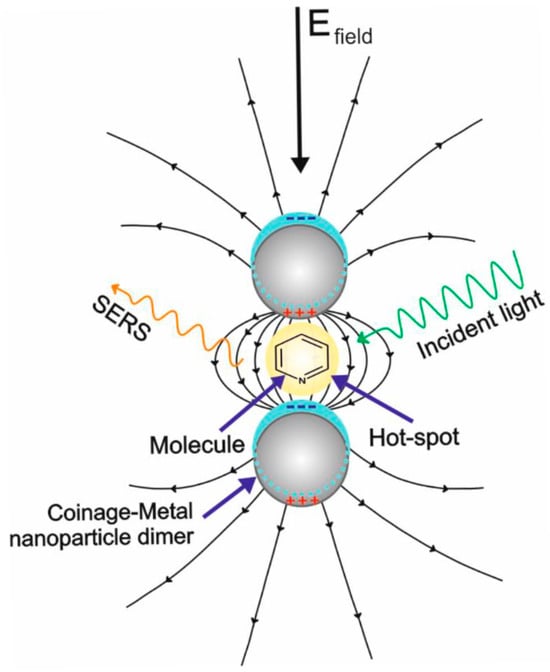
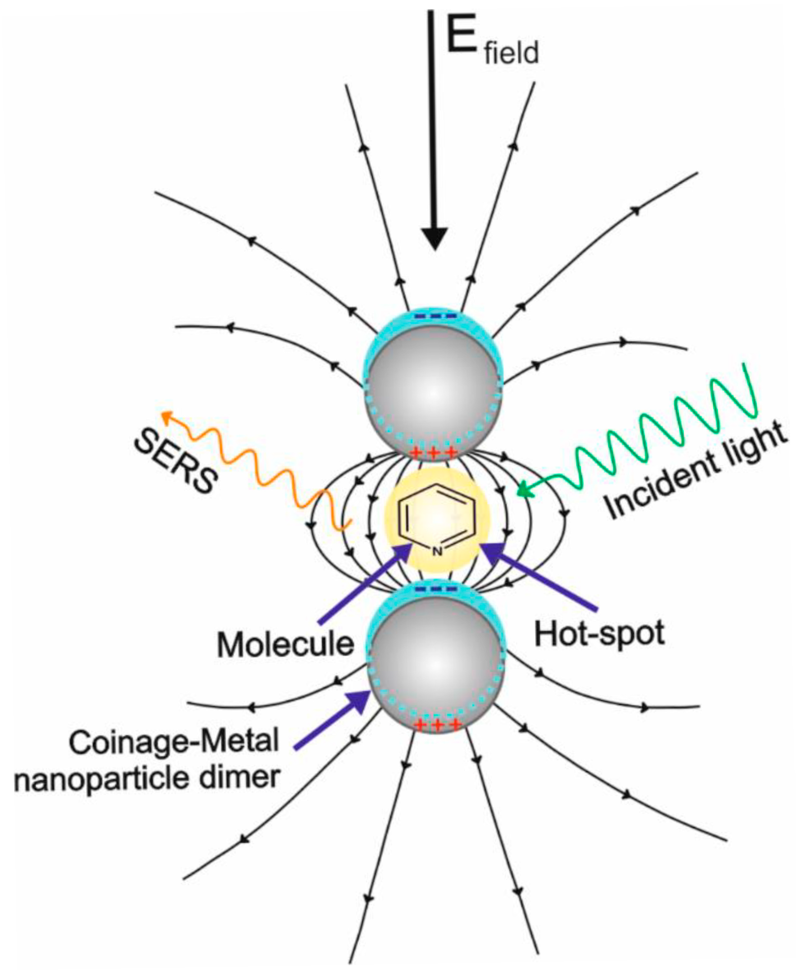
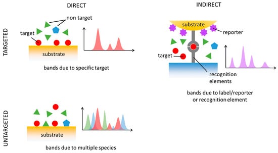
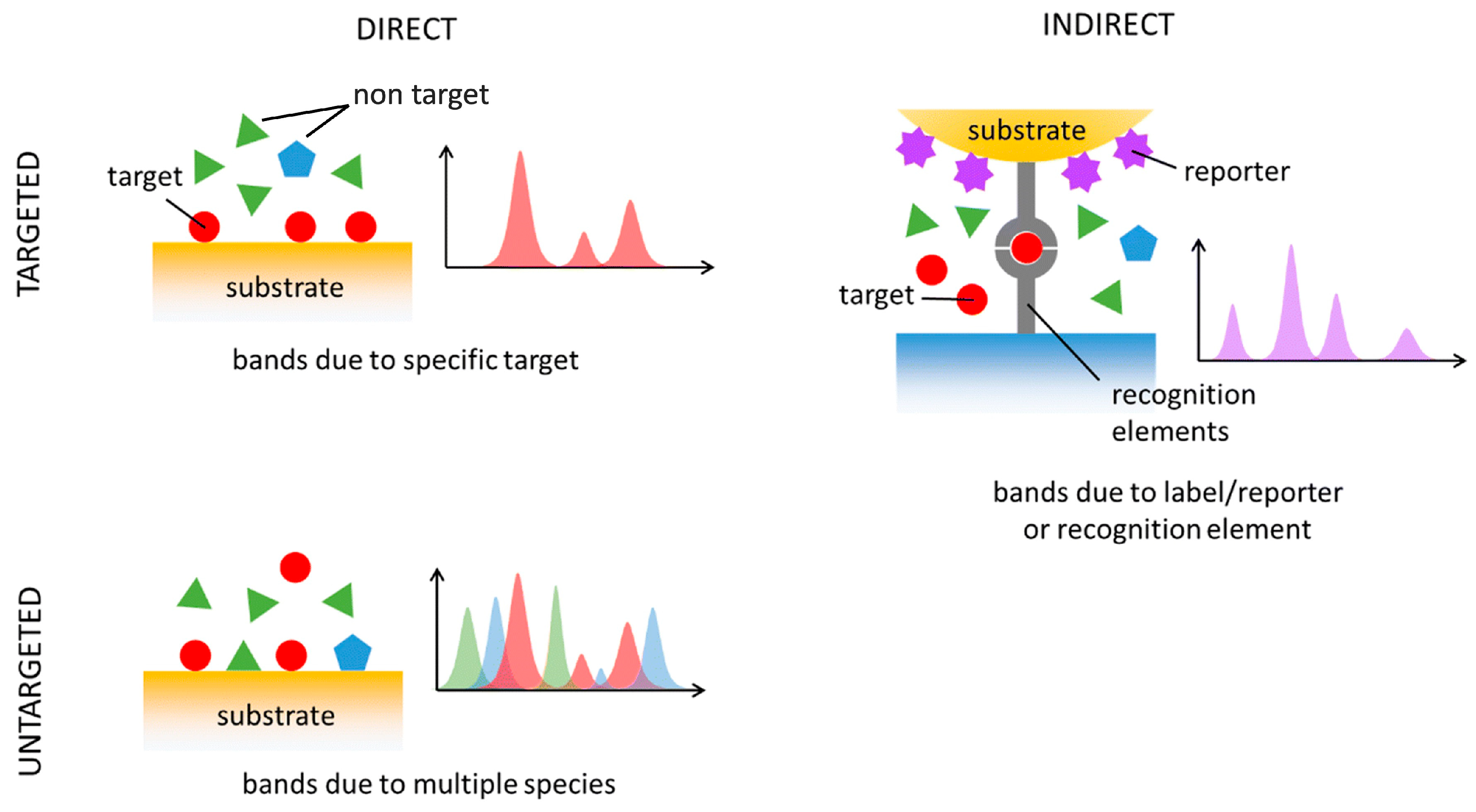
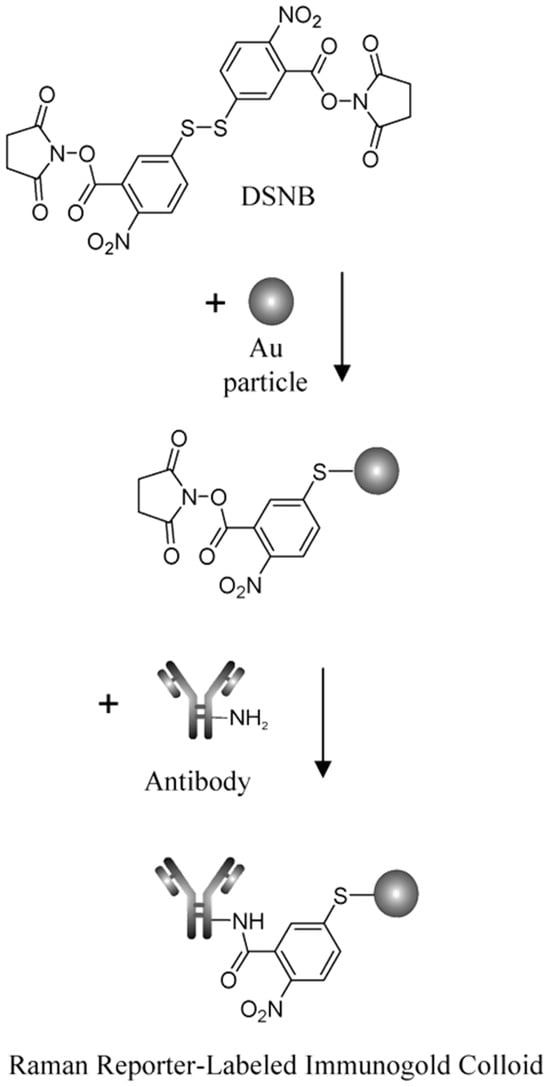
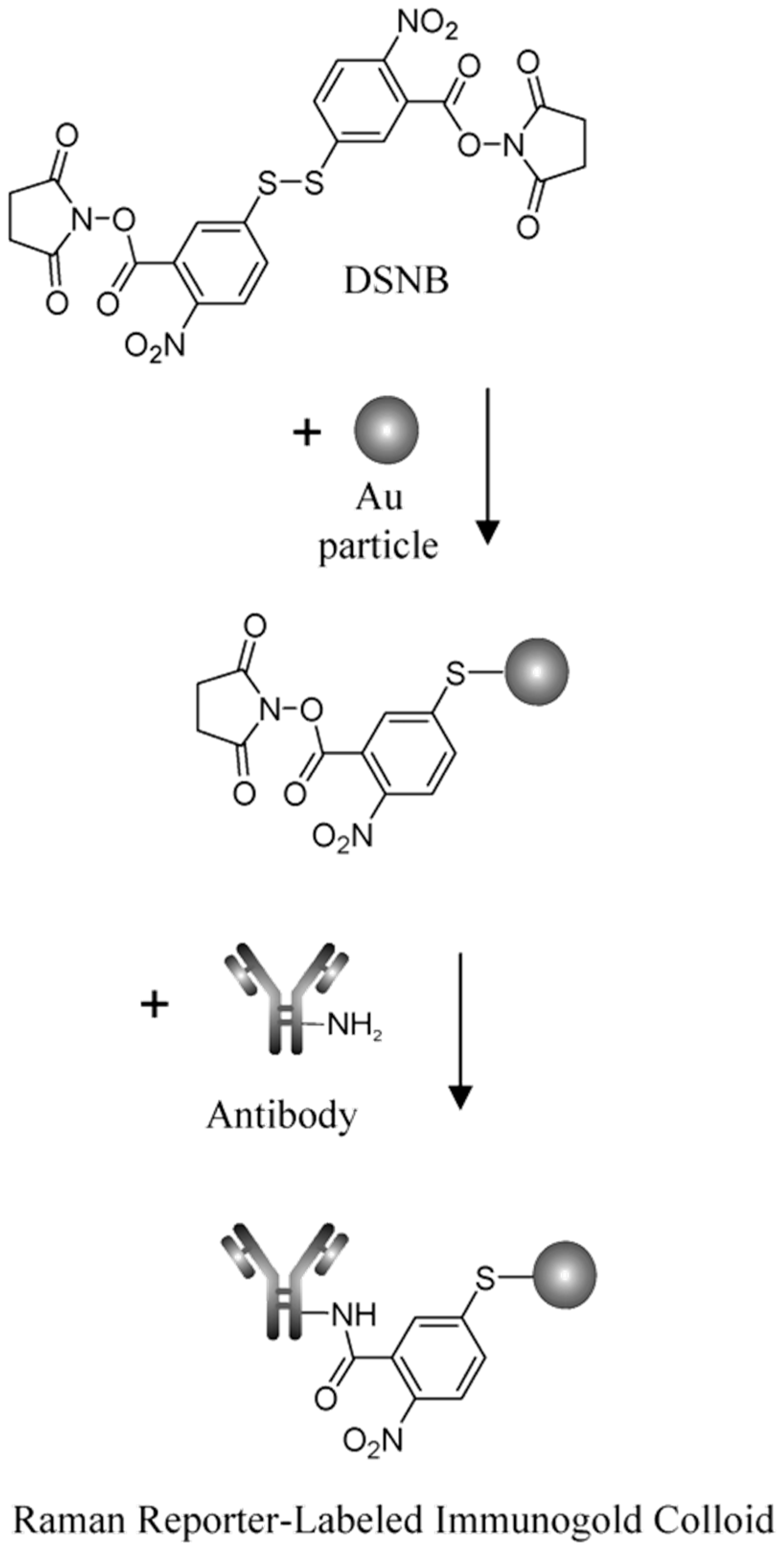
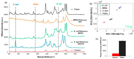
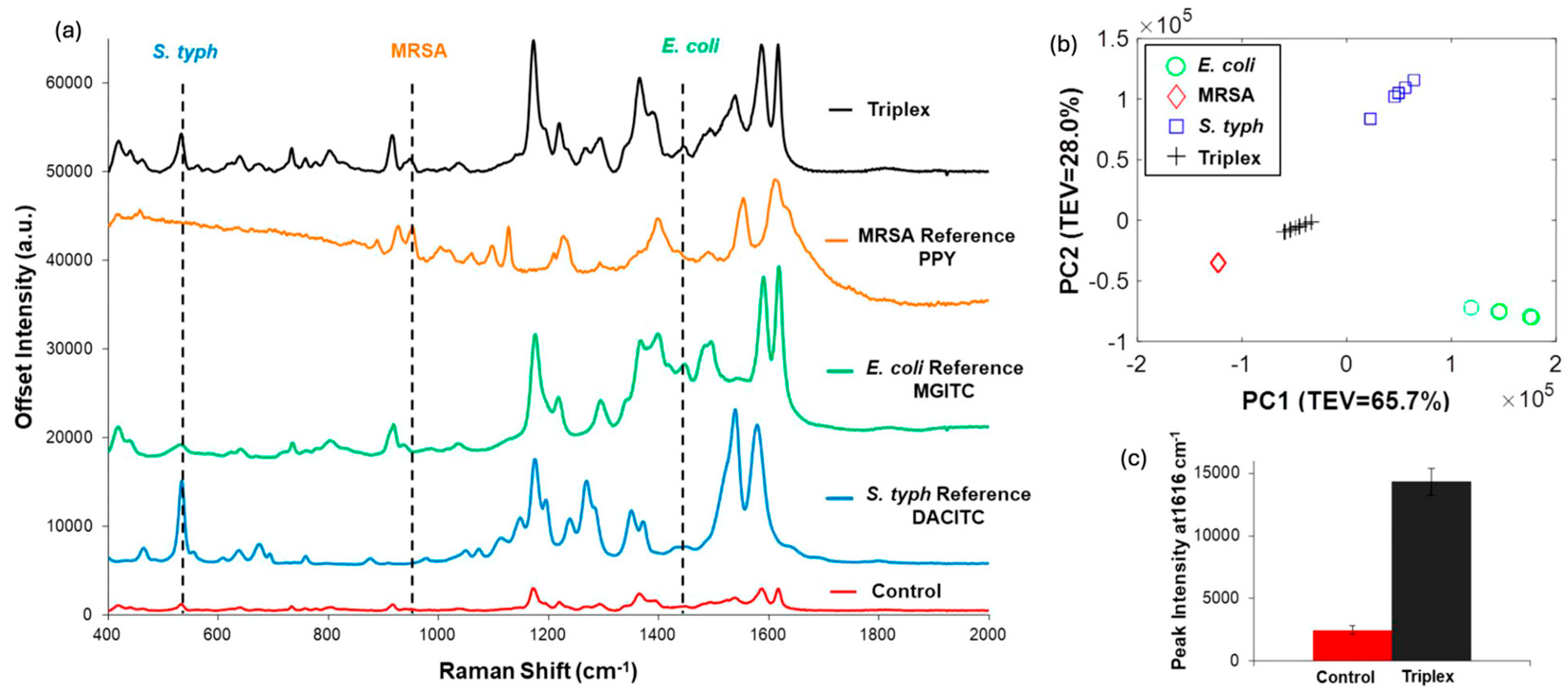
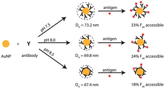
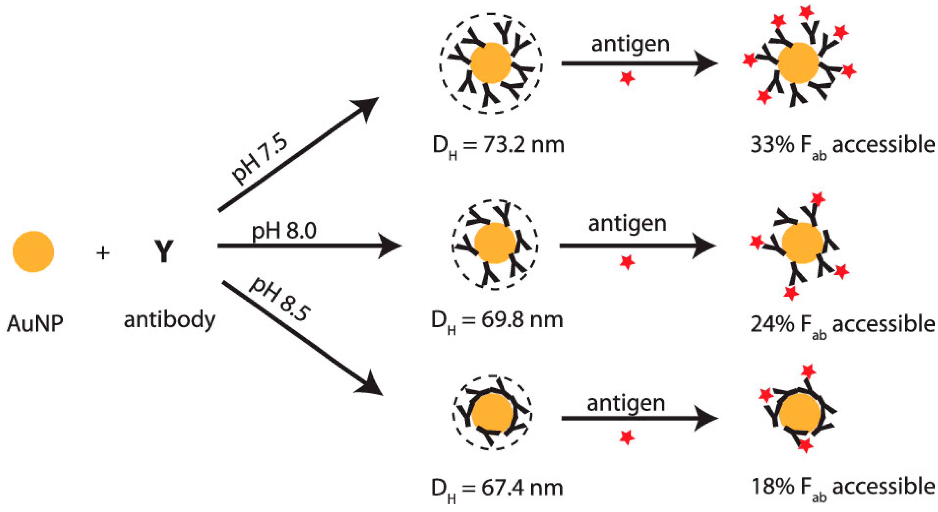
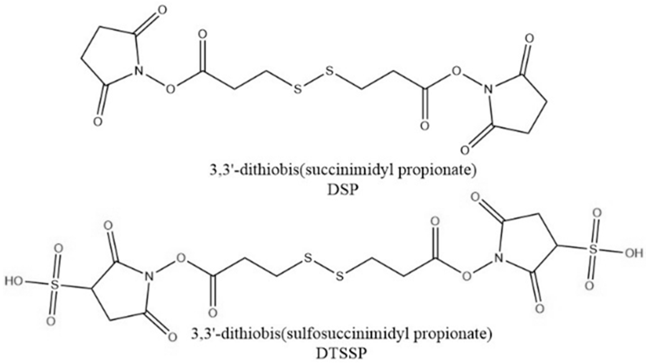
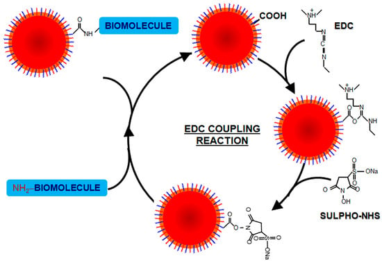
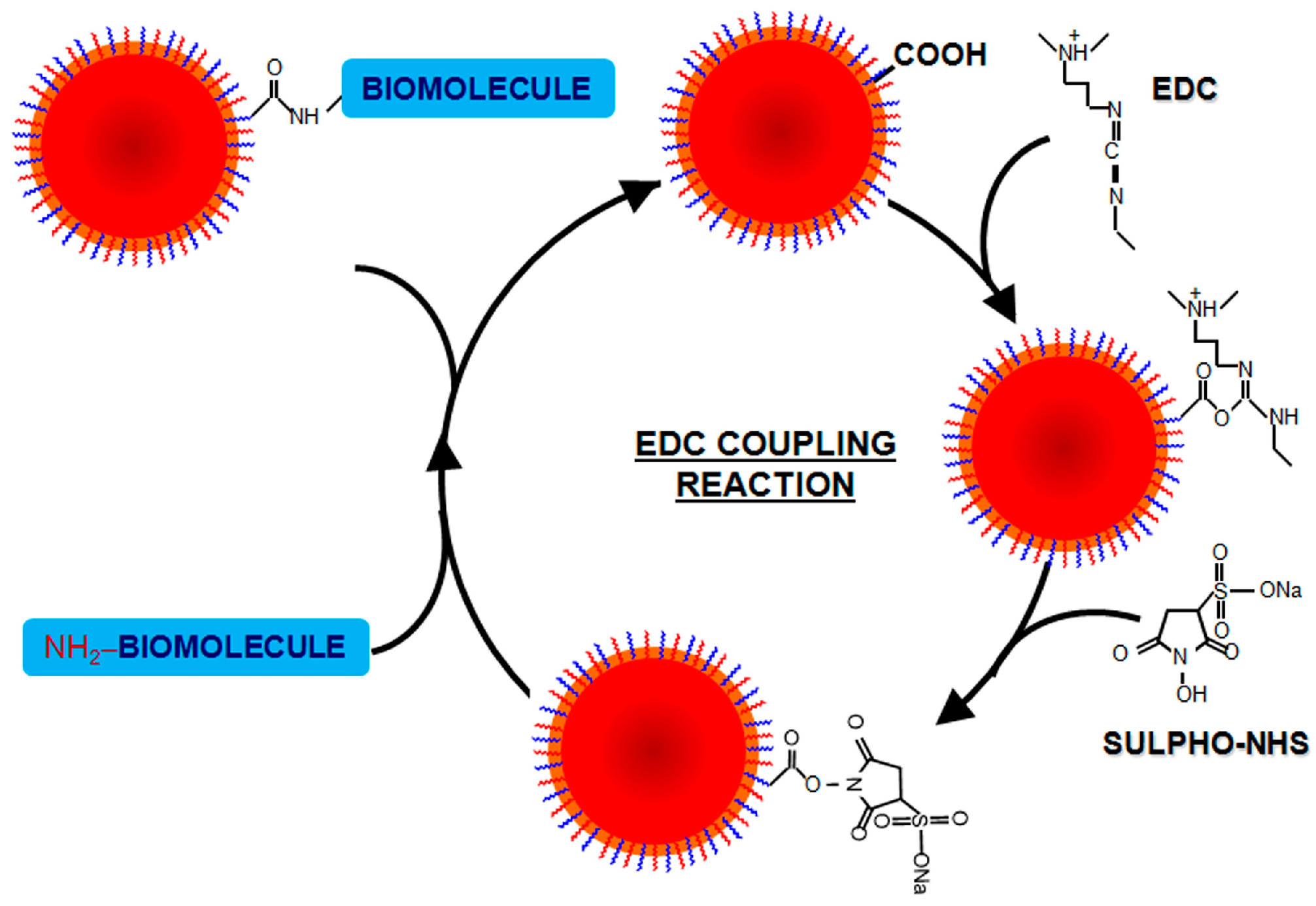
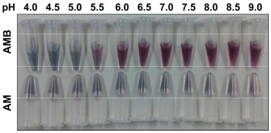
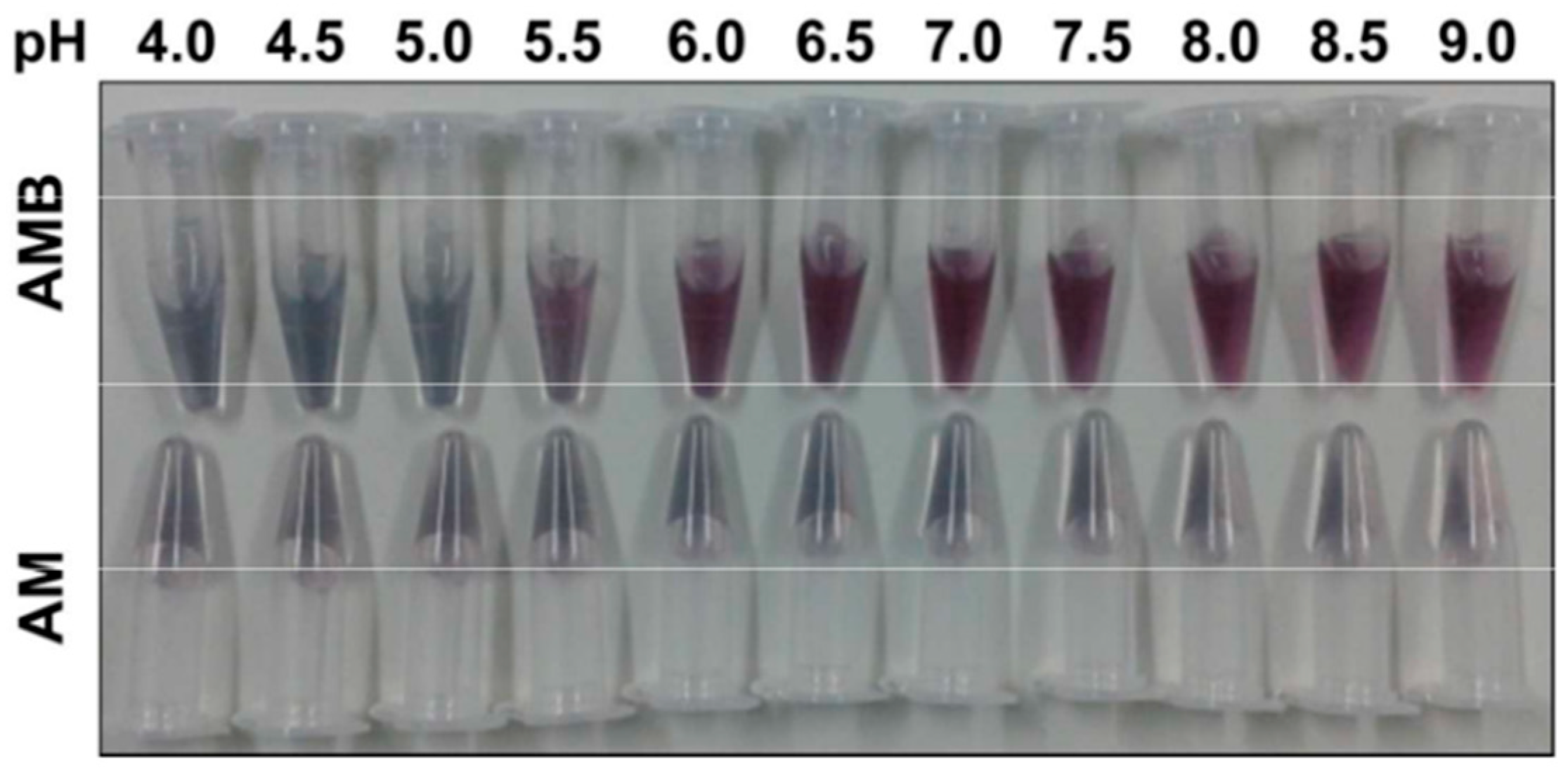
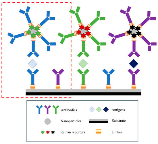
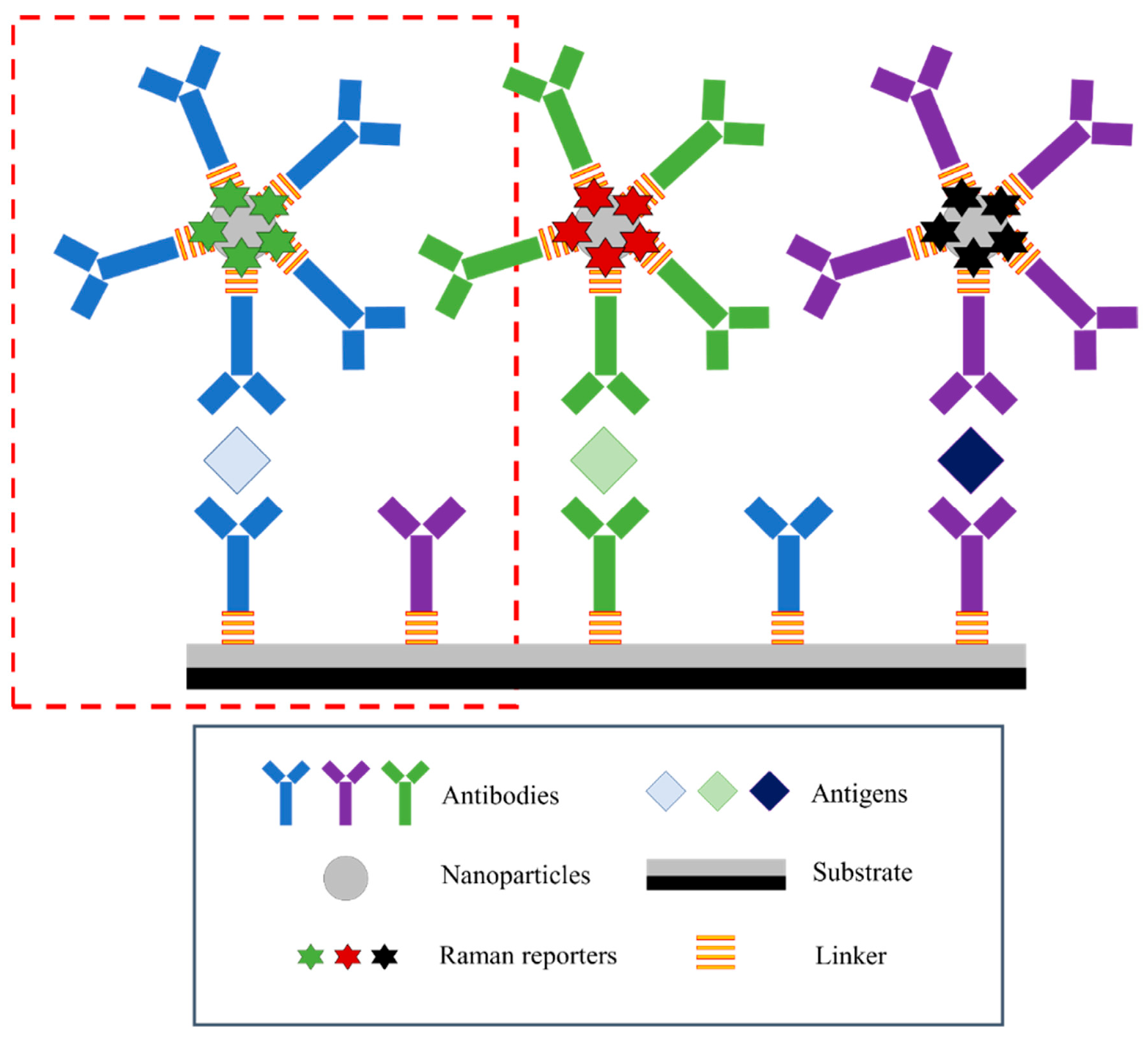
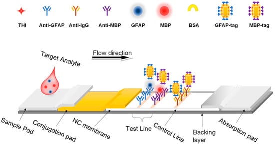
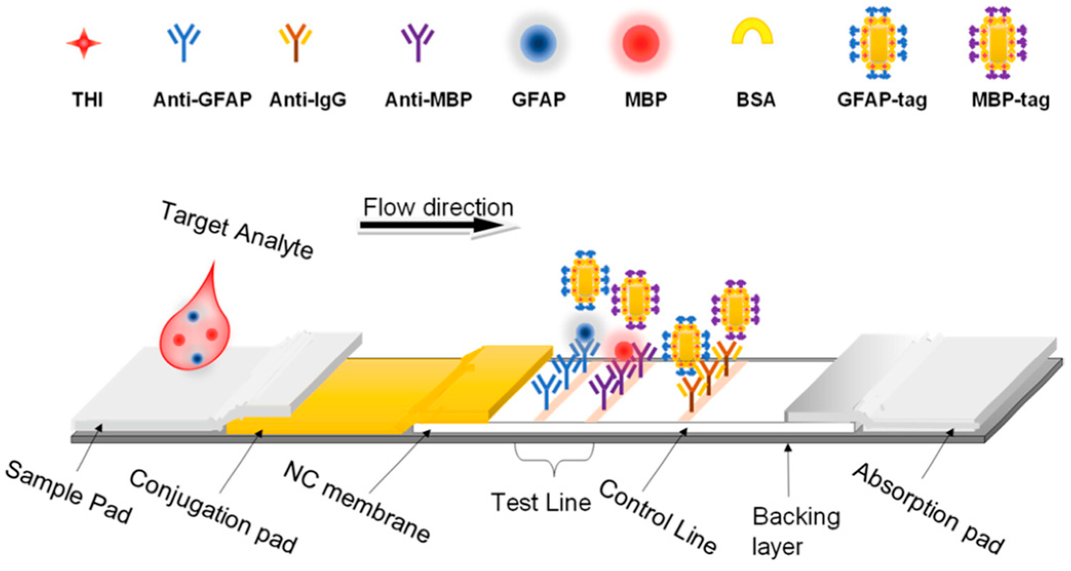
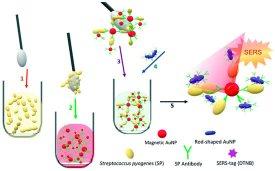
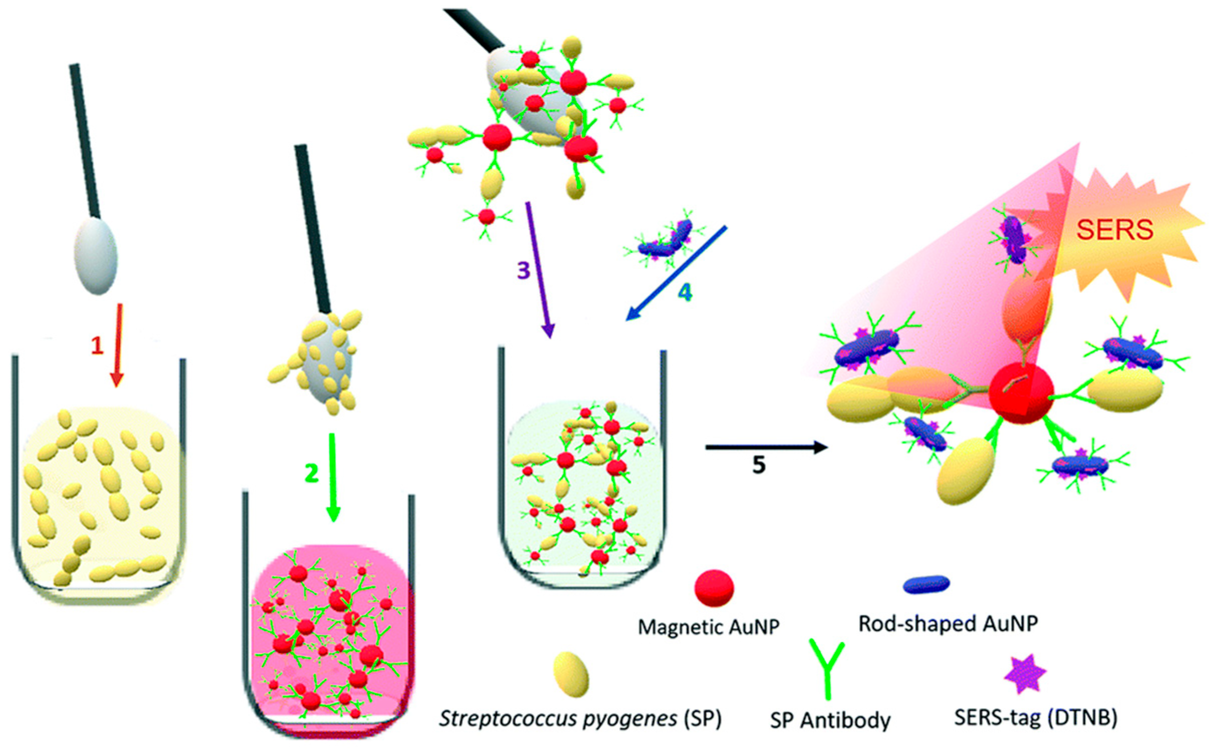
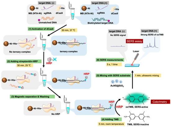
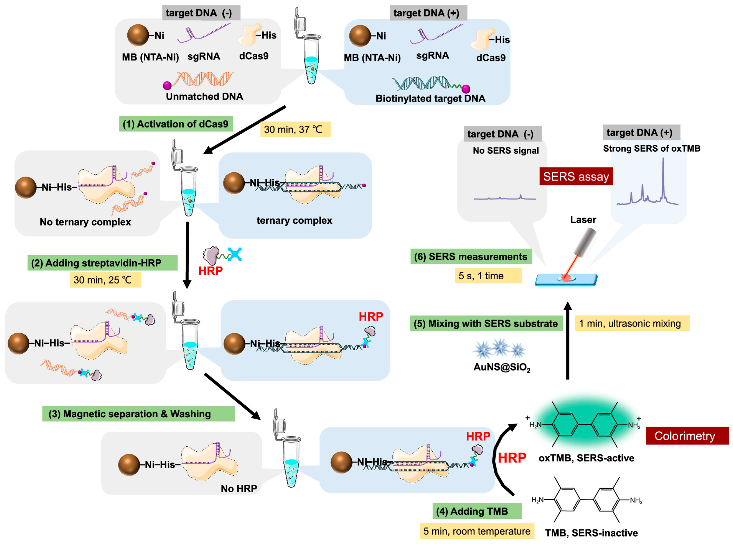
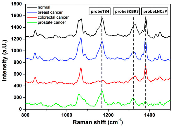
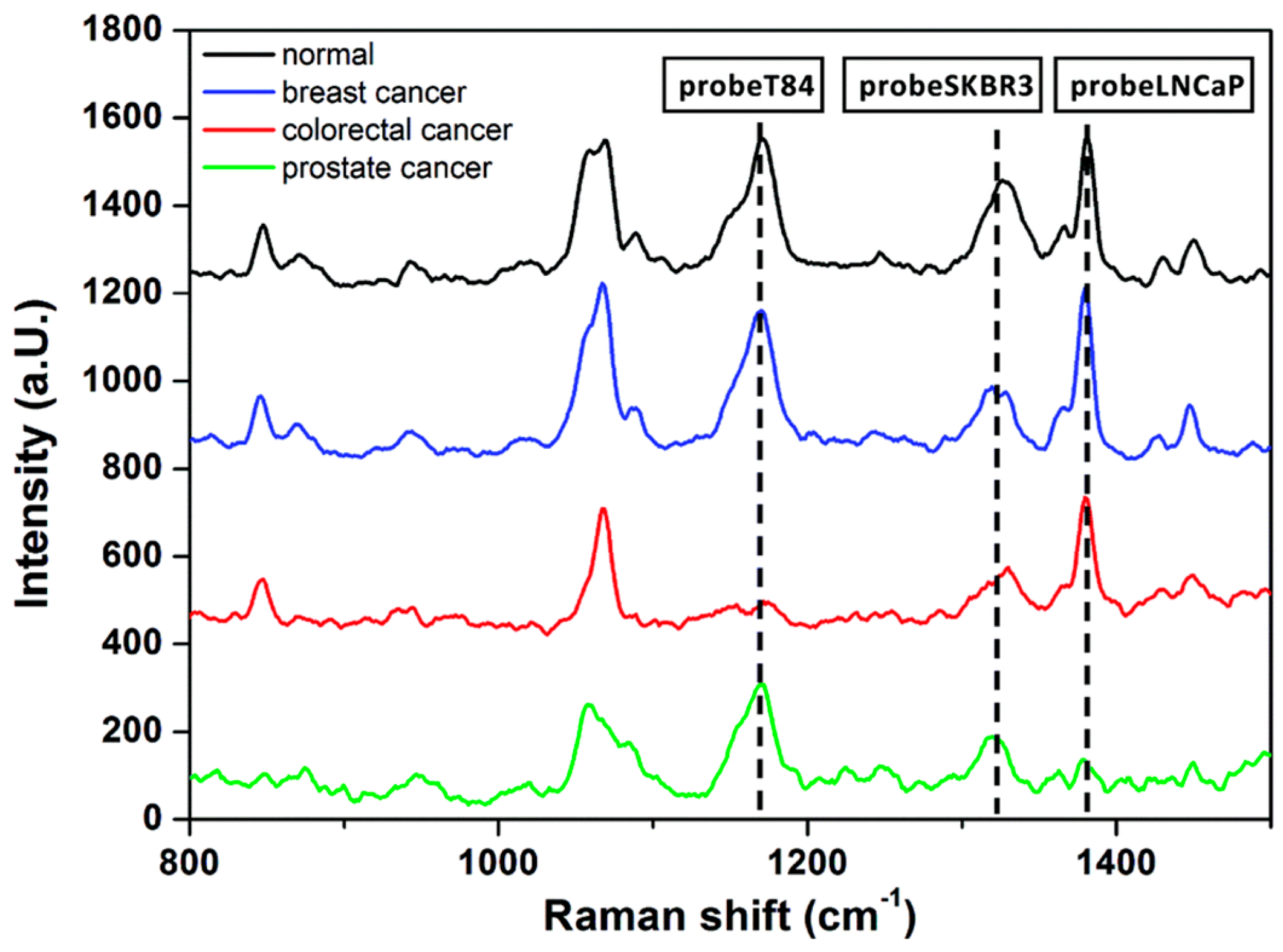
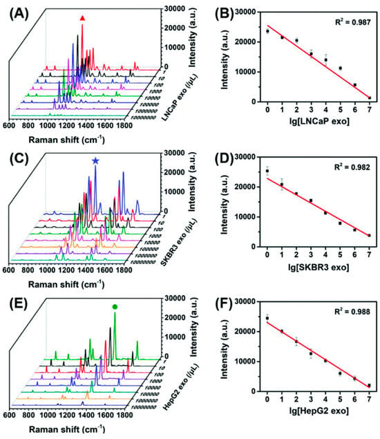
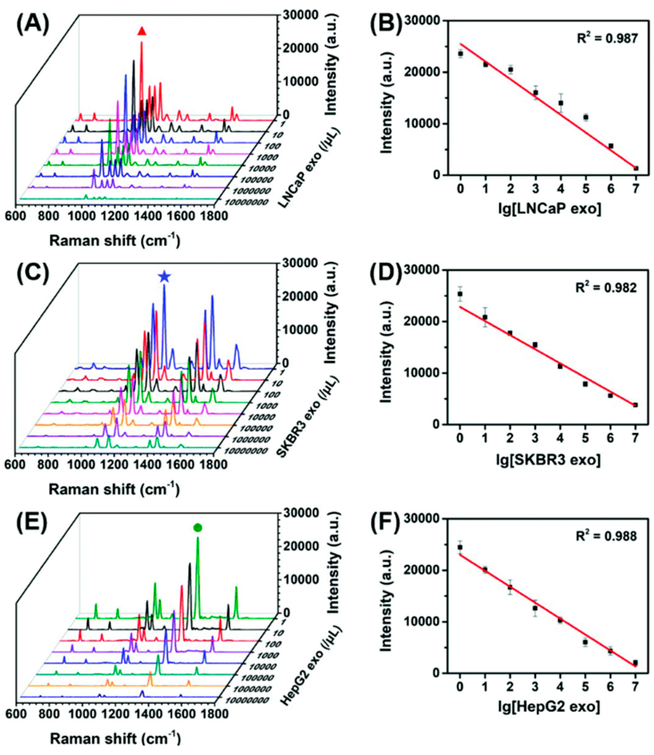
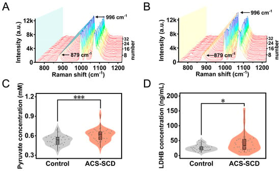
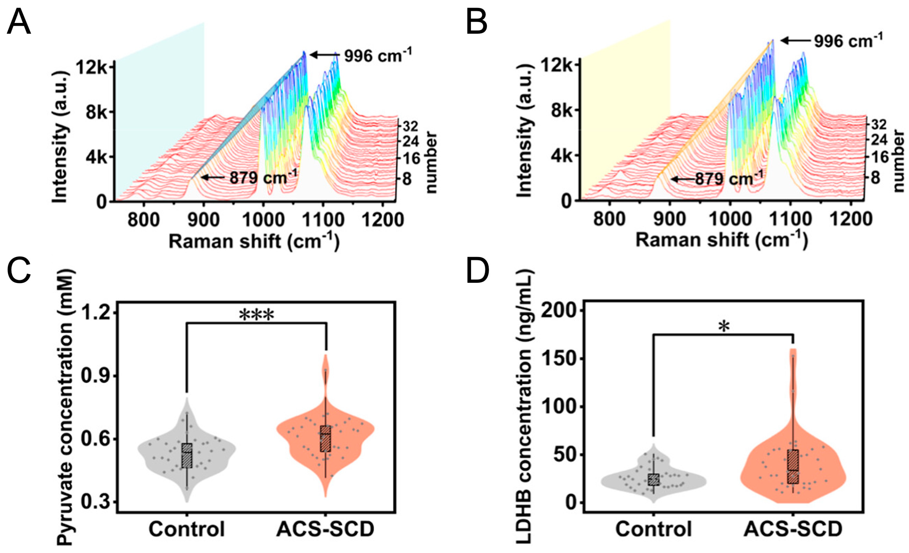
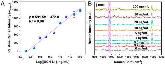
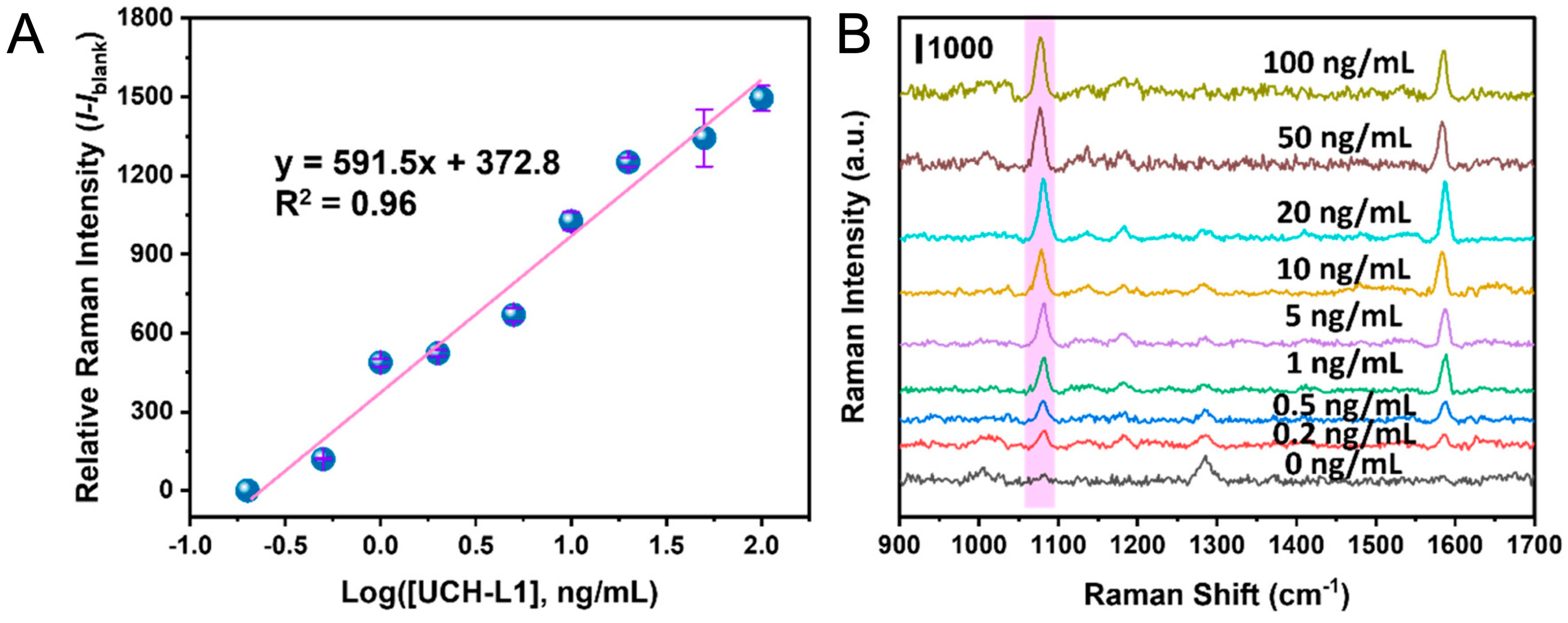
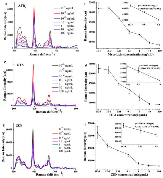
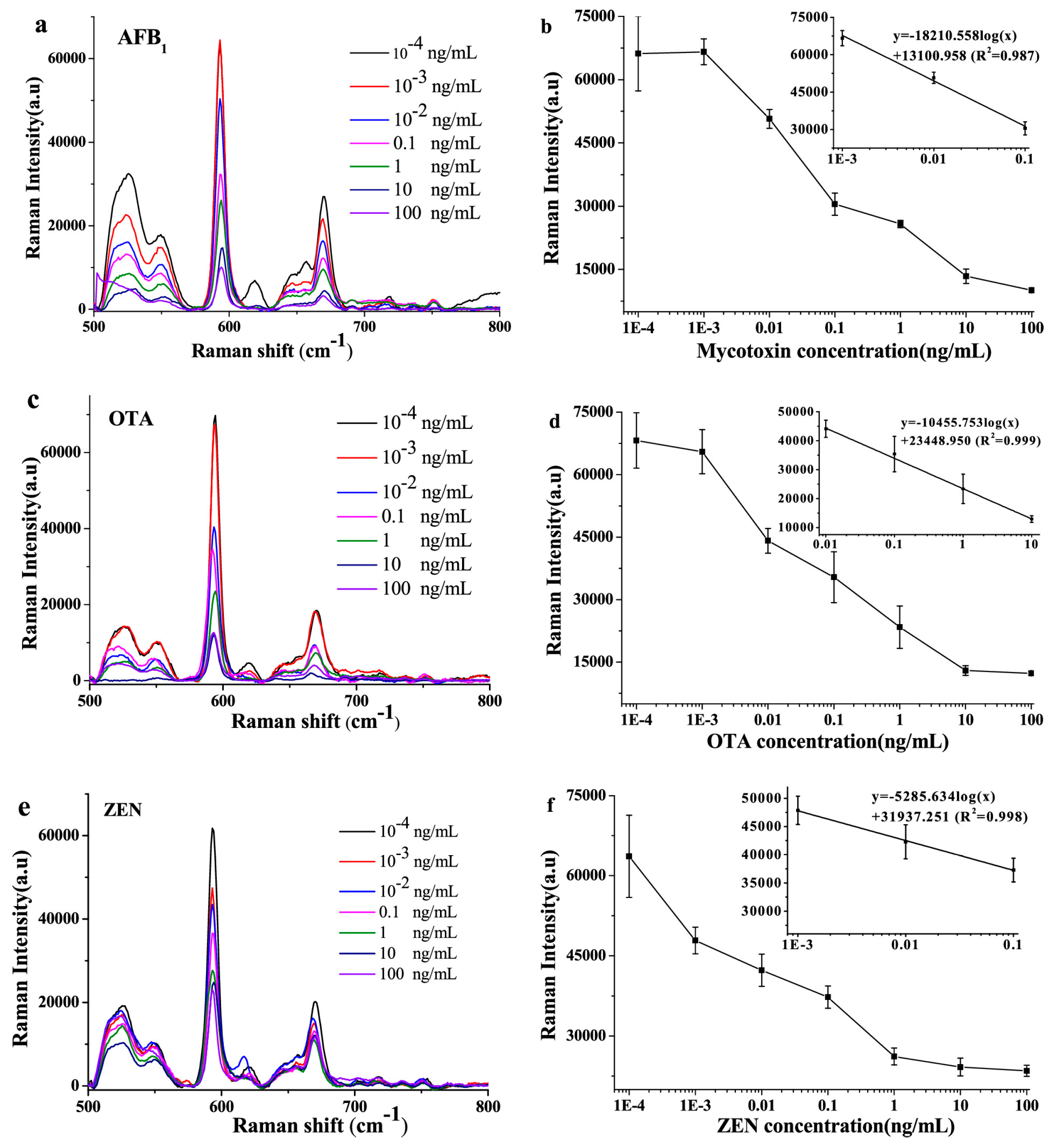
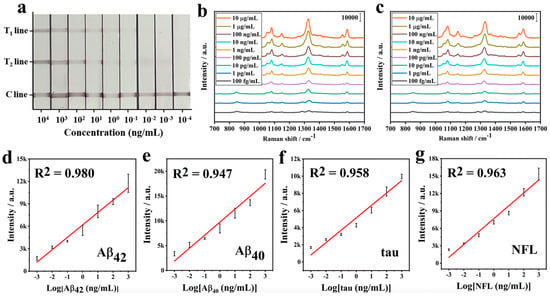
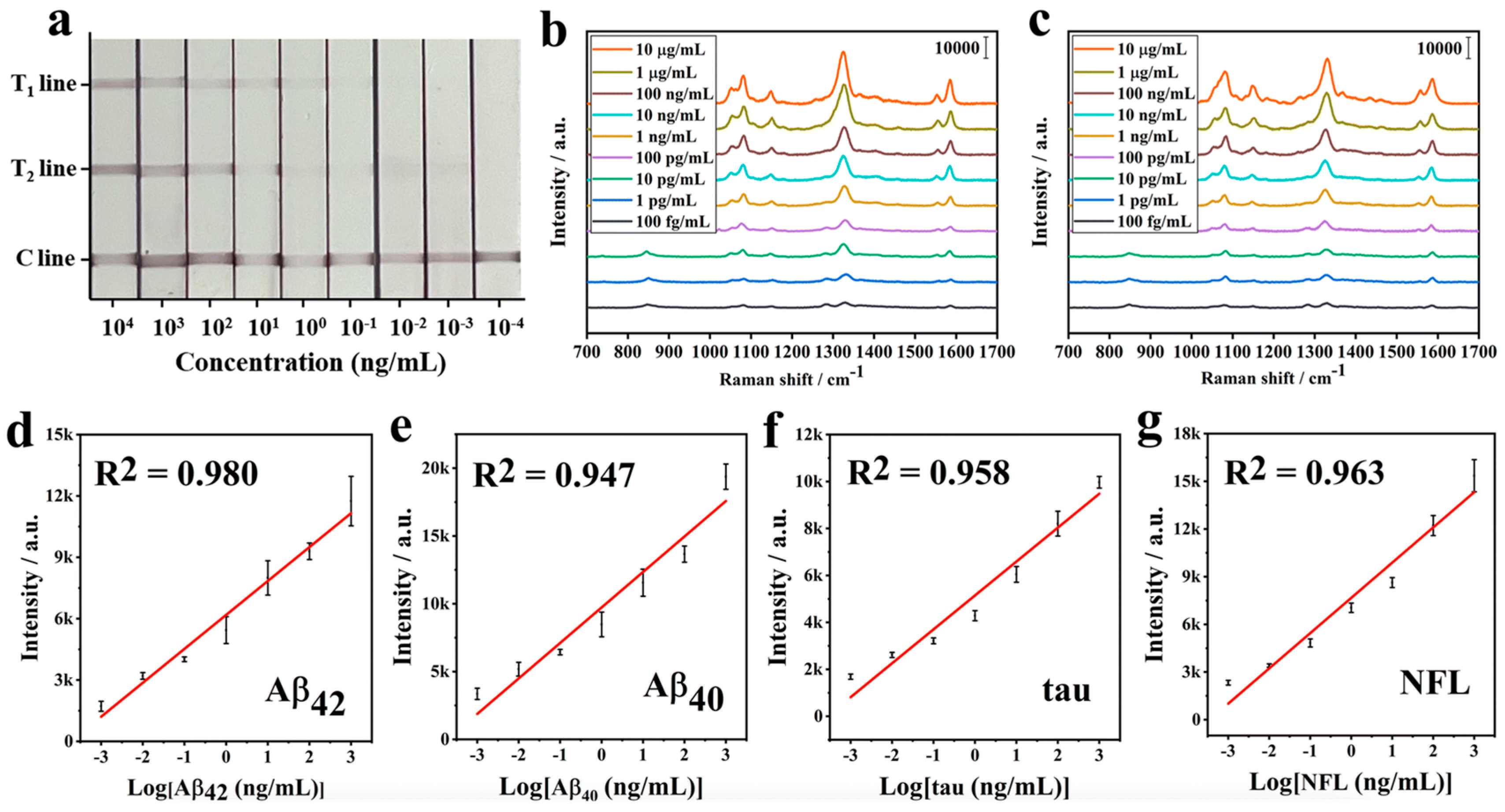
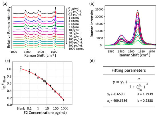
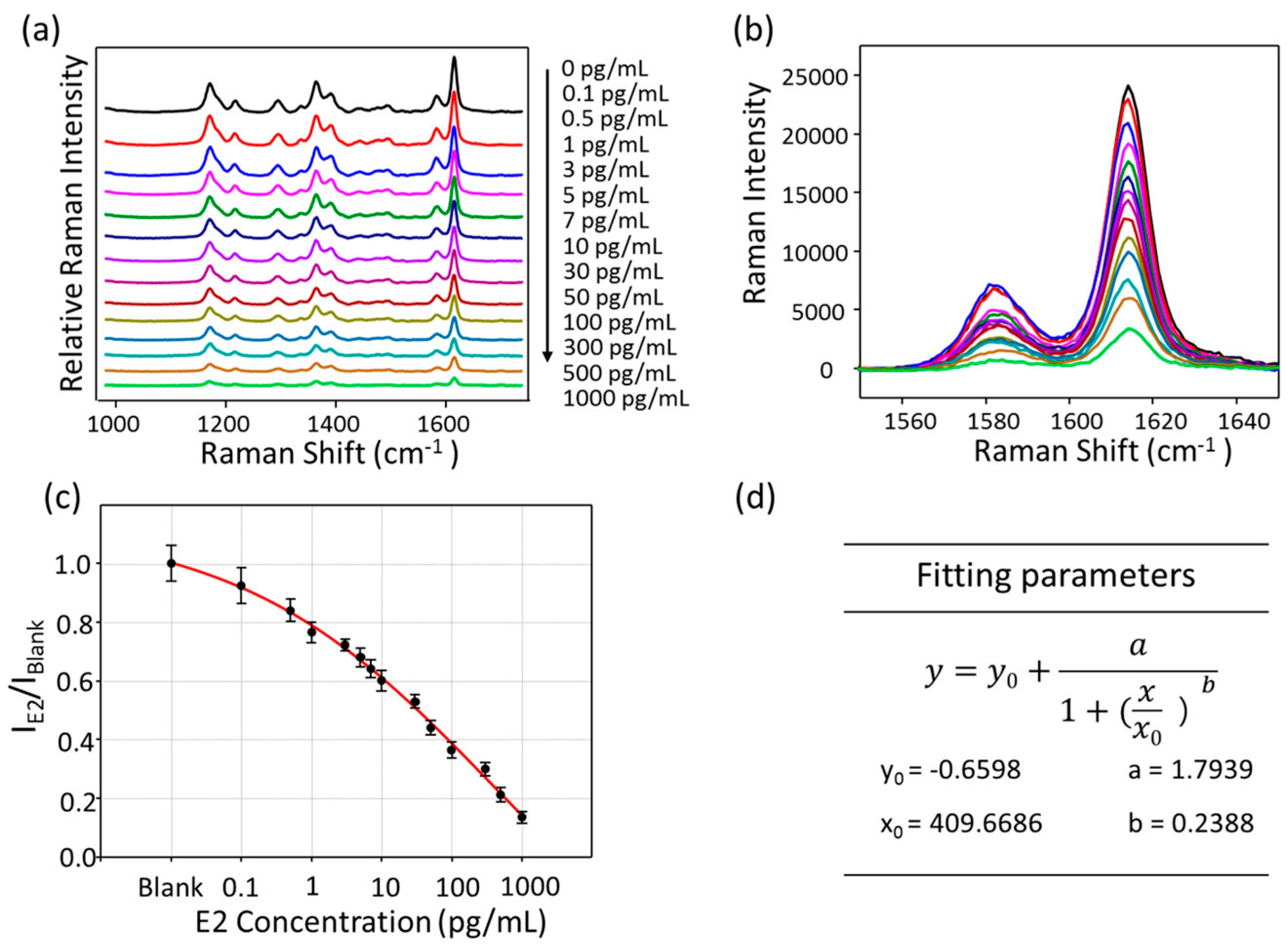
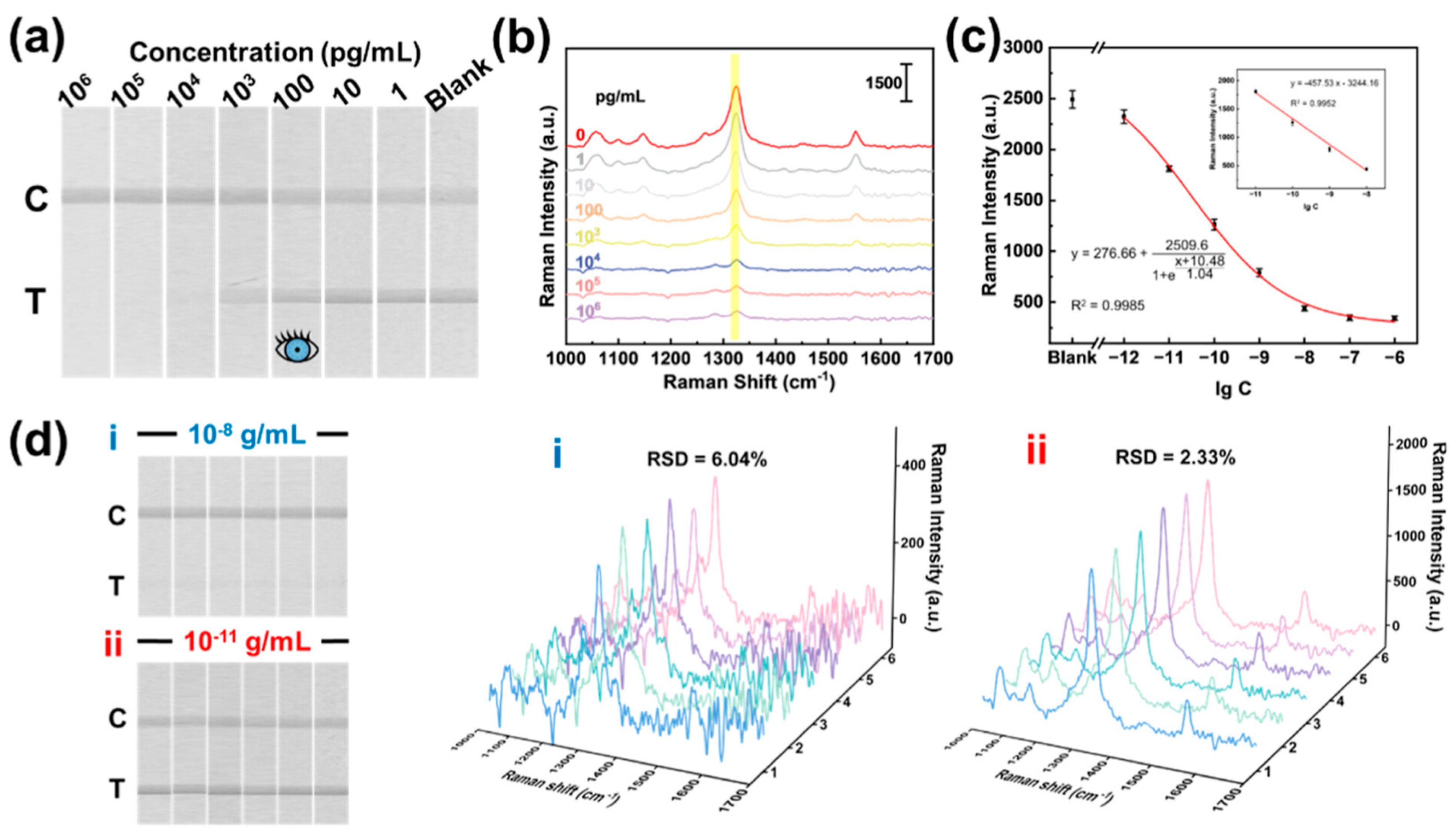

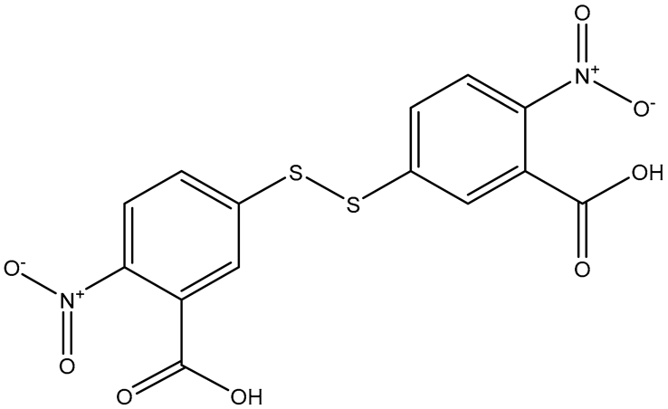
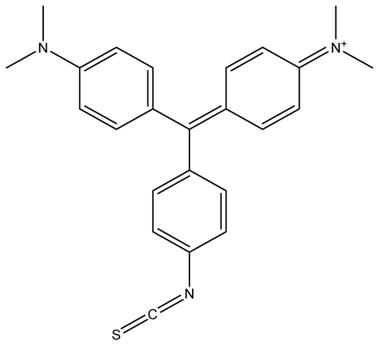

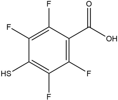

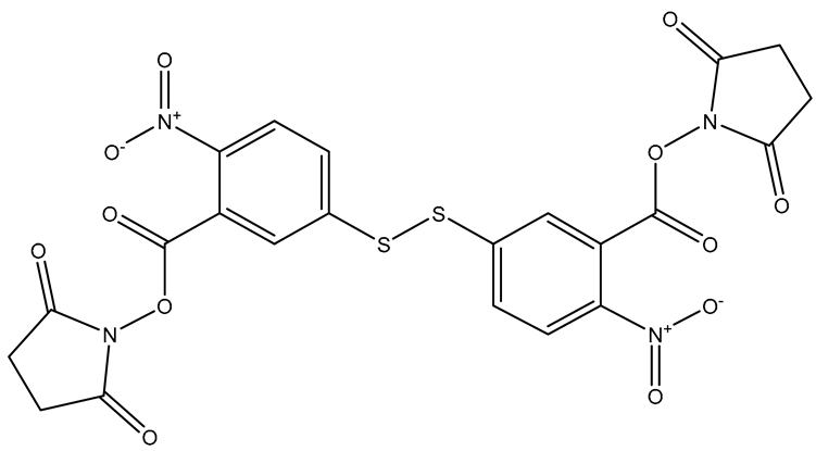
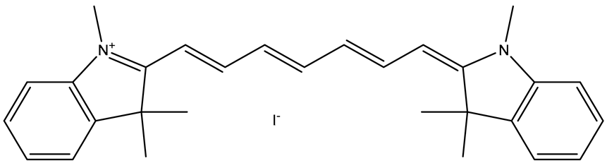
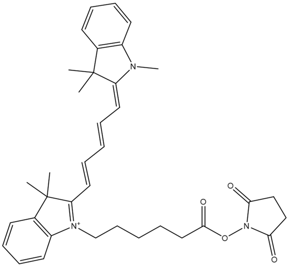
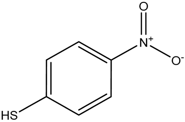
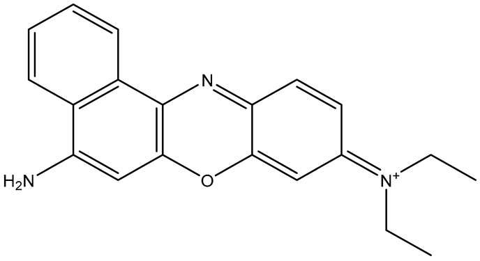
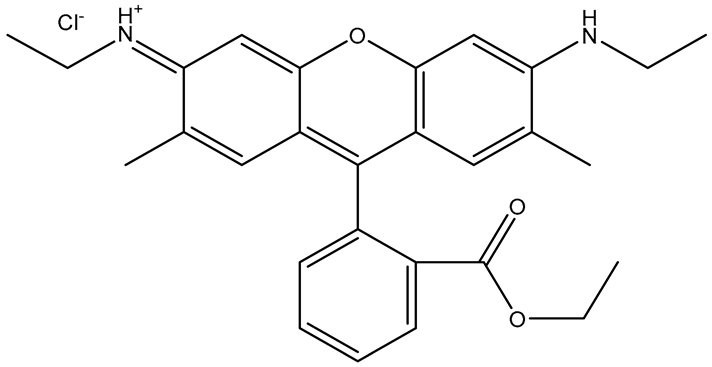
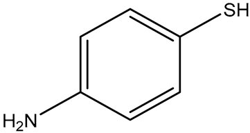
Disclaimer/Publisher’s Note: The statements, opinions and data contained in all publications are solely those of the individual author(s) and contributor(s) and not of MDPI and/or the editor(s). MDPI and/or the editor(s) disclaim responsibility for any injury to people or property resulting from any ideas, methods, instructions or products referred to in the content. |
© 2026 by the authors. Licensee MDPI, Basel, Switzerland. This article is an open access article distributed under the terms and conditions of the Creative Commons Attribution (CC BY) license.