


Highlights
What are the main findings?
- There is a lack of standardized and systematic descriptions for limb shortening and compensatory movements in hemiparetic gait.
What is the implication of the main finding?
- We proposed a set of relevant biomarkers to enhance standardization by systematically describing the deficit of shortening and the compensatory movements.
Abstract
Background: Hemiparetic gait is characterized by reduced limb shortening during swing, increasing the risk of tripping and leading to compensatory strategies. Despite 3D gait analysis being the gold standard for gait assessment, there is no consensus on relevant kinematic biomarkers for limb shortening and compensatory movements. Methods: Systematic review querying five databases (PubMed, Cochrane, Scopus, PEDro, and Web of Science). We included articles that described at least one kinematic biomarker of the lower limb in the sagittal plane and at least one biomarker of the lower limb or pelvis in the transversal or frontal plane, or pelvis in the sagittal plane. Then, we collected kinematic biomarkers from these studies and identified those that seemed relevant to describe limb shortening and compensatory movements during the swing phase. Results: We included 40 studies and collected 385 biomarkers. Among them, 15 described limb shortening, 22 compensations, and 3 toe clearance. Analysis of 12 interventional studies showed that some biomarkers of shortening and compensation were more sensitive to change than others. Conclusions: This review highlights the lack of standardized description for limb shortening and compensatory movements in hemiparetic gait. A set of 13 relevant biomarkers is proposed to improve the interpretation of gait analysis and support consistent evaluation of therapeutic interventions.
1. Introduction
Hemiparesis is a motor disturbance caused by a central nervous system injury that usually leads to gait disorders [1]. Among post-stroke subjects, 64% achieve independent ambulation after 11 months of rehabilitation, while 14% still require assistance [2]. Hemiparetic subjects commonly exhibit abnormal gait patterns, including insufficient limb shortening during the swing phase of gait on the affected side [3,4], mainly due to muscle weakness and spastic overactivity [5]. Under physiological conditions, foot clearance during the swing phase of gait primarily results from limb shortening in the sagittal plane, enabled by the coordinated flexion of the hip, knee, and ankle joints [6]. Pelvic movements are minimal during the swing phase and mainly serve to limit the vertical displacement of the body’s center of mass, reducing energy expenditure [7]. In stroke survivors, the impaired ability to shorten the paretic limb in the sagittal plane often necessitates the use of compensatory movements in other planes of motion to ensure foot clearance and avoid tripping or falling [8]. However, these adaptations typically come at the cost of increased energy expenditure [9]. In the sagittal plane, compensatory strategies may include a steppage gait, characterized by exaggerated hip and knee flexion. In the frontal plane, hip hiking—elevation of the ipsilateral hip joint center—can facilitate swing phase clearance. In the transverse plane, circumduction and external rotation of the lower limb are commonly observed, primarily driven by hip abduction and external rotation. Compensatory strategies can also involve the contralateral limb, such as vaulting, where the stance leg performs plantarflexion during push-off to help the paretic limb clear the ground [10,11].
Improving limb shortening during the swing phase has been identified as a key therapeutic target for enhancing gait patterns of hemiparetic subjects, either by enhancing the recruitment of paretic flexor muscles or by weakening hypertonic extensor muscles. Various therapeutic strategies have been used, such as strengthening of the hip flexors and ankle dorsiflexors through rehabilitation programs [12], functional electrical stimulation (FES) of ankle dorsiflexors [13], botulinum toxin injections targeting knee extensors [14,15,16], orthotic devices such as ankle–foot orthoses (AFOs) [17,18,19], and neuro-orthopaedic surgery [20].
A comprehensive view of limb shortening is essential to evaluate the overall impact of these interventions on gait and to define relevant therapeutic goals. For instance, the FES of ankle dorsiflexors has been shown to have a detrimental effect on maximum knee flexion during swing [13]. However, synthesis studies rarely provide this broader kinematic perspective—either due to a lack of available data, as reported by some authors [20,21], or because they focus exclusively on a single joint level [17,18,19].
Moreover, evaluating limb shortening and compensatory movements simultaneously appears particularly valuable. The effect of botulinum toxin injections into the rectus femoris on improving knee flexion during swing—by approximately 5°—has been well documented [14,15,16]. However, their impact on compensatory movements has not been investigated. Yet reducing compensatory movements may help decrease the energy cost of walking and contribute to patients’ perceived improvement, just as much as improving limb shortening reduces the risk of tripping.
Three-dimensional instrumental gait analysis (3D-IGA) is considered the gold standard for investigating gait abnormalities, notably offering a standardized and detailed kinematic dataset that enables thorough evaluation of both limb shortening mechanisms and compensatory strategies. Its use is now recommended by the International Stroke Recovery and Rehabilitation Alliance for evaluating gait after stroke, particularly through global indices such as the Gait Deviation Index (GDI) and Gait Profile Score (GPS). These recommendations also emphasize the need for further work on biomechanical metrics—especially those aimed at identifying compensatory strategies—which is expected to be addressed in upcoming consensus efforts [22]. However, up to now, the selection of kinematic biomarkers to characterize gait abnormalities is left to the discretion of the investigator, which limits consistency across studies and clinical applications. Moreover, currently available systematic reviews on instrumental gait assessment in post-stroke individuals have not specifically focused on the swing phase or on compensatory movements associated with insufficient limb shortening [23,24,25]. This gap highlights the need for a more targeted and comprehensive synthesis of relevant biomarkers addressing these specific aspects of post-stroke gait impairment.
This systematic review aimed to establish how limb shortening and compensatory movements during the swing phase are currently conceptualized and measured in the existing literature. The secondary objective was to identify the most relevant 3D-IGA-derived, kinematic biomarkers for characterizing limb shortening and compensatory movements in hemiparetic subjects. Biomarker validity was assessed by reviewing the contexts in which they were used, and their sensitivity to change was analyzed through pre/post-intervention or with/without orthosis comparisons.
This investigation aimed to enhance practitioners’ understanding of the relationship between limb-shortening deficits and compensatory movements, potentially facilitating the evaluation of therapeutic interventions by assessing reductions in compensatory movements alongside improvements in limb shortening.
2. Materials and Methods
This systematic review followed the Preferred Reporting Items for Systematic Reviews and Meta-Analyses (PRISMA) statement and used a predefined research protocol.
2.1. Search Strategy
A comprehensive search was conducted on 31 March 2023 across MEDLINE, COCHRANE, Web of Science, PEDro, and Scopus using MeSH and non-MeSH terms, including hemiplegia, stroke, brain injury, gait, and kinematics. Detailed search strategies for all the databases are available in Appendix A. No filters, date restrictions, or additional searches through cited references were applied.
2.2. Eligibility Criteria
The study inclusion criteria were as follows: adults (>18) with hemiparesis due to central nervous system disease, who underwent a 3D gait analysis using an optoelectronic system either on a treadmill or overground. At least one kinematic data point for limb shortening and compensatory movements was required. Exclusion criteria were non-gold-standard systems, non-hemiplegic conditions, children, animals, and spinal injuries. Robot-assisted studies were included if the system was used for rehabilitation without assistance during gait analysis. Eligible studies were published in English, Spanish, German, or French and included observational, interventional, and case series designs.
2.3. Study Selection Process
Two independent reviewers (EM and LL) used the Rayyan online tool (https://www.rayyan.ai, accessed on 31 March 2023) for duplicate removal and screening of titles and abstracts. Full texts were reviewed for eligibility, and disagreements were resolved through consensus. A third reviewer (DG) was consulted if necessary.
2.4. Data Collection Process and Data Items
Two independent reviewers (EM and LL) extracted data, without contacting authors for missing information. Collected data included: the name of the first author, year of publication, study design, study objective, number of subjects, population characteristics (mean age, disease, time since stroke for post-stroke subjects), inclusion and exclusion criteria, system analysis, kinematic model, experimental conditions (e.g., treadmill or overground walking), intervention characteristics and outcome measures.
For interventional studies, intervention characteristics were extracted from those describing a pre-/post-protocol or a with/without orthotic device comparison. When available, information on protocol duration, frequency, and therapeutic target (e.g., muscle, joint, or movement strategy) was recorded.
Outcome measures included all quantitative kinematic biomarkers described in the study. Only biomarkers derived from three-dimensional optoelectronic motion capture systems were retained. Spatiotemporal, kinetic, or EMG parameters were not considered.
Kinematic biomarkers were categorized into: (1) limb-shortening biomarkers (hip, knee, ankle in the sagittal plane); and (2) compensatory movements biomarkers (hip, knee, ankle and pelvic movements in the frontal and transversal planes as well as pelvic movements in the sagittal plane). Toe clearance biomarkers were also collected as a result of both limb shortening and compensations. Biomarkers were identified by direction of movement, quantitative description, and gait cycle timing. Only swing-phase-specific biomarkers with quantitative data were included. Biomarkers lacking these criteria or not describing limb shortening or compensatory movements were excluded. Finally, we compiled a list of relevant kinematic biomarkers for describing limb shortening (e.g., hip flexion, knee flexion, ankle dorsiflexion, toe clearance, and global limb shortening) or compensatory movements (e.g., hip hiking, circumduction, posterior pelvic tilt, posterior pelvic rotation, and vaulting). Studies reporting at least one limb shortening and one compensatory movement biomarker were selected for qualitative analysis.
To complement the descriptive synthesis, we conducted a secondary quantitative synthesis. For this purpose, we identified a subset of interventional studies that included pre- and post-intervention data or with/without orthosis comparisons. From these studies, relevant swing-phase-specific kinematic biomarkers were extracted with their associated numerical values, when reported, to assess the sensitivity of these biomarkers to intervention.
2.5. Study Risk of Bias Assessment
For the qualitative analysis, risk of bias was deemed irrelevant as the focus was on biomarkers rather than study outcomes. Consequently, no formal bias assessment was conducted. For the quantitative analysis, two independent reviewers (EM and LL) evaluated methodological quality using JBI’s critical appraisal tools.
2.6. Synthesis Methods
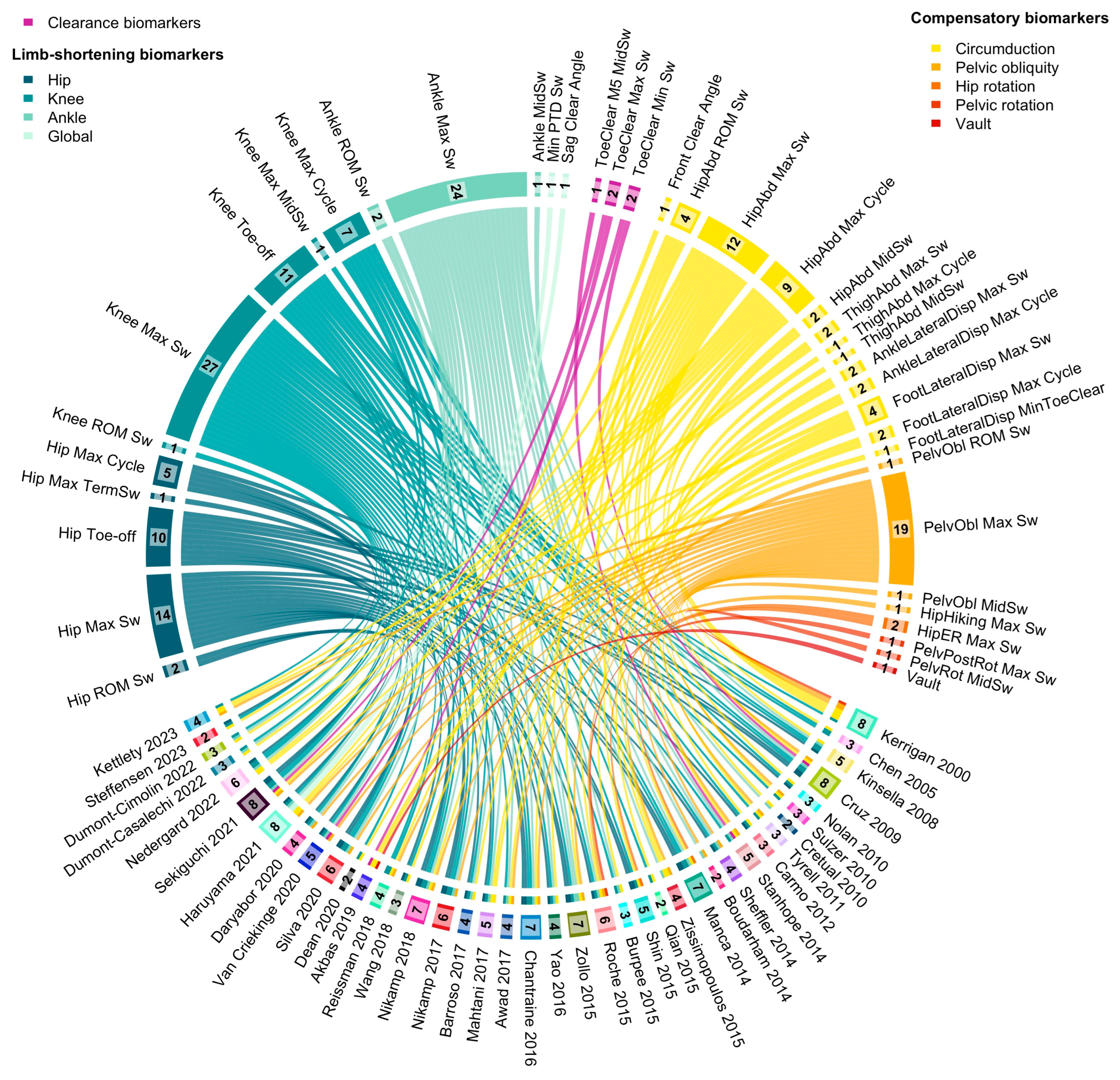
A descriptive synthesis summarized study characteristics and biomarker usage. A Circos plot was created using R (version 4.3.2, Circlize package 0.4.16) [26,27] to visually represent the distribution and frequency of biomarker usage. In this plot, each study was linked to the biomarkers it described, allowing for immediate visual identification of (1) which biomarkers were most frequently reported, and (2) the heterogeneity in marker selection across studies.
2.7. Effect Size
To assess whether interventions or orthotic devices increased limb shortening while reducing compensatory movements, effect sizes were calculated. If necessary, standard deviation was derived from standard error based on sample size. For paired pre/post-intervention comparisons, Hedges’ g with 95% confidence intervals was used. Confidence intervals excluding zero were considered statistically significant [28]. Effect sizes were classified as small (0.2), moderate (0.2–0.8), or large (>0.8) [29].
3. Results
3.1. Study Selection
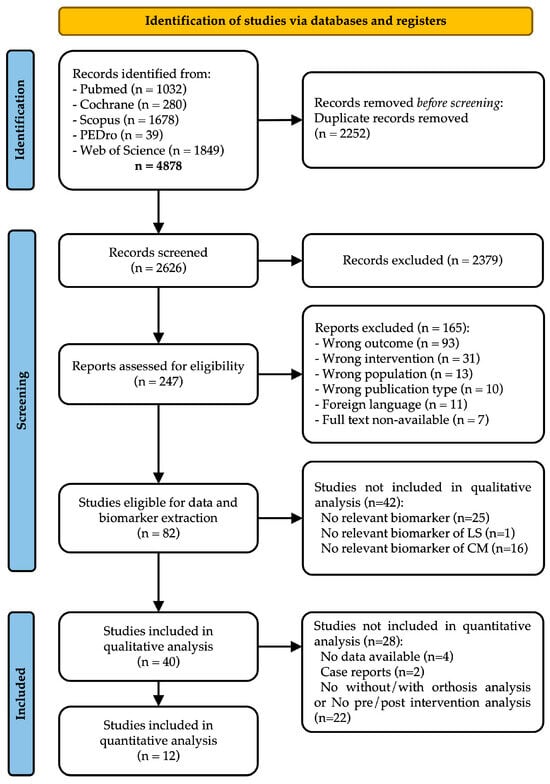
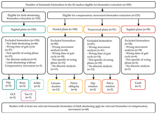
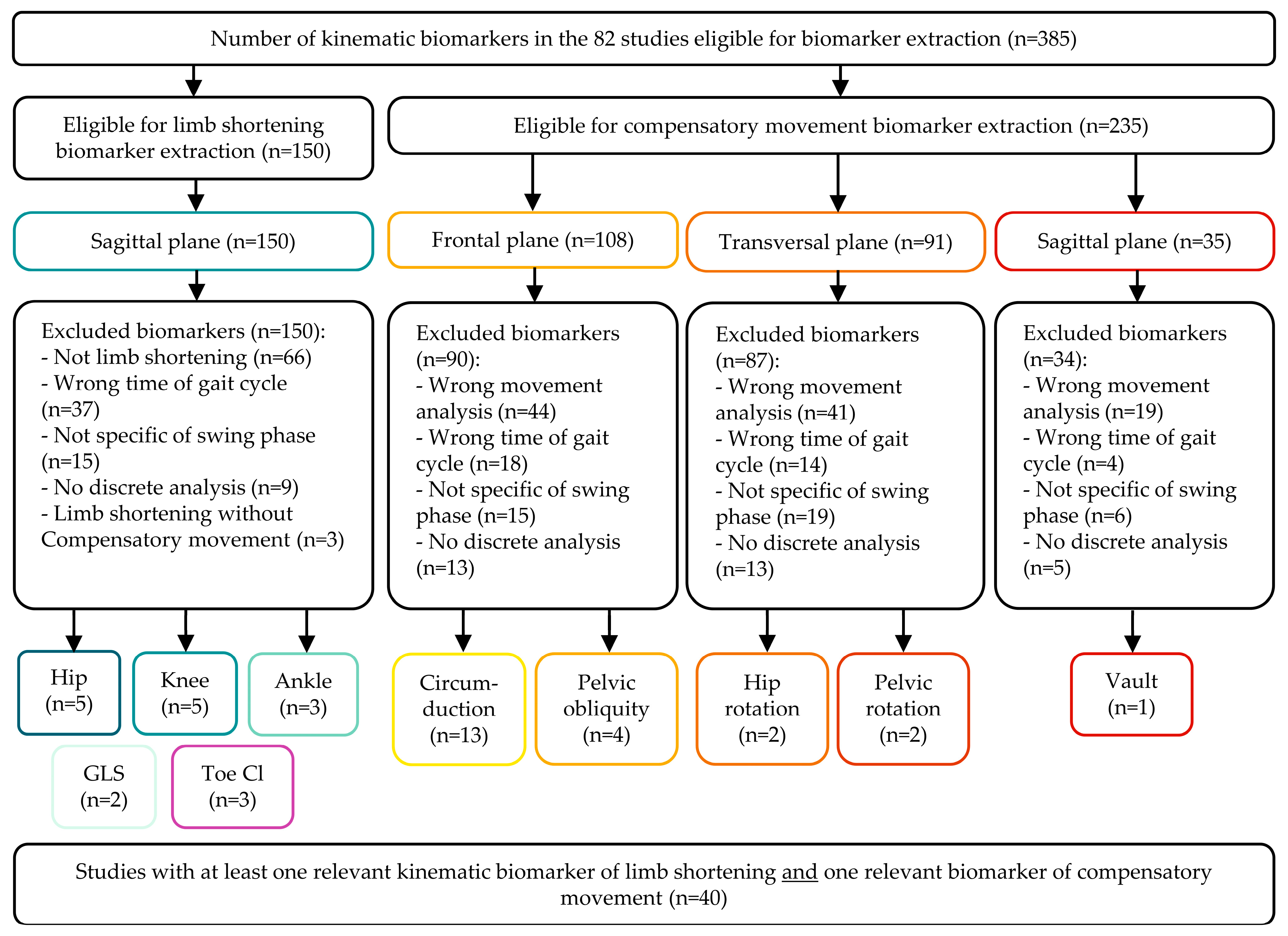
The selection process is presented in the PRISMA flowchart (Figure 1). A database search identified 4878 records. 2626 unique records were identified since this value excluded the duplicates removed, from which 82 studies met the inclusion criteria after abstract and full-text screening. The main reasons for exclusion were the absence of frontal/transversal plane data, sagittal pelvic analysis, or optoelectronic system use. Biomarker extraction followed (Figure 2), identifying 385 biomarkers: 15 for describing limb shortening, 24 for compensatory movements, and 3 for clearance. Irrelevant biomarkers and exclusion reasons are listed in Supplemental Digital Content Tables S1 and S2. Exclusions mainly resulted from incorrect gait cycle timing (n = 37, stance phase data) and inappropriate compensatory movement analysis (n = 104, lacking directional data). Following biomarker selection, 40 of the 82 eligible studies were included in the qualitative analysis. Characteristics and reasons for excluding 42 studies are detailed in Supplemental Digital Content Table S3. No additional records were found in manual reference searches.
Figure 1.
PRISMA flowchart of studies’ selection process.
Figure 2.
Flow chart of biomarkers’ selection process. Biomarkers related to limb shortening are outlined in blue-green, those reflecting compensatory movements in red-orange, and those associated with toe clearance in purple.
3.2. Population and Studies Characteristics
Among the 40 studies included, 22 were observational and 18 were interventional. A total of 891 participants were analyzed, with a mean (SD) of 22.3 (19.8) participants per study. The mean (SD) age across studies was 54.8 (7.6) years. Of these participants, 878 were post-stroke individuals, 8 had cerebral palsy, and 5 had traumatic brain injury. Within the stroke population, 616 participants (70.2%) were evaluated in the chronic phase, 182 (20.7%) in the subacute phase, 31 (3.5%) in mixed subacute-chronic cohorts [30], and the time since stroke was not specified for 49 participants (5.6%) [31,32,33].
Most studies (n = 31) were conducted in motion analysis laboratories, while the remaining nine used treadmill-based assessments. A total of 33 studies employed optoelectronic motion capture systems. Among these, the most commonly used system was Vicon (n = 22), followed by Motion Analysis (n = 7). Other systems included Qualisys, Dvideo, BTS, SMART-D140, Mac 3D, Oqus, and PhaseSpace, each used in one or two studies.
Kinematic modeling approaches varied considerably across studies. Custom-built models focusing solely on the pelvis and lower limbs were used in eight studies, while four studies employed custom models including the trunk, and one study included both trunk and head. Full-body custom models were reported in four studies. Standardized models were frequently used, including Plug-In Gait (n = 12), Helen Hayes or its modified versions (n = 5), and the Modified Cleveland Clinic model (n = 3). Less commonly used models included SAFLo, Leardini, and Total3Dgait (one study each). One study did not specify the model used.
Among the interventional studies, eight investigated kinematic parameters with and without assistive devices: mainly AFOs [19,34,35,36,37,38,39], but also a powered knee orthosis [40] and a soft wearable robot [41]. Five studies assessed changes in kinematics before and after different types of rehabilitation programs, including: functional electrical stimulation [33], rhythmic auditory stimulation [42], treadmill rehabilitation with cross-tilt [43], transcranial direct current stimulation combined with treadmill training [44], and photobiomodulation therapy combined with a static magnetic field [45]. The included studies’ characteristics are summarized in Table 1.
Table 1.
Studies characteristics.
Prior to biomarker exclusion, the studies highlighted a median (min–max) of 11 (2–91) biomarkers, including 7 (1–32) limb-shortening biomarkers and 3 (1–59) compensatory biomarkers.
Following biomarker exclusion, the studies highlighted a median (min–max) of 5 (2–10) biomarkers, including 2 (1–6) limb-shortening biomarkers and 2 (1–7) compensatory biomarkers.
3.3. Selected Biomarkers
3.3.1. Limb Shortening
We identified 15 kinematic biomarkers as relevant for describing limb shortening during the swing phase. These included five hip flexion biomarkers—maximum hip flexion during swing, hip flexion at toe-off, maximum flexion at terminal swing, maximum hip flexion during the gait cycle, and hip flexion range of motion during swing. Similarly, five knee flexion biomarkers were reported, including maximum knee flexion during swing, knee flexion at toe-off, knee flexion at mid-swing, maximum knee flexion during the cycle, and knee flexion range of motion during swing. Three ankle dorsiflexion biomarkers were included: maximum dorsiflexion during swing, ankle angle at mid-swing, and ankle dorsiflexion range of motion during swing. Additionally, two biomarkers assessed global limb shortening: minimal pelvic–toe distance and sagittal clearance angle (Figure 3).
Among these, maximum knee flexion during swing was the most frequently used biomarker (reported in 27 studies) [10,19,30,32,34,37,38,39,40,41,43,44,45,46,47,48,52,53,54,55,56,57,58,62,64,65,67], followed by maximum ankle dorsiflexion during swing (24 studies) [10,19,30,31,32,34,35,37,38,39,41,44,45,48,52,53,54,55,56,57,58,59,64,65], and maximum hip flexion during swing (14 studies) [10,19,32,34,37,38,39,53,55,56,57,58,64,65]. Other joint-specific biomarkers, such as hip flexion at toe-off (10 studies) [32,34,38,39,42,48,49,53,57,65] or knee flexion at toe-off (11 studies) [32,34,38,39,43,47,48,49,53,57,65], were less consistently used. Composite or segmental measures like the minimal pelvic–toe distance and sagittal clearance angle were [14,25,27,29,32,33,34,35,36,38,39,40,41,42,43,47,48,49,50,51,52,53,54,58,60,61,63] reported in only one study [47].
Moreover, temporal parameters—such as toe-off (11 studies) [32,34,38,39,43,47,48,49,53,57,65], mid-swing (4 studies) [36,42,46,52], or terminal swing (1 study) [42]—were not consistently specified across studies. Range-of-motion biomarkers (e.g., hip or ankle ROM during swing) were rarely used (1 study) [37].
Figure 3.
Circos-plot linking the included studies with their relevant kinematic biomarkers for limb shortening and compensatory movement description. Biomarkers are displayed in the upper part of the figure: limb-shortening biomarkers are shown on the left in blue-green, compensatory movement biomarkers on the right in yellow-orange, and clearance-related biomarkers in the centre in purple. The studies using these biomarkers are listed in the lower part of the figure. The number of relevant biomarkers per study and the number of times each biomarker was used are reported in black for each study or biomarker, respectively. The list of biomarker abbreviations is available in Table 2.
Table 2.
List of relevant biomarkers.
3.3.2. Toe Clearance
Five studies reported toe clearance using five different biomarkers (Figure 3) [34,52,61,63,65]. These included the vertical displacement of the fifth metatarsal head at mid-swing, the maximal vertical displacement of the toe marker during swing, and its minimal vertical displacement, which were used in one or two studies each. Only one study reported sagittal and frontal clearance angles, both aiming to reflect the combined contribution of limb shortening and compensatory movements to overall limb clearance.
3.3.3. Compensatory Movements
Among the 22 kinematic biomarkers relevant for compensatory movements (Figure 3), 13 quantified circumduction and 4 quantified hip hiking. Circumduction was the most frequently reported compensatory movement (13 biomarkers, 32 studies), assessed via maximum hip abduction during swing (12 studies) [30,31,34,39,40,43,48,52,53,55,57,59] or maximum hip abduction during cycle (9 studies) [30,44,45,46,51,58,60,66,67]. It was also quantified using lateral paretic limb displacement in the frontal plane with two biomarkers based on ankle or heel marker (4 studies) [36,50,58,62] and three biomarkers based on foot or toe marker (6 studies) [33,41,47,54,61,67]. Three studies proposed quantifying circumduction by measuring thigh abduction angle [46,64,65] instead of hip abduction. Hip hiking was the second most reported movement (4 biomarkers, 21 studies), mainly via maximum pelvic obliquity during swing [10,19,32,34,35,36,38,39,40,43,46,50,52,55,57,63,64,65,67], with one study measuring hip marker elevation in centimeters in the frontal plane [41].
Posterior pelvic rotation [46,55] and hip external rotation [42,53] were poorly described. Vaulting was quantified in one study [61] by measuring the maximum of non-paretic ankle dorsiflexion during stance. Posterior pelvic tilt was not considered a compensatory movement, as it primarily facilitates forward limb progression rather than compensating for limb shortening.
3.4. Quantitative Analysis
The JBI critical appraisal tools were used to assess risk of bias (Supplemental Digital Content Table S4). The primary concern in quasi-experimental studies was the absence of a control group, while in RCTs, the main issue was non-concealed treatment allocation. However, such biases did not provide sufficient grounds to exclude studies since our primary objective was to analyze 3D-IGA biomarkers, which were not influenced by study bias.
A 12-study quantitative analysis assessed effect sizes for therapeutic interventions (pre- and post-intervention and with/without orthosis) on kinematic biomarkers to evaluate which were the most sensitive to change (Table 3; full effect sizes in Supplemental Digital Content Table S5). Studies are listed in Table 1.
Table 3.
Classes of effect size of the limb-shortening and compensatory movement biomarkers based on the Hedges’ g effect size of therapeutic interventions (pre/post intervention and with/without AFO comparison). Data are shown as the number of studies (percentage of studies) [number of statistically significant Hedges’ g].
Across the 101 limb-shortening biomarkers analyses, moderate to large effect size were observed for: maximum ankle dorsiflexion increase (20/27 analyses, nine are significant) [34,35,37,38,39,41,44,45,58], maximum knee flexion increase (9/27 analyses, three are significant) [39,42,44,58], maximum knee flexion decrease (3/27, one is significant) [34,37], and hip flexion at toe-off decrease (5/11 analyses, three are significant) [39].
Across the 47 compensatory movement biomarkers analyses, intervention effects on circumduction biomarkers were heterogeneous. Positive moderate to large effect sizes were observed for: maximum hip abduction during the cycle (8/15 analyses, two are significant) [44,45,58], maximum lateral displacement of the ankle during the cycle (1/3 analysis, significant) [58]. Negative moderate to large effect sizes were observed for: maximum hip abduction during swing (1/9 analyses, significant) [39], maximum hip abduction during the cycle (1/15 analyses, significant) [60], and maximal lateral displacement of the foot during the cycle also decreased (1/1 analysis, significant) [33]. Pelvic obliquity and hip hiking biomarkers had poor to moderate effect sizes.
4. Discussion
This systematic review analysed the use of 3D-IGA biomarkers to assess limb shortening and compensatory movements specifically during the swing phase of hemiplegic gait. The objective was to evaluate how these phenomena are defined in the literature and identify the most relevant kinematic biomarkers for their quantification. The review highlighted a critical gap: only 40 out of 2626 studies (1.52%) measured both limb shortening and compensatory movements concurrently, revealing a lack of comprehensive descriptions and standardization in kinematic biomarkers. Although 3D-IGA protocols adhere to standardization, the selection and interpretation of biomarkers vary according to clinician preference, contributing to significant heterogeneity in kinematic data.
4.1. Respective Contributions of Joints on Limb Shortening
A precise description of limb shortening in hemiparetic gait requires a detailed kinematic analysis focused on the swing phase. However, many studies did not emphasize this phase, often lacking quantitative biomarkers (Supplemental Digital Content Table S3).
Definitions of mid-swing varied considerably; some identified it as the midpoint of the swing phase [46], aligning with minimal toe clearance in healthy subjects [6], while others defined it based on thigh advancement [7]. Notably, studies suggest that minimal toe clearance in hemiparetic gait may occur earlier than in healthy individuals, making it an unreliable indicator of limb shortening [68,69]. Instead, mid-swing can be defined as the moment when the toe marker aligns beneath the hip marker, corresponding to maximum limb advancement and offering a reliable reference for assessing both limb shortening and compensatory movements [6,64]. This alignment ensures that limb clearance is at its most critical phase, making it an optimal point for evaluation [68].
Selecting hip and knee biomarkers during pre-swing is relevant, as they predict flexion movements in swing. This phase influences maximum knee flexion, with muscle forces at pre-swing determining knee flexion velocity at toe-off, which strongly correlates with maximum knee flexion during swing [70].
Accordingly, and in line with the literature, we selected biomarkers that describe limb shortening at maximum flexion for the hip, knee, and ankle simultaneously to identify its origin. Additionally, we included biomarkers assessing limb shortening at maximum global limb shortening or when the vertical projection of the toe marker aligns with the hip marker. Although mid-swing biomarkers were included, they are not recommended due to variability in their concordance with limb shortening in hemiparetic gait [68].
Most studies assessed limb shortening via segmental kinematic angular analysis, quantifying the decrease in hip, knee, and ankle flexion during the swing phase (Figure 3). While informative, this approach does not fully capture the contribution of each joint to global limb shortening. An alternative method proposed by Prado-Medeiros et al. [71] involves measuring elevation angles of the thigh, shank, and foot relative to a vertical reference line, eliminating dependency on above and below-segment positioning. These relevant biomarkers are not illustrated in Figure 3 because the study did not describe any compensatory movement. Another interesting approach is to measure in millimeters both global limb shortening and the shortening of the hip–toe distance and the component of this shortening due to the hip, knee, and ankle [68].
4.2. Toe Clearance and Global Limb Shortening
Toe clearance, representing foot elevation during swing, was inconsistently measured across studies, with only five reporting on this parameter using three different biomarkers. Toe clearance could be estimated from the vertical displacement of the second or the fifth metatarsal head marker, depending on the kinematic model. The height during swing was corrected by the height during stance [34,68]. The relevance of this parameter for assessing post-stroke intervention efficacy is questionable, as it appears to be insensitive to change. For instance, Cruz et al. demonstrated an improvement in limb shortening by wearing an AFO, resulting in increased maximum ankle dorsiflexion during swing and decreased compensatory movements such as hip hiking. However, the intervention did not improve toe clearance [34]. This lack of improvement could be attributed to minimal toe clearance, which may not exceed measurement error or represent global limb clearance. Thus, improvements in limb shortening or compensations do not necessarily correlate with changes in toe clearance, although patients may experience improved comfort from reduced compensations. Quantifying maximal toe clearance may not be relevant, as it occurs late in the swing phase and does not reflect limb shortening in the critical part of the swing.
Haruyama et al. proposed minimum pelvic–toe distance as a measure of global limb shortening, defined as the linear distance between the anterior superior iliac spine and a toe marker [64]. This measure accounts for hip flexion, knee flexion, ankle dorsiflexion, and anterior pelvic tilt. This biomarker correlated strongly with gait quality, speed, and maximum knee flexion, remaining unaffected by frontal plane compensations, probably due to the omission of hip hiking in this index. A suitable biomarker for measuring global limb shortening would be limb length from hip to toe, eliminating pelvic tilt effects on apparent shortening.
Additionally, Haruyama et al. introduced the Sagittal Clearance angle, summing hip, knee, and ankle flexion, and the Frontal Clearance angle, summing hip hiking and circumduction [64]. The maximal Sagittal Clearance angle correlated positively with global limb shortening, while the Frontal Clearance angle showed a negative correlation. Although the term “clearance” may seem confusing, the distinction highlights the respective contributions to functional limb shortening. Assessing these biomarkers post-intervention could provide valuable insights.
4.3. Compensatory Movements
Circumduction was the most frequently quantified compensatory movement, with 12 biomarkers identified. Maximum abduction during the gait cycle was commonly measured, though its role in limb shortening is debated [64]. Some studies measured lateral heel displacement, while others used hip abduction angles, both of which were influenced by pelvic motion and hip hiking (Figure 3). Kerrigan et al. proposed quantifying circumduction via the coronal thigh angle relative to a vertical line, eliminating pelvic influence and allowing independent measurement [46].
Hip hiking was consistently defined using pelvic obliquity angles, with most studies assessing maximum obliquity during swing. However, obliquity during the entire gait cycle is less relevant due to its physiological role in stance. Only one study proposed quantifying hip elevation in centimeters [41]. Our review revealed that a clear definition of hip hiking and circumduction is still lacking, thus emphasizing the need for independent quantification. It seems especially pertinent to describe thigh abduction [46] and pelvic elevation [41] in conjunction.
Posterior pelvic rotation and hip external rotation were rarely examined. Additionally, pelvic motion biomarkers in the transversal and sagittal planes lacked directional information, complicating the determination of compensation direction. Measures of pelvic range of motion were often irrelevant, as they covered the entire gait cycle rather than focusing on swing-phase compensations.
For pelvic compensations, it is crucial to select biomarkers specific to the swing phase to distinguish them from other compensations, to describe them quantitatively, and provide directional information.
A promising approach by Pongpipatpaiboon et al. [68] described compensatory movements and their components (e.g., paretic side pelvic obliquity, non-paretic hip elevation, and foot elevation due to circumduction) in millimeters, facilitating direct comparison of shortening and compensatory changes accounting for proportional changes in each joint. The change in dorsiflexion in millimeters can be compared to the change in pelvic obliquity in millimeters. Although this study was excluded due to non-optoelectronic methodology, its approach remains highly relevant.
4.4. Quantitative Analysis
The quantitative analysis identified biomarkers with a moderate to large effect size following intervention. A meta-analysis was unfeasible due to heterogeneity in interventions, biomarkers and populations.
Among the 12 studies, biomarkers for limb shortening and compensatory movements showed varying degrees of responsiveness to intervention. Three limb-shortening biomarkers showed significant effect sizes post-intervention. Maximum ankle dorsiflexion during swing demonstrated the highest sensitivity, with moderate to large effect sizes in 20 of 27 analyses (9 statistically significant). Maximum knee flexion exhibited mixed effects, increasing in pre- and post-intervention studies but decreasing in with/without orthosis studies, suggesting that AFOs did not increase knee flexion or may even worsen it due to altered propulsion [72]. Hip flexion at toe-off, a key determinant of the maximum knee flexion during swing [73], showed significant responsiveness in five analyses.
For compensatory movements, circumduction biomarkers displayed substantial effect sizes post-intervention, though the direction varied. Some interventions reduced maximum hip abduction during swing or during cycle and lateral foot displacement during cycle, aligning with post-intervention or orthosis goals to minimize compensatory movements. Positive effect sizes were found for maximum hip abduction during swing and ankle lateral displacement during the cycle. All significant results came from one study on intensive stepping training in post-stroke patients, where increased frontal plane strategies likely resulted from higher walking speed or compensatory adaptation [58]. Although the direction of effect sizes varied, circumduction biomarkers were sensitive to changes due to interventions.
Conversely, pelvic obliquity and hip hiking biomarkers were largely insensitive to intervention-induced changes, potentially due to measurement variability in 3D gait analysis, where frontal plane errors can reach 2° [74].
These findings highlight limb shortening and compensatory biomarkers as sensitive to change and valuable for assessing intervention efficacy in gait analysis, reinforcing their importance in future studies.
4.5. Limitations
This study’s primary limitation was the heterogeneity of included studies, preventing a meta-analysis of intervention effects on kinematic biomarkers. Variability in study objectives also led to differences in extracted biomarkers, as some studies did not explicitly focus on limb shortening or compensatory movements. Furthermore, restricting the review to studies analyzing both limb shortening and compensatory movements may have excluded valuable research examining only one aspect.
Additionally, this review focused exclusively on 3D-IGA using optoelectronic systems, the gold standard for gait analysis. However, alternative gait analysis methods, more commonly used in clinical practice, were not included, potentially limiting the generalizability of findings. Differences in kinematic models likely influenced biomarker availability across studies. While technological advances may have facilitated access to full-body models, we observed no clear temporal trend—possibly because research teams tend to reuse familiar models for consistency and comparability. Nonetheless, all models used were sufficient to capture key features of limb shortening and compensatory strategies. Thus, improvements over time may reflect a growing awareness and evolving analytical focus rather than advances in modeling alone.
4.6. Perspectives
The absence of a standard definition for limb shortening and compensation in 3D-IGA complicates their role in global limb shortening. Akbas et al. suggest that circumduction may result from abnormal muscle coordination rather than a compensatory strategy [61]. We hypothesize that improving foot-floor clearance via limb shortening interventions (e.g., orthotics, rehabilitation, botulinum toxin) could reduce compensatory movements and energy expenditure during gait in hemiplegic individuals. Quantitative analysis suggests that both limb shortening and compensatory movement biomarkers could be useful in evaluating clinical practices. However, kinematic data variability prevents definitive conclusions, and meta-analysis was unfeasible.
Standardizing kinematic data measurement in 3D-IGA is crucial to understanding limb shortening, compensations, and gait quality. We propose key biomarkers in Table 4 to guide future studies, facilitating standardization and potential meta-analysis. We did not propose a toe clearance biomarker, as existing ones inadequately reflect tripping risk, capturing only combined limb shortening and compensatory effects. Future research should identify a more precise biomarker that better represents the subjective experience of tripping.
Table 4.
Recommendation of kinematic biomarkers to describe limb shortening and compensatory movements in hemiparetic gait. The italic font refers to hip biomarkers and indicates that the thigh biomarker should be used.
5. Conclusions
In conclusion, this systematic review identified biomarkers currently used in 3D-IGA to quantify limb shortening and compensatory movements. The lack of standardization in data extraction hinders the understanding of their relationship and impact on gait patterns. To address these gaps, we recommend a core set of biomarkers for defining global limb shortening, circumduction, and hip hiking, aiming to improve data standardization and enhance gait analysis in hemiplegic patients.
Supplementary Materials
The following supporting information can be downloaded at: https://www.mdpi.com/article/10.3390/s25154598/s1. Table S1: Lower limb biomarkers in the sagittal plane deemed irrelevant for describing limb shortening during the swing phase. Table S2: Lower limb and pelvis biomarkers in the frontal and transversal planes, and pelvis biomarkers in the sagittal plane deemed irrelevant for describing compensatory movements. Table S3: Characteristics of studies excluded from qualitative analysis. Table S4: Bias assessed according to JBI’s critical appraisal tools. Table S5: Quantitative analysis based on the Hedges’ g effect size of therapeutic interventions (pre- and post-intervention and with/without AFO comparison).
Author Contributions
Data collection, L.L. and E.M.; visualization, E.M.; writing—original draft preparation, L.L., E.M. and C.C.; writing—review and editing, M.S., C.C. and D.G.; supervision, C.C. and D.G. All authors have read and agreed to the published version of the manuscript.
Funding
This research received no external funding.
Data Availability Statement
Data extraction tables can be provided by the authors upon reasonable request.
Conflicts of Interest
The authors declare no conflicts of interest.
Abbreviations
The following abbreviations are used in this manuscript:
| 3D-IGA | Three-dimensional instrumental gait analysis |
| AFO | Ankle–foot orthosis |
| ASIS | Anterior superior iliac spine |
| DAFO | Dynamic ankle–foot orthosis |
| FES | Functional electrical stimulation |
| M2 | Second metatarsal |
| M5 | Fifth metatarsal |
| MOCAP | Motion capture system |
| PBMT-SMF | Photobiomodulation therapy combined with static magnetic field. |
| PTD | Pelvic–toe distance |
| RCT | Randomised controlled trial |
| ROM | Range of motion |
| SKG | Stiff–knee gait |
| TBI | Traumatic brain injury |
Appendix A
Research Strategy
Pubmed: (hemipleg*[tw] OR hemipare*[tw] OR hemiparaly*[tw] OR stroke[tw] OR “Cerebrovascular Accident”[tw] OR CVA[tw] OR “brain vascular disease”[tw] OR “brain injury”[tw]) AND (Gait[tw]) AND (“three-dimensional”[tw] OR “quantitative gait analysis”[tw] OR kinematics[tw] OR kinematic[tw])
Cochrane, Scopus, PEDro, and Web of Science: (hemipleg* OR hemipare* OR hemiparaly* OR stroke OR “cerebrovascular accident” OR CVA OR “brain vascular disease” OR “brain injury”) AND gait AND (“three-dimensional” or “quantitative gait analysis” OR kinematics OR kinematic)
References
- Whittle, M. Gait Analysis: An Introduction; Butterworth-Heinemann: Oxford, UK; Boston, MA, USA, 1991; ISBN 978-0-7506-0045-3. [Google Scholar]
- Jørgensen, H.S.; Nakayama, H.; Raaschou, H.O.; Olsen, T.S. Recovery of Walking Function in Stroke Patients: The Copenhagen Stroke Study. Arch. Phys. Med. Rehabil. 1995, 76, 27–32. [Google Scholar] [CrossRef] [Scilit] [PubMed]
- Balaban, B.; Tok, F. Gait Disturbances in Patients with Stroke. PM R 2014, 6, 635–642. [Google Scholar] [CrossRef] [Scilit] [PubMed]
- Woolley, S.M. Characteristics of Gait in Hemiplegia. Top. Stroke Rehabil. 2001, 7, 1–18. [Google Scholar] [CrossRef] [Scilit] [PubMed]
- Olney, S.J.; Richards, C. Hemiparetic Gait Following Stroke. Part I: Characteristics. Gait Posture 1996, 4, 136–148. [Google Scholar] [CrossRef] [Scilit]
- Moosabhoy, M.A.; Gard, S.A. Methodology for Determining the Sensitivity of Swing Leg Toe Clearance and Leg Length to Swing Leg Joint Angles during Gait. Gait Posture 2006, 24, 493–501. [Google Scholar] [CrossRef] [Scilit] [PubMed]
- Perry, J. Gait Analysis: Normal and Pathological Function; SLACK: San Francisco, CA, USA, 1992; ISBN 978-1-55642-192-1. [Google Scholar]
- Zhang, F.; Bohlen, P.; Lewek, M.D.; Huang, H. Prediction of Intrinsically Caused Tripping Events in Individuals with Stroke. IEEE Trans. Neural Syst. Rehabil. Eng. 2017, 25, 1202–1210. [Google Scholar] [CrossRef] [Scilit] [PubMed]
- Kramer, S.; Johnson, L.; Bernhardt, J.; Cumming, T. Energy Expenditure and Cost During Walking after Stroke: A Systematic Review. Arch. Phys. Med. Rehabil. 2016, 97, 619–632.e1. [Google Scholar] [CrossRef] [Scilit] [PubMed]
- Sheffler, L.R.; Bailey, S.N.; Gunzler, D.; Chae, J. Effect of Body Mass Index on Hemiparetic Gait. PM R 2014, 6, 908–913. [Google Scholar] [CrossRef] [Scilit] [PubMed]
- Whittle, M. Gait Analysis: An Introduction; Butterworth-Heinemann: Oxford, UK, 2007; ISBN 978-0-7506-8883-3. [Google Scholar]
- Milot, M.-H.; Nadeau, S.; Gravel, D.; Bourbonnais, D. Effect of Increases in Plantarflexor and Hip Flexor Muscle Strength on the Levels of Effort during Gait in Individuals with Hemiparesis. Clin. Biomech. (Bristol, Avon) 2008, 23, 415–423. [Google Scholar] [CrossRef] [Scilit] [PubMed]
- Kesar, T.M.; Perumal, R.; Jancosko, A.; Reisman, D.S.; Rudolph, K.S.; Higginson, J.S.; Binder-Macleod, S.A. Novel Patterns of Functional Electrical Stimulation Have an Immediate Effect on Dorsiflexor Muscle Function during Gait for People Poststroke. Phys. Ther. 2010, 90, 55–66. [Google Scholar] [CrossRef] [Scilit] [PubMed]
- Stoquart, G.G.; Detrembleur, C.; Palumbo, S.; Deltombe, T.; Lejeune, T.M. Effect of Botulinum Toxin Injection in the Rectus Femoris on Stiff-Knee Gait in People with Stroke: A Prospective Observational Study. Arch. Phys. Med. Rehabil. 2008, 89, 56–61. [Google Scholar] [CrossRef] [Scilit] [PubMed]
- Bleyenheuft, C.; Cockx, S.; Caty, G.; Stoquart, G.; Lejeune, T.; Detrembleur, C. The Effect of Botulinum Toxin Injections on Gait Control in Spastic Stroke Patients Presenting with a Stiff-Knee Gait. Gait Posture 2009, 30, 168–172. [Google Scholar] [CrossRef] [Scilit] [PubMed]
- Roche, N.; Boudarham, J.; Hardy, A.; Bonnyaud, C.; Bensmail, B. Use of Gait Parameters to Predict the Effectiveness of Botulinum Toxin Injection in the Spastic Rectus Femoris Muscle of Stroke Patients with Stiff Knee Gait. Eur. J. Phys. Rehabil. Med. 2015, 51, 10. [Google Scholar]
- Wada, Y.; Otaka, Y.; Mukaino, M.; Tsujimoto, Y.; Shiroshita, A.; Kawate, N.; Taito, S. The Effect of Ankle-Foot Orthosis on Ankle Kinematics in Individuals after Stroke: A Systematic Review and Meta-Analysis. PM R 2022, 14, 828–836. [Google Scholar] [CrossRef] [Scilit] [PubMed]
- Choo, Y.J.; Chang, M.C. Effectiveness of an Ankle–Foot Orthosis on Walking in Patients with Stroke: A Systematic Review and Meta-Analysis. Sci. Rep. 2021, 11, 15879. [Google Scholar] [CrossRef] [Scilit] [PubMed]
- Daryabor, A.; Arazpour, M.; Aminian, G.; Baniasad, M.; Yamamoto, S. Design and Evaluation of an Articulated Ankle Foot Orthosis with Plantarflexion Resistance on the Gait: A Case Series of 2 Patients with Hemiplegia. J. Biomed. Phys. Eng. 2020, 10, 119–128. [Google Scholar] [CrossRef] [Scilit] [PubMed]
- de l’Escalopier, N.; Voisard, C.; Michaud, M.; Moreau, A.; Jung, S.; Tervil, B.; Vayatis, N.; Oudre, L.; Ricard, D. Evaluation Methods to Assess the Efficacy of Equinovarus Foot Surgery on the Gait of Post-Stroke Hemiplegic Patients: A Literature Review. Front. Neurol. 2022, 13, 1042667. [Google Scholar] [CrossRef] [Scilit] [PubMed]
- Varvarousis, D.N.; Martzivanou, C.; Dimopoulos, D.; Dimakopoulos, G.; Vasileiadis, G.I.; Ploumis, A. The Effectiveness of Botulinum Toxin on Spasticity and Gait of Hemiplegic Patients after Stroke: A Systematic Review and Meta-Analysis. Toxicon 2021, 203, 74–84. [Google Scholar] [CrossRef] [Scilit] [PubMed]
- Van Criekinge, T.; Heremans, C.; Burridge, J.; Deutsch, J.E.; Hammerbeck, U.; Hollands, K.; Karthikbabu, S.; Mehrholz, J.; Moore, J.L.; Salbach, N.M.; et al. Standardized Measurement of Balance and Mobility Post-Stroke: Consensus-Based Core Recommendations from the Third Stroke Recovery and Rehabilitation Roundtable. Int. J. Stroke 2024, 19, 158–168. [Google Scholar] [CrossRef] [Scilit] [PubMed]
- Wonsetler, E.C.; Miller, E.L.; Huey, K.L.; Frye, S.E.; Bowden, M.G. Association between Altered Hip Extension and Kinetic Gait Variables. Am. J. Phys. Med. Rehabil. 2018, 97, 131–133. [Google Scholar] [CrossRef] [Scilit] [PubMed]
- Figueiredo, J.; Moreno, J.C.; Matias, A.C.; Pereira, F.; Santos, C.P. Outcome Measures and Motion Capture Systems for Assessing Lower Limb Orthosis-Based Interventions after Stroke: A Systematic Review. Disabil. Rehabil. Assist. Technol. 2021, 16, 674–683. [Google Scholar] [CrossRef] [Scilit] [PubMed]
- Li, J.; Kwong, P.W.; Lin, W.; Fong, K.N.; Wu, W.; Sidarta, A. Assessment of Ambulation Functions through Kinematic Analysis in Individuals with Stroke: A Systematic Review. Eur. J. Phys. Rehabil. Med. 2025, 61, 28–40. [Google Scholar] [CrossRef] [Scilit] [PubMed]
- Gu, Z.; Gu, L.; Eils, R.; Schlesner, M.; Brors, B. Circlize Implements and Enhances Circular Visualization in R. Bioinformatics 2014, 30, 2811–2812. [Google Scholar] [CrossRef] [Scilit] [PubMed]
- Krzywinski, M.; Schein, J.; Birol, I.; Connors, J.; Gascoyne, R.; Horsman, D.; Jones, S.J.; Marra, M.A. Circos: An Information Aesthetic for Comparative Genomics. Genome Res. 2009, 19, 1639–1645. [Google Scholar] [CrossRef] [Scilit] [PubMed]
- Cumming, G. The New Statistics: Why and How. Psychol. Sci. 2014, 25, 7–29. [Google Scholar] [CrossRef] [Scilit] [PubMed]
- Borenstein, M.; Hedges, L.V.; Higgins, J.P.T.; Rothstein, H.R. Introduction to Meta-Analysis; John Wiley & Sons, Inc.: Hoboken, NJ, USA, 2009. [Google Scholar]
- Nedergård, H.; Schelin, L.; Liebermann, D.G.; Johansson, G.M.; Häger, C.K. Core Sets of Kinematic Variables to Consider for Evaluation of Gait Post-Stroke. Front. Hum. Neurosci. 2022, 15, 820104. [Google Scholar] [CrossRef] [Scilit] [PubMed]
- Cretual, A.; Bervet, K.; Ballaz, L. Gillette Gait Index in Adults. Gait Posture 2010, 32, 307–310. [Google Scholar] [CrossRef] [Scilit] [PubMed]
- Silva, M.R.E.; Jacinto, J. Velocity Determinants in Spastic Patients after Stroke-A Gait Analysis Study. Neurol. Int. 2020, 12, 48–54. [Google Scholar] [CrossRef] [Scilit] [PubMed]
- Qian, J.; Rong, K.; Qian, Z.; Wen, C.; Zhang, S. Effects of a Multichannel Dynamic Functional Electrical Stimulation System on Hemiplegic Gait and Muscle Forces. J. Phys. Ther. Sci. 2015, 27, 3541–3544. [Google Scholar] [CrossRef] [Scilit] [PubMed]
- Cruz, T.H.; Dhaher, Y.Y. Impact of Ankle-Foot-Orthosis on Frontal Plane Behaviors Post-Stroke. Gait Posture 2009, 30, 312–316. [Google Scholar] [CrossRef] [Scilit] [PubMed]
- Boudarham, J.; Pradon, D.; Roche, N.; Bensmail, D.; Zory, R. Effects of a Dynamic-Ankle-Foot Orthosis (Liberté®) on Kinematics and Electromyographic Activity during Gait in Hemiplegic Patients with Spastic Foot Equinus. NeuroRehabilitation 2014, 35, 369–379. [Google Scholar] [CrossRef] [Scilit] [PubMed]
- Zissimopoulos, A.; Fatone, S.; Gard, S. Effects of Ankle-Foot Orthoses on Mediolateral Foot-Placement Ability during Post-Stroke Gait. Prosthet. Orthot. Int. 2015, 39, 372–379. [Google Scholar] [CrossRef] [Scilit] [PubMed]
- Zollo, L.; Zaccheddu, N.; Ciancio, A.L.; Morrone, M.; Bravi, M.; Santacaterina, F.; Milazzo, M.L.; Guglielmelli, E.; Sterzi, S. Comparative Analysis and Quantitative Evaluation of Ankle-Foot Orthoses for Foot Drop in Chronic Hemiparetic Patients. Eur. J. Phys. Rehabil. Med. 2015, 51, 12. [Google Scholar]
- Nikamp, C.D.M.; Hobbelink, M.S.H.; van der Palen, J.; Hermens, H.J.; Rietman, J.S.; Buurke, J.H. A Randomized Controlled Trial on Providing Ankle-Foot Orthoses in Patients with (Sub-)Acute Stroke: Short-Term Kinematic and Spatiotemporal Effects and Effects of Timing. Gait Posture 2017, 55, 15–22. [Google Scholar] [CrossRef] [Scilit] [PubMed]
- Nikamp, C.D.M.; van der Palen, J.; Hermens, H.J.; Rietman, J.S.; Buurke, J.H. The Influence of Early or Delayed Provision of Ankle-Foot Orthoses on Pelvis, Hip and Knee Kinematics in Patients with Sub-Acute Stroke: A Randomized Controlled Trial. Gait Posture 2018, 63, 260–267. [Google Scholar] [CrossRef] [Scilit] [PubMed]
- Sulzer, J.S.; Gordon, K.E.; Dhaher, Y.Y.; Peshkin, M.A.; Patton, J.L. Preswing Knee Flexion Assistance Is Coupled with Hip Abduction in People with Stiff-Knee Gait after Stroke. Stroke 2010, 41, 1709–1714. [Google Scholar] [CrossRef] [Scilit] [PubMed]
- Awad, L.N.; Bae, J.; Kudzia, P.; Long, A.; Hendron, K.; Holt, K.G.; O’Donnell, K.; Ellis, T.D.; Walsh, C.J. Reducing Circumduction and Hip Hiking During Hemiparetic Walking Through Targeted Assistance of the Paretic Limb Using a Soft Robotic Exosuit. Am. J. Phys. Med. Rehabil. 2017, 96, S157–S164. [Google Scholar] [CrossRef] [Scilit] [PubMed]
- Shin, Y.-K.; Chong, H.J.; Kim, S.J.; Cho, S.-R. Effect of Rhythmic Auditory Stimulation on Hemiplegic Gait Patterns. Yonsei Med. J. 2015, 56, 1703. [Google Scholar] [CrossRef] [Scilit] [PubMed]
- Reissman, M.E.; Gordon, K.E.; Dhaher, Y.Y. Manipulating Post-Stroke Gait: Exploiting Aberrant Kinematics. J. Biomech. 2018, 67, 129–136. [Google Scholar] [CrossRef] [Scilit] [PubMed]
- Dumont, A.J.L.; Cimolin, V.; Parreira, R.B.; Armbrust, D.; Fonseca, D.R.P.; Fonseca, A.L.; Cordeiro, L.; Franco, R.C.; Duarte, N.A.C.; Galli, M.; et al. Effects of Transcranial Direct Current Stimulation Combined with Treadmill Training on Kinematics and Spatiotemporal Gait Variables in Stroke Survivors: A Randomized, Triple-Blind, Sham-Controlled Study. Brain Sci. 2022, 13, 11. [Google Scholar] [CrossRef] [Scilit] [PubMed]
- Dumont, A.J.L.; Casalechi, H.L.; Tomazoni, S.S.; Grecco, L.C.; Galli, M.; Oliveira, C.S.; Leal-Junior, E.C.P. Photobiomodulation Therapy Combined with Static Magnetic Field (PBMT–SMF) on Spatiotemporal and Kinematics Gait Parameters in Post-Stroke: A Pilot Study. Life 2022, 12, 186. [Google Scholar] [CrossRef] [Scilit] [PubMed]
- Kerrigan, D.C.; Frates, E.P.; Rogan, S.; Riley, P.O. Hip Hiking and Circumduction: Quantitative Definitions. Am. J. Phys. Med. Rehabil. 2000, 79, 247–252. [Google Scholar] [CrossRef] [Scilit] [PubMed]
- Chen, G.; Patten, C.; Kothari, D.H.; Zajac, F.E. Gait Differences between Individuals with Post-Stroke Hemiparesis and Non-Disabled Controls at Matched Speeds. Gait Posture 2005, 22, 51–56. [Google Scholar] [CrossRef] [Scilit] [PubMed]
- Kinsella, S.; Moran, K. Gait Pattern Categorization of Stroke Participants with Equinus Deformity of the Foot. Gait Posture 2008, 27, 144–151. [Google Scholar] [CrossRef] [Scilit] [PubMed]
- Nolan, K.J.; Savalia, K.K.; Yarossi, M.; Elovic, E.P. Evaluation of a Dynamic Ankle Foot Orthosis in Hemiplegic Gait: A Case Report. NeuroRehabilitation 2010, 27, 343–350. [Google Scholar] [CrossRef] [Scilit] [PubMed]
- Tyrell, C.M.; Roos, M.A.; Rudolph, K.S.; Reisman, D.S. Influence of Systematic Increases in Treadmill Walking Speed on Gait Kinematics after Stroke. Phys. Ther. 2011, 91, 392–403. [Google Scholar] [CrossRef] [Scilit] [PubMed]
- Carmo, A.A.; Kleiner, A.F.R.; da Costa, P.H.L.; Barros, R.M.L. Three-Dimensional Kinematic Analysis of Upper and Lower Limb Motion during Gait of Post-Stroke Patients. Braz. J. Med. Biol. Res. 2012, 45, 537–545. [Google Scholar] [CrossRef] [Scilit] [PubMed]
- Stanhope, V.A.; Knarr, B.A.; Reisman, D.S.; Higginson, J.S. Frontal Plane Compensatory Strategies Associated with Self-Selected Walking Speed in Individuals Post-Stroke. Clin. Biomech. (Bristol, Avon) 2014, 29, 518–522. [Google Scholar] [CrossRef] [Scilit] [PubMed]
- Manca, M.; Ferraresi, G.; Cosma, M.; Cavazzuti, L.; Morelli, M.; Benedetti, M.G. Gait Patterns in Hemiplegic Patients with Equinus Foot Deformity. BioMed Res. Int. 2014, 2014, 939316. [Google Scholar] [CrossRef] [Scilit] [PubMed]
- Burpee, J.L.; Lewek, M.D. Biomechanical Gait Characteristics of Naturally Occurring Unsuccessful Foot Clearance during Swing in Individuals with Chronic Stroke. Clin. Biomech. (Bristol, Avon) 2015, 30, 1102–1107. [Google Scholar] [CrossRef] [Scilit] [PubMed]
- Roche, N.; Bonnyaud, C.; Geiger, M.; Bussel, B.; Bensmail, D. Relationship between Hip Flexion and Ankle Dorsiflexion during Swing Phase in Chronic Stroke Patients. Clin. Biomech. (Bristol, Avon) 2015, 30, 219–225. [Google Scholar] [CrossRef] [Scilit] [PubMed]
- Yao, D.; Jakubowitz, E.; Tecante, K.; Lahner, M.; Ettinger, S.; Claassen, L.; Plaass, C.; Stukenborg-Colsman, C.; Daniilidis, K. Restoring Mobility after Stroke: First Kinematic Results from a Pilot Study with a Hybrid Drop Foot Stimulator. Musculoskelet. Surg. 2016, 100, 223–229. [Google Scholar] [CrossRef] [Scilit] [PubMed]
- Chantraine, F.; Filipetti, P.; Schreiber, C.; Remacle, A.; Kolanowski, E.; Moissenet, F. Proposition of a Classification of Adult Patients with Hemiparesis in Chronic Phase. PLoS ONE 2016, 11, e0156726. [Google Scholar] [CrossRef] [Scilit] [PubMed]
- Mahtani, G.B.; Kinnaird, C.R.; Connolly, M.; Holleran, C.L.; Hennessy, P.W.; Woodward, J.; Brazg, G.; Roth, E.J.; Hornby, T.G. Altered Sagittal- and Frontal-Plane Kinematics Following High-Intensity Stepping Training Versus Conventional Interventions in Subacute Stroke. Phys. Ther. 2017, 97, 320–329. [Google Scholar] [CrossRef] [Scilit] [PubMed]
- Barroso, F.O.; Torricelli, D.; Molina-Rueda, F.; Alguacil-Diego, I.M.; Cano-de-la-Cuerda, R.; Santos, C.; Moreno, J.C.; Miangolarra-Page, J.C.; Pons, J.L. Combining Muscle Synergies and Biomechanical Analysis to Assess Gait in Stroke Patients. J. Biomech. 2017, 63, 98–103. [Google Scholar] [CrossRef] [Scilit] [PubMed]
- Wang, H.-Q.; Dong, G.-R.; Bao, C.-L.; Jiao, Z.-H. Immediate Effect of Scalp Acupuncture on the Gait of Patients with Subacute Intracerebral Haemorrhage Analysed by Three-Dimensional Motion: Secondary Analysis of a Randomised Controlled Trial. Acupunct. Med. 2018, 36, 71–79. [Google Scholar] [CrossRef] [Scilit] [PubMed]
- Akbas, T.; Prajapati, S.; Ziemnicki, D.; Tamma, P.; Gross, S.; Sulzer, J. Hip Circumduction Is Not a Compensation for Reduced Knee Flexion Angle during Gait. J. Biomech. 2019, 87, 150–156. [Google Scholar] [CrossRef] [Scilit] [PubMed]
- Dean, J.C.; Bowden, M.G.; Kelly, A.L.; Kautz, S.A. Altered Post-Stroke Propulsion is Related to Paretic Swing Phase Kinematics. Clin. Biomech. (Bristol, Avon) 2020, 72, 24–30. [Google Scholar] [CrossRef] [Scilit] [PubMed]
- Van Criekinge, T.; Wim, S.; Nolan, H.; Patricia, V.D.W.; Willem, D.H.; Steven, T.; Ann, H. Trunk Biomechanics during Walking after Sub-Acute Stroke and Its Relation to Lower Limb Impairments. Clin. Biomech. (Bristol, Avon) 2020, 75, 105013. [Google Scholar] [CrossRef] [Scilit]
- Haruyama, K.; Kawakami, M.; Okada, K.; Okuyama, K.; Tsuzuki, K.; Liu, M. Pelvis-Toe Distance: 3-Dimensional Gait Characteristics of Functional Limb Shortening in Hemiparetic Stroke. Sensors 2021, 21, 5417. [Google Scholar] [CrossRef] [Scilit] [PubMed]
- Sekiguchi, Y.; Honda, K.; Izumi, S.-I. Effect of Walking Adaptability on an Uneven Surface by a Stepping Pattern on Walking Activity After Stroke. Front. Hum. Neurosci. 2022, 15, 762223. [Google Scholar] [CrossRef] [Scilit] [PubMed]
- Steffensen, E.A.; Magalhães, F.; Knarr, B.A.; Kingston, D.C. Comparison of Markerless and Marker-Based Motion Capture of Gait Kinematics in Individuals with Cerebral Palsy and Chronic Stroke: A Case Study Series. Res. Sq. 2023. [Google Scholar] [CrossRef] [Scilit]
- Kettlety, S.A.; Finley, J.M.; Reisman, D.S.; Schweighofer, N.; Leech, K.A. Speed-Dependent Biomechanical Changes Vary across Individual Gait Metrics Post-Stroke Relative to Neurotypical Adults. J. Neuroeng. Rehabil. 2023, 20, 14. [Google Scholar] [CrossRef] [Scilit] [PubMed]
- Pongpipatpaiboon, K.; Mukaino, M.; Matsuda, F.; Ohtsuka, K.; Tanikawa, H.; Yamada, J.; Tsuchiyama, K.; Saitoh, E. The Impact of Ankle–Foot Orthoses on Toe Clearance Strategy in Hemiparetic Gait: A Cross-Sectional Study. J. Neuroeng. Rehabil. 2018, 15, 41. [Google Scholar] [CrossRef] [Scilit] [PubMed]
- Begg, R.; Best, R.; Dell’Oro, L.; Taylor, S. Minimum Foot Clearance during Walking: Strategies for the Minimisation of Trip-Related Falls. Gait Posture 2007, 25, 191–198. [Google Scholar] [CrossRef] [Scilit] [PubMed]
- Goldberg, S.R.; Ounpuu, S.; Delp, S.L. The Importance of Swing-Phase Initial Conditions in Stiff-Knee Gait. J. Biomech. 2003, 36, 1111–1116. [Google Scholar] [CrossRef] [Scilit] [PubMed]
- Prado-Medeiros, C.L.; Sousa, C.O.; Souza, A.S.; Soares, M.R.; Barela, A.M.F.; Salvini, T.F. Effects of the Addition of Functional Electrical Stimulation to Ground Level Gait Training with Body Weight Support after Chronic Stroke. Rev. Bras. Fisioter. 2011, 15, 436–444. [Google Scholar] [CrossRef] [Scilit] [PubMed]
- Daryabor, A.; Arazpour, M.; Aminian, G. Effect of Different Designs of Ankle-Foot Orthoses on Gait in Patients with Stroke: A Systematic Review. Gait Posture 2018, 62, 268–279. [Google Scholar] [CrossRef] [Scilit] [PubMed]
- Fox, M.D.; Delp, S.L. Contributions of Muscles and Passive Dynamics to Swing Initiation over a Range of Walking Speeds. J. Biomech. 2010, 43, 1450–1455. [Google Scholar] [CrossRef] [Scilit] [PubMed]
- McGinley, J.L.; Baker, R.; Wolfe, R.; Morris, M.E. The Reliability of Three-Dimensional Kinematic Gait Measurements: A Systematic Review. Gait Posture 2009, 29, 360–369. [Google Scholar] [CrossRef] [Scilit] [PubMed]

Disclaimer/Publisher’s Note: The statements, opinions and data contained in all publications are solely those of the individual author(s) and contributor(s) and not of MDPI and/or the editor(s). MDPI and/or the editor(s) disclaim responsibility for any injury to people or property resulting from any ideas, methods, instructions or products referred to in the content. |
© 2025 by the authors. Licensee MDPI, Basel, Switzerland. This article is an open access article distributed under the terms and conditions of the Creative Commons Attribution (CC BY) license (https://creativecommons.org/licenses/by/4.0/).