


Abstract
The tobacco ground beetle, Mesomorphus villiger (Blanchard, 1853) (Coleoptera: Tenebrionidae), is a widely distributed species across Asia, Africa, and Australia, recognized for its economic importance as a pest of tobacco seedlings and other crops in India. Despite its pest status and characteristic aggregation behavior, information on its antennae and antennal sensory structures remains limited. In this study, the morphology of the antennae and the types and distribution of antennal sensilla in M. villiger males were examined using scanning electron microscopy. The antennae had 11 segments, including a scape, a pedicel, and a flagellum, with the last three flagellomeres forming a club. Six types of sensilla were identified: Böhm’s bristles, sensilla chaetica (two subtypes), sensilla trichodea, sensilla basiconica (three subtypes), sensilla auricillica (two subtypes) and grooved peg sensilla. The structural characteristics and distributional patterns of the sensilla suggest functional specializations for olfactory, mechanoreceptive, and chemioreceptive roles. Sensilla diversity progressively increased from proximal to distal antennal regions, with the highest number of types observed on the club. This study provides the first comprehensive ultrastructural description of the antennae and antennal sensilla in M. villiger males, establishing a morphological foundation for future electrophysiological and behavioral investigations to be extended to females and supporting the development of innovative management strategies for this species.
1. Introduction
The family Tenebrionidae (Coleoptera) includes several economically important species that feed on plants, dead wood, and stored grains. This family includes approximately 15,000 species, classified into 10 subfamilies and 96 tribes [1]. Mesomorphus villiger (Blanchard, 1853) (Coleoptera: Tenebrionidae), commonly known as the tobacco ground beetle, is widely distributed across Asia (India, Afghanistan and Siberia), Africa, and Australia [2,3]. In southern India, this species feeds on the stems of newly planted tobacco seedlings, often causing seedling death. It also nibbles on fallen young and tender leaves of other plants, including mango, Mangifera indica L. (Sapindales: Anacardiaceae), rubber, Hevea brasiliensis Müll. Arg. (Malpighiales: Euphorbiaceae), cashew, Anacardium occidentale L. (Sapindales: Anacardiaceae), and rain tree, Samanea saman (Jacq.) Merr. (Fabales: Fabaceae) [2]. In addition, during the monsoon season, M. villiger often enters homes and buildings in large aggregations, causing serious disturbances because of nocturnal activity and release of an irritating quinonic secretion, which may cause mild skin burns [2].
In northern and central India, this beetle has been observed forming large aggregations also on old Sal trees, Shorea robusta Roth (Malvales: Dipterocarpaceae) [4].
The sensillar array on insect antennae plays a pivotal role in detecting chemical, mechanical, and thermal stimuli. The morphological specificity of these sensilla is considered an evolutionary adaptation to different ecological roles, enabling insects to effectively perceive and respond to environmental cues [5,6]. In the Tenebrionidae family, the antennal ultrastructure in adults have been previously investigated. Currently available data concern species belonging to the subfamilies Diaperinae, Pimeliinae and Tenebrioninae. For each subfamily, the species for which these data have been reported are as follows: Diaperinae: Ulomoides dermestoides (Chevrolat, 1878) [7]; Pimeliinae: Asida jurinei subsp. jurinei Solier, 1836, Asida jurinei subsp. pyrenaea Baudi di Selve, 1875 Asida marmottani C. Brisout de Barneville, 1863 [8]; Adelostoma sulcatum (Duponchel, 1827) [9]; Pachychila (Pachychila) impunctata Fairmaire, 1860 [10]; Tenebrioninae: Tribolium confusum (Herbst, 1797) [11,12]; Tribolium castaneum Herbst, 1797 [13,14,15,16]; Tenebrio molitor Linnaeus, 1758 [17]; Alphitobius diaperinus (Panzer, 1797) [18]; and Scaurus gigas Walt, 1835 [19].
However, no studies have yet been conducted on the antennae of the genus Mesomorphus. The present study examines the morphology of the antenna and the types, number, and distribution of sensillar structures of the antenna in males of M. villiger using scanning electron microscopy. This study focuses on males as a first step toward characterizing the antennal sensory system of this species. Providing a detailed ultrastructural description of the male antennae represents a necessary morphological baseline for future comparative investigations including females, as well as for electrophysiological and behavioral studies aimed at clarifying the functional role of the sensilla. These data may also be useful to develop suitable management strategies against this insect pest species in India.
2. Materials and Methods
2.1. Insect Collection
Adult males of Mesomorphus villiger were collected in June 2022 from a Sal forest at the Forest Research Institute, Dehradun, Uttarakhand, India, during aggregation events on trees of S. robusta. The individuals were collected using tweezers, preserved in absolute ethanol (99.9%) and identified through morphological criteria using taxonomical keys [20]. Sex identification of M. villiger males was based on the diagnostic characters of the first three dilated protarsal segments and the presence of dense micro-setae on the ventral side [20]. The sex identification of males was confirmed by gently squeezing the abdomen of the collected specimens and exposing the genitalia.
2.2. Scanning Electron Microscopy
For scanning electron microscopy (SEM) examination, the heads of eight adult males were dissected under a stereomicroscope (Nikon SMZ1500, Tokyo, Japan) using fine surgical blades. Five heads were used for morphological analyses of the antennae and antennal sensilla, and three were used for the analysis of sensillar distribution. The detached heads were rinsed in a liquid soap solution (1 part dishwashing liquid:99 parts distilled water) to remove surface dust and debris, followed by immersion in phosphate-buffered saline (PBS, pH 7.0) for a final rinsing. Samples were dehydrated in a graded ethanol series (50%, 70%, 80%, and 100%) for 15 min at each step. Each antenna was then transferred to a vial containing acetone (99%; Loba Chemie Pvt. Ltd., Pune, Maharashtra, India) for 3 min to ensure complete dehydration. The samples were air-dried and mounted on aluminum stubs using double-sided conductive adhesive tape, then sputter-coated with gold in a Quorum Q150 ES Plus coater (Quorum Technologies, Laughton, East Sussex, UK). Prepared specimens were observed using a Zeiss Gemini 1 Sigma 500 SEM (Carl Zeiss, Jena, Germany) operated with EHT = 3–5 kV.
2.3. Antennal and Sensillar Terminology
The terminology of the antennal segments of M. villiger males followed that of previously published studies [21,22], regarding the different types and subtypes of sensilla found on the scape, pedicel and flagellum [23,24,25], based on the following criteria: shape, presence of microperforations and/or grooves, sharp or blunt tips, articulated base, and presence of pores at the base of the sensillum.
2.4. Analysis of Sensillar Distribution
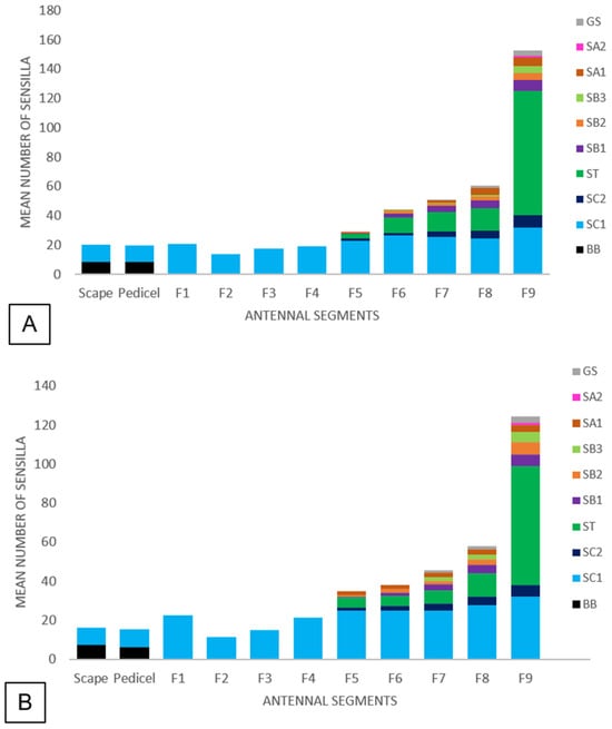
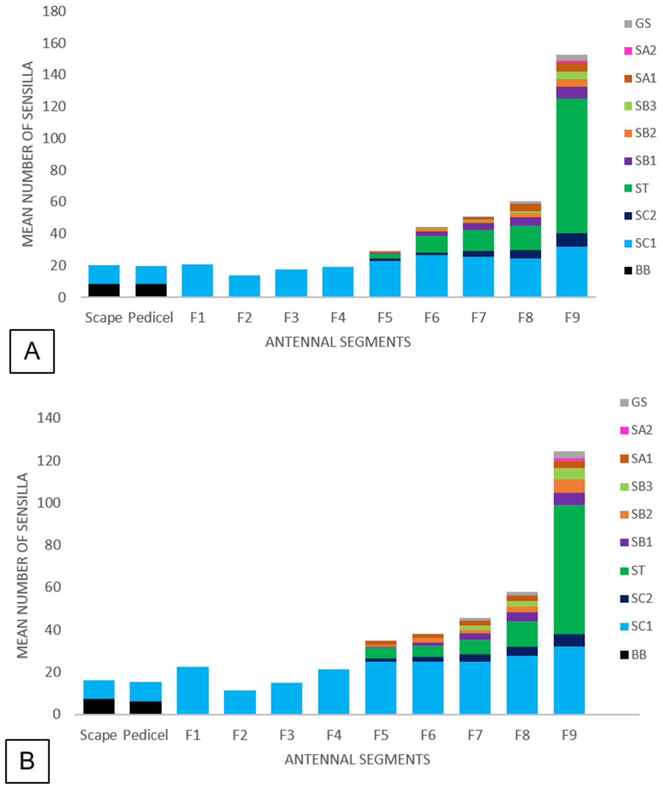
In order to count the sensilla on the dorsal and ventral sides, both antennae were detached from three individuals and placed on SEM stubs, always counting the sensilla on the dorsal side of the left antenna and the ventral side of the right one. All types and subtypes of sensilla visible in SEM micrographs were recorded for each antennal segment (scape, pedicel, and flagellomeres F1–F9) in three individuals. Sensillar counts were performed separately for each antennal segment and surface, counting only clearly visible sensilla on each examined surface. The overlapping sensilla were counted individually using high-magnification SEM images from multiple viewing angles. The mean numerical values were calculated for all types and subtypes of sensilla on the dorsal and ventral side of each segment of the antenna and reported as stacked bar plots obtained by Microsoft Excel (version 2026).
The distribution of types and subtypes of sensilla on segments was reported also as a manual drawing of the image of the antenna in toto in Microsoft PowerPoint (version 2016), filling each segment with the sensillar types and distribution according to the SEM images. The lengths of the antenna, the segments and the different types and subtypes of sensilla were measured using ImageJ software (version 1.54g; National Institutes of Health, Bethesda, MD, USA) and expressed as mean with standard deviation.
3. Results
3.1. Antennal and Sensillar Morphology
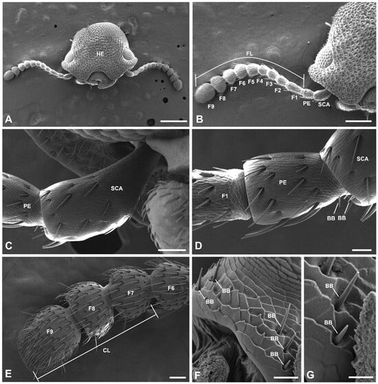
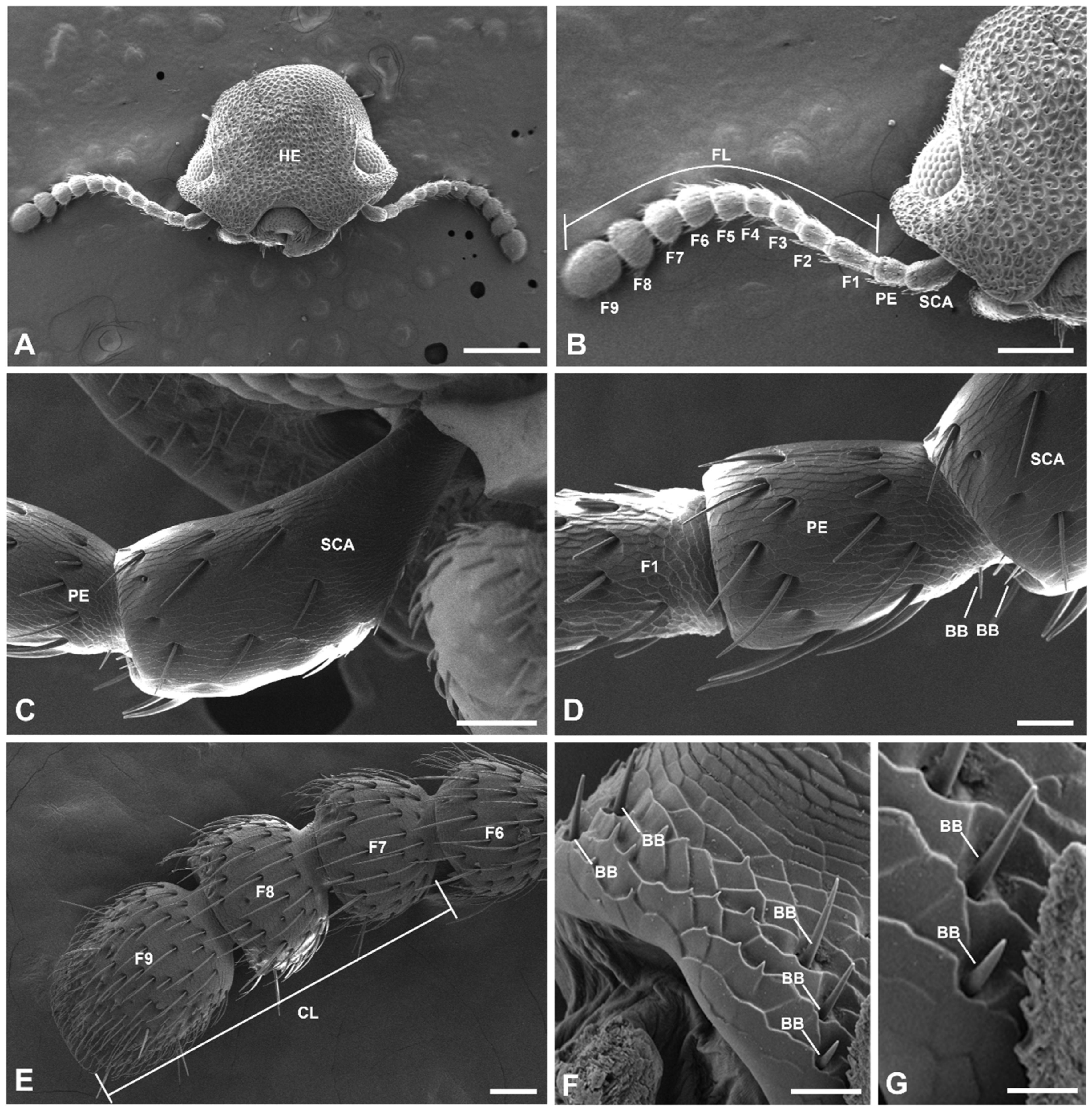
The antennae of the adult male of Mesomorphus villiger are shown in toto in Figure 1A. The antennae, clavate, had lengths of 1.78 ± 0.02 mm (measured on both antennae of five individuals) and were composed of 11 segments (Figure 1A,B), with the last three segments forming a club. The antenna had three parts: the scape, the pedicel, and the flagellum. The scape (length = 0.18 ± 0.06 mm, n = 5) was clavate and had a thinner proximal end, articulated with the head (Figure 1C). The pedicel (length = 0.14 ± 0.04 mm, n = 5) was more or less cylindrical (Figure 1D). The flagellum (length = 1.49 ± 0.01 mm, n = 5) had nine flagellomeres (Figure 1B,E), of which the last three formed a terminal club (Figure 1E). During this study, six types of antennal sensilla and their subtypes were identified: Böhm’s bristles (BB); sensilla chaetica (SC) with two subtypes (SC1 and SC2); sensilla trichodea (ST); sensilla basiconica (SB) with three subtypes (SB1, SB2, and SB3); sensilla auricillica (SA) with two subtypes (SA1 and SA2); and grooved peg sensilla (GS).
Figure 1.
Scanning electron micrographs of the antenna of an adult male of M. villiger. (A) Frontal view of the head, showing the antennae in toto. (B) Dorsal view of the right antenna. (C) Dorsal view of the scape. (D) Dorsal view of the pedicel. (E) The four terminal flagellomeres, of which F7, F8 and F9 form the club. (F) Proximal region of the scape. (G) Detail of the lower left region of the scape, showing two Böhm’s bristles. Scale bars: (A) = 500 μm; (B) = 250 μm; (C) = 50 μm; (D) = 50 μm; (E) = 25 μm; (F) = 10 μm; (G) = 5 μm. Abbreviations: BB, Böhm’s bristles; CL, club; FL, flagellum; F1 to F9, flagellomeres 1–9; HE, head; PE, pedicel; SCA, scape.
The diversity of sensilla progressively increased from the proximal to the distal regions of the antenna, with the highest number of types observed on the club. The sensilla of the antennae are described in sequence, starting from the scape. The lengths of all types or subtypes of sensilla are reported in Table 1.
Table 1.
Types and subtypes of sensilla and their mean length measured on the dorsal and ventral surface of the antenna in males of Mesomorphus villiger. Abbreviations: n, number of sensilla measured. Abbreviations: BB, Böhm’s bristles; GS, grooved peg sensillum; SA1, sensillum auricillicum of subtype 1; SA2, sensillum auricillicum of subtype 2; SB1, sensillum basiconicum of subtype 1; SB2, sensillum basiconicum of subtype 2; SB3, sensillum basiconicum of subtype 3; SC1, sensillum chaeticum of subtype 1; SC2, sensillum chaeticum of subtype 2; ST, sensillum trichodeum.
Böhm’s bristles (BB) (Figure 1D,F,G) were short structures, roughly triangular and articulated at the base.
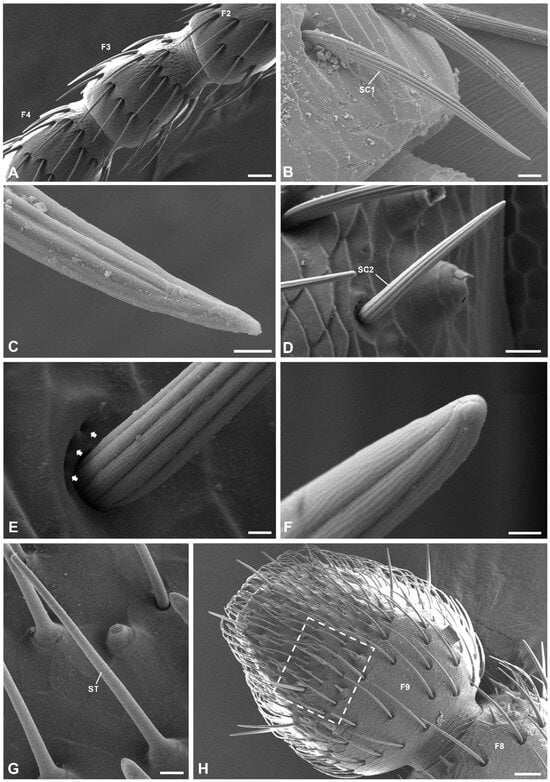
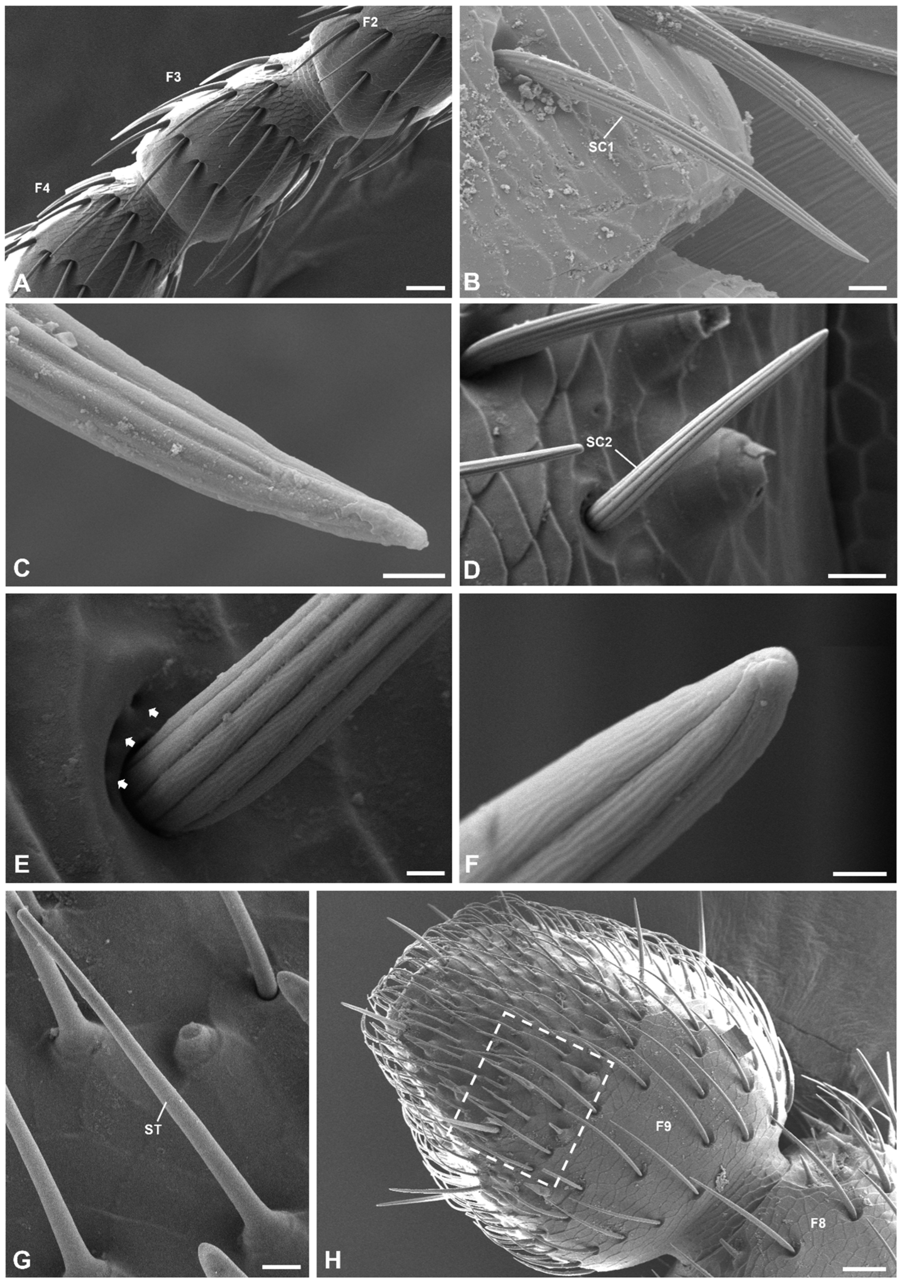
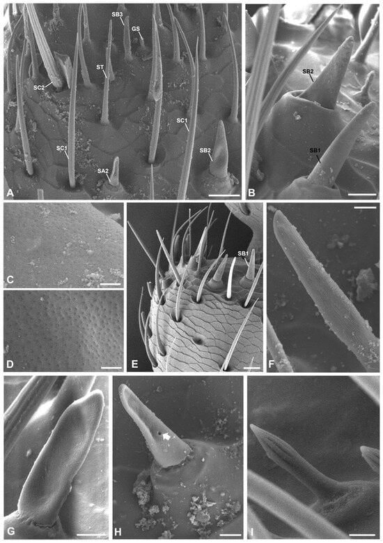
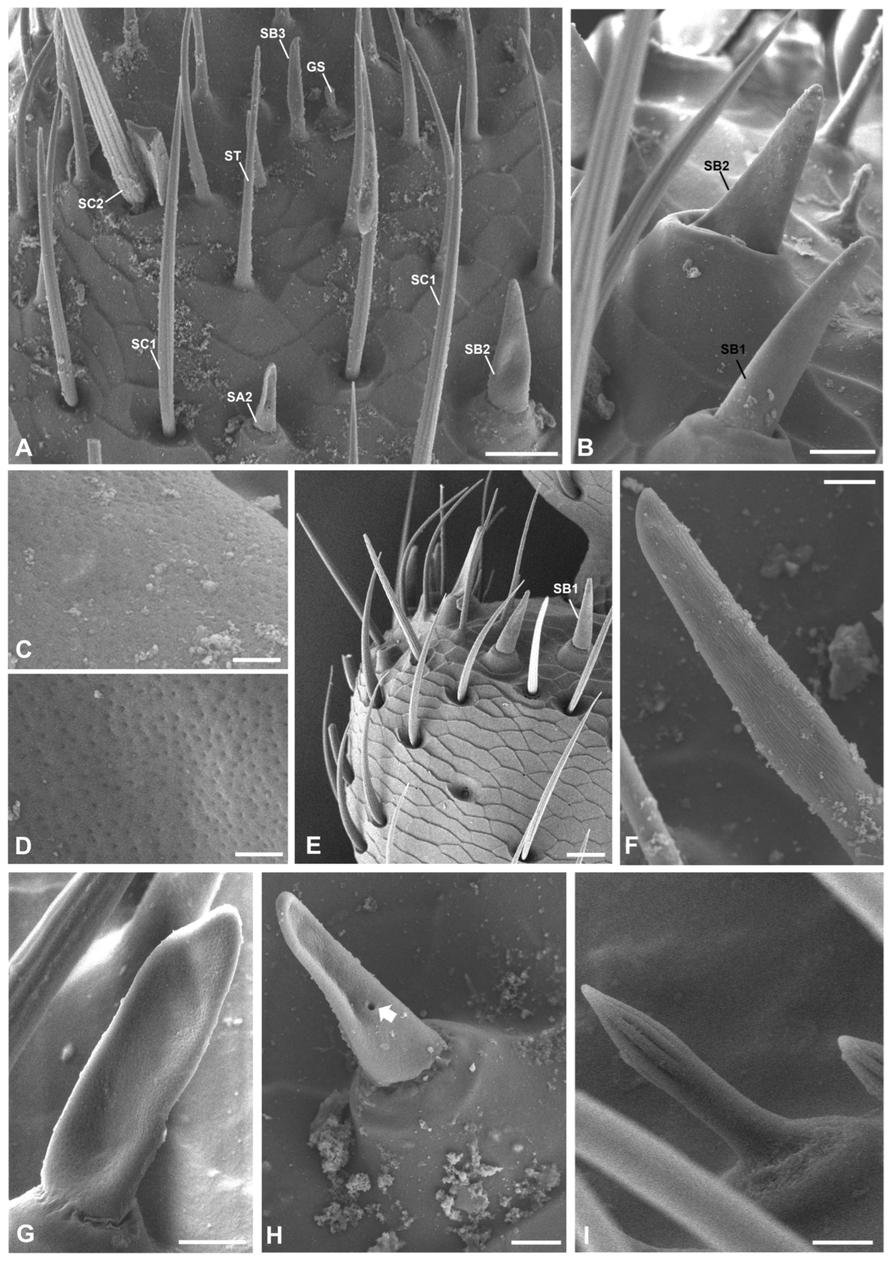
The sensilla chaetica (SC) (Figure 2A) were straight, robust, bristle-like sensilla with deep longitudinal grooves and pointed tips. Each sensillum arose from a deep, wide basal socket. Two subtypes were identified: SC1 and SC2. They were deeply grooved longitudinally and oriented toward the antennal apex. The SC2 were thicker than SC1 and deeply grooved longitudinally (Figure 2A–F). The SC2 were often surrounded at their base by a variable number of cuticular pores (Figure 2D–F). The sensilla trichodea (ST) were characterized by blunt, rounded tips, microperforated walls and a raised base (Figure 2G). The sensilla basiconica (SB) appeared as cone-shaped pegs with blunt tips and microperforated walls (Figure 2H and Figure 3A–F). Three subtypes were observed: SB1, SB2 and SB3. The SB1 were microperforated and arose from raised bases (Figure 3B,C,E). The SB2 were stout with broader raised bases and had microperforations more evident and more numerous than SB1 (Figure 3B,D). The SB3 had longitudinal grooves with microperforations on the bottom and a slight inverse curvature (Figure 3A,F). The sensilla auricillica (SA), rare, were characterized by a shallow concave depression in their middle region and microperforations, had blunt ends, and arose from slightly elevated bases (Figure 3G,H). Two subtypes of sensilla auricillica were found: AS1 and AS2. The AS1 and AS2 had similar morphology, but AS2 exhibited a large pore in the concave depression (Figure 3H). The grooved peg sensilla (GS) were rare and arose from slightly raised cuticular areas with non-articulated bases. They were characterized by longitudinal grooves in their distal region and were smooth in the basal one (Figure 3A,I).
Figure 2.
Scanning electron micrographs of the flagellomeres of an adult male of M. villiger. (A) Overview of flagellomere F3 and parts of flagellomeres F2 and F4. (B) Detail of a flagellomere showing a sensillum chaeticum of subtype 1 (SC1). (C) Apical part of the SC1 shown in (B). (D) Detail of a flagellomere showing a sensillum chaeticum of subtype 2 (SC2) in toto. (E) Base of the SC2 shown in (D), showing cuticle pores (arrows). (F) Apical part of the SC2 shown in (D). (G) Detail of one of the three flagellomeres forming the club, showing a sensillum trichodeum (ST) in toto. (H) Overview of the terminal flagellomere (F9). The rectangle outlines an area showed in detail in Figure 3A. Scale bars: (A) = 25 μm; (B) = 5 μm; (C)= 1 μm; (D) = 5 μm; (E) = 1 μm; (F) = 0.5 μm; (G) = 2.5 μm; (H) = 25 μm. Abbreviations: F2, flagellomere 2; F3, flagellomere 3; F4, flagellomere 4; F8, flagellomere 8; F9, flagellomere 9; SC1, sensillum chaeticum of subtype 1; SC2, sensillum chaeticum of subtype 2; ST, sensillum trichodeum.
Figure 3.
Scanning electron micrographs of the club of an adult male of M. villiger. (A) Detail of the surface of the terminal flagellomere (F9) within the rectangle in Figure 2H. (B) Detail of a flagellomere of the club showing two sensilla basiconica of subtype 1 (SB1) and of subtype 2 (SB2), respectively. (C) Detail of the surface of SB1. (D) Detail of the surface of SB2. (E) Detail of the distal region of flagellomere 8 (F8). (F) Detail of a flagellomere of the club, showing a sensillum basiconicum of subtype 3 (SB3). (G) Detail of the distal region of a flagellomere of the club, showing a sensillum auricillicum of subtype 1 (SA1). (H) Detail of the distal region of a flagellomere of the club, showing a sensillum auricillicum of subtype 2 (SA2). The arrow indicates the large pore in the concave depression. (I) Detail of a flagellomere of the club showing a grooved peg sensillum (GS). Scale bars: (A) = 10 μm; (B) = 5 μm; (C) = 0.5 μm; (D) = 0.5 μm; (E) = 10 μm; (F) = 1.5 μm; (G) = 2 μm; (H) = 2.5 μm; (I) = 1 μm. Abbreviation: GS, grooved peg sensillum; SA2, sensillum auricillicum of subtype 2; SB1, sensillum basiconicum of subtype 1; SB2, sensillum basiconicum of subtype 2; SB3, sensillum basiconicum of subtype 3; SC1, sensillum chaeticum of subtype 1; SC2, sensillum chaeticum of subtype 2; ST, sensillum trichodeum.
3.2. Sensillar Distribution
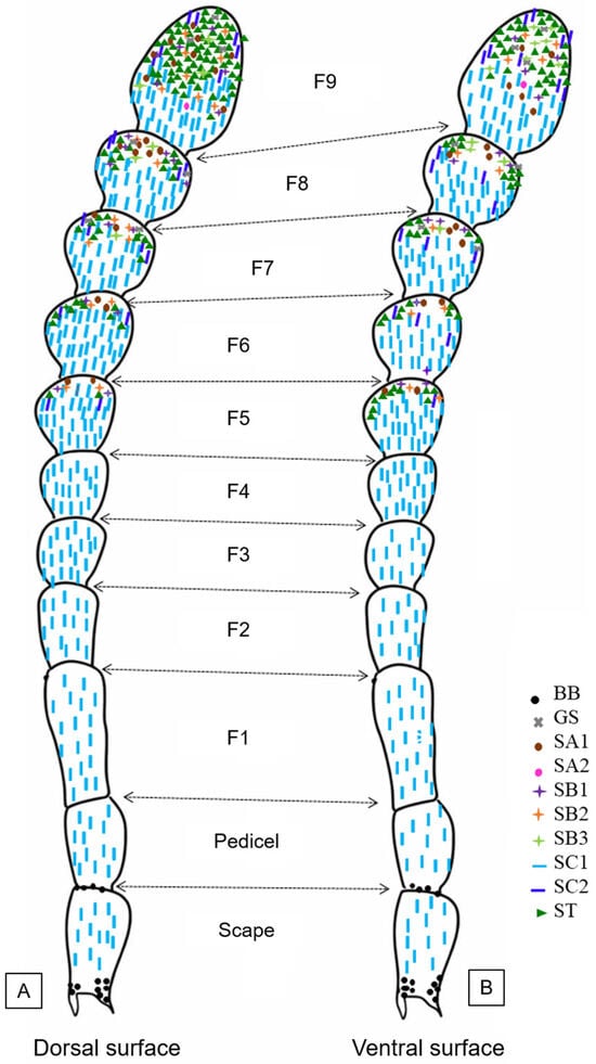
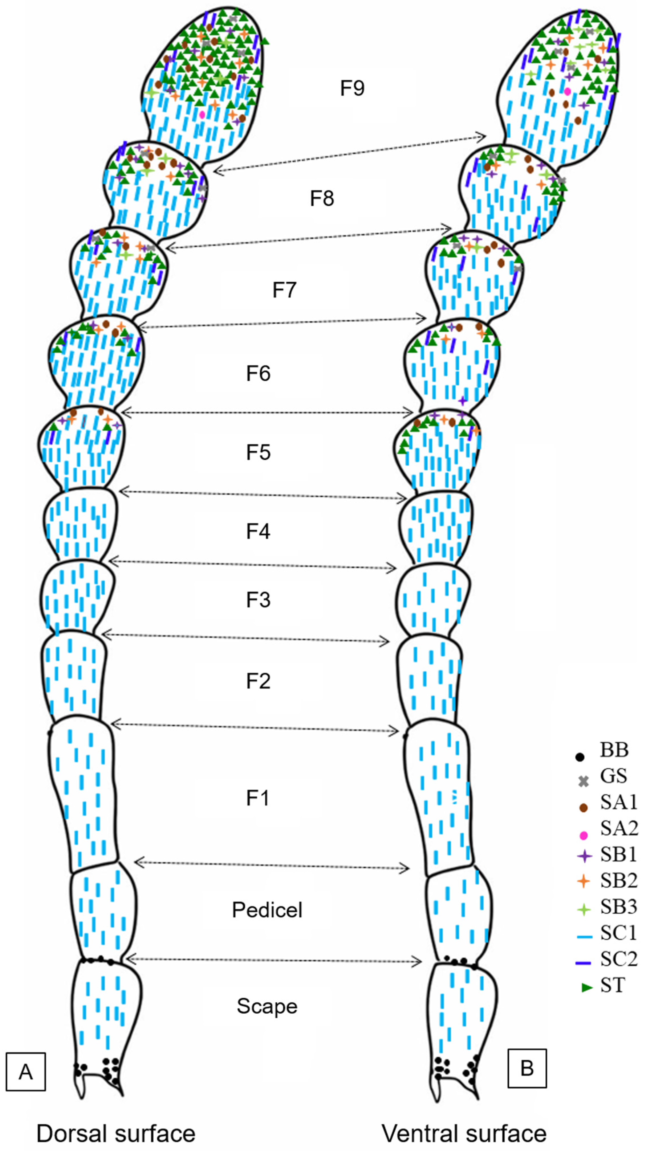
The distribution of the different types and subtypes of sensilla in the antenna of M. villiger male is shown as a drawing in Figure 4, with the dorsal side represented in Figure 4A and the ventral one in Figure 4B. Table 1 reports the length of all types or subtypes of sensilla.
Figure 4.
Manual drawing based on the image of the antenna in toto showing the distribution of types and subtypes of sensilla in the dorsal (A) and ventral (B) of antennal segments in males of M. villiger, based on SEM images. Abbreviations: BB, Böhm’s bristles; F1 to F9, flagellomeres 1–9; GS, grooved peg sensilla; SA1, sensilla auricillica of subtype 1; SA2, sensilla auricillica of subtype 2; SB1, sensilla basiconica of subtype 1; SB2, sensilla basiconica of subtype 2; SB3, sensilla basiconica of subtype 3; SC1, sensilla chaetica of subtype 1; SC2, sensilla chaetica of subtype 2; ST, sensilla trichodea.
The BB were found on the proximal region of the scape and of the pedicel (Figure 1D,F,G and Figure 4).
For SC, the subtype SC1, the most abundant sensillar subtype, was distributed across all antennal segments (Figure 2A–C). From the scape to flagellomere F4, the SC1 were distributed along all the dorsal and ventral surface of the segments, but from F5 to F9 they tended to concentrate on the proximal half of the segment. Moreover, the SC1 were the only sensillar subtype found in flagellomeres F1–F4 (Figure 4).
The subtype SC2 (Figure 2D) was distributed only in the distal half of flagellomeres F5-F9, on both the dorsal and the ventral side (Figure 4).
The ST (Figure 2G and Figure 3A) were found from F5 to F9, distributed on the distal part of F5–F8 in both dorsal and ventral sides. In F9, these sensilla occupied the entire distal half of the segment in both dorsal and ventral sides (Figure 4).
Concerning the three subtypes of SB, SB1 (Figure 3B,C,E and Figure 4) and SB2 (Figure 3B,D and Figure 4) shared the same distribution of ST. SB3, rare, were found only from F7 to F9: in F7 and F8 they were present in the distal part, while in F9 they occupied the entire distal half of the segment (Figure 3A,F and Figure 4).
The SA had two subtypes, SA1 and SA2. The SA1 were distributed in the distal part of flagellomeres from F5 to F8, and on the entire distal half of F9 (Figure 3G and Figure 4). The SA2 were found only on F9 (Figure 3H and Figure 4).
Finally, the GS (Figure 3I) were located only in the three last flagellomeres, in the distal part of F7 and F8, and in the entire distal half of F9 (Figure 4). Based on the stacked bar plots in Figure 5A,B, the number of sensilla and their types and subtypes increased along the segments from the proximal to the distal ones.
Figure 5.
Distribution and mean number of types and subtypes of antennal sensilla in the dorsal (A) and ventral (B) side of the antennal segments of M. villiger males, represented as stacked bar plots. Abbreviations: BB, Böhm’s bristles; F1 to F9, flagellomeres 1–9; GS, grooved peg sensilla; SA1, sensilla auricillica of subtype 1; SA2, sensilla auricillica of subtype 2; SB1, sensilla basiconica of subtype 1; SB2, sensilla basiconica of subtype 2; SB3, sensilla basiconica of subtype 3; SC1, sensilla chaetica of subtype 1; SC2, sensilla chaetica of subtype 2; ST, sensilla trichodea.
4. Discussion
The present study describes for the first time the ultrastructural morphology of the antennae of adult males of Mesomorphus villiger and provides the first data about the antennal ultrastructure and sensilla of the genus Mesomorphus Miedel, 1880 (Coleoptera: Tenebrionidae).
In M. villiger males, the antennae show a general morphology similar to that of other species of Tenebrionidae: the antennae are clavate with 11 segments, the three distal ones forming a club. Starting from the scape, the sensilla found in the antennal segments are Böhm’s bristles (BB), the sensilla chaetica (CS) with two subtypes (CS1 and CS2), the sensilla trichodea (ST), the sensilla basiconica (BS) with three subtypes (BS1, BS2 and BS3), the sensilla auricillica (AS) with two subtypes (AS1 and AS2), and the grooved peg sensilla (GS). The morphology and function of each type and subtype of sensilla will be discussed, comparing the data obtained in M. villiger males with those available in the Tenebrionidae family, in other families of the order Coleoptera, and in other insect orders.
4.1. Böhm’s Bristles
Böhm’s bristles (BB), short, roughly triangular and articulated structures, were detected at the base of the scape and pedicel in M. villiger males. These sensilla were found on the scape and pedicel of the antennae of many insect orders including Coleoptera [25]. The morphology of Böhm’s bristles found in M. villiger males is similar to that of other Coleoptera families, such as Chrysomelidae [26,27], Cleridae [28], Cerambycidae [25,29], Coccinellidae [30], Curculionidae [31] and Erotylidae [32]. In Tenebrionidae, Böhm’s bristles have been described in the antennae of Ulomoides dermestoides [7]. Several studies, not only in Coleoptera but also in other orders such as Lepidoptera, Orthoptera and Hymenoptera, identify the function of these sensilla as mechanoreceptory, being able to provide information on antennal bending position. In Daphnis nerii (Linnaeus, 1758) (Lepidoptera: Sphingidae), Böhm’s bristles specifically mediate antennal positioning during flight, avoiding dangerous collisions of antennae with wings [33]. In Achaeta domesticus L. 1758 (Orthoptera: Gryllidae), these antennal sensilla are bent when the scape and pedicel are deflected; thus, they signal the bending position of the antenna [34]. In Dolichogenidea gelechiidivoris (Marsh, 1975) (Hymenoptera: Braconidae), Böhm’s bristles are suggested to play a role in antennal perception related to the body and to orientation adjustments during flight [35]. The location of these sensilla on the proximal regions of scape and pedicel of M. villiger males suggests a similar functional role as proprioceptive mechanoreceptors.
4.2. Sensilla Chaetica
In M. villiger males, the sensilla chaetica (SC) were straight, robust and bristle-like, with deep longitudinal grooves, pointed tips, and a deep, wide basal socket. Two subtypes were identified: the SC1, the most abundant, were distributed in the dorsal and ventral parts of all segments and were oriented toward the antennal apex; the SC2, thicker and surrounded at their base by cuticular pores, were distributed only in the distal half of flagellomeres F5-F9. In Tenebrionidae, similar sensilla have been described in the antennae of U. dermestoides [7] and T. castaneum [14,15], but in both cases they were identified as sensilla trichodea. Sensilla chaetica with a similar morphology, including the cuticular pores at their base, were identified in Platynus dorsalis (Pontoppidan, 1763) (Coleoptera, Carabidae) [36]. “Stout sensilla chaetica”, some with pores clustered at their base, were found in Monochamus galloprovincialis (Olivier, 1795) (Coleoptera: Cerambycidae) [37] and were similar to those previously described in Monochamus notatus (Drury, 1773) and Monochamus scutellatus (Say, 1824) (Coleoptera: Cerambycidae) [38]. In insects, sensilla chaetica are generally considered as tactile receptors [23] based on their morphology. In Mo. galloprovincialis the distal sensilla chaetica respond with spike trains when manually bent, suggesting their role as mechanoreceptors [37]. The presence of “gland pores” around the base of these sensilla in Mo. notatus and Mo. scutellatus [38] apparently suggests an association between a mechanoreceptory and a secretory function. In Elater ferrugineus Linnaeus, 1758 (Coleoptera: Elateridae), sensilla chaetica were found with clusters of small pores at their base and were considered to be involved in the secretion of lubricating substances [39]. Based on these studies, similar functions could also be hypothesized in M. villiger males.
4.3. Sensilla Trichodea
In M. villiger males, the sensilla trichodea (ST) had blunt, rounded tips, microperforated walls, and raised non-articulated bases. Their distribution was similar in both dorsal and ventral sides of the antenna from F5 to F9, on the distal part of F5-F8, and in the entire distal half of the segment in F9. In previous studies on Tenebrionidae, similar non-articulated multiporous sensilla have been reported in Sc. gigas [19], A. sulcatum [9] and P. impunctata [10] and are considered olfactory sensilla.
In insects, a general function suggested for sensilla trichodea is chemiosensory [23,40]. In Coleoptera, besides Tenebrionidae, “sensilla trichodea II and III” were also reported in Dendroctonus frontalis (Zimmerman, 1868) (Coleoptera: Curculionidae). The sensilla trichodea II, with cuticular pores, were considered olfactory sensilla, while the sensilla trichodea III, without pores, were considered mechanosensitive ones [41]. Sensilla trichodea have also been identified in the recently discovered new species of nocturnal firefly, Phausis christineae Hodson & Branham 2024 (Coleoptera: Lampyridae), and interpreted as being related to chemiosensory activity in pheromone perception [42]. In other insect orders, sensilla trichodea, called “trichoideum sensillum (TSp)”, with porous wall and non-articulated base were reported in Heteroptera, in the damsel bug Nabis rugosus (Linnaeus, 1758) (Nabidae). In this species, sensilla trichodea and sensilla basiconica were considered related to perception of chemical stimuli [43]. Sensilla trichodea (“trichoid sensilla”) are also very common in the antennae of many species of Diptera, and they are thought to be involved in the perception of sexual pheromones and olfactory function [44,45]. Based on the literature, we could advance the hypothesis that these sensilla are involved in the perception of pheromones also in M. villiger males. More studies on electrophysiological aspects of these sensilla are necessary, aimed also to develop an integrated pest management strategy of this species by sexual confusion and mass trapping.
4.4. Sensilla Basiconica
In M. villiger males, the sensilla basiconica (SB) appeared as cone-shaped pegs with blunt tips and microperforated walls, with three subtypes (SB1, SB2 and SB3). The SB1 and SB2 had the same distribution as the sensilla trichodea: they were found on the apical and lateral regions of the last three flagellomeres, while the SB3, very rare, were present on flagellomeres F7–F9.
Multiple subtypes of sensilla basiconica have been described across various insect orders, reflecting functional and structural diversity [24,38,46,47]. Within the subfamily Tenebrioninae, subtypes of sensilla basiconica have been reported in T. castaneum, with different numbers according to the authors: six [13], four [14] and two [15]. In the subfamily Pimeliinae, two subtypes were reported in A. sulcatum [9] and four in P. impunctata [10]. In M. villiger males three subtypes were found. Sensilla basiconica characterized by a surface covered by microperforations are found in many insect taxa [48].
Concerning their function, in T. castaneum the sensilla basiconica were considered to play an olfactory role in pheromone and host plant odor perception [13]. In the Australian longhorn beetle Phoracantha semipunctata (Fabricius, 1775) (Coleoptera: Cerambycidae), studies using SEM, electrophysiology and gas chromatography showed that sensilla basiconica of subtypes I and II basiconic sensilla of types I and II were involved in olfactory recognition of plant odors [49]. A similar role of sensilla basiconica in detecting host tree volatiles and/or pheromones was suggested in the invasive Asian beetle Aromia bungii (Faldermann, 1835) (Coleoptera: Cerambycidae) [50]. In these two species of Cerambycidae and in Mo. galloprovincialis, no sexual dimorphism was detected in the distribution and abundance of sensilla basiconica; thus, a similar olfactory role was suggested in both sexes [37]. In Diptera, the sensilla basiconica (“basiconic sensilla”), very common in the antennae of many species, are considered to be involved in olfactory function [51]. In Hymenoptera, in two ant species, the black carpenter ant Camponotus pennsylvanicus (De Geer, 1773) and the Indian jumping ant Harpegnathos saltator (T. C. Jerdon, 1851), the detection of cuticular hydrocarbons through sensilla basiconica (“basiconic sensilla”) is known to play a key role in eusocial communication [52]. Based on their SEM structure and on the above literature data, an olfactory role of sensilla basiconica could also be hypothesized in M. villiger males for perception of pheromones and plant odors.
4.5. Sensilla Auricillica
In M. villiger males, the rare sensilla auricillica (SA) showed a concave depression in their middle region, microperforations, and blunt ends and were divided into two subtypes, SA1 and SA2. The SA1 were found in flagellomeres F5 to F9, while the SA2 were localized only in F9. Up to date, sensilla auricillica have never been previously reported in Tenebrionidae. However, sensilla of similar shape were detected in Cerambycidae, and their suggested function was olfactory [25]. In other insect orders, studies on the morphology and physiology of sensilla auricillica in the primitive moth genus Eriocrania spp. Zeller, 1851 (Lepidoptera: Eriocraniidae) showed a strong similarity to those of the order Trichoptera (caddisflies), supporting a homology of pheromone detection systems in the two orders which share a common ancestry within the superorder Amphiesmenoptera [53].
4.6. Grooved Peg Sensilla
In M. villiger males, the rare, grooved peg sensilla (GP) were non-articulated at their base and were located only in the three terminal flagellomeres of the antenna, in the distal part of F7 and F8, and in the entire distal half of F9. In insect orders, grooved peg sensilla have been variously reported as “multiporous grooved sensilla” [23,48], “sensilla styloconica” [54] or a subtype of “multiporous sensilla basiconica, B3” [10]. Concerning their function, a general chemosensory role [23,48] and a hygrothermal one [40] were hypothesized. Electrophysiological studies on the cabbage-stem flea beetle, Psylliodes chrysocephala Linnaeus, 1758 (Coleoptera: Chrysomelidae) showed that grooved peg sensilla were innervated by five neurons, thus considered to perform both olfactory and thermoreceptive functions [55]. In Diptera, “grooved pegs” were studied at the ultrastructural level in the antennae of Aedes aegypti Linnaeus, 1762 (Diptera: Culicidae). These sensilla were found connected to 3–5 neurons and were identified as olfactory receptors of air-borne stimuli [56]. In Mantodea, grooved peg sensilla were recently described in antennae of males and females of the Chinese flower mantis, Creobroter nebulosa Zheng, 1988 (Mantodea: Hymenopodidae). Their number was higher in males in comparison to females, in relation to the need for detection of sex pheromones [57]. Based on these data, an olfactory role of the grooved peg sensilla could also be tentatively suggested in M. villiger males.
5. Conclusions
In the present study, the morphology, types, and distribution of antennal sensilla in males of the coleopteran Mesomorphus villiger were described for the first time using scanning electron microscopy. This study is also the first one conducted on the antennae of the genus Mesomorphus. Although any functional interpretation of the identified sensilla should be considered preliminary because the study is limited and involves only males, these sensilla could be hypothesized to be involved in the perception of mechanical and chemical stimuli and probably in the aggregation phenomena known for this species as well. The structure of the antenna and the different types of sensilla in M. villiger males apparently reflect broad sensory adaptation probably related to its ecological requirements. These findings extend the understanding of chemosensory diversity in Tenebrionidae and establish a morphological basis for future electrophysiological and behavioral studies.
The morphological and functional characterization of antennal sensilla in pest species represent a relevant step to develop management strategies safe for the environment, animals, and humans. These investigations may therefore provide the foundation for electroantennography studies evaluating the responses of antennal sensilla to specific odors, aimed at developing techniques of sexual confusion and possible mass capture, which could reduce economic damages and indoor disturbances caused by M. villiger infestations. Moreover, considering the key role of antennal sensory structures in insect communication, the data obtained in this preliminary study on M. villiger males could represent the foundation for further studies on mate recognition, sexual selection and ecological adaptations.
Author Contributions
Conceptualization, M.B.; Methodology, M.B. and M.P.; Formal Analysis, M.B., S.S., H.B. and M.P.; Investigation, M.B., S.S., H.B. and M.P.; Data Curation, M.B., T.B., H.B. and M.P.; Writing—Original Draft Preparation, M.B., S.S. and M.P.; Writing—Review and Editing, M.B., S.S., T.B., H.B. and M.P.; Supervision, M.B. and M.P.; Funding Acquisition, M.B. All authors have read and agreed to the published version of the manuscript.
Funding
Financial support from the Department of Science and Technology (DST), Government of India, is gratefully acknowledged (Project No. WOS-A/LS-187/2019 & No. DST/INSPIRE Fellowship/2023/IF230060). The publication costs were covered by “Fondi Ricerca di Base” (“Basic Research Funds”) assigned to Professor Teresa Bonacci (Department of Biology, Ecology and Earth Sciences, University of Calabria).
Institutional Review Board Statement
Not Applicable.
Data Availability Statement
The original contributions presented in this study are included in the article. Further inquiries can be directed to the corresponding authors.
Acknowledgments
The authors express their sincere gratitude to the Director, Forest Research Institute (FRI), Dehradun, for providing field access during the study. The authors also thank the Central Instrumentation Facility, Thapar Institute of Engineering and Technology (TIET), Patiala (Punjab), for providing access to the scanning electron microscope (Zeiss Gemini 1 Sigma 500) and technical assistance during imaging.
Conflicts of Interest
The authors declare no conflicts of interest.
References
- Bouchard, P.; Lawrence, J.F.; Davies, A.E.; Newton, A.F. Synoptic classification of the world Tenebrionidae (Insecta: Coleoptera) with a review of family-group names. Ann. Zool. 2005, 55, 499–530. [Google Scholar]
- Seena, C.M.; Thomas, S.K. Defensive glands of the darkling beetle Mesomorphus villiger Blanchard (Coleoptera: Tenebrionidae). Psyche J. Entomol. 2013, 2013, 201769. [Google Scholar] [CrossRef] [Scilit]
- Catalogue of Life. Available online: https://www.catalogueoflife.org/data/taxon/QRCRV (accessed on 7 January 2026).
- Hegde, V.D.; Lal, B.; Kushwaha, R.K. Darkling beetles (Tenebrionidae: Coleoptera) of Dudhwa National Park, Uttar Pradesh, India. J. New Biol. Rep. 2013, 2, 130–141. [Google Scholar] [CrossRef] [Scilit]
- Hansson, B.S.; Stensmyr, M.C. Evolution of insect olfaction. Neuron 2011, 72, 698–711. [Google Scholar] [CrossRef] [Scilit] [PubMed]
- Elgar, M.A.; Zhang, D.; Wang, Q.; Wittwer, B.; Thi Pham, H.; Johnson, T.L.; Freelance, C.B.; Coquilleau, M. Insect antennal morphology: The evolution of diverse solutions to odorant perception. Yale J. Biol. Med. 2018, 91, 457–469. [Google Scholar] [PubMed]
- Qian, J.; Wang, Y.; Guo, H.; Chi, D. Distribution and ultrastructure of sensilla on Ulomoides dermestoides (Coleoptera: Tenebrionidae) antennae. J. Entomol. Sci. 2018, 53, 123–140. [Google Scholar] [CrossRef] [Scilit]
- Soldati, F.; Soldati, L. Species delimitation using morphological and molecular tools in the Asida (Polasida) jurinei Solier, 1836 species-complex. Preliminary results. (Coleoptera: Tenebrionidae: Tentyriinae). Cah. Sci. Muséum D’histoire Nat. Lyon—Cent. Conserv. D’étude Collect. 2006, 10, 111–116. [Google Scholar] [CrossRef] [Scilit]
- Faucheux, M.J. The tactile antennae with an olfactive apex of Adelostoma sulcatum Duponchel, 1827 (Coleoptera: Tenebrionidae: Pimeliinae: Adelostomini): Characteristics in liaison with the life in termite nests. Bull. l’Institut Sci. Rabat Sect. Sci. Vie 2023, 45, 55–65. [Google Scholar]
- Faucheux, M.J. Les sensilles antennaires de Pachychila (Pachychila) impunctata Fairmaire, 1860 (Coleoptera: Tenebrionidae: Pimeliinae: Tentyriini). Bull. Soc. Sci. Nat. Ouest Fr. (N.S.) 2023, 45, 54–62. [Google Scholar]
- Zohry, N.M.H. Scanning electron morphological studies of Tribolium confusum Jacquelin du Val (Coleopteran: Tenebrionidae). J. Basic Appl. Zool. 2017, 78, 6. [Google Scholar] [CrossRef] [Scilit]
- Thakare, P.R.; Kakde, V.R. Scanning electronic microscopic study of antenna of Tribolium confusum (confused floor beetle). Int. J. Zool. Stud. 2022, 7, 69–73. [Google Scholar]
- Ali, S.A.I.; Diakite, M.M.; Ali, S.; Wang, M.-Q. Effects of the antennal sensilla distribution pattern on the behavioral responses of Tribolium castaneum (Coleoptera: Tenebrionidae). Fla. Entomol. 2016, 99, 52–59. [Google Scholar] [CrossRef] [Scilit]
- Seada, M.A.; Hamza, A.M. Differential morphology of the sensory sensilla and antennae, palpi, foretarsi and ovipositor of adult Tribolium castaneum (Herbst) (Coleoptera: Tenebrionidae). Ann. Agric. Sci. 2018, 63, 1–8. [Google Scholar] [CrossRef] [Scilit]
- Lokesh, M.; Anand, A.; Sreekrishnakumar, A.K.; Subramanian, E.V. Morphological characteristics of the antenna and its sensilla of three stored-product beetle species. J. Basic Appl. Zool. 2025, 86, 36. [Google Scholar] [CrossRef] [Scilit]
- Tungjitwitayakul, J.; Yasanga, T.; Tatun, N. Impact of UV-C radiation on morphology of the antenna and antennal sensilla in Tribolium castaneum (Coleoptera: Tenebrionidae). J. Radiat. Res. Appl. Sci. 2020, 13, 648–656. [Google Scholar] [CrossRef] [Scilit]
- Harbach, R.E.; Larsen, J.R. Fine structure of antennal sensilla of the adult mealworm beetle, Tenebrio molitor L. (Coleoptera: Tenebrionidae). Int. J. Insect Morphol. Embryol. 1977, 6, 41–60. [Google Scholar] [CrossRef] [Scilit]
- da Silva Soares, C.E.; Weber, A.; Scussel, V.M. Stereo and scanning electron microscopy characteristics of poultry breeding beetle (Alphitobius diaperinus)—A filamentous toxigenic fungi carrier. Emir. J. Food Agric. 2018, 30, 150–156. [Google Scholar] [CrossRef] [Scilit]
- Faucheux, M.J. Morphologie fonctionnelle des sensilles antennaires de Scaurus gigas Walt, 1835 (Coleoptera: Tenebrionidae). Bull. Soc. Sci. Nat. Ouest Fr. (N.S.) 2013, 35, 109–125. [Google Scholar]
- Ferrer, J. Révision des espèces asiatiques du genre Mesomorphus Seidlitz, 1893 (Coleoptera: Tenebrionidae: Opatrinae: Opatrini). Ann. Zool. 2002, 52, 533–557. [Google Scholar]
- Chapman, R.F. The Insects: Structure and Function, 4th ed.; Cambridge University Press: New York, NY, USA, 1998; pp. 3–11. [Google Scholar]
- Beutel, R.G.; Friedrich, F.; Ge, S.-Q.; Yang, X.-K. Insect Morphology and Phylogeny: A Textbook for Students of Entomology; Walter de Gruyter GmbH: Berlin, Germany; Boston, MA, USA, 2014; pp. 1–103. [Google Scholar]
- Zacharuk, R.Y. Antennae and sensilla. In Comprehensive Insect Physiology, Biochemistry and Pharmacology; Kerkut, G.A., Gilbert, L.I., Eds.; Pergamon: Oxford, UK, 1985; Volume 6, pp. 1–69. [Google Scholar]
- Faucheux, M.J.; Németh, T.; Hoffmannova, J.; Kundrata, R. Scanning electron microscopy reveals the antennal micromorphology of Lamprodila (Palmar) festiva (Coleoptera: Buprestidae), an invasive pest of ornamental Cupressaceae in Western Palaearctic. Biology 2020, 9, 375. [Google Scholar] [CrossRef] [Scilit] [PubMed]
- Haddad, S.; Clarke, D.J.; Jeong, S.H.; Mitchell, R.F.; McKenna, D.D. Antennal sensilla in longhorn beetles (Coleoptera: Cerambycidae). Ann. Entomol. Soc. Am. 2023, 116, 83–113. [Google Scholar] [CrossRef]
- Zhang, J.; Hou, L.; Cheng, B.; Xi, J. Morphology and distribution of the antennal sensillia of the leaf beetle, Ambrostoma quadriimpressum (Coleoptera: Chrysomelidae). Trans. Am. Entomol. Soc. 2017, 143, 757–772. [Google Scholar] [CrossRef] [Scilit]
- Chen, Q.; Li, S.; Wang, Y.; Jia, D.; Wang, Y.; Ma, R. Morphological characteristics of the sensilla in a monophagous insect: Agasicles hygrophila Selman and Vogt (Coleoptera: Chrysomelidae, Halticinae). Insects 2023, 14, 501. [Google Scholar] [CrossRef] [Scilit] [PubMed]
- Zhang, J.; Cheng, B.; Fu, L.; Sun, S. Morphology and distribution of antennal sensilla of the predatory clerid beetle, Thanasimus substriatus (Coleoptera: Cleridae). J. Entomol. Sci. 2021, 56, 441–455. [Google Scholar] [CrossRef] [Scilit]
- Tong, X.; Wu, W.-J.; Liu, S.-Y.; Wang, L. Morphology and sexual dimorphism of antennal sensilla in longhorn beetle Moechotypa diphysis (Pascoe, 1871) (Coleoptera: Cerambycidae: Lamiinae). Zool. Anz. 2026, 321, 238–247. [Google Scholar] [CrossRef] [Scilit]
- Hao, Y.N.; Sun, Y.X.; Liu, C.Z. Functional morphology of antennae and sensilla of Hippodamia variegata (Coleoptera: Coccinellidae). PLoS ONE 2020, 15, e0237452. [Google Scholar] [CrossRef] [Scilit] [PubMed]
- Wang, L.; Wang, Q.; Luo, Y.; Yan, S. Ultrastructural morphology and descriptive analysis of cuticular sensilla in adult Tomicus pilifer (Coleoptera: Curculionidae). Insects 2025, 16, 890. [Google Scholar] [CrossRef] [Scilit] [PubMed]
- Hou, X.L.; Huang, S.H.; Hong, B.; Yang, M.F.; Luo, C.Q. Functional morphology of antennae and sensilla of the fungivore beetle, Triplax ainonia Lewis (Coleoptera: Erotylidae). PLoS ONE 2024, 19, e0309670. [Google Scholar] [CrossRef] [Scilit] [PubMed]
- Krishnan, A.; Prabhakar, S.; Sudarsan, S.; Sane, S.P. The neural mechanisms of antennal positioning in flying moths. J. Exp. Biol. 2012, 215, 3096–3105. [Google Scholar] [CrossRef] [Scilit] [PubMed]
- McCarter, M.G.; Loudon, C. The spatial arrangement of Böhm’s bristles and resolution of joint position in cricket antennae (Acheta domesticus) (Orthoptera: Gryllidae). J. Insect Sci. 2025, 25, 22. [Google Scholar] [CrossRef] [Scilit] [PubMed]
- Yan, S.-Y.; Yang, H.-S.; Huang, C.; Zhang, G.-F.; Arnó, J.; Collatz, J.; Li, C.-R.; Wan, F.-H.; Liu, W.-X.; Zhang, Y.-B. The olfactory system of Dolichogenidea gelechiidivoris (Marsh) (Hymenoptera: Braconidae), a natural enemy of Tuta absoluta (Meyrick) (Lepidoptera: Gelechiidae). Int. J. Mol. Sci. 2025, 26, 7312. [Google Scholar] [CrossRef] [Scilit] [PubMed]
- Merivee, E.; Ploomi, A.; Luik, A.; Rahi, M.; Sammelselg, V. Antennal sensilla of the ground beetle Platynus dorsalis (Pontoppidan, 1763) (Coleoptera, Carabidae). Microsc. Res. Tech. 2001, 55, 339–349. [Google Scholar] [CrossRef] [Scilit] [PubMed]
- Álvarez, G.; Ammagarahalli, B.; Hall, D.R.; Pajares, J.A.; Gemeno, C. Smoke, pheromone and kairomone olfactory receptor neurons in males and females of the pine sawyer Monochamus galloprovincialis (Olivier) (Coleoptera: Cerambycidae). J. Insect Physiol. 2015, 82, 46–55. [Google Scholar] [CrossRef] [Scilit] [PubMed]
- Dyer, L.J.; Seabrook, W.D. Sensilla on the antennal flagellum of the sawyer beetles Monochamus notatus (Drury) and Monochamus scutellatus (Say) (Coleoptera: Cerambycidae). J. Morphol. 1975, 146, 513–531. [Google Scholar] [CrossRef] [Scilit] [PubMed]
- Zauli, A.; Maurizi, E.; Carpaneto, G.M.; Chiari, S.; Merivee, E.; Svensson, G.P.; Di Giulio, A. Scanning electron microscopy analysis of the antennal sensilla in the rare saproxylic beetle Elater ferrugineus (Coleoptera: Elateridae). Ital. J. Zool. 2016, 83, 338–350. [Google Scholar] [CrossRef] [Scilit]
- Keil, T.A. Morphology and development of the peripheral olfactory organs. In Insect Olfaction; Hansson, B.S., Ed.; Springer: Berlin/Heidelberg, Germany, 1999; pp. 5–47. [Google Scholar]
- Dickens, J.C.; Payne, T.L. Structure and function of the sensilla on the antennal club of the southern pine beetle, Dendroctonus frontalis (Zimmerman) (Coleoptera: Scolytidae). Int. J. Insect Morphol. Embryol. 1978, 7, 251–265. [Google Scholar] [CrossRef] [Scilit]
- Pacheco, Y.M.; Mann, E.; Da Silveira, L.F.L.; Bybee, S.M.; Branham, M.A.; McHugh, J.V.; Stanger-Hall, K.F. Antennal sensilla diversity in diurnal and nocturnal fireflies (Coleoptera, Lampyridae). PLoS ONE 2025, 20, e0323722. [Google Scholar] [CrossRef] [Scilit] [PubMed]
- Chakilam, S.; Brożek, J.; Chajec, Ł.; Poprawa, I.; Gaidys, R. Ultra-morphology and mechanical function of the trichoideum sensillum in Nabis rugosus (Linnaeus, 1758) (Insecta: Heteroptera: Cimicomorpha). Insects 2022, 13, 799. [Google Scholar] [CrossRef] [Scilit] [PubMed]
- Pezzi, M.; Whitmore, D.; Chicca, M.; Semeraro, B.; Brighi, F.; Leis, M. Ultrastructural morphology of the antenna and maxillary palp of Sarcophaga tibialis (Diptera: Sarcophagidae). J. Med. Entomol. 2016, 53, 807–814. [Google Scholar] [CrossRef] [Scilit] [PubMed]
- Pezzi, M.; Leis, M.; Chicca, M.; Falabella, P.; Salvia, R.; Scala, A.; Whitmore, D. Morphology of the antenna of Hermetia illucens (Diptera: Stratiomyidae): An ultrastructural investigation. J. Med. Entomol. 2017, 54, 925–933. [Google Scholar] [CrossRef] [Scilit] [PubMed]
- Euzébio, D.E.; Martins, G.F.; Fernandes-Salomão, T.M. Morphological and morphometric studies of the antennal sensilla from two populations of Atta robusta (Borgmeier 1939) (Hymenoptera: Formicidae). Braz. J. Biol. 2013, 73, 663–668. [Google Scholar] [CrossRef] [Scilit] [PubMed]
- Roh, G.H.; Lee, Y.J.; Park, C.G. Morphology and distribution of antennal sensilla in a parasitoid fly, Gymnosoma rotundatum (Diptera: Tachinidae). Microsc. Res. Tech. 2020, 83, 589–596. [Google Scholar] [CrossRef] [Scilit] [PubMed]
- Zacharuk, R.Y. Ultrastructure and function of insect chemosensilla. Annu. Rev. Entomol. 1980, 25, 27–47. [Google Scholar] [CrossRef] [Scilit]
- Lopes, O.; Barata, E.N.; Mustaparta, H.; Araújo, J. Fine structure of antennal sensilla basiconica and their detection of plant volatiles in the eucalyptus woodborer, Phoracantha semipunctata Fabricius (Coleoptera: Cerambycidae). Arthropod Struct. Dev. 2002, 31, 1–13. [Google Scholar] [CrossRef] [Scilit] [PubMed]
- Di Palma, A.; Pistillo, M.; Griffo, R.; Garonna, A.P.; Germinara, G.S. Scanning electron microscopy of the antennal sensilla and their secretion analysis in adults of Aromia bungii (Faldermann, 1835) (Coleoptera, Cerambycidae). Insects 2019, 10, 88. [Google Scholar] [CrossRef] [Scilit] [PubMed]
- Pezzi, M.; Scapoli, C.; Mamolini, E.; Leis, M.; Bonacci, T.; Whitmore, D.; Krčmar, S.; Furini, M.; Giannerini, S.; Chicca, M.; et al. Ultrastructural characterization of sensilla and microtrichia on the antenna of female Haematopota pandazisi (Diptera: Tabanidae). Parasitol. Res. 2018, 117, 959–970. [Google Scholar] [CrossRef] [Scilit] [PubMed]
- Gellert, H.R.; Halley, D.C.; Sieb, Z.J.; Smith, J.C.; Pask, G.M. Microstructures at the distal tip of ant chemosensory sensilla. Sci. Rep. 2022, 12, 19328. [Google Scholar] [CrossRef] [Scilit] [PubMed]
- Larsson, M.C.; Hallberg, E.; Kozlov, M.V.; Francke, W.; Hansson, B.S.; Löfstedt, C. Specialized olfactory receptor neurons mediating intra- and interspecific chemical communication in leafminer moths Eriocrania spp. (Lepidoptera: Eriocraniidae). J. Exp. Biol. 2002, 205, 989–998. [Google Scholar] [CrossRef] [Scilit] [PubMed]
- Zhang, Y.; Ren, L.; Zhang, L.; Luo, Y. Ultrastructure of antennal and posterior abdominal sensilla in Chlorophorus caragana females. Micron 2015, 75, 45–57. [Google Scholar] [CrossRef] [Scilit] [PubMed]
- Bartlet, E.; Romani, R.; Williams, I.H.; Isidoro, N. Functional anatomy of sensory structures on the antennae of Psylliodes chrysocephala L. (Coleoptera: Chrysomelidae). Int. J. Insect Morphol. Embryol. 1999, 28, 291–300. [Google Scholar] [CrossRef] [Scilit]
- McIver, S.B. Fine structure of antennal grooved pegs of the mosquito, Aedes aegypti. Cell Tissue Res. 1974, 153, 327–337. [Google Scholar] [CrossRef] [Scilit] [PubMed]
- Wang, Y.; Wan, T.; Wang, Y.; Zhao, P.; Liu, Y. Ultrastructure of the antennal sensilla of the praying mantis Creobroter nebulosa Zheng (Mantedea: Hymenopodidae). PLoS ONE 2024, 19, e0301445. [Google Scholar] [CrossRef] [Scilit] [PubMed]

Disclaimer/Publisher’s Note: The statements, opinions and data contained in all publications are solely those of the individual author(s) and contributor(s) and not of MDPI and/or the editor(s). MDPI and/or the editor(s) disclaim responsibility for any injury to people or property resulting from any ideas, methods, instructions or products referred to in the content. |
© 2026 by the authors. Licensee MDPI, Basel, Switzerland. This article is an open access article distributed under the terms and conditions of the Creative Commons Attribution (CC BY) license.