


Abstract
Diatoms are microscopic algae with silica walls; they evolved relatively recently compared to other algal groups but have the greatest diversity of any and are abundant enough to contribute a fifth of the oxygen in the atmosphere and to have accumulated fossil mineral deposits (diatomaceous earth). Among the most recent and most diverse diatom genera is Mastogloia, with over 370 valid taxa but so far no pattern of phylogenetic relationships among the species. The genus is defined by the presence of chambers (partecta) on the girdle bands, which are as varied as the valve shapes and pore patterns. Mastogloia decussata was described in 1892, but its structure is still poorly understood, including whether it has axial costae and the nature of a V-shaped structure reported in 1970. A similar species, M. singaporensis, differs in having narrower, quadrate partecta that occupy only the middle half of the band; it has axial costae. We have observed several populations with quadrate partecta extending nearly to the apices in samples from Micronesian and Caribbean islands; such specimens have also been reported in the literature misidentified as M. decussata. The objective was to establish the ultrastructure of these species. Four major findings are as follows: (1) We establish the correct description of M. decussata with a neotype and show that it has axial costae; (2) we show that it has compound partecta with intrapartectal cylinders; (3) we describe one new species, M. paddocketkempii Lobban & J.A.B.Valencia, which also has compound partecta; (4) we show that all have dual pseudoloculate valve walls with interior and exterior pseudoloculi in slightly offset hexagonal arrays. The new species occurred in both ocean basins and may include simulacra, but we could not separate them morphologically. Detailed SEM of M. singaporensis shows its structural similarity to the other two species.
1. Introduction
Diatoms are unicellular eukaryotic microbes that all have silica shells (frustules) elaborately perforated for gas exchange, which are the primary means of their identification and taxonomy. They are by far the most speciose group of algae, with an estimated 30,000–100,000 species [1] extant in marine and freshwaters and as fossils in marine and freshwater deposits. The deposits are mined as diatomaceous earth, which was key to Alfred Nobel’s discovery of dynamite [2], but their shells are now being explored as sources of slab photonic crystals for quantum computing [3,4]. The living populations contribute about 20% of the world’s oxygen and are the major primary producers in the open ocean. Yet, they are relatively recent in the tree of life, with major radiation taking place after the KT mass extinction [5]. They were first described in the 19th Century from light microscope observations recorded by drawings but in the last 75 years have also been studied by electron microscopy, which has revealed much more structural detail and repeatedly shown that where a single species was thought to exist, several similar species can often be distinguished. The number of recognized genera has greatly expanded since Round et al. [6] first redefined genera on the basis of electron microscopy, especially scanning electron microscopy (SEM) (most recent catalogue in [7]).
Among the diatoms, one of the youngest and morphologically most diverse genera is Mastogloia Thwaites ex W. Smith, branching off about 52 mya [5]. There are at least 376 taxa of Mastogloia currently accepted (my own draft list edited from a 2024 compilation from [8], Supplemental Material), but few have yielded molecular data, so progress in phylogeny has been slow. However, the genus has a distinctive synapomorphy, the presence of a series of chambers (partecta) along each side of the cell on the first girdle band (valvocopula). Hustedt 1933 [9] broke up his key to the entire genus by using features easily recognized in light microscopy (LM), one of those being decussate (hexagonal) arrangement of the lines of pores on the valve face, and he named that key-group Decussatae after the species M. decussata Grunow, emphatically stating that his groups had no phylogenetic value. Mastogloia decussata was keyed out by large (>50 µm long) lanceolate valves, the apices not protracted, finely striated (>20 in 10 µm), the partectal ring unusually wide (4 µm), and the partecta very short (i.e., in the apical direction). No other diatom known at the time fit the same criteria, and even today, there is only one similar species with such wide partecta, and it is not decussate (M. viperina M. Voigt [10]). However, M. singaporensis M. Voigt [10] was described as similar to M. decussata, differing in the dimensions and in having a much shorter series of narrower, nearly square partecta. Voigt understood the status of Hustedt’s key-groups and did not assign his species to any. He also reported that M. singaporensis had thickened ribs (axial costae) on each side of the raphe (SEM in [11], Figure 224), a feature that Hustedt observed in multiple species scattered across several key-groups but not in M. decussata. A description by Hendey [12] also did not mention them and his LM image is ambiguous. SEM images in [13], a seminal paper on ultrastructure of the genus, clearly show axial costae in a species identified as M. decussata, and we have observed them in our specimens assigned to that species. However, in exploring these species, we encountered some species with quadrate partecta like M. singaporensis but chambered to the apices like M. decussata. Similar species have been misidentified in the literature as M. decussata (e.g., [14], pl. 38, Figure 6; Ref. [15], pl. 8a; Ref. [16], Figure 45). These species—perhaps not close enough to qualify as simulacra sensu Sterrenburg [17]—all have axial costae, and the specimens in our collections also proved to have a structure within the partectal ring, the intrapartectal cylinder, first observed with TEM by Stoermer et al. [18] in a freshwater species, and later observed in SEM by Paddock & Kemp [13] and Novarino [19,20] in marine and brackish species. The objective of this study was to clarify the definition of M. decussata, describe the ultrastructure of M. singaporensis more fully, and describe one new species.
2. Materials and Methods
For this study, we re-examined our own materials from Micronesia and St. Thomas, USVI, plus samples from Florida (ongoing collaboration with Tom Frankovich) and a subset of samples from the Indonesian study by Risjani et al. [21,22]. We also had a sample from Hawaii, rich in M. decussata, “HI 2010” from Kaneohe Bay, Oahu, Hawai‘i, collector’s name now lost, given to the late Frithjof Sterrenburg and passed to us by Wulf Herwig. This sample will be designated as the neotype for the species. Potential simulacra among the specimens were studied using samples with substantial populations of the putative species to establish characters; specimens from other samples were then compared to those parameters.
We requested type material of M. decussata from W but were informed that as no herbarium folder with the name M. decussata exists, nor is there one of Grunow’s drawings that could yield information to ascertain his sample number and thus locate material. Grunow’s material must be considered lost (Tanya Schuster, pers. comm., 5 September 2023, 22 July 2026).
Preparation, microscopy and terminology were described in detail in [11]. However, we would add that the partectal ring is a modification of the septum, which includes both the part bearing partecta, here appressed to the valvocopula wall but in a few other species offset to the edge of the septum and, in these species, the rest of the septum extending around the cell apex and often interlocking with the valve pseudoseptum. The wavy ridge on the advalvar surface of the partectal ring (Ref. [20], fig. 22) is the pars interior of the valvocopula, which interlinks with the valve and is particularly rippled in valves with transverse costae.
3. Results
3.1. Mastogloia decussata Grunow in Cleve 1892 (Figure 1, Figure 2, Figure 3, Figure 4, Figure 5 and Figure 6)
Cleve [23] Le Diatomiste 1892 1: 162, pl. 23, Figure 17; Cleve [24] 1895 Synopsis 2: 147.
Original localities: Seychelles, Hawaii (neither specified as type locality).
Claimed records: Pantropical (“widespread” [9]).
References: Ref. [25], pl. 186, figs 40, 41 (see Figure 1); Ref. [26], p. 55, pl. 3, figs 23–25; Ref. [13], figs 52c, d, 81, 93; Ref. [27], pl. 27, figs 8, 9.
Figure 1.
Drawings of Mastogloia decussata from Schmidt [25], pl. 186, figs 40–44, with original caption. [Public domain].
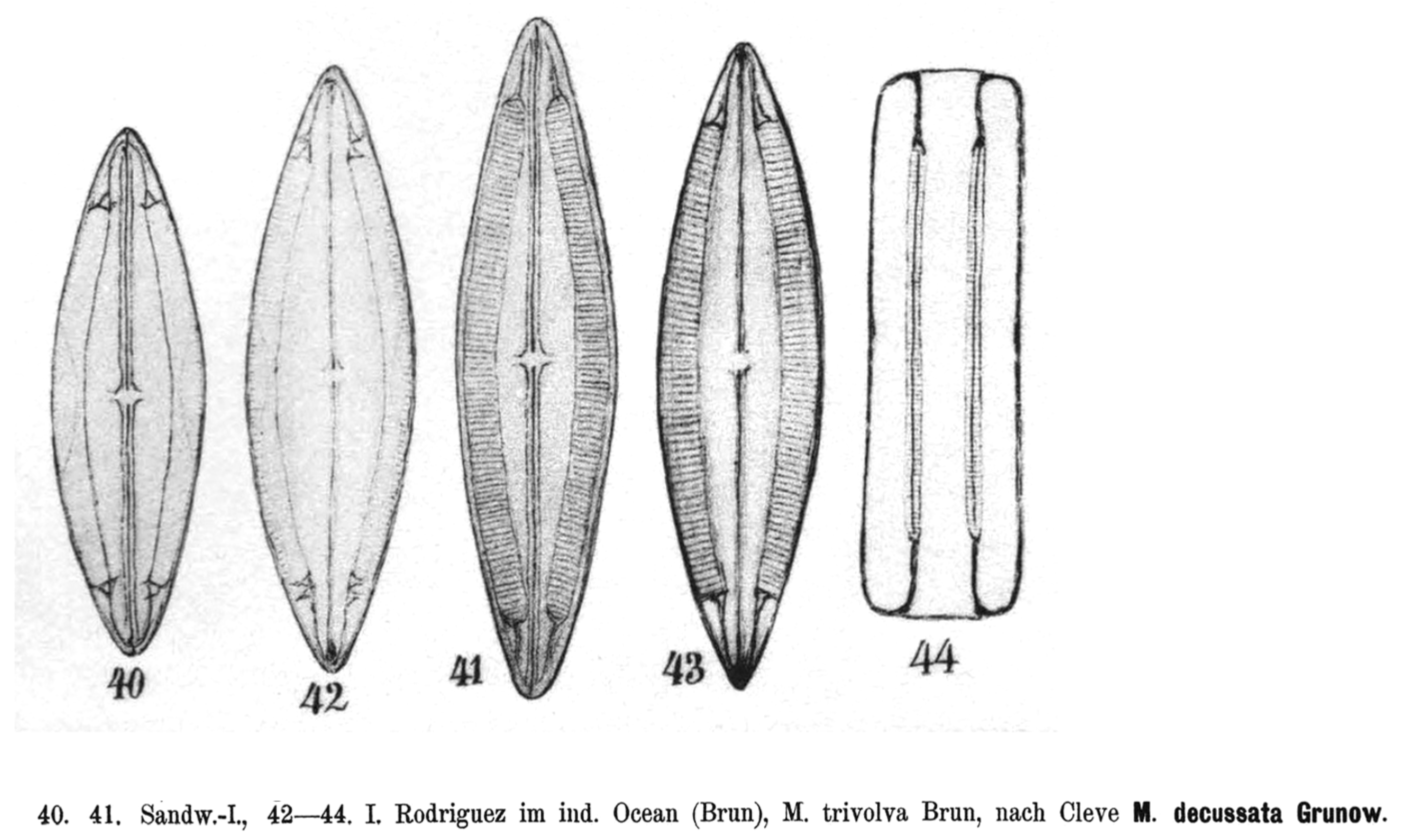
Figure 2.
General views of Mastogloia decussata in LM (a) and SEM (b–d). (a) Guam specimen (GU44I-4) at two focal planes, valve focus showing decussate striae and axial costae, VC focus showing partecta and diagonal exit ducts. (b) Exterior valve surface (GU12Y). (c) Interior view of valve with attached VC (GU12Y). (d) Three valves in different views, HI2010 population, showing sinuous raphe, central area, axial costae and partecta. Scale bars: 20 µm.
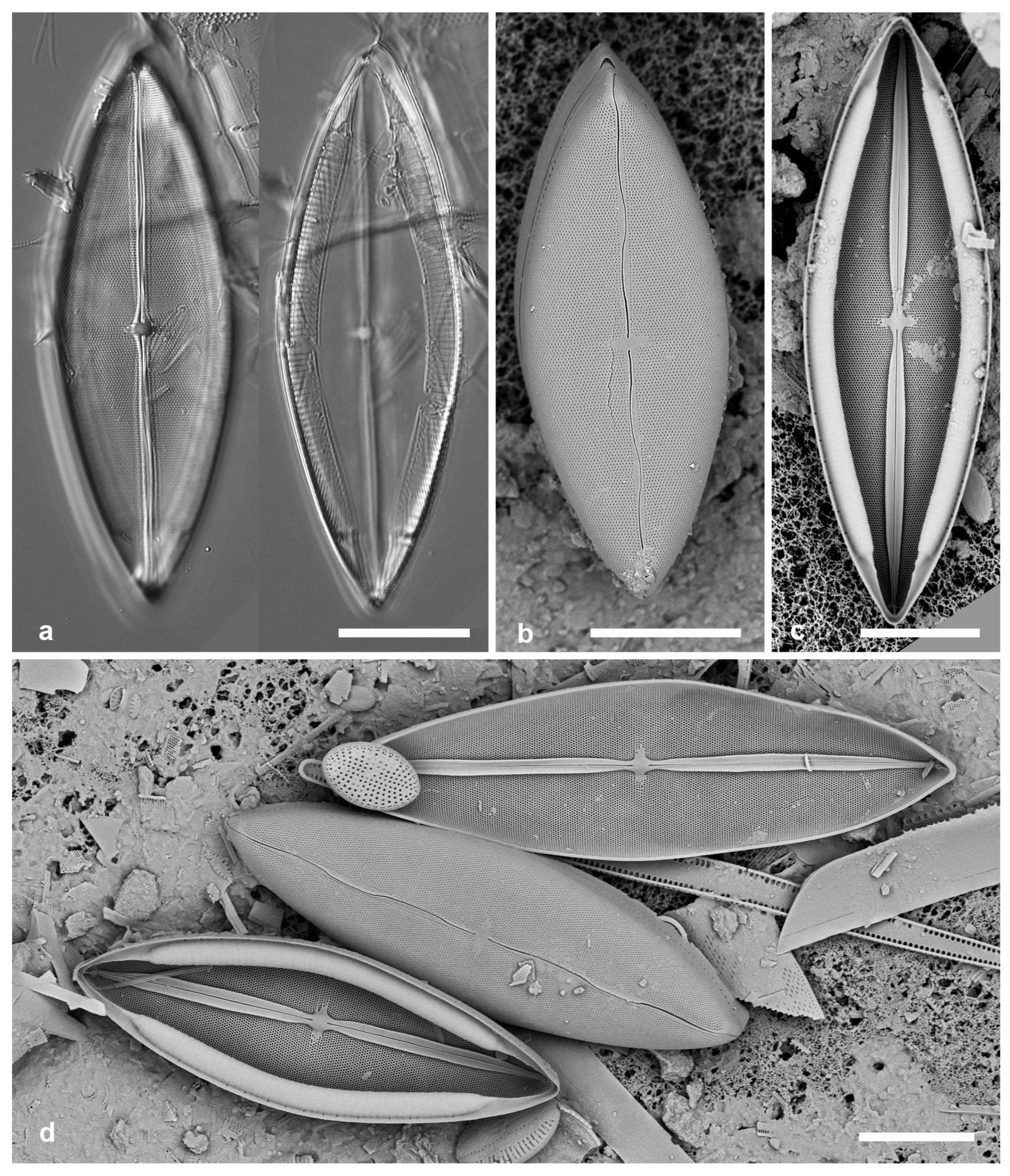
Description from LM and general SEM views (based on the literature and new observations) (Figure 2a–d): Dimensions given by Hustedt [9] were length 70–130 µm, width 22–27 µm, stria density 26–30 in 10 µm and the coin-like partecta, 9–10 in 10 µm, terminated at some distance from the ends; the original description [23] specified stria density 22–25 in 10 µm and the chambers continuing to the apices. Cleve [23] described the raphe as straight; Hustedt [9] said it had slight lateral dilations; we describe it as sinuous (Figure 2b,d). The axial costae are somewhat ambiguous in LM (Figure 2a) but very clear in SEM of interiors (Figure 2c,d). Population in Guam (Table 1): length 54–106 μm, width 18–32 μm; striae 23–26; partecta 4.0–4.7 µm wide, 10 in 10 µm. Population in Hawaii (Table 1): length 83–110 μm, width 29–34 μm; striae 24 in 10 µm; partecta 3.8–4.8 µm wide, 10 in 10 µm. The stria densities fit Cleve’s account rather than Hustedt’s. Each partectum has a diagonal exit duct (Figure 2a); Hendey [12] measured the angle of this V as about 150° but did not know what they were (see below, Figure 4a,b; also drawn in Schmidt’s fig. 40, see Figure 1). Every partectum has a duct, and the openings leave a gap in the middle of the band while continuing beyond the end of the partectum to the very apex of the valve (see also Figure 3a and Figure 5a below).
Table 1.
Morphometrics of Mastogloia populations studied.
SEM description: Valve: Sternum wide where raphe curves but narrow near central area and apices (Figure 3a–f). External raphe path clearly sinuous, central raphe endings at the end of the asymmetrical central area, raphe sometimes bordered by slight thickening (Figure 3c,d); terminal ends making slight turn at the valve–mantle boundary before dropping straight to the valve border with a small bend near the margin (Figure 3b,e,f). External foramina typically circular, but sometimes linear or narrowly lanceolate longitudinal slits in same population (Figure 3e,f). Wall structure is dual pseudoloculate (Figure 3g,i) (category 2.2.3 in [13], p. 87, figs 52a–f), i.e., with internal pseudoloculi offset from external pseudoloculi within each stria and joined by four small pores from two different pseudoloculi (cf. Figure 9d–g, M. paddocketkempii, sp. nov., below). Internally, the axial costae are prominent extensions of the sternum, arising asymmetrically at the apices (Figure 3h and Figure 4d and Figure 5a) and interrupted by the thickened central nodule (Figure 3i,j). Valvocopula and partecta: The partectal walls have no visible perforations (Figure 4b,d and Figure 5c,d). As seen in girdle view, there are two sets of exit pores with a wide gap mid-cell but continuing very close to the apex beyond the partecta, showing that exit ducts are diagonal (Figure 5a). These can be directly observed in carefully focused LM (Figure 2a and Figure 4a) and advalvar views of the ring (Figure 4c) but not in the abvalvar surface of the flat portion of the septum in this species (Figure 4d). The septum between the diverging ducts is solid and sometimes remains when the partecta and exit ducts are broken away (Figure 4b; see also M. davisii Lobban: Ref. [11], fig. 123); most of the septum in this and similar species is hollow, made up of the series of ducts. Each apical partectum has a characteristic conical extension containing the end of the intrapartectal cylinder (Figure 2a and Figure 4d) (it resembles the exhaust cone of a jet engine). The intrapartectal cylinder was directly observed in specimens from Guam and Hawaii (Figure 5a–c) and was also recorded by Paddock (unpublished SEM, Figure 5d); it lies in the angle between the valvocopula wall and the advalvar surface of the partectal ring. The cylinder is perforated, and a hyaline line can be seen with uniseriate striae on one side and biseriate striae on the other (Figure 5b,c).
Figure 3.
Mastogloia decussata frustule structure, SEM. (a) Oblique view of frustule including girdle bands: VC with exit pores and two plain pleurae (STT-7). (b) Polar view of valve showing deflection of terminal raphe ending (HI2010). (c,d) Raphe and central area; raphe with raised edges along the deflection and near the central area (GU12Y, HI2010, respectively). (e,f) Apices showing circular and linear foramina, GU12Y and STT-7, respectively. (g) Detail of broken wall, internal side, show pairs of pores opening into internal pseudoloculi (GU12Y). (h) Apex of valve showing raphe ending in helictoglossa (HI2010). (i,j) Central area with axial costae oblique and perpendicular views, GU12Y and HI2010, respectively. (Compare (i) with [13], fig. 81.) Scale bars: (a) 10 µm; (b–f,h,j) 5 µm; (g,i) 2 µm.
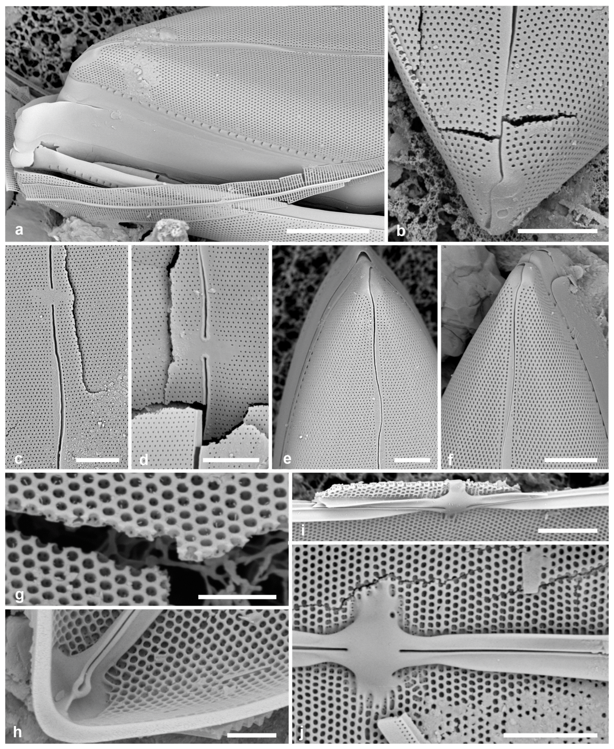
Figure 4.
Mastogloia valvocopula structure. (a) Isolated valvocopula, probably from M. paddocketkempii, in LM at two focal planes, showing IPC and solid septum between diverging exit ducts (GU44L-A). Concave arrows here and on (b) show the solid portion of the septum; black plain arrow shows the characteristic cone over end of IPC, white plain arrow shows probable edge of IPC. (b–d) SEM of M. decussata. (b) Frustule with upper valve and underlying partecta broken away, leaving the triangle of solid septum on that VC, and the complete lower valve and VC (GU52O-2). (c) Portion of isolated VC, advalvar side, showing layer of exit ducts and external pores (STT-7). (d) Apex of VC on valve with continuation of exit ducts within the septum; both here and in (b), partecta show no visible pores; black arrow points to conical cap over IPC, white plain arrow points to last exit pore (HI2010). Scale bars: (a,b) 10 µm, (c,d) 5 µm.
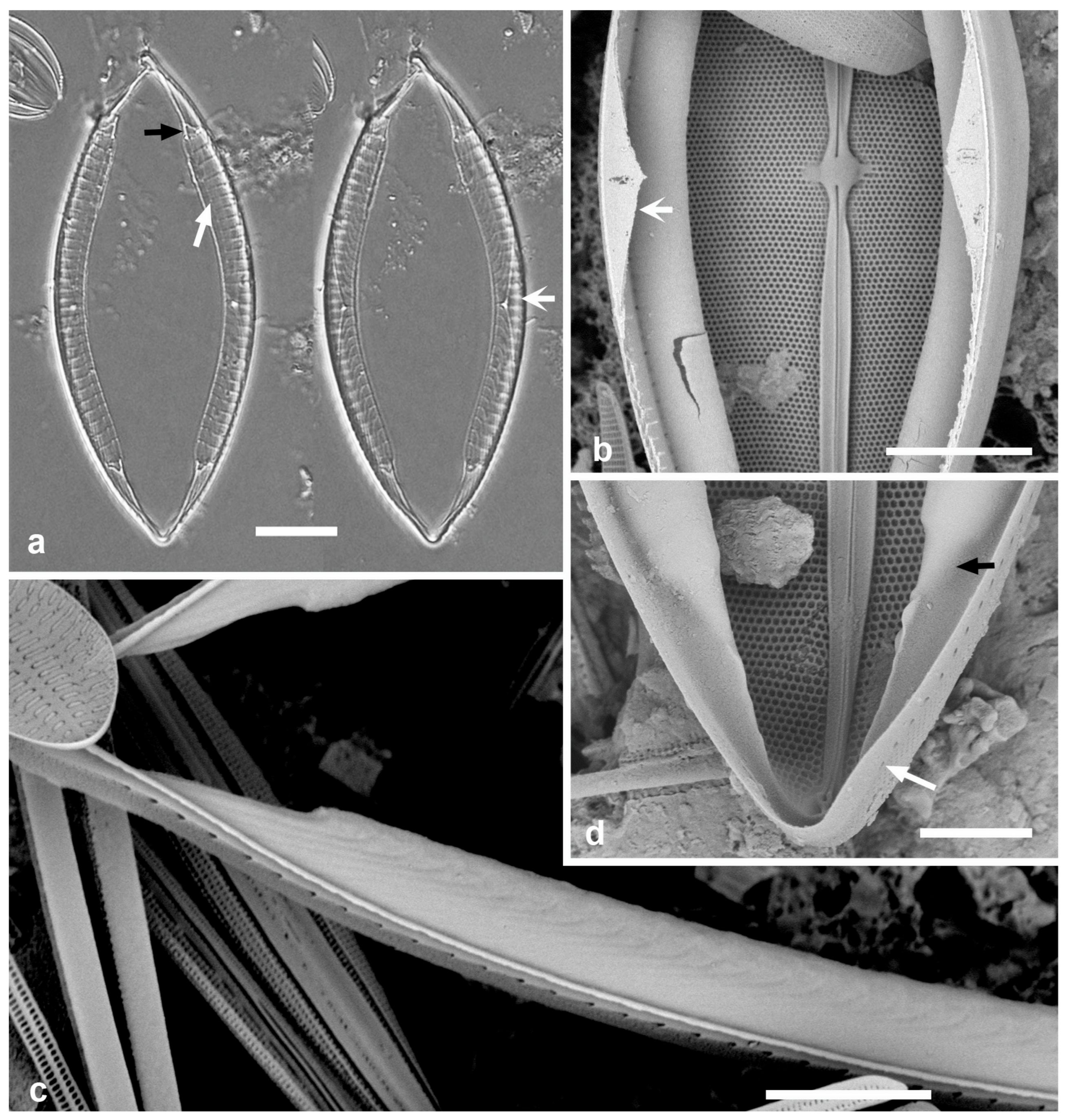
Figure 5.
Mastogloia decussata compound partecta structure. (a) Tilted view of broken frustule looking into the cross-sectioned partecta, also showing gap between exit pores in middle of cell. Specimen appears to be a newly divided cell with no VC yet on the hypotheca; partectal ring shows advalvar face with exit ducts (GU12Y). (b) Same specimen in perpendicular view showing poration of the IPC; arrow shows hyaline line between uniseriate and biseriate striae (compare with (c)). The break is near the middle, so no ducts show. (c) Collapsed partectum exposing IPC within, its uniseriate and biseriate striae clear on opposite side of hyaline line (arrow) (HI2010). (d) Paddock proof from sheet KK56, showing IPC in broken end of M. decussata VC. Natural History Museum, London collection, used with permission. Lack of pores in the partectal walls clear in (c,d). Scale bars: (a) 10 µm, (c) 5 µm, (b) 2 µm, (d), 1 µm.
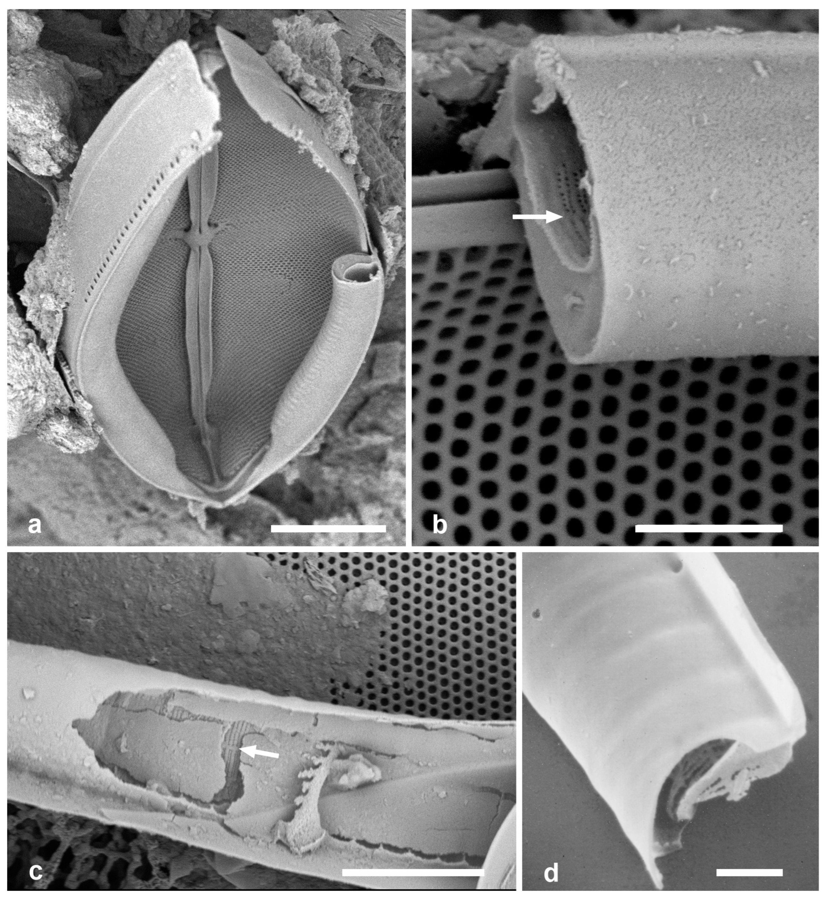
Figure 6.
Mastogloia decussata, neotype specimen from Hawaii sample HI2010. Scale bar 20 µm.
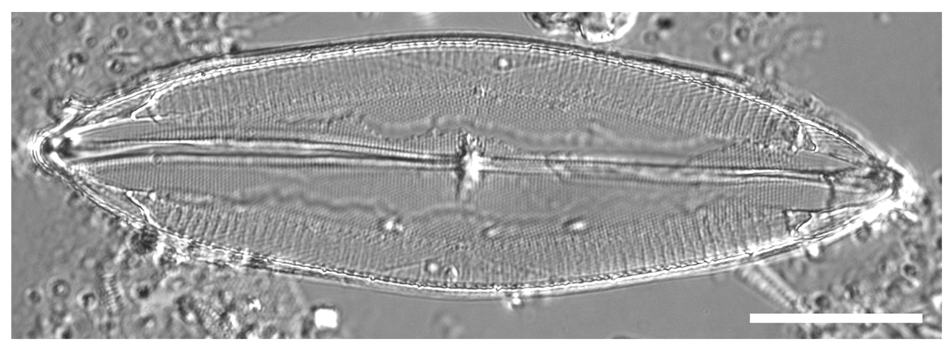
Typification: As explained in Materials and Methods, Grunow’s material is considered lost. The Madrid Code [28], Art. 9.16 states, “When a holotype or a previously designated lectotype has been lost or destroyed and it can be shown that all the other original material differs taxonomically from the lost or destroyed type, a neotype may be selected to preserve the usage established by the previous typification”. Thus, we propose the HI2010 specimen as a neotype. We were guided in choosing this collection by figs 40 and 41 in Schmidt’s Altas [25] (Figure 1), which show the species as understood today and are from one of the collection locations specified by Cleve [23]. If Grunow’s original material is ever found, it can be used to supersede the neotype (Ref. [28], Art. 9.18).
Neotype here designated: Specimen at 11.0 mm E, 3.9 mm S of the mark on slide 4232, Kaneohe Bay, Oahu, Hawaii, 2010, collector and precise coordinates not recorded; Figure 6. Deposited at Drexel University Academy of Natural Sciences of Philadelphia, catalog number ANSP GC20124.
Phycobank registration: http://phycobank.org/107499
Hustedt (1933) [9] listed as illustrations, in addition to the original Cleve (1892) [23], p. 23, fig. 17—a figure that is inadequate to address the question, Schmidt’s Atlas [25], pl. 186, figs 40, 41; he did not include figs 42 or 43, which in the Atlas were captioned as M. trivolva Brun, “nach Cleve M. decussata Grunow”, i.e., synonymous (Figure 1). Those figures do not show axial costae. Hustedt’s drawing (Ref. [9], fig. 917) is indistinguishable from neotype M. decussata except for absence of axial costae. Although he showed axial costae in several species and used them as a key character, they can be hard to see in LM, and we assume he overlooked them. However, records of “M. decussata” that clearly show a lack of axial costae must be re-evaluated, e.g., Güttinger’s SEM [29], pl. 2.05 29-9.
Biogeography hypothesis: Pantropical.
3.2. Mastogloia singaporensis M. Voigt 1942 (Figure 7)
Voigt [10], J. Roy. Microsc. Soc. 62: 17, fig. 25
Type locality: Singapore.
Additional references: [30], figs 88, 89; [11], pp. 392–395, figs 222–224 (with SEM)
Population studied: Yap, Y39A, scraping from mangrove root in Tagireeng Channel between Yap and Tamil-Gagil.
LM description (based on the literature, especially [10]): Valves lanceolate with rounded apices, length 46–78 µm, width 13–17 µm; raphe straight; striae 28–29 in 10 µm, decussate; axial costae present alongside raphe–sternum; prominent central area; partecta 5–7 in 10 µm, 2–3 µm wide, quadrate, extending over only the middle 46–50% of the valve length (Figure 7a). Voigt noted the similarity to M. decussata, except for the shape and extent of the partecta and the larger central area. He particularly pointed out the axial costae, assuming, from Hustedt [9], that M. decussata lacked them.
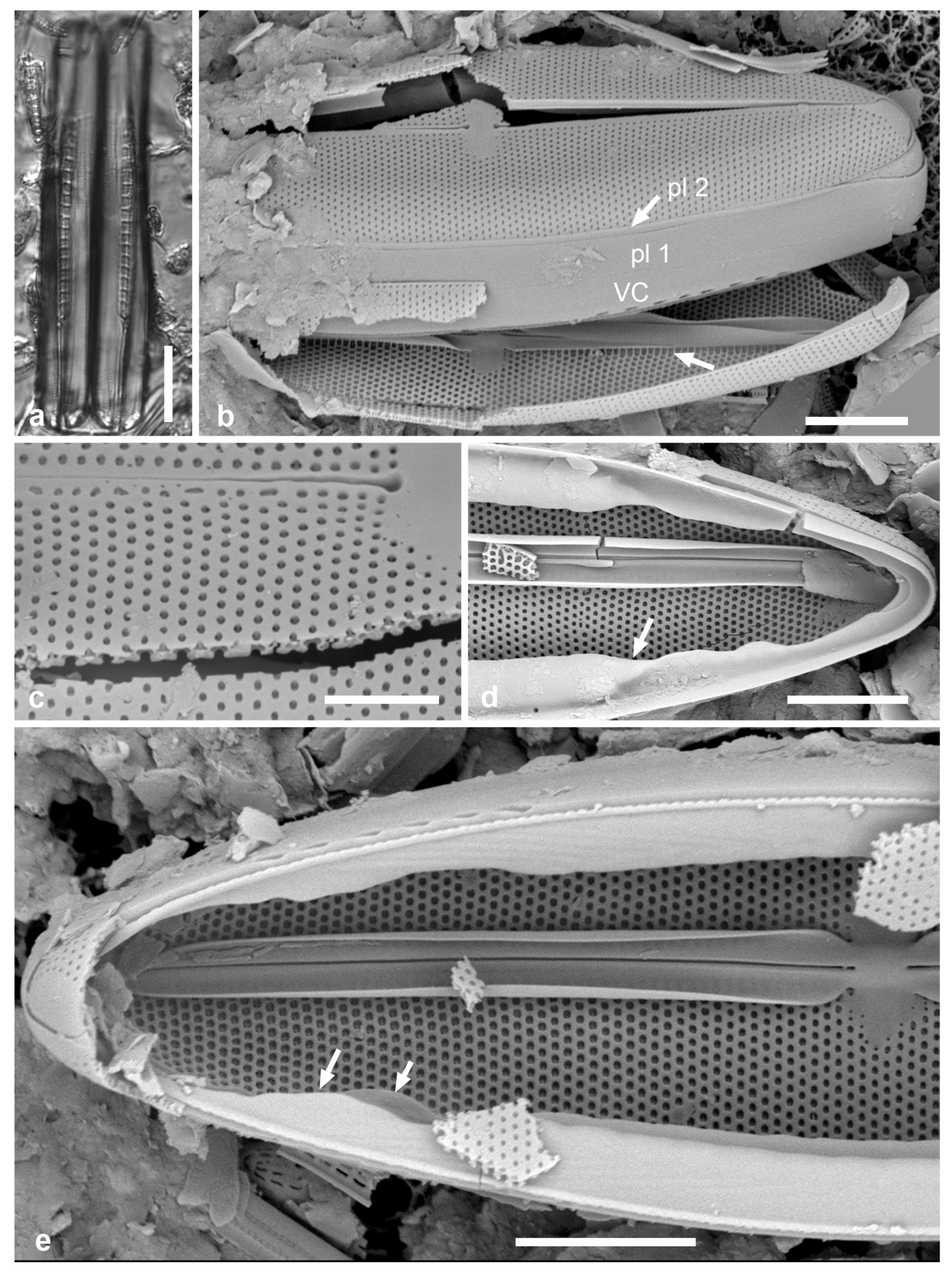
SEM description: Valve: Frustules with hyaline VC and two pleurae (Figure 7b). Walls dual pseudoloculate (Figure 7c), external foramina circular, internal foramina circular to quadrate (Figure 7e), similarly to M. decussata. Axial costae wider and curled upward compared to M. decussata and taper more abruptly at the central nodule (Figure 7b,e vs. Figure 3h) but arise from the apical nodule, as in M. decussata. There is a row of longer internal foramina beneath them (Figure 7b arrow). Valvocopula and partectal ring: Partecta without visible pores and ending in conical apices (Figure 7d), as in M. decussata, but we were unable to find a damaged ring with a direct view of the IPC. Septum continuing around the apex, with a broad lateral extension beyond ends of exit ducts (Figure 7e).
Biogeography hypothesis: Regional endemic. So far, rarely found only from Indo-West Pacific, i.e., Singapore [10], within Micronesia only in Yap [11], and from stromatolites in Western Australia [30].
Figure 7.
Mastogloia singaporensis. (a) LM girdle view of recently divided cell, each daughter cell hypotheca still without VC (cf. Figure 5a). (b–e) SEM. (b) Frustule showing girdle bands (valvocopula and 1st and 2nd pleurae), straight raphe, and row of larger internal foramina under the curled axial costae (unlabeled arrow). (c) Broken valve showing dual pseudoloculate wall structure. (d) Apex of valve interior and abvalvar side of VC, closed end, showing conical cover of IPC (arrow) and lack of visible pores in partecta walls. (e) Frustule with both VCs but missing the upper valve, showing advalvar side of partecta with exit pore system and septa extended beyond the exit ducts apically and laterally (arrows). Scale bars (a) 10 µm, (b,d,e) 5 µm, (c) 2 µm.
3.3. Mastogloia paddocketkempii Lobban et J.A.B.Valencia, sp. nov. (Figure 8, Figure 9 and Figure 10)
Diagnosis: Differing from M. decussata in the nearly quadrate partecta and finer striae, from M. singaporensis in the partecta reaching almost to the apices and wavy raphe, and from both in the rostrate apices.
Holotype: Specimen at 25.6 mm E and 2.5 mm S of the mark on slide 805 in the Diatom Collection, Academy of Natural Sciences of Drexel University, Philadelphia, accession number ANSP GC20125. Figure 8a.
Type locality: GU32B, Sella Bay, Guam, 13°19”41′ N, 144°39”06′ E, seagrass leaves and seaweeds from shallow seagrass bed. Collected by C. Lobban and M. Schefter, 25 March 1989.
Figure 8.
Populations of Mastogloia paddocketkempii studied. LM views of specimens at two focal planes: (a) Guam, GU32B, holotype; (b) Guam, GU44I-1 “species 19”; and (c) St. Thomas, USVI, STT-7. Scale bars: 10 µm.
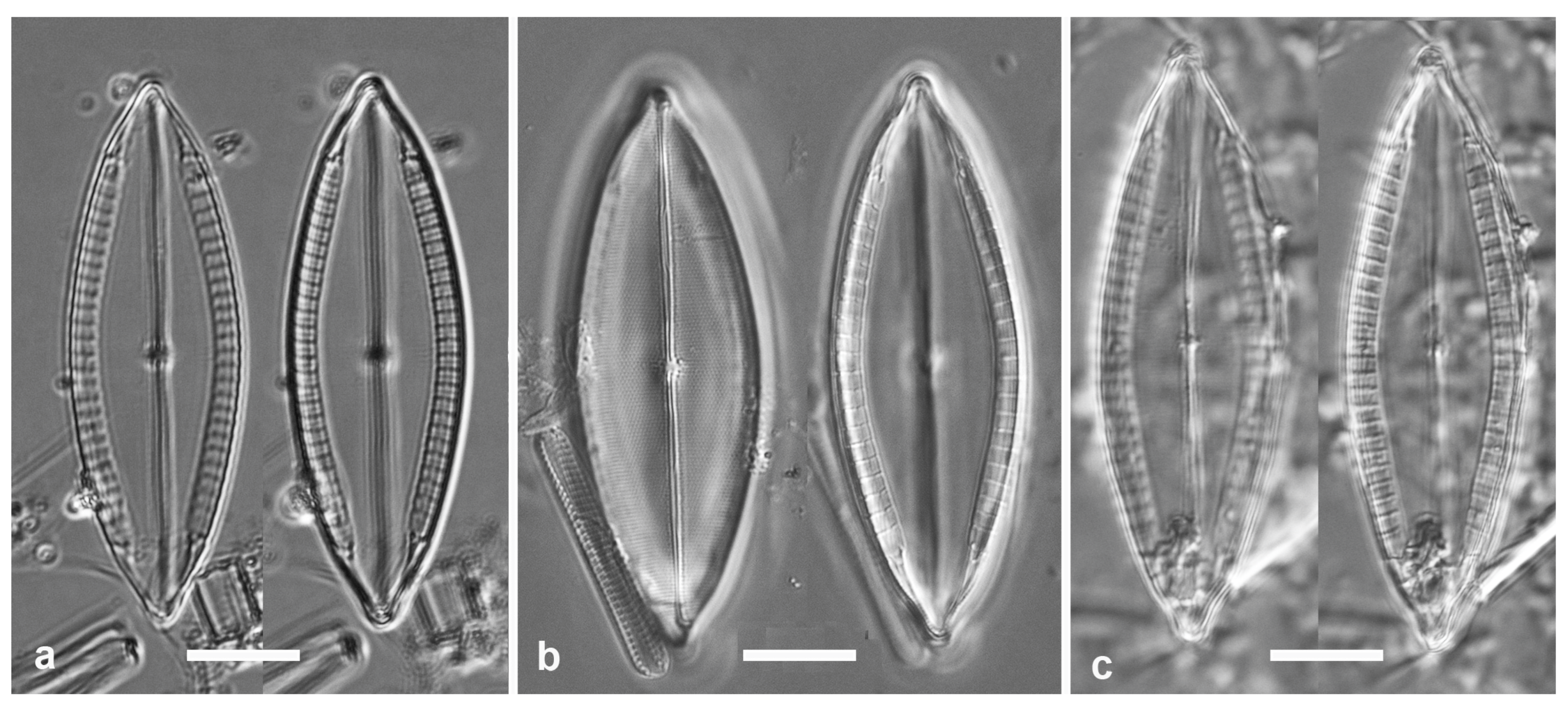
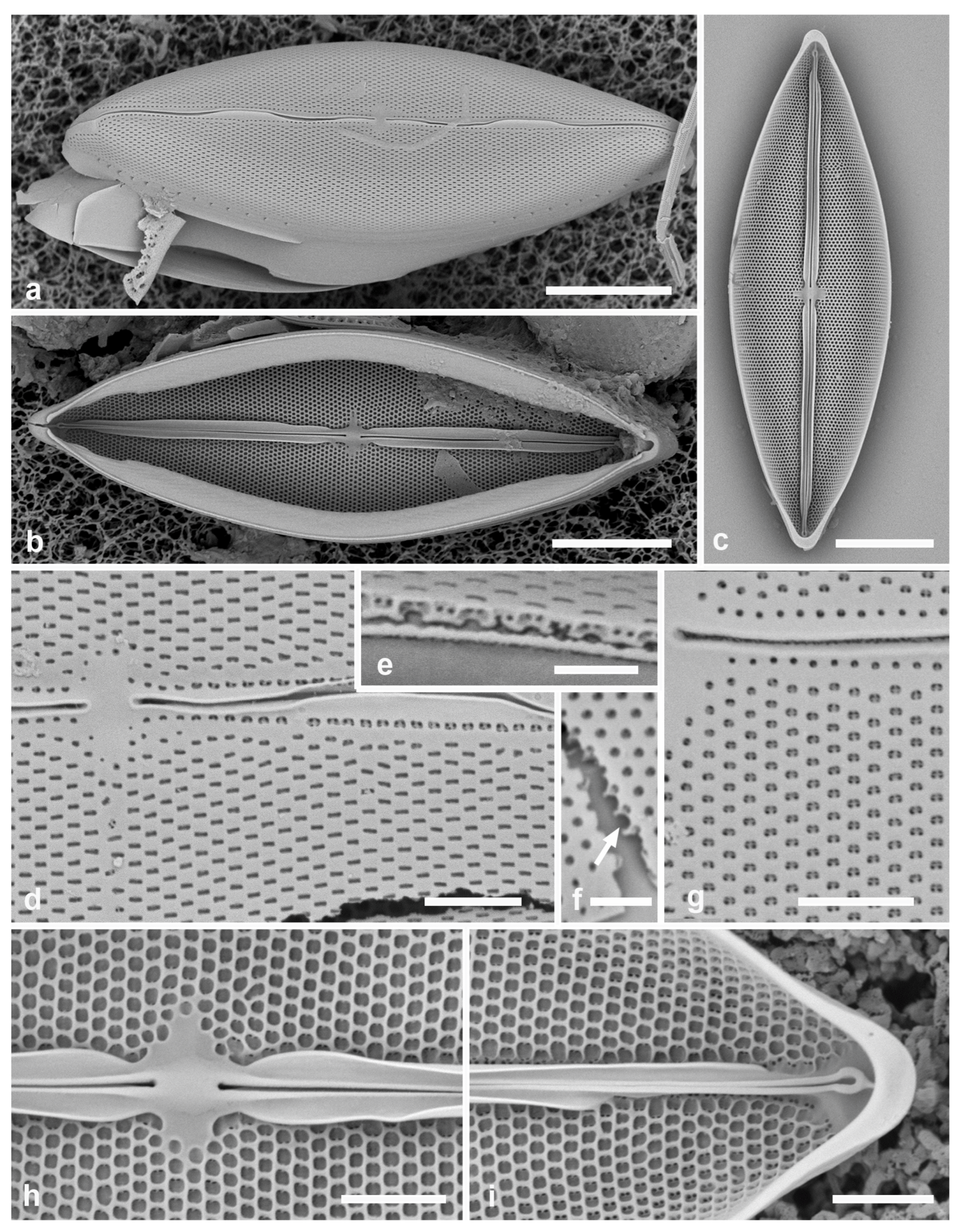
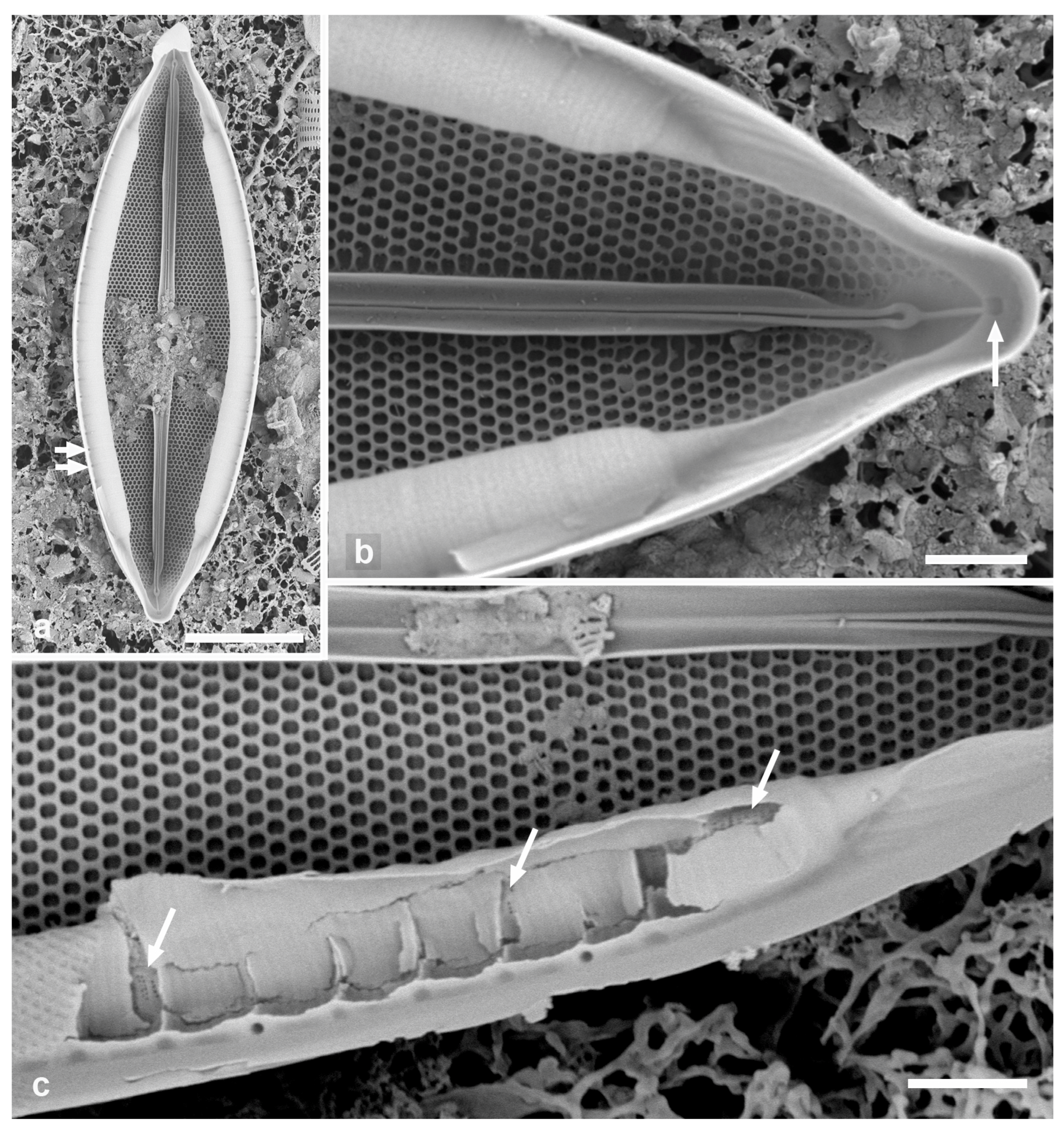
Morphology: Valves lanceolate, slightly rostrate, length 30–76 µm, width 15–27 µm, decussate striae 26–28 in 10 µm (Table 1, Figure 8a–c and Figure 9a–c), external foramina usually linear, showing two pores within and a slightly separate row along each side of the sternum (Figure 9d); areolae dual pseudoloculate (Figure 9e); external foramina more circular in USVI sample (Figure 9f), but in both cases, two pairs of pores in external pseudoloculus leading to two different internal pseudoloculi, as in M. decussata. Likewise, the two hexagonal arrays are offset from one another in the striae (Figure 9f). Small, irregular central area. Raphe sinuous, slightly raised, hooked at the terminal ends and bordered internally by axial costae that arise asymmetrically below the apices (contrast M. decussata) and are interrupted at the central area (Figure 9h,i). Valvocopula (Figure 10a–c): Partecta slightly ribbed but without visible pores, transapically rectangular to quadrate, 2.4–3.3 µm wide, 7–9 in 10 µm, occupying 68–73% of the valve length, the ring ending in a characteristic small cone, and ends of 2–3 exit ducts visible on abvalvar side of septum (Figure 10b). IPC visible in a specimen from Chuuk (Figure 10c), where the biseriate striae show in several places through the broken partectal walls. The partectal density and rostrate apex in this specimen show that it is M. paddocketkempii, not M. decussata.
Figure 9.
Mastogloia paddocketkempii in SEM. (a) Frustule in oblique view showing sinuous raphe, spacing of partectal exit pores on VC, and 1st copula (GU32B). (b) Valve with VC, the partectal ring in advalvar view showing exit duct system, and this from recently divided cell (GU32B). (c) Isolated valve, internal view (GU32B). (d) Detail of central area and external foramina (Guam: GU6I-3). (e,f) Details of wall cross-section showing dual pseudoloculate structure (cf. [13], fig. 52f) and offset hexagonal arrays ((f), arrow) (STT-7). (g) Contrasting external foramina in USVI sample (STT-7). (h,i) Internal details of isolated valve, central area and apex, respectively (GU55B-4). Scale bars: (a–c) 10 µm, (d,g–i) 2 µm, (e,f) 1 µm.
Additional records: Guam: GU6I-3!, GU44I-1!, GU44Y-13!, GU56A-2!, GU32B!; GU44I-2!; Chuuk records in [16] (figs 45a, b) as M. decussata and CHB1-2. Observed in SEM of Indonesian sample LM4-2! [21], on Padina, Melano Berat, 0°43′56” N, 108°43′09” E. Also, slide 031! in the Kemp set of specimens from Indonesia at BM (not identified to species and no collection data).
Figure 10.
Mastogloia paddocketkempii partecta and IPC in SEM. (a,b) Valve with abvalvar view of partecta on VC, (a) whole specimen with tall partecta indicated by the pores at the crosswalls (arrows). (b) Detail of apex showing characteristic IPC cones, lack of partectal pores, and exit ducts visible on abvalvar surface of extended septum; closed end of band showing lacuna (arrow). (c) Chuuk specimen revealing IPC striae visible through broken partecta (cf. Figure 5c) (CHB1-2). Scale bars: (a) 10 µm, (c) 5 µm, (b) 2 µm.
Etymology: The species name commemorates the collaboration of T.B.B. (Barrie) Paddock and Klaus-Dieter Kemp on Mastogloia, including several new species and a seminal study of the ultrastructure [13], from which sets of Paddock’s proof sheets with additional images (e.g., Figure 5d here), arranged by species, are in the BM collection. Kemp left a set of slides with individually mounted Mastogloia specimens from Indonesia.
PhycoBank Registration: http://phycobank.org/107500.
Biogeography hypothesis: Possibly pantropical but may be a species complex.
4. Discussion
The results clarify the morphology of M. decussata and establish the presence of intrapartectal cylinders, as seen in a few apparently unrelated species to date [11,13,18,19,20]. The IPC in M. decussata seems to occupy a larger fraction of the partecta than in M. punctifera (Ref. [10], fig.117a) or M. lanceolata (Ref. [19], fig. 3). While the IPC itself can be directly observed only through suitable breaks in the partectal walls, such compound partecta are also characterized by a lack of visible pores in the partectal walls (though they are undoubtedly porous) and a characteristic cone over the tips of the IPC, which can be seen in LM and was drawn by Schmidt [25], as shown in Figure 1. From the species reported with compound partecta to date, we were sufficiently confident that these more visible characters indicated the presence of an IPC to claim it for M. singaporensis, where it was not directly observed. An extensive study of the distribution of these characters across the genus has increased that confidence to virtual certainty (Lobban et al. in prep.).
We have resolved some of the questions about M decussata, including whether there are axial costae and what was the V-shaped structure observed by Hendey. Hendey [12], p. 145, pl. 1, fig. 2, found this species to be the commonest Mastogloia in his Kuwait samples and gave a detailed description, yet made no comment about the axial costae which seem to be present. He reported lengths of 100–120 μm, width 20–30 μm, striae 24 in 10 μm. Light micrographs in Foged [31], pl. 11, fig. 1; Witkowski et al. [32], pl. 78, figs 1, 2; Hein et al. [14], pl. 38, fig. 6; and Lobban et al. [27], pl. 27, figs 8, 9 for this species also show specimens that appear to have axial costae; yet there is no mention of costae, ribs or thickening in M. decussata as described by Cleve [23] or Hustedt [9], p. 493, fig. 917. Ricard [26], p. 55, figs 23–25, showed an SEM image of a specimen with axial costae identified as M. decussata. The identities of tightly cropped SEM images in Paddock & Kemp [13] are often unverifiable, but their fig. 93 of a Bermuda specimen identified as M. decussata is recognizable (we have also checked Paddock’s unpublished proof sheets, grouped by species), and it clearly shows the axial costae and central nodule.
M. singaporensis: This species was well defined by Voigt [10] and compared to M. decussata and was first illustrated in SEM by Lobban [11]. This species is clearly related to M. decussata by the close structural similarity, differing in the sizes and shapes of the valve and partecta. From the limited material available, we have been able to add only details of the areolae and the septum beyond the partecta. The comparison with M. paddocketkempii partecta is also important.
M. paddocketkempii: Two populations from Guam and one from St. Thomas, USVI, were compared, and no significant differences could be found to distinguish them (Table 1). The species was typed on a Guam population, and other Guam populations extended the morphometrics. The Chuuk specimens, also from Micronesia, were confidently placed in the species, but the Atlantic specimens less so, as are Kemp slides, labeled as M. decussata and our observations of a few specimens from Indonesia. No molecular data were obtained but might distinguish among potential simulacra. Other populations that warrant re-examination include those, misidentified as M. decussata, from The Bahamas [14], pl. 38, fig. 6, and the French Antilles [15], pl. 8a, among other potential simulacra. A culture from the Red Sea has been identified in the UTEX Culture Collection, and its sequences will be reported with other Mastogloias in a subsequent paper.
Dual pseudoloculate wall structure is rare in Mastogloia but not unique to this group, being also reported from M. paradoxa [13], which has areolae in a square pattern rather than hexagonal. It results in two arrays slightly offset from one another and could have interesting photonic properties, so far explored only in the large, single-loculate girdle bands of some centric diatoms [4].
The addition of this group of decussate species, differing from the few but diverse species of Mastogloia already known to have compound partecta with intrapartectal cylinders, has led us to search widely for this character. These results will be reported in a subsequent paper.
Supplementary Materials
The following supporting information can be downloaded at: https://www.mdpi.com/article/10.3390/d18080457/s1; Spreadsheet with draft list of currently accepted Mastogloia species.
Author Contributions
Conceptualization and methodology C.S.L.; microscopy: C.S.L., K.D. and J.A.B.V.; writing—reviewing and editing: C.S.L. All authors have read and agreed to the published version of the manuscript.
Funding
This material is based upon work supported by the National Science Foundation under the Guam NSF EPSCoR GECCO program Award Number OIA-1946352 and E-RISE program Award Number OIA-2445825. K.D. was supported by a GECCO internship in 2023, and J.A.B.V. was supported by an E-CORE internship in 2026. Collection of the St. Thomas samples was carried out during a trip funded by Guam NSF INCLUDES SEAS Alliance Award Number HRD-1930857.
Institutional Review Board Statement
Not applicable.
Data Availability Statement
Additional images are available upon request from C.S.L.
Acknowledgments
C.S.L. thanks María Schefter and the many students who have contributed to the development of the GUAM Herbarium Diatom Collection over the years. He especially thanks Wulf Herwig for the HI2010 sample important to this study and Tanja Schuster and Wolf-Henning Kusber for advice on issues around M. decussata typification.
Conflicts of Interest
The authors declare no conflicts of interest.
Abbreviations
The following abbreviations are used in this manuscript:
| IPC | intrapartectal cylinder |
| LM | light microscopy |
| SEM | scanning electron microscopy |
| TEM | transmission electron microscopy |
| VC | valvocopula |
References
- Mann, D.G.; Vanormelingen, P. An inordinate fondness? The number, distributions, and origins of diatom species. J. Eukaryot. Microbiol. 2013, 60, 414–420. [Google Scholar] [CrossRef] [Scilit]
- Harwood, D.M. Diatomite. In The Diatoms: Applications for the Environmental and Earth Sciences, 2nd ed.; Smol, J.P., Stoermer, E.F., Eds.; Cambridge University Press: Cambridge, UK, 2011; pp. 570–574. [Google Scholar]
- Goessling, J.W.; Wardley, W.P.; Lopez-Garcia, M. Highly reproducible, bio-based slab photonic crystals grown by diatoms. Adv. Sci. 2020, 7, 1903726. [Google Scholar] [CrossRef] [Scilit] [PubMed]
- Ashworth, M.P.; Lam, D.W.; Lopez-Garcia, M.; Manning, S.R.; Goessling, J.W. Adaptive evolution and early diversification of photonic nanomaterials in marine diatoms. Sci. Rep. 2025, 15, 6290. [Google Scholar] [CrossRef] [Scilit] [PubMed]
- Alverson, A.J.; Roberts, W.R.; Ruck, E.C.; Nakov, T.; Ashworth, M.P.; Bryłka, K.; Downey, K.M.; Kociolek, J.P.; Parks, M.; Pinseel, E.; et al. Phylogenomics reveals the slow-burning fuse of diatom evolution. Proc. Natl. Acad. Sci. USA 2025, 122, e2500153122. [Google Scholar] [CrossRef] [Scilit] [PubMed]
- Round, F.E.; Crawford, R.M.; Mann, D.G. The Diatoms: Biology and Morphology of the Genera; Cambridge University Press: Cambridge, UK, 1990; 747p. [Google Scholar]
- Fourtanier, E.; Kociolek, J.P. Catalogue of the diatom genera. Diatom Res. 1999, 14, 1–190. [Google Scholar] [CrossRef] [Scilit]
- Guiry, M.D.; Guiry, G.M. AlgaeBase; World-wide electronic publication; National University of Ireland: Galway, Ireland, 2024; Available online: http://www.algaebase.org.
- Hustedt, F. Die Kieselalgen Deutschlands, Österreichs und der Schweiz. In Rabenhorst’s Kryptogamenflora; Johnson Reprint: New York, NY, USA, 1933; Volume 7, Reprint 1962. [Google Scholar]
- Voigt, M. Contribution to the knowledge of the diatom genus Mastogloia. J. R. Microsc. Soc. Ser. 1942, 62, 1–20. [Google Scholar] [CrossRef] [Scilit]
- Lobban, C.S. Towards a biogeography of Mastogloia (Diatomeae: Bacillariophyceae): A preliminary checklist for Yap, Micronesia, with new ultrastructure images and a review of problems. Nova Hedwig. 2025, 120, 313–417. [Google Scholar] [CrossRef] [Scilit]
- Hendey, N.I. Some littoral diatoms of Kuwait. Nova Hedwig. Beih. 1970, 31, 107–167. [Google Scholar] [CrossRef] [Scilit]
- Paddock, T.B.B.; Kemp, K.D. An illustrated survey of the morphological features of the diatom genus Mastogloia. Diatom Res. 1990, 5, 73–103. [Google Scholar] [CrossRef] [Scilit]
- Hein, M.K.; Winsborough, B.M.; Sullivan, M.J. Bacillariophyta (Diatoms) of the Bahamas. In Iconographia Diatomologica; Gantner Verlag: Ruggell, Liechtenstein, 2008; Volume 19, 303p, (Distributed by Koeltz Scientific Books, Koenigstein, Germany). [Google Scholar]
- Loir, M.; Novarino, G. Marine Mastogloia Thwaites ex W.Sm and Stigmaphora Wallich species from the French Lesser Antilles. Diatom Monogr. 2013, 16, 1–133. [Google Scholar]
- Park, J.S.; Lobban, C.S.; Lee, K.-W. Diatoms associated with seaweeds from Moen Island in Chuuk Lagoon, Micronesia. Phytotaxa 2018, 351, 101–140. [Google Scholar] [CrossRef] [Scilit]
- Sterrenburg, F.A.S. Studies on the genera Gyrosigma and Pleurosigma (Bacillariophyceae): The identity of Pleurosigma obscura W. Smith. Diatom Res. 1993, 8, 447–456. [Google Scholar] [CrossRef] [Scilit]
- Stoermer, E.F.; Pankratz, H.S.; Drum, R.W. The fine structure of Mastogloia grevillea Wm. Smith. Protoplasma 1964, 59, 1–13. [Google Scholar] [CrossRef] [Scilit]
- Novarino, G. A note on the internal construction of the partectal ring of Mastogloia lanceolata. Diatom Res. 1987, 2, 213–217. [Google Scholar] [CrossRef] [Scilit]
- Novarino, G. Observations on the frustule architecture of Mastogloia smithii, with particular reference to the valvocopula and its integration with the valve. Diatom Res. 1990, 5, 373–385. [Google Scholar] [CrossRef] [Scilit]
- Risjani, Y.; Witkowski, A.; Kryk, A.; Yunianta; Gorecka, E.; Krzywda, M.; Safitri, I.; Sapar, A.; Dąbek, P.; Arsad, S.; et al. Indonesian coral reef habitats reveal exceptionally high species richness and biodiversity of diatom assemblages. Estuar. Coast. Shelf Sci. 2021, 261, 107551. [Google Scholar] [CrossRef] [Scilit]
- Lobban, C.S.; Jerao, K.R.; Frankovich, T.A. Contribution to taxonomy and biogeography of Mastogloia (Diatomeae, Bacillariophyceae): A global species and a potential regional endemic. Diversity 2026, 18, 37. [Google Scholar] [CrossRef] [Scilit]
- Cleve, P.T. Sur quelques nouvelles formes du genre Mastogloia. Le Diatomiste 1892, 1, 159–163. [Google Scholar]
- Cleve, P.T. Synopsis of the naviculoid diatoms. Part II. Kongl. Sven. Vetensk.-Akad. Handl. 1895, 27, 1–219. [Google Scholar]
- Schmidt, A. Atlas der Diatomaceen-Kunde; Series I–X; O.R. Reisland: Leipzig, Germany, 1874–1959; (Reprint 1974, Koeltz: Koenigstein). [Google Scholar]
- Ricard, M. Ultrastructure de quelques Mastogloia (diatomées benthiques) marines d’un lagon de Tahiti. Protistologica 1975, 11, 49–60. [Google Scholar]
- Lobbban, C.S.; Schefter, M.; Jordan, R.W.; Arai, Y.; Sasaki, A.; Theriot, E.C.; Ashworth, M.; Ruck, E.C.; Pennesi, C. Coral-reef diatoms (Bacillariophyta) from Guam: New records and preliminary checklist, with emphasis on epiphytic species from farmer-fish territories. Micronesica 2012, 43, 237–479. [Google Scholar]
- Turland, N.J.; Wiersema, J.H.; Barrie, F.R.; Gandhi, K.N.; Gravendyck, N.; Greuter, W.; Hawksworth, D.L.; Herendeen, P.S.; Klopper, R.R.; Knapp, S.; et al. International Code of Nomenclature for Algae, Fungi, and Plants (Madrid Code); University of Chicago Press: Chicago, IL, USA, 2025; Available online: https://www.iaptglobal.org/_functions/code/madrid (accessed on 21 July 2026).
- Güttinger, W. Collection of SEM-Micrographs of Diatoms; Series 8; Koeltz Scientific Books: Koenigstein, Switzerland, Privately printed; 1996; 50p. [Google Scholar]
- John, J. Mastogloia species associated with active stromatolites in Shark Bay, west coast of Australia. In Proceedings of the 11th International Diatom Symposium; Kociolek, J.P., Ed.; California Academy of Sciences: San Francisco, CA, USA, 1994; pp. 189–209. [Google Scholar]
- Foged, N. Diatoms from Viti Levu, Fiji Islands. Bibl. Diatomol. 1987, 14, 1–195. [Google Scholar]
- Witkowski, A.; Lange-Bertalot, H.; Metzeltin, D. Diatom Flora of Marine Coasts I; Gantner Verlag: Ruggell, Liechtenstein, 2000; 925p, (Distributed by Koeltz Scientific Books, Koenigstein, Germany). [Google Scholar]

Disclaimer/Publisher’s Note: The statements, opinions and data contained in all publications are solely those of the individual author(s) and contributor(s) and not of MDPI and/or the editor(s). MDPI and/or the editor(s) disclaim responsibility for any injury to people or property resulting from any ideas, methods, instructions or products referred to in the content. |
© 2026 by the authors. Licensee MDPI, Basel, Switzerland. This article is an open access article distributed under the terms and conditions of the Creative Commons Attribution (CC BY) license.