


Abstract
The rhabdocoel family Trigonostomidae has been the subject of recent phylogenetic revisions. However, due to the limited number of sequenced taxa, questions regarding intrafamilial and intrageneric relationships remain unresolved. To address these gaps, we collected trigonostomids from the North Sea, the Mediterranean, and the Atlantic Ocean, and examined them using an integrative approach. Nineteen specimens, representing ten species, were sequenced for phylogenetic analyses. Eight species were sequenced for the first time, including the first molecular data for Lutheriella. All genera were recovered as monophyletic; however, Ceratopera falcata (type species of Messoplana) and a new closely related species were found to cluster with species of Beklemischeviella, while the remaining species of Ceratopera formed a distinct clade. Based on these findings, we propose the resurrection of Messoplana, which had previously been synonymised with Ceratopera. Notably, genetic divergence and differences in the bursal mouthpiece between specimens of M. falcata suggest the possible presence of cryptic diversity or geographical isolation within this taxon. The inclusion of Trigonostomum breitfussi provided molecular support for its morphologically defined group. Furthermore, L. diplostyla was found to be closely related to species of Ptychopera. In addition to phylogenetic analyses, we describe four new species: Ceratopera bransoni sp. nov., Messoplana judithae sp. nov., Parapharyngiella scotti sp. nov., and Ptychopera woodyi sp. nov. The discussion of the new species of Parapharyngiella and Ptychopera motivated the reclassification of Pt. scutulifera as Pa. scutulifera comb. nov. We record for the first time eight species from Florida, seven from the Indian River Lagoon, and two from the Florida Keys National Marine Sanctuary, in the USA. This study refines the phylogenetic framework and internal classification of Trigonostomidae by providing new molecular evidence for previously unsampled taxa and by re-evaluating generic boundaries within the family.
1. Introduction
The family Trigonostomidae Graff, 1905 is a species-rich group of rhabdocoel microturbellarians, including 12 genera and 100 species. Most of this diversity is classified within the genera Proxenetes Jensen, 1878 (27 species), Ceratopera Den Hartog, 1964 (26 species), Trigonostomum Schmidt, 1852 (22 species), and Ptychopera Den Hartog, 1964 (13 species) [1]. The family has a cosmopolitan distribution, with members inhabiting both marine and brackish environments. However, several genera are restricted to temperate to cold waters of the northern hemisphere (including the Mediterranean and the Black Sea), such as Beklemischeviella Luther, 1943, Brederveldia van der Velde & van de Winkel, 1975, Lutheriella Den Hartog, 1966, Petaliella Ehlers, 1974, and Proxenetes [2,3,4,5,6,7]. This family also includes some taxa with apparent worldwide distributions, which possibly constitute complexes of cryptic species, i.e., Ceratopera axi (Riedl, 1954) Den Hartog, 1964 [8,9], and Trigonostomum armatum (Jensen, 1878) Graff, 1905 [10,11].
The family Trigonostomidae was previously split into the subfamilies Trigonostominae Graff, 1905 and Mariplanellinae Ax & Heller, 1970. However, its classification has changed after several recent revisions. Perhaps the most substantial systematic change was the reclassification of Mariplanellinae as an early branching taxon within Rhabdocoela, now Mariplanellida Ax & Heller, 1970 [12]. As such, all current representatives of Trigonostomidae were previously classified within the suppressed subfamily Trigonostominae. Another major taxonomic update was the synonymisation of Messoplana Den Hartog, 1966 with Ceratopera Den Hartog, 1964 [9]. Two other recent contributions explored the internal phylogenetic relationships of the speciose genus Trigonostomum [11,13], supporting the monophyly of three morphologically distinct clades, with a fourth group pending confirmation. Recently developed scanning electron microscopy (SEM) techniques have also contributed to understanding the morphology of sclerotised structures and their taxonomic implications in Trigonostomum [14]. These recent systematics revisions highlight the need for additional molecular sampling to test generic boundaries and evaluate the evolutionary significance of diagnostic morphological traits.
In this study, we specifically test whether Messoplana falcata (Ax, 1953) Den Hartog, 1966 is phylogenetically nested within Ceratopera, evaluate the placement of Lutheriella, and assess the monophyly of previously recognised morphological groups within Trigonostomidae.
2. Materials and Methods
2.1. Sampling and Morphological Study
The specimens studied herein were collected in the Canary Islands, Spain (2011), Greece (2002), Portugal (2012), Cuba (2020–2021), Sardinia Island, Italy (2015 & 2024), the German island Sylt (2015 & 2024), and Florida, United States of America (2025–2026). They were extracted from sediment, hydrozoan colonies, and algae using the MgCl2 method [15] and studied alive. Morphological voucher specimens were whole mounted with lactophenol on microscope slides. The specimens used for molecular phylogenetic analyses were preserved in ethanol (99%) and stored at −20 °C. One specimen of Trigonostomum breitfussi (Graff, 1905) Meixner, 1924 collected in Sylt was cut in half; the posterior part containing the sclerotised structures was whole mounted (ZMH V13882), and the anterior part was used for molecular analyses. Drawings of sclerotised structures were made with a camera lucida on an Olympus BH-2 microscope (Olympus Optical Co., Ltd., Tokyo, Japan), using Nomarski interference contrast. Measurements were taken along the central axis of the measured structure.
Confocal laser microscopy (CLM) was employed to obtain additional morphological information, following the methodology of Hochberg [16]. The original mounting medium was replaced with Fluoromont-G TM (Electron Microscopy Sciences, Hatfield, PA, USA). Specimens were examined using a Zeiss LSM 800 confocal Laser Microscope (Carl Zeiss Microscopy GmbH, Jena, Germany). Image stacks were processed using ZEN 2.6 software (Carl Zeiss Microscopy GmbH, Jena, Germany) to generate orthogonal projections.
2.2. DNA Extraction, PCR Amplification, and Sequencing
Total DNA was extracted using a protocol for low-input tissues [17]. The specimens were processed with 195 µL of the TNES lysis buffer and 5 µL of proteinase K (Invitrogen, Carlsbad, CA, USA) at 55 °C for 1 h. Subsequently, 1.5 µL yeast tRNA (Invitrogen, Carlsbad, CA, USA) as a coprecipitant, 65 µL 5M NaCl, and 290 µL 96% EtOH were added to the lysates. The samples were then stored at −20 °C for 2 h, the lysate was centrifuged (18,000 rpm), and the supernatant was discarded. Purification was performed with two 70% EtOH wash steps, and the DNA was resuspended overnight at 4 °C in 0.1X TE buffer with 0.02% Tween™ 20 (Thermo Fisher Scientific Inc., Waltham, MA, USA).
PCR reactions were carried out using 0.2 mL PuReTaq Ready-To-Go PCR beads (GE Healthcare, Little Chalfont, UK). Each reaction included 2.5 μL of each primer (5 μM), 1 μL of DNA, and 19 μL of purified water for a final volume of 25 μL. PCR reactions were carried out on a PCR T-100 Touch thermal cycler (Bio-Rad Laboratories, Inc., Hercules, CA, USA). The primer pairs TimA (5′ AMCTGGTTGATCCTGCCAG 3′) and Poly18SR2 (5′ GCMRGKTCACCTACRGAAACCTTGTT 3′) [18], and LSU5 (5′ TAGGTCGACCCGCTGAAYTTA 3′) and LSU6-3B (5′ GAGAAGGGTTCCATGTGAACAGC 3′), were used to partially amplify subunits 18S and 28S rDNA, consecutively [19]. Both genes were amplified with the following PCR conditions: 95 °C for 3 min, 9 × (94 °C for 30 s, 60 °C for 30 s (−0.5 °C/cycle), 72 °C for 90 s), 30 × (94 °C for 30 s, 55 °C for 30 s, 72 °C for 90 s), 72 °C for 5 min. PCR products were verified on a 1.4% agarose gel, stained with GelRed® (Biotium, Inc., Fremont, CA, USA). PCR products were commercially purified and sequenced by Macrogen Europe B.V. (Amsterdam, The Netherlands) or Psomagen (Rockville, MD, USA) under BigDye™ terminator cycling conditions on an ABI3730XL DNA sequencer.
2.3. Molecular Phylogenetic Analysis
An overview of all used sequences, including the newly obtained ones, and corresponding accession numbers is presented in Table 1. Raw reads were quality-trimmed (error probability = 0.05) and assembled in Geneious Prime v2025.0 [20]. Consensus sequences were subjected to a BLAST v. 2.17.0 search [21] on the NCBI website (ncbi.nlm.nih.gov) to check for signs of contamination. Available trigonostomid sequences were mined from GenBank [22], and a separate dataset was compiled for both 18S (68 sequences) and 28S rDNA (56 sequences). Five species of Adenorhynchidae, the sister taxon of Trigonostomidae [11], were included as outgroups: Cilionema hawaiiensis Karling, Mack-Fira & Dörjes, 1972, Coronhelmis lutheri Ax, 1951, C. multispinosus Luther, 1948, Litucivis serpens Ax & Heller, 1970, and Tvaerminnea karlingi Luther, 1943.
Table 1.
Sequences used for phylogenetic analyses. Specimens in bold were sequenced for this study.
Both datasets were aligned using MAFFT v7, as implemented in Geneious [23]. The resulting alignments were concatenated in Geneious. An initial partitioning scheme defining gene boundaries was manually constructed. The concatenated alignment and partition files were used as input for the ModelFinder tool [24] on the IQ-TREE webserver [25]. Model fit was evaluated using the Akaike Selection Criterion, and partition merging was enabled [26]. The latter feature determines the best-fit partitioning scheme for a particular dataset, while also calculating the best-fitting evolutionary models for each selected subset.
Maximum likelihood (ML) analyses were conducted using the ‘Tree Inference’ tool on the IQ-TREE server [27], using edge-linked partitions. Branch support was assessed by ultrafast bootstrapping (UFboot) [28] and the nonparametric approximate likelihood-ratio test (SH-aLRT) [29], both with 1000 replicates. Bayesian inference (BI) was carried out using the Metropolis-coupled Markov Chain Monte Carlo (MC3) algorithm, implemented in MrBayes v3.2.6 [30] and available as a plugin in Geneious. Best-fitting partitions and substitution models were specified where possible; otherwise, the second most complex model implemented in MrBayes was chosen. Two independent runs were conducted simultaneously for 10,000,000 generations, each including one cold and three heated chains. Trees were sampled every 1000th generation, the first 25% being discarded as burn-in. Chain convergence was confirmed by the average standard deviation of split frequencies dropping below 0.01, the potential scale reduction factor approaching 1.0, and the log probability reaching a stationary distribution. The obtained topologies were summarised in a majority-rule consensus tree. Inferred posterior probabilities (pp) were employed as support values. ML and BI trees were visualised and rooted in FigTree v1.4.4 [31]. Weakly supported clades (pp < 0.95, SH-aLRT < 80, and UFboot < 95) were collapsed.
3. Results
3.1. Molecular Phylogenetic Relationships
The analysed datasets had an extension of 1854 bp and 1692 bp for 18S and 28S rDNA partial genes, respectively (concatenated dataset of 3546 bp). Missing data represented 4.3% of the concatenated dataset. Bayesian and ML topologies were congruent. The family Trigonostomidae (pp = 1; SH-aLRT = 99.8; UFboot = 100) encompasses two major groups, one containing species of Parapharyngiella Willems, Artois, Vermin, Backeljau & Schockaert, 2005 (pp = 1; SH-aLRT = 100; UFboot = 100), and the other the remaining taxa (pp = 1; SH-aLRT = 99.8; UFboot = 100) (Figure 1). The internal relationships within the sampled species of Parapharyngiella are not fully resolved as P. caribbaea, P. mackfirae, and an undescribed species from Curaçao form a polytomy (pp = 1; SH-aLRT = 100; UFboot = 100). The other group, containing most trigonostomids, splits into two subgroups: Ptychopera spp. + Lutheriella diplostyla Den Hartog, 1966 (pp = 1; SH-aLRT = 100; UFboot = 100) and the remaining taxa (pp = 0.98; SH-aLRT = 86.1; UFboot = 78). Within the last group, Ceratopera (pp = 1; SH-aLRT = 100; UFboot = 100) is the sister taxon to the remaining species (pp = 0.98; SH-aLRT = 80; UFboot = 75).
Figure 1.
Molecular phylogenetic relationships of Trigonostomidae based on the majority-rule consensus tree from the Bayesian analysis of the concatenated 18S + 28S rDNA. Branches with support values below the thresholds (pp < 0.95, bootstrap < 80, and UFboot < 95) were collapsed. Branches without symbols have pp = 1, bootstrap = 100, and UFboot = 100. Taxa from which new sequences were obtained for this study are highlighted in bold.
Species of Trigonostomum form a highly supported clade (pp = 1; SH-aLRT = 99; UFboot = 100), which is the sister taxon to a group containing Proxenetes + Beklemischeviella + Messoplana (pp = 1; SH-aLRT = 100; UFboot = 100). The clade conformed by Trigonostomum penicillatum (Schmidt, 1857) Graff, 1905 + T. breitfussi (pp = 1; SH-aLRT = 100; UFboot = 100) is the sister group to the other species of Trigonostomum, which form three other fully supported groups, corresponding to Clades 1, 2, and 3 in Diez et al. [11]. Species of Proxenetes (pp = 1; SH-aLRT = 100; UFboot = 100) are the sister taxon to Beklemischeviella + Messoplana (pp = 1; SH-aLRT = 98.6; UFboot = 100). Within Messoplana, three specimens of M. falcata form a cluster (pp = 1; SH-aLRT = 92.3; UFboot = 99), in which two specimens from Sylt, Germany, are more closely related (pp = 1; SH-aLRT = 100; UFboot = 100) than the specimen from the Canary Islands.
3.2. New Species and Distribution Records
Ceratopera Den Hartog, 1964
Ceratopera axi (Riedl, 1954) Den Hartog, 1964
Figure 2.
Ceratopera axi (Riedl, 1954) Den Hartog, 1964, specimens collected in Florida, USA. (A,B) Live specimens (NMNH 1774224 & 1774225). (C) Details of the atrial systems (NMNH 1774225). (D,E) Male Sclerotised structures (NMNH 1774224 & 1774225). (F) Bursal appendage (NMNH 1774226). Scale bars: 100 μm (A,B); 50 μm (C); 20 μm (D–F).
Figure 3.
Ceratopera axi (Riedl, 1954) Den Hartog, 1964, specimen collected in Sardinia, Italy (ZMH V13883). (A) Habitus of a live specimen. (B) Sclerotised atrial organs in a live specimen. Scale bars: 50 μm (B); 25 μm (C,D).
Known distribution. Species originally described from Sorrento and Sicily, Italy (type locality) [32]. Atlantic Ocean: Falklands Islands [33], and Departamento de Rocha, Uruguay [34]. Indian Ocean: La Reunion [35]. Pacific Ocean: Galapagos Islands [36], California and Oregon, USA [33], British Columbia, Canada [9], and New South Wales, Australia [8]. Antarctica and subantarctic territories: Weddell Sea [35] and Kerguelen [37].
Material. Italy • Observations in one live and whole mounted specimen; Sardinia, near Stintino, Punta Negra, sublittoral, 0.5 m deep, silty algae under a pier, salinity 37 ‰; 40.95323, 8.228437; 11 August 2024; ZMH V13883. United States of America • Three specimens studied alive and whole mounted; Florida, Fort Pierce, just south of Jim Island, Indian River Lagoon (IRL), on algae (Hypnea and red filamentous) and hydrozoans on mangrove roots, salinity 35 ‰; 27.47345, −80.31249; 1 March 2025; NMNH 1774224–1774226.
New morphological information. The newly collected specimens (Figure 2 and Figure 3) largely align with descriptions of C. axi [8]. In the specimens from Florida (Figure 2), the male sclerotised structures include a 92–101-long stylet (x̄ = 95 µm; n = 3) (Figure 2A–E: st) and a 52–56-µm-long mantle (x̄ = 54 µm; n = 3) (Figure 2C–E: m). The bursal appendage (Figure 2A,C,F: ba) measures 68–69 µm in length (n = 2) and exhibits a proximal cup-shaped part connecting to the bursa, and a distal elongated, tubular part, which terminally splits into two narrower tubes. The specimen from Sardinia (Figure 3) exhibits a 126-µm-long stylet (Figure 3B,C: st), the mantle (Figure 3B,C: m) is 64 µm long, and the bursal appendage (Figure 3A–D: ba) is 126 µm long. The bursal appendage in this specimen is proximally funnel shaped, and the distal tubular part coils (Figure 3B: ba), ending in a single tube (Figure 3D: tb).
Remarks. This species exhibits a wide range of variability in stylet length depending on the geographic region, from as short as 70 µm in Australia to as long as 180 µm in the Falkland Islands (see summary in Willems et al. [8], with additional records in Willems et al. [37,38] and Van Steenkiste et al. [34]). The bursal appendage similarly varies, ranging from 56 µm in some specimens from Uruguay [34] to 118 µm in specimens from British Columbia, Canada [9]. Here, we record the longest known bursal appendage for this species in the specimen collected in Sardinia (126 µm long).
Beyond size variation in the male sclerotised structures and bursal appendage across populations of C. axi, the morphology of the bursal appendage shows a level of variation that could be taxonomically significant. Ehlers and Ax [36] described C. bifida Ehlers & Ax, 1974—now considered a synonym of C. axi—based on the shape and distal bifurcation of the bursal appendage. The bursal appendage in Floridian specimens (68–69 µm long) closely resembles those from the Galapagos Islands (67–87 µm), characterised by a bursal appendage that is proportionally much longer than wide and distally splits into two short tubes. In contrast, specimens from other regions, such as the type locality in the Mediterranean (see Riedl [32]: Figure 27), typically have a broader bursal appendage, with a proximally funnel-shaped portion that tapers distally into a single tube, as is the case in the specimen recently collected in Sardinia. The latter specimen also exhibits the distal tubular part of the bursal appendage rolled up, whereas the tube does not coil in the specimens from Florida or the Galapagos.
Although these observations are consistent with the cryptic species hypothesis, the evidence currently available is still insufficient to distinguish between cryptic speciation, geographic structuring, and phenotypic plasticity. However, the morphological variability in the atrial structures of C. axi suggests the possibility of multiple cryptic species within what is currently considered a single taxon, a hypothesis broadly supported [8,9,34,37,38]. Nevertheless, as noted by Karling [33] and Artois et al. [35], the apparent variability in this species may partly stem from difficulties in observing the distal portion of the bursal appendage, as the bifurcation is not always visible. Additionally, these structures can be distorted by squeezing or affected by the developmental stage of the copulatory organs. Consequently, resolving this taxonomic ambiguity requires a more comprehensive morphological and phylogenetic investigation.
Ceratopera sellai (Steinböck, 1933) Den Hartog, 1964
Figure 4.
Ceratopera sellai (Levinsen, 1879) Den Hartog, 1964. (A) Sclerotised structures observed in the squeezed live specimen. (B) Sclerotised structures in the whole mount; one plate of the mantle is missing. (C) Proximal part of the stylet and one mantle plate. (D) Second mantle plate. (B–D) From the whole mount (NMNH 1774227). Scale bars: 40 μm (A); 20 μm (B–D); 50 μm (E,F).
Known distribution. Until now, this species was only known from its type locality: Rovinj region, Croatia, Adriatic Sea [39].
Material. United States of America • One specimen studied alive and whole mounted; Florida, Key West, Key Haven, subtidal, 0.5 m deep, fine-grained sand rich in organic matter, salinity 40 ‰; 24.586693, −81.742208; 24 March 2025; NMNH 1774227.
New morphological information. The general habitus of the newly collected specimen could not be thoroughly examined due to damage sustained immediately after mounting. However, the sclerotised atrial structures remained and were available for study. The stylet (Figure 4A–C,E: st) measures 96 µm in length. The mantle consists of two plates. Following fixation, one of the plates became detached. One mantle plate (Figure 4A–C,E: m2) measures 66 µm in length, is tubular in shape, bent proximally at approximately 45°, and terminates in a rounded distal tip. The second plate (Figure 4A,C–E: m1) is 71 µm long, flattened, roughly S-shaped, and tapers to a sharp distal point. The bursal appendage (Figure 4A,B: ba, Figure 4F) is funnel-shaped, with a distal tubular region that bifurcates into two narrower tubes. The total length of the appendage is 40 µm, with the distal tube measuring 11 µm in length and 2 µm in width.
Remarks. The fact that the single examined specimen damaged after mounting does not preclude the identification of C. sellai. All species of Ceratopera show a homogeneous habitus, and species differentiation relies on the study of the sclerotised structures. Fortunately, in the newly collected specimen, the sclerotised structures are well preserved (Figure 4). The morphology of the sclerotised structures in the Floridian specimen is consistent with the description of C. sellai from the Mediterranean, as reported by Steinböck [39]. In both populations, the mantle is composed of two plates, and the bursal appendage is relatively short, funnel-shaped, and terminates in a tube. However, Steinböck [39] does not mention the distal bifurcation of the tube observed in our specimen. This discrepancy could reflect observational limitations, as previously noted for C. axi, where the morphology of the tube can be difficult to interpret and is affected by its orientation and compression during preparation.
Ceratopera sellai is a poorly known species, previously only reported from its type locality and original description by Steinböck [39], with additional remarks by Den Hartog [40], who reclassified the species within Ceratopera. Steinböck [39] mentioned that C. sellai was highly similar to C. gracilis (Graff, 1882) Den Hartog, 1964. However, as both described and illustrated (Steinböck [39]: Figure 2), C. sellai possesses a mantle composed of two plates—an arrangement that is absent in C. gracilis. In fact, a mantle formed by two plates is only known in C. flabellifera (Levinsen, 1879) Den Hartog, 1964, C. reisingeri (Riedl, 1959) Den Hartog, 1964, and C. bransoni sp. nov. (see further description). These species, however, are distinguishable from C. sellai based on the morphology of the mantle plates. In C. flabellifera, the plates are nearly straight; one distally terminates in a rounded tip, while the other ends in a curved hook [40,41]. In C. reisingeri, the plates are arched, measuring 82 µm and 68 µm respectively (based on Riedl’s drawing), they are not tubular, and both end in obliquely cut (in lateral view), rounded tips [42].
Unfortunately, no measurements are available for the sclerotised structures of C. flabellifera or the original specimens of C. sellai described by Steinböck [39], limiting direct morphological comparisons. Additionally, the bursal appendage of C. reisingeri remains undescribed, but this structure provides a key differentiation between C. flabellifera (distal tube long and coils approximately twice) and C. sellai (distal tube short and uncoiled). Ceratopera sellai is further discussed with C. bransoni sp. nov. in the remarks on the new species.
Ceratopera bransoni Diez sp. nov.
Ocean Census species No. OC-SP-0002618
Figure 5.
Ceratopera bransoni sp. nov. (A,B) live specimen. (C,D) Micrographs of male copulatory hard structures (NMNH 1769577 and 1769578, respectively). (E,F) Micrographs of the bursal appendage (NMNH 1769577 and 1769578, respectively). Scale bars: 100 μm (A); 50 μm (B–D,G); 20 μm (E,F,H).
Type material. Holotype. United States of America • One whole mount; Florida, offshore, 6.75 km SE from the mouth of Fort Pierce Inlet, medium-grained and well-sorted sand, 10.6 m deep, salinity 37 ‰; 27.429300; −80.241083 (Type locality); 11 June 2025; NMNH 1769577. Paratype. United States of America • One whole mount; same data as for the holotype; NMNH 1769578.
Additional material. Observations on live specimens.
Diagnosis. Species of Ceratopera with male sclerotised organs, including a stylet and a two-plated mantle. Tubular stylet ~105 µm long. Mantle S-shaped plate ~86 µm long, tapering to a distal sharp tip. Mantle tubular plate 70 µm long, distally rounded. Bursal appendage ~73 µm long, with distal tube 8 µm long and 2 µm wide, splitting into two narrower tubes.
Etymology. Species named after David Branson, captain of the R/V Sunburst at the Smithsonian Marine Station at Fort Pierce, who kindly assisted in collecting the new species and supported the field work.
Description. The specimens (Figure 5A) are 772–846 µm long (x̄ = 809 µm; n = 2), translucent, and with a pair of eyes (Figure 5A: e). Strong adenal rhabdite tracts (Figure 5A: ar) open anteriorly. The pharynx (Figure 5A: ph) is located about midbody.
The testes (Figure 5A,B: t) are located posterior to the pharynx. The male and female genital organs are positioned in the posterior third of the body. The tubular stylet (Figure 5A–F: st) measures 100–110 µm in length (x̄ = 105 µm; n = 2) and is accompanied by a two-plated mantle. The mantle encompasses an S-shaped plate (Figure 5B–F: m1), 83–88 µm long (x̄ = 86 µm; n = 2), distally tapering to a sharp tip, which does not sheath the stylet; and a 70-µm-long tubular plate (n = 2) (Figure 5B–F: m2), which is proximally bent about 45°, and distally sheaths the stylet. The two plates of the mantle are connected proximally by a ring, which surrounds the stylet and connects to its proximal part. Distally, the tubular plate is closely associated with the stylet; in the holotype, they overlap, and it is difficult to differentiate their exact boundaries; in the paratype, the distal part of the mantle coats the stylet, which shows a distally oblique aperture.
The vitellaria run along the body’s sides, posterior to the brain to the level of the atrial organs. The bursal appendage (Figure 5B,H: ba, Figure 5G) is 58–87 µm in total length (x̄ = 73 µm; n = 2), displaying a bipartite, sac-shaped, proximal part, and a distal tube. The distal tube is 8 µm long and 2 µm wide in both specimens, and splits into two narrower tubes (Figure 5G,H: tb). The bursal stalk (Figure 5B: bs) is short and slightly sclerotised.
Remarks. As previously noted, a two-plated mantle is known in only three other species of Ceratopera—C. flabellifera, C. reisingeri, and C. sellai—with C. bransoni sp. nov. representing the fourth described species exhibiting this characteristic. Consequently, our comparative discussion focuses on its morphologically closest congeners, while acknowledging that the phylogenetic significance of this mantle structure remains uncertain. The S-shaped mantle plate in C. bransoni sp. nov. is comparable only to that of C. sellai, though larger (86 µm vs. 68 µm, respectively). In the other two species, the mantle plate, which does not sheath the stylet, ends in a rounded tip, being arched in C. reisingeri and nearly straight in C. flabellifera. The second, tubular mantle plate is highly similar in C. bransoni sp. nov. and C. sellai, both showing a proximal 45° bend and measuring 70 µm and 71 µm, respectively. Conversely, in C. flabellifera, this plate ends in a curved hook, and in C. reisingeri, it is arched and 68 µm long.
The bursal appendage morphology clearly distinguishes C. bransoni sp. nov. from C. sellai. In the new species, the appendage comprises two distinct chambers: a more proximal one (connected to the bursa) with longitudinal striations, and a distal one with zigzag striations. Ceratopera sellai possesses only a single differentiated chamber with longitudinal striations. The appendage is also considerably larger in C. bransoni sp. nov. (~73 µm) than in C. sellai (40 µm). The distal tubular portion is similar in both species, consisting of a short tube branching into two narrower distal tubes; however, relative to the total appendage length, this tubular section is proportionally longer in C. sellai (25%) than in C. bransoni sp. nov. (11%). The long, coiled distal tube found in C. flabellifera is markedly different from that in C. bransoni sp. nov., while the structure remains undescribed in C. reisingeri.
Taken together, the morphological differentiation of both male and female sclerotised structures provides robust support for the recognition of C. bransoni sp. nov. as a distinct species.
Messoplana Den Hartog, 1966 gen. rev.
Messoplana falcata (Ax, 1953) Den Hartog, 1966
Figure 6.
Messoplana falcata Ax, 1953. (A,B) Specimen from Sylt, Germany (XXVII.2.04). (C,D) Specimen from Faro, Portugal (HU XIII.2.34). (E,F) Specimen from the Canary Islands, Spain (XXVII.2.08). (G,H) Specimen from Sardinia, Italy (XXVII.2.05). (A,C,E,G) Male sclerotised structures. (B,D,F,H) Bursal appendage. Scale bars: 20 μm (A–F); 25 μm (G,H).
Known distribution. Ax [43] described the species based on material collected in Kiel Bay, Germany (Baltic Sea) and Sicily, Italy (Mediterranean). Black Sea: Turkiye [44]. North Sea: Sylt Island, Germany [4], Grindöy, near Tromsø, Norway [45]. Atlantic Ocean: White Oak River, North Carolina, United States of America [46]. Pacific Ocean: Santa Cruz, Archipielago de Colon, Galapagos Islands [36,47].
New material. Germany • Observations on live specimens; two whole mounts; Sylt, List, intertidal, coarse superficial sand, salinity 30 ‰; 55.02117, 8.439817; 22 June 2024; ZMH V13884–V13886 • two specimens used for molecular analyses; same data as for the previous ones • one whole mount; Sylt, sublittoral, fairly close to shore, fine-grained sand; 55.05322, 8.439500; 25 August 2015; HU XXVII.2.04. Greece • one whole mount; Macedonia, Thermaikos Gulf, Nea Michaniona, broad beach, fine green algae just beneath the waterline; 18 July 2002; leg. Willems, Willems & Schockaert; HU II.2.13. Italy • one whole mount; Sardinia, Cala Sant’Andrea, 2 m deep; 41.013322, 8.248974; 27 September 2014; HU XXVII.2.05. Portugal • two whole mounts; Faro, lower eulitoral, fine to medium sand with some detritus but little silt; 19 September 2012; HU XIII.2.34–XIII.2.35 • two whole mounts; Faro, Ilha de Culatra, side of Rio, mid-eulitoral, relative fine-grained sand with some silt; 19 September 2012; HU XIII.2.36–XIII.2.37. Spain • Observations on live specimens; two whole mounts; Canary Islands, Lanzarote, Orzola, sheltered bay with sandy beach, 0.5 m deep, coarse-grained sand, salinity 35 ‰; 29.216661, −13.442223; 6 October 2011; HU XXVII.2.06–XXVII.2.07• three whole mounts; Canary Islands, Lanzarote, Mala, large sand patch, 20 m deep, medium to coarse-grained, calcareous sand, salinity 35 ‰; 29.083481, −13.449749; 8 October 2011; HU XXVII.2.08–XXVII.2.10• one specimen used for molecular analyses; Canary Islands, Lanzarote, Mala, small patches among small rocks and algae, coarse-grained, calcareous sand, 18 m deep, salinity 35 ‰; 29.083481, −13.449749; 8 October 2011 • one whole mount; Canary Islands, Lanzarote, Punta Jameos del Agua, off shore, 38 m deep, medium-grained, clean sand, salinity 35 ‰; 29.156972, −13.427191; 15 October 2011; HU XXVII.2.11.
New morphological information. The male sclerotised structures in the newly collected specimens from Sylt are stylet (Figure 6A: st) 85–100 µm long (x̄ = 91 µm; n = 3), mantle (Figure 6A: ma) 86–94 µm long (x̄ = 91 µm; n = 3), and mantle hook (Figure 6A: h) 41–43 µm long (x̄ = 42 µm; n = 3). The bursal appendage (Figure 6B) is 20–31 µm long (x̄ = 26 µm; n = 3). The distal tubes (Figure 6B: dt) are slender, without thickened rims and measure 13–19 µm long (x̄ = 16 µm; n = 3). Measurements in the specimens from the Canary Islands are stylet (Figure 6C: st) 73–90 µm long (x̄ = 82 µm; n = 5), mantle (Figure 6C: ma) 74–89 µm long (x̄ = 81 µm; n = 5), and mantle hook (Figure 6C: h) 34–40 µm long (x̄ = 36 µm; n = 5). The bursal appendage (Figure 6D) is 28–36 µm long (x̄ = 32 µm; n = 4). The distal tubes (Figure 6D: dt) are slender, distally tapering and without thickened rims, measuring 25–28 µm long (x̄ = 26 µm; n = 4). Those structures in the specimens from Faro measure as follows: stylet (Figure 6E: st) 90–113 µm long (x̄ = 107 µm; n = 4), mantle (Figure 6E: ma) 93–106 µm long (x̄ = 100 µm; n = 4), and mantle hook (Figure 6E: h) 40–50 µm long (x̄ = 47 µm; n = 4). The bursal appendage (Figure 6F) is 32–42 µm long (x̄ = 36 µm; n = 4). The distal tubes (Figure 6F: dt) are slender, distally tapering and without thickened rims, measuring 23–30 µm long (x̄ = 26 µm; n = 4).
In the specimen from Greece, only the male hard structures are observable: the stylet is 119 µm long, the mantle 109 µm long, and the mantle hook 50 µm long. In the single specimen studied from Italy, the stylet is 74 µm long, the mantle 77 µm long, the mantle hook 44 µm long, the bursal appendage 25 µm long, and the distal tubes 19 µm long. A summary of the measurements of the hard structures in all species of Messoplana is provided in Table 2.
Table 2.
Measurements of the hard structures in species of Messoplana. Abbreviations: L, length.
Remarks. All species of Messoplana, including the type species of the genus M. falcata, were transferred to the genus Ceratopera by Van Steenkiste and Leander [9], who synonymised the two genera based on the phylogenetic position of M. pacifica Karling, 1986 (now C. pacifica). However, our phylogenetic analysis recovered M. falcata and a related new species as the sister taxa to Beklemischeviella, while species of Ceratopera, including the type species C. gracilis, formed a distinct clade. As discussed by Van Steenkiste and Leander [9], distinguishing between species of Messoplana and Ceratopera is challenging due to the apparent convergent evolution of the male sclerotised structures and the bursal appendage. Consequently, some species assigned to Messoplana exhibit morphological traits typical of Ceratopera, and vice versa—for instance, regarding the position of the pharynx.
Den Hartog [48] distinguished species of Messoplana by the presence of a pharynx located in the mid to posterior part of the body, a bursal appendage formed by a cuticular ring, and both the stylet and the mantle (‘additional duct’) proximally connected. In contrast, species of Ceratopera are characterised by an anteriorly positioned pharynx, a bursal appendage lacking a cuticular ring, and a mantle closely associated with and coating the stylet [40]. However, there are numerous exceptions to these morphological definitions. For example, several species previously assigned to Messoplana lack a ring in the bursal appendage, such as C. armonica (Ehlers & Sopott-Ehlers, 1989) Van Steenkiste & Leander, 2018, C. floralis (Ehlers, 1974) Van Steenkiste & Leander, 2018, C. globulifera (Artois, Vermin & Schockaert, 2000) Van Steenkiste & Leander, 2018, C. helgolandica (Ax, 1971) Van Steenkiste & Leander, 2018, C. minuta (Artois, Vermin & Schockaert, 2000) Van Steenkiste & Leander, 2018, C. pacifica (Karling, 1986) Van Steenkiste & Leander, 2018, C. rugata (Ehlers, 1974) Van Steenkiste & Leander, 2018, and C. spiralis (Artoi, Vermin & Schockaert, 2000) Van Steenkiste & Leander, 2018. Additionally, the close association between the mantle and stylet is not evident in C. axi, C. ehlersi Karling, 1986, C. pilifera Karling, 1986, and C. steinboecki (Riedl, 1959) Den Hartog, 1964. In these species, the mantle appears to coat, or be associated with, the stylet only at its distal end.
In the light of our phylogenetic analysis, it is clear, however, that Ceratopera and Messoplana form two distinct monophyletic lineages. Therefore, we propose to reinstate the validity of the genus Messoplana and undertake a comprehensive reexamination of the morphology of all species currently assigned to Ceratopera, with the aim of clarifying the morphological characters that differentiate the two genera. The recognition of both genera is based primarily on the phylogenetic positions of their respective type species, while newly identified morphological diagnostic characteristics allowed the classification of species that have not been included in molecular analyses.
Messoplana falcata shares a nearly identical construction of the male sclerotised structures with Ceratopera roscoffensis. In both species, the stylet and mantle are proximally connected but not distally associated (the mantle does not coat the stylet). The mantle in both cases shows a proximally flattened region with thickened lateral rims and a distally claw-shaped portion. Additionally, both species possess a ring on the bursal appendage and a lateral spine, the latter being recorded for the first time in M. falcata in this study (Figure 6F: sp). Despite these similarities, C. roscoffensis can be readily distinguished from M. falcata by its distinctly elongated bursal appendage. An additional character that has not previously been considered in the differentiation between Messoplana and Ceratopera is the position of the testes relative to the pharynx. Notably, M. falcata and C. roscoffensis are the only species within these genera in which the testes lie anterior to the pharynx. The two species also share a similar position of the pharynx, located in the posterior third of the body. In contrast, all other species currently assigned to Ceratopera exhibit a pharynx positioned either in the anterior region or in the proximal portion of the posterior body region, never extending into the posterior third, and the testes lie posterior to the pharynx. This set of characteristics observed in M. falcata and C. roscoffensis is also corroborated in M. judithae sp. nov., a new species of Messoplana further described in this manuscript.
The present findings suggest that a specific combination of characters, such as the position of the testes anterior to the pharynx, the location of the pharynx in the posterior third of the body, the mantle not sheathing the stylet, and the presence of a ring and lateral spine on the bursal appendage, serves as a reliable diagnostic suite for Messoplana. Based on this evidence, we formally reinstate the genus Messoplana to include M. falcata, M. roscoffensis, and M. judithae sp. nov. Given the current morphological and molecular data, we propose retaining the remaining species within Ceratopera, at least provisionally, pending further integrative studies. This conservative approach is warranted, as some species exhibit a mix of traits associated with both genera. For instance, C. globulata, C. minuta, and C. spiralis—originally described as species of Messoplana—possess male hard structures typical of Messoplana, specifically a mantle that does not sheath the stylet [35]. However, these same species have testes located posterior to the pharynx and lack a ring on the bursal appendage, as characteristic of Ceratopera.
The inconsistencies in the morphological boundaries between Messoplana and Ceratopera highlight the need for a more integrative taxonomic framework, combining detailed morphological reassessments with expanded phylogenetic analyses. Additionally, the relationship between Messoplana and Beklemischeviella requires further scrutiny, particularly given the apparent absence of clear synapomorphies linking the two genera. Species of Beklemischeviella are the only known trigonostomids lacking a mantle in the male copulatory organ, a condition interpreted by Van Steenkiste and Leander [9] as a secondary reduction. Nonetheless, such species of Messoplana, Beklemischeviella also possess testes located anterior to the pharynx. Moreover, species of Beklemischeviella exhibit a poorly differentiated and weakly sclerotised bursal appendage, a trait shared with members of Ptychopera and Lutheriella. However, phylogenetic evidence places Ptychopera and Lutheriella in a more basal clade within the family, whereas Messoplana and Beklemischeviella form a sister group to Proxenetes (Figure 1).
As shown in Table 2, the length of hard structures in M. falcata varies notably among different populations. Direct comparisons with measurements reported for other populations, however, are complicated by differences in measurement approaches adopted by various authors. For example, Ax [43] reported the total length of the male copulatory organ as 80–95 µm and the bursal appendage as 24 µm. Likewise, Ehlers and Ax [36] described specimens of M. falcata valida from the Galapagos Islands with a male organ measuring 80–90 µm and a bursal appendage 17–21 µm long. In contrast, in the material examined here, stylet length ranges from 74 µm in the specimen from Italy to 119 µm in specimens from Greece. The mantle hook also displays substantial variation, accounting for approximately 50% of the total mantle length in the Greek specimens, compared with 36% in those from the Canary Islands. The total length of the bursal appendage also varies among populations, reaching its maximum in specimens from Faro and the Canary Islands (36 µm) and its minimum in those from the Sardinia (25 µm).
As previously described, a further point of interest is the potential presence of cryptic species or geographically isolated lineages within M. falcata, given its wide distribution across the North Sea, the eastern Atlantic, the Mediterranean, and the Black Sea, as well as the documented genetic divergence between populations from the North Sea and the Canary Islands. In addition, the taxonomic status of M. falcata valida from the Galapagos Islands must be reassessed once new material becomes available. Unfortunately, the original description of this subspecies was based solely on photographs, and no additional material is currently accessible. Nevertheless, considering the morphological variability observed among M. falcata populations from different localities, and the description of a new closely related species from Florida (see below), we suspect that the Pacific population may in fact represent a distinct species.
Emended diagnosis of Messoplana (after Den Hartog [48]). Trigonostomids with paired testes in front of the pharynx. Pharynx located in the posterior body third. Male sclerotised structures including a stylet and a mantle, both proximally connected. Mantle never sheathes the stylet. Bursal appendage with a basal ring and lateral spine.
Included species. Messoplana falcata (Ax, 1953) Den Hartog, 1966 (type species), M. roscoffensis Ehlers, 1980, and M. judithae sp. nov.
Emended diagnosis of Ceratopera (after Den Hartog [40] and Van Steenkiste & Leander [9]). Trigonostomids with paired testes posterior to the pharynx. Pharynx located within the anterior body two-thirds. Male sclerotised structures including a stylet and a mantle. Mantle differentiated into one or several pieces, proximally connected to and mostly sheathing the stylet. Bursal appendage consisting of a funnel-shaped tube, two slender tubes or a combination of both, never surrounded by a ring.
Included species. Ceratopera gracilis (Graff, 1882) Den Hartog, 1964 (type species), C. aremorica (Ehlers & Sopott-Ehlers, 1989) Van Steenkiste & Leander, 2018, C. axi (Riedl, 1954) Den Hartog, 1964, C. bermudensis Karling, 1986, C. bransoni sp. nov., C. canariensis (Ehlers & Ehlers, 1980) Van Steenkiste & Leander, 2018, C. cascadiensis Van Steenkiste & Leander, 2018, C. complicata Van Steenkiste & Leander, 2018, C. ehlersi Karling, 1986, C. elegans (Luther, 1948) Van Steenkiste & Leander, 2018, C. flabellifera (Levinsen, 1879) Den Hartog, 1964, C. floralis (Ehlers, 1974) Van Steenkiste & Leander, 2018, C. geminata (Hartog, 1966) Van Steenkiste & Leander, 2018, C. globulifera (Artois, Vermin & Schockaert, 2000) Van Steenkiste & Leander, 2018, C. gracilis (Graff, 1882) Den Hartog, 1964, C. helgolandica (Ax, 1971) Van Steenkiste & Leander, 2018, C. minuta (Artois, Vermin & Schockaert, 2000) Van Steenkiste & Leander, 2018, C. pacifica (Karling, 1986) Van Steenkiste & Leander, 2018, C. paradoxa (Pereyaslawzewa, 1892) Den Hartog, 1964, C. paragracilis Ehlers & Ax, 1974, C. pilifera Karling, 1986, C. reisingeri (Riedl, 1959) Den Hartog, 1964, C. rugata (Ehlers, 1974) Van Steenkiste & Leander, 2018, C. sellai (Steinböck, 1933) Den Hartog, 1964, C. spiralis (Artois, Vermin & Schockaert, 2000) Van Steenkiste & Leander, 2018, and C. steinboecki (Riedl, 1959) Den Hartog, 1964.
Messoplana judithae Diez sp. nov.
Ocean Census species No. OC-SP-0002619
Figure 7.
Messoplana judithae sp. nov. (A,B) Live specimen. (C,E,F) Male sclerotised structures. (D,G) Bursal appendage. (C–G) From the holotype (NMNH 1774228). Scale bars: 200 μm (A); 50 μm (B,F,G); 20 μm (C–E).
Figure 8.
Messoplana judithae sp. nov. (A,C) Male sclerotised structures (NMNH 1774234 and 1774252). (B,D) Bursal appendage (NMNH 1774234 and 1774232). (E) F-actin-stained specimen studied by confocal laser microscopy. Scale bar: 25 μm (A–D); 20 μm (E).
Type material. Holotype. United States of America • One whole mount; Florida, Jupiter, Dubois Park, IRL, subtidal, 0.3 m deep, medium-grained sand with organic matter, salinity 38 ‰; 26.943401, −80.074212 (Type locality); 15 April 2025; NMNH 1774228. Paratypes. United States of America • One whole mounts; Florida, Jupiter, Dubois Park, IRL, intertidal, upper layer, medium-grained sand, with organic matter, salinity 28 ‰, and shallow subtidal, 0.3 m deep, medium-coarse sand with organic matter, salinity 38 ‰; 26.94158, −80.07341; 1 February 2025; NMNH 1774229–1774230 • One whole mount; same data as for the holotype; NMNH 1774231.
Other material. United States of America • Observations on live specimens; three whole mounts; Florida, Fort Pierce, little beach near South Causeway Beach, IRL, intertidally, coarse-shelly sand, upper 5–10 cm, salinity 35 ‰; 27.45854, −80.31697; 21 January 2025; NMNH 1774232–1774234 • One whole mount; Florida, Fort Pierce, South Causeway Beach, IRL, subtidally, 0.3 m deep at low tide, fine-grained, silty sand, salinity 35 ‰; 27.458528, −80.316972; 21 January 2025; NMNH 1774235 • Two whole mounts; Florida, Fort Pierce, South Causeway Beach, IRL, subtidally, 0.5 m deep, on filamentous red algae, salinity 35 ‰; 27.458528, −80.316972; 27 January 2025; NMNH 1774236–1774237 • One specimen used for molecular analyses; Florida, Fort Pierce, South Causeway Beach, IRL, subtidally, 0.5 m deep, on filamentous red algae, salinity 35 ‰; 27.458528, −80.316972; 5 April 2025 • Two whole mounts; Florida, Fort Pierce, just south of Jim Island, IRL, intertidal, fine-grained sand, upper layer, salinity 35 ‰; 27.47345, −80.31249; 02 Feb 2025; NMNH 1774238–1774239 • One whole mount; Florida, Fort Pierce, just south of Jim Island, IRL, intertidal, fine-grained sand with organic matter, upper layer, salinity 35 ‰; 27.47345, −80.31249; 4 March 2025; NMNH 1774240 • Two whole mounts; Florida, Fort Pierce, just south of Jim Island, IRL, intertidal, medium-grained, silty sand, upper layer, salinity 35 ‰; 27.47345, −80.31249; 20 November 2025; NMNH 1774241–1774242 • Two whole mounts; Florida, Fort Pierce, near the Smithsonian Marine Station dock, IRL, intertidal, upper 5 cm of coarse-grained, clean sand, salinity 37 ‰; 27.45638, −80.30979; 13 March 2025; NMNH 1774243–1774244 • One whole mount; Florida, Florida Keys, Key West, Stock Island, subtidal, 0.6 m deep, medium-grained sand, salinity 40 ‰; 24.576852, −81.723871; 24 March 2025; NMNH 1774245 • Five whole mounts; Florida, Wabasso, Wabasso Island, IRL, subtidal, 0.5 m deep, medium-grained sand with organic matter, salinity 36 ‰; 27.756715, −80.422162; 12 April and 8 July 2025; NMNH 1774246–1774250; voucher 1774249 containing three specimens • One whole mount; Florida, Port Salerno, Sandsprit Park, IRL, subtidal, 0.5 m deep, fine-grained sand with organic matter, salinity 37 ‰; 27.162735, −80.193504; 16 April 2025; NMNH 1774251 • One whole mount; Florida, Palm Beach, Peanut Island, subtidal, 1 m deep, fine-grained sand with Syringodium filiforme Kützing, salinity 38 ‰; 26.77673, −80.04436; 4 June 2025; NMNH 1774252 • One CLM mount; Florida, Fort Pierce, Old Ford Park, IRL, sublittoral, 0.5 m deep, fine-grained, silty sand, salinity 35 ‰; 27.437535, −80.319533; 11 December 2025 • One whole mount; Florida, Tampa, Tampa Bay, sublittoral, 0.5 m deep, fine-grained sand with some organic matter, salinity 32 ‰; 27.5837, −82.6158; 21 January 2026; NMNH 1774253.
Diagnosis. Species of Messoplana with a ~75-µm-long stylet and mantle ~84 µm long. Bursal appendage ~18 µm long, with very short tubes (~9 µm), distally forming nozzles.
Etymology. Species named after Dr. Judith Winston, prominent researcher on bryozoan taxonomy and systematics, and Research Associate at the Smithsonian Marine Station at Fort Pierce.
Description. The general habitus of the new species (Figure 6A,B) is as that of M. falcata.
The testes (Figure 7A,B: t) are located anterior to the pharynx, and the seminal ducts run posteriorly to open in a pair of seminal vesicles. The seminal vesicles (Figure 7A,B and Figure 8E: sv) are lined by circular muscles. Both seminal vesicles join distally in a common seminal duct that enters the male bulb proximally. The male bulb (Figure 8: mb) is lined by circular muscles. Proximally, it contains the prostate vesicle (Figure 7A,B: pv) and distally the sclerotised structures (stylet and mantle). Strong longitudinal muscles (Figure 8: ms) anchor the male sclerotised structures to the distal part of the bulb. The male atrium (Figure 8: ma) is internally lined by circular muscles and externally by more developed longitudinal ones. The stylet (Figure 7A–C,E and Figure 8A,C: st) measures 69–86 µm in length (x̄ = 75 µm; n = 15), accompanied by a mantle 75–90 µm long (x̄ = 84 µm; n = 15) (Figure 7B,C,E and Figure 8A,C: m). The distal hook of the mantle measures 32–46 µm long (x̄ = 40 µm; n = 20), which represents about 48% of the mantle’s length.
The ovaries are located posterior to the pharynx. The vitellaria extend between the brain and the ovaries. The bursa (Figure 7A,B and Figure 8E: b) and the bursal stalk (Figure 8E: bs) are lined by circular muscles. The bursal appendage (Figure 7A,B,D,G and Figure 8B,D: ba) is 14–21 µm long (x̄ = 18 µm; n = 12) and shows a proximal ring with a lateral spine (Figure 7D,G and Figure 8D: sp). The distal tubes (Figure 7D,G and Figure 8B: dt) show thickened rims (nozzles) and measure 7–10 µm long (x̄ = 9 µm; n = 11).
Remarks. As previously discussed, M. judithae sp. nov. shows all diagnostic characters of species of Messoplana and clusters in the phylogenetic analysis with M. falcata. The general habitus and morphology of the male atrial organs are almost identical in the three species recognised within the genus (Table 2). The male sclerotised structures of M. judithae sp. nov. are similar in size to those in the populations of M. falcata from the Canary Islands and Sardinia but smaller than in the specimens from Sylt, Faro and Macedonia. The male sclerotised structures are only considerable smaller in M. roscoffensis.
The bursal appendage, however, exhibits sufficient variability to allow species differentiation. The bursal appendage of M. judithae sp. nov. (~18 µm long) is the smallest between species of Messoplana (26–36 µm in M. falcata and 39 µm in M. roscoffensis). The distal tubes of the bursal appendage are very small in M. judithae sp. nov. (9 µm) compared to those in M. falcata (16–26 µm) and M. roscoffensis (19 µm). Furthermore, the distal rim of the tubes of the bursal appendage are thickened in M. judithae sp. nov., forming a nozzle-like structure, which is not present in the other known species of Messoplana.
Parapharyngiella Willems, Artois, Vermin, Backeljau & Schockaert, 2005
Parapharyngiella caribbaea Diez, Reygel & Artois, 2018
Figure 9.
Parapharyngiella caribbaea Diez, Reygel & Artois, 2018. (A,B) Habitus of live specimens. (C) Atrial structures in a live specimen. (D) Male sclerotised structures in the holotype (SMNH 8946). (E,F) Male sclerotised structures in specimens from Florida (NMNH 1769507 and 1769506). (G) Bursal appendage in a Floridian specimen (NMNH 1769506). Scale bars: 200 μm (A,B); 50 μm (C); 20 μm (D–G).
Figure 10.
(A,B) Parapharyngiella caribbaea Diez, Reygel & Artois, 2018. F-actin-stained specimen studied by confocal laser microscopy. Scale bars: 20 μm (A); 10 μm (B).
Known distribution. Until now, the species was only recorded from Eastern Cuba [49].
New material. Cuba • Two specimens studied alive and whole mounted; Santiago de Cuba, Siboney, sublittoral, 0.6 m deep, fine-grained sand with organic matter, salinity 32 ‰; 19.95960, −75.70201; 11 May 2021; HU XXVII.2.12–XXVII.2.13. United States of America • Observations on live animals, one whole mount; Florida, Fort Pierce, small beach near South Causeway Beach, IRL, intertidal, coarse-grained sand, upper 5 cm, salinity 35 ‰; 27.45854, −80.31697; 23 January 2025; NMNH 1774254 • Two whole mounts from the previous locality, intertidal, upper 10 cm of medium-grained sand, salinity 35 ‰; 27.46241, −80.30975; 23 June 2025; NMNH 1769506–1769507 • Two whole mounts from the previous locality, intertidal, upper 10 cm of coarse-grained sand, salinity 35 ‰; 27.46241, −80.30975; 26 January 2026; NMNH 1774255–1774256 • One CLM mount from the previous locality, intertidal, upper 10 cm of coarse-grained sand, salinity 36 ‰; 27.46241, −80.30975; 14 January 2026 • One whole mount; Florida, Wabasso, Wabasso Island, IRL, subtidal, 0.5 m deep, coarse-shelly sand, salinity 35 ‰; 27.75647, −80.42265; 8 July 2025; NMNH 1769512.
New morphological information. Both the Floridian and the newly collected Cuban specimens of P. caribbaea are morphologically identical to those originally described from Cuba (Figure 9A,B).
Both the seminal vesicles (Figure 9A–C and Figure 10A,B: sv) and the male bulb (Figure 10A,B: mb) are lined by a layer of oblique muscles. The male atrium is lined by an internal circular and an external longitudinal muscle layer. The sclerotised structures of the male copulatory organ present the same construction described for the species. The stylet (Figure 9D–F: st) is arched, measuring 86–99 µm in length (x̄ = 91 µm; n = 8). The mantle (Figure 9D–F: m) measures 36–42 µm (x̄ = 38 µm; n = 8), and the comma-shaped protuberance (Figure 9D–F: p) is 52–58 µm long (x̄ = 56 µm; n = 8).
The bursal appendage (Figure 9C and Figure 10A,B: ba, Figure 9G) measures 42 µm long in one specimen (NMNH 1769506, Figure 9G) and presents the three parts described for the species: the proximal muscular mouthpiece (Figure 9G: ba1), a middle tube (Figure 9G: ba2), and a distal cup-shaped part (Figure 9G: ba3). However, what Diez et al. [49] described as a cup-shaped part is indeed a distal bifurcation into two tubes. The bursal canal is seen to be partially sclerotised in live specimens (Figure 9C: bc), while the CLM study revealed that it is lined by circular muscles (Figure 10B: bc).
Remarks. The measures of the male structures in the new specimens are similar to those previously recorded by Diez et al. [49], which are ~97 µm, ~46 µm, and ~61 µm for the stylet, the mantle, and the protuberance, respectively. The bursal appendage, otherwise, is shorter in the newly measured specimen (42 µm vs. 52 µm).
Parapharyngiella caribbaea is primarily distinguished from its congeners by the morphology of the protuberance and the bursal appendage. Until now, the comma-shaped protuberance, as in the newly collected specimens, is unique for P. caribbaea. However, the bursal appendage of P. caribbaea resulted of the same type of that documented for the first time in P. mackfirae and P. scotti sp. nov., which will be discussed in the remarks on the latter species.
Parapharyngiella mackfirae (Karling, 1978) Diez, Reygel & Artois, 2018
Syn. Parapharyngiella sp. in Diez et al. [50]
Figure 11.
Parapharyngiella mackfirae (Karling, 1978) Diez, Reygel & Artois, 2018. (A,B) Live specimens. (C) Male sclerotised structures in the holotype (SMNH 2964). (D) Male sclerotised structures in the Cuban specimen (HU XXVII.2.14). (E) Male sclerotised structures in a Floridian specimen (NMNH 1774259). (F) Bursal appendage, specimen from Florida (NMNH 1774260). Scale bars: 200 μm (A); 50 μm (B); 20 μm (C–F).
Known distribution. Mullet Bay and Tucker’s Town Cove, Bermuda [51].
New material. Cuba • One specimen studied alive and whole mounted; Santiago de Cuba, Guamá, near Hotel Guamá, sublittoral, 0.8 m deep, fine-grained and silty sand, surrounded by the seagrass Thalassia testudinum K. D. Koenig, salinity 35 ‰; 19.96525, −76.37184; 21 October 2020; HU XXVII.2.14. United States of America • Observations on live specimens • One whole mount; Florida, Jupiter, Dubois Park, Loxahatchee River, intertidal, upper layer of medium-grained sand, with organic matter, salinity 28 ‰; 26.94158, −80.07341; 1 February 2025; NMNH 1774257 • One whole mount; Florida, Florida Keys, Long Key, Layton, in front of the Keys Marine Laboratory, subtidal, 0.7 m deep, coarse sand, rich in organic matter, salinity 40 ‰; 24.826222, −80.814502; 23 March 2025; NMNH 1774258 • One specimen cut in half, the posterior part whole mounted and the anterior used for molecular analyses; Florida, Florida Keys, Key Haven, subtidal, 0.6 m deep, medium-grained sand, salinity 40 ‰; 24.576852, −81.723871; 24 March 2025; NMNH 1774259 • One whole mount; Florida, Wabasso, Wabasso Island, IRL, subtidal, 0.5 m deep, coarse-shelly sand, salinity 35 ‰; 27.75647, −80.42265; 8 July 2025; NMNH 1774260.
New morphological information. The newly collected specimens of P. mackfirae (Figure 11A) are similar to those from Bermuda. The arched stylet (Figure 11B–E: st) is 115–138 µm long (x̄ = 126 µm; n = 4). The mantle (Figure 11C–E: m) measures 46–47 µm (x̄ = 47 µm; n = 4), and the protuberance (Figure 11C–E: p) is 54–64 µm long (x̄ = 58 µm; n = 4). The bursal appendage (Figure 11F) measures 47 µm long in one Floridian specimen (NMNH 1774260, Figure 11F) and shows the same three-part structure (Figure 11F: ba1, ba2, ba3) as that in P. caribbaea. However, the differentiation of the proximal muscular part is less evident in this species.
Remarks. Until now, P. mackfirae was only known from Bermuda, and its description lacked relevant information such as the detailed bursal appendage morphology [51]. Therefore, the study of the new material contributes to completing the morphological description of this species. The stylet is slightly larger in the new specimens compared to those from Bermuda (~126 μm vs. ~118 μm); however, the mantle (~47 μm vs. ~46 μm) and the protuberance (~58 μm vs. ~59 μm) are almost identical. Regarding the bursal appendage, Karling [51] described a 30 μm long structure, consisting of two parts: a proximal cup-shaped part and a thin distal tube. It is difficult to understand from Karling’s which of those parts correspond to the ones observed in our specimen, and the original depiction of that structure (Karling [51]: Figure 33) slightly differs from our observations. However, we suspect that those differences are due to interpretation bias. As such, the distal, funnel-shaped part described by Karling [51] corresponds to the proximal, muscular part in our description (considering the bursa as the most proximal structure), whereas the tubular part bifurcates distally into two tubes, a characteristic not observed by Karling.
From the previous comparison, it is clear that there is a close relationship between P. mackfirae and P. caribbaea, the only two species, until now, with a protuberance ending in a sharp tip and displaying lateral projections [49]. Now, we also know that both species present an almost identical bursal appendage, which distally bifurcates into two tubes, as is also the case in P. scotti sp. nov. (see further remarks on that species). The other two known species of Parapharyngiella, P. involucrum and P. steenkistei, present protuberances lacking such lateral expansions and ending in rounded tips, and the bursal appendage does not bifurcate distally [37,52].
Parapharyngiella scutulifera (Ehlers & Ax, 1974) comb. nov.
Syn. Ptychopera scutulifera Ehlers & Ax, 1974
Distribution. Santa Cruz, Archipielago de Colon, Galapagos Islands [6]. Mogadishu, Somalia [53]. Coffs Harbour, New South Wales, and Amity Point, North Stradbroke Island, Queensland, Australia [8].
Remarks. Ptychopera scutulifera Ehlers & Ax, 1974 is an aberrant species within the genus Ptychopera that exhibits morphological traits more characteristic of species belonging to Parapharyngiella. The pharynx is positioned in the posterior half or even the posterior third of the body, with the testes located anterior to it [36,53]. The copulatory organ bears a long, narrow stylet that is proximally bent. At its base, the stylet fuses with a hook-shaped, curved structure resembling the protuberance typical of species of Parapharyngiella and is accompanied by a funnel-shaped mantle. The bursa has a sclerotised canal that widens distally to form a cup-shaped structure [36] and a non-sclerotised, bursal appendage [36,53], traits also observed in species of Parapharyngiella.
All known species of Ptychopera exhibit the pharynx positioned from midbody toward the anterior region, with the testes invariably located posterior to the pharynx. The construction of the male copulatory organ in species of Ptychopera is markedly different from that of Pt. scutulifera. In species of Ptychopera, the copulatory organ is generally cylindrical to conical, with the mantle strongly integrated with the stylet—making the boundaries between the two difficult to discern—and may bear several stylets or spiny elements, but never a protuberance as in Parapharyngiella [49]. The architecture of the afferent portion of the female system in species of Ptychopera is likewise distinct: the bursa includes a seminal duct incorporating a seminal receptacle (the bursal appendage) and a broad connection to the common atrium that does not form a sclerotised tube [9]. Consequently, both the male and female genital systems of Pt. scutulifera differ from those of all other species of Ptychopera but are identical to those found in species of Parapharyngiella. We therefore transfer Pt. scutulifera to the genus Parapharyngiella, as Pa. scutulifera comb. nov.
To date, species of Parapharyngiella have been recorded only from their type localities and nearby regions. Parapharyngiella scutulifera comb. nov. appears to be an exception, with a broad distribution across the Galapagos Islands, East Africa, and Australia. Willens et al. [8] reported two Australian populations with male sclerotised structures differing substantially in size (54 μm vs. 98 μm; measured axially and including the entire structure—stylet proper, mantle, and protuberance); however, those specimens were not illustrated. Populations from the Galapagos and Somalia also show size variation in the male copulatory organ (38–40 μm vs. 45–47 μm, respectively) [36,53]. Additionally, Schockaert and Martens [53] described the Somalian population as having a more convex male copulatory organ compared to that depicted for the Galapagos population (Ehlers and Ax [36]: Figure 1D, mst). Although Schockaert and Martens [53] stated that the bursal appendage was not observed, the structure is clearly distinguishable in their Figure 7 (a funnel-shaped, striated, bent tube located in the posterior part of the bursa). Furthermore, the pharynx is positioned more posteriorly in the Galapagos specimens than in the Somalian ones (75% vs. 60%).
This variability in atrial structures among the four known populations of Pa. scutulifera comb. nov. calls into question their conspecific status. We therefore suspect that, as in many purportedly ‘cosmopolitan’ microturbellarians, these populations may represent distinct species—a hypothesis that requires further examination of specimens from these localities in order to be confirmed.
Parapharyngiella scotti Diez sp. nov.
Figure 12.
Parapharyngiella scotti sp. nov. (A–D) Micrographs of the male sclerotised structures. (E) Micrograph of the bursal appendage. (F) Drawing of the male sclerotised structures. (G) Drawing of the bursal appendage. (H) Drawing of the bursal canal. All from the holotype (NMNH 1774261). Scale bars: 20 μm (A–E); 50 μm (F–H).
Type material. Holotype. United States of America • One whole mount; Florida, Sandsprit Park, Port Salerno, IRL, subtidal, 0.5 m deep, fine-grained sand with organic matter, salinity 37 ‰; 27.162750, −80.193500 (Type locality); 16 April 2025; NMNH 1774261.
Additional material. Observations on the live specimen.
Etymology. Species named after Scott Jones, Scientific Program Coordinator at the SMS, who helped in the collection of the new species.
Diagnosis. Species of Parapharyngiella with the pharynx located in the second body half. Stylet 115 μm long. Mantle forming two curved, hook-shaped plates, which taper to blunt tips, 51 μm and 30 μm long, respectively. Protuberance slightly arched, tapering to a distal sharp tip, 35 μm long. Bursal canal 95 μm long. Bursal appendage 40 μm long, showing three distinct parts: proximal muscular, middle tube, and distal splitting two tubes.
Description. The live specimen was damaged immediately after being mounted; therefore, no pictures are available. However, its habitus was as that of other species of the genus. The specimen was translucent, with a pair of eyes and strong adenal rhabdite tracts opening in the anterior margin. The pharynx was located at 60%.
A pair of testes was located anterior to the pharynx. Posterior to the pharynx were the male and female atrial organs. The stylet (Figure 12A,B,D,F: st) is 115 μm long, tubular, and curved. The mantle (Figure 12A–D,F: m) forms two distal, curved, and hook-shaped plates, which taper to blunt tips. The larger plate is 51 μm long, and it is more curved than the smaller, 30-μm-long plate. The protuberance (Figure 12A–D,F: p) is 35 μm long, hook-shaped, slightly arched, with a proximal broad aperture and tapers to a distal sharp tip.
The vitellaria were not discernible. The ovaries were located in front of the male copulatory organ. The globular bursa was located posterior to the male bulb. The bursal canal (Figure 12C: bc, Figure 12H) is a 95-μm-long sclerotised tube. The bursal appendage (Figure 12E,G) has the same three-part construction (Figure 12E,G: ba1, ba2, ba3) as that previously described for P. caribbaea and P. mackfirae. It measures 40 μm in total, with the proximal muscular part (Figure 12E,G: ba1) measuring 20 μm long, the tube (Figure 12E,G: ba2) 15 μm long, and the distal bifurcating tubes (Figure 12E,G: ba3) 5 μm long.
Remarks. As previously mentioned, the now three known species of Parapharyngiella from the Western Atlantic (P. caribbaea, P. mackfirae, and P. scotti sp. nov.) are characterised by a same morphology of the bursal appendage, showing a proximal muscular part and a tube that distally bifurcates into two other tubes. In the other three known species of the genus, P. involucrum, P. steenkistei, and P. scutulifera comb. nov., the bursal appendage is proximally barrel or funnel-shaped, and the tube does not bifurcate [8,36,52]. The sclerotised bursal canal (bursal stalk in Van Steenkiste and Leander [9]) does not show any peculiarity in the known species. However, P. scotti sp. nov. has the largest duct (95 μm), followed by P. mackfirae (80 μm), P. involucrum (77 μm), P. caribbaea (60 μm), P. steenkistei (52 μm), and P. scutulifera comb. nov. (20 μm).
The male sclerotised structures provide the main characteristics to differentiate between species of Parapharyngiella. The two-plated mantle of P. scotti sp. nov. is unique in the genus, while all other species present a non-splitting mantle, ending in a hook (except in P. scutulifera comb. nov., which lacks the hook). The protuberance in the new species is also peculiar; while it ends in a sharp tip, it does not show lateral expansions, as occurs in P. caribbaea and P. mackfirae. Furthermore, the 35-μm-long protuberance in P. scotti sp. nov. is longer than that in P. involucrum and P. steenkistei (29 μm and 23 μm, respectively) but much shorter than that in P. caribbaea (~61 μm), P. mackfirae (58–59 μm), and P. scutulifera comb. nov. (45 μm; measured over the drawing of Schockaert and Martens 1985: Figure 8B). The stylet of P. scotti sp. nov. (115 μm) is only shorter than that of P. mackfirae (~126 μm), whereas it varies from 69 μm in P. involucrum to 100 μm in P. mackfirae (see Diez et al. [49]: Table 2), and it is 71 μm in P. scutulifera comb. nov. [53]. Considering the uniqueness of the sclerotised parts of P. scotti sp. nov., its classification as a new species is warranted.
Ptychopera Den Hartog, 1964
Ptychopera woodyi Diez sp. nov.
Figure 13.
Ptychopera woodyi sp. nov. (A) Habitus of a live specimen. (B) Posterior part of the body with gonads and atrial organs. (C–E) Micrographs of the stylet in the holotype (NMNH 1774262). (F,I) Bursa in the holotype. (G,H) Stylet in the holotype and paratype (NMNH 1774262 and 1774263, respectively). Scale bars: 200 μm (A); 50 μm (B); 20 μm (C–F); 25 μm (G–I).
Type material. Holotype. United States of America • One whole mount; Florida, Fort Pierce, near the SMS dock, IRL, intertidal, upper 10 cm of coarse-grained sand with organic matter, salinity 35 ‰; 27.45639, −80.30984 (type locality); 3 March 2025; NMNH 1774262. Paratype. United States of America • One whole mount; same data as for the holotype; NMNH 1774263.
Additional material. Observations on live specimens.
Etymology. Species named after William (Woody) Lee, recently retired Research Assistant at the SMS. Woody provided support to the station’s researchers for over 40 years, contributing significantly to the projects.
Diagnosis. Species of Ptychopera with eyes. Male copulatory organ 52–53 μm long, encompassing a 27-µm-long mantle, sheathing four spines. Spines slightly curved and tapering to distal sharp tips.
Description. The specimens are 532–804 μm long (x̄ = 668 µm; n = 2), translucent, with a pair of eyes (Figure 13A: e), and strong adenal rhabdite tracts (Figure 13A: ar). The pharynx (Figure 13A: ph) is located at the end of the body’s anterior part.
The testes (Figure 13A: t) are located posterior to the pharynx. The seminal ducts form a pair of seminal vesicles, which enter proximally into the male bulb. The male copulatory organ (Figure 13A,B: mco) is positioned almost at the posterior end of the body and encompasses the prostate vesicle (Figure 13A,B: pv) and the sclerotised structures (stylet and mantle). As in most species of Ptychopera, the male sclerotised structures (Figure 13C–E,G,H) of the new species are very complex, and identifying a single structure as a stylet is difficult. The whole structure in the new species measures 52–53 μm long (n = 2) and includes a proximal mantle (Figure 13C,D,G,H: m), sheathing four spines (Figure 13C–E,G,H: sp). The mantle is 23–31 μm long (x̄ = 27 µm; n = 2) and proximally fuses with the spines. The four spines are slightly curved and taper to distal, sharp tips.
The vitellaria (Figure 13A: vi) extend at both body’s sides, between the brain and the ovaries. The ovaries (Figure 13A,B: ov) are located posterior to the testes. The bursa (Figure 13B,F,I: b) is located beside the male bulb, and its afferent system (homologous to the bursal appendage of other trigonostomids, according to Van Steenkiste and Leander [9]) consists of the spermatic duct, which opens into the seminal receptacle (Figure 13F,I: sr). A sphincter (Figure 13F,I: sph) constricts the connection between the seminal duct and the seminal receptacle.
Remarks. Until now, 12 species of Ptychopera were known [1], excluding the previously reclassified Pt. scutulifera. Their general habitus is very similar, while P. ehlersi Ax, 1971, and P. subterranea Ax, 1971 are the only two species that lack eyes. All species of Ptychopera present complex male sclerotised organs without strong differentiation of a stylet(s) and mantle. Ptychopera ehlersi is also remarkable due to the presence of two hooks in the bursa, as is the case in P. hartogi Ax, 1971. Therefore, we exclude P. ehlersi and P. hartogi from our discussion. However, the Mediterranean species P. subterranea shows a subtriangular male copulatory organ that resembles that of P. woodyi sp. nov. and deserves detailed comparison.
In addition to the lack of eyes in P. subterranea, Ax [4] described the singular position of the ovaries, which are posterior to the male copulatory organ, in the most posterior part of the body, and a subtringular male copulatory organ measuring 36–37 μm in length. These three traits are distinct in P. woodyi sp. nov.: eyes are present, and the ovaries lie anterior to the male copulatory organ, which is 52–53 μm long. The description of the male cuticular structures provided by Ax [4] is not as detailed as to fully understand the structure of the male in P. subterranea. However, he mentioned that on one side of the structure, there are several longitudinal spines. That morphology is similar in P. woodyi sp. nov.; however, in the new species, the mantle is restricted to the anterior half of the organ, and in the distal half, only the spines are visible. Differently, according to Ax ([4]: Figure 9), the mantle covers the spines until their distal part in P. subterranea.
The other known species of Ptychopera present a male copulatory organ that forms one or two hooks (only in P. japonica Ax, 2008) surrounded by the mantle [4,9,54], and, therefore, they do not deserve comparison with P. woodyi sp. nov.
Trigonostomum Schmidt, 1852
Trigonostomum franki Willems, Artois, Vermin & Schockaert, 2004
Figure 14.
Trigonostomum franki Willems, Artois, Vermin & Schockaert, 2004, specimens from Florida. (A,B) Live specimen. (C,D) Male sclerotised structures (NMNH 1774264). (E,F) Bursal appendage (NMNH 1774264 and 1774271, respectively). Scale bars: 100 μm (A,B); 20 μm (C–F).
Known distribution. Dam di Cabicuchi, Curaçao; Florida, USA; Mombasa, Kenya; Zanzibar, Tanzania; Nouméa, New Caledonia [10]. Santiago de Cuba, Cuba [11].
New material. United States of America • Observations on live specimens, eight whole mounts; Florida, Fort Pierce, just south of Jim Island, IRL, on hydrozoan colonies and algae on mangrove roots, and floating algae mats, salinity 35 ‰; 27.45639, −80.30984; 3 February, 1 March, and 19 April 2025); NMNH 1774264–1774271 • One whole mount; Florida, Fort Pierce, Fort Pierce Inlet, IRL, subtidal, 0.5 m deep, hydrozoan colony, salinity 35 ‰; 27.468333, −80.298333; 8 April 2025; NMNH 1774272.
New morphological information. The general habitus of the studied specimens (Figure 14A) largely fits that described by Willems et al. [10] for this species. It is remarkable in this species and the studied population, the middle dorsal dark-pigmented band, which is the result of the accumulation of coarse pigmented granules in the epidermis. In the newly studied specimens, the stylet (Figure 14B–D: st) is 90–121 µm long (x̄ = 106 µm; n = 9). The mantle (Figure 14C,D: m) sheaths the stylet over its entire length and ends in two distal hooks. The bursal appendage (Figure 14B: ba, Figure 14E–F) is 112–139 µm long (x̄ = 121 µm; n = 4), very coiled and not measurable in most specimens.
Remarks. This species was first recorded in Fort Pierce, Florida, by Willems et al. [10] after studying one whole mounted specimen. Therefore, we herein expand the characterization of the species in that area. The size of the sclerotised structures in the new material is in the range recorded by Willems et al. [10] and Diez et al. [11]: 86–111 µm long for the stylet, and 61–166 µm long for the bursal appendage. These authors mentioned that the bursal appendage in the species proximally curved over 270°; however, in some specimens from Fort Pierce, the appendage makes two proximal coils. Furthermore, the pigmentation of the specimens from Florida is similar to that recorded for most populations (see Willems et al. [10]: Figure 8G). Otherwise, in the specimen recorded from Cuba, the pigmentation appears in the form of middorsal stripes that are more visible anteriorly, between the eyes [11]. The high variability in the size of the sclerotised structures and pigmentation pattern in this species has been interpreted as a potential case of cryptic speciation. However, whether or not there is confirmation of this hypothesis is still pending a broad molecular coverage of all known populations.
4. General Discussion
Our phylogenetic findings are consistent with previous studies by Van Steenkiste et al. [19], Van Steenkiste and Leander [9], and Diez et al. [11]. The newly sequenced taxa allowed us to establish the phylogenetic position of Lutheriella diplostyla as the sister taxon of Ptychopera. Additionally, Trigonostomum breitfussi clustered with T. penicillatum, providing phylogenetic support for a species group (Type 1) proposed by Hu et al. [13] based on atrial structures’ morphology.
Species belonging to the two most basal trigonostomid groups (Parapharyngiella and Ptychopera + Lutheriella) exhibit a poorly differentiated, non-sclerotised bursal appendage. Therefore, we interpret this character as ancestral within the family, with secondary evolution in species of Beklemischeviella. The close phylogenetic relationship between Ptychopera and Lutheriella is morphologically supported by the identical organisation of the female reproductive system in representatives of both genera. Specifically, they lack sclerotised structures, and the large bursa is connected to a small seminal receptacle by a narrow seminal duct [9,48]. A similar female reproductive system occurs in species of Beklemischeviella, although members of this genus possess a more developed seminal receptacle and are closer related to Messoplana. Den Hartog [48] also highlighted the similarities between the female systems of Ptychopera and Lutheriella and noted the anterior position of the pharynx in both genera. Furthermore, species of both genera have the testes located posterior to the pharynx.
The genus Parapharyngiella appears to be a promising taxon with numerous species yet to be described. In this study, we describe a new species—the third recorded from the Western Atlantic—while two undescribed species from Qatar and Curaçao remain known only from molecular data [9,19,55]. Additionally, we transferred Pt. scutulifera to the genus Parapharyngiella (now Pa. scutulifera comb. nov.) based on an exhaustive comparative analysis of its atrial organs. Parapharyngiella scutulifera comb. nov. could also include multiple species; therefore, the diversity of the genus is still underappreciated and needs more investigation.
Our analysis also supports the validity of the genus Messoplana after its type species, M. falcata, clustered with species of Beklemischeviella. This finding prompted a morphological reassessment of all species of Messoplana (previously transferred to Ceratopera). Based on this revision, the genus Messoplana has been re-diagnosed and currently includes three species: M. falcata, M. roscoffensis, and M. judithae sp. nov. However, further examination of species retained in Ceratopera is needed, particularly in a broader molecular phylogenetic framework. A deeper analysis is also required to clarify potential cryptic speciation within M. falcata.
Regarding the genus Proxenetes, our results corroborate phylogenetic support for Section I, as defined by Den Hartog [56]. Two species of that section are included in our phylogeny, P. flabellifer and P. deltoides (here sequenced by the first time), and they cluster together. Both species possess a bursal canal with a comb-shaped, spiny apparatus, a characteristic likely representing synapomorphy of the group [56]. In contrast, species belonging to Sections II and III do not form monophyletic groups.
Within Trigonostomidae, we identified the position of the testes as a reliable genus-level diagnostic character. All species of Ceratopera exhibit testes located posterior to the pharynx, whereas species of Messoplana (and its sister genus Beklemischeviella) have anterior testes. This character also supported the transfer of Pt. scutulifera to Parapharyngiella, which exhibits anterior testes relative to the pharynx, a condition shared by species of Proxenetes and Feanora De Clerck & Schockaert, 1995. All remaining genera of Trigonostomidae, including Ptychopera, possess testes positioned posterior to the pharynx.
Our phylogenetic analyses refine the understanding of relationships within Trigonostomidae and corroborate previous hypotheses while providing new insights into generic boundaries and character evolution. The recognition of L. diplostyla as a sister to Ptychopera, the clustering of species of Trigonostomum, and the recovery of synapomorphic traits within Proxenetes reinforce the importance of combining molecular and morphological data. The validation and re-diagnosis of Messoplana and the reclassification of Pa. scutulifera comb. nov. underscore the need for continued taxonomic revisions. Furthermore, the position of the testes emerges as a reliable diagnostic trait at the genus level. These findings not only clarify species boundaries but also reveal potential cryptic diversity and highlight Parapharyngiella as a genus likely to encompass numerous undescribed taxa. Future studies integrating broader molecular sampling, and detailed morphological analyses will be crucial to resolve remaining uncertainties and increase understanding of the evolutionary diversification within this family. Particularly, we emphasise the necessity of sequencing the genera and type species not represented in this study, as this is essential for genus-level classification, as demonstrated by the taxonomic delimitation of Ceratopera and Messoplana.
This study underscores the critical importance of conducting intensive sampling in poorly explored or previously unstudied regions, such as the Floridian coast and the Indian River Lagoon, alongside extensively sampled areas, such the North Sea and the Mediterranean. Through just a few months of systematic sampling in Florida’s coastal habitats, we collected and documented nine species of Trigonostomidae—four of which are new to science. Prior to our work, knowledge of marine microturbellarians in Florida was extremely limited, with only seven species recorded, and merely three of them reported from the IRL [57,58,59,60]. The only trigonostomid known from Florida before our study was T. franki, collected from an unspecified location near Fort Pierce [10], a species we also encountered during our survey. As a result of this effort, we now report, for the first time, eight additional species of marine microturbellarians for Florida and seven for the IRL. Furthermore, this study provides the first record of trigonostomids within the Florida Keys National Marine Sanctuary, representing an essential initial step toward documenting this poorly known group in one of the most biodiverse marine areas in the United States.
Author Contributions
Conceptualization, Y.L.D.; methodology, Y.L.D., M.M. and T.A.; formal analysis, Y.L.D., M.M., T.A. and L.H.S.; resources, Y.L.D., M.M., T.A. and L.H.S.; writing—original draft preparation, Y.L.D. and M.M.; writing—review and editing, T.A. and L.H.S.; funding acquisition, Y.L.D., T.A. and L.H.S. All authors have read and agreed to the published version of the manuscript.
Funding
This research was funded by the Alexander von Humboldt Foundation, Germany, through grant number 3.2-CUB-1226121-GF-P awarded to Y.L.D. and the Smithsonian Institution through the SMS Postdoctoral Fellowship Program, which is partially funded by the Link Foundation, granted to Y.L.D. The Nippon Foundation-Nekton Ocean Census supported Y.L.D. with the acquisition of infrastructure. The research leading to the results presented in this publication was partially carried out with infrastructure funded by EMBRC Belgium-FWO project GOH3817N.
Data Availability Statement
The data generated by this study is included in the manuscript. GenBank accession numbers are provided in Table 1.
Acknowledgments
Scott Jones, David Branson, Jay Houk and the members of the Benthic Ecology Laboratory at the Smithsonian Marine Station at Fort Pierce (SMS) are thanked for their assistance during the collection in Florida. Iris Segura-García provided the samples from Tampa. Jhoe Reyes helped document some specimens. Part of the Floridian studied material was collected during the Indian River Lagoon Bioblitz organised by Jessica Zehnpfennig, L. Holly Sweat and Christopher Meyer as part of the Ocean DNA Initiative of the NMNH and hosted at the SMS in June 2025. Collecting permission in Florida was covered by the Special Activity License SAL-24-2639B-SR issued by the Florida Fish and Wildlife Conservation Commission to L. Holly Sweat. Collecting permission in the Florida Keys was issued by the National Oceanic and Atmospheric Administration (NOAA) to Valerie Paul (FKNMS-2023-037). This is Smithsonian Marine Station at Fort Pierce Contribution No. 1260.
Conflicts of Interest
The authors declare no conflicts of interest.
Abbreviations
The following abbreviations are used in this manuscript:
| ar | Adenal rhabdite tracts |
| b | Bursa |
| ba | Bursal appendage |
| ba1, ba2, ba3 | Bursal appendage parts 1, 2, and 3 |
| bs | Bursal stalk |
| bw | Body wall |
| dt | Bursal appendage distal tubes |
| e | Eyes |
| h | Mantle hook |
| HU | Hasselt University |
| m | Mantle |
| ma | Male atrium |
| mb | Male bulb |
| mco | Male copulatory organ |
| ms | Muscles anchoring the sclerotised structures to the male bulb |
| m1, m2 | Mantle plates 1 and 2 |
| NMNH | National Museum of Natural History |
| ov | Ovary |
| ph | Pharynx |
| pv | Prostate vesicle |
| rh | Rhabdite |
| SMNH | Swedish Museum of Natural History |
| sph | Sphincter |
| sr | Seminal receptacle |
| st | Stylet |
| sv | Seminal vesicle |
| t | Testi |
| vi | Vitellaria |
| ZMH | Museum of Nature Hamburg |
References
- Tyler, S.; Artois, T.; Schilling, S.; Hooge, M.; Bush, F. World List of Turbellarian Worms: Acoelomorpha, Catenulida, Rhabditophora. Trigonostomidae Graff, 1905. 2006–2026. Available online: https://www.marinespecies.org/aphia.php?p=taxdetails&id=142106 (accessed on 6 February 2026).
- Luther, A. Untersuchungen an rhabdocoelen Turbellarien. IV. Über einige Repräsentanten der Familie Proxenetidae. Acta Zool. Fenn. 1943, 38, 3–100. [Google Scholar]
- Ax, P. Turbellarien aus salzdurchtränkten Wiesenböden der deutschen Meeresküsten. Z. Wiss. Zool. 1960, 163, 210–235. [Google Scholar]
- Ax, P. Zur Systematik und Phylogenie der Trigonostominae (Turbellaria, Neorhabdocoela). Mikrof. Meeresb. 1971, 4, 143–220. [Google Scholar]
- Den Hartog, C. A preliminary revision of the Proxenetes group. Supplement. In Proceedings of the Koninklijke Nederlandse Academie van Wetenschappen. Series C, Biological and Medical Sciences; Elsevier BV: Amsterdam, The Netherlands, 1966; Volume 69, pp. 557–570. [Google Scholar]
- Ehlers, U. Interstitielle Typhloplanoida (Turbellaria) aus dem litoral der nordseeinsel Sylt. Mikrof. Meeresb. 1974, 49, 1–102. [Google Scholar]
- van der Velde, G.; van de Winkel, J.L.M.W. Brederveldia bidentata gen. n., sp. n., a new species of the family Trigonostomidae (Turbellaria, Neorhabdocoela) from the Netherlands. Bull. Zool. Mus. Univ. Amst. 1975, 4, 197–198. [Google Scholar]
- Willems, W.R.; Artois, T.J.; Vermin, W.; Backeljau, T.; Schockaert, E.R. Reports on the free-living Plathelminthes from Australia: Typhloplanoida, with description of three new taxa. Zool. Sci. 2004, 21, 333–341. [Google Scholar] [CrossRef] [Scilit] [PubMed]
- Van Steenkiste, N.W.L.; Leander, B.S. Molecular phylogeny of trigonostomine turbellarians (Platyhelminthes: Rhabdocoela: Trigonostomidae), including four new species from the Northeast Pacific Ocean. Zool. J. Linn. Soc. 2018, 182, 237–257. [Google Scholar] [CrossRef] [Scilit]
- Willems, W.R.; Artois, T.J.; Vermin, W.; Schockaert, E.R. Revision of Trigonostomum Schmidt, 1852 (Platyhelminthes, Typhloplanoida, Trigonostomidae) with the description of seven new species. Zool. J. Linn. Soc. 2004, 141, 271–296. [Google Scholar] [CrossRef] [Scilit]
- Diez, Y.L.; Monnens, M.; Wuyts, A.; Brendonck, L.; Reygel, P.; Schmidt-Rhaesa, A.; Artois, T. Taxonomy and phylogeny of Dalytyphloplanida Willems et al., 2006 (Platyhelminthes: Rhabdocoela), with the description of a new family, a new genus, and sixteen new species from Cuba and Panama. Org. Divers. Evol. 2023, 23, 631–681. [Google Scholar] [CrossRef] [Scilit]
- Van Steenkiste, N.W.L.; Leander, B.S. The molecular phylogenetic position of Mariplanella piscadera sp. nov. reveals a new major group of rhabdocoel flatworms: Mariplanellida status novus (Platyhelminthes: Rhabdocoela). Org. Div. Evol. 2022, 22, 577–584. [Google Scholar] [CrossRef] [Scilit]
- Hu, X.Z.; Chen, Y.S.; Zhong, L.H.; Xie, Y.H.; Feng, W.T.; Zhang, Y.U.; Wang, A.T. Two new species of Rhabdocoela (Polycystididae and Trigonostomidae) from China. Zootaxa 2019, 4695, 351–366. [Google Scholar] [CrossRef] [Scilit] [PubMed]
- Vanstraelen, L.; Artois, T.; Backeljau, T.; Kmentová, N.; Geraerts, N.; Monnens, M. A Scanning Electron Microscopy Method to Visualise the Copulatory Organ Morphology of Microturbellarian Flatworms: Trigonostomum Schmidt, 1852 as a Case Study. J. Morphol. 2025, 286, e70040. [Google Scholar] [CrossRef] [Scilit] [PubMed]
- Schockaert, E.R. Turbellarians. In Methods for the Examination of Organismal Diversity in Soils and Sediments; Hall, G.S., Ed.; CAB International: Wallingford, UK, 1996; pp. 211–225. [Google Scholar]
- Hochberg, R. Smithsoniarhynches, a new genus of interstitial Gnathorhynchidae (Platyhelminthes: Kalyptorhynchia) from Mosquito Lagoon and Indian River Lagoon, Florida. J. Mar. Biol. Assoc. U. K. 2004, 84, 1143–1149. [Google Scholar] [CrossRef] [Scilit]
- Laumer, C. Protocols.io. Picogram Input Multimodal Sequencing (PiMmS) V.1. 2023. Available online: https://www.protocols.io/view/picogram-input-multimodal-sequencing-pimms-rm7vzywy5lx1/v1 (accessed on 10 January 2024).
- Tessens, B.; Janssen, T.; Artois, T. Molecular phylogeny of Kalyptorhynchia (Rhabdocoela, Platyhelminthes) inferred from ribosomal sequence data. Zool. Scr. 2014, 43, 519–530. [Google Scholar] [CrossRef] [Scilit]
- Van Steenkiste, N.W.L.; Tessens, B.; Willems, W.; Backeljau, T.; Jondelius, U.; Artois, T. A comprehensive molecular phylogeny of Dalytyphloplanida (Platyhelminthes: Rhabdocoela) reveals multiple escapes from the marine environment and origins of symbiotic relationships. PLoS ONE 2013, 8, e59917. [Google Scholar] [CrossRef] [Scilit] [PubMed]
- Kearse, M.; Moir, R.; Wilson, A.; Stones-Havas, S.; Cheung, M.; Sturrock, S.; Buxton, S.; Cooper, A.; Markowitz, S.; Duran, C.; et al. Geneious Basic: An integrated and extendable desktop software platform for the organization and analysis of sequence data. Bioinformatics 2012, 28, 1647–2164. [Google Scholar] [CrossRef] [Scilit] [PubMed]
- Altschul, S.F.; Gish, W.; Miller, W.; Myers, E.W.; Lipman, D.J. Basic local alignment search tool. J. Mol. Biol. 1990, 215, 403–410. [Google Scholar] [CrossRef] [PubMed]
- Benson, D.A.; Karsch-Mizrachi, I.; Clark, K.; Lipman, D.J.; Ostell, J.; Sayers, E.W. GenBank. Nucleic Acids Res. 2012, 40, D48–D53. [Google Scholar] [CrossRef] [Scilit] [PubMed]
- Katoh, K.; Rozewicki, J.; Yamada, K.D. MAFFT online service: Multiple sequence alignment, interactive sequence choice and visualization. Brief. Bioinform. 2017, 20, 1160–1166. [Google Scholar] [CrossRef] [Scilit] [PubMed]
- Kalyaanamoorthy, S.; Minh, B.Q.; Wong, T.K.F.; von Haeseler, A.; Jermiin, L.S. ModelFinder: Fast model selection for accurate phylogenetic estimates. Nat. Meth. 2017, 14, 587–589. [Google Scholar] [CrossRef] [Scilit] [PubMed]
- Trifinopoulos, J.; Nguyen, L.-T.; von Haeseler, A.; Minh, B.Q. W-IQ-TREE: A fast online phylogenetic tool for maximum likelihood analysis. Nucleic Acids Res. 2016, 44, W232–W235. [Google Scholar] [CrossRef] [Scilit] [PubMed]
- Lanfear, R.; Calcott, B.; Ho, S.Y.W.; Guindon, S. Partition Finder: Combined selection of partitioning schemes and substitution models for phylogenetic analyses. Mol. Biol. Evol. 2012, 29, 1695–1701. [Google Scholar] [CrossRef] [Scilit] [PubMed]
- Nguyen, L.-T.; Schmidt, H.A.; von Haeseler, A.; Minh, B. IQ-TREE: A fast and effective stochastic algorithm for estimating maximum-likelihood phylogenies. Mol. Biol. Evol. 2015, 32, 268–274. [Google Scholar] [CrossRef] [Scilit] [PubMed]
- Hoang, D.T.; Chernomor, O.; Von Haeseler, A.; Minh, B.Q.; Vinh, L.S. UFBoot2: Improving the ultrafast bootstrap approximation. Mol. Biol. Evol. 2017, 35, 518–522. [Google Scholar] [CrossRef] [Scilit] [PubMed]
- Guindon, S.; Dufayard, J.-F.; Lefort, V.; Anisimova, M.; Hordijk, W.; Gascuel, O. New algorithms and methods to estimate maximum-likelihood phylogenies: Assessing the performance of PhyML 3.0. Syst. Biol. 2010, 59, 307–321. [Google Scholar] [CrossRef] [Scilit]
- Ronquist, F.; Teslenko, M.; Van Der Mark, P.; Ayres, D.L.; Darling, A.; Hohna, S.; Larget, B.; Liu, L.; Suchard, M.A.; Huelsenbeck, J.P. MrBayes 3.2 efficient Bayesian phylogenetic inference and model choice across a large model space. Syst. Biol. 2012, 61, 539–542. [Google Scholar] [CrossRef] [Scilit] [PubMed]
- Rambaut, A. FigTree: Tree Figure Drawing Tool. 2006–2019. Available online: http://tree.bio.ed.ac.uk/software/figtree/ (accessed on 15 February 2024).
- Riedl, R. Neue Turbellarien aus dem mediterranen Felslitoral-Ergebnisse der “Unterwasser Expedition AUSTRIA 1948–1949”. Zool. Jahr. Syst. 1954, 82, 157–244. [Google Scholar]
- Karling, T.G. Free-living marine Rhabdocoela (Platyhelminthes) from the N. American Pacific coast. With remarks on species from other areas. Zool. Scr. 1986, 15, 201–219. [Google Scholar] [CrossRef] [Scilit]
- Van Steenkiste, N.W.L.; Volonterio, O.; Schockaert, E.; Artois, T. Marine Rhabdocoela (Platyhelminthes, Rhabditophora) from Uruguay, with the description of eight new species and two new genera. Zootaxa 2008, 1914, 1–33. [Google Scholar] [CrossRef] [Scilit]
- Artois, T.; Vermin, W.; Schockaert, E. Rhabdocoela (Platyhelminthes) from the Weddell Sea (Antarctica) with the description of eight new species. Belg. J. Zool. 2000, 130, 103–110. [Google Scholar]
- Ehlers, U.; Ax, P. Interstitielle Fauna von Galapagos VIII. Trigonostominae (Turbellaria, Typhloplanoida). Mikrof. Meeresb. 1974, 30, 641–671. [Google Scholar]
- Willems, W.R.; Artois, T.J.; Vermin, W.; Backeljau, T.; Schockaert, E.R. “Typhloplanoida” (Platyhelminthes: Rhabdocoela) from the Indian Ocean, with the description of six new taxa. J. Nat. Hist. 2005, 39, 1561–1582. [Google Scholar] [CrossRef] [Scilit]
- Willems, W.R.; Artois, T.J.; Backeljau, T.; Schockaert, E.R. Typhloplanoida (Platyhelminthes, Rhabdocoela) from New Caledonia and eastern Australia, with the description of six new taxa. N. Z. J. Zool. 2005, 32, 79–98. [Google Scholar] [CrossRef] [Scilit]
- Steinböck, O. Die Turbellarienfauna der Umgebung von Rovigno. Thalassia 1933, 1, 1–33. [Google Scholar]
- Den Hartog, C. A preliminary revision of the Proxenetes group (Trigonostomidae, Turbellaria). I. In Proceedings of the Koninklijke Nederlandse Academie van Wetenschappen. Series C, Biological and Medical Sciences; Elsevier BV: Amsterdam, The Netherlands, 1964; Volume 67, pp. 371–407. [Google Scholar]
- Levinsen, G.M.R. Bidrag til Kundskab om Grønlands Turbellariefauna. Vidensk. Meddel. Naturhist. Foren. Kjöbenhavn 1879, 1879–1880, 165–204. Available online: https://www.marinespecies.org/turbellarians/aphia.php?p=sourceget&id=2307 (accessed on 6 February 2026).
- Riedl, R. Turbellarien aus submarinen Höhlen. 1. Archoophora Ergebnisse d. Osterreichischen Tyrrhenia-Expedition 1952. Teil VII. Publ. Staz. Zool. Napoli 1959, 30, 178–208. [Google Scholar]
- Ax, P. Proxenetes falcatus nov. spec. (Turbellaria Neorhabdocoela) aus dem Mesopsammal der Ostsee und der Mittelmeerküste. Kiel. Meeresforsch. 1953, 9, 238–240. [Google Scholar]
- Ax, P. Zur Systematik, Ökologie und Tiergeographie der Turbellarienfauna in den ponto-kaspischen Brackwassermeeren. Zool. Jahrb. Syst. 1959, 87, 43–187. [Google Scholar]
- Schmidt, P. Zonierung und jahreszeitliche Fluktuationen der interstitiellen Fauna in Sandstranden des Gebiets von Tromsoe (Norwegen). Mikrof. Meeresb. 1972, 12, 1–86. [Google Scholar] [CrossRef] [Scilit]
- Tyler, S. Comparative Ultrastructure of Adhesive Systems in the Turbellaria and Other Interstitial Animals. Ph.D. Thesis, UNC, Chapel Hill, NC, USA, 1975. [Google Scholar]
- Ax, P. Problems of speciation in the interstitial fauna of the Galapagos. Mikrof. Meeresb. 1977, 61, 29–43. [Google Scholar]
- Den Hartog, C. A preliminary revision of the Proxenetes group. IX. In Proceedings of the Koninklijke Nederlandse Academie van Wetenschappen. Series C, Biological and Medical Sciences; Elsevier BV: Amsterdam, The Netherlands, 1966; Volume 69, pp. 139–154. [Google Scholar]
- Diez, Y.L.; Reygel, P.; Artois, T. Parapharyngiella caribbaea n. sp., a new species of Trigonostomidae (Rhabdocoela; Platyhelminthes) from Cuba, with a taxonomical reassessment of the genus. Trop. Zool. 2018, 31, 34–43. [Google Scholar] [CrossRef] [Scilit]
- Diez, Y.L.; Sanjuan, C.; Bosch, C.; Catalá, A.; Monnens, M.; Curini-Galletti, M.; Artois, T. Diversity of free-living flatworms (Platyhelminthes) in Cuba. Biol. J. Linn. Soc. 2023, 140, 423–433. [Google Scholar] [CrossRef] [Scilit]
- Karling, T.G. Anatomy and Systematics of Marine Turbellaria from Bermuda 1. Zool. Scr. 1978, 7, 225–248. [Google Scholar] [CrossRef] [Scilit]
- Van Steenkiste, N.W.L.; Tessens, B.; Krznaric, K.; Artois, T. Dalytyphloplanida (Platyhelminthes: Rhabdocoela) from Andalusia, Spain, with the description of four new species. Zootaxa 2011, 2791, 1–29. [Google Scholar] [CrossRef] [Scilit]
- Schockaret, E.R.; Martens, P.M. Turbellaria from Somalia: III. Lecithoepitheliata and Typhloplanoida. Mon. Zool. Ital. 1985, 20, 27–41. [Google Scholar] [CrossRef] [Scilit]
- Ax, P.; Armonies, W. Brackish water Plathelminthes from Alaska as evidence for the existence of a boreal brackish water community with circumpolar distribution. Microf. Mar. 1990, 6, 7–109. [Google Scholar]
- Van Steenkiste, N.W.L.; Wakeman, K.C.; Söderström, B.; Leander, B.S. Patterns of host-parasite associations between marine meiofaunal flatworms (Platyhelminthes) and rhytidocystids (Apicomplexa). Sci. Rep. 2023, 13, 21050. [Google Scholar] [CrossRef] [Scilit] [PubMed]
- Den Hartog, C. A preliminary revision of the Proxenetes group (Trigonostomidae, Turbellaria). IV. In Proceedings of the Koninklijke Nederlandse Academie van Wetenschappen. Series C, Biological and Medical Sciences; Elsevier BV: Amsterdam, The Netherlands, 1965; Volume 68, pp. 98–111. [Google Scholar]
- Martens, P.M.; Curini-Galletti, M. Revision of Promonotus Beklemischev, 1927 (Platyhelminthes: Proseriata), with description of two new species from the Mediterranean. Hydrobiologia 1999, 412, 131–142. [Google Scholar] [CrossRef] [Scilit]
- Artois, T.; Schockaert, E.R. Interstitial fauna of the Galapagos: Duplacrorhynchinae, Macrorhynchinae, Polycystidinae, Gyratricinae (Plathelminthes, Polycystididae). Trop. Zool. 2001, 14, 63–85. [Google Scholar] [CrossRef] [Scilit]
- Gobert, S.; Armonies, W.; Diez, Y.L.; Jouk, P.; Monnens, M.; Revis, N.; Reygel, P.; Smith, J., III; Van Steenkiste, N.W.L.; Artois, T. Orostylis gen. nov., a new genus of Dalytyphloplanida with seven new species (Platyhelminthes: Rhabdocoela). Zootaxa 2022, 5115, 29–46. [Google Scholar] [CrossRef] [Scilit] [PubMed]
- Diez, Y.L.; Reyes, J.; Hermans, S.; Sweat, H.; Monnens, M.; Artois, T. Phylogenetic relationships of a new schizorhynch genus refute the monophyly of Schizochilus Boaden, 1963 (Platyhelminthes, Rhabdocoela). Mar. Biol. 2026, 56, 26. [Google Scholar] [CrossRef] [Scilit]
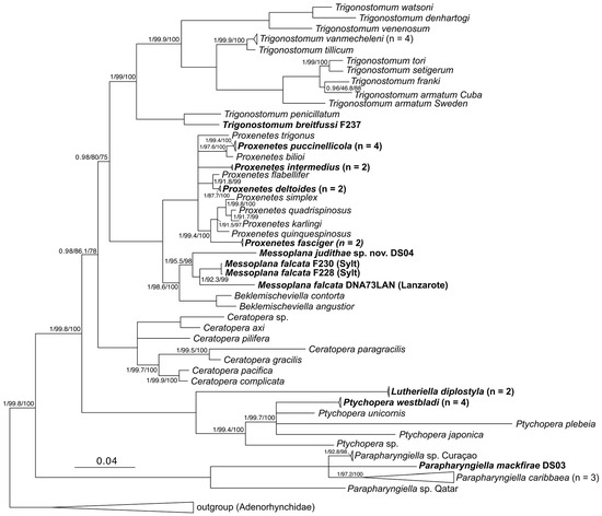
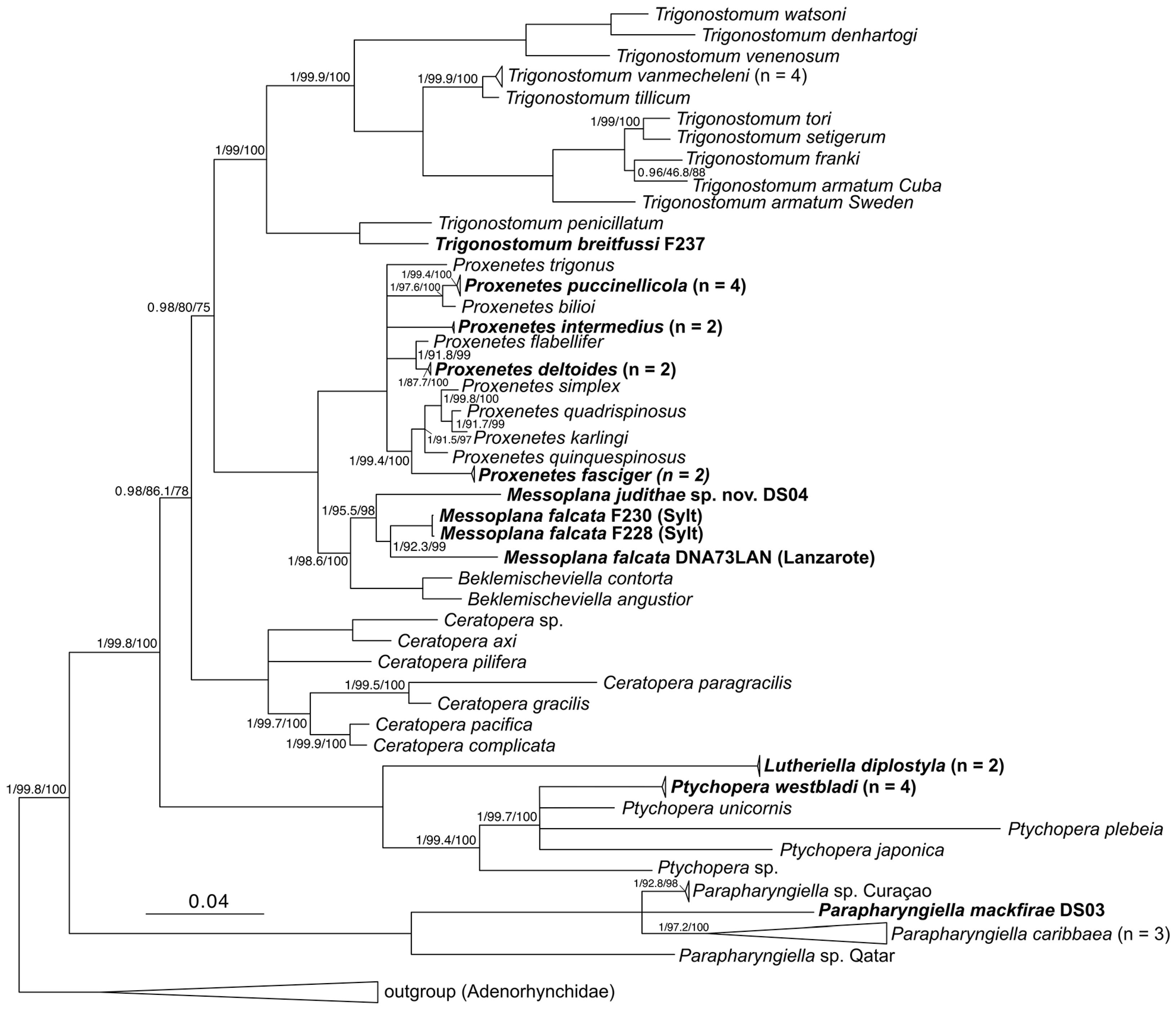
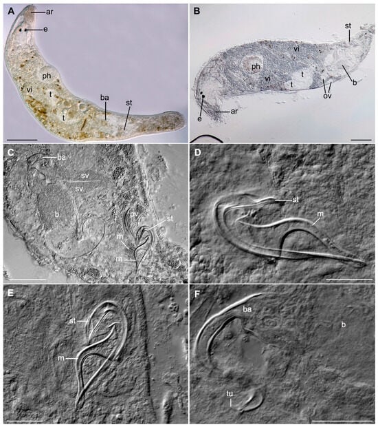
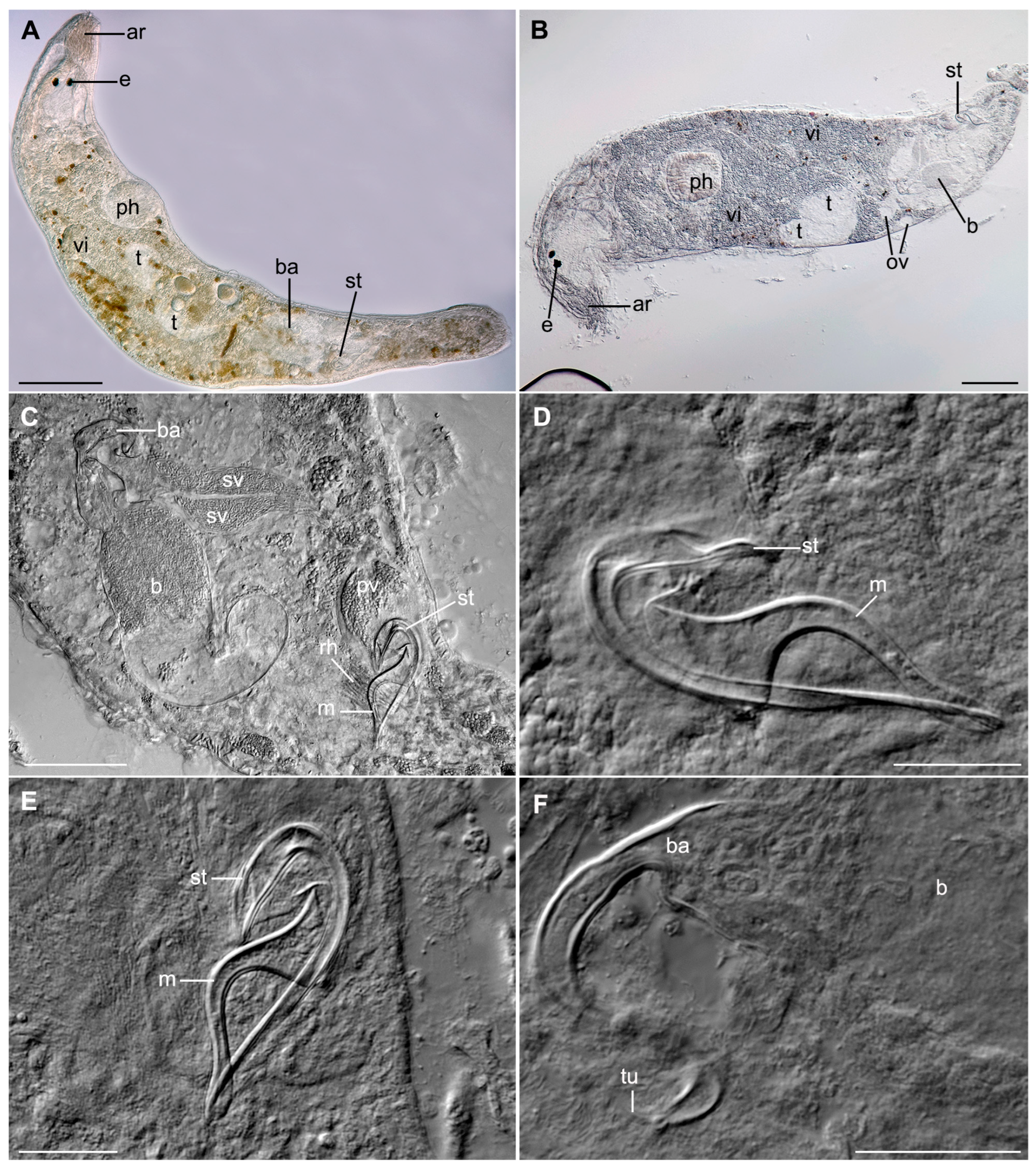
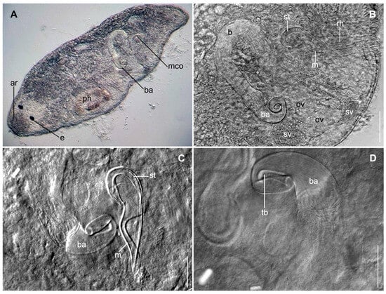
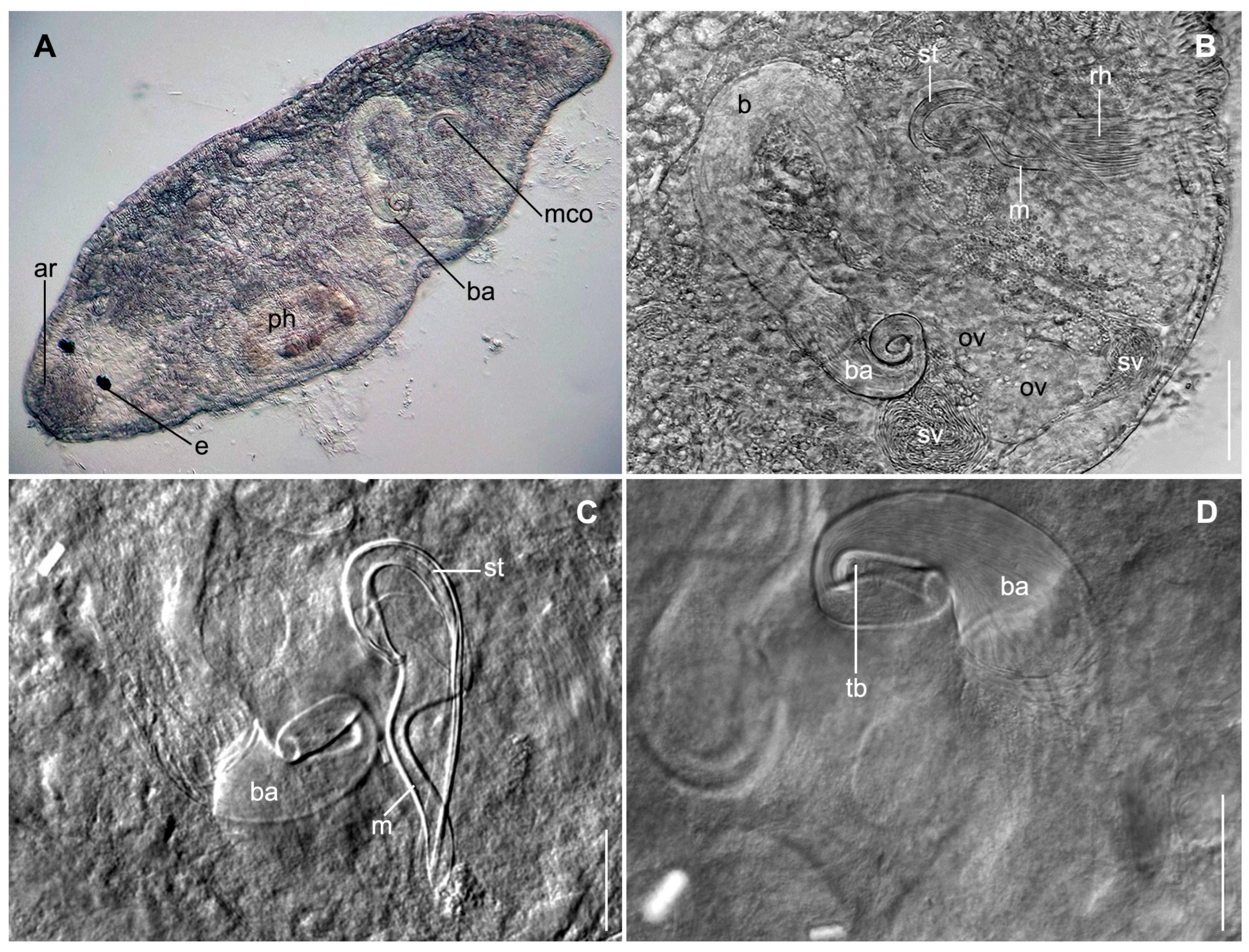
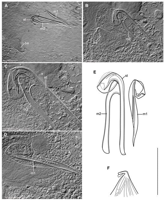
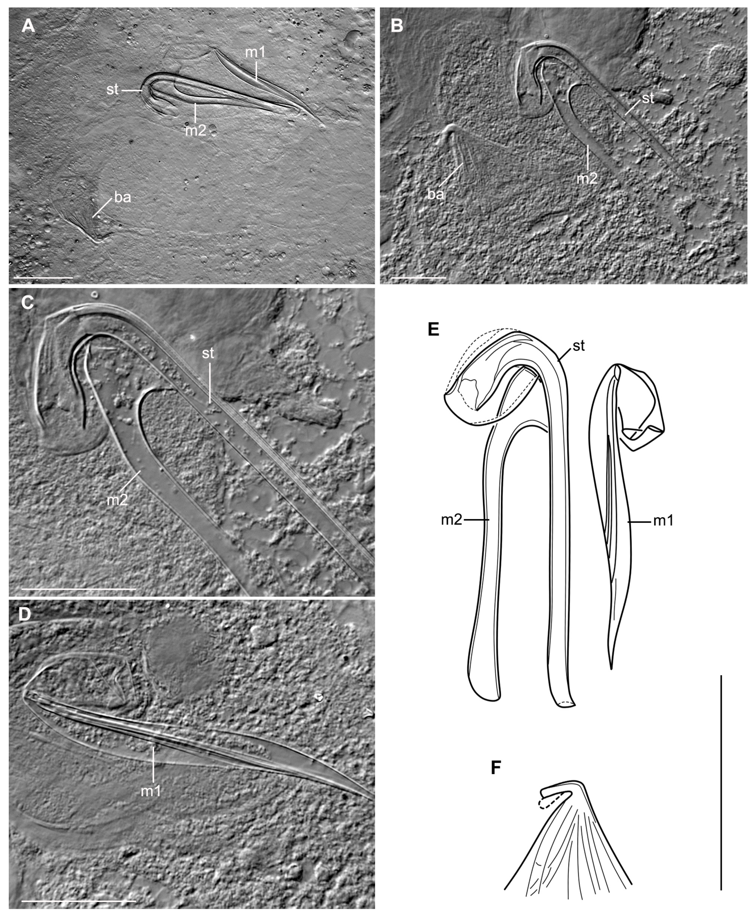
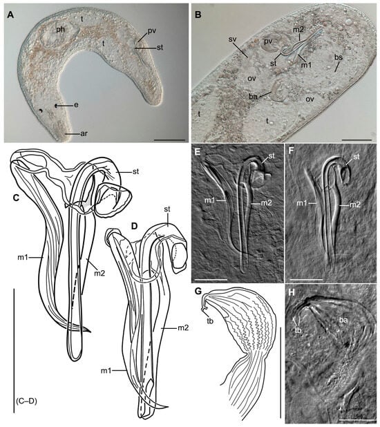
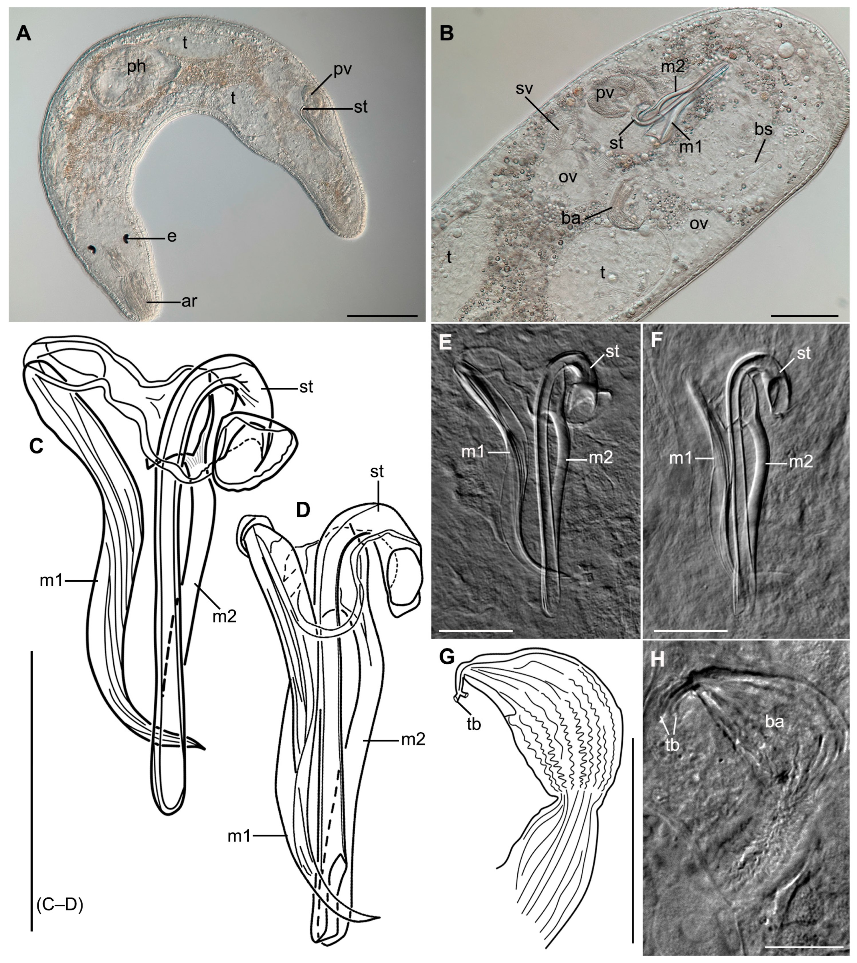
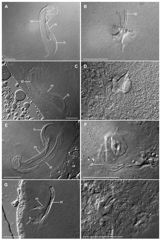
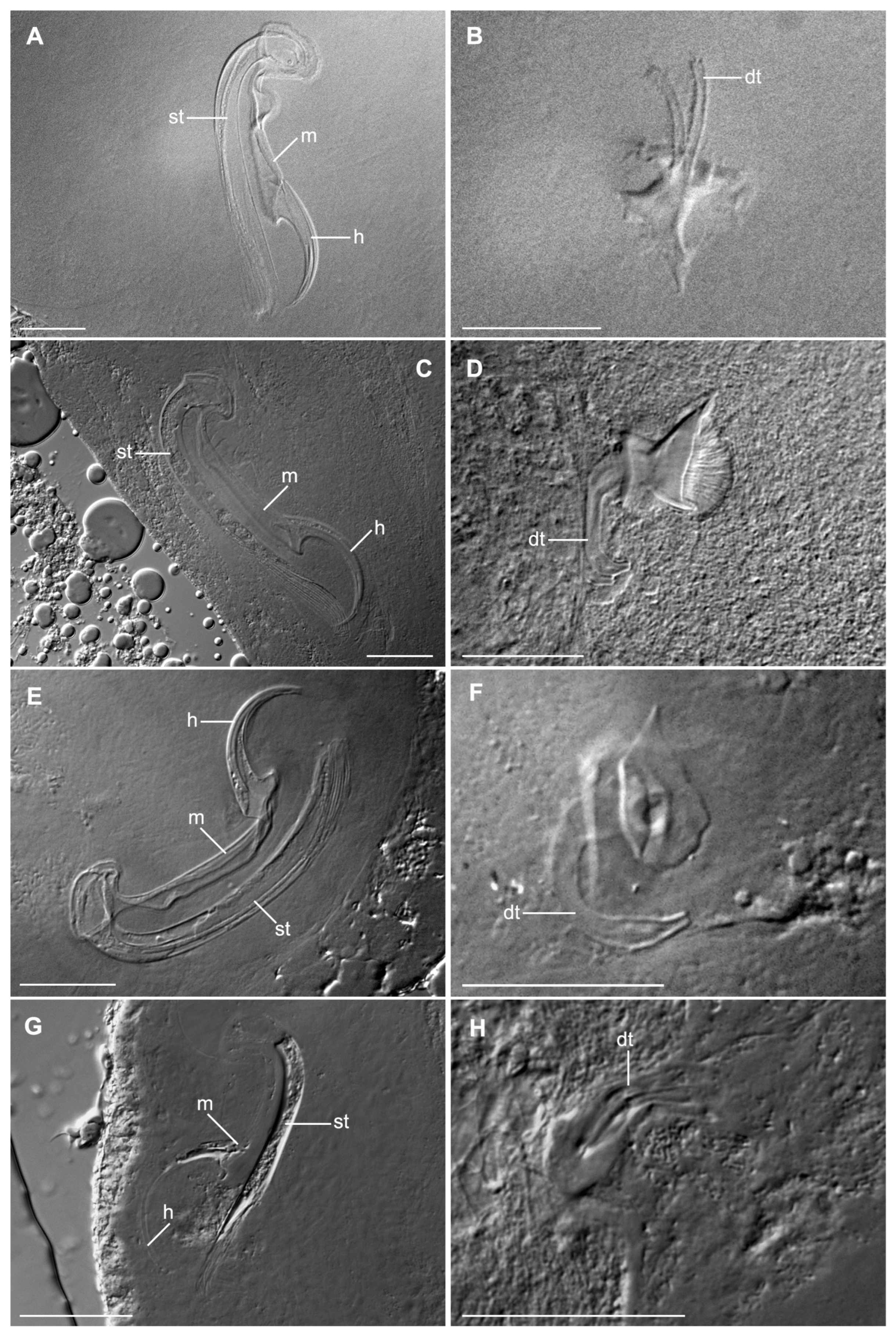
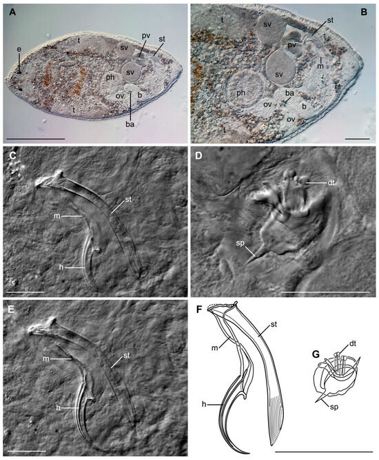
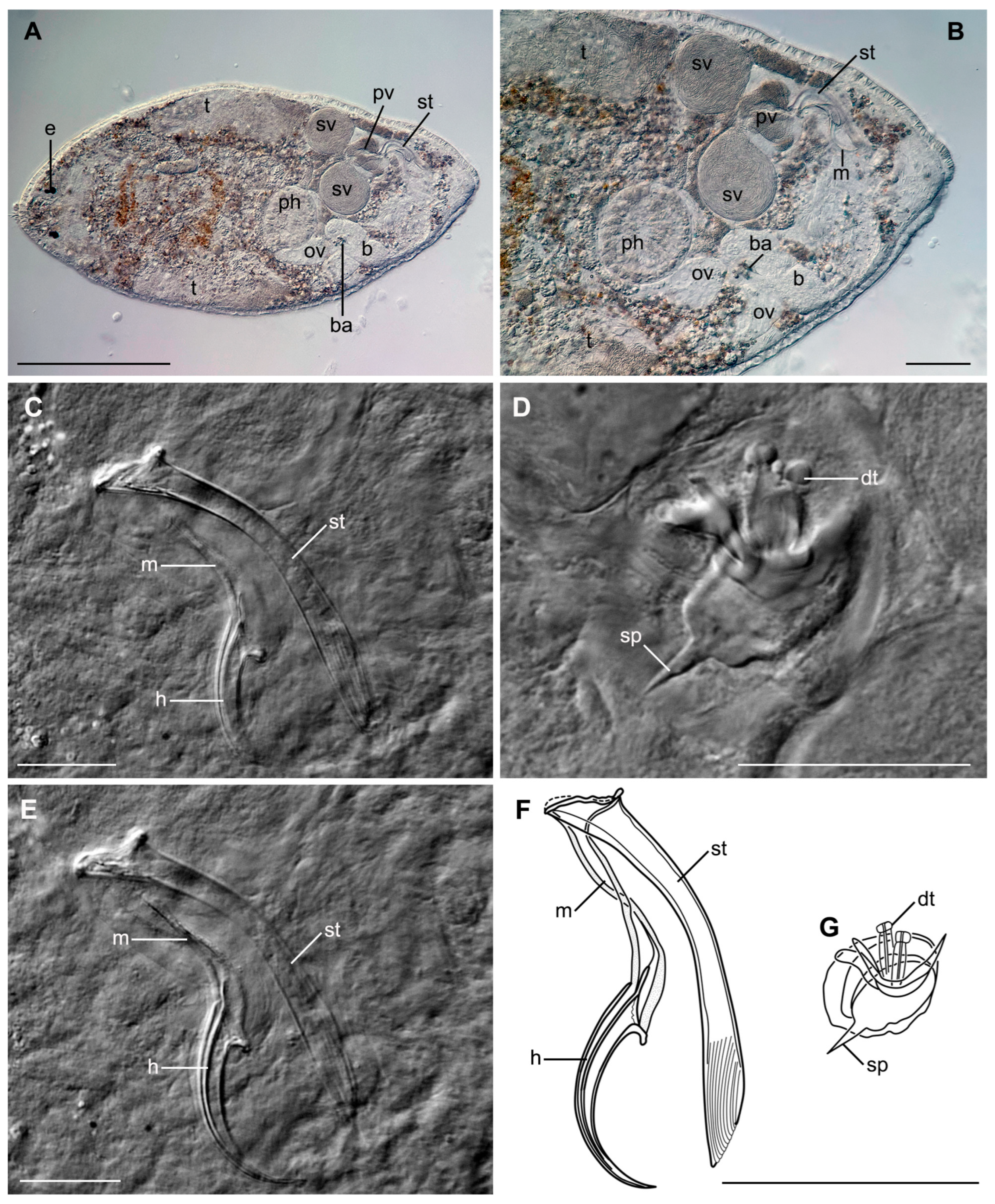
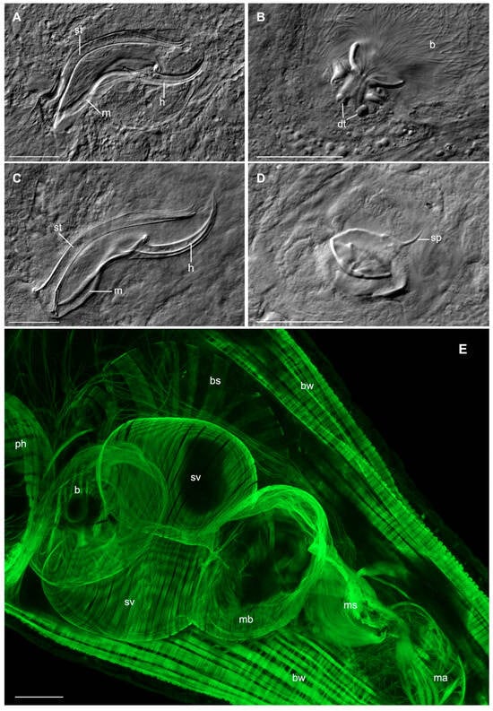
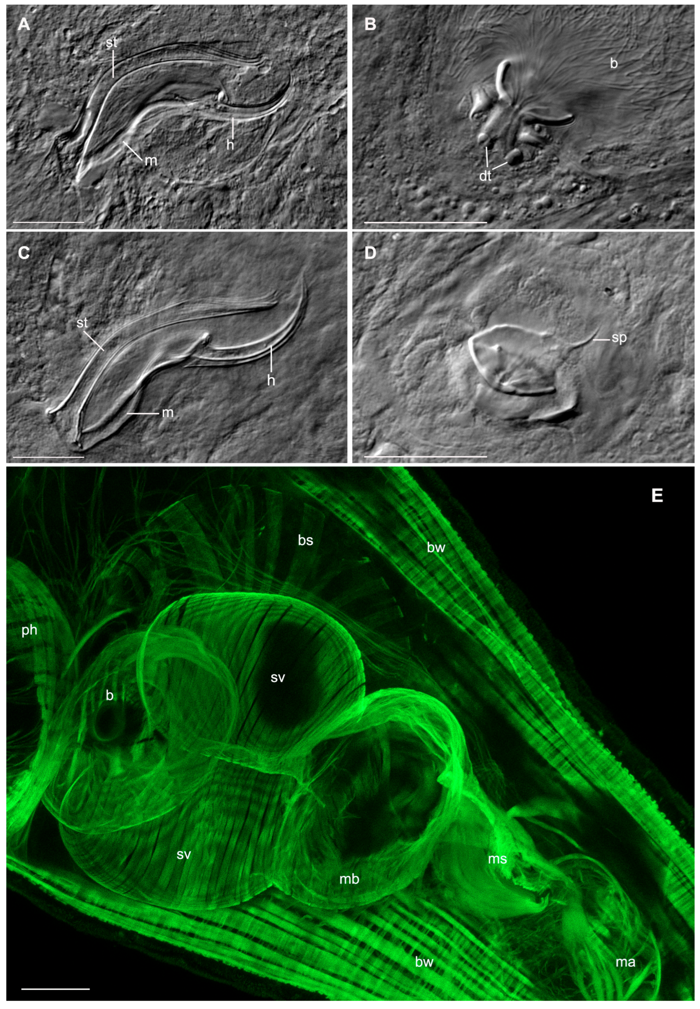
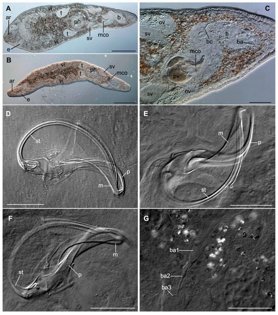
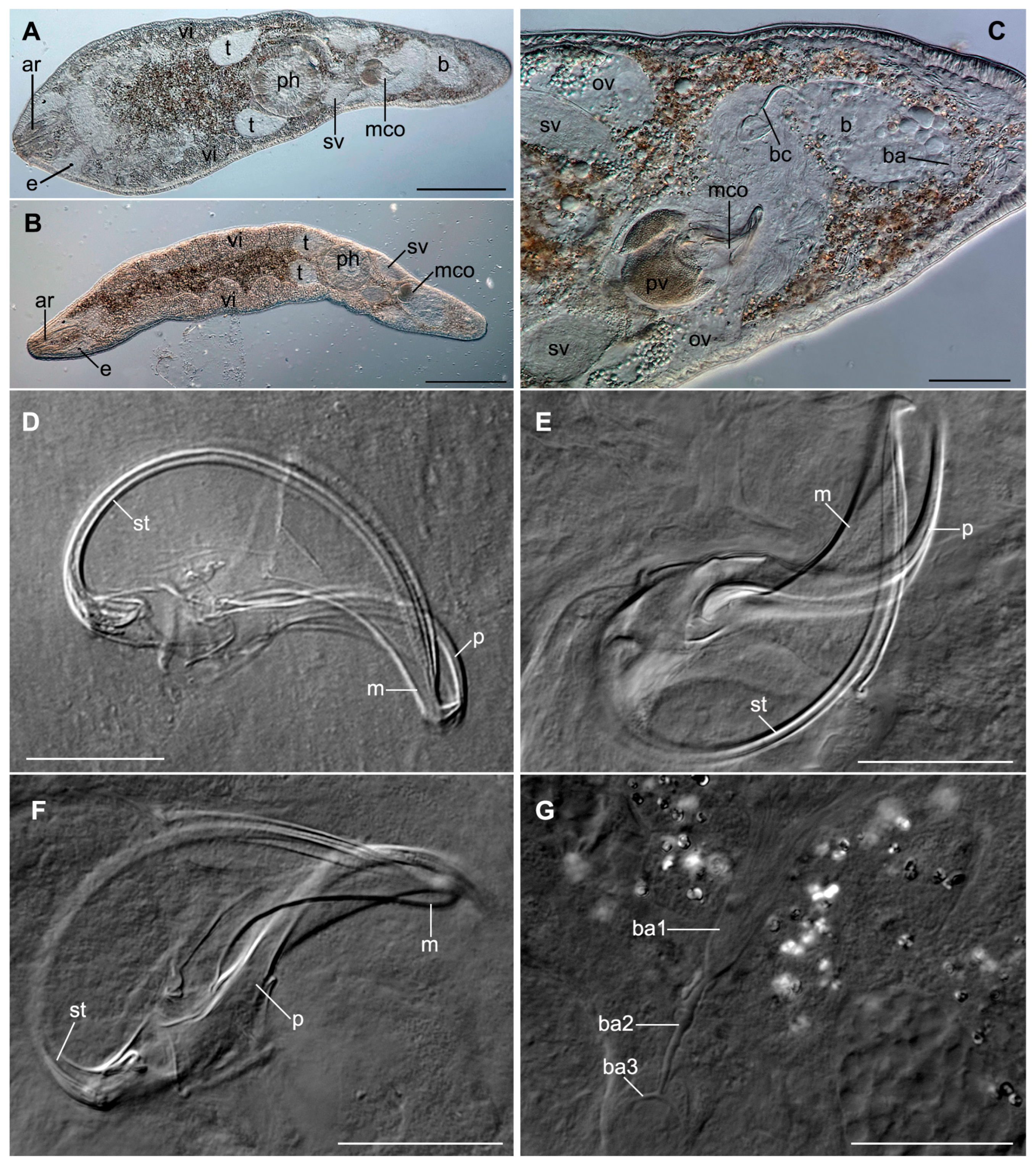
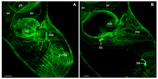
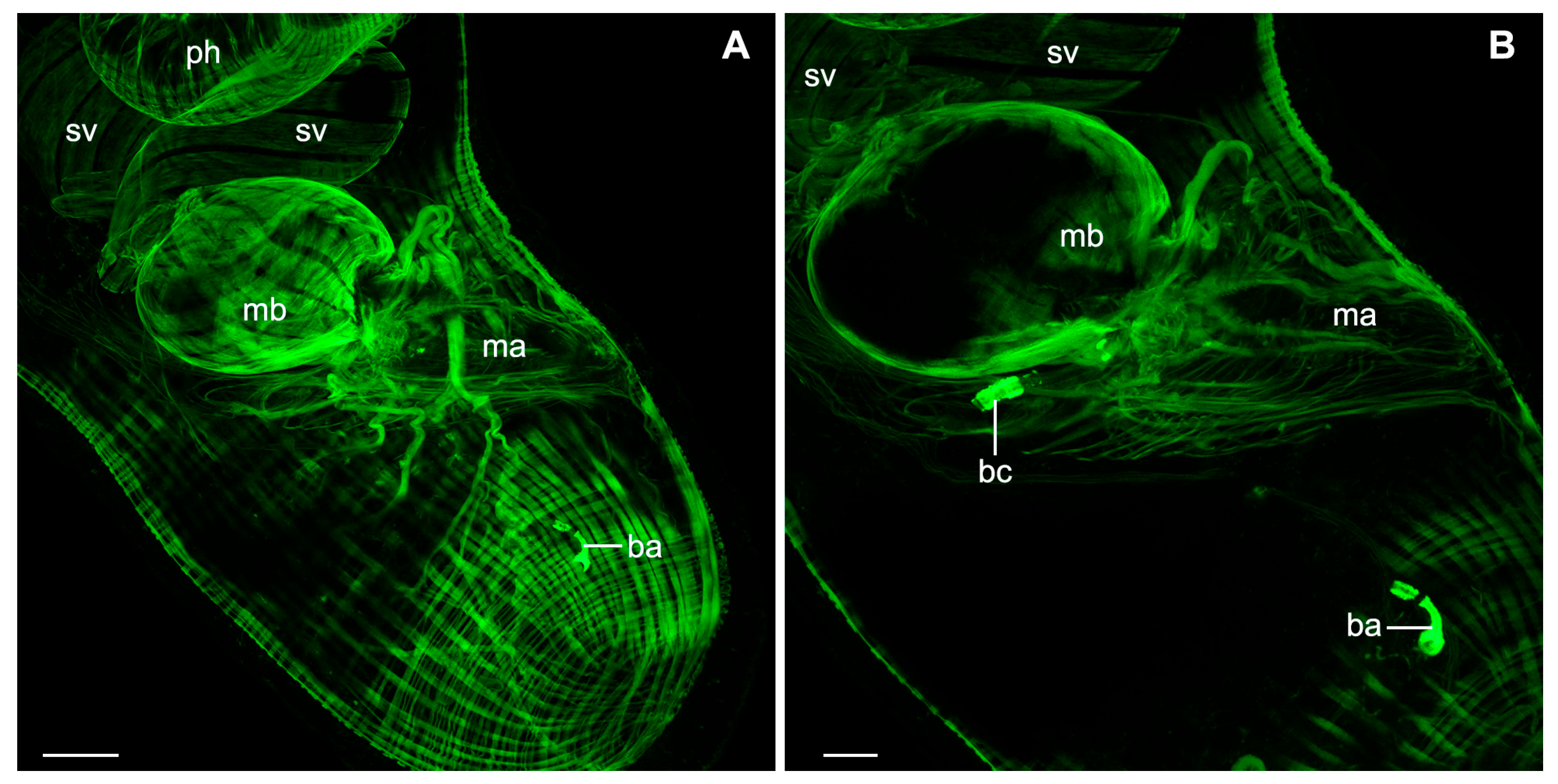
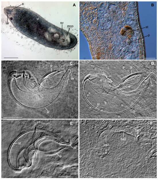
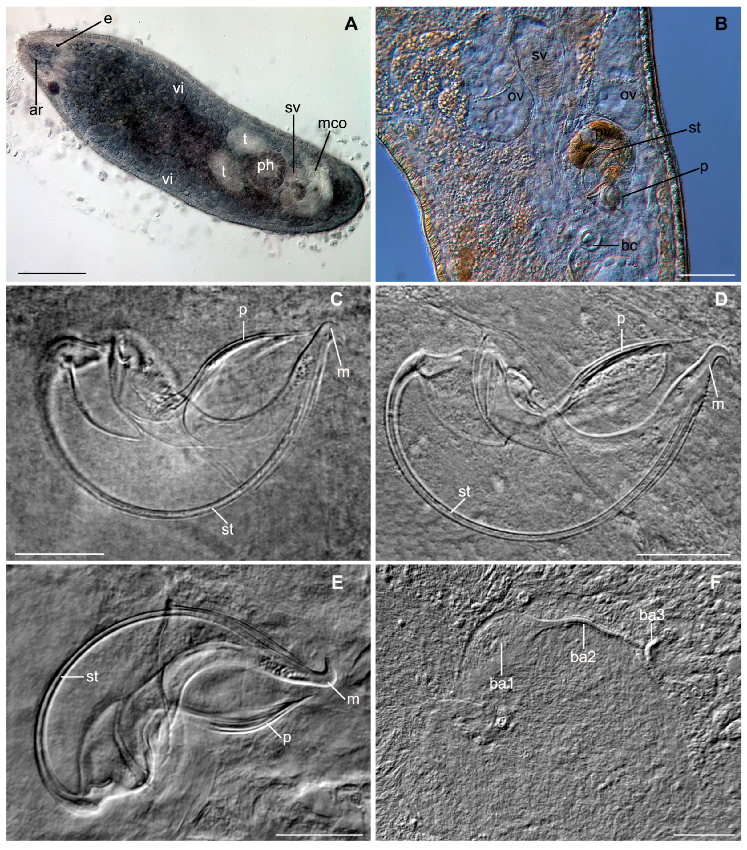
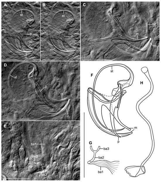
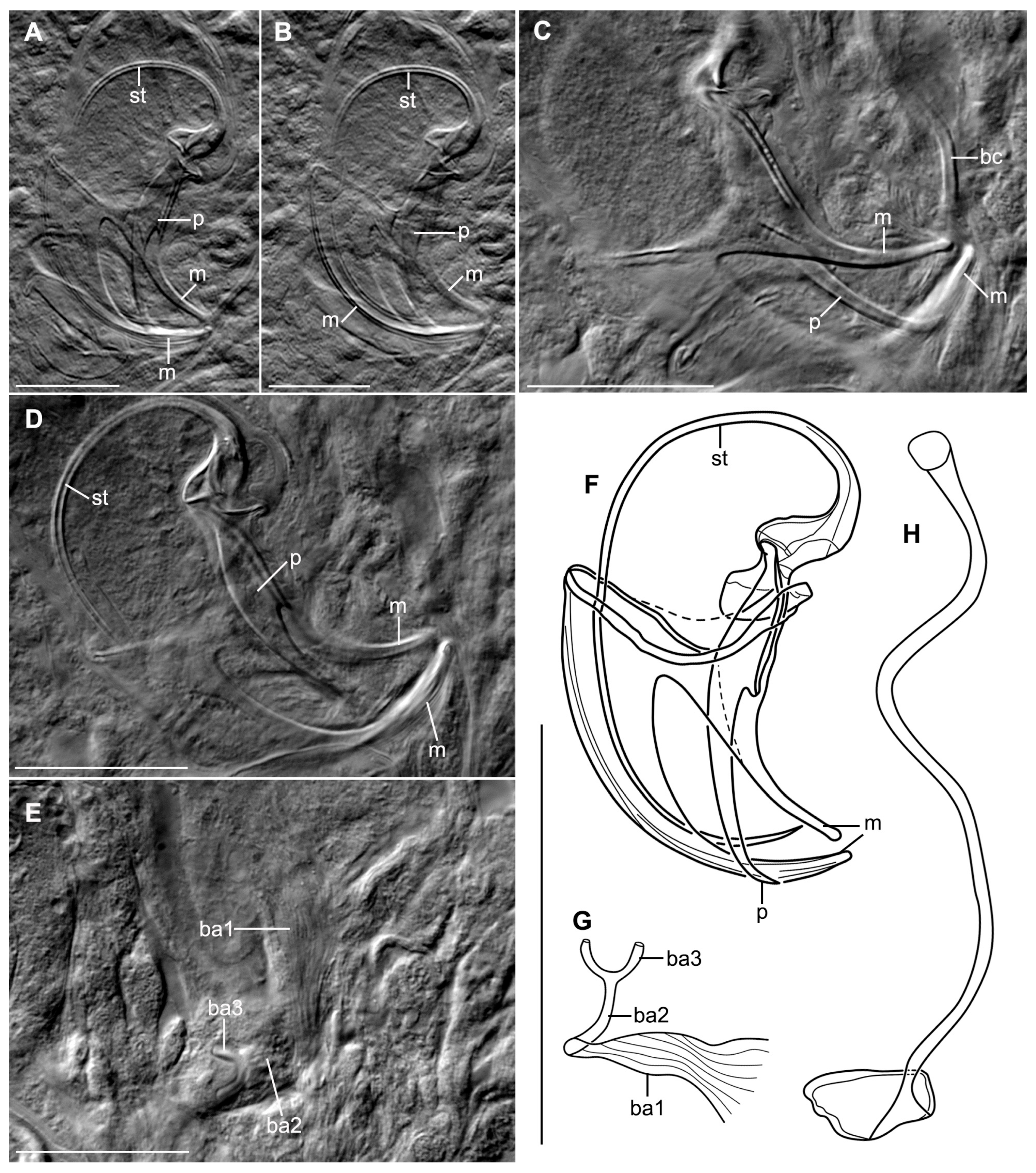
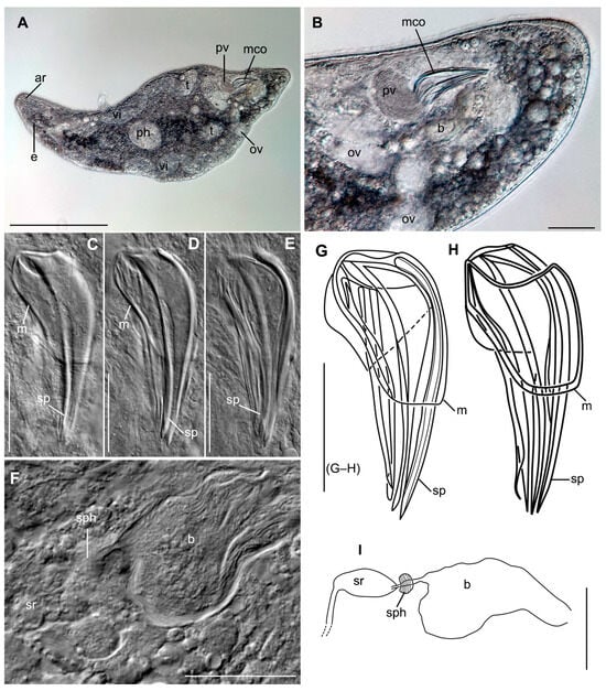
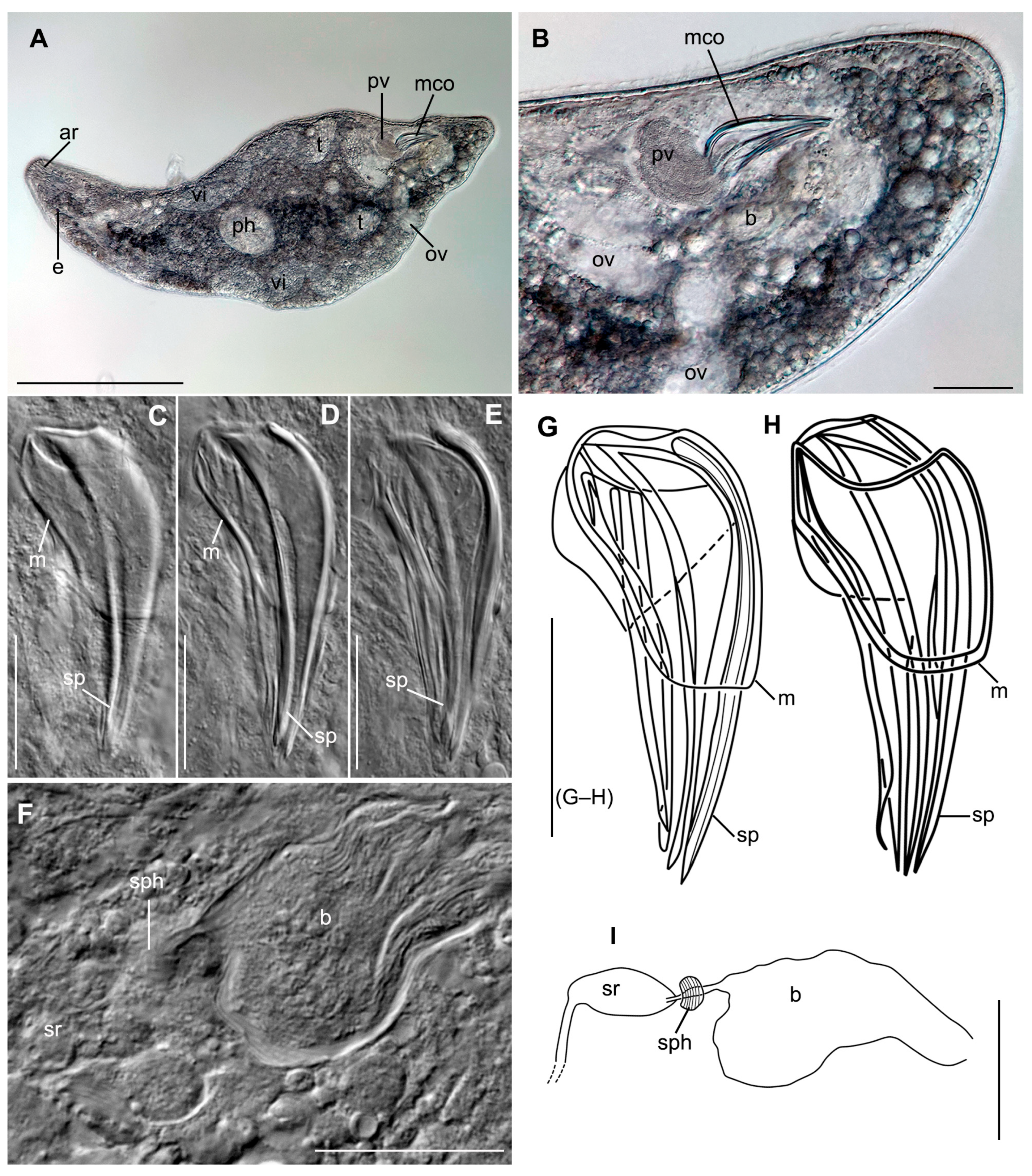
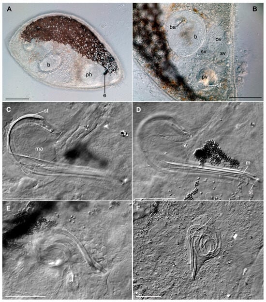
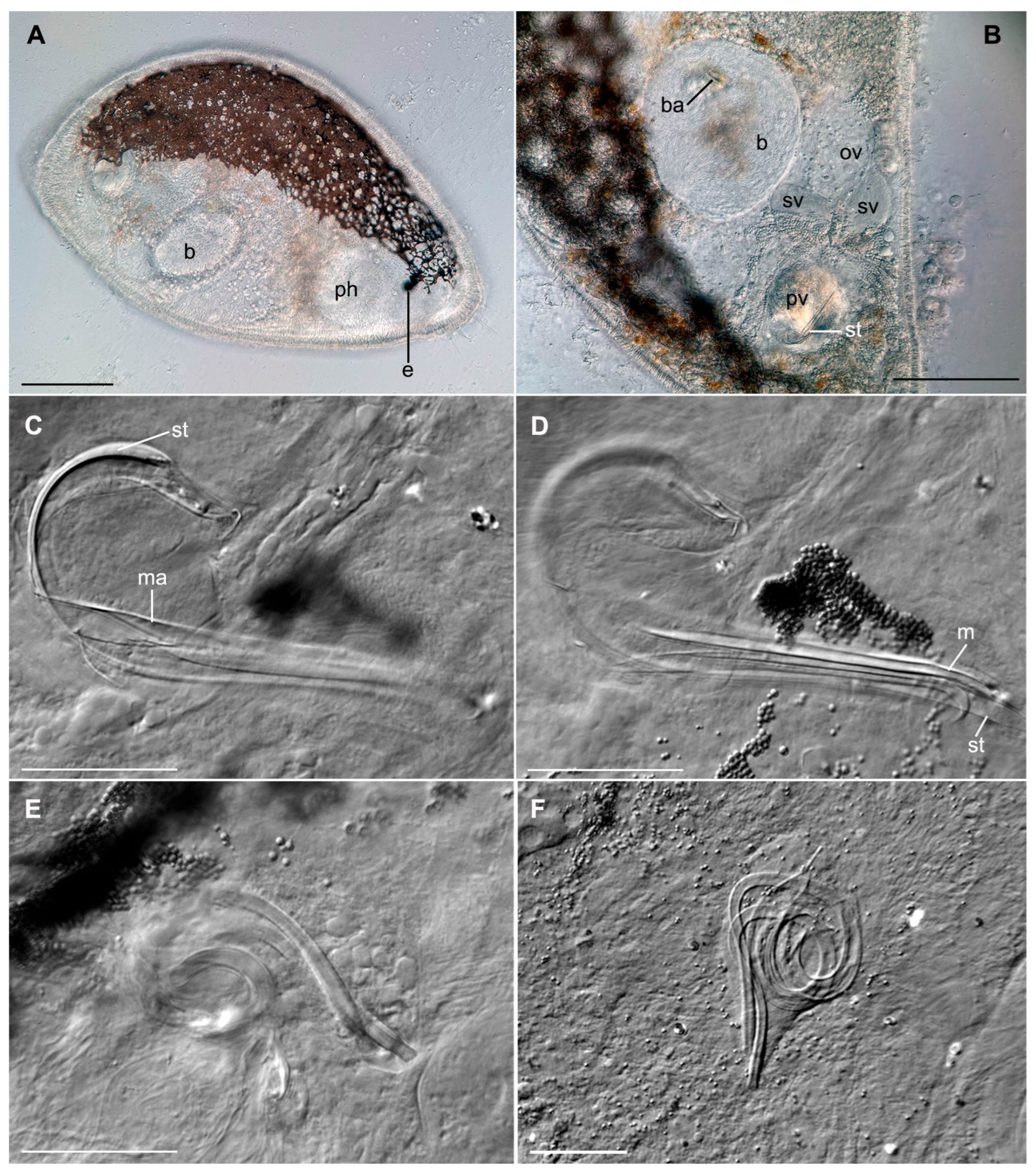

Disclaimer/Publisher’s Note: The statements, opinions and data contained in all publications are solely those of the individual author(s) and contributor(s) and not of MDPI and/or the editor(s). MDPI and/or the editor(s) disclaim responsibility for any injury to people or property resulting from any ideas, methods, instructions or products referred to in the content. |
© 2026 by the authors. Licensee MDPI, Basel, Switzerland. This article is an open access article distributed under the terms and conditions of the Creative Commons Attribution (CC BY) license.