


Abstract
During a survey of myxozoan diversity in fishes from Hunan Province, two new Myxidium species were discovered infecting the gallbladder of Sarcocheilichthys kiangsiensis Nichols, 1930 and Sarcocheilichthys parvus Nichols, 1930, in Dongting Lake, China. In both cases, myxospores were observed freely floating in the biles, with no typical plasmodia detected. Morphologically, both of them can be differentiated from previously described congeners by a combination of features, including myxospore dimensions, polar capsule shape, number of polar tubule coils and shell valve striations. BLASTn research indicated that neither species matched any available species in GenBank. The highest sequence similarity for Myxidium kiangsiensis n. sp. was 98.54% with M. asiaticum Chen et al., 2020 (PQ776264), and that for Myxidium parvusis n. sp. was 93.06% with Zschokkella guelaguetza Alama-Bermejo et al., 2023 (OQ888223). This study represents the first record of Myxidiidae infection in Sarcocheilichthys hosts. Phylogenetic analysis based on the obtained SSU rDNA sequences placed the two species in separate subclades interspersed with other Myxidium and Zschokkella species. This topology further corroborates the polyphyletic nature of these two genera.
1. Introduction
Myxozoans are a highly diversified group within the phylum Cnidaria [1], characterized by a simplified body organization and a complex, two-host life cycle typically involving annelids (definitive hosts) and fish (primary intermediate hosts) [2,3]. Myxidium Bütschli, 1882 (Myxozoa: Myxidiidae) are primarily coelozoic parasites that infect the gallbladder, kidney, and urinary bladder of fish [2,4]. Morphologically, the genus is defined by typically fusiform, pyriform, or crescent-shaped myxospores with slightly pointed ends, and two polar capsules (pyriform or spherical) positioned at opposite ends of the myxospore [2,5]. In traditional taxonomy, myxozoa identification relied almost exclusively on myxospore measurements and schematic drawings, an approach that resulted in numerous species being reported from a wide range of hosts and infection sites. Many of these nominal species are now recognized as cryptic species complexes [6,7]. A notable example is Myxidium rhodei (Leger, 1905), which was originally documented from over 40 host species across multiple tissues [8]. However, a recent integrative study by Baiko et al. (2024), combining myxospore morphology and SSU rDNA data from ten cyprinid fishes, revealed that these records actually represent four distinct species, highlighting the critical role of molecular evidence in resolving taxonomic boundaries within the Myxozoa [9]. The application of integrative taxonomic approaches [10,11] has stimulated a marked increase in studies on Myxidium. Currently, over 250 species have been documented globally, surpassing all other genera within Myxidiidae [4,12,13]. In China, more than 80 Myxidium species have been reported [14,15,16], the majority of which were described by Chen and Ma (1998) [14]. At that time, species identification relied primarily on mature myxospores, with little consideration of host identity or infection site, and molecular evidence was largely unavailable. This historical reliance on morphology alone has greatly complicated the accurate delimitation of morphologically similar Myxidium species [17].
Hunan Province is situated in the middle reaches of the Yangtze River in China and is characterized by abundant aquatic resources. Its major river systems (the Xiang, Zi, Yuan, and Li Rivers) converge into Dongting Lake, creating diverse aquatic habitats that support a rich fish fauna [15,18]. To date, 218 fish species assigned to 28 families and 102 genera have been recorded [19]. And approximately 150 myxozoan species have been reported from Hunan, with only 13 belonging to the genus Myxidium, most lacking molecular data [14,15,20,21]. The cyprinid genus Sarcocheilichthys Bleeker, 1860 is widely distributed across rivers, lakes, and reservoirs in Hunan Province and plays an important ecological role in freshwater ecosystems [15,19]. Only three Myxidium species have been reported from Sarcocheilichthys, including Myxidium sarcocheilichthysi (Feng et al., 1990) from the gallbladder of Sarcocheilichthys nigripinnis (Günther, 1873), M. rhodei from the gills and kidneys of S. nigripinnis, and M. sinensis (Wei, 2024) from the gallbladder of S. sinensis (Bleeker, 1871) [14,15]. This highlights that the diversity of Myxidium species parasitizing fishes in Hunan Province remains largely unexplored.
During a survey of myxozoan diversity in fishes from Hunan Province, two Myxidium species were discovered independently parasitizing the gallbladders of S. kiangsiensis and S. parvus in the East Dongting Lake. Based on an integrative approach incorporating morphological observations, SSU rDNA sequence data, and phylogenetic analyses, these parasites were identified as two new species, which are described herein as Myxidium kiangsiensis n. sp. and Myxidium parvusis n. sp. This represents the first record of Myxidium infection in S. kiangsiensis and S. parvus from East Dongting Lake, thereby expanding our understanding of Myxidiidae diversity in freshwater fishes of this region.
2. Materials and Methods
2.1. Collection of Fish Specimens and Morphological Examination
A total of 10 individuals of Sarcocheilichthys parvus (Nichols, 1930), measuring 5.9 (4.8–6.4) cm in length and weighing 3.9 (2.7–5.3) g, were collected in May 2023, and 15 individuals of Sarcocheilichthys kiangsiensis (Nichols, 1930), measuring 12.9 (11.8–14.3) cm in length and weighing 23.4 (18.5–33.8) g, were collected in May 2024. All host specimens were obtained from the Pingjiang section of East Dongting Lake (28°42′13″ N, 113°35′16″ E), Yueyang, Hunan Province, China. Freshly captured fish were examined immediately after collection. The fins, gills, and selected internal organs, including the gallbladder, kidney, and intestine, were examined for myxozoan infections. Bile samples were examined as fresh preparations under an Olympus BX53 light microscope (Olympus, Tokyo, Japan), and myxospores were recorded and photographed at magnifications of 400× and 1000×. Morphometric analysis was conducted on fresh myxospores using digital micrographs, with measurements (spore length, width, thickness, and polar capsule dimensions) performed using calibrated tools in Adobe Photoshop 2021. For each species, 50 randomly selected mature myxospores were measured. Morphological terminology and measurement criteria adhered to established standards for myxosporean taxonomy [22].
A bile sample was aseptically collected from each infected host, preserved in absolute ethanol, maintained at low temperature, and transported to the laboratory for subsequent molecular analyses.
2.2. Genomic DNA Extraction, SSU rDNA Amplification, Cloning, and Sequencing
Stored bile samples were pelleted by centrifugation at 12,000 rpm for 1 min and rinsed twice with phosphate-buffered saline (PBS) to remove residual ethanol. Genomic DNA was isolated using a commercial DNA genomic extraction kit (Qiagen, Hilden, Germany). A partial SSU rDNA fragment (1689–1709 bp) was amplified using primers MyxospecF [10] and 18R [23] in 25 μL PCR reactions as reported by Zhao et al. [24]. Amplified PCR products (10 μL) were examined on 1% agarose gels, excised, and purified using a DNA Gel Extraction kit (CWBiotech, Taizhou, China). Subsequently, they were inserted into the pMD-18T vector (TaKaRa, Kusatsu, Japan) and transformed into Escherichia coli DH5α. Positive clones were sequenced by Tsingke Biotechnology Co., Ltd. (Beijing, China) to ensure high-quality, accurate sequences. Sequencing multiple clones allowed verification of sequence consistency and minimized potential PCR-induced errors. The resulting sequences were assembled. The obtained sequences were deposited in GenBank.
2.3. Phylogenetic Analyses
The two newly generated SSU rDNA sequences were subjected to BLASTn similarity searches against the GenBank database. A total of 32 closely related sequences (≥85% similarity) were selected for phylogenetic analysis, with Tetracapsuloides bryosalmonae (KF731712) designated as the outgroup. All sequences were imported into PhyloSuitev1.2.3 [25] and aligned using MAFFTv7.505 [26], resulting in a final alignment of 1153 sites. The optimal nucleotide substitution model was selected using ModelFinderv2.2.0 [27] based on the Bayesian Information Criterion (BIC). Phylogenetic trees were constructed under both Maximum Likelihood (ML) and Bayesian inference (BI) methods. ML analysis was performed in IQ-TREE v2.2.0 [28] using the TIM3 + F + R3 model with 1000 standard (non-parametric) bootstrap replicates to evaluate node support, whereas BI analysis was conducted in MrBayesv3.2.7a [29] under the GTR + I + G + F model with 2,000,000 generations, and the first 25% of sampled data were discarded as burn-in.
Based on SSU rDNA sequence similarity, six closely related reference sequences were selected for each newly identified species. Sequence similarity values ranged from 91.08% to 98.54% for M. kiangsiensis n. sp., and from 92.22% to 93.06% for M. parvusis n. sp. Pairwise genetic distances were calculated in MEGA 11 [30] using the p-distance model with pairwise deletion of gaps. GC content was calculated based on the obtained sequences using an online tool provided by NovoPro (https://www.novopro.cn/tools/gc-content.html, accessed on 20 December 2025).
3. Results
3.1. Species Descriptions
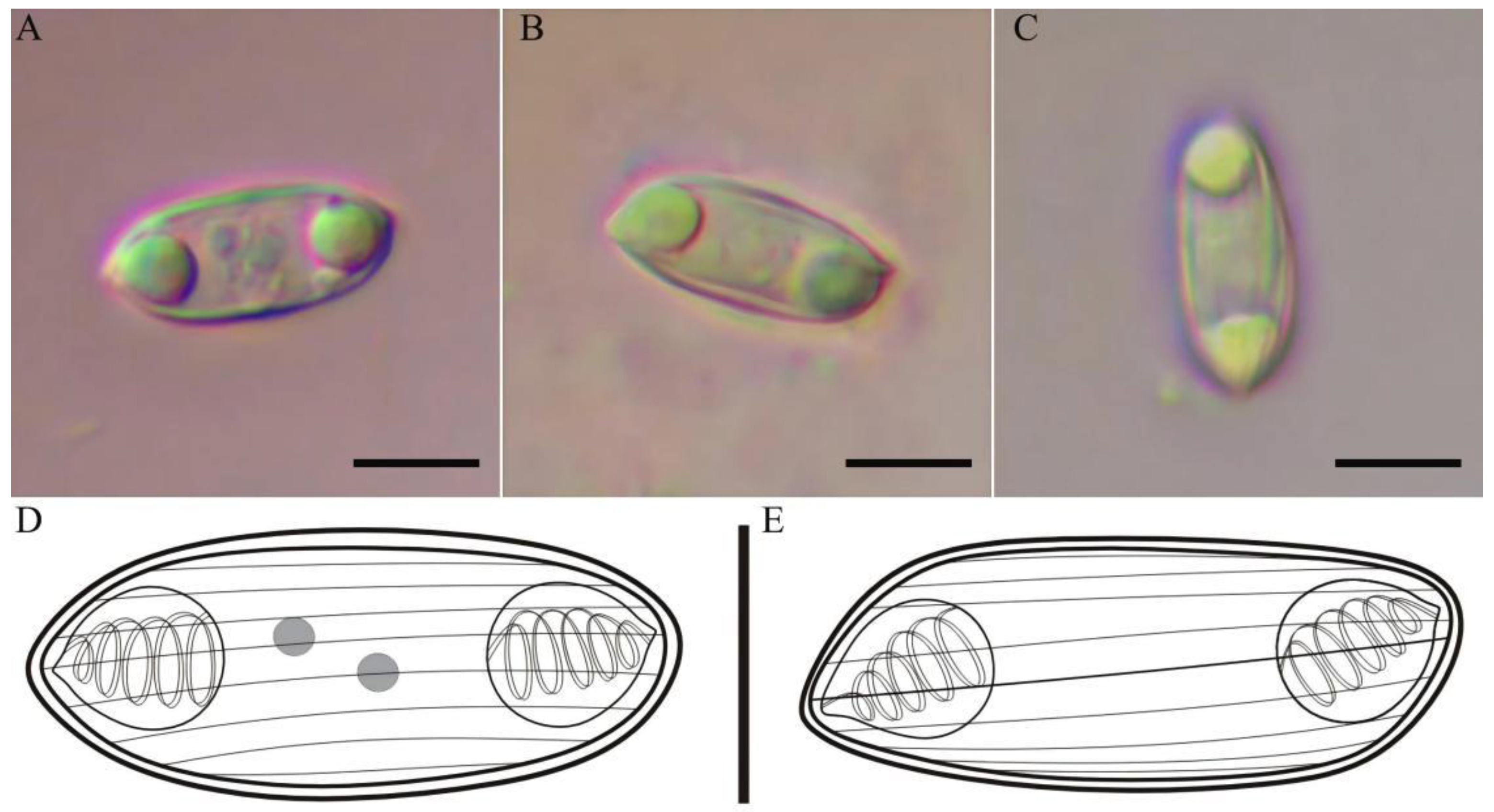
3.1.1. Myxidium kiangsiensis n. sp. (Figure 1A–E)
Morphological description: No plasmodia and trophozoites were observed during examination. Abundant mature myxospores were present in the bile. Mature myxospores are fusiform in the valvular and sutural view, measuring 11.7 ± 0.5 (10.9–12.7) μm long, 5.2 ± 0.4 (4.5–6.6) μm wide, and 4.8 ± 0.2 (4.3–5.7) μm thick. Two equal-sized, pyriform polar capsules are typically located one at each end of the myxospore and their openings are generally situated in the sutural plane and open in opposite directions. They measure 3.1 ± 0.2 (2.6–3.5) μm long and 2.8 ± 0.2 (2.4–3.3) μm wide. The myxospore surface exhibits 6–9 longitudinal striations. The sporoplasm contains two nuclei, located between two polar capsules. The distance between polar capsules is 4.6 ± 0.3 (4.1–5.2) μm. Polar tubules form 4–5 coils.
Figure 1.
Myxospore of Myxidium kiangsiensis n. sp. isolated from the gallbladder of Sarcocheilichthys kiangsiensis. Myxospore in valvular view (A); myxospore in sutural view (B); myxospore showing longitudinal striations on the shell valves (C); Line drawing of a myxospore in the valvular view (D) and in the sutural view (E). Scale bar: 5 μm.
Type host: Sarcocheilichthys kiangsiensis Nichols, 1930.
Type locality: Dongting Lake, Pingjiang section, Yueyang City, Hunan Province, China (28°42′13″ N, 113°35′16″ E).
Site of infection: Gallbladder.
Date of sampling: May 2024.
Prevalence of infection (%): 13.3% (2/15).
Deposited materials: The bile containing myxospores has been preserved in a tube of alcohol, and the syntype (No. SK-GB-202405) has been stored at Hunan Agricultural University in China.
Etymology: The specific epithet follows that of the host species, as this is the first Myxidium species reported from the gallbladder of S. kiangsiensis.
ZooBank number: urn:lsid:zoobank.org:pub:EE838D71-5B55-4091-BA89-5BEC49E63AFA.
Molecular characteristics: One SSU rDNA sequence (1709 bp) with a GC content of 48.04% was obtained and deposited in GenBank (accession number PV364357).
Remarks: Myxidium kiangsiensis n. sp. from Sarcocheilichthys kiangsiensis is the first Myxidium reported from this host. Based on a GenBank BLAST search, M. kiangsiensis n. sp. showed the highest sequence similarity to M. asiaticum Chen et al., 2020 (PQ776264, 98.54%), followed by M. spinibarba Chen et al., 2020 (MN922568, 98.45%) and M. hoabinhense Chinh et al., 2019 (MK913426, 97.67%). The sequence similarity between M. kiangsiensis n. sp. and both M. asiaticum and M. spinibarba falls close to the intraspecific level (generally >98%).
Based on the morphology of these myxospores, this new species is superficially similar to M. chuatsis (Yin et al., 2025), M. asiaticum, M. onychostomatis (Zhao et al., 1994), and M. spinibarba. It can be distinguished from most of these species by myxospore dimensions (Table 1), except M. chuatsi and M. spinibarba, for which reliable differentiation requires host order (Cypriniformes versus Centrarchiformes) and SSU rDNA sequence data. The myxospore of M. kiangsiensis n. sp. is fusiform in valvular view and sutural view, similar to M. chuatsi, M. asiaticum, and M. spinibarba, whereas M. onychostomatis is oval in valvular view and fusiform in sutural view. The myxospore size of M. kiangsiensis n. sp. (10.9–12.7 µm × 4.5–6.6 µm) is broadly comparable to that of M. chuatsi (10.0–11.4 × 5.4–6.1 µm) and M. spinibarba (10.6–12.4 × 5.5–7.2 µm), and clearly smaller than that of M. onychostomatis (13.6–14.4 × 6.0–7.2 µm ) and M. asiaticum (12.9–15.1 × 5.7–8.5 µm). The polar capsule size of M. kiangsiensis n. sp. (2.6–3.5 × 2.4–3.3 µm) is comparable to M. chuatsi (2.9–3.7 × 2.6–3.2 µm), but smaller than M. spinibarba (3.0–4.4 × 2.7–3.2 µm), M. asiaticum (2.8–4.0 × 2.8–4.0 µm), and M. onychostomatis (3.9–4.0 × 2.3–2.4 µm). The number of polar tubule coils (4–5) and the number of striations (6–9) in M. kiangsiensis n. sp. are similar to M. chuatsi (6–8), differing from M. spinibarba (8–10 striations) and M. onychostomatis (6–7 striations). Taken together, these morphological and molecular features support the distinction of M. kiangsiensis n. sp. from closely related Myxidium species.
Table 1.
Comparative analysis of Myxidium kiangsiensis n. sp. with morphologically similar species.
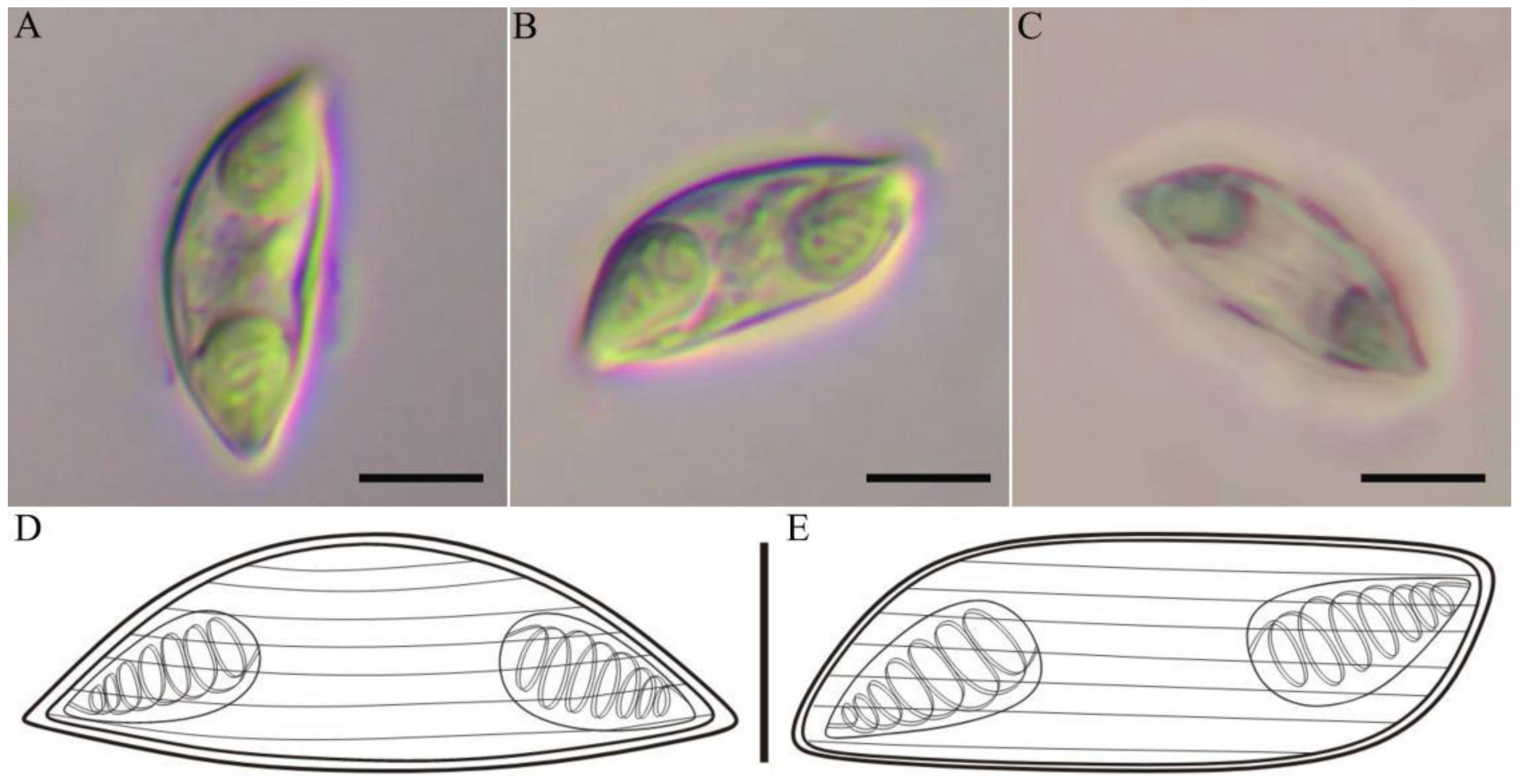
3.1.2. Myxidium parvusis n. sp (Figure 2A–E)
Morphological description: Numerous mature myxospores were observed floating in the bile. No plasmodia were observed. Myxospores are slightly crescentic in the valvular view and fusiform in the sutural view, with both ends slightly protruding. Measurement of mature myxospores was 16.2 ± 0.7 (15.1–17.8) μm long, 6.0 ± 0.2 (5.7–6.3) μm wide, and 5.8 ± 0.4 (4.9–6.3) μm thick. Six to seven longitudinal striations were clearly observed in the shell valves of myxospores. Two equal pyriform polar capsules are located one at each end of the myxospore, measuring 6.2 ± 0.3 (5.3–7.0) μm long and 3.7 ± 0.2 (3.5–4.0) μm wide, with their openings situated in the sutural plane and open in opposite directions. The distance between the two opposite polar capsules measures 3.6 ± 0.3 (3.5–4.2) μm. The polar capsule tubule coiled into 6–7 coils.
Figure 2.
Myxospore of Myxidium parvusis n. sp. isolated from the gallbladder of Sarcocheilichthys parvus. Myxospore in valvular view (A); myxospore in sutural view (B); myxospore showing longitudinal striations on the shell valves (C); line drawings of myxospore of the valvular view (D) and the sutural view (E). Scale bar: 5 μm.
Type host: Sarcocheilichthys parvus Nichols, 1930.
Type locality: Dongting Lake, Pingjiang section, Yueyang City, Hunan Province, China (28°42′13″ N, 113°35′16″ E).
Site of infection: Gallbladder.
Date of sampling: May 2023.
Prevalence of infection (%): 10.0%, (1/10).
Deposited materials: The bile containing myxospores has been preserved in a tube of alcohol, and the syntype (No. SP-GB-202305) has been stored at Hunan Agricultural University in China.
Etymology: The specific epithet derives from the specific epithet of the host species, as this species is the first Myxidium species found in the gallbladder of S. parvus.
ZooBank number: urn:lsid:zoobank.org:pub:EE838D71-5B55-4091-BA89-5BEC49E63AFA.
Molecular characteristics: One SSU rDNA sequence (1689 bp) with a GC content of 47.42% was obtained and deposited in GenBank (accession number PV364360).
Remarks: BLASTn analysis showed that M. parvusis n. sp. exhibited the highest similarity to Zschokkella guelaguetza Alama-Bermejo et al., 2023 (OQ888223, 93.06%), followed by M. sharmai Gupta et al., 2025 (OP174299, 92.95%). Based on the morphology of these myxospores, this new species is similar to M. cuneiforme (Fujita, 1924), M. sarcocheilichthysi (Feng et al., 1990), M. pseudocuneiforme (Chen et al., 2021) and M. kagayamai (Kudo, 1919), but can be differentiated from them by a combination of myxospore dimensions and polar capsule morphology (Table 2). In terms of myxospore size, M. parvusis n. sp. (15.1–17.8 × 5.7–6.3 μm) is distinctly larger than M. cuneiforme (12.0–13.1 × 4.8–6.1 μm) and M. pseudocuneiforme (12.0–13.1 × 4.8–6.1 μm), slightly smaller than M. sarcocheilichthysi (16.0–18.5 × 5.1–6.0 μm), and overlaps partly with M. kagayamai (15–18 × 6–7 μm). With respect to polar capsule dimensions, M. parvusis n. sp. has polar capsules measuring 5.3–7.0 × 3.5–4.0 μm, which are larger than those reported for M. cuneiforme (4.0–4.6 × 2.6–3.4 μm), M. pseudocuneiforme (3.5–4.2 × 2.5–2.8 μm), and M. sarcocheilichthysi (4.0–6.0 × 3.2–4.0 μm). No detailed measurements are available for M. kagayamai. Regarding the number of polar tubule coils and shell valve striations, M. parvusis is characterized by 6–7 coils and 6–7 striations, differing from M. cuneiforme and M. pseudocuneiforme (5–6 coils, 6–8 striations) as well as from M. sarcocheilichthysi (4–5 coils, 9–10 striations). Myxidium parvusis n. sp. can further be distinguished from M. cuneiforme (MT014004) and M. pseudocuneiforme (MH497019) by its low interspecific SSU rDNA sequence similarities to these species, namely 92.22% and 91.43%, respectively. In contrast, comparison with M. kagayamai is constrained by the lack of molecular and detailed morphological data. When compared more closely, the myxospores of M. kagayamai are slightly larger than M. parvusis n. sp., and the two species are currently distinguished by their hosts, with M. parvus n. sp. infecting Sarcocheilichthys parvus and M. kagayamai infecting Misgurnus anguillicaudatus Cantor, 1842 (Cobitidae). If more detailed data become available for M. kagayamai in the future, further comparison and differentiation can be made.
Table 2.
Comparative analysis of Myxidium parvusis n. sp. with morphologically similar species.
3.2. Distance Estimation and Phylogenetic Analysis
Genetic distances from Myxidium kiangsiensis n. sp. to its six closely related reference species ranged from 0.0106 to 0.1047, and those from Myxidium parvusis n. sp. to its six closely related reference species ranged from 0.0800 to 0.1000 (Table 3).
Table 3.
Pairwise nucleotide sequence identity (upper right) values and evolutionary distances (left bottom) among Myxidium kiangsiensis n. sp. and M. parvusis n. sp. isolates and other species with high sequence similarity based on SSU rDNA sequences.
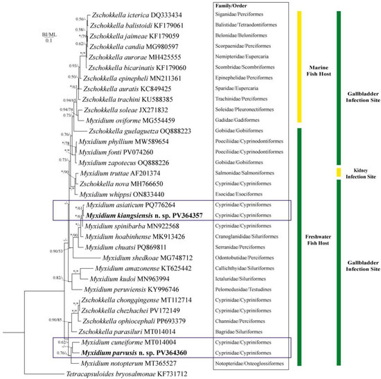
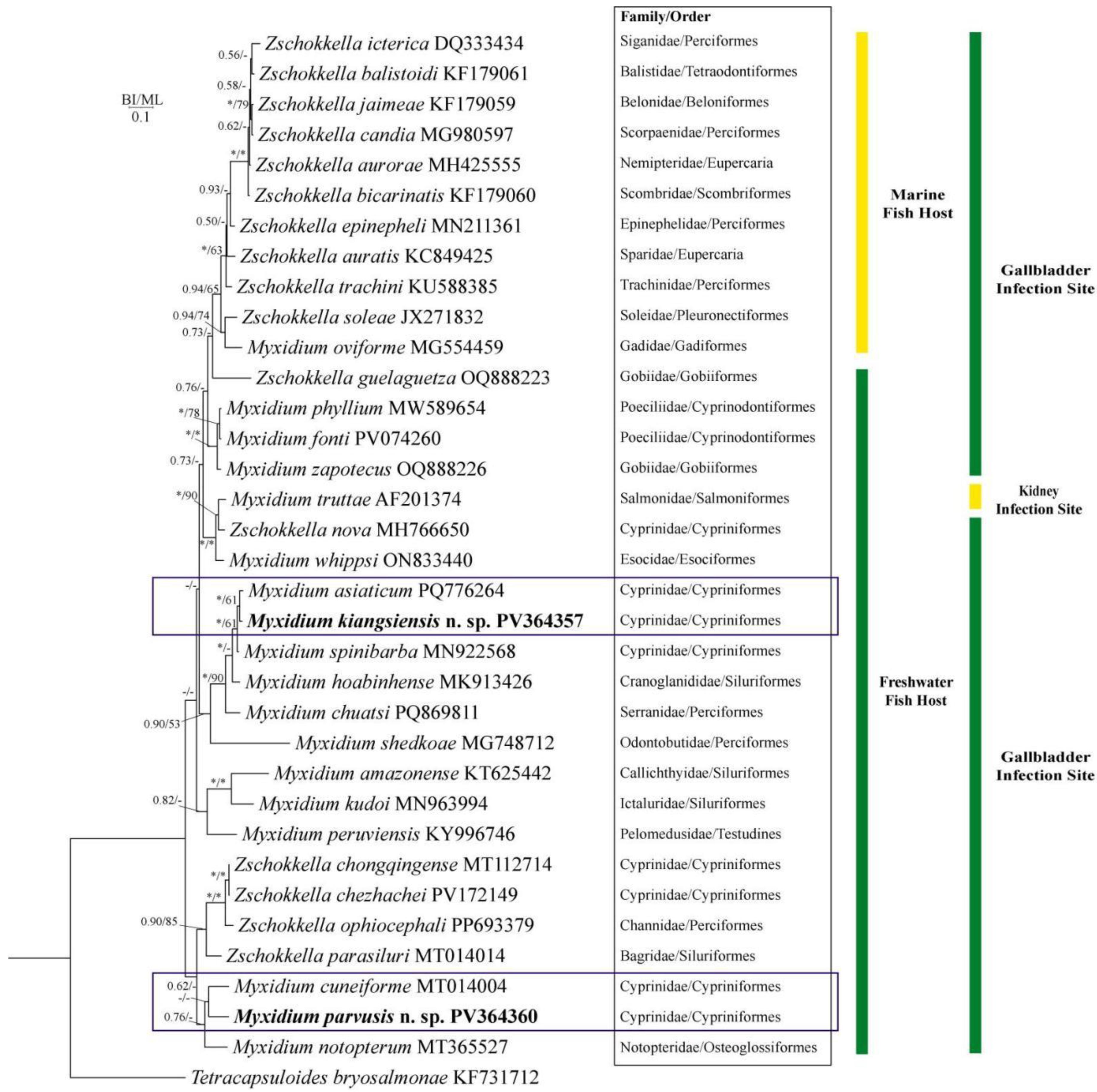
Phylogenetic analyses showed that species of Zschokkella and Myxidium did not form reciprocally monophyletic clades, but were interspersed across multiple lineages (Figure 3). In the upper portion of the tree, Zschokkella species predominated, with M. oviforme nested among them. Conversely, the middle to lower portions were dominated by Myxidium species, within which several Zschokkella species were embedded. From an ecological perspective, species in the upper clade were associated with marine hosts, whereas those in the lower clade were exclusively parasitic in freshwater hosts. All analyzed species were coelozoic parasites, with the gallbladder as the predominant infection site; only M. truttae (AF201374) has been reported to infect the kidney.
Figure 3.
Phylogenetic position of Myxidium kiangsiensis n. sp. and M. parvusis n. sp. based on SSU rDNA gene analysis using the Bayesian Inference (BI) method. Tetracapsuloides bryosalmonae (KF731712) was used as the outgroup. Posterior probabilities and bootstrap support values are indicated at the nodes; “*” denotes values above 95%, and “-” indicates values below 50%. The new species described in this study is shown in bold.
Within the phylogenetic tree, M. kiangsiensis n. sp. clustered with M. asiaticum and constituted a well-supported subclade within a freshwater lineage dominated by Myxidium. M. parvusis n. sp. clustered as a sister species to M. cuneiforme and together with M. notopterum formed a sister clade to several Zschokkella species.
4. Discussion
The traditional taxonomy of Myxozoa was mainly based on morphological criteria including myxospore and polar capsule dimensions and shape, number and arrangement of polar capsules, polar tubule coils, and shell valve striations [35,36]. Although M. kiangsiensis n. sp. and M. parvusis n. sp. share several morphological features with related species, including the number of polar tubule coils and shell valve striations, they can be clearly distinguished by differences in myxospore and polar capsule dimensions. However, as highlighted by Li et al. (2016) [32] and Yin et al. (2025) [21], many morphological characters in myxozoans have limited diagnostic value due to high phenotypic plasticity. This is particularly evident between Myxidium and Zschokkella, where morphology-based taxonomic boundaries are often ambiguous [21,32]. Accurate species identification becomes challenging when relying solely on myxospore morphology, as morphologically similar species cannot be correctly delimited without molecular data [37,38]. Therefore, integrative approaches combining morphological characters, biological traits (host species, infection site, habitat), and molecular data are widely accepted as the gold standard in myxozoan systematics [39,40].
The phylogenetic tree reveals that species assigned to Myxidium and Zschokkella do not form two reciprocally monophyletic clades. Instead, species of both genera are distributed across several distinct phylogenetic lineages. This aligns with previous studies suggesting a polyphyletic origin of the family Myxidiidae [12,35]. The phylogeny is clearly divided into marine and freshwater clades, indicating that Myxidium and Zschokkella are not monophyletic but cluster primarily according to habitat or type of invertebrate. This suggests that the ecological environment of the host is an important factor shaping phylogenetic relationships [41]. Overall, our findings concur with Chen (2021), who questioned the taxonomic independence of Myxidium and Zschokkella [16], indicating that the generic delimitation between these two genera remains unresolved. The low support values obtained in our phylogenetic study may be improved by increasing taxon sampling and incorporating additional markers (e.g., 28S rDNA or ITS) in future analyses.
Previous studies indicate that parasite populations within the same aquatic ecosystem may experience ongoing gene flow, while adaptive divergence can be maintained by host-related selective pressures [42,43,44]. In this study, although M. parvusis n. sp. and M. kiangsiensis n. sp. both parasitize Sarcocheilichthys species in Dongting Lake, phylogenetic analyses place them in distinct positions, with M. parvusis occupying a basal branch. This suggests they are not recently diverged sister species but likely originate from different ancestral lineages. Host-related differences in body size, physiology, and ecology may impose selective pressures within the gallbladder microenvironment, contributing to the maintenance of genetic and morphological divergence despite potential gene flow. These results support the role of host-related ecological factors in adaptive parasite evolution and highlight the need for further research on gene flow and host-associated evolutionary processes, particularly given the limited genomic data available for myxozoans [45].
5. Conclusions
In conclusion, Myxidium kiangsiensis n. sp. from S. kiangsiensis and Myxidium parvusis n. sp. from S. parvus are described here as two distinct new species based on integrative morphological characteristics and SSU rDNA data. This study increases the known diversity of Myxidiidae in Hunan Province and highlights the importance of combining morphological and molecular data for accurate species delimitation.
Author Contributions
Conceptualization, X.L.; methodology, S.R. and Q.Y.; validation, Y.L. and X.Z.; formal analysis, S.R. and W.D.; investigation, W.D.; writing—original draft preparation, W.D.; writing—review and editing, S.R. and X.L.; funding acquisition, X.L. All authors have read and agreed to the published version of the manuscript.
Funding
This work was supported by the Natural Sciences Foundation of Hunan (2024JJ6241), the earmarked fund for HARS (HARS-06), the National Key Research and Development Program of China (2023YFD2400900), Hunan Provincial Education Department (23B0211).
Institutional Review Board Statement
Not applicable.
Informed Consent Statement
Not applicable.
Data Availability Statement
The morphological data supporting the findings of this study are included in the article. The molecular data (SSU rDNA sequences) have been deposited in GenBank (NCBI), and the corresponding accession numbers are provided in the article. The nomenclatural acts have been registered in ZooBank, and the corresponding LSID is provided in the article.
Acknowledgments
This is a short text to acknowledge the contributions of specific colleagues, institutions, or agencies that aided the efforts of the authors.
Conflicts of Interest
The authors declare no conflicts of interest.
References
- Atkinson, S.D.; Bartholomew, J.L.; Lotan, T. Myxozoans: Ancient metazoan parasites find a home in phylum Cnidaria. Zoology 2018, 129, 66–68. [Google Scholar] [CrossRef] [Scilit] [PubMed]
- Lom, J.; Dyková, I. Myxozoan genera: Definition and notes on taxonomy, life-cycle terminology and pathogenic species. Folia Parasitol. 2006, 53, 1–36. [Google Scholar] [CrossRef] [Scilit] [PubMed]
- Kent, M.L.; Andree, K.B.; Bartholomew, J.L.; El-Matbouli, M.; Desser, S.S.; Devlin, R.H.; Feist, S.W.; Hedrick, R.P.; Hoffmann, R.W.; Khattra, J.; et al. Recent advances in our knowledge of the Myxozoa. J. Eukaryot. Microbiol. 2001, 48, 395–413. [Google Scholar] [CrossRef] [Scilit] [PubMed]
- Eiras, J.C.; Saraiva, A.; Cruz, C.F.; Santos, M.J.; Fiala, I. Synopsis of the species of Myxidium Bütschli, 1882 (Myxozoa: Myxosporea: Bivalvulida). Syst. Parasitol. 2011, 80, 81–116. [Google Scholar] [CrossRef] [Scilit]
- Fariya, N.; Kaur, H.; Abidi, R. Myxidium tictoi n. sp., a myxozoan parasite infecting kidney of fresh water barb Puntius ticto (Hamilton, 1822) from river Gomti, Lucknow (U.P). J. Parasit. Dis. 2020, 44, 126–130. [Google Scholar] [CrossRef] [Scilit]
- Sanil, N.K.; Chandran, A.; Shamal, P.; Binesh, C.P. Molecular and morphological descriptions of Ceratomyxa collarae n. sp. and Ceratomyxa leucosternoni n. sp. from marine ornamental fishes of Indian waters. Parasitol. Res. 2017, 116, 529–537. [Google Scholar] [CrossRef] [Scilit]
- Thabet, A.; Mansour, L.; Al Omar, S.Y.; Tlig-Zouari, S. Ceratomyxa tunisiensis n. sp. (Myxosporea: Bivalvulida) from the Gallbladders of Two Carangid Fish Caught Off the Coast of Tunisia. J. Eukaryot. Microbiol. 2016, 63, 86–92. [Google Scholar] [CrossRef] [Scilit]
- Batueva, M.D.; Pan, X.; Zhang, J.; Liu, X.; Wei, W.; Liu, Y. Morphological, histological and molecular characterization of Myxidium cf. rhodei infecting the kidney of Rutilus rutilus. Dis. Aquat. Organ. 2020, 141, 39–46. [Google Scholar] [CrossRef] [Scilit]
- Baiko, D.; Lisnerová, M.; Bartošová-Sojková, P.; Holzer, A.S.; Blabolil, P.; Schabuss, M.; Fiala, I. Solving the Myxidium rhodei (Myxozoa) puzzle: Insights into its phylogeny and host specificity in Cypriniformes. Parasite 2024, 31, 35. [Google Scholar] [CrossRef] [Scilit]
- Fiala, I. The phylogeny of Myxosporea (Myxozoa) based on small subunit ribosomal RNA gene analysis. Int. J. Parasitol. 2006, 36, 1521–1534. [Google Scholar] [CrossRef] [Scilit]
- Atkinson, S.D.; Bartošová-Sojková, P.; Whipps, C.M.; Bartholomew, J.L. Approaches for Characterising Myxozoan Species. In Myxozoan Evolution, Ecology and Development; Okumura, B., Gruhl, A., Bartholomew, J.L., Eds.; Springer: Cham, Germany, 2015; pp. 111–123. [Google Scholar] [CrossRef] [Scilit]
- Heiniger, H.; Adlard, R.D. Relatedness of novel species of Myxidium Bütschli, 1882, Zschokkella Auerbach, 1910 and Ellipsomyxa Køie, 2003 (Myxosporea: Bivalvulida) from the gall bladders of marine fishes (Teleostei) from Australian waters. Syst. Parasitol. 2014, 87, 47–72. [Google Scholar] [CrossRef] [Scilit]
- Elloumi, A.; Rangel, L.F.; Santos, M.J.; Bahri, S. Myxidium tunisiensis n. sp. (Myxosporea: Myxidiidae) infecting the rough skate Raja radula Delaroche, 1908 (Rajiformes: Rajidae) from North East Tunisia. Parasitol. Res. 2023, 122, 19–33. [Google Scholar] [CrossRef] [Scilit] [PubMed]
- Chen, Q.L.; Ma, C.L. Fauna Sinica, Myxozoa: Myxosporea, 1st ed.; Science Press: Beijing, China, 1998. (In Chinese) [Google Scholar]
- Wei, Q. A preliminary Survey of Myxosporean Infection of Fish from the Hunan Section of the Xiang River. Master’s Dissertation, Hunan Agricultural University, Changsha, China, 2024. (In Chinese) [Google Scholar] [CrossRef]
- Chen, W. Taxonomy and Systematics of the Myxidiid Myxosporean in China. Ph.D. Dissertation, Southwest University, Chongqing, China, 2021; pp. 17–96. (In Chinese) [Google Scholar]
- Zhai, Y.; Whipps, C.M.; Gu, Z.; Guo, Q.; Wu, Z.; Wang, H.; Liu, Y. Intraspecific morphometric variation in myxosporeans. Folia Parasitol. 2016, 63, 011. [Google Scholar] [CrossRef] [Scilit] [PubMed]
- Liu, K.J. Study on the Distribution Patterns and Genetic Diversity of Crucian Carp Populations in Hunan Province. Master’s Dissertation, Hunan Agricultural University, Changsha, China, 2020. (In Chinese) [Google Scholar] [CrossRef]
- Wu, Y.A.; Li, H.; Liao, F.C.; Yang, X.; Xie, Z.G. Fishes of Hunan, 1st ed.; Science Press: Beijing, China, 2021. (In Chinese) [Google Scholar]
- Jin, X.L.; Dai, Z.Y.; Liu, X.Y.; Zeng, G.C.; Zhang, B.K.; He, S.L.; Xiang, J.G. Investigation and studies of fish parasites and pathogen flora in Hunan province. J. Hunan Agric. Coll. 1993, 4, 297–389. (In Chinese) [Google Scholar] [CrossRef]
- Yin, Q.; Yang, Y.J.; Liu, Y.L.; Xiang, J.G.; Li, D.L.; Liu, X.H.; Yu, J.B. Identification and phylogenetic analysis of Myxidium chuatsis n. sp. (Myxozoa: Myxidiidae) from the gallbladder of Siniperca chuatsi in China. Acta Hydrobiol. Sin. 2025, 49, 24–31. (In Chinese) [Google Scholar] [CrossRef] [Scilit]
- Lom, J.; Arthur, J.R. A guideline for the preparation of species descriptions in Myxosporea. J. Fish Dis. 1989, 12, 151–156. [Google Scholar] [CrossRef] [Scilit]
- Whipps, C.M.; Adlard, R.D.; Bryant, M.S.; Lester, R.J.; Findlay, V.; Kent, M.L. First report of three Kudoa species from eastern Australia: Kudoa thyrsites from mahi mahi (Coryphaena hippurus), Kudoa amamiensis and Kudoa minithyrsites n. sp. from sweeper (Pempheris ypsilychnus). J. Eukaryot. Microbiol. 2003, 50, 215–219. [Google Scholar] [CrossRef] [Scilit]
- Zhao, X.; Yin, Q.; Cai, J.; Wei, Q.; Li, D.; Yu, J.; Xiang, J.; Zhang, J.; Liu, X. Myxobolus dabryi n. sp. (Myxozoa: Myxobolidae) infecting the gills of Chanodichthys dabryi, Bleeker, 1871 (Cypriniformes: Cyprinidae) in Hunan Province, China. Animals 2024, 14, 2487. [Google Scholar] [CrossRef] [Scilit]
- Zhang, D.; Gao, F.; Jakovlić, I.; Zou, H.; Zhang, J.; Li, W.X.; Wang, G.T. PhyloSuite: An integrated and scalable desktop platform for streamlined molecular sequence data management and evolutionary phylogenetics studies. Mol. Ecol. Resour. 2020, 20, 348–355. [Google Scholar] [CrossRef] [Scilit]
- Katoh, K.; Standley, D.M. MAFFT multiple sequence alignment software version 7: Improvements in performance and usability. Mol. Biol. Evol. 2013, 30, 772–780. [Google Scholar] [CrossRef] [Scilit]
- Kalyaanamoorthy, S.; Minh, B.Q.; Wong, T.K.F.; von Haeseler, A.; Jermiin, L.S. ModelFinder: Fast model selection for accurate phylogenetic estimates. Nat. Methods 2017, 14, 587–589. [Google Scholar] [CrossRef] [Scilit] [PubMed]
- Nguyen, L.T.; Schmidt, H.A.; von Haeseler, A.; Minh, B.Q. IQ-TREE: A fast and effective stochastic algorithm for estimating maximum-likelihood phylogenies. Mol. Biol. Evol. 2015, 32, 268–274. [Google Scholar] [CrossRef] [Scilit]
- Ronquist, F.; Teslenko, M.; van der Mark, P.; Ayres, D.L.; Darling, A.; Höhna, S.; Larget, B.; Liu, L.; Suchard, M.A.; Huelsenbeck, J.P. MrBayes 3.2: Efficient Bayesian phylogenetic inference and model choice across a large model space. Syst. Biol. 2012, 61, 539–542. [Google Scholar] [CrossRef] [Scilit] [PubMed]
- Tamura, K.; Stecher, G.; Kumar, S. MEGA11: Molecular Evolutionary Genetics Analysis Version 11. Mol. Biol. Evol. 2021, 38, 3022–3027. [Google Scholar] [CrossRef] [Scilit] [PubMed]
- Dai, W.J.; Yin, Q.; Liu, Y.L.; Liu, Y.C.; Yu, J.B.; Liu, X.H. New host records and molecular phylogeny of Myxidium asiaticum Chen, 2020 (Myxozoa: Myxidiidae Thélohan). Freshw. Fish. 2025, 55, 48–55. (In Chinese) [Google Scholar] [CrossRef]
- Li, C.; Suo, D.; Yang, C.Z.; Zhao, Y.J. Redescription of Myxidium cuneiforme Fujita, 1924 (Myxosporea: Bivalvulida) and molecular phylogeny with its relative species. Sichuan J. Zool. 2016, 35, 384–390. (In Chinese) [Google Scholar]
- Chen, W.; Zhang, D.D.; Whipps, C.M.; Yang, C.Z.; Zhao, Y.J. Description of Myxidium pseudocuneiforme n. sp. (Myxosporea: Myxidiidae) from Cyprinus carpio in China, with the resolution on a taxonomic dilemma of Myxidium cuneiforme. J. Eukaryot. Microbiol. 2021, 68, e12859. [Google Scholar] [CrossRef] [Scilit]
- Kudo, R.R. Studies on Myxosporidia: A synopsis of genera and species of Myxosporidia, 1st ed.; Illinois Biological Monographs: Urbana, IL, USA, 1920. [Google Scholar]
- Fiala, I.; Bartošová-Sojková, P.; Whipps, C.M. Classification and Phylogenetics of Myxozoa. In Myxozoan Evolution, Ecology and Development; Okumura, B., Gruhl, A., Bartholomew, J.L., Eds.; Springer: Cham, Germany, 2015; pp. 85–105. [Google Scholar] [CrossRef] [Scilit]
- Liu, Y.; Whipps, C.M.; Liu, W.S.; Zeng, L.B.; Gu, Z.M. Supplemental diagnosis of a myxozoan parasite from common carp Cyprinus carpio: Synonymy of Thelohanellus xinyangensis with Thelohanellus kitauei. Vet. Parasitol. 2011, 178, 355–359. [Google Scholar] [CrossRef] [Scilit]
- Gunter, N.L.; Adlard, R.D. Bivalvulidan (Myxozoa: Myxosporea) parasites of damselfishes with description of twelve novel species from Australia’s Great Barrier Reef. Parasitology 2008, 135, 1165–1178. [Google Scholar] [CrossRef] [Scilit]
- Bartosová, P.; Fiala, I.; Hypsa, V. Concatenated SSU and LSU rDNA data confirm the main evolutionary trends within myxosporeans (Myxozoa: Myxosporea) and provide an effective tool for their molecular phylogenetics. Mol. Phylogenet Evol. 2009, 53, 81–93. [Google Scholar] [CrossRef] [Scilit]
- Liu, X.H.; Weng, M.Q.; Song, R.; Zhao, Y.L.; Li, A.H.; Zhang, J.Y. Redescription and phylogenetic analysis of Myxobolus tricostatus (Myxozoa: Myxobolidae). J. Fish. China 2021, 45, 1340–1349. (In Chinese) [Google Scholar] [CrossRef]
- Zhao, Y.J.; Li, N.N.; Tang, F.H.; Dong, J.L. Remarks on the validity of Myxobolus ampullicapsulatus and Myxobolus honghuensis (Myxozoa: Myxosporea) based on SSU rDNA sequences. Parasitol. Res. 2013, 112, 3817–3823. [Google Scholar] [CrossRef] [Scilit]
- Azizi, R.; Rangel, L.F.; Castro, R.; Santos, M.J.; Bahri, S. Morphology, seasonality and phylogeny of Zschokkella trachini n. sp. (Myxozoa, Myxosporea) infecting the gallbladder of greater weever Trachinus draco (L.) from Tunisian waters. Parasitol. Res. 2016, 115, 4129–4138. [Google Scholar] [CrossRef] [Scilit]
- Ye, C.; Zhang, L.; Tang, L.; Duan, Y.; Liu, J.; Zhou, H. Host genetic backgrounds: The key to determining parasite-host adaptation. Front. Cell Infect. Microbiol. 2023, 13, 1228206. [Google Scholar] [CrossRef] [Scilit]
- Gandon, S.; Nuismer, S.L. Interactions between genetic drift, gene flow, and selection mosaics drive parasite local adaptation. Am. Nat. 2009, 173, 212–224. [Google Scholar] [CrossRef] [Scilit]
- Guo, Q.; Atkinson, S.D.; Xiao, B.; Zhai, Y.; Bartholomew, J.L.; Gu, Z. A myxozoan genome reveals mosaic evolution in a parasitic cnidarian. BMC Biol. 2022, 20, 51. [Google Scholar] [CrossRef] [Scilit]
- Nazarizadeh, M.; Nováková, M.; Vlček, J.; Štefka, J. Host-Associated Genetic Differentiation in the Face of Ongoing Gene Flow: Ecological Speciation in a Pathogenic Parasite of Freshwater Fish. Mol. Biol. Evol. 2025, 42, msaf163. [Google Scholar] [CrossRef] [Scilit]

Disclaimer/Publisher’s Note: The statements, opinions and data contained in all publications are solely those of the individual author(s) and contributor(s) and not of MDPI and/or the editor(s). MDPI and/or the editor(s) disclaim responsibility for any injury to people or property resulting from any ideas, methods, instructions or products referred to in the content. |
© 2026 by the authors. Licensee MDPI, Basel, Switzerland. This article is an open access article distributed under the terms and conditions of the Creative Commons Attribution (CC BY) license.