


Abstract
Integrative taxonomy is important for accurate identification of ciliate species and understanding of their evolutionary relationships. This study investigated two freshwater hypotrich ciliates of the family Oxytrichidae, subfamily Stylonychinae, isolated in Austria, using morphology, cyst structure and two molecular markers (rDNA and COI gene). Stylonychia ammermanni is characterized as follows: body size 164 × 70 µm in vivo, 15–21 left and 24–35 right marginal cirri, 30–50 adoral membranelles, and no ring-shaped structures on cysts. Stylonychia vorax resembles Tetmemena pustulata in morphology, rDNA and COI and we therefore synonymize it. Phylogenetic analyses of the SSU rDNA and the internal transcribed spacer region 2 (ITS2) secondary structure nest T. pustulata within the Tetmemena complex and S. ammermanni in a Stylonychia complex, respectively. The SSU rDNA, including the V9 region, was useful for delimiting species assigned to Stylonychia, but not for Tetmemena. ITS and COI genes are more variable than the SSU rRNA gene and allow for species discrimination. Further, this study shows morphological differences in the cysts and genetic divergence between S. ammermanni populations originating from Italy and Austria. Therefore, we recommend the reexamination of the cyst structure and molecular data of S. ammermanni, particularly the type material and the Chinese population of S. harbinensis, which was synonymized earlier with S. ammermanni.
1. Introduction
The genus Stylonychia Ehrenberg, 1830 comprises twelve species, namely S. mytilus Ehrenberg, 1838 (type species), S. lemnae Ammermann and Schlegel, 1983, S. curvata (Kahl, 1932) Borror, 1972, S. gibbera Foissner and Heber, 2016, S. notophora Stokes, 1885, S. pseudograndis Wang and Nie, 1935, S. putrina Stokes, 1885, S. pusilla Stokes, 1878, S. stylomuscorum Foissner et al., 1991, S. ammermanni Gupta et al., 2001, S. koreana Kumar et al., 2016, and S. vorax Stokes, 1885. Presently four species, S. mytilus, S. lemnae, S. ammermanni, and S. koreana, are confirmed by morphological, ontogenetic, and molecular data to belong to Stylonychia sensu stricto [1,2,3,4,5,6,7,8,9,10,11,12]. These species form a monophyletic cluster and are morphologically characterized by a curved body with a transversely truncated posterior end, wide frontal region, transverse cirri in two distinct groups, and widely spaced caudal cirri [1,2,3,4,5,6,8,9,10,11,12]. However, several taxa provisionally assigned to Stylonychia sensu stricto probably belong to other genera based on their morphology [13,14,15]. For example, S. notophora and S. gibbera have a rounded posterior end and narrowly spaced caudal cirri, whereas S. pseudograndis, S. putrina, S. pusilla, S. stylomuscorum, and S. vorax have the transverse cirri in one group, which is a diagnostic characteristic commonly observed in Tetmemena [4,14,15]. Furthermore, these taxa remain poorly characterized because they lack ontogenetic or molecular data or both [14]. Stylonychia mytilus and S. lemnae are well-studied sibling species including morphological, ontogenetic, mating-type characterization, biochemical, and molecular approaches [1,8,9,12,16,17]. Other, only morphologically studied and similar species are S. ammermanni and S. koreana [3,4,5,6,11] which possess an anteriorly shortened dorsal kinety four (DK4) compared with the bipolar DK4 in S. mytilus and S. lemnae [1,2,3,4,5,6,11,12]. Stylonychia ammermanni and S. koreana are distinguished by their cyst morphology and molecular data [6]. Cyst morphology has been reported to be species-specific; however, in S. ammermanni such a diagnostic character is yet to be confirmed in the type or any other populations [6,18]. We discovered an Austrian population of S. ammermanni enabling us to investigate its cyst morphology and molecular sequences.
Tetmemena Eigner, 1999, is a stylonychid genus comprising four species: the type species T. pustulata (Müller, 1786) Eigner, 1999, T. indica Bharti et al., 2019, T. saprai Gupta et al., 2020, and T. polymorpha Omar et al., 2023 [18,19,20,21,22,23]. Except for T. polymorpha, the remaining species form two complexes, i.e., the T. pustulata and the T. saprai/T. indica complex, neither of which is monophyletic in the SSU rDNA phylogenetic tree [22]. Each complex comprises morphologically similar and genetically cryptic species [19,20,21,22]. For instance, the Austrian and the North American populations assigned to the T. pustulata complex are morphologically similar but exhibit significant genetic variations and distinct phylogenetic placements in the SSU rDNA phylogenetic trees [21,22]. On the other hand, the isolation of an S. vorax-like T. pustulata by Wirnsberger et al. [13] represents its morphological plasticity, although no molecular data were provided. These findings highlight the importance of integrative approaches for delimitations in Tetmemena and its species complexes. Moreover, the inclusion of multigene markers, such as the internal transcribed spacer sequences (ITS), the large subunit ribosomal rRNA gene (LSU), and the cytochrome c oxidase subunit 1 (COI) gene, has been reported as more delimiting [24,25,26].
Here, we investigated an S. ammermanni population isolated from the shoreline of the Danube River near Linz, Austria, in 2024, and a population of S. vorax isolated from Lake Mondsee, Austria, in 2022. For both, detailed morphology, cyst structure and multigene molecular data (ribosomal operon and COI) were used for their delimitation.
2. Materials and Methods
Stylonychia ammermanni was isolated from the shoreline of the Danube River near Linz, Austria (48°18′52.2″ N, 14°19′44.1″ E), on 17 June 2024. The sampling site was characterized by grassland and riparian forest at approximately 250 m above sea level. In the water, we measured a pH of 7.97, a temperature of 15.2 °C, an electrical conductivity of 354 µS/cm, a dissolved oxygen concentration of 9.32 mg/L, and an oxygen saturation of 97% with multiprobes (HQ40d, Hach, Düsseldorf, German). Stylonychia vorax was collected at the shoreline of Lake Mondsee in July 2022 (47°48′28″ N, 13°23′15″ E). Details of sample handling and maintenance of the cultures are provided in Owuor et al. [22].
The infraciliature and the nuclear apparatus were studied from living and protargol-stained cells at magnifications of up to 1000× [19,20]. Cyst morphology was also documented. Further details can be found in Owuor et al. [22]. Terminology and literature keys were according to Berger [2], Kumar et al. [6], Omar and Jung [14], and Owuor et al. [22].
A single living specimen from each population was isolated under a stereomicroscope (Olympus SZ61, Vienna, Austria), washed five times in sterilized water, and transferred to 1.5 mL microcentrifuge tubes (Eppendorf, Vienna, Austria). Genomic DNA extraction was performed following the procedures described in Owuor et al. [22]. The SSU was amplified according to Frantal et al. [27], and the COI with the primer set and conditions as described in Park et al. [27]. Briefly, the DNA extracts were amplified using the same Taq-PCR-Mastermix and the primers CiCO1 Fv2 (5′-GWTGRGCKATGATYACACC-3′) and CiCO1 Rv2 (5′-ACCATRTACATATGATGWCC-3′). The conditions for this PCR were as follows: 2 min at 94 °C followed by 40 cycles (95 °C for 15 s, 53 °C for 30 s, and 72 °C for 60 s) and a final extension at 72 °C for 5 min. Sequencing was done by Eurofins Genomics LLC, Europe.
The sequences of S. ammermanni and S. vorax were analyzed together with other 27 ciliate taxa for the concatenated SSU rDNA + ITS phylogeny. Log-likelihood values of about 56 models were calculated using the automated tool implemented in PAUP version 4.0b169 [28] to test which evolutionary model best fit the dataset. The best model according to the Akaike criterion by PAUP was the GTR + I + G model (detailed settings are given in the legends of Figure 5) as calculated with the automated model-selection tool implemented in PAUP. We used PAUP version 4.0b169 for phylogenetic analyses, including distance, maximum parsimony, and maximum likelihood. The robustness of tree topology was tested with different bootstrap methods as described in Frantal et al. [28]. In addition, to find molecular signatures for Stylonychia species, the secondary structures of the hypervariable V9 region of the SSU rDNA were constructed using the program mfold [29,30]. We compared five sequences of the COI gene with the sequence belonging to T. saprai strain Y1 retrieved from the NCBI database. However, due to the very low number of available COI gene sequences for this group of ciliates, it was not possible to compile a molecular phylogenetic tree. Additionally, the nucleotide sequences were translated into amino acid sequences with the help of Translate Tool (web.expasy.org) using the mitochondrial genetic code of molds, protists, and coelenterates for comparison.
3. Results
3.1. Stylonychia ammermanni (Figure 1A–E and Figure 2A–C; Table S1)
Description
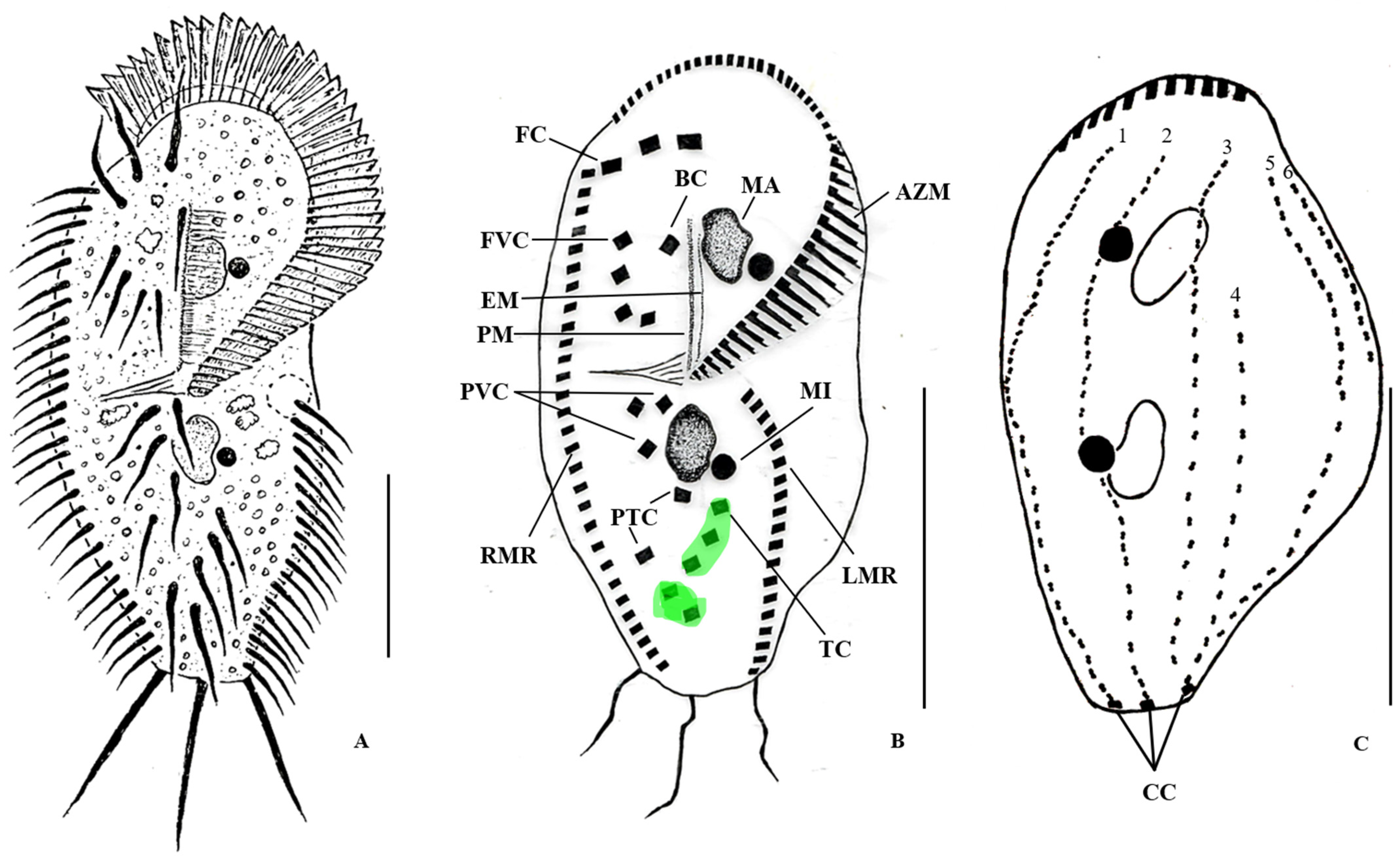
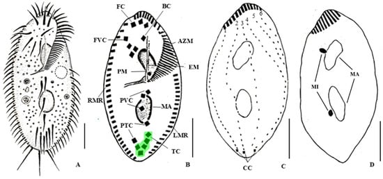
Body size 127–196 × 50–85 µm, on average 164 × 70 µm in vivo and 75–123 × 32–65 µm, on average 99 × 45 µm in protargol preparations; length:width ratio 2.2:1 in protargol preparations (Table S1). Body rigid, shape asymmetrical, with the right margin convex and the left slightly concave, posterior end truncated; dorsoventrally flattened; peristome large with no conspicuous peristomal bulge (Figure 1A,D and Figure 2B). Nuclear apparatus consists of two ellipsoidal macronuclei, about 57–34 × 9–17 µm in protargol preparations; 2–4 ellipsoidal micronuclei with an average size of 5 × 4 µm, always located near the macronuclei. Contractile vacuole located near left cell margin, approximately at the level of the posterior end of the adoral zone of membranelles. Cortical granules absent. Food vacuoles scattered through the cell containing bacteria, algae and other small ciliates (Figure 1A). The adoral zone of membranelles occupies about 51% of the cell length, with about 30–50 membranelles. Undulating membranes in Stylonychia pattern. Eighteen frontal-ventral-transverse cirri: 3 frontal cirri; 1 buccal cirrus at the level of the anterior end of the endoral membrane; 4 fronto-ventral cirri in an L-shaped pattern; 3 postoral ventral cirri; 2 pretransverse ventral and 5 transverse cirri arranged in two distinct groups with the two rightmost cirri, i.e., V/I and VI/I protruding beyond the posterior end of the cell (Figure 1D and Figure 2A). Marginal rows are composed of an average of 15–21 cirri in the left and 24–35 cirri in the right row. The dorsal ciliature composed of 6 dorsal kineties, with kineties 1–3 being bipolar, kinety 4 shortened anteriorly, and kineties 5 and 6 (= dorsomarginal rows 1 and 2) (Figure 1E and Figure 2C); 3 widely spaced caudal cirri (Figure 1A,B and Figure 2A,B). Resting cyst 50 µm in diameter; with wavy surface, which, when pressed a little, got folded, and the wavy structure disappeared, making the cyst appearance smoother; some cysts had a mucus layer on the surface, and few flagellates were attached; on protargol-stained cysts only, some few spines were visible on the surface (Figure 1C and Figure S1). Locomotion fast and irregular. Three voucher slides with protargol-stained specimens have been deposited at the Biologiezentrum at Linz (LI), Austria (accession numbers OLML 2026/126–128). The monoclonal culture of S. ammermanni is maintained at the Culture Collection of Algae and Protozoa (accession number CCAP 1681/6).
Figure 1.
Stylonychia ammermanni from Austria, in vivo (A,C), and after protargol staining (B,D,E). (A,D) Ventral views of typical specimen. (B,E) dorsal views. (A,B) the contractile vacuole, the caudal cirri, the macronuclear nodules and the micronuclei are shown. (C) Resting cyst showing two micronuclei, with opposed arrowheads indicating the cyst wall. (D,E) Details on the adoral zone of membranelles, the cirri pattern and the dorsal kineties are presented. AZM, adoral zone of membranelles; BC, buccal cirrus; CC, caudal cirri; CV, contractile vacuole; FC, frontal cirri; FVC, fronto-ventral cirri; LMR, left marginal row; MA, macronuclear nodules; MI, micronuclei; PTC, pretransverse ventral cirri; PVC, postoral ventral cirri; RMR, right marginal row; TC, transverse cirri; 1–6, dorsal kineties 1–6. Scale bars = 30 µm.
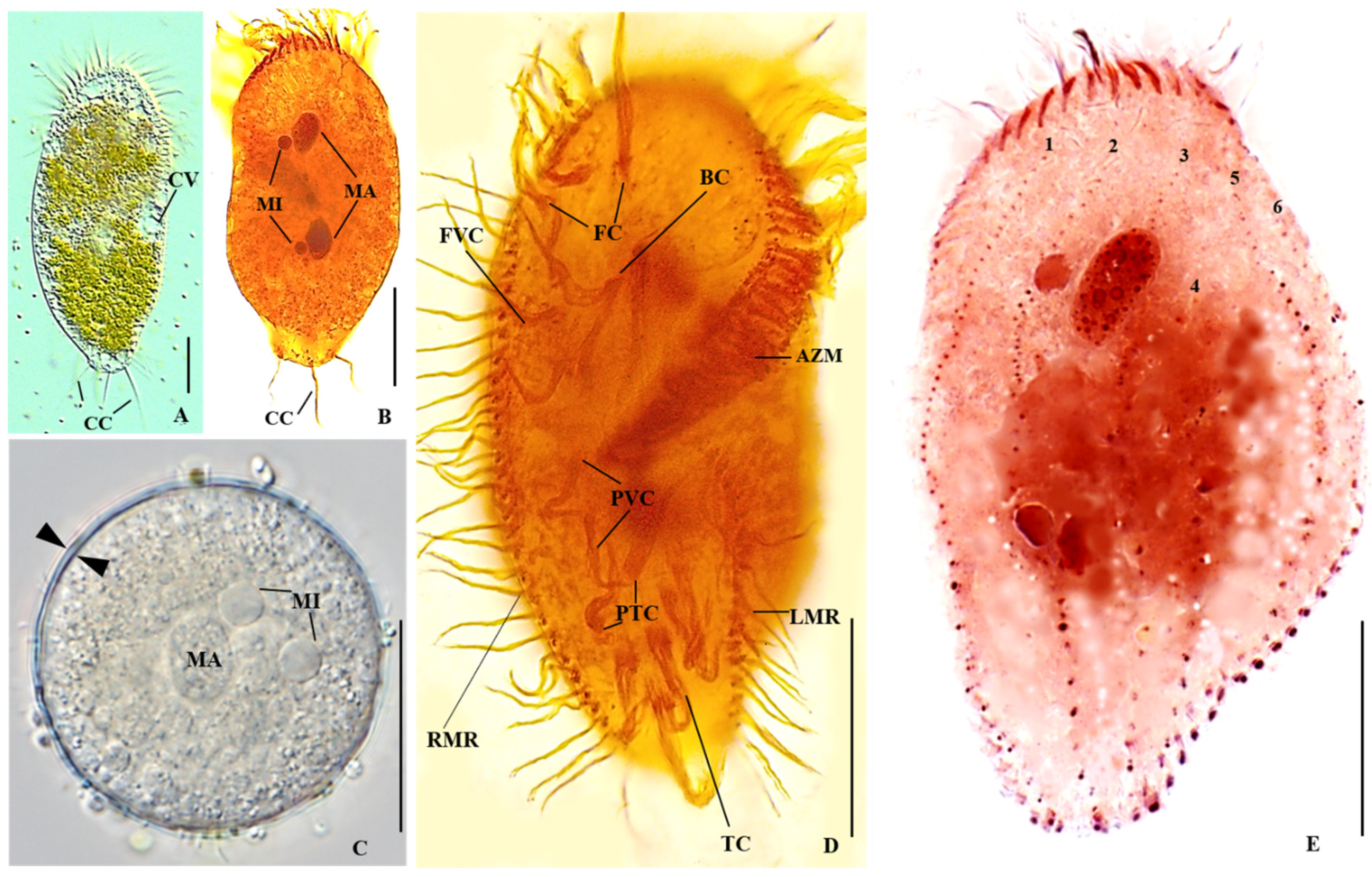
Figure 2.
Stylonychia ammermanni from Austria, in vivo (A) and after protargol staining (B,C). (A,B) Ventral views of typical specimens. The two groups of genus-specific transverse cirri are highlighted in green. (C) Dorsal view. AZM, adoral zone of membranelles; BC, buccal cirrus; CC, caudal cirri; EM, endoral membrane; FC, frontal cirri; FVC, fronto-ventral cirri; LMR, left marginal row; MA, macronucleus; MI, micronucleus; PM, paroral membrane; PTC, pretransverse ventral cirri; PVC, postoral ventral cirri; RMR, right marginal row; TC, transverse cirri; 1–6, dorsal kineties 1–6. Scale bars = 50 µm.
3.2. Tetmemena pustulata (Müller, 1786) Eigner 1999 [Synonym: Stylonychia vorax Stokes, 1885)
Remarks: It is noteworthy that the population identified as S. vorax by Wirnsberger et al. [13] does not represent the original S. vorax Stokes, 1885 [2]. Instead, it represented S. bifaria Stokes, 1887, which was subsequently recognized as a misidentification of Antetmemena bifaria Omar and Jung, 2024. The original S. vorax described by Stokes [31] and the Austrian S. vorax investigated in this study both differ from the population reported by Wirnsberger et al. [13], particularly in having a single group of transverse cirri rather than two groups. Having one group of transverse cirri is the main distinguishing characteristic of the closely related stylonychine genus Tetmemena [14,15].
Description
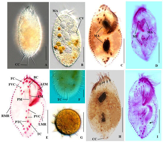
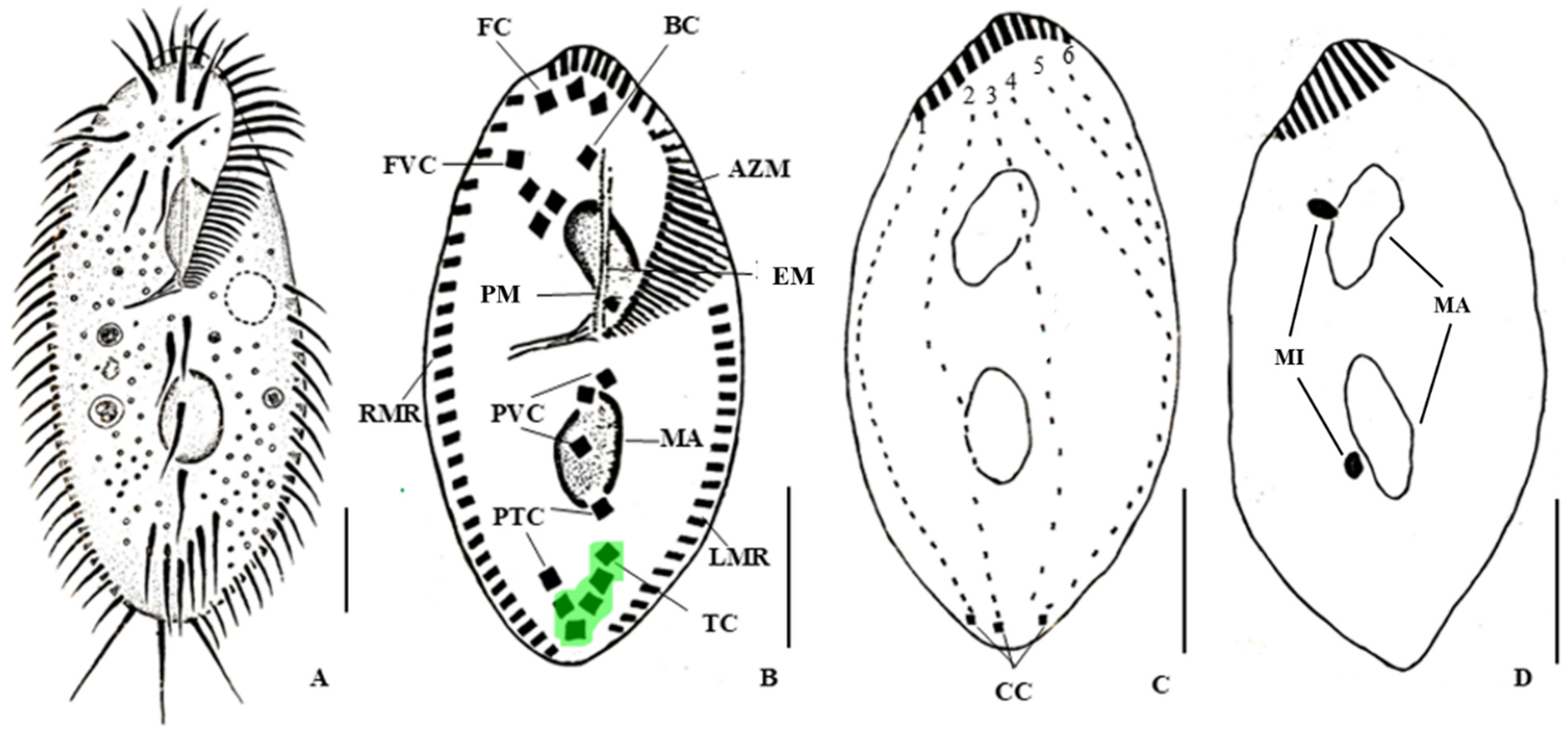
Body size 74–142 × 44–66 µm, on average 110 × 56 µm in vivo; 62–91 × 41–51 µm, on average 77 × 50 µm in protargol preparations; length:width ratio 2.2:1 in protargol preparations (Table S1). Body rigid; dorsoventrally flattened; shape obovoid with a pointed anterior body end and the posterior end rounded or also pointed (Figure 3A–E,H,I). Nuclear apparatus comprises 2 ellipsoidal macronuclear nodules; 2 micronuclei each associated with a macronuclear nodule, well visible in middle dividers after protargol-staining, less visible in non-dividers (Figure 3B–D and Figure 4C,D). Contractile vacuole near the posterior end of the adoral zone of membranelles close to the left cell margin (Figure 3B and Figure 4A). Cortical granules absent. Cytoplasm is colorless, with food vacuoles containing bacteria, cryptomonads (yellowish), green algae and other small protists (Figure 3B). Adoral zone of membranelles covers on average 47% of cell length, comprises 22–40 membranelles; endoral and paroral membranes are straight and closely spaced, i.e., in Stylonychia-like pattern (Figure 3C,E and Figure 4B). Eighteen frontal-ventral-transverse cirri: 3 frontal cirri; 1 buccal cirrus; 4 fronto-ventral cirri; 3 postoral ventral cirri; 2 pretransverse ventral and 5 transverse cirri arranged in a hook-like pattern, of which the two right transverse cirri protrude slightly beyond the posterior end of the cell; left marginal row composed of 16–21 cirri, right row of 26–34 cirri (Figure 3C,E,F and Figure 4A,B). Resting cyst with processes and a diameter of 37 µm on average (Figure 3G). Dorsal ciliature is composed of 6 kineties with kineties 1–4 being bipolar and kineties 5 and 6 (= dorsomarginal rows 1 and 2) located in the anterior third of the dorsal side (Figure 3F and Figure 4C). Caudal cirri are moderately widely spaced, located at the posterior end of dorsal kineties 1, 2 and 4 (Figure 3A,H and Figure 4A,C). Movement is rapid and erratic. One voucher slide with protargol-stained specimens has been deposited at the Biologiezentrum at Linz, Austria (accession number OLML 2026/129). The monoclonal culture of T. pustulata is maintained at the Culture Collection of Algae and Protozoa (accession number CCAP 1674/2).
Figure 3.
Tetmemena pustulata from Austria, in vivo (A,B,F,G) and after protargol staining (C–E,H,I). (A–C) Ventral views of typical specimens showing the moderately widely spaced caudal cirri; the contractile vacuole, and the macronuclear nodules. (D) Dorsal view of a middle divider, showing the two micro- and macronuclei. (E,F) Ventral view showing the adoral zone of membranelles; the buccal cirrus; the paroral and endoral membranes; the frontal cirri; the fronto-ventral cirri; the right and left marginal rows; the pretransverse and postoral ventral cirri, and the transverse cirri with two cirri protruding beyond the posterior cell end. (G) Process-bearing cyst. (H) Dorsal view showing dorsal kineties 1–6. (I) Ventral view of a middle divider showing the anlagen formation and the de novo development of the opisthe oral primordium (arrow). AZM, adoral zone of membranelles; BC, buccal cirrus; CC, caudal cirri; CV, contractile vacuole; EM, endoral membrane; FC, frontal cirri; FVC, fronto-ventral cirri; LMR, left marginal row; MA, macronuclear nodules; MI, micronuclei; PM, paroral membrane; PTC, pretransverse ventral cirri; PVC, postoral ventral cirri; RMR, right marginal row; TC, transverse cirri; 1–6, dorsal kineties 1–6. Scale bars = 30 µm.
Figure 4.
Tetmemena pustulata from Austria, in vivo (A) and after protargol-staining (B–D). (A,B) Ventral views of typical specimens. The single group of the genus-specific transverse cirri is highlighted in green. (C,D) Dorsal views. AZM, adoral zone of membranelles; BC, buccal cirrus; CC, caudal cirri; EM, endoral membrane; FC, frontal cirri; FVC, fronto-ventral cirri; LMR, left marginal cirri; MA, macronuclear nodules; MI, micronucleus; PM, paroral membrane; PTC, pretransverse ventral cirri; PVC, postoral ventral cirri; RMR, right marginal row; TC, transverse cirri; 1–6, dorsal kineties 1–6. Scale bars = 20 µm.
3.3. Molecular Data
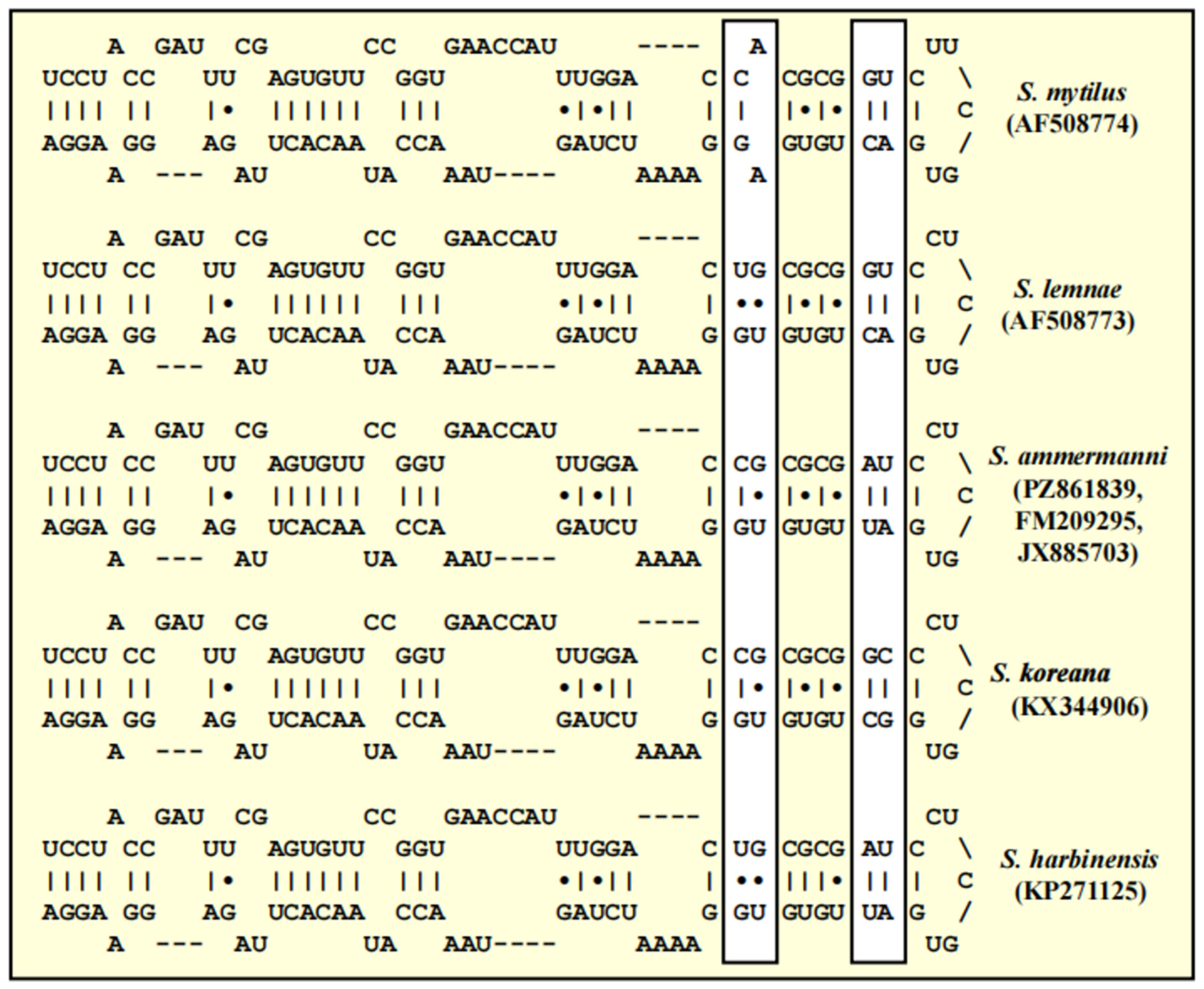
GenBank accession numbers for all newly deposited sequences in this study, and those used in phylogenetic analyses, are presented in Table S2. The two stylonychids were initially identified as S. ammermanni and S. vorax and are registered under the accession numbers PZ861839 and PZ861840, respectively. SSU rDNA comparisons showed that the sequence of S. vorax was identical to T. pustulata (PP760152), supporting its assignment to T. pustulata. In the concatenated SSU rDNA + ITS phylogeny, S. ammermanni clusters within an S. mytilus complex, while T. pustulata is placed within the T. pustulata complex (Figure 5). The sequence of the Austrian population of S. ammermanni differed by 22 nucleotides from S. mytilis (AF508774), 15 nucleotides from S. lemnae (AF508773), and 3 nucleotides from Stylonychia harbinensis (KP271125). The variable (V9) region of the SSU rDNA shows that the five Stylonychia species are distinguishable by compensatory base changes (Figure 6).
Figure 5.
Molecular phylogeny of the spirotrich taxa based on SSU and ITS rDNA sequence comparisons. The phylogenetic tree shown was inferred using the maximum likelihood method based on the datasets (2263 aligned positions of 27 taxa) using PAUP 4.0a build169. For the analyses, the best model was calculated by the automated model selection tool implemented in PAUP. The setting of the best model was given as follows: GTR + I + G (base frequencies: A 0.2756; C 0.2085; G 0.2392; U 0.2767; rate matrix A-C 1.5456; A-G 2.5077; A-U 1.7955; C-G 0.6907; C-U 4.8217; G-U 1.0000) with the proportion of invariable sites (I = 0.8030) and gamma shape parameter (G = 0.6824). The branches in bold are highly supported in all analyses (bootstrap values >50% calculated with PAUP using maximum likelihood, neighbor joining, maximum parsimony, and maximum likelihood). The newly sequenced strains are highlighted in bold.
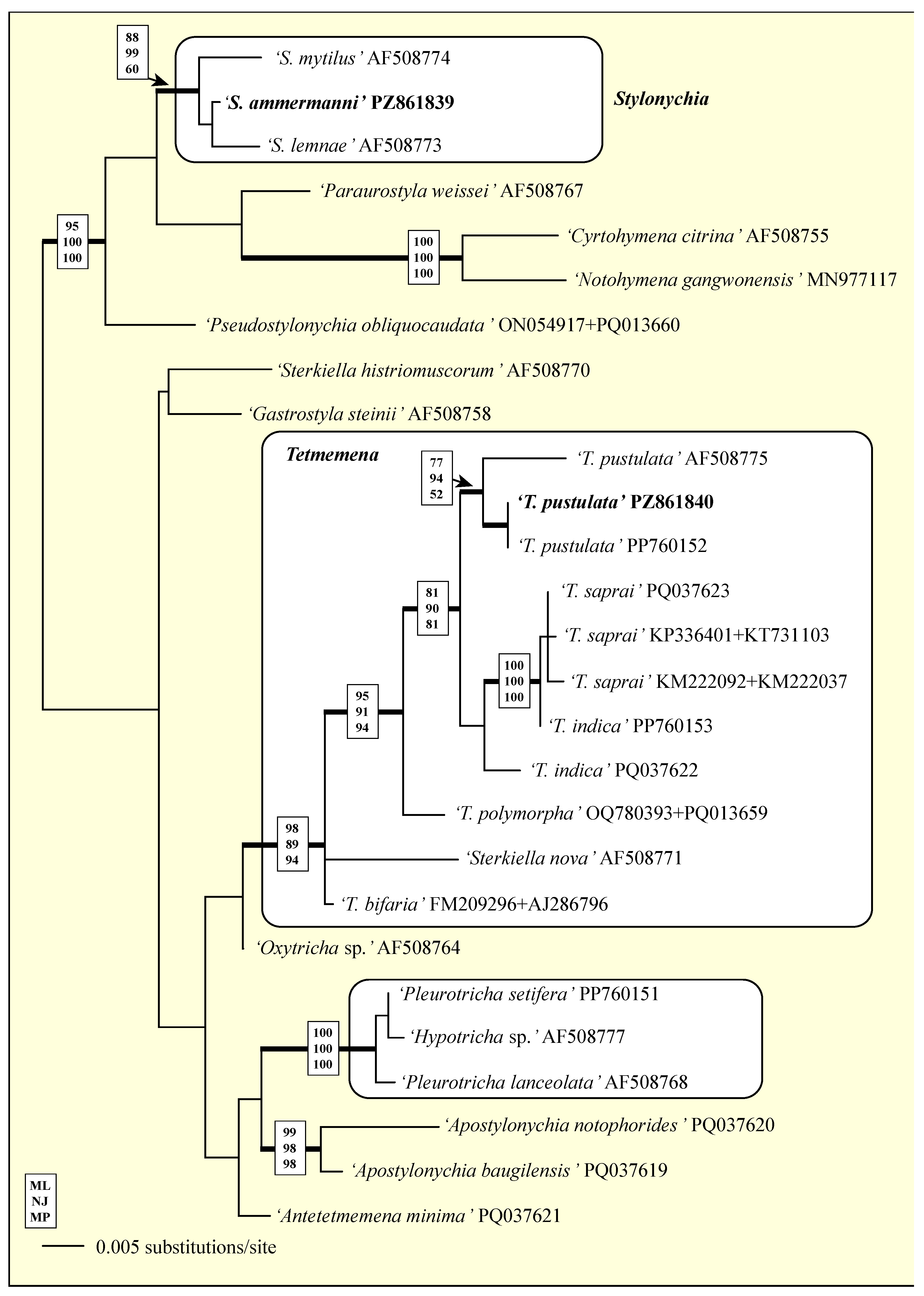
Figure 6.
Comparison of the V9 secondary structures among species of the genus Stylonychia sensu stricto. Variable regions are marked in white boxes.
To validate our findings, we pairwise compared available sequences, including different barcode markers of S. ammermanni (PZ446743), T. indica (PZ446739), T. pustulata (PZ446740), the neotype of T. pustulata (PZ446741), and Pleurotricha setifera (PZ446742) among each other (Table 1). On the COI gene, the two T. pustulata strains showed 98.95% identity, whereas the putative Indian T. saprai sequence (MN027505) showed 98.74% identity with the Kenyan T. indica strain. Stylonychia ammermanni, and P. setifera were used as outgroups and differed from Tetmemena by 17–20%.
Table 1.
Variability of SSU rDNA, SSU + ITS rDNA, ITS rDNA, and COI mtDNA sequences between T. pustulata and other stylonchine taxa.
To assess the effect of nucleotide divergence at the amino acid level, we compared translated proteins (Table 1). No differences were found in the T. saprai/T. indica complex and between the two T. pustulata strains including the former S. vorax (Table 2). The genus Tetmemena is characterized by five amino acids (highlighted in blue) and the two species differed in two amino acids (highlighted in yellow) from each other. Pleurotricha setifera and S. ammermanni showed one, respectively two synapomorphies in the COI.
Table 2.
Comparison of the COI amino acid sequence among species of Tetmemena, Pleurotricha setifera and Stylonychia ammermanni. Differences in the amino acid sequences are highlighted in blue, the diagnostic residues for the T. pustulata and the T. saprai/T. indica complexes in yellow boxes. * marks the strain investigated in this study.
4. Discussion
Five described Stylonychia species having one group of transverse cirri were insufficiently described [14], and their morphology suggests that they likely belong to the genus Tetmemena [14,15]. Furthermore, studies indicated that the presence of one or two groups of transverse cirri is a main generic character delimiting members of the genus Tetmemena from Stylonychia [15].
4.1. Synonymizing S. vorax with T. pustulata
A T. pustulata with S. vorax-like characteristics was first reported by Wirnsberger et al. [13] from a population collected from pasture pools in the area of Mondsee, Austria. Here, we present another T. pustulata population from Lake Mondsee resembling S. vorax and members of the T. pustulata complex [22]. Morphologically, our isolate is similar to populations of S. vorax described by Stokes [31], Kahl [32], Šrámek-Hušek [33], Dragesco [34], and Foissner et al. [35] having a slightly tapered anterior and posterior end, long, stout and narrowly arranged caudal cirri, and the transverse cirri arranged in one group [2]. However, there are significant differences between our population and S. vorax (former S. ovalis) described by Dragesco [34] who reported a body size of 71–85 vs. 74–142 µm in our strain, a number of adoral membranelles of 41–60 vs.22–40, and 5 vs.2 transverse cirri protruding beyond the posterior end of the cell (Table S3). All T. pustulata populations have two main morphological characteristics, i.e., the bipolar dorsal kinety 4 and process-bearing cysts [13,22,23,36]. Nonetheless, according to Wirnsberger et al. [13] and our own investigations, the two Austrian populations differed in body size, i.e., 48–124 vs. 74–142, body shape, i.e., obovoid vs. elliptical, the location of the micronuclei, i.e., embedded in the macronucleus vs. associated, and the number of transverse cirri protruding beyond the posterior cell end, i.e., 5 vs. 2 (Table S3).
The SSU rDNA phylogeny confirmed the non-monophyletic relationship of T. pustulata populations in many studies [19,20,21,22]. However, the concatenated SSU rDNA + ITS phylogeny in this study also confirms the monophyly of Tetmemena [14], including the position of the Austrian neotype of T. pustulata in the SSU rDNA phylogenetic tree [22]. Although the North American T. pustulata clusters differently with other populations in their SSU rDNA phylogeny, the concatenated SSU rDNA + ITS phylogeny confirms their monophyletic relationship (Figure 5). This monophyly in the T. pustulata complex is likely to represent a synapomorphy, as monophyletic groups are expected to have similar morphological characters, in this case particularly the bipolar dorsal kinety 4 and a process-bearing cyst [14,15].
There was no sequence variation in our former S. vorax and the neotype of T. pustulata in the SSU rDNA and ITS marker genes. In contrast, the North American population had a higher (1.29%, 28 nucleotide differences) genetic variability to both Austrian populations. Schmidt et al. [8] suggested that a genetic variation of >1% in the SSU rDNA was enough as a standard threshold for the designation of species, which was used to assign T. saprai [19]. Therefore, based on this high genetic variability, inter-population studies within the T. pustulata complex are required from different geographical regions. On the other hand, the COI sequence of our former S. vorax showed low (1.1%, 6 nucleotide differences) genetic variability to the neotype (Table 1), and these differences did not affect amino acid sequences (Table 2). Nevertheless, COI is a very sensitive marker and delimits most hypotrichs [24,25,26], but not some other ciliates [37,38]. Additionally, the threshold of species delimitation is taxon-specific [39,40,41,42]. However, based on molecular data in the present study, the Austrian population of the former S. vorax is likely T. pustulata and we therefore synonymize it.
4.2. Characterization of the Austrian Population of S. ammermanni
The morphology of our Austrian population of S. ammermanni is consistent with that of recently described and redescribed Stylonychia species, including having two groups of transverse cirri, a truncated posterior end, and widely spaced caudal cirri [1,2,3,4,5,6,11,12]. Stylonychia ammermanni is a widely distributed stylonychid ciliate whose morphological characteristics considerably overlap with those of several congeners, resulting in repeated taxonomic revisions and synonymisations [3,4,5,6,11]. Our population is larger than previously described, i.e., 127–196 × 50–85 µm vs. 126–165 × 46–77 µm, with other characters overlapping (Ref. [6]; Table S4). On the other hand, it differs from the populations of S. koreana in having a bigger size, i.e., 127–196 × 50–85 µm vs. 75–128 × 32–58 µm, more marginal cirri on the left (15–21 vs. 11–18) and on the right (24–35 vs. 16–24), and more adoral membranelles (30–50 vs. 33–44) (Table S3).
The Italian population of S. ammermanni was reported to be morphologically similar to the Indian type population and S. harbinensis; thus, it was synonymized [4,11]. Recently, cyst morphology has been considered an important diagnostic character for distinguishing S. ammermanni from morphologically similar Stylonychia species, particularly those characterized by an anteriorly shortened dorsal kinety 4. For instance, the Italian population of S. ammermanni differs from S. koreana in possessing small and many ring-shaped structures on the cyst surface, which were absent in the latter [6]. However, our findings highlight the limitation of relying on cyst morphology in the absence of comparison with the type population, thereby rendering the assignment of the Italian population to S. ammermanni less conclusive. This is corroborated by the absence of ring-shaped structures on the cysts of the Austrian S. ammermanni population. This inter-population variation emphasizes the potential diagnostic value of cyst morphology, while also demonstrating the need for more integrative approaches. Moreover, synonymization of S. harbinensis with S. ammermanni without integration of cyst morphology and molecular data suggests potential misidentification of the two possibly cryptic species. Cyst morphology has emerged as one of the most important characters for delimiting most hypotrichs, including a process-bearing vs. a smooth cyst surface, macronuclear nodules fused vs. separated, and presence vs. absence of lepidosomes on the surface [18,22,43]. Stylonychia koreana and S. ammermanni were delimited based on the absence and presence of ring-shaped structures on the cyst surface, respectively [6]. Consequently, Omar et al. [44] raised S. ammermanni koreana to the species level, i.e., S. koreana. The cyst of our Austrian S. ammermanni shares separated macronuclear nodules with both S. koreana and the Italian population of S. ammermanni (Table S4). The Austrian population had a wavy surface, which upon pressure appeared smoother and showing some folds. In protargol-stained cysts, some few spines were visible on the surface (Figure S1).
We analyzed the Austrian S. ammermanni using multigene sequences. Paucity of SSU rDNA + ITS and COI sequences makes SSU rDNA sequences readily available for analysis in several phylogenies [6]. Only two sequences, AF508773 and AF508774, representing S. lemnae and S. mytilus, respectively, and another sequence, AF508754, designated as Stylonychia sp., were available with the complete rDNA region sequenced. Their respective sequence variations were 99.33% (14 nucleotides), 99.02% (19 nucleotides), and 98.62% (29 nucleotides) compared to the Austrian S. ammermanni. This variation elucidates a distant relationship between S. ammermanni and the sibling species S. lemnae and S. mytilus, both reported to have a single-base pair difference [8], and monophyletically separated from S. ammermanni [20]. This study further highlights their differences in V9 SSU rDNA sequences.
To date, no SSU rDNA sequence is available from the neotype or the type population of S. ammermanni. However, for future phylogenetic analyses, Schmidt et al. [8] provided standardized SSU rDNA sequences for S. lemnae (AJ310496) and S. mytilus (AJ310499) which showed 99.54% (8 nucleotides) and 99.49% (7 nucleotides) sequence similarity, respectively, to the SSU rDNA of the Austrian S. ammermanni. Furthermore, our SSU rDNA sequence is 100% identical to the German sequence (FM209295) and differing by one nucleotide (99.9% similarity) from the Indian (JX885703) and three (99.8% similarity) from the Italian sequences (KP271125). Elsewhere, the Italian S. ammermanni is designated as S. harbinensis in both the phylogenetic tree and GenBank entries and they cluster apart from S. mytilus and S. lemnae [20]. This study corroborates the results by differences observed in the V9 region of the SSU rDNA for S. ammermanni, S. harbinensis, S. koreana, S. lemnae, and S. mytilus (Figure 6). The high similarity of the Austrian, Indian, and German sequences suggests conspecificity with S. ammermanni. In contrast, the molecular divergence of the Italian population, together with its cyst morphology, raises questions regarding its current identification and assignment and suggests it may represent a separate species, potentially corresponding to S. harbinensis as designated by the sequence. However, this interpretation is putative, considering that detailed and integrative taxonomy of the Indian S. ammermanni and the Chinese S. harbinensis are currently lacking. The Austrian S. ammermanni population differed from S. koreana (KX344906), showing 99.09% sequence similarity, corresponding to 15 nucleotides in the SSU rDNA + ITS sequence. In conclusion, the molecular and morphological evidence supports the distinctiveness of the present population from S. koreana and indicates that the taxonomic status of the Italian population attributed to S. ammermanni needs further investigation using an integrative comparison of ciliate and cyst morphology, and molecular sequences with material from the type locality and S. harbinensis.
5. Conclusions
There was a high variability in the ITS for delimitation of Tetmemena and its species complexes, revealing that ITS genes are more reliable for species delimitation in Tetmemena and Stylonychia and confirming the monophyletic relationships in the species complexes. However, SSU rDNA + ITS genes could not delimit the two Austrian populations of S. vorax and T. pustulata and we therefore synonymized them based also on earlier observations mentioning low morphological differences. The COI gene sequence showed low variations for two Austrian populations of T. pustulata comparable to differences between T. indica strains originating from India and Austria. This variation in the COI gene confirms its importance and high resolutive capacity at short evolutionary distances for hypotrich ciliates. For S. ammermanni, we were able to distinguish all four species of Stylonychia sensu stricto, including S. ammermanni and S. koreana based on the V9 region. Thus, we have demonstrated the importance of the V9 region for the diagnosis within Stylonychia, as well as the high variability of SSU rDNA + ITS and COI genes in Tetmemena, Pleurotricha, and Stylonychia. For the discovery of S. ammermanni in Austria and its distinct cyst morphology and molecular divergence from the Italian population, we recommend an integrative taxonomic reassessment against the type species to confirm its designation. This is because the German sequence for S. ammermanni was 100% identical to the Austrian sequences.
Supplementary Materials
The following supporting information can be downloaded at https://www.mdpi.com/article/10.3390/d18110705/s1, Figure S1: Resting cysts of the Stylonychia ammermanni population from Austria. Table S1: Morphometric data of Stylonychia ammermanni (Sa) and Tetmemena pustulata (Tp), formerly known as Stylonychia vorax of the Austrian populations obtained from live observation (1) and protargol-impregnated cells (2). Measurements in µm. CV, coefficient of variation in %; M, median; Max, maximum; Mean, arithmetic mean; Min, minimum; n, number of individuals investigated; SD, standard deviation; SE, standard error of arithmetic mean. Table S2: Taxonomic and molecular sequence information for ciliate taxa included in the phylogenetic analyses. Table S3: Comparison of Tetmemena pustulata and Stylonychia vorax congeners. Arithmetic mean and minimum and maximum values are given in brackets. Table S4: Comparison of Stylonychia ammermanni and Stylonychia koreana populations. Arithmetic mean and minimum and maximum values given in brackets.
Author Contributions
M.J.O.: conceptualization; data curation; formal analysis; investigation. methodology; writing—original draft, review and editing. U.K.: formal analysis; investigation; methodology; resources; validation; writing—review and editing. N.K.: formal analysis; investigation; methodology; writing—original draft, review and editing. E.L.: conceptualization; data curation; validation; methodology; writing—review and editing. T.P.: conceptualization; data curation; formal analysis; investigation; methodology; writing—original draft, review and editing. G.O.O.: conceptualization; data curation; funding acquisition; methodology; project administration; resources; supervision; validation; writing—original draft; review and editing. A.P.: conceptualization; data curation; investigation; supervision; validation; writing—review and editing. B.S.: conceptualization; data curation; project administration; resources; supervision; validation; visualization; writing—original draft, review and editing. All authors have read and agreed to the published version of the manuscript.
Funding
This study was supported by the Austrian Science Fund (FWF; grant-DOI 10.5576/I2238-B25) and the Horizon 2020 EU program RISE (project Next generation taxonomy—Ciliophora and their bacterial endosymbiont as a proof of concept; NGTax 872767-DOI 10.3030/872767).
Institutional Review Board Statement
Not applicable.
Data Availability Statement
The original contributions presented in this study are included in the article and its Supplementary Materials. Further inquiries can be directed to the corresponding author.
Acknowledgments
We thank Valentina Schmidt who provided cyst images and measurements of S. ammermanni.
Conflicts of Interest
The authors declare no conflicts of interest.
References
- Ammermann, D. Cell size and dorsal cilia: Are they useful features for the identification of Stylonychia mytilus (Ciliophora, Spirotrichea) and its subpopulations? Eur. J. Protistol. 2008, 44, 263–268. [Google Scholar] [CrossRef] [Scilit] [PubMed]
- Berger, H. Monograph of the Oxytrichidae (Ciliophora, Hypotrichia). Monogr. Biol. 1999, 78, 1–1080. [Google Scholar]
- Foissner, V.W. Ökologie and Taxonomie der Hypotrichida (Protozoa: Ciliophora) einiger österreichischer Böden. Arch. Protistenkd. 1982, 126, 19–143. [Google Scholar] [CrossRef] [Scilit]
- Foissner, W. Terrestrial and semiterrestrial ciliates (Protozoa, Ciliophora) from Venezuela and Galápagos. Denisia 2016, 35, 1–912. [Google Scholar]
- Gupta, R.; Kamra, K.; Arora, S.S.; Sapra, G.R. Stylonychia ammermanni sp. n., a new oxytrichid [Ciliophora: Hypotrichida] ciliate from the River Yamuna, Delhi, India. Acta Protozool. 2001, 40, 75–79. [Google Scholar]
- Kumar, S.; Bharti, D.; Quintela-Alonso, P.; Shin, M.K.; La Terza, A. Fine-tune investigations on three stylonychid (Ciliophora, Hypotricha) ciliates. Eur. J. Protistol. 2016, 56, 200–218. [Google Scholar] [CrossRef] [Scilit] [PubMed]
- Kumar, S.; Foissner, W. Morphology and ontogenesis of Stylonychia (Metastylonychia) nodulinucleata nov. subgen. (Ciliophora, Hypotricha) from Australia. Eur. J. Protistol. 2017, 57, 61–72. [Google Scholar] [CrossRef] [Scilit] [PubMed]
- Schmidt, S.L.; Ammermann, D.; Schlegel, M.; Bernhard, D. Stylonychia lemnae strains from a North American site differ in a single nucleotide within the small subunit rDNA from Eurasian strains. J. Eukaryot. Microbiol. 2006, 53, 308–309. [Google Scholar] [CrossRef] [Scilit] [PubMed]
- Schmidt, S.L.; Bernhard, D.; Schlegel, M.; Foissner, W. Phylogeny of Stichotrichia (Ciliophora; Spirotrichea) reconstructed with nuclear small subunit rDNA gene sequences: Discrepancies and accordances with morphological data. J. Eukaryot. Microbiol. 2007, 54, 201–209. [Google Scholar] [CrossRef] [Scilit] [PubMed]
- Shi, X.; Li, H. Discovery of Stylonychia nodulinucleata sp. nov. (Ciliophora, Hypotrichida, Oxytrichidae) and comparison of its neighboring species. Zool. Res. 1993, 14, 10–14. [Google Scholar]
- Shi, X.-B.; Ammermann, D. Stylonychia harbinensis sp. n., a new oxytrichid ciliate (Ciliophora, Hypotrichia) from the Heilongjiang Province, China. Protistology 2004, 3, 215–221. [Google Scholar]
- Wirnsberger, E.; Foissner, W.; Adam, H. Biometric and morphogenetic comparison of the sibling species Stylonychia mytilus and S. lemnae, including a phylogenetic system for the oxytrichids (Ciliophora, Hypotrichida). Arch. Protistenkd. 1986, 132, 167–185. [Google Scholar] [CrossRef] [Scilit]
- Wirnsberger, E.; Foissner, W.; Adam, H. Morphological, biometric and morphogenetic comparison of two closely related species, Stylonychia vorax and S. pustulata (Ciliophora: Oxytrichidae). J. Protozool. 1985, 32, 261–268. [Google Scholar] [CrossRef] [Scilit]
- Omar, A.; Jung, J.H. Disentangling the stylonychine ciliates (Ciliophora: Hypotricha): The establishment of two new genera using an integrative taxonomic approach. Zool. J. Linn. Soc. 2024, 202, zlae144. [Google Scholar] [CrossRef]
- Shao, C.; Lu, X.; Ma, H. A general overview of the typical 18 frontal-ventral-transverse cirri Oxytrichidae sl genera (Ciliophora, Hypotrichia). J. Ocean Univ. China 2015, 14, 522–532. [Google Scholar] [CrossRef] [Scilit]
- Ammermann, D.; Schlegel, M. Characterization of Two Sibling Species of the Stylonychia mytilus Complex (Ciliophora, Hypotricha): Stylonychia mytilus Ehrenberg, 1838 and Stylonychia lemnae n. sp. J. Protozool. 1983, 30, 290–294. [Google Scholar] [CrossRef] [Scilit]
- Haentzsch, M.; Schmidt, S.L.; Bernhard, D.; Ammermann, D.; Berendonk, T.U.; Schlegel, M. A PCR-Based Method to Distinguish the Sibling Species Stylonychia mytilus and Stylonychia lemnae (Ciliophora, Spirotrichea) Using Isocitrate Dehydrogenase Gene Sequences. J. Eukaryot. Microbiol. 2006, 53, 343–347. [Google Scholar] [CrossRef] [Scilit] [PubMed]
- Bharti, D.; Kumar, S.; La Terza, A.; Chandra, K. Morphology and ontogeny of Tetmemena pustulata indica nov. subspec. (Ciliophora, Hypotricha), from the Thane Creek, Mumbai, India. Eur. J. Protistol. 2019, 71, 125629. [Google Scholar] [CrossRef] [Scilit] [PubMed]
- Gupta, R.; Abraham, J.S.; Sripoorna, S.; Maurya, S.; Toteja, R.; Makhija, S.; Al-Misned, F.A.; El-Serehy, H.A. Description of a new species of Tetmemena (Ciliophora, Oxytrichidae) using classical and molecular markers. J. King Saud Univ. Sci. 2020, 32, 2316–2328. [Google Scholar] [CrossRef] [Scilit]
- Kaur, H.; Shashi; Warren, A.; Negi, R.K.; Kamra, K. Taxonomic resolution of Stylonychia notophora (Alveolata, Ciliophora, Hypotricha) sensu Sapra and Dass 1970 using an integrative approach. Int. J. Syst. Evol. Microbiol. 2020, 70, 4199–4208. [Google Scholar] [CrossRef] [Scilit] [PubMed]
- Omar, A.; Jang, S.W.; Jung, J.H. Polymorphism, gigantism, and cannibalism, one stylonychid ciliate (Ciliophora, Hypotricha) to rule them all. Front. Microbiol. 2023, 14, e1159634. [Google Scholar] [CrossRef] [Scilit] [PubMed]
- Owuor, M.J.; Koll, U.; Kostner, N.; Lelmen, E.; Pröschold, T.; Ong’ondo, G.O.; Sonntag, B. Shedding new light on stylonychid ciliates: A case study of Tetmemena and Pleurotricha (Ciliophora, Spirotrichea, Hypotrichia, Dorsomarginalia). Eur. J. Protistol. 2026, 104, 126193. [Google Scholar] [CrossRef] [Scilit] [PubMed]
- Shao, C.; Pan, X.; Jiang, J.; Ma, H.; Al-Rasheid, K.A.S.; Warren, A.; Lin, X. A redescription of the oxytrichid Tetmemena pustulata (Müller, 1786) Eigner, 1999 and notes on morphogenesis in the marine urostylid Metaurostylopsis salina Lei et al., 2005 (Ciliophora, Hypotrichia). Eur. J. Protistol. 2013, 49, 272–282. [Google Scholar] [CrossRef] [Scilit] [PubMed]
- Fan, X.; Yao, S.; Luo, X.; Dong, T.; Xu, Y.; Chen, L.; Bourland, W.; Zhao, Y.; Huang, J. Some morphologically distinguishable hypotrich ciliates share identical 18S rDNA gene sequences–taxonomic insights from a case study on Oxytricha species (Protista, Ciliophora). Zool. J. Linn. Soc. 2021, 193, 356–379. [Google Scholar] [CrossRef] [Scilit]
- Kim, K.S.; Ji, S.J.; Min, G.S. COI DNA barcoding for Sterkiella multicirrata (Ciliophora: Oxytrichidae) from South Korea. Anim. Syst. Evol. Divers. 2020, 36, 7–9. [Google Scholar] [CrossRef]
- Park, M.H.; Jung, J.H.; Jo, E.; Park, K.M.; Baek, Y.S.; Kim, S.J.; Min, G.S. Utility of Mitochondrial CO1 Sequences for Species Discrimination of Spirotrichea Ciliates (Protozoa, Ciliophora). Mitochondrial DNA Part A DNA Mapp. Seq. Anal. 2019, 30, 148–155. [Google Scholar] [CrossRef] [Scilit] [PubMed]
- Frantal, D.; Agatha, S.; Beisser, D.; Boenigk, J.; Darienko, T.; Dirren-Pitsch, G.; Filker, S.; Gruber, M.; Kammerlander, B.; Nachbaur, L.; et al. Molecular data reveal a cryptic diversity in the genus Urotricha (Alveolata, Ciliophora, Prostomatida), a key player in freshwater lakes, with remarks on morphology, food preferences, and distribution. Front. Microbiol. 2022, 12, e787290. [Google Scholar] [CrossRef] [Scilit] [PubMed]
- Swofford, D.L. PAUP: Phylogenetic Analysis Using Parsimony (and Other Methods), version 4.0b10; Sinauer Associates: Sunderland, MA, USA, 2002.
- Wolf, M.; Friedrich, J.; Dandekar, T.; Müller, T. CBC Analyzer: Inferring phylogenies based on compensatory base changes in RNA secondary structures. In Silico Biol. 2005, 5, 291–294. [Google Scholar] [CrossRef] [Scilit]
- Zuker, M. Mfold web server for nucleic acid folding and hybridization prediction. Nucleic Acids Res. 2003, 31, 3406–3415. [Google Scholar] [CrossRef] [Scilit] [PubMed]
- Stokes, A.C. Some new infusoria. Am. Nat. 1885, 19, 433–443. [Google Scholar] [CrossRef] [Scilit]
- Kahl, A. Urtiere oder Protozoa: Wimpertiere oder Ciliata (Infusoria); Fischer: Jena, Germany, 1935. [Google Scholar]
- Šrámek-Hušek, R. K poznání nálevníků Ostravského kraje [Zur Kenntnis der Ciliaten des Ostrauer-Gebietes (Tschechoslowakei)]. Věstn. Ceskoslov. Zool. Spol. 1957, 21, 1–24. [Google Scholar]
- Dragesco, J. Ciliés libres de Thonon et ses environs. Protistologica 1966, 2, 59–95. [Google Scholar]
- Foissner, W.; Blatterer, H.; Berger, H.; Kohmann, F. Taxonomische und ökologische Revision der Ciliaten des Saprobiensystems—Band I: Cyrtophorida, Oligotrichida, Hypotrichia, Colpodea; Informationsberichte des Bayer. Landesamtes für Wasserwirtschaft; Bayerisches Landesamt für Wasserwirtschaft: München, Germany, 1991; pp. 1–478. [Google Scholar]
- Bell, A.J.; Satir, P.; Grimes, G.W. Mirror-imaged doublets of Tetmemena pustulata: Implications for the development of left–right asymmetry. Dev. Biol. 2008, 314, 150–160. [Google Scholar] [CrossRef] [Scilit] [PubMed]
- Jung, J.H.; Omar, A.; Kim, K.S.; Kang, S.C.; Kwak, D.Y.; Sun, J.H.; Min, G.S. A study on the non-monophyletic genera Australothrix and Holostichides based on multigene and morphological analyses with a reexamination of type materials (Protozoa: Ciliophora). Mol. Phylogenet. Evol. 2019, 139, 106538. [Google Scholar] [CrossRef] [Scilit] [PubMed]
- Shazib, S.U.A.; Vďačný, P.; Slovak, M.; Gentekaki, E.; Shin, M.K. Deciphering phylogenetic relationships and delimiting species boundaries using a Bayesian coalescent approach in protists: A case study of the ciliate genus Spirostomum (Ciliophora, Heterotrichea). Sci. Rep. 2019, 9, 16360. [Google Scholar] [CrossRef] [Scilit] [PubMed]
- Chantangsi, C.; Lynn, D.H.; Brandl, M.T.; Cole, J.C.; Hetrick, N.; Ikonomi, P. Barcoding ciliates: A comprehensive study of 75 isolates of the genus Tetrahymena. Int. J. Syst. Evol. Microbiol. 2007, 57, 2412–2423. [Google Scholar] [CrossRef] [Scilit] [PubMed]
- Obert, T.; Zhang, T.; Rurik, I.; Vďačný, P. First molecular evidence of hybridization in endosymbiotic ciliates (Protista, Ciliophora). Front. Microbiol. 2022, 13, 1067315. [Google Scholar] [CrossRef] [Scilit] [PubMed]
- Shazib, S.U.A.; Cote-L’Heureux, A.; Ahsan, R.; Muñoz-Gómez, S.A.; Lee, J.; Katz, L.A.; Shin, M.K. Phylogeny and species delimitation of ciliates ins the genus Spirostomum (class Heterotrichea) using single-cell transcriptomes. BMC Ecol. Evol. 2025, 25, 17. [Google Scholar] [CrossRef] [Scilit] [PubMed]
- Strüder-Kypke, M.C.; Lynn, D.H. Comparative analysis of the mitochondrial cytochrome c oxidase subunit I (COI) gene in ciliates (Alveolata, Ciliophora) and evaluation of its suitability as a biodiversity marker. Syst. Biodivers. 2010, 8, 131–148. [Google Scholar] [CrossRef] [Scilit]
- Kim, K.-S.; Min, G.-S. Morphology and molecular phylogeny of Oxytricha seokmoensis sp. nov. (Hypotrichia: Oxytrichidae), with notes on its morphogenesis. Eur. J. Protistol. 2019, 69, 125641. [Google Scholar] [CrossRef] [Scilit] [PubMed]
- Omar, A.; Yeo, J.H.; Jung, J.-H. A new “flagship” ciliate, Pseudostylonychia obliquocaudata n. gen., n. sp. (Ciliophora, Hypotricha), from South Korea. Eur. J. Protistol. 2022, 84, 125893. [Google Scholar] [CrossRef] [Scilit] [PubMed]

Disclaimer/Publisher’s Note: The statements, opinions and data contained in all publications are solely those of the individual author(s) and contributor(s) and not of MDPI and/or the editor(s). MDPI and/or the editor(s) disclaim responsibility for any injury to people or property resulting from any ideas, methods, instructions or products referred to in the content. |
© 2026 by the authors. Licensee MDPI, Basel, Switzerland. This article is an open access article distributed under the terms and conditions of the Creative Commons Attribution (CC BY) license.