


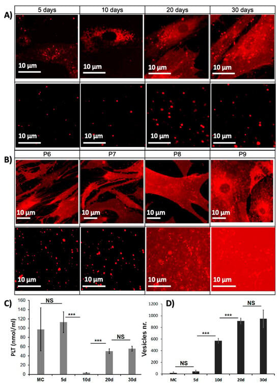
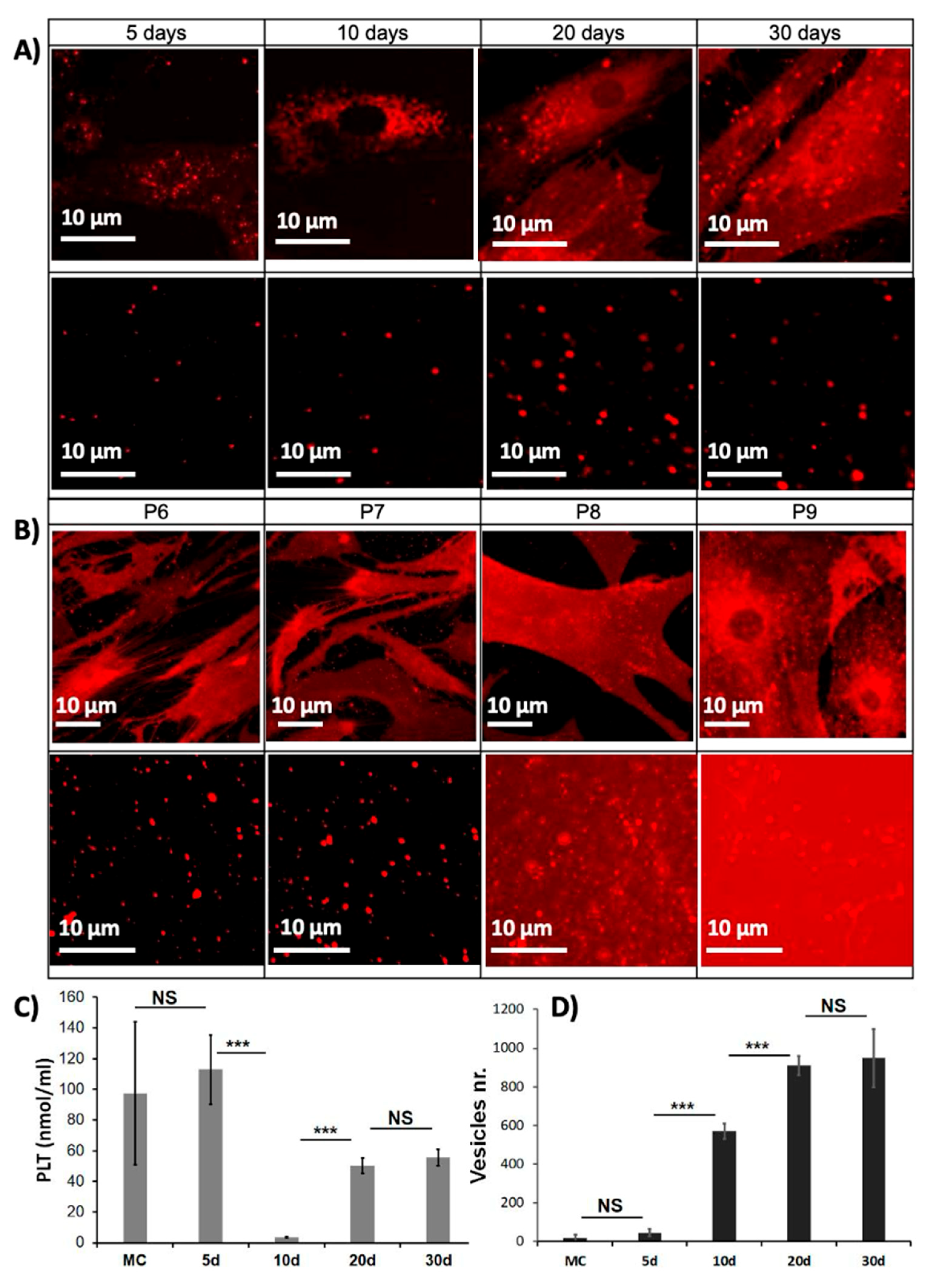
In the original publication [1], there was a mistake in Figure 1 as published. The original figure contains two duplicate figures, initially intended to show that the patients who were tested in the kinetic part (part A of the figure) and those who were tested in the cell passage part (part B of the figure) are the same. However, this duplication caused an error because P6 should be set at 20 days and not 10 days as shown in the original image. The corrected Figure 1 appears below. The authors state that the scientific conclusions are unaffected. This correction was approved by the Academic Editor. The original publication has also been updated.
Figure 1.
Selection of the most appropriate conditions for the structural and mechanical characterization. (A) Cells (top) and supernatants (bottom) confocal micrographs at 5, 10, 20, and 30 days of cell culture (X63 immersion oil microscope objective) after passage 6. (B) Cells (top) and supernatants (bottom) micrographs from passage 6 to 9 each after 20 days. Labeling of the different components was done by lipophilic fluorescent label “DiI” (λex~549 nm). (C) PLT concentration (µg/mL) in the supernatant and (D) number of vesicles per image as a function of cell culture day. MC, medium of culture. Bars represent mean ± SD. *** p < 0.0001, and NS non-significant.
Reference
- Filali, S.; Darragi-Raies, N.; Ben-Trad, L.; Piednoir, A.; Hong, S.-S.; Pirot, F.; Landoulsi, A.; Girard-Egrot, A.; Granjon, T.; Maniti, O.; et al. Morphological and Mechanical Characterization of Extracellular Vesicles and Parent Human Synoviocytes under Physiological and Inflammatory Conditions. Int. J. Mol. Sci. 2022, 23, 13201. [Google Scholar] [CrossRef] [Scilit] [PubMed]
Disclaimer/Publisher’s Note: The statements, opinions and data contained in all publications are solely those of the individual author(s) and contributor(s) and not of MDPI and/or the editor(s). MDPI and/or the editor(s) disclaim responsibility for any injury to people or property resulting from any ideas, methods, instructions or products referred to in the content. |
© 2026 by the authors. Licensee MDPI, Basel, Switzerland. This article is an open access article distributed under the terms and conditions of the Creative Commons Attribution (CC BY) license.