


Abstract
Compensatory mechanisms are thought to maintain sufficient dopamine (DA) signaling to mitigate locomotor impairment during progressive nigrostriatal neuron loss in Parkinson’s disease (PD). Recent evidence indicated augmented DA tissue content in the substantia nigra (SN), not striatum, compensates for tyrosine hydroxylase (TH) and neuronal loss, and alleviates the severity of hypokinesia during neuronal loss. Here, we determined if increased extracellular DA in the SN may also be a compensatory mechanism to augment DA signaling. Following unilateral 6-hydroxydopamine (6-OHDA) lesion or sham-operation, we contemporaneously evaluated extracellular DA against both DA tissue and TH levels in striatum and SN at 7 and 28 days. At 7 days post-lesion, TH loss exceeded ~90% in striatum, and ~70% in the SN. The severity of DA tissue loss coincided with TH protein loss only in striatum (>90%) on both days after lesion, whereas in the SN, DA loss was absent on day 7 and significantly less than TH loss by day 28. Whereas there was a robust increase in extracellular DA in striatum in our sham-operation group, the severe TH and DA tissue loss in striatum practically abolished KCl (K+)-stimulated extracellular DA by day 7. In contrast, whereas striatal K+-stimulation had no effect on extracellular DA in the SN in sham-operation group, extracellular DA levels increased in the SN 7 days after nigrostriatal lesion: an increase no longer apparent by day 28. Thus, despite significant loss of TH protein loss in the SN, extracellular and tissue DA tissue levels were augmented during neuronal loss. These results build upon evidence that compensatory mechanisms to augment DA signaling are not engaged in striatum, and point to the SN as the locus of augmented DA signaling to offset loss of TH during nigrostriatal neuron loss.
1. Introduction
Motor impairment in Parkinson’s disease (PD) first occurs when nigrostriatal dopamine (DA) neuron loss exceeds 50%, coincident with 70–80% loss of tyrosine hydroxylase (TH) in the striatum [1,2,3]. Well after PD diagnosis, 10–50% of neurons remain in the SN, whereas in the striatum, TH and DA transporter proteins are virtually undetectable [3,4]. Paradoxically, motor impairment still increases in severity even though striatal DA markers are undetectable [3,4,5,6]. Therefore, in human PD, the metrics of striatal DA loss are not linear with motor impairment, making our understanding of how nigrostriatal DA signaling affects motor impairment far from complete.
Given that severe striatal TH loss is present at the onset of motor impairment, engagement of compensatory mechanisms in DA signaling have been proposed to maintain motor function despite nigrostriatal neuron loss [7,8,9,10,11,12]. However, current evidence does not point to a compensatory mechanism occurring in striatum to augment DA signaling. Increased striatal DA metabolism and/or turnover occurs only after onset of motor impairment, accompanied by severe TH protein loss in striatum [2,7,11,13,14,15]. Increased DA biosynthesis, by increased TH phosphorylation as a compensatory mechanism, does not occur in striatum [13,14]. While evidence supports that post-synaptic DA receptor expression can upregulate in response to DA depletion [16,17], the increased expression of DA D1 receptors has not been substantiated in striatum [13]. The lack of evidence for dopaminergic plasticity in striatum has led to the idea that non-dopaminergic mechanisms serve as possible compensatory processes, including increased basal ganglia circuit activity and engagement of circuits from the motor cortex or thalamic nuclei [8,18]. Endogenous DA must still be available to drive basal ganglia circuitry. As such, others have reported that extracellular DA levels may increase in response to neuronal loss [19,20,21,22] to compensate against striatal TH protein and DA tissue loss [3,13]. However, it is currently unknown whether DA tissue loss, at the magnitude seen at onset of motor impairment, would directly limit DA release capacity in striatum.
Historically, the loss of striatal DA has been argued to be central to disruption of normal basal ganglia output that produces impoverished motor function. However, an often-overlooked component of DA signaling in the nigrostriatal pathway is also present in the SN, bearing the same regulation steps of DA signaling that are present in striatum [15,23,24,25]. As loss of DA markers within the SN occur at a slower rate compared to the striatum in human PD [2,3,4,5,6,26], it is plausible that remaining cell bodies in SN could be a site of compensatory mechanisms to influence motor function. Indeed, it is plausible that somatodendritic release of DA in the SN as shown in previous studies [25,27,28,29,30,31,32] may drive basal ganglia output to influence motor function through local release in the SN [33,34,35,36,37,38] and subsequent DA binding to post-synaptic DA D1 receptors [15,39,40]. It is also feasible that nigral DA signaling may play a dominant role in regulating motor function when striatal DA deficits are severe, as recently suggested in a mouse PD model featuring loss of mitochondrial complex I [41].
Contralateral or inter-hemispheric compensation may also play a role in maintaining DA signaling and locomotion during striatal DA loss. In fact, the transition to Hoehn–Yahr stage 1 to 2 is characterized by progression from unilateral to bilateral motor impairment. Evidence in both preclinical and clinical literature suggests hemisphere asymmetry in neuronal processes which drive the motor symptom lateralization [42,43]. For example, in unilaterally 6-hydroxydopamine (6-OHDA) lesioned rats, stimulating the contralateral SN increases ipsilateral striatal DA release, whereas stimulating the ipsilateral SN does not [44]. Following unilateral 6-OHDA lesion, reorganization of inter-hemispheric pathways within higher cortical regions may also influence movement [45]. Arguably, the unbalanced degeneration and dysfunction that characterizes PD, at least in early stages, may provide an avenue for therapeutic targeting [46]. However, it is not known exactly how, where, and the extent to which tissue DA and TH protein loss during nigrostriatal neuron loss would affect extracellular DA availability. Understanding the nature of inherent responses that maintain DA signaling, in response to nigrostriatal neuron loss, can reveal which intervention points would be most effective to improve motor function.
We used a unilateral 6-OHDA regimen that produces a progressive loss of nigrostriatal neurons over 28 days, with hypokinesia onset present by day 21 [13,47], to contemporaneously evaluate extracellular DA levels in the striatum and SN under baseline and depolarizing conditions at 7 and 28 days. We previously reported that extracellular DA levels were greatly diminished in lesioned striatum [47] within 7 days after 6-OHDA, replicating the severity of DA and TH loss seen upon PD diagnosis. We now extend this study to evaluate how loss of TH protein and DA tissue levels in striatum and SN would affect extracellular DA levels, ipsilateral or contralateral to lesion, to identify potential compensatory mechanisms against TH protein loss. To answer to this, we included a sham-operation group as a relevant control comparison, and also evaluated if potential sex differences in extracellular DA profiles existed within this group [48,49]. In the SN, wherein TH protein decreases at a slower rate than in striatum [13], we expected progressively diminished DA levels. Our results show that extracellular DA levels were transiently augmented in the DA lesioned group, but only in the SN, not striatum, following depolarizing stimulation in the striatum. This study adds new insight into the emerging evidence that compensatory mechanisms that are engaged in the nigrostriatal pathway during neuronal loss occur selectively in SN, further highlighting a still underappreciated role of DA signaling in SN.
2. Results
2.1. Establishment of Forelimb Akinesia Threshold
Experimental design depicting the double cannulation procedure and collection times of dialysate and tissue is shown in Figure 1. This 6-OHDA hemi-lesion targets the medial forebrain bundle (mfb) to produce progressive TH+ cell loss between 7 (~25%) and 28 (75%) days (Supplementary Figure S1) [13]. The double cannulation procedure affected forelimb usage on the side contralateral to the cannula implant (Supplementary Figure S2). This observation compelled us to report the impact of 6-OHDA on forelimb use as a percentage of baseline (pre-lesion) noted as percent loss of forelimb use, instead of relative to forelimb use associated with the contralateral side of lesion. Average decline in forelimb use was not significantly different amongst the four 6-OHDA-lesioned treatment groups considering (side of cannulation X day post lesion (F(1,18) = 1.33, p = 0.26; Supplementary Figure S3)). The percent loss of forelimb use for each group was as follows (as mean ± SEM); D7 Ipsil, 45 ± 2%; D7 Contra, 47 ± 6%; D28 Ipsil, 48 ± 3%; D28 Ipsil, 39 ± 4%) (Day post lesion F(1,18) = 0.39, p = 0.54; side of cannulation F(1,18) = 0.59, p = 0.45). Notwithstanding the impact of double cannulation on forelimb use, sham-operation itself had no effect on forelimb use at either time point, as seen by no difference in forelimb use on the same side as the side of cannulation (Supplementary Figure S2).
Figure 1.
Experimental design and in vivo microdialysis timeline. Male and female Sprague Dawley rats (n = 45) were acclimated to handling and the forepaw adjusting steps (FAS) test prior to surgery. During surgery, animals received either 6-hydroxydopamine (6-OHDA; n = 21) or sham (vehicle; n = 24) medial forebrain bundle (MFB) lesion. In the same surgery, animals were implanted with striatal and substantia nigra (SN) microdialysis cannulae, either ipsilateral or contralateral, to the MFB lesion or the sham-operation. Cohorts were designated for one-time microdialysis on either day 7 or day 28 post-lesion. During testing, artificial cerebrospinal fluid (aCSF) was perfused through the striatal and nigral cannula and samples were collected every 20 min. Animals underwent 1 h of habituation and 2 h of baseline collection. With 10 min remaining of baseline (170-min timepoint) collection, striatal aCSF was replaced with a potassium chloride (KCl) spiked aCSF to evoke DA release. This infusion remained on for 20 min and replaced with aCSF again at 190 min for the remainder of testing. On day 8 or 29 post-lesion, motor impairments were assessed with the forepaw adjusting steps test (FAS) testing followed by striatal and nigral tissue collection for analysis for DA tissue content and quantification of TH protein.
2.2. TH Expression, Tissue and Extracellular DA Levels in SN and Striatum in Sham-Operated Rats
Sham-operation had no effect on TH protein expression levels in either striatum or SN at either time point (Supplementary Figure S4). Dopamine tissue content was not significantly affected in striatum or SN at either time point (Supplementary Figure S5), with the exception of a modest 30% decrease in the striatum 28 days after sham-operation. There were substantially greater (~10-fold) tissue DA levels in striatum than in SN (Supplementary Figure S5), consistent with previous studies [13,14,15]. Extracellular DA levels at baseline were not affected in the SN or striatum between cannulation sides (ipsil v contra (Day 7, F(1,19) = 1.47, p = 0.24; Day 28, F(1,20) = 0.09, p = 0.77). However, time of aCSF infusion was a factor in the SN on day 7 (time, Day 7, F(5,87) = 2.98, p = 0.016; but not Day 28 (F(5,81) = 0.69, p = 0.63; Supplementary Figure S6A). Across all time points collected during aCSF infusion, extracellular DA levels were lower by ~2-fold in the SN vs. striatum at day 7 (F(1,19) = 8.72, p = 0.008; Supplementary Figure S6A) and day 28 (F(1,20) = 13.2, p = 0.002; Supplementary Figure S6B). This difference was reflected in the average DA levels at day 7 ((F(3,20) = 32.0, p < 0.0001; Figure 2A) and day 28 ((F(3,19) = 41.8, p < 0.0001; Figure 2B) post-sham-operation. Notably, the disparity in DA levels between SN and striatum is far greater at the tissue than the extracellular level. At day 7 only, DA levels increased in the SN, ipsilateral to the sham-operation, which we attribute to a transient response to tissue injury from the cannulation of the SN.
Figure 2.
Baseline extracellular dopamine levels in substantia nigra and striatum at days 7 and 28 in sham group. (A,B). Extracellular dopamine (DA) levels were greater in the striatum compared with substantia nigra (SN) post-sham-operation (sham-op) at day 7 (A) and day 28 (B). (A,B). Average extracellular DA levels, Striatum v. SN A. Day 7. Average extracellular DA levels were significantly different between striatum and SN in both hemispheres relative to sham-op; ipsilateral (I) (t = 4.47, ### p = 0.0009, df = 20), contralateral (C) (t = 7.98, #### p < 0.0001, df = 20). In the SN, there was also a significant difference in DA between the hemispheres, with extracellular DA levels ipsilateral to sham-op greater than contralateral side (t = 4.24, ** p = 0.002, df = 20). (B). Day 28 Average extracellular DA levels were significantly different between striatum and SN in both hemispheres respective to lesion; ipsilateral (t = 7.74, #### p = 0.0009, df = 19), contralateral (t = 7.89, #### p < 0.0001, df = 19). Closed triangles represent data from the substantia nigra (SN) and open triangle represent data from striatum (Str). ns = not significant.
Following infusion of K+ into striatum, extracellular DA levels increased in striatum at both the day 7; BL v K+ (F(1,10) = 37.0, p < 0.0001), time of infusion x BL v K+ (F(5,39) = 22.1, p < 0.0001) and time of infusion x BL v K+ x side of infusion (F(5,39) = 2.5 p = 0.045; Figure 3A) and day 28; BL v K+ (F(1,10) = 34.9, p < 0.0001) time of infusion x BL v K+ (F(5,42) = 17.5, p < 0.0001; Figure 3C) and time of infusion x BL v K++ x side of infusion (F(5,42) = 0.45 p = 0.81; Figure 3B). However, at the same time in the SN, K+-infusion in striatum had no effect on extracellular DA either at day 7; BL v K+ (F(1,10) = 2.7, p = 0.13), time of infusion x BL v K+ (F(5,43) = 1.00, p = 0.43), and time of infusion x BL v K+ x side of infusion (F(5,43) = 2.36 p = 0.056; Figure 3B) or day 28; BL v K+ (F(1,10) = 1.6, p = 0.24), time of infusion x BL v K+ (F(5,27) = 1.31, p = 0.29), and time of infusion x BL v K+ x side of infusion (F(5,27) = 1.13, p = 0.36) after sham-operation (Figure 3D).
Figure 3.
Timeline of K+-stimulated increase in extracellular dopamine levels in striatum and substantia nigra in sham group. (A,B). Day 7 after sham-operation. A. Striatum. There was a highly significant interaction between time past infusion and baseline (BL) vs. K+ on extracellular dopamine (DA) levels occurring either ipsilateral or contralateral to the side of sham-operation (F(5,39) = 22.1, p < 0.0001), with a 15- to 35-fold increase in extracellular DA that was maintained out to at least 40 min after initiating K+ infusion. Significant differences in extracellular DA at 20 min (t = 4.65, *** p = 0.0002, df = 20) and 40 min (t = 5.28, *** p< 0.0001, df = 19), but not 60 min (t = 1.49, p = 0.15, df = 19). (B). SN. Striatal infusion of K+ 7 days after operation had no effect on nigral extracellular DA levels and there was no significant interaction between time of infusion and BL vs. K extracellular DA levels occurring either ipsilateral or contralateral to the side of sham-operation (F(5,43) = 1.00, p = 0.43). (C,D). Day 28 after sham-operation. (C). Striatum. There was a highly significant interaction between time into infusion and BL vs. K+ extracellular DA levels occurring either ipsilateral or contralateral to the side of sham-operation 28 days after operation (F(5,42) = 17.5, p < 0.0001), with a 15- to 25-fold increase in extracellular DA, maintained out to at least 40 min after initiating K+ infusion. Significant differences in extracellular DA at 20 min (t = 4.81, *** p = 0.0001, df = 20) and 40 min (t = 7.38, *** p < 0.0001, df = 20), but not 60 min (t = 1.71, p = 0.10, df = 20). (D). SN. Striatal infusion of K+ 28 days after operation had no effect on nigral extracellular DA and there was no significant interaction between time of infusion and BL vs. K+ extracellular DA levels occurring either ipsilateral or contralateral to the side of sham-operation (F(5,27) = 1.13, p = 0.29).
By 60 min after K+-infusion, the increase in extracellular DA seen in striatum returned to baseline levels (Figure 3A,C). The side of double cannulation relative to sham-operation (ipsilateral or contralateral) had no influence on striatal DA levels (day 7, (F(1,39) = 3.0, p = 0.091); day 28 (F(1,42) = 0.01, p = 0.91) or nigral DA levels (day 7, (F(1,43) = 2.2, p = 0.15); day 28 (F(1,27) = 0.07, p = 0.79). Together, these results confirm that in the absence of nigrostriatal lesion, basal extracellular DA levels are ~2-fold greater in striatum vs. SN, and that the depolarizing influence of striatal K+-infusion to increase extracellular DA is restricted to the striatum.
2.3. Determination of Sex Differences in Extracellular DA Under Baseline and K+-Infusion
Potential sex differences in extracellular DA were evaluated in the sham-operation group. Extracellular DA levels in the striatum were not affected by day after sham-operation (F = (1,19) = 0.07, p = 0.79), nor were there interactions with the other two variables (dialysate collection time or sex). Collapsing the results from both days after sham-operation, we did not find any significant differences due to sex (F = (1,21) = 1.02, p = 0.32), or interaction with time after dialysate collection (F = (5,91) = 0.75, p = 0.59). Given no effect of time of dialysate collection, results from each rat were averaged. No sex differences existed in striatum for extracellular DA levels (Supplementary Figure S7A). In the SN, extracellular DA levels were not affected by day after sham-operation (F = (1,20) = 1.92, p = 0.18), nor were there interactions with the other two variables (dialysate collection time or sex). Collapsing the results from both days after sham-operation, we did not find any significant differences due to sex (F = (1,22) = 3.48, p = 0.075), or interaction with time after dialysate collection (F = (5,98) = 0.82, p = 0.54). Comparison of average extracellular DA levels between sexes, we found no significant differences between sexes in the SN (Supplementary Figure S7B).
Following K+-infusion, DA levels in the striatum were not affected by day after sham-operation (F = (1,19) = 0.30, p = 0.59), nor were there interactions with the other two variables (dialysate collection time or sex). Collapsing the results from both days after sham-operation, we did not find any significant differences due to sex (F = (1,21) = 1.29, p = 0.27), or interaction with time after K+-infusion (F = (2,39) = 0.61, p = 0.55), although there was, expectedly, an effect of time past K+-infusion (F = (2,39) = 35.4, p < 0.0001) (Supplementary Figure S7C). In the SN, DA levels in the striatum were not affected by day after sham-operation (F = (1,18) = 3.00, p = 0.10), though there was an interaction between time past K+-infusion and sex (F = (2,31) = 6.35, p = 0.005). Collapsing the results from both days after sham-operation, time past K+-infusion was significant (F = 2,34) = 5.02, p = 0.012), and although no significant differences were seen between sexes (F = (1,20) = 2.12, p = 0.16), there was significant interaction between time after K+-infusion and sex (F = (2,34) = 6.02, p = 0.006) due to differences isolated at day 28 post sham-operation, specifically and only at 40 min past K+-infusion (Supplementary Figure S7D).
2.4. Lesion Impact on TH Protein Expression and DA Tissue Content
The 6-OHDA lesion predictably produced major TH protein loss in both striatum and SN, but with differential impact in these regions. Loss in striatum exceeded 86% by day 7, reaching 97% by day 28 (F(1,16) = 133, p < 0.0001), although there was no effect of time post-lesion in TH quantity in either contralateral or ipsilateral to lesioned side (F(1,16) = 1.24, p = 0.28; Figure 4A). In the SN, TH loss was ~70% by day 7 (F(1,15) = 24.9, p = 0.0002), with no further decrease by day 28 (F(1,19) = 0.14, p = 0.72; Figure 4B).
Figure 4.
Loss of TH protein expression vs. DA tissue loss in striatum vs. SN during lesion progression. (A,B), TH protein loss, (C,D), DA tissue loss A. Striatum TH. There was substantial TH protein loss after 6-OHDA lesion induction at 7 (t = 6.0, **** p< 0.0001, df = 19, (86%)) and 28 days (t = 8.3, **** p< 0.0001, df = 15 (97%)). No difference in TH protein was observed between days post lesion for either the contralateral ((contra) t = 0.17, p = 0.87, df = 19) or ipsilateral ((ipsil) t = 1.87, p = 0.078, df = 17) side to lesion. (B). SN TH. There was TH protein loss after 6-OHDA lesion induction at 7 (t = 2.9, * p= 0.011, df = 19, (74%)) and 28 days (t = 2.8, * p =0.013, df = 14, (72%)). No difference in TH protein was observed between days post lesion for either the contralateral ((contra) t = 0.17, p = 0.87, df = 19) or ipsilateral ((ipsil) t = 1.87, p = 0.078, df = 17) side to lesion. (C). striatum DA. There was substantial (>90%) DA loss after 6-OHDA lesion induction at 7 (t = 9.2, #### p< 0.0001, df = 18), and 28 days (t = 6.0, #### p< 0.0001, df = 19). No difference in DA was observed between days post lesion for either ipsilateral ((ipsil) t = 0.75, p = 0.46, df = 18), or contralateral ((contra) t = 1.26, p = 0.22, df = 18) side to lesion. (D). SN DA. There was no DA loss after 6-OHDA lesion induction at 7 days (t = 0.80, p = 0.43, df = 16), but a 36% reduction at 28 days (t = 2.53, # p = 0.023, df = 15) as compared to the side contralateral to lesion. The loss of DA on the lesioned side at day 28 vs. day 7 was also significantly greater (t = 2.67, * p = 0.019, df = 19).
The 6-OHDA lesion also decreased DA tissue levels, but in a disparate way between striatum and SN, with more severe loss in striatum on both days after lesion. Loss in striatum exceeded 90% by day 7 and did not worsen by day 28 (F(1,18) = 116, p < 0.0001; Figure 4C). However, in the SN, no DA tissue loss occurred at day 7, but by day 28, there was moderate (~30%) DA loss (F(1,13) = 5.8, p = 0.031; Figure 4D).
The magnitude of TH loss in striatum exceeded that in the SN during the course of lesion progression (F(1,30) = 9.6, p = 0.004; Figure 5A,C,D). This differential loss between TH and DA was reported in our recent study [13] and reflects the more rapid rate of striatal TH loss compared to that in the SN in human PD and established PD models [2,3]. The loss of tissue DA levels in SN was far less than that in striatum during the course of lesion progression (F(1,14) = 56.6, p < 0.0001; Figure 5B). Thus, despite significant TH protein loss by day 7, engagement of a compensatory process augmented DA signaling to diminish the impact of TH protein loss on DA levels; however, this disparate degree of DA vs. TH loss was isolated to the SN, whereas comparable loss of TH and DA occurred in striatum.
Figure 5.
(A). Tyrosine hydroxylase protein remaining striatum vs. substantia nigra. Remaining tyrosine hydroxylase (TH) protein trended less loss in SN vs. striatum by day 7 (t = 2.00, p = 0.061, df = 18), and TH protein loss in SN was significantly less by day 28 (t = 3.0, * p = 0.011, df = 12). (B). Dopamine remaining striatum vs. SN. Remaining dopamine (DA) tissue levels were greatly different between striatum vs. SN at both time points, with substantially more remaining in SN at day 7 ((85 v 5.6%), t = 6.4, #### p < 0.0001, df = 17), day 28 ((74 v 11.3%), t = 2.0, ### p = 0.0005, df = 16). (C). Representative blot of striatal TH expression. A total of 4 µg protein (as depicted by Ponceau stain image below TH image) was loaded to quantify total TH protein in lesioned (L) vs. contralateral to lesioned (C) striatum against a calibrated TH protein standard curve, ranging from 0.7 to 2.5 ng total TH protein. TH loss in striatum was severe by day 7, even though this 6-OHDA produces only 25% TH+ cell loss by day 7 after lesion induction [13]. (D). Representative blot of nigral TH expression. A total of 8 µg protein (as depicted by Ponceau stain image below TH image) was loaded to quantify total TH protein in lesioned (L) vs. contralateral (C) to lesioned SN against a calibrated TH protein standard curve, ranging from 0.01 to 2.0 ng total TH protein. TH loss in SN was by day 7, even though this 6-OHDA produces only 25% TH+ cell loss by day 7 after lesion induction (Kasanga et al., 2023) [13].
2.5. Differential Impact of Lesion on Baseline and K+-Evoked Extracellular DA
With the establishment of baseline extracellular DA levels between striatum and SN (Figure 2) and ascertaining that K+-dependent depolarization in striatum increased extracellular DA only in striatum in the sham-operation group (Figure 3), we determined the effect of progressive nigrostriatal neuron loss on BL and K+-evoked extracellular DA levels in the lesioned and contralateral to lesioned striatum and SN.
Baseline extracellular DA levels did not differ between the two time points after lesion, respective to side of lesion, in striatum; ipsil (F(1,53) = 0.00, p = 0.98); contra (F(5,39) = 0.89, p = 0.50), nor in the SN; ipsil (F(1,54) = 0.09, p = 0.75); contra (F(1,39) = 1.03, p = 0.31). However, there were significant differences in baseline extracellular DA levels between the ipsilateral and contralateral sides relative to lesion in the striatum and SN at day 7 (striatum, (F(2,69) = 11.8, p < 0.0001; Supplementary Figure S8A); SN, (F(2,67) = 3.9, p = 0.026; Supplementary Figure S8B) and in only in striatum at day 28 post lesion (striatum, (F(2,69) = 17.2, p < 0.0001; Supplementary Figure S8C); SN, (F(2,68) = 0.96, p = 0.39; Supplementary Figure S8D). Specifically, in striatum, on day 7 after 6-OHDA lesion, extracellular DA levels in the side contralateral to lesion were increased compared to the sham-operated group (q = 3.98, p = 0.017) and to a greater extent compared to striatum ipsilateral to lesion (q = 6.75, p < 0.0001). Given the differences in baseline levels in striatum between contralateral to lesion v sham-operated group, we evaluated the first 60 min following 20 min of K+-infusion across both time periods after lesion or sham-operation. There was a substantial effect of time after infusion (F (2,18) = 50.8, p < 0.0001), but time after lesion or sham-operation was not a significant factor (F (1,9) = 1.0, p = 0.34), nor were there differences between extracellular DA levels contralateral to lesion v sham-operation group, although there was a notable trend toward an increase in striatum contralateral to lesion (F (1,9) = 4.1, p = 0.073).
Following K+-infusion 7 days after nigrostriatal lesion, extracellular DA levels increased in the striatum contralateral to lesion (F(1,8) = 61.2, p < 0.0001), time of infusion x lesion (F(11,83) = 25.0, p < 0.0001; Figure 6A). Extracellular DA was 30-fold greater than baseline conditions in the striatum contralateral to lesion within the first 60 min after K+-infusion (F(1,22) = 52.1, p < 0.0001; Figure 6B). In contrast, on the lesioned side, differences in extracellular DA versus baseline after K+ were diminished >90%, with only a modest 2-fold difference above baseline within the first 60 min after K+-infusion (F(1,22) = 8.6, p = 0.012; Figure 6C), and no significant difference at any 20 min interval. These disparities were similar to the stark loss in DA tissue levels resulting from lesion. These results indicate that a compensatory response to augment extracellular DA levels does not exist to counteract severe DA tissue loss, and that tissue DA levels exert control over extracellular levels.
Figure 6.
Depolarization-stimulated dopamine levels, 7 days post-lesion, aCSF v K+ matched comparison. Striatum (A–C), Substantia nigra (D–F). Striatum. (A). K+-evoked extracellular dopamine (DA) levels were intact in the striatum, contralateral to lesion, increasing ~30-fold within the first 20 min after K+-infusion. However, in striatum ipsilateral to lesion, K+-evoked extracellular DA levels were virtually eliminated (Ipsil vs. contra, (F(1,8) = 61.2, p < 0.0001)). (B). Contralateral to lesion. 20 min (t = 6.87, **** p < 0.0001); 40 min (t = 5.39, **** p < 0.0001); 60 min (t = 0.01, p = 0.99). (C). Ipsilateral to lesion. 20 min (t = 2.68, p = 0.059); 40 min (t = 2.33, p = 0.115); 60 min (t = 0.08, p = 0.99). SN. (D). K+-evoked extracellular DA levels increased following striatal K+-infusion were restricted to the SN, ipsilateral to lesion (Ispil vs. contra (F(1,9) = 5.56, p = 0.043)). (E). Contralateral to lesion. No significant differences in extracellular DA were observed at any time point after striatal K+-infusion. (F). Ipsilateral to lesion. 20 min (t = 5.25, *** p = 0.0008); 40 min (t = 2.28, p = 0.132); 60 min (t = 1.70, p = 0.35).
In the SN, there were significant differences between the lesioned and contralateral to lesion SN extracellular DA following striatal K+-infusion (F(1,9) = 5.6, p = 0.043; Figure 6D). However, the differences were opposite to those seen contemporaneously in the striatum. Similar to the lack of effect of K+-infusion in the SN of the sham-operation group, extracellular DA levels in the SN contralateral to lesion were not different between baseline and after striatal K+-infusion (F(1,8) = 2.41, p = 0.157; Figure 6E). However, in the SN ipsilateral to lesion, there were highly significant increases in extracellular DA in the SN after K+-infusion in striatum (F(1,11) = 27.9, p = 0.0003; Figure 6F), with extracellular DA increasing by ~7-fold within the first 20 min.
By 28 days post-lesion, following striatal K+-infusion, extracellular DA levels still increased substantially in the striatum, contralateral to lesion compared to lesioned side (F(1,8) = 82.9, p < 0.0001; Figure 7A). Notably, extracellular DA was still 30-fold greater than baseline conditions, contralateral to lesion, within the first 60 min after K+-infusion (F(1,8) = 91.6, p < 0.0001; Figure 7B). In contrast, in the ipsilateral lesioned side, differences in extracellular DA versus baseline after K+ were still diminished by >90%, with no significant difference in levels between BL and following K+-infusion (F(1,25) = 3.77, p = 0.064; Figure 7C).
Figure 7.
Depolarization-stimulated dopamine levels, 28 days post-lesion, aCSF v K+ matched comparison. Striatum (A–C), Substantia nigra (D–F). Striatum. (A). K+-evoked extracellular dopamine (DA) levels were intact in the striatum, contralateral to lesion, increasing ~30-fold within the first 20 min after K+-infusion. However, in striatum ipsilateral to lesion, K+-evoked extracellular DA levels were virtually eliminated. Ipsil vs. contra (F(1,8) = 82.9, p < 0.0001). (B). Contralateral to lesion. 20 min (t = 10.3, **** p < 0.0001); 40 min (t = 6.69, *** p = 0.0005); 60 min (t = 0.01, p = 0.99). (C). Ipsilateral to lesion. 20 min (t = 0.97, p = 0.99); 40 min (t = 2.19, p = 0.113); 60 min (t = 0.04, p = 0.99). Substantia nigra. (D). K+-evoked extracellular DA levels did not change following striatal K+- infusion in either side relative to lesion (F(1,9) = 0.20, p = 0.67). (E). Contralateral to lesion. No significant differences in extracellular DA were observed at any time point after striatal K+-infusion. (F). Ipsilateral to lesion. No significant differences in extracellular DA were observed at any time point after striatal K+-infusion.
In the SN, by day 28 post-lesion, the significant differences between the lesioned and contralateral to lesion following striatal K+-infusion seen at day 7 were no longer apparent (F(1,8) = 0.20, p = 0.67; Figure 7D). Extracellular DA levels in the SN contralateral to lesion were not significantly different between baseline and after striatal K+-infusion (F(1,8) = 1.64, p = 0.24; Figure 7E). On the lesioned side, unlike at day 7, there was no longer a difference between extracellular DA levels at baseline vs. that following K+-infusion (F(1,12) = 0.94, p = 0.35; Figure 7F).
To compare across the two time points after lesion, responses to K+ in striatum and SN ipsilateral to lesion were compared on day 7 vs. day 28. There were no differences in extracellular DA levels within the first 60 min after striatal K+-infusion between the day 7 and day 28 cohorts (F(1,25) = 0.66, p = 0.42; Figure 8A), suggesting there was little change in the major decrease in extracellular DA during nigrostriatal lesion progression. However, in the SN, the difference in depolarization-stimulated DA levels as lesion progressed was substantial, with significantly greater DA levels after striatal K+-infusion early (day 7) versus late after lesion (day 28) (F(1,25) = 22.0, p < 0.0001; Figure 8B).
Figure 8.
Compensatory increase in extracellular dopamine is transient and restricted to the substantia nigra. (A). Striatum. There was no significant difference in extracellular dopamine (DA) in striatum after depolarizing stimulation therein as a function of time past lesion induction. (B). Substantia nigra. There is a transient increase in extracellular DA in the substantia nigra (SN) after depolarizing stimulation, as a function of time past lesion induction. 20 min (t = 4.19, *** p = 0.0003); 40 min (t = 2.34, * p = 0.027); 60 min (t = 1.55, p = 0.13).
2.6. Differential Impact of Lesion on Baseline and K+-Evoked Extracellular DOPAC
To analyze whether differences in DA turnover factored into the differences in extracellular DA observed between the two time points in the SN, we evaluated extracellular DOPAC levels and found that there was diminished DOPAC at the 28 day timepoint in the side contralateral to lesion (F(1,15) = 4.62, p = 0.047) (Supplementary Figure S10A). In contrast, time past lesion was not a factor in extracellular DOPAC in the striatum (F(1,15) = 0.53, p = 0.47) (Supplementary Figure S10B). In striatum, the side of 6-OHDA lesion substantially decreased DOPAC on both days post-lesion ((F(1,15) = 48.6, p < 0.0001) (Supplementary Figure S10B). Notably, the levels of DOPAC in striatum contralateral to lesion were not significantly different vs. sham-operated control group (F(1,15) = 0.28 p = 0.60).
3. Discussion
It has been well established that the motor impairments driven by nigrostriatal neuron loss in PD do not occur until there is ~50% loss of these neurons, which is associated with 80% loss of TH protein in the striatum [2,3,6,10]. Compensatory mechanisms have been thought to be engaged to augment DA signaling in the striatum against neuron or TH protein loss [7,8,11,12]; this includes augmentation of DA uptake through catecholamine transporters other than the DA transporter [50,51]. Our results now show that extracellular DA levels are not augmented when TH protein loss in striatum exceeds 80%, even under depolarizing conditions. Notably, loss of extracellular DA under depolarizing conditions was commensurate with drastic loss of tissue DA and TH protein. We recently reported that TH phosphorylation at either ser31 or ser40 did not increase in striatum at any time point after lesion induction [13]. Thus, striatal TH protein loss and the lack of any increase in its activity through phosphorylation, appears to dictate the limits of DA signaling in remaining terminals in striatum. In contrast, both extracellular and tissue DA levels in the SN did not coincide with significant TH protein loss therein, suggesting that both post-translational modification of TH [13,14,52], likely through increased ser31 TH phosphorylation [13], and DA release capacity were augmented to diminish the impact of TH protein loss on DA signaling in the SN. Perhaps most strikingly, this increase in extracellular DA in the SN following depolarizing stimuli occurred only during the earlier stages of progressive nigrostriatal neuron loss. This indicates that compensatory mechanisms to augment DA signaling in the nigrostriatal pathway during progressive neuronal loss occur at the tissue and extracellular level only in the SN and not striatum, even though the origin of depolarizing stimulation occurred in striatum. Finally, we point out that hypokinetic behavior in this 6-OHDA model does not occur until 21 days after lesion induction and is maintained at 28 days [13]. Thus, the eventual loss of tissue and extracellular DA by day 28, but not day 7, is consistent with the idea that eventual loss of nigral DA compensation plays a major role in onset of hypokinesia.
Our results show that in addition to SN-restricted increased DA biosynthesis (through increased ser31 TH phosphorylation) during nigrostriatal neuron loss [13], plasticity in DA signaling therein extends to regulation at the extracellular level in SN, with a transient increase in synaptic levels of DA. Importantly, this increase only arises from depolarizing stimulating in the lesioned striatum, suggesting that the trigger to increase DA levels in the SN results from alterations in basal ganglia circuitry that begin in the striatum resulting from major DA loss therein. Augmentation of DA signaling is thought to prevent onset of parkinsonian signs until there is at least 70–80% loss of TH or dopamine transporter (DAT) in the striatum [4,8,10,11,12]. However, in the striatum, DA tissue levels decrease to the same magnitude as TH protein [13,14], suggesting no augmentation of DA biosynthesis therein when TH loss is at the magnitude seen at PD diagnosis [3,53]. In the current study, we found essentially the same outcome in that comparable TH protein and DA tissue loss severely limits a possible compensatory increase in extracellular DA. It is plausible, by virtue of our study design, that we may have missed augmented extracellular DA levels earlier than 7 days after lesion induction. That said, our data indicate that possible enhancement of DA release does not occur when TH protein loss meets the threshold of loss that occurs at PD diagnosis. Whereas there were robust increases in DA in the striatum under depolarizing conditions in the sham group and contralateral to lesion in the 6-OHDA group, there was a decrease in extracellular DA at the baseline which was greatly magnified under depolarizing conditions; in fact, DA levels were essentially no different (~1.5-fold) than levels at baseline after K+-infusion. We also did not find a significant difference in extracellular DA after depolarizing stimulation in the striatum contralateral to lesion compared to sham-operation group at either time point, indicating that the slight increase in baseline levels did not augment potential DA release capacity therein. This new finding suggests that if release capacity was augmented, as previously reported [19,22], it appears that the inherent DA tissue content in striatum ultimately dictates extracellular levels, including release capacity. Thus, even if DA release mechanisms were autonomously affected from biosynthesis or vesicular packaging, the increase would be minimal, potentially far below the levels needed for normal motor function.
The source of elevated levels of extracellular DA in the SN may be explained by changes in basal ganglia circuitry induced by the loss of nigrostriatal DA [8,54,55]. The neuronal pathways that comprise the indirect pathway include the final neuronal pathway which targets the SN, originating from the subthalamic nucleus (STN); the subthalamonigral pathway. This glutamatergic pathway innervates the GABAergic output neurons from the SN pars reticulata (SNr) and becomes overactive following the loss of DA in striatum [56,57,58]. As a result, there is an increase in the tonic inhibitory output of the basal ganglia by the STN, which may also overexcite the remaining nigrostriatal neurons, leading to increased DA release. Several lines of evidence indicate that this pathway does innervate the nigral DA neurons [59,60], and extracellular glutamate levels increase in the SN in a mouse PD model [61]. A glutamatergic basis for augmented tissue and extracellular DA in the SN may be initiated from the influence of depolarizing stimulation arising from the STN [62]. In response to depolarizing stimulation, modulation of glutamate receptors or its uptake, Ser19 TH phosphorylation levels change in accordance with the direction of glutamatergic modulation or depolarization [63,64,65,66]. In fact, ser19 TH phosphorylation levels increase in the SN within a week after the induction of 6-OHDA lesion [14]. Therefore, the source of increased extracellular DA and tissue DA, despite TH protein loss, may arise from overactive glutamate release from the subthalamic nucleus. Furthermore, while it is unlikely storage capacity is increased in the striatum [53], DA vesicular storage may slightly increase in the SN for somatodendritic release early post-lesion, resulting in the increase of release during striatal K+-infusion seen here. Future assessments of this circuit change could incorporate an analyses of vesicular monoamine transport 2 (VMAT2) levels, which could indicate if an upregulation of storage is occurring, in addition to increased DA synthesis and release [67].
Lastly, cross-hemispheric collaterals are an interesting component in disease models. In the present data we observed significant increases in contralateral striatal DA at baseline and following depolarizing stimulation, suggesting an intact contralateral side untouched by our MFB lesion. Work assessing cross-hemispheric projections from the SN to the striatum in a 6-OHDA lesioned model suggests a portion of these contralateral projections that survive may protect against certain medication related side effects such as L-DOPA-induced dyskinesia [68]. Knowing this, targeting these surviving neurons and respective projections may provide additional compensation for motor dysfunction. Indeed, our results indicate DA catabolism is reduced at the latter stages of nigrostriatal neuron loss, by way of less extracellular DOPAC in the SN, contralateral to lesion, 28 days after 6-OHDA lesion induction.
Limitations
This is the first study to our knowledge that has four coincident measures of DA signaling within the same tissues, making it possible to determine where compensatory mechanisms might be engaged, evidenced by inconsistencies in the magnitude of loss of TH protein, DA tissue content, extracellular DA, and DA catabolism in striatum and SN. However, given the nature of the experimental design to ascertain extracellular striatal and SN DA levels under both BL and K+-infusion, we were unable to collect tissue at the time of K+-striatal infusion. Thus, it remains unknown whether tissue DA levels would be further augmented along with extracellular DA under depolarizing conditions. It is established that depolarizing stimuli can augment DA biosynthesis [66,69]. Notably, the quantitation of TH protein in our study matches previously reported levels in striatum and SN [13,14,15,70], adding an additional degree of certainty that changes in extracellular DA levels would also be consistent across in vivo studies. Despite that we did not observe any increase in extracellular DA in striatum after lesion induction by day 7, it is plausible that extracellular DA levels may be less influenced by DA tissue or TH protein loss occurring earlier during neuron loss. It would be important to determine if there is a breaking point between the magnitude of TH protein loss and any increase in DA release potential, which could identify a compensation mechanism in striatum operational at least in the very early stages of neuronal loss. Another limitation of our study is that microdialysis does not capture DA release and uptake dynamics as does voltametric methods (fast-scan cyclic voltammetry) [71]. Capturing release and DA uptake dynamics during nigrostriatal neuron loss would further enable us to interpret, for example, how much of the increase in extracellular DA in the SN early after lesion at 7 days is due to augmented DA release vs. decreased DA uptake.
An additional limitation of our study is that open-field locomotor data was not collected. Thus, with only forelimb step data, we do not have the full scope of interrogation of motor function. This is critical for comparison of our nigral DA data, as our previous study using the same 6-OHDA model did show significant correlation between nigral DA tissue loss and the onset of hypokinesia; notably, this study showed deficits in forelimb use were critical for the eventual onset of hypokinesia [13]. Another limitation is that we did not ascertain cognitive function, as possible non-motor effects may have been present as deficits in executive function, particularly in the earliest stages of DA loss [72,73]. Finally, in the sham-operation group, depolarizing stimulation was associated with an increase in extracellular DA in the SN that was isolated at 28, but not 7, days after sham-operation in the female rats. Even more unexpected was that this increase occurred only 40 min after striatal K+-infusion. We speculate that this may be a delayed response to tissue injury, but it is interesting that this increase was seen only in females, which have resiliency against perturbations in DA loss [48,49]. While not the subject of our study, this preliminary finding may be a piece of evidence of why females are at less risk for PD, with inherent resiliency against CNS insults not necessarily specific to nigrostriatal neurons.
4. Materials and Methods
4.1. Animals
Upon arrival, adult male and female Sprague Dawley (n = 52; 30 F, 22 M) rats were housed in reverse light/dark cycle (lights on at 3:00 p.m., off at 3:00 a.m.) in a temperature-controlled room (22–23 °C). Initially, animals were pair housed in plastic cages (22 cm high, 45 cm deep, and 23 cm wide) with ad libitum food (Rodent Diet 5001; Lab Diet, Brentwood, MO, USA) and water. To ensure sufficient habituation to experimenter handling, animals were handled at least 2 times for 5-min periods before any surgery or experimentation. After surgeries, animals were ~5 months old and were single housed to prevent housemates from chewing on in-dwelling cannulae. Rats were maintained in accordance with the guidelines of the Institutional Animal Care and Use Committee of Binghamton University and the “Guide for the Care and Use of Laboratory Animals, 8th edition”, Institute for Laboratory Animal Research, National Academies Press, 2011 and the Animal Care and Use Review Office of the Department of Defense, protocol PD180098.e002.
4.2. Surgical Procedures
As shown in Figure 1, all rats received either a unilateral sham (0.9% NaCl + 0.1% ascorbic acid) or 6-OHDA (Sigma, St. Louis, MO, USA) lesion to the left medial forebrain bundle (mfb) to deplete striatal DA [13,74]. To do so, rats were anesthetized with inhalant isoflurane (2–3%; Sigma) in oxygen (2.5 L/min) and placed in a stereotaxic instrument (David Kopf Instruments) with the incisor bar positioned at 5 mm below the interaural line. A 10-µL Hamilton syringe attached to a 26-gauge needle was lowered into the site (AP: −1.8 mm; ML: +2.0 mm; DV: −8.6 mm, relative to bregma). Either vehicle or 6-OHDA (3 μg/μL) was infused at a rate of 2 µL/min for a total volume of 4 µL. The needle remained in the injection site for 5 additional min before removal to allow for optimal diffusion. This 6-OHDA hemi-lesion approach, by targeting the mfb at the same coordinates, produces progressive TH+ cell loss between 7 (~25%) and 28 (75%) days (Supplementary Figure S1) [13].
During the same surgery, rats received a guide cannula (CMA 12, CMA Microdialysis, Holliston, MA, USA) lowered into the striatum (ML: ±2.5; AP: +1.0; DV: −3.5) and substantia nigra pars compacta (ML: ±2.5; AP: −5.7; DV: −7.0) and secured to the skull with surgical screws (Plastics One, Roanoke, VA, USA) and Jet Denture Repair Acrylic (Lang Dental). To assess potential contralateral effects, we counterbalanced the placement of the guide cannula with half of the rats receiving cannula placements ipsilateral to lesion and the other half receiving them contralateral to lesion. Dummy cannulae were placed in the guide to maintain patency during the recovery period. All animals that underwent surgery received one pre-operative injection of the analgesic buprenex (Buprenorphine HCl; 0.03 mg/kg, i.p; Reckitt Benckiser Pharmaceuticals, Slough, UK) and 2 post-operative injections of carprofen (5 mg/kg, s.c.; Zoetis, Inc, Fort Worth, TX, USA) 6–12 h apart to reduce pain and discomfort.
4.3. Forepaw Adjusting Steps Test
To ascertain motor impairment in relation to the duration of lesion (or progressive nigrostriatal neuron loss as previously shown [13], we evaluated use of forelimbs. Prior to surgical intervention, all animals underwent 2 days of habituation to the forepaw adjusting steps test (FAS) followed by baseline FAS measures. Briefly, an experimenter held both back legs and one front paw, forcing weight onto the free paw. Rats were then moved laterally across a table’s surface at a rate of 90 cm/10 s. In total there are six trials for each paw: three trials for the forehand movement and three trials for the backhand movement. At either Day 8 or Day 29 post-lesion, FAS was done to assess post-lesion motor deficits. This test is sensitive to predict eventual 6-OHDA induced hypokinesia [13,75,76] and is valuable as an assay to approximate extent of striatal DA nigrostriatal lesion.
4.4. In Vivo Microdialysis
To examine the effects of nigrostriatal 6-OHDA lesion timing on extracellular DA release dynamics, rats underwent in vivo microdialysis procedures at Day 7 or 28 post-lesion (Figure 1). On test days during the rats’ dark cycle (from 7:00 a.m.–1:00 p.m.), microdialysis probes (CMA 12 Elite probe) were inserted into targeted striatal and nigral sites. Probes for the striatal site extended 3 mm beyond the cannula tip (molecular weight cut-off 100 kDA) and the nigral probes extended 1 mm beyond (molecular weight cut-off 20 kDA). In both sites, artificial cerebral spinal fluid (aCSF; 147 mM NaCl, 2.8 mM KCl (K+), 1.2 mM CaCl2, 1.2 mM MgCl2, pH 7.40 ± 0.02, sterile filtered) were perfused with a syringe pump (model 400, CMA Microdialysis) at 2 μL/min. Rats underwent 1 h habituation collection followed by a 2 h baseline (BL) collection period. At 170 min into microdialysis, potassium-chloride (K+) spiked aCSF (47.7 mM NaCl, 100 mM K+, 1.2 mM CaCl2, 1.2 mM MgCl2) was infused into the striatal site to evoke DA release. This K+-infusion remained on for 20 min, after which it was removed and replaced with aCSF again for the remainder of testing [77]. Dialysate samples were taken every 20 min for 5 h and stored at −80 °C until analysis. After a 24 h washout, brains were flash frozen in cold 2-methylbutane (EMD Millipore) for later tissue and protein analysis.
4.5. Tissue Processing
Following microdialysis, striatal and nigral tissue were flash frozen for later dissection for quantification of DA tissue and TH protein levels according to established methodology [13,14,15,70]. Fresh-frozen tissues were sonicated in ice-cold perchloric acid solution and centrifuged to segregate precipitated protein from the supernatant. The supernatant was analyzed in-house by HPLC (UltiMate 3000, Thermofisher Scientific, West Palm Beach, FL, USA) for DA (cat #H8502, Millipore Sigma, St. Louis, MO, USA), dihydroxyphenylacetic acid (DOPAC) (cat #11569, Millipore Sigma, St. Louis, MO, USA) for assessment of DA turnover. A standard curve, ranging from 1.56 to 800 ng/mL was used to quantify DA and DOPAC. The resulting values for tissue DA were calculated to total recovery in the sample and normalized against total protein recovered.
The precipitated protein was sonicated in 1% sodium dodecyl sulfate solution and aliquots were assayed for total protein by the bicinchoninic acid method against an albumin standard curve. Sample buffer with dithiothreitol as a reducing agent was then added to the sonicated samples for SDS-PAGE for quantitative Western blot assessment of TH protein [13,70]. The samples for all treatments and groups were represented and balanced within each blot, so that individual differences in treatments were captured within each blot.
Tyrosine hydroxylase protein was quantified against a calibrated standard traceable over multiple studies in rats and mice [13,14,38,64,65,66,70,78]. Optimal total protein load for TH determination in striatum and SN was 4 and 8 µg, respectively, using the primary antibody obtained from Millipore (cat #AB152, Temecula, CA, USA).
4.6. Statistics
We used GraphPad Prism (v. 10) for statistical analyses. For motor assessment, in the sham-operation group, forelimb use was evaluated using an unpaired t-test to determine whether cannulation itself affected use of the forelimb. In the 4 6-OHDA groups (2 time points post-lesion, 2 sides (ipsilateral and contralateral to side of lesion) of cannulation), a 2-way ANOVA was used to assess if day post-lesion or side of cannulation affected percent loss of forelimb use against respective baseline (BL) use that was obtained pre-lesion.
For all neurochemical analyses, appropriate statistical approaches were used to determine if significant differences existed among multiple different independent variables, depending upon the comparisons made. These variables included the following: sham-operation, 6-OHDA lesion, impact of time of collecting microdialysate every 20 min, side of cannulation, and aCSF (BL) vs. post K+-infusion conditions. In the sham-operation groups, several statistical methods were used. To analyze TH and DA tissue content in the SN and striatum, a paired t-test was used matching side of sham surgery against respective intact side within each subject. To determine impact of sham-operation on extracellular DA levels, side of cannulation, and time of microdialysate collection on extracellular DA levels, a 3-way repeated measures ANOVA was used, matching time of collection for each subject. To determine impact of K+-infusion in this group, we used a 3-way ANOVA with time after infusion, side of cannulation, and baseline vs. K+ as independent variables. Post-hoc paired t-tests were run with results obtained from the different sides of cannulation collapsed, given minimal to no impact at both day 7 (F(1,39) = 3.00, p = 0.091) and day 28 (F(1,42) = 0.01, p = 0.91) after sham-operation, to evaluate BL vs. K+ differences at each 20 min.
As we used both male and female rats for these studies, we also determined if there were differences in extracellular DA levels in the SN and striatum in the sham-operation group using a 3-way repeated measures ANOVA, matching dialysate collection times for each rat with independent variables being days after sham surgery, sex, and time of dialysate collection. Notably, we did not find that the day after sham surgery was a significant variable in either striatum or SN, either under aCSF or following K+-infusion. We therefore collapsed the days together to evaluate if sex differences existed using repeated measures 2-way ANOVA.
For determination of lesion impact on extracellular DA, tissue DA, and total TH protein in striatum and SN, the results from each rat had to meet two of three inclusion criteria: >30% loss of forelimb use, >70% striatal DA tissue loss, and >70% striatal TH protein loss. Of 28 successfully cannulated rats that underwent 6-OHDA lesion, 7 did not meet the full criterion, likely due to minor variations in stereotactic placement, spread of toxin around the mfb, and individual animal susceptibility to the toxic effects of the dopamine-depleting agent. These rats were excluded from statistical analysis. To ascertain impact of lesion and effect of time past lesion, a repeated measures (using time of microdialysate collection matched to each subject), a 2-way ANOVA was used including both male and female rats in the analyses as we did not find a significant effect of sex in the analyses conducted in the sham-operation groups. This approach was also used to ascertain differences in percent TH and DA remaining in striatum and SN ipsilateral to 6-OHDA lesion against contralateral tissue. After verifying rats met inclusion criterion described above, the Grubb’s test was used to identify any remaining outliers with alpha set at <0.05.
5. Conclusions
Our results support the idea that pre-symptomatic compensation mechanisms, engaged during nigrostriatal neuron loss and long suspected to occur in PD, involve increased DA signaling in the SN. Although DA compensation may have occurred much earlier in striatum after lesion induction in our model, previous work by others has not shown evidence that such compensation occurs prior to motor decline [2,7]. Our study reveals that dopaminergic compensatory mechanisms do exist in the nigrostriatal pathway in the form of increased tissue and extracellular DA levels in the SN, despite substantial TH protein loss; notably none of these are seen in striatum. The contemporaneous collection of extracellular DA from both striatum and the SN enabled time-matched observations of DA regulation during TH protein loss and also enabled comparison against tissue DA and TH protein levels. The severity of DA tissue loss continued to be less than TH protein loss in the SN throughout the time course of our study, whereas increased extracellular DA occurred early, but not late, after lesion induction. As the results represent changes in DA signaling of the entire nigrostriatal pathway, the disparity of TH loss between striatum and SN in our study emulates conditions in human PD, as there is also greater TH loss in striatum than in SN upon PD diagnosis [3]. Our findings also align with confirmation of loss of one component of motor function (forelimb use) against TH protein loss in both striatum and SN. We have shown in previous work that loss of tissue DA in the SN coincides with the onset of hypokinesia, which occurs 2 weeks after forelimb loss [13], and studies in human PD support dopaminergic components in the SN coinciding with motor impairment [4,5,6]. Preclinical results corroborate a critical role for nigral DA in motor function [33,34,35,36,37,38,39,41]. As to the mechanisms that increase DA signaling in the SN, we speculate the well-known changes in basal ganglia circuit function that occurs during DA loss in PD may contribute to engagement of DA compensation in the SN, as we previously reported increased TH phosphorylation in the SN in this 6-OHDA model [13,14], which may arise from increased activity from STN efferents to the SN [56,57,58,59,60,61]. This possibility should be evaluated in future studies. Our results provide a new and important piece of the puzzle to optimize therapeutic strategies; in this case, the therapeutic potential for targeting regions outside the depleted striatum. It is conceivable that therapeutic targeting of DA biosynthesis, release, or turnover in the SN could potentially provide critical levels of DA needed to preserve motor function or delay motor impairment.
Supplementary Materials
The following supporting information can be downloaded at: https://www.mdpi.com/article/10.3390/ijms27093923/s1.
Author Contributions
Study design; M.F.S. and C.B.; Study conduct; A.G., R.M. and W.N. Data collection and analysis; M.F.S., R.M., W.N., A.G. and S.C. Interpretation of results; M.F.S., R.M., C.B. and A.G. Writing manuscript; A.G., M.F.S. Editing manuscript; M.F.S., C.B., A.G. and R.M. All authors have read and agreed to the published version of the manuscript.
Funding
This study was funded by the US Department of Defense U.S. Army Medical Research and Material Command Congressionally Directed Medical Research Program (Award Number W81XWH-19-1-0757). The funders had no role in the study design, in the collection, analysis, and interpretation of data, or writing of the manuscript.
Institutional Review Board Statement
All animals were used in compliance with the Office of Laboratory Animal Welfare guidelines and protocols approved by the Institutional Animal Care and Use Committees and at the University of North Texas Health Science Center, Binghamton University, and the Animal Care and Use Review Office, US Army Medical Research and Development Command (approval codes: IACUC-885-23 (for Binghamton University) and PD180098 (for U.S. Army ACURO 23), approval date: 1 July 2019).
Informed Consent Statement
Not applicable.
Data Availability Statement
The original contributions presented in this study are included in the article/Supplementary Material. Further inquiries can be directed to the corresponding author.
Conflicts of Interest
The authors declare that they have no competing interests relevant to the content of this article, no financial or proprietary interests in any material discussed in this article, or affiliations with or involvement in any organization or entity with any financial interest or non-financial interest in the subject matter or materials discussed in this manuscript.
References
- Bernheimer, H.; Birkmayer, W.; Hornykiewicz, O.; Jellinger, K.; Seitelberger, F. Brain dopamine and the syndromes of Parkinson and Huntington. Clinical, morphological and neurochemical correlations. J. Neurol. Sci. 1973, 20, 415–455. [Google Scholar] [CrossRef] [Scilit]
- Bezard, E.; Dovero, S.; Prunier, C.; Ravenscroft, P.; Chalon, S.; Guilloteau, D.; Crossman, A.R.; Bioulac, B.; Brotchie, J.M.; Gross, C.E. Relationship between the appearance of symptoms and the level of nigrostriatal degeneration in a progressive 1-methyl-4-phenyl-1,2,3,6-tetrahydropyridine-lesioned macaque model of Parkinson’s disease. J. Neurosci. 2001, 21, 6853–6861. [Google Scholar] [CrossRef] [Scilit]
- Kordower, J.H.; Olanow, C.W.; Dodiya, H.B.; Chu, Y.; Beach, T.G.; Adler, C.H.; Halliday, G.M.; Bartus, R.T. Disease duration and the integrity of the nigrostriatal system in Parkinson’s disease. Brain 2013, 136, 2419–2428. [Google Scholar] [CrossRef] [Scilit] [PubMed]
- Furukawa, K.; Shima, A.; Kambe, D.; Nishida, A.; Wada, I.; Sakamaki, H.; Yoshimura, K.; Terada, Y.; Sakato, Y.; Mitsuhashi, M.; et al. Motor progression and nigrostriatal neurodegeneration in Parkinson disease. Ann. Neurol. 2022, 92, 110–121. [Google Scholar] [CrossRef] [Scilit] [PubMed]
- Perlmutter, J.S.; Norris, S.A. Neuroimaging biomarkers for Parkinson’s disease: Fact and fantasy. Ann. Neurol. 2014, 76, 769–783. [Google Scholar] [CrossRef] [Scilit]
- Tabbal, S.D.; Tian, L.; Karimi, M.; Brown, C.A.; Loftin, S.K.; Perlmutter, J.S. Low nigrostriatal reserve for motor parkinsonism in nonhuman primates. Exp. Neurol. 2012, 237, 355–362. [Google Scholar] [CrossRef] [Scilit]
- Blesa, J.; Pifl, C.; Sánchez-González, M.; Juri, C.; García-Cabezas, M.; Adánez, R.; Iglesias, E.; Collantes, M.; Peñuelas, I.; Sánchez-Hernández, J.; et al. The nigrostriatal system in the presymptomatic and symptomatic stages in the MPTP monkey model: A PET, histological, and biochemical study. Neurobiol. Dis. 2012, 48, 79–91. [Google Scholar] [CrossRef] [Scilit]
- Blesa, J.; Trigo-Damas, I.; Dileone, M.; Lopez-Gonzalez del Rey, N.; Hernandez, L.F.; Obeso, J.A. Compensatory mechanisms in Parkinson’s disease: Circuit adaptations and role in disease modification. Exp. Neurol. 2017, 298, 148–161. [Google Scholar] [CrossRef] [Scilit]
- Collier, T.J.; Lipton, J.; Daley, B.F.; Palfi, S.; Chu, Y.; Sortwell, C.; Bakay, R.A.E.; Sladek, J.R.; Kordower, J.H. Aging-related changes in the nigrostriatal dopamine system and the response to MPTP in nonhuman primates: Diminished compensatory mechanisms as a prelude to parkinsonism. Neurobiol. Dis. 2007, 26, 56–65. [Google Scholar] [CrossRef] [Scilit] [PubMed]
- Fearnley, J.M.; Lees, A.J. Ageing and Parkinson’s disease: Substantia nigra regional selectivity. Brain 1991, 114, 2283–2301. [Google Scholar] [CrossRef] [Scilit]
- Pifl, C.; Hornykiewicz, O. Dopamine turnover is upregulated in the caudate/putamen of asymptomatic MPTP-treated rhesus monkeys. Neurochem. Int. 2006, 49, 519–524. [Google Scholar] [CrossRef] [Scilit]
- Zigmond, M.J. Do compensatory processes underlie the preclinical phase of neurodegenerative disease? Insights from an animal model of parkinsonism. Neurobiol. Dis. 1997, 4, 247–253. [Google Scholar] [CrossRef] [Scilit]
- Kasanga, E.A.; Han, Y.; Shifflet, M.K.; Navarrete, W.; McManus, R.; Parry, C.; Barahona, A.; Nejtek, V.A.; Manfredsson, F.P.; Kordower, J.H.; et al. Nigral-specific increase in Ser31 phosphorylation compensates for tyrosine hydroxylase protein and nigrostriatal neuron loss: Implications for delaying parkinsonian signs. Exp. Neurol. 2023, 368, 114509. [Google Scholar] [CrossRef] [Scilit]
- Salvatore, M.F. Ser31 tyrosine hydroxylase phosphorylation parallels differences in dopamine recovery in nigrostriatal pathway following 6-OHDA lesion. J. Neurochem. 2014, 129, 548–558. [Google Scholar] [CrossRef] [Scilit]
- Salvatore, M.F. Dopamine signaling in substantia nigra and its impact on locomotor function—Not a new concept, but neglected reality. Int. J. Mol. Sci. 2024, 25, 1131. [Google Scholar] [CrossRef] [Scilit]
- Araki, T.; Tanji, H.; Fujihara, K.; Kato, H.; Imai, Y.; Mizugaki, M.; Itoyama, Y. Sequential changes of cholinergic and dopaminergic receptors in brains after 6-hydroxydopamine lesions of the medial forebrain bundle in rats. J. Neural Transm. 2000, 107, 873–884. [Google Scholar] [CrossRef] [Scilit]
- Feyder, M.; Bonito-Oliva, A.; Fisone, G. L-DOPA-induced dyskinesia and abnormal signaling in striatal medium spiny neurons: Focus on dopamine D1 receptor-mediated transmission. Front. Behav. Neurosci. 2011, 5, 71. [Google Scholar] [CrossRef] [Scilit]
- Bezard, E.; Boraud, T.; Bioulac, B.; Gross, C.E. Involvement of the subthalamic nucleus in glutamatergic compensatory mechanisms. Eur. J. Neurosci. 1999, 11, 2167–2170. [Google Scholar] [CrossRef] [Scilit] [PubMed]
- Perez, X.A.; Parameswaran, N.; Huang, L.Z.; O’Leary, K.T.; Quik, M. Pre-synaptic dopaminergic compensation after moderate nigrostriatal damage in non-human primates. J. Neurochem. 2008, 105, 1861–1872. [Google Scholar] [CrossRef] [Scilit] [PubMed]
- Spigolon, G.; Fisone, G. Signal transduction in L-DOPA-induced dyskinesia: From receptor sensitization to abnormal gene expression. J. Neural Transm. 2018, 125, 1171–1186. [Google Scholar] [CrossRef] [Scilit] [PubMed]
- Perez-Taboada, I.; Alberquilla, S.; Martin, E.D.; Anand, R.; Vietti-Michelina, S.; Tebeka, N.N.; Cantley, J.; Cragg, S.J.; Moratalla, R.; Vallejo, M. Diabetes causes dysfunctional dopamine neurotransmission favoring nigrostriatal degeneration in mice. Mov. Disord. 2020, 35, 1636–1648. [Google Scholar] [CrossRef] [Scilit] [PubMed]
- Cramb, K.M.L.; Beccano-Kelly, D.; Cragg, S.J.; Wade-Martins, R. Impaired dopamine release in Parkinson’s disease. Brain 2023, 146, 3117–3132. [Google Scholar] [CrossRef] [Scilit]
- Cragg, S.J.; Greenfield, S.A. Differential autoreceptor control of somatodendritic and axon terminal dopamine release in substantia nigra, ventral tegmental area, and striatum. J. Neurosci. 1997, 17, 5738–5746. [Google Scholar] [CrossRef] [Scilit]
- Cragg, S.J.; Rice, M.E.; Greenfield, S.A. Heterogeneity of electrically evoked dopamine release and reuptake in substantia nigra, ventral tegmental area, and striatum. J. Neurophysiol. 1997, 77, 863–873. [Google Scholar] [CrossRef] [Scilit]
- Geffen, L.B.; Jessell, T.M.; Cuello, A.C.; Iversen, L.L. Release of dopamine from dendrites in rat substantia nigra. Nature 1976, 260, 258–260. [Google Scholar] [CrossRef] [Scilit]
- Matuskey, D.; Tinaz, S.; Wilcox, K.C.; Naganawa, M.; Toyonaga, T.; Dias, M.; Henry, S.; Pittman, B.; Ropchan, J.; Nabulsi, N.; et al. Synaptic changes in Parkinson disease assessed with in vivo imaging. Ann. Neurol. 2020, 87, 329–338. [Google Scholar] [CrossRef] [Scilit]
- Orosz, D.; Bennett, J.P. Simultaneous microdialysis in striatum and substantia nigra suggests that the nigra is a major site of action of L-DOPA in the hemiparkinsonian rat. Exp. Neurol. 1992, 115, 388–393. [Google Scholar] [CrossRef] [Scilit] [PubMed]
- Gerhardt, G.A.; Cass, W.A.; Yi, A.; Zhang, Z.; Gash, D.M. Changes in somatodendritic but not terminal dopamine regulation in aged rhesus monkeys. J. Neurochem. 2002, 80, 168–177. [Google Scholar] [CrossRef] [Scilit] [PubMed]
- Bergquist, F.; Shahabi, H.N.; Nissbrandt, H. Somatodendritic dopamine release in rat substantia nigra influences motor performance on the accelerating rod. Brain Res. 2003, 973, 81–91. [Google Scholar] [CrossRef] [Scilit]
- Sarre, S.; Vandeneede, D.; Ebinger, G.; Michotte, Y. Biotransformation of L-DOPA to dopamine in the substantia nigra of freely moving rats: Effect of dopamine receptor agonists and antagonists. J. Neurochem. 1990, 70, 1730–1739. [Google Scholar] [CrossRef] [Scilit]
- Sarre, S.; Yuan, H.; Jonkers, N.; Van Hemelrijck, A.; Ebinger, G.; Michotte, Y. In vivo characterization of somatodendritic dopamine release in the substantia nigra of 6-hydroxydopamine-lesioned rats. J. Neurochem. 2004, 90, 29–39. [Google Scholar] [CrossRef] [Scilit]
- Sarre, S.; Herregodts, P.; Deleu, D.; Devrieze, A.; De Klippel, N.; Ebinger, G.; Michotte, Y. Biotransformation of L-DOPA in striatum and substantia nigra of rats with a unilateral nigrostriatal lesion: A microdialysis study. Naunyn-Schmiedebergs Arch. Pharmacol. 1992, 346, 277–285. [Google Scholar] [CrossRef] [Scilit]
- Jackson, E.A.; Kelly, P.H. Role of nigral dopamine in amphetamine-induced locomotor activity. Brain Res. 1983, 278, 366–369. [Google Scholar] [CrossRef] [Scilit] [PubMed]
- Robertson, G.S.; Robertson, H.A. Evidence that L-DOPA-induced rotational behavior is dependent on both striatal and nigral mechanisms. J. Neurosci. 1999, 9, 3326–3331. [Google Scholar] [CrossRef] [Scilit] [PubMed]
- Robertson, G.; Damsma, G.; Fibiger, H. Characterization of dopamine release in the substantia nigra by in vivo microdialysis in freely moving rats. J. Neurosci. 1991, 11, 2209–2216. [Google Scholar] [CrossRef] [Scilit]
- Tekriwal, A.; Lintz, M.J.; Thompson, J.A.; Felsen, G. Disrupted basal ganglia output during movement preparation in hemiparkinsonian mice is consistent with behavioral deficits. J. Neurophysiol. 2021, 126, 1248–1264. [Google Scholar] [CrossRef] [Scilit]
- Salvatore, M.F.; McInnis, T.R.; Cantu, M.A.; Apple, D.M.; Pruett, B.S. Tyrosine hydroxylase inhibition in substantia nigra decreases movement frequency. Mol. Neurobiol. 2019, 56, 2728–2740. [Google Scholar] [CrossRef] [Scilit]
- Salvatore, M.F.; Kasanga, E.A.; Kelley, D.P.; Venable, K.E.; McInnis, T.R.; Cantu, M.A.; Terrebonne, J.; Lanza, K.; Meadows, S.M.; Centner, A.; et al. Modulation of nigral dopamine signaling mitigates parkinsonian signs of aging. GeroScience 2023, 45, 45–63. [Google Scholar] [CrossRef] [Scilit]
- Kliem, M.A.; Maidment, N.T.; Ackerson, L.C.; Chen, S.; Smith, Y.; Wichmann, T. Activation of nigral and pallidal dopamine D1-like receptors modulates basal ganglia outflow in monkeys. J. Neurophysiol. 2007, 98, 489–500. [Google Scholar] [CrossRef] [Scilit] [PubMed]
- Kliem, M.A.; Pare, J.F.; Khan, Z.U.; Wichmann, T.; Smith, Y. Ultrastructural localization and function of dopamine D1-like receptors in basal ganglia. Eur. J. Neurosci. 2010, 31, 836–851. [Google Scholar] [CrossRef] [Scilit]
- González-Rodríguez, P.; Zampese, E.; Stout, K.A.; Guzman, J.N.; Ilijic, E.; Yang, B.; Tkatch, T.; Stavarache, M.A.; Wokosin, D.L.; Gao, L.; et al. Disruption of mitochondrial complex I induces progressive parkinsonism. Nature 2021, 599, 650–656, Correction in Nature 2022, 603, E1. [Google Scholar] [CrossRef] [Scilit]
- Riederer, P.; Jellinger, K.A.; Kolber, P.; Hipp, G.; Sian-Hülsmann, J.; Krüger, R. Lateralisation in Parkinson disease. Cell Tissue Res. 2018, 373, 297–312. [Google Scholar] [CrossRef] [Scilit] [PubMed]
- Li, P.; Ensink, E.; Lang, S.; Marshall, L.; Schilthuis, M.; Lamp, J.; Vega, I.; Labrie, V. Hemispheric asymmetry in the human brain and in Parkinson’s disease is linked to divergent epigenetic patterns in neurons. Genome Biol. 2020, 21, 61. [Google Scholar] [CrossRef] [Scilit] [PubMed]
- Fox, M.E.; Mikhailova, M.A.; Bass, C.E.; Takmakov, P.; Gainetdinov, R.R.; Budygin, E.A.; Wightman, R.M. Cross-hemispheric dopamine projections have functional significance. Proc. Natl. Acad. Sci. USA 2016, 113, 6985–6990. [Google Scholar] [CrossRef] [Scilit] [PubMed]
- Jávor-Duray, B.N.; Vinck, M.; van der Roest, M.; Bezard, E.; Berendse, H.W.; Boraud, T.; Voorn, P. Alterations in functional cortical hierarchy in hemiparkinsonian rats. J. Neurosci. 2017, 37, 7669–7681. [Google Scholar] [CrossRef] [Scilit]
- Slevin, J.T.; Gerhardt, G.A.; Smith, C.D.; Gash, D.M.; Kryscio, R.; Young, B. Improvement of bilateral motor functions in patients with Parkinson disease through the unilateral intraputaminal infusion of glial cell line–derived neurotrophic factor. J. Neurosurg. 2005, 102, 216–222. [Google Scholar] [CrossRef] [Scilit]
- Kasanga, E.A.; Soto, I.; Centner, A.; McManus, R.; Shifflet, M.K.; Navarrete, W.; Han, Y.; Lisk, J.; Ehrhardt, T.; Wheeler, K.; et al. Moderate intensity aerobic exercise alleviates motor deficits in 6-OHDA lesioned rats and reduces serum levels of biomarkers of Parkinson’s disease severity without recovery of striatal dopamine or tyrosine hydroxylase. Exp. Neurol. 2024, 379, 114875. [Google Scholar] [CrossRef] [Scilit]
- Bourque, M.; Dluzen, D.E.; Di Paolo, T. Male/female differences in neuroprotection and neuromodulation of brain dopamine. Front. Endocrinol. 2011, 2, 35. [Google Scholar] [CrossRef] [Scilit]
- Rodriguez-Perez, A.I.; Valenzuela, R.; Joglar, B.; Garrido-Gil, P.; Guerra, M.J.; Labandeira-Garcia, J.L. Renin angiotensin system and gender differences in dopaminergic degeneration. Mol. Neurodegener. 2011, 6, 58. [Google Scholar] [CrossRef] [Scilit]
- Chotibut, T.; Apple, D.M.; Jefferis, R.; Salvatore, M.F. Dopamine transporter loss in 6-OHDA Parkinson’s model is unmet by parallel reduction in dopamine uptake. PLoS ONE 2012, 7, e52322. [Google Scholar] [CrossRef] [Scilit]
- Chotibut, T.; Fields, V.; Salvatore, M.F. Norepinephrine transporter inhibition with desipramine exacerbates L-DOPA-induced dyskinesia: Role for synaptic dopamine regulation in denervated nigrostriatal terminals. Mol. Pharmacol. 2014, 86, 675–685. [Google Scholar] [CrossRef] [Scilit]
- Kolacheva, A.; Alekperova, L.; Pavlova, E.; Bannikova, A.; Ugrumov, M.V. Changes in tyrosine hydroxylase activity and dopamine synthesis in the nigrostriatal system of mice in an acute model of Parkinson’s disease as a manifestation of neurodegeneration and neuroplasticity. Brain Sci. 2022, 12, 779. [Google Scholar] [CrossRef] [Scilit]
- Nandhagopal, R.; Kuramoto, L.; Schulzer, M.; Mak, E.; Cragg, J.; McKenzie, J.; McCormick, S.; Ruth, T.J.; Sossi, V.; de la Fuente-Fernandez, R.; et al. Longitudinal evolution of compensatory changes in striatal dopamine processing in Parkinson’s disease. Brain 2011, 134, 3290–3298. [Google Scholar] [CrossRef] [Scilit]
- Galvan, A.; Devergnas, A.; Wichmann, T. Alterations in neuronal activity in basal ganglia-thalamocortical circuits in the parkinsonian state. Front. Neuroanat. 2015, 9, 2015. [Google Scholar] [CrossRef] [Scilit]
- Blesa, J.; Foffani, G.; Dehay, B.; Bezard, E.; Obeso, J.A. Motor and non-motor circuit disturbances in early Parkinson disease: Which happens first? Nat. Rev. Neurosci. 2022, 23, 115–128. [Google Scholar] [CrossRef] [Scilit] [PubMed]
- Remple, M.S.; Bradenham, C.H.; Kao, C.C.; Charles, P.D.; Neimat, J.S.; Konrad, P.E. Subthalamic nucleus neuronal firing rate increases with Parkinson’s disease progression. Mov. Disord. 2011, 26, 1657–1662. [Google Scholar] [CrossRef] [Scilit]
- Jourdain, V.A.; Morin, N.; Grégoire, L.; Morissette, M.; Di Paolo, T. Changes in glutamate receptors in dyskinetic parkinsonian monkeys after unilateral subthalamotomy. J. Neurosci. 2015, 123, 1383–1393. [Google Scholar] [CrossRef] [Scilit] [PubMed]
- Ellens, D.J.; Leventhal, D.K. Review: Electrophysiology of basal ganglia and cortex in models of Parkinson disease. J. Parkinon’s Dis. 2013, 3, 241–254. [Google Scholar] [CrossRef] [Scilit]
- Iribe, Y.; Moore, K.; Pang, K.C.; Tepper, J.M. Subthalamic stimulation-induced synaptic responses in substantia nigra pars compacta dopaminergic neurons in vitro. J. Neurophysiol. 1999, 82, 925–933. [Google Scholar] [CrossRef] [Scilit]
- Lee, C.R.; Tepper, J.M. Basal ganglia control of substantia nigra dopaminergic neurons. J. Neural Transm. Suppl. 2009, 71–90. [Google Scholar] [CrossRef] [Scilit]
- Meredith, G.E.; Totterdell, S.; Beales, M.; Meshul, C.K. Impaired glutamate homeostasis and programmed cell death in a chronic MPTP mouse model of Parkinson’s disease. Exp. Neurol. 2009, 219, 334–340. [Google Scholar] [PubMed]
- Mintz, I.; Hammond, C.; Guibert, B.; Leviel, V. Stimulation of the subthalamic nucleus enhances the release of dopamine in the rat substantia nigra. Brain Res. 1986, 376, 406–408. [Google Scholar] [CrossRef] [Scilit] [PubMed]
- Lindgren, N.; Xu, Z.Q.; Linskog, M.; Herrera-Marschitz, M.; Goiny, M.; Haycock, J.W.; Goldstein, M.; Hökfelt, T.; Fisone, G. Regulation of tyrosine hydroxylase activity and phosphorylation at Ser(19) and Ser(40) via activation of glutamate NMDA receptors in rat striatum. J. Neurochem. 2000, 74, 2470–2477. [Google Scholar] [CrossRef] [Scilit]
- Chotibut, T.; Davis, R.W.; Arnold, J.C.; Frecheck, A.; Gurwara, S.; Bondada, V.; Geddes, J.W.; Salvatore, M.F. Ceftriaxone increases glutamate uptake and reduces striatal tyrosine hydroxylase loss in 6-OHDA Parkinson’s model. Mol. Neurobiol. 2014, 49, 1282–1292. [Google Scholar] [CrossRef] [Scilit] [PubMed]
- Salvatore, M.F.; Davis, R.W.; Arnold, J.C.; Chotibut, T. Transient striatal GLT-1 blockade increases EAAC1 expression, glutamate reuptake, and decreases tyrosine hydroxylase phosphorylation at Ser(19). Exp. Neurol. 2012, 234, 428–436. [Google Scholar] [CrossRef] [Scilit]
- Salvatore, M.F.; Waymire, J.C.; Haycock, J.W. Depolarization-stimulated catecholamine biosynthesis: Involvement of protein kinases and tyrosine hydroxylase phosphorylation sites in situ. J. Neurochem. 2001, 79, 349–360. [Google Scholar] [CrossRef] [Scilit] [PubMed]
- Tokuoka, H.; Muramatsu, S.; Sumi-Ichinose, C.; Sakane, H.; Kojima, M.; Aso, Y.; Nomura, T.; Metzger, D.; Ichinose, H. Compensatory regulation of dopamine after ablation of the tyrosine hydroxylase gene in the nigrostriatal projection. J. Biol. Chem. 2011, 286, 43549–43558. [Google Scholar] [CrossRef] [Scilit]
- Iyer, V.; Venkiteswaran, K.; Savaliya, S.; Lieu, C.A.; Handly, E.; Gilmour, T.P.; Kunselman, A.R.; Subramanian, T. The cross-hemispheric nigrostriatal pathway prevents the expression of levodopa-induced dyskinesias. Neurobiol. Dis. 2021, 59, 105491. [Google Scholar] [CrossRef] [Scilit]
- Lindgren, N.; Goiny, M.; Herrera-Marschitz, M.; Haycock, J.W.; Hokfelt, T.; Fisone, G. Activation of extracellular signal-regulated kinases 1 and 2 by depolarization stimulates tyrosine hydroxylase phosphorylation and dopamine synthesis in rat brain. Eur. J. Neurosci. 2002, 15, 769–773. [Google Scholar] [CrossRef] [Scilit]
- Salvatore, M.F.; Pruett, B.S. Dichotomy of tyrosine hydroxylase and dopamine regulation between somatodendritic and terminal field areas of nigrostriatal and mesoaccumbens pathways. PLoS ONE 2012, 7, e29867. [Google Scholar] [CrossRef] [Scilit]
- Grinevich, V.P.; Zakirov, A.N.; Berseneva, U.V.; Gerasimova, E.V.; Gainetdinov, R.R.; Budygin, E.A. Applying a Fast-Scan Cyclic Voltammetry to Explore Dopamine Dynamics in Animal Models of Neuropsychiatric Disorders. Cells 2022, 11, 1533. [Google Scholar] [CrossRef] [Scilit]
- Doshier, K.; Ryals, A.J.; Nejtek, V.A.; Salvatore, M.F.; Lisk, J. Eye movements during the Iowa Gambling Task in Parkinson’s disease: A brief report. Front. Psychol. 2025, 16, 1478500. [Google Scholar] [CrossRef] [Scilit]
- Soto, I.; Nejtek, V.A.; Siderovski, D.P.; Salvatore, M.F. PINK1 knockout rats show premotor cognitive deficits measured through a complex maze. Front. Neurosci. 2024, 18, 1390215. [Google Scholar] [CrossRef] [Scilit]
- Lanza, K.; Perkins, A.E.; Deak, T.; Bishop, C. Late aging–associated increases in L-DOPA–induced dyskinesia are accompanied by heightened neuroinflammation in the hemi-parkinsonian rat. Neurobiol. Aging 2019, 81, 190–199. [Google Scholar] [CrossRef] [Scilit] [PubMed]
- Chang, J.-W.; Wachtel, S.R.; Young, D.; Kang, U.-J. Biochemical and anatomical characterization of forepaw adjusting steps in rat models of Parkinson’s disease: Studies on medial forebrain bundle and striatal lesions. Neuroscience 1999, 88, 617–628. [Google Scholar] [CrossRef] [Scilit]
- Conti, M.M.; Meadows, S.M.; Melikhov-Sosin, M.; Lindenbach, D.; Hallmark, J.; Werner, D.F.; Bishop, C. Monoamine transporter contributions to L-DOPA effects in hemi-parkinsonian rats. Neuropharmacology 2016, 110, 125–134. [Google Scholar] [CrossRef] [Scilit] [PubMed]
- Centner, A.; Del Priore, I.; Chambers, N.; Cohen, S.R.; Terry, M.L.; Coyle, M.; Glinski, J.; Stoll, A.C.; Patterson, J.R.; Kemp, C.J.; et al. Deficits in basal and evoked striatal dopamine release following alpha-synuclein preformed fibril injection: An in vivo microdialysis study. Eur. J. Neurosci. 2024, 59, 1585–1603. [Google Scholar] [CrossRef] [Scilit] [PubMed]
- Salvatore, M.F.; Nejtek, V.A.; Khoshbouei, H. Prolonged increase in Ser31 tyrosine hydroxylase phosphorylation in substantia nigra following cessation of chronic methamphetamine. Neurotoxicology 2018, 67, 121–128. [Google Scholar] [CrossRef] [Scilit] [PubMed]
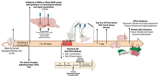
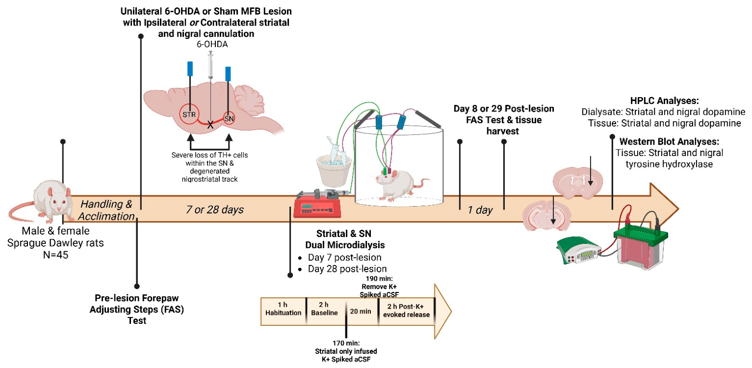
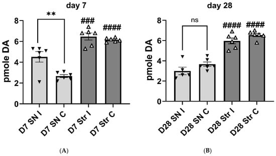
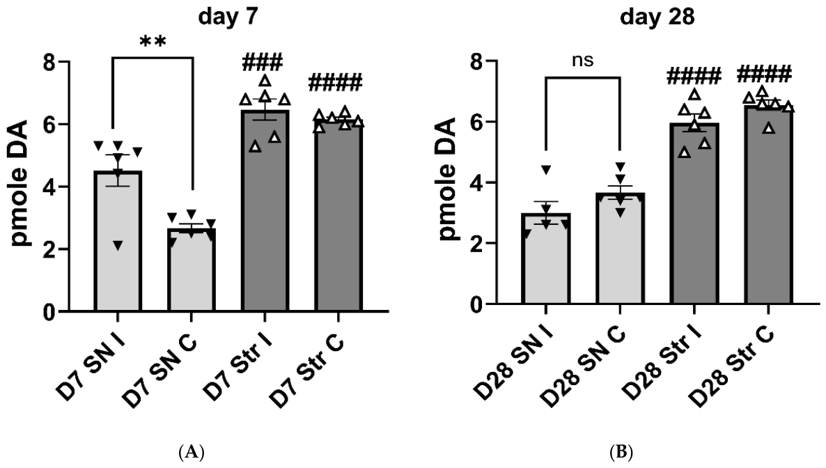
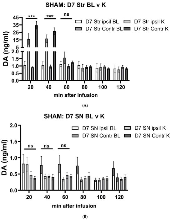
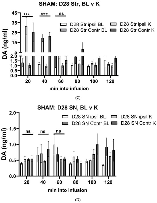
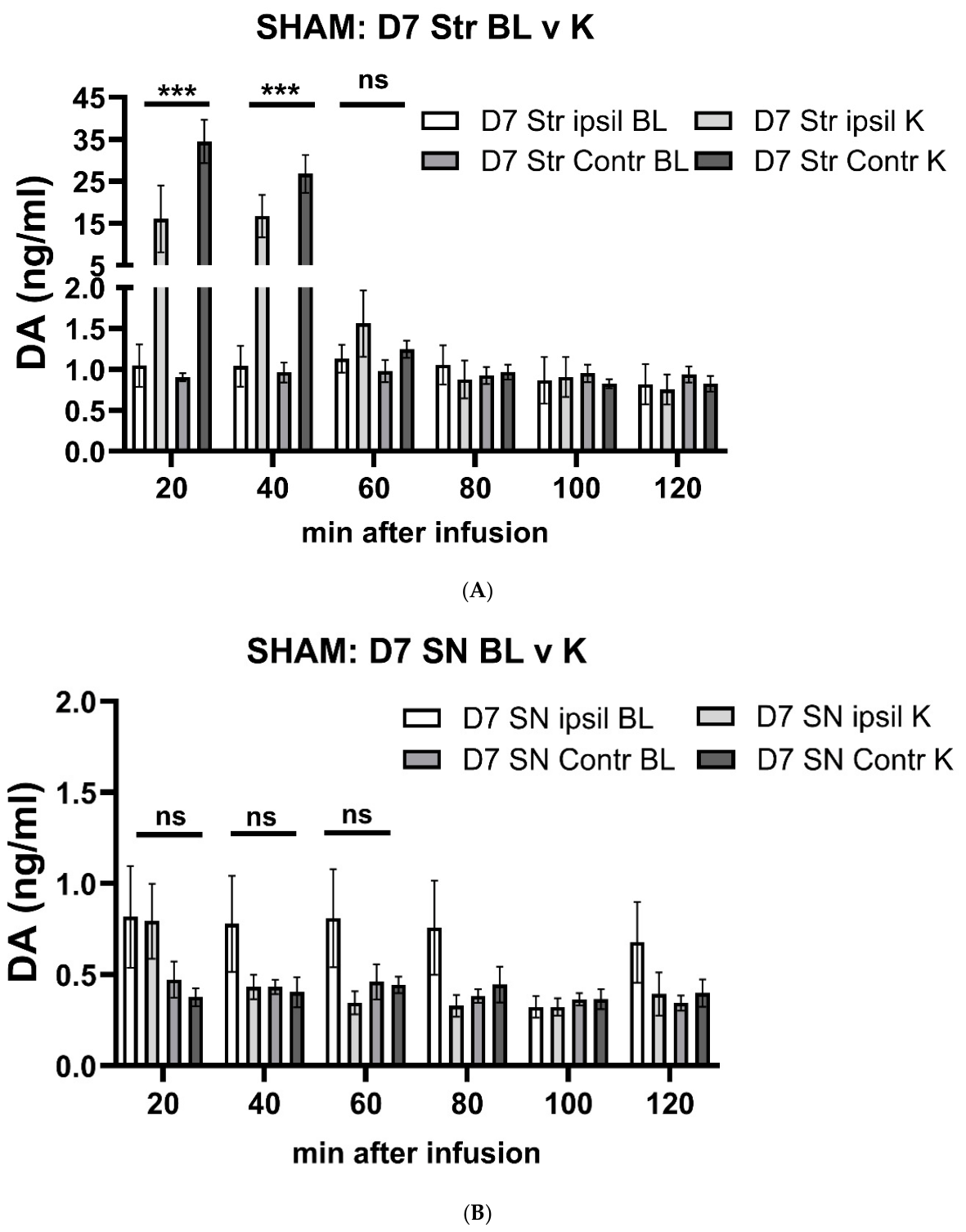
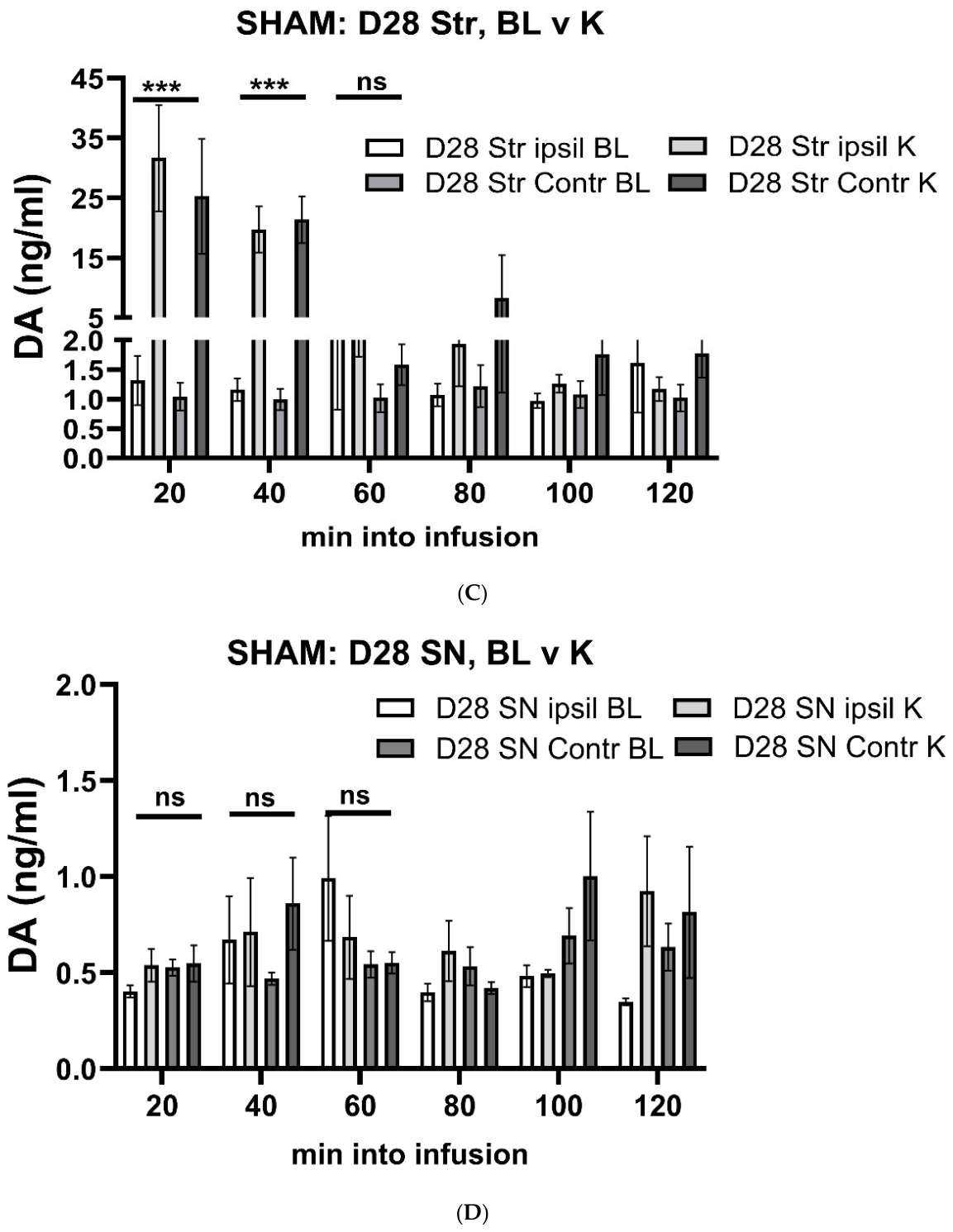
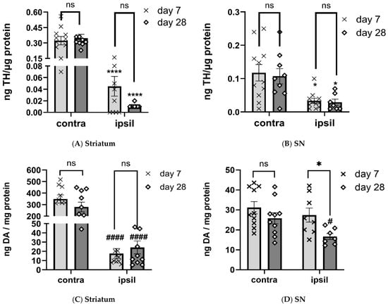
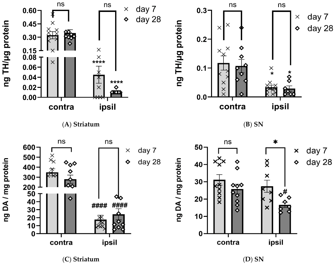
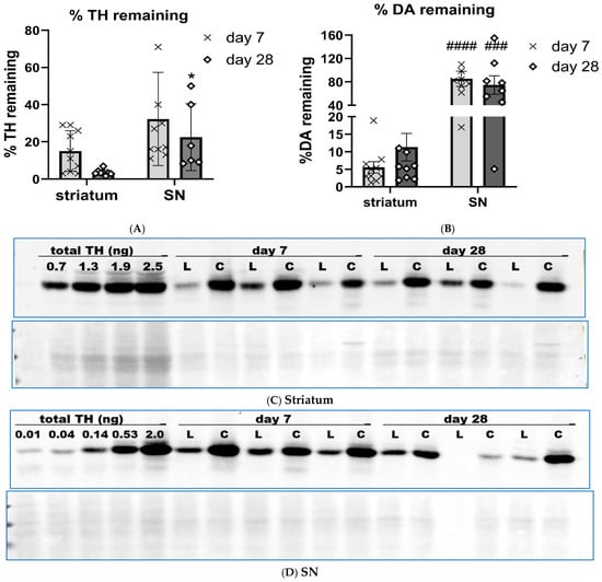
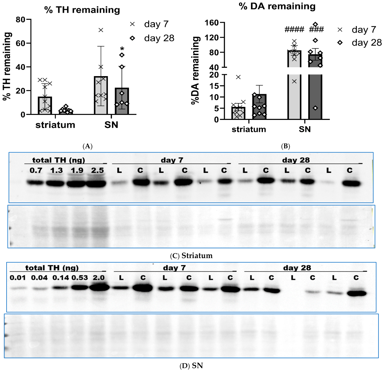
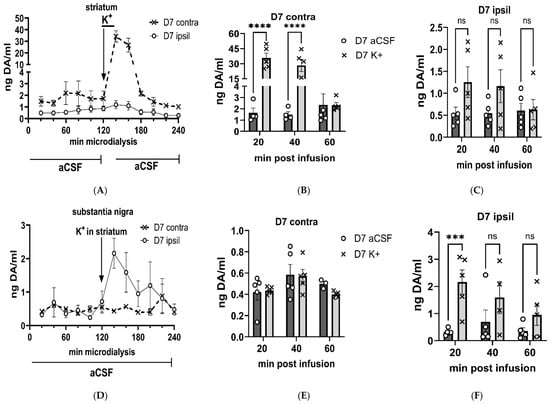
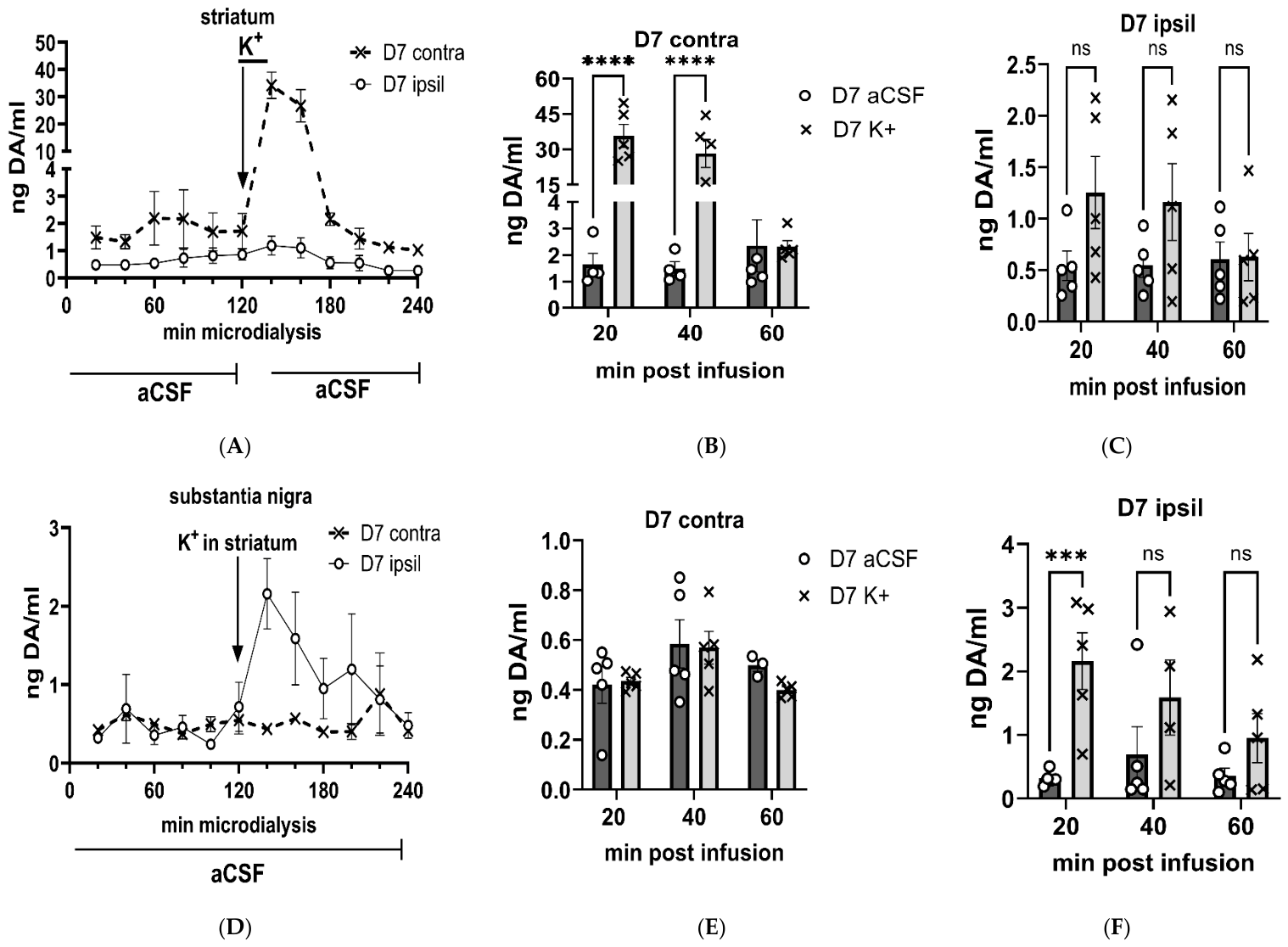
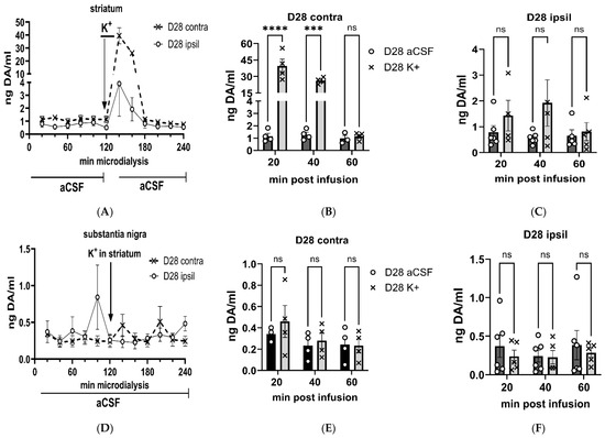
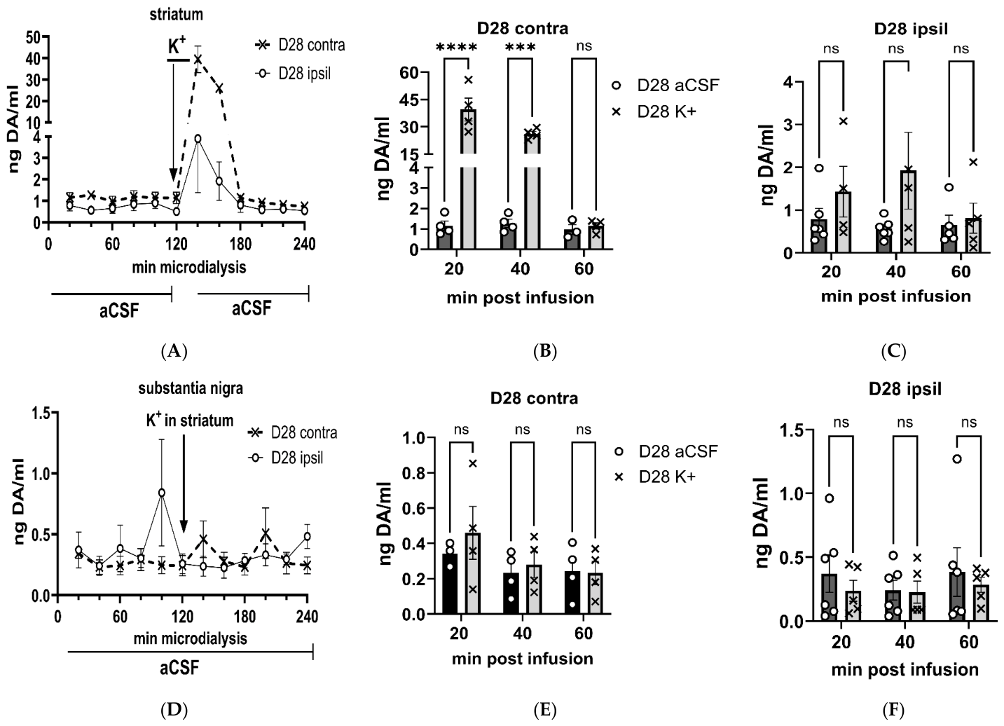
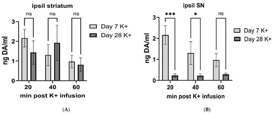
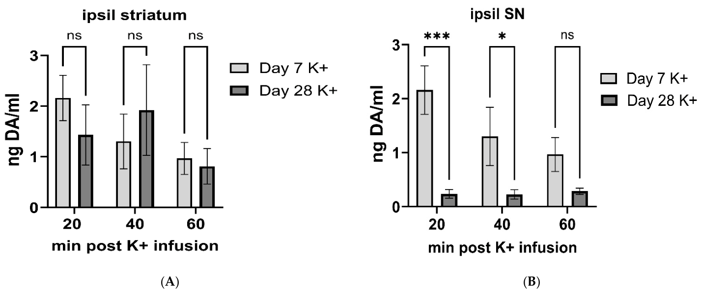
Disclaimer/Publisher’s Note: The statements, opinions and data contained in all publications are solely those of the individual author(s) and contributor(s) and not of MDPI and/or the editor(s). MDPI and/or the editor(s) disclaim responsibility for any injury to people or property resulting from any ideas, methods, instructions or products referred to in the content. |
© 2026 by the authors. Licensee MDPI, Basel, Switzerland. This article is an open access article distributed under the terms and conditions of the Creative Commons Attribution (CC BY) license.