


Abstract
Acute lung injury (ALI) is a severe inflammatory condition associated with high morbidity and mortality, and there are currently no specific pharmacological treatments available. In this context, plants and natural products have emerged as promising therapeutic alternatives. SteLL (Schinus terebinthifolia Raddi leaf lectin) has demonstrated several biological activities, including anti-inflammatory and immunomodulatory effects. This study evaluated the anti-inflammatory effects of SteLL in a murine model of lipopolysaccharide (LPS)-induced ALI. Female BALB/c mice received intraperitoneal (i.p.) administration of SteLL (1, 5, or 10 mg/kg), dexamethasone (2 mg/kg), or vehicle (PBS). Sixty minutes later, ALI was induced by intranasal instillation of 25 µL of LPS (1 μg/μL). After 24 h, the animals were euthanized. Bronchoalveolar lavage fluid (BALF) was obtained to evaluate inflammatory parameters and lungs were collected for histopathological analysis. The tested doses of SteLL resulted in a 45–66% lower leukocyte infiltration. The group treated with 5 mg/kg exhibited a lower proportion of neutrophils and a higher proportion of mononucleated cells. Pre-treatment with SteLL also minimized plasma leakage and myeloperoxidase (MPO) activity. Furthermore, SteLL attenuated the release of pro-inflammatory cytokines at all tested doses as well as prevented nitric oxide (NO) production at the highest dose (10 mg/kg). Histopathological analysis showed that SteLL (5 and 10 mg/kg) attenuated LPS-induced lung injury. Overall, SteLL demonstrated significant anti-inflammatory effects, showing its potential as a plant-derived compound for modulating pulmonary inflammation.
1. Introduction
Acute lung injury (ALI) is a condition of respiratory failure resulting from multifactorial damage to the alveolar epithelium. The underlying inflammatory process is characterized by the migration of leukocytes to the pulmonary parenchyma, accumulation of interstitial fluid, and increased release of pro-inflammatory mediators such as cytokines and nitric oxide (NO), as well as the formation of reactive oxygen species mediated by myeloperoxidase (MPO) [1,2,3,4]. Acute respiratory distress syndrome (ARDS) represents the most severe clinical manifestation of ALI and constitutes a significant clinical challenge due to its rapid progression and high rates of morbidity and mortality [4,5,6].
It is estimated that approximately 3 million people worldwide are affected by ALI each year [7,8,9]. ALI generally has an infectious origin, with respiratory viruses and Gram-negative bacteria being particularly prominent. During the COVID-19 pandemic, the SARS-CoV-2 virus emerged as a new trigger for ALI and its most severe outcome, ARDS, contributing significantly to an exorbitant increase in mortality and morbidity rates among patients affected by the disease [6,10]. However, although SARS-CoV-2 drew global attention to viral causes of ALI, Gram-negative bacteria such as Escherichia coli and Klebsiella pneumoniae have long represented a major and persistent threat. This is largely due to lipopolysaccharide (LPS), a structural endotoxin of their outer membrane that has been recognized for decades as a potent inducer of ALI [11,12].
Current conventional therapies for ALI and ARDS primarily consist of mechanical ventilatory support and the administration of anti-inflammatory drugs, such as corticosteroids, vasodilators, and modulators of the inflammatory cascade [13,14,15,16]. Despite these interventions, hospital mortality rates can reach 40%, representing a significant clinical challenge [17,18].
Schinus terebinthifolia Raddi (Anacardiaceae), popularly known as aroeira or Brazilian pepper tree, has widespread use in traditional medicine. Due to its anti-inflammatory, antipyretic, analgesic, and depurative activities, it is used in the treatment of urinary tract infections, gastrointestinal disorders, and other health conditions [19,20,21]. Leaf preparations are popularly used as anti-inflammatory agents [22], relieving conditions such as bronchitis, ulcers, and wounds [23]. Among the bioactive constituents of S. terebinthifolia leaves, there is a lectin called SteLL (S. terebinthifolia leaf lectin) [24].
Lectins are carbohydrate-binding proteins that can selectively and reversibly bind to carbohydrates [25], exhibiting distinct biological activities, including antibacterial [26], antifungal [27], antibiofilm [28], nematicidal [29], and insecticidal [30] effects, as well as pharmacological properties such as antinociceptive, anti-inflammatory, and antitumor activities [31]. SteLL is a glycoprotein with a molecular mass of 12.4 kDa and hemagglutinating activity that is resistant to variations in pH (5.0–8.0) and temperature (up to 100 °C), as well as exhibiting functional stability in the presence of Ca2+ and Mg2+ ions [24,32]. The safety of SteLL administered intraperitoneally or orally at a single dose of 100 mg/kg was demonstrated in mice [33]. Studies have already demonstrated in vitro antimicrobial [24] and immunomodulatory [32] effects of SteLL. In vivo, SteLL has shown antitumor activity against sarcoma 180 [34], anti-infective effects [35], antidepressant and anxiolytic effects in different contexts [36,37], and antinociceptive activity [38,39], all in mice.
Marinho et al. [33] reported that SteLL demonstrated significant anti-inflammatory activity in acute inflammation models, including peritonitis and paw edema, both induced by carrageenan in mice. The results indicated the modulatory potential of SteLL on cellular inflammatory responses and edematous processes, as evidenced by reductions in leukocyte migration, MPO activity, protein extravasation, and the levels of NO and pro-inflammatory cytokines.
The traditional use of S. terebinthifolia leaves in respiratory inflammatory conditions suggests the presence of bioactive compounds capable of modulating pulmonary inflammatory responses. In addition, the diverse biological activities described for SteLL highlight its broad pharmacological potential, including an ability to modulate inflammatory processes. Therefore, this study aimed to investigate the preventive effects of SteLL on the pulmonary inflammatory response in an experimental model of LPS-induced ALI.
2. Results
SteLL was successfully purified following the original protocol described by Gomes et al. [24], and the batch used in this study showed functional activity, with a specific hemagglutinating activity of 6336, indicating the presence of active SteLL molecules.
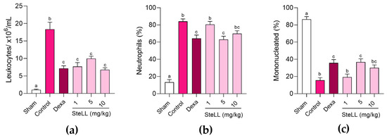
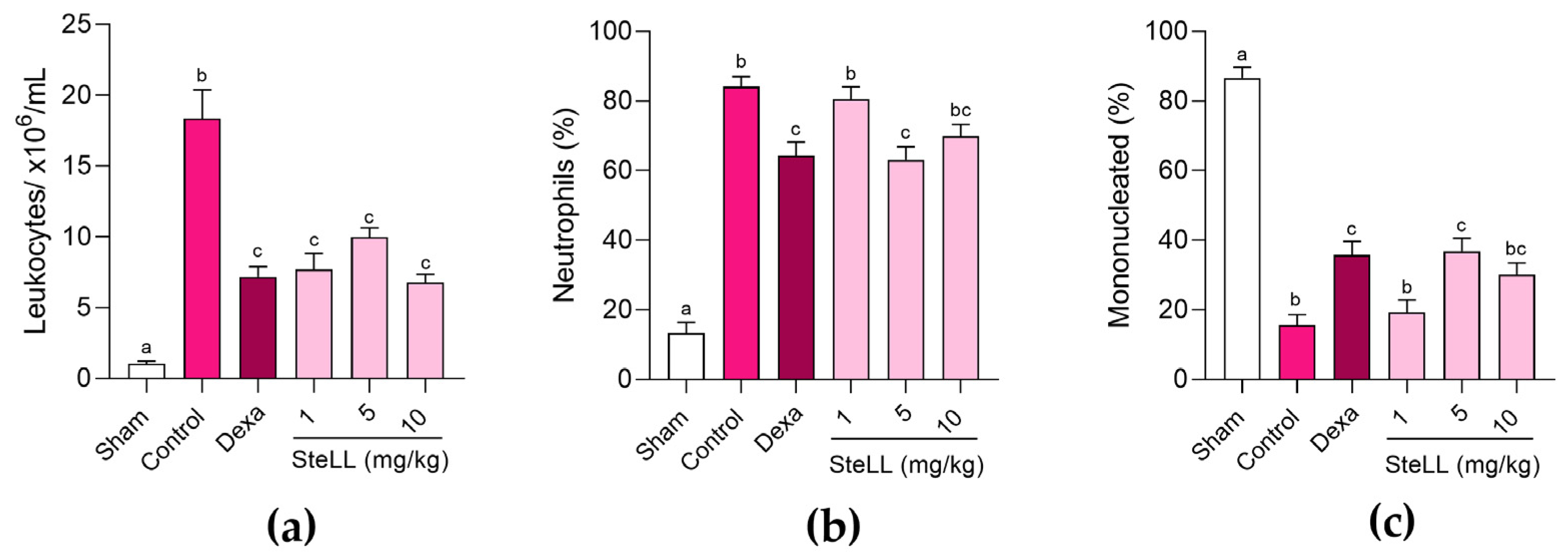
Figure 1a shows the total leukocyte counts in the bronchoalveolar lavage fluid (BALF) of healthy (Sham) mice and of animals with LPS-induced ALI previously treated with SteLL (1, 5, or 10 mg/kg), dexamethasone (2 mg/kg), or vehicle (phosphate-buffered saline, PBS; control), revealing significant differences between the groups (F5,42 = 29.13; p < 0.0001). The control group exhibited markedly higher (p < 0.0001) leukocyte migration (18,360 ± 2020 cells/mL) compared with the Sham group (1025 ± 190 cells/mL), confirming the validity of the experimental model. SteLL-treated groups demonstrated an anti-inflammatory effect, with leukocyte migration 58.07 ± 6.15% (1 mg/kg), 45.55 ± 3.48% (5 mg/kg), and 63.11 ± 3.11% (10 mg/kg) lower than in the control group (p < 0.0001). There were no significant differences (p > 0.05) among the groups treated with distinct doses of SteLL. Dexamethasone-treated group showed leukocyte migration that was 60.86 ± 3.81% lower than the control group (p < 0.0001) but similar (p > 0.05) to SteLL treatments.
Figure 1.
Evaluation of leukocyte migration in the bronchoalveolar fluid of healthy mice (Sham) and those subjected to acute lung inflammation induced by lipopolysaccharide and previously treated intraperitoneally with PBS (control), SteLL (1, 5, and 10 mg/kg), or dexamethasone (Dexa, 2 mg/kg). (a) Leukocyte count. (b) Percentage of neutrophils. (c) Percentage of mononucleated cells. The results are expressed as mean ± standard error of the mean (SEM) (n = 8 per group). Different letters indicate significant differences (p < 0.05) between the groups, based on analysis of variance (ANOVA) followed by Tukey’s test.
Differential leukocyte counts in the BALF was performed to evaluate the proportion of neutrophils (Figure 1b; F5,42 = 53.87; p < 0.0001) and mononucleated cells (Figure 1c; F5,42 = 53.82; p < 0.0001). SteLL at 5 mg/kg resulted in a significantly (p = 0.0015) lower percentage of neutrophils (63.13 ± 3.74%) compared with the control group (84.13 ± 2.94%), whereas the other doses did not significantly reduce (p > 0.05) neutrophil counts compared to the control group (Figure 1b). Correspondingly, the group pre-treated with SteLL at 5 mg/kg showed higher (p = 0.0013) proportion of mononucleated cells (36.88 ± 3.74%) than in control (15.63 ± 2.96%) (Figure 1c). Dexamethasone group also showed lower (p = 0.0029) neutrophil proportion (64.25 ± 3.98%) (Figure 1b) and higher (p = 0.0026) mononucleated cells proportion (35.75 ± 3.98%) (Figure 1c) relative to the control group, while showed an effect similar (p > 0.999) to that of SteLL at 5 mg/kg.
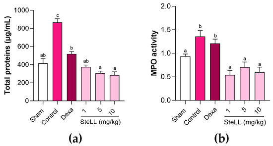
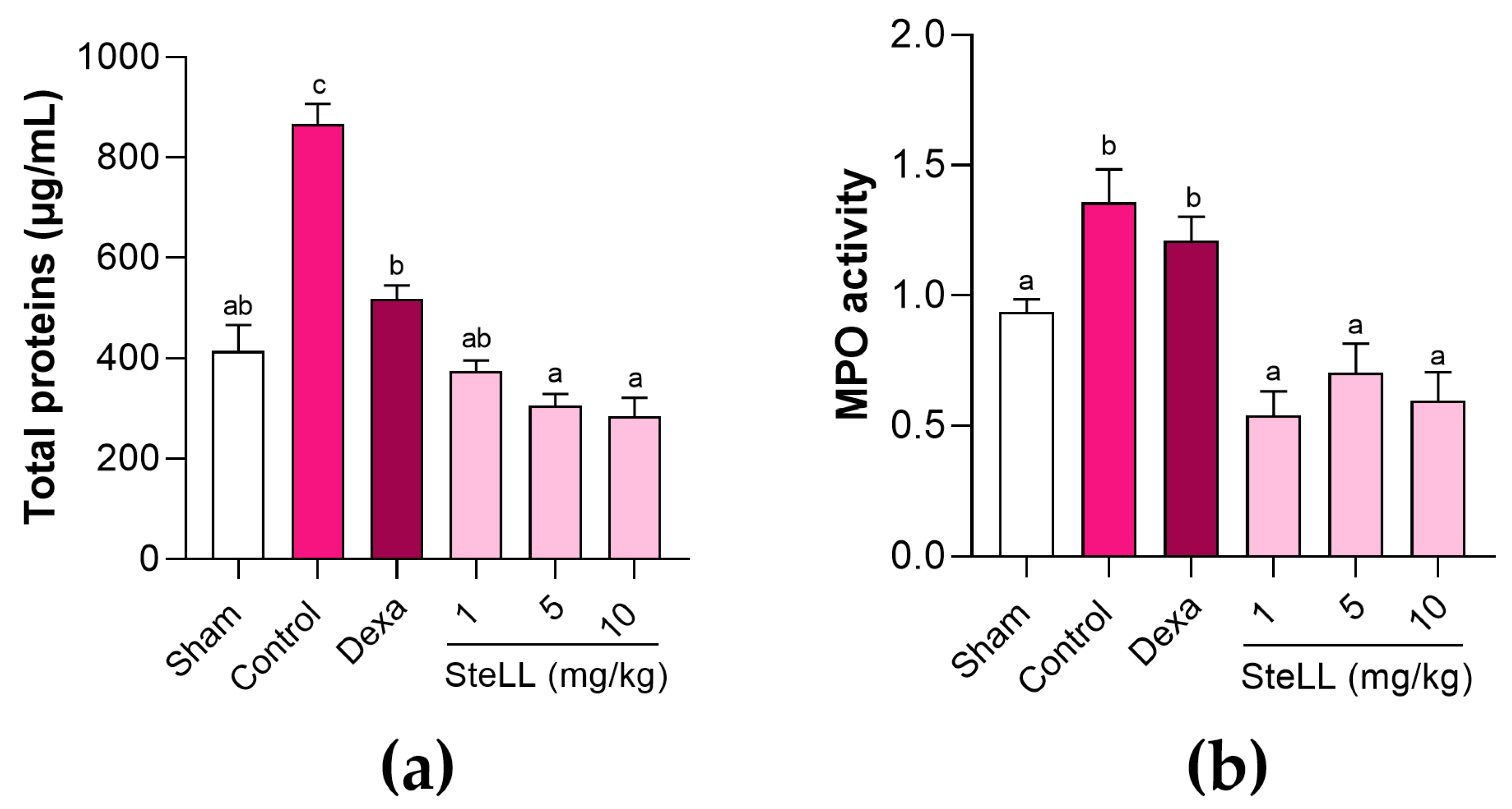
Plasma extravasation into the BALF was evaluated by assessing the total protein content (Figure 2a; F5,42 = 38.03; p < 0.0001). In comparison with control group, the animals pre-treated with SteLL (1, 5, and 10 mg/kg) showed significantly less (p < 0.0001) plasma extravasation into the BALF, being the values 57.36 ± 2.48%, 65.12 ± 2.69%, and 67.57 ± 4.26% lower, respectively. In turn, dexamethasone treatment led to plasma extravasation 40.85 ± 3.07% lower than in control (p < 0.0001). The data also showed that total protein content in the SteLL- and dexamethasone-treated groups was similar (p > 0.05) to that in the BALF of healthy (Sham) animals, whereas SteLL at 5 and 10 mg/kg resulted in lower protein content (p < 0.001) compared with the dexamethasone group.
Figure 2.
Total protein levels (µg/mL) (a) and myeloperoxidase (MPO) activity (b), in the bronchoalveolar fluid of healthy mice (Sham) and those subjected to acute lung inflammation induced by lipopolysaccharide and previously treated intraperitoneally with PBS (control), SteLL (1, 5, and 10 mg/kg), or dexamethasone (Dexa, 2 mg/kg). Results are expressed as mean ± standard error of the mean (SEM) (n = 8 per group). Different letters indicate significant differences (p < 0.05) between the groups, based on analysis of variance (ANOVA) followed by Tukey’s test.
Figure 2b shows the MPO activity in the BALF of the studied groups (F5,42 = 9.241; p < 0.0001). Treatment with SteLL (1, 5, and 10 mg/kg) also led to lower (p ≤ 0.0005) MPO activity in the BALF compared with the negative control group, differently of dexamethasone (p = 0.8960). SteLL doses showed similar levels (p > 0.85) of MPO activity, with enzyme activity values equivalent (p > 0.05) to the healthy (Sham) group.
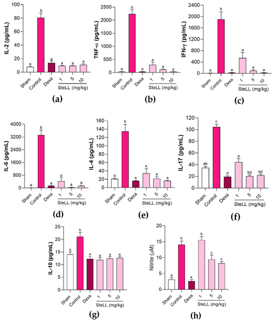
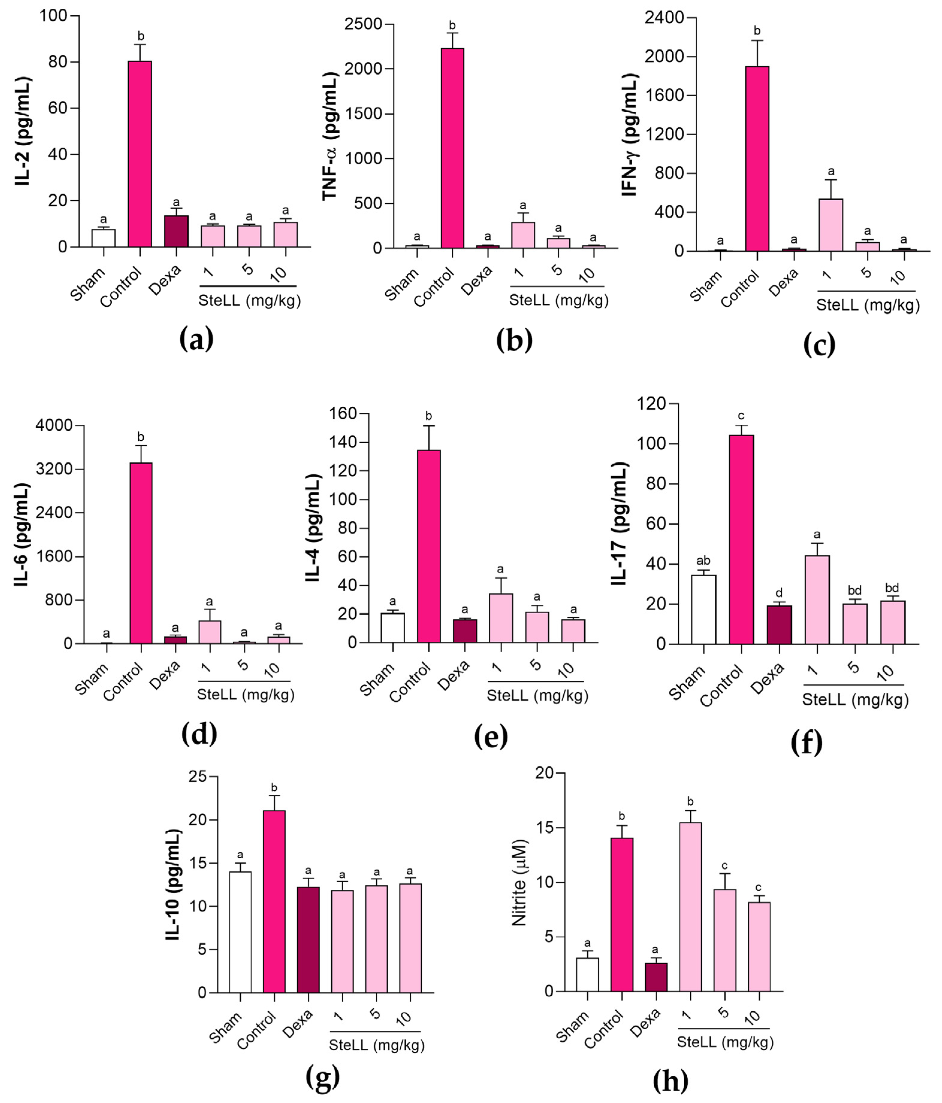
Significant differences were detected for the levels of the interleukin (IL) 2 (Figure 3a, F5,42 = 80.81; p < 0.0001), tumor necrosis factor (TNF) α (Figure 3b, F5,42 = 123.6; p < 0.0001), interferon (IFN)-γ (Figure 3c, F5,42 = 31.35; p < 0.0001), IL-6 (Figure 3d, F5,42 = 39.70; p < 0.0001), IL-4 (Figure 3e, F5,42 = 31.31; p < 0.0001), IL-17 (Figure 3f, F5,42 = 86.73; p < 0.0001) and IL-10 (Figure 3g, F5,42 = 11.10; p < 0.0001) in BALF among the different groups. The LPS administration was highly immunogenic, causing a significant increase in all measured cytokines in the control group compared with the Sham group. Pre-treatment with SteLL (1, 5, and 10 mg/kg) or dexamethasone (2 mg/kg) resulted in significantly lower (p < 0.0001) levels of both pro-inflammatory (IL-2, TNF-α, IFN-γ, IL-6, and IL-17) and anti-inflammatory (IL-4 and IL-10) cytokines. There were significant differences among SteLL doses only for IL-17, with the animals that received the lowest dose (1 mg/kg) showing a higher level (p ≤ 0.0006) of this cytokine in the BALF (Figure 3f). In addition, SteLL at 5 and 10 mg/kg and dexamethasone groups showed lower NO levels relative to the control group (Figure 3h, F5,42 = 32.11; p < 0.0001).
Figure 3.
Cytokines (a–g) and nitric oxide (h) levels in the bronchoalveolar fluid of healthy mice (Sham) and those subjected to acute lung inflammation induced by lipopolysaccharide and previously treated intraperitoneally with PBS (control), SteLL (1, 5, and 10 mg/kg), or dexamethasone (Dexa, 2 mg/kg). Bars represent the mean ± standard error of the mean (SEM) of data from animals (n = 8 per group). Different letters indicate significant differences (p < 0.0001) between the groups, based on analysis of variance (ANOVA) followed by Tukey’s test and false discovery rate (FDR) correction.
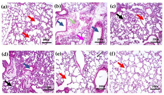
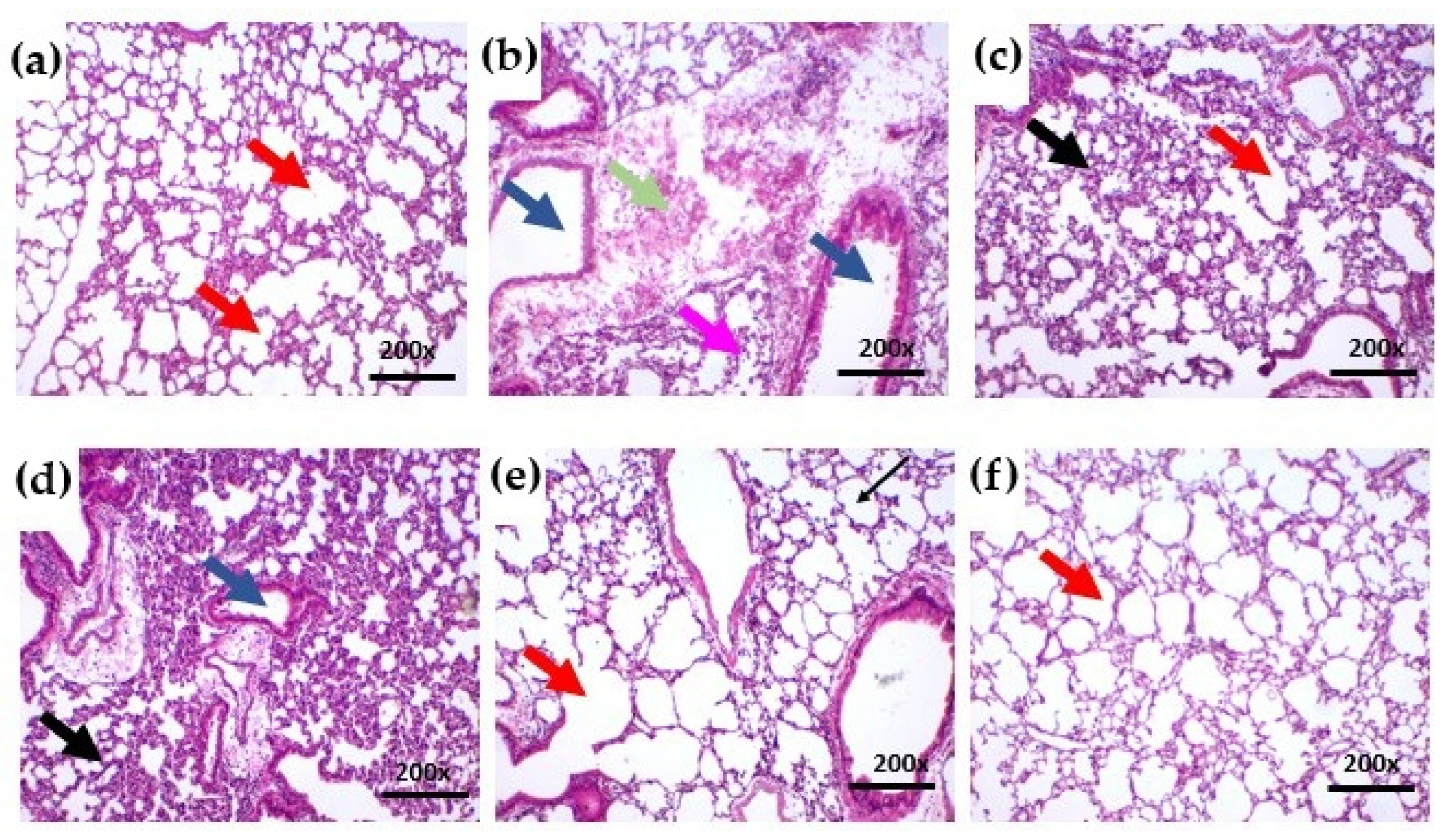
Histopathological analysis showed that lungs from the healthy (Sham) group had a regular epithelium of normal thickness and alveolar chambers of normal size, without edema or congestion (Figure 4a). In contrast, the control group exhibited massive infiltration of inflammatory cells throughout the pulmonary parenchyma, accompanied by venous congestion (Figure 4b). In the dexamethasone-treated group, a reduction in alveolar chambers was observed, suggesting a potential decrease in respiratory capacity (Figure 4c). The SteLL 1 mg/kg group also showed inflammatory signs, which were more pronounced and associated with epithelial thickening (Figure 4d).
Figure 4.
Histopathological analysis of the right lung from healthy (Sham) mice (a) as well as mice with LPS-induced acute lung inflammation previously treated intraperitoneally with PBS, control (b), dexamethasone at 2 mg/kg (c), or SteLL at 1 mg/kg (d), 5 mg/kg (e), or 10 mg/kg (f). Hematoxylin–eosin staining; magnification 200×. Key features: alveolar chambers (red arrows), bronchioles (blue arrows), congested vessels (green arrows), thickened epithelium (black arrows), and inflammatory infiltrate (pink arrows).
Conversely, lungs from animals treated with SteLL at 5 and 10 mg/kg (Figure 4e,f) resembled those of the healthy group, displaying regular alveolar epithelium, preserved thickness, normal alveolar spaces, and reduced inflammatory infiltration compared with control. In the SteLL 10 mg/kg group (Figure 4f), erythrocytes were occasionally observed in some alveolar spaces; however, these represent minor changes likely due to transient vascular permeability induced by LPS instillation and do not reflect impaired lung function. Importantly, no structural alterations or significant tissue injury were detected, indicating that SteLL treatment substantially mitigated ALI while maintaining essentially normal pulmonary architecture.
3. Discussion
ALI is characterized by severe hypoxemic respiratory failure resulting from immune hyperactivation, oxidative stress, and cellular damage, leading to increased vascular permeability, pulmonary edema, and impaired gas exchange [40,41]. Despite advances in supportive care, current therapies have limited efficacy and safety, highlighting the need for novel interventions. In this study, pre-treatment with SteLL effectively mitigated key pathological features of LPS-induced ALI mice, including leukocyte and neutrophil infiltration, protein-rich edema, MPO activity, and pro-inflammatory cytokine release, while preserving lung architecture. Importantly, these parameters were comprehensively assessed in BALF, which directly reflects the local inflammatory milieu of the lung.
LPS is widely used in rodent ALI models because it induces an intense inflammatory response closely resembling human pathology. LPS administration disrupts the alveolar epithelium, promotes endothelial activation, upregulates adhesion molecules (P-selectin, E-selectin, ICAM-1, VCAM-1), and drives neutrophil recruitment. This cascade leads to barrier leakage, protein-rich edema, and amplification of inflammatory signaling through enzymes, reactive oxygen species, and cytokines [13,42,43]. SteLL treatment mitigated total leukocyte and neutrophil migration into BALF, suggesting attenuation of this inflammatory cascade. Lower MPO activity further indicates mitigation of neutrophil-mediated oxidative damage, contributing to preservation of alveolar-capillary integrity and limiting edema formation. By attenuating protein extravasation, SteLL can help maintain surfactant function, alveolar stability, lung compliance, and gas exchange [44,45]. Together, the findings demonstrate a consistent modulation of key local inflammatory processes central to ALI pathophysiology.
Notably, the cytokine panel analyzed provides a broad view of the inflammatory response modulated by SteLL within the pulmonary compartment. This anti-inflammatory effect is clinically relevant, as uncontrolled cytokine release, such as in COVID-19-associated cytokine storm, can drive severe pulmonary dysfunction and respiratory failure [46,47]. By dampening both neutrophil-mediated tissue damage and cytokine production, SteLL addresses central mechanisms underlying ALI. Histological analysis confirmed that higher doses of SteLL preserved alveolar architecture and limited inflammatory alterations, while lower doses showed mild epithelial thickening and residual inflammation.
The previously reported effects of SteLL in carrageenan-induced peritonitis [33] complement and reinforce its protective role observed in LPS-induced ALI. In both models, SteLL significantly reduced leukocyte migration, particularly neutrophil infiltration, and decreased markers of oxidative damage, such as MPO activity. Cytokine modulation was also consistent across models as SteLL limited excessive pro-inflammatory responses, including TNF-α. These results suggest a capacity to control immune activation while minimizing tissue injury, highlighting the broad immunomodulatory potential of SteLL. Taken together, these findings support that SteLL exerts anti-inflammatory effects across distinct inflammatory contexts, as evaluated in compartment-specific settings.
Unlike highly toxic lectins such as abrin, which cause severe lung injury, excessive inflammation, oxidative stress, and pulmonary edema even after a single inhalation exposure in mice [48], SteLL is a non-toxic lectin in rodents [33,34]. In the present work, pre-treatment of mice with SteLL did not induce acute toxicity or lung tissue damage, while effectively modulating neutrophil infiltration, cytokine release, and vascular permeability. It is important to note that absence of systemic inflammation can be inferred from the lack of clinical and histopathological alterations. This highlights that SteLL’s activity confers beneficial immunomodulatory and anti-inflammatory effects without the ribotoxicity or cytotoxicity observed with lectins like abrin [48].
The anti-inflammatory effect of lectins on lung inflammation models remains underexplored. However, the Urtica dioica agglutinin (UDA) demonstrated protection in a SARS-CoV mouse model, but this effect was attributed to direct antiviral activity [49] rather than modulation of host inflammation. While UDA inhibits viral adsorption and replication by binding viral glycoproteins, the beneficial effects of SteLL observed in ALI are anti-inflammatory and immunomodulatory, as SteLL has not yet been evaluated in infectious models.
The TIP peptide, which mimics the lectin-like domain of TNF, improved barrier integrity, enhanced alveolar fluid clearance, and reduced inflammatory cytokines and bacterial translocation in ex vivo human lungs [50]. TIP acts by directly modulating epithelial ion channels, suggesting avenues for future mechanistic investigations of SteLL.
The dose–response profile of SteLL in the preventive treatments analyzed suggests that its anti-inflammatory effects do not follow a simple linear relationship. When administered prior to ALI induction, the 5 mg/kg dose showed the most consistent effect in modulating the proportion of neutrophils and mononucleated cells, whereas lower (1 mg/kg) or higher (10 mg/kg) doses produced comparable effects in other parameters, indicating the presence of an optimal preventive therapeutic window. In contrast, control of plasma extravasation and MPO activity reached maximal effect even at the lowest preventive dose, suggesting a saturation of response. These results indicate that SteLL exhibits dose-dependent immunomodulatory activity in a preventive setting, but the relationship is not proportional, a pattern common for agents targeting complex inflammatory processes. Furthermore, the differences in dose–response profiles across the various parameters suggest that SteLL’s preventive effects likely involve distinct mechanisms or pathways for each outcome, reflecting the complexity and potentially independent regulation of leukocyte migration, cytokine production, and vascular permeability prior to ALI onset.
This work offers a new perspective on the development of natural products as potential therapeutics for ALI, addressing a research gap regarding the use of plant lectins in pulmonary inflammatory disorders. Nevertheless, as an initial investigation, further studies are required to fully elucidate the mechanisms through which lectins confer protection in ALI. Here, the anti-inflammatory phenotype of SteLL in ALI was demonstrated, paving the way for future research aiming at uncovering the underlying molecular mechanisms. This includes exploring canonical pathways such as Toll-like receptor 4/nuclear factor kappa B (TLR4/NF-κB) signaling, mitogen-activated protein kinase (MAPK) pathway, pyroptosis, and oxidative stress [51,52,53], to determine whether SteLL mediates its effects via direct inhibition of inflammatory signaling, modulation of immune cell activity, or alternative mechanisms.
As this study was specifically designed to investigate local pulmonary inflammation, systemic inflammatory responses were not evaluated. The use of BALF as the primary biological matrix is well established in ALI models, as it provides a direct and sensitive assessment of inflammatory processes occurring within the lung microenvironment. Nevertheless, given the recognized interplay between pulmonary and systemic inflammation, future studies should incorporate circulating biomarkers to further characterize the systemic impact of SteLL and strengthen translational interpretation.
Reductions in MPO activity and pro-inflammatory cytokines observed in BALF suggest that these molecules could serve as early indicators of therapeutic efficacy of SteLL. Compared to conventional anti-inflammatory therapies, SteLL offers the advantage of simultaneously reducing neutrophil-mediated damage, cytokine release, and vascular leakage, suggesting a multi-protective effect. These results warrant further preclinical and clinical investigations to evaluate SteLL as a potential adjunct therapy for ALI and ARDS.
While these findings are promising, extrapolation to human ALI requires caution. LPS-induced models do not fully replicate the complexity of clinical disease, including infectious triggers, heterogeneous lung injury, and comorbidities. In addition, further investigation is needed regarding the optimal administration routes and treatment windows before advancing. For instance, the impact of SteLL on both the acute and recovery phases of ALI and the potential of delivery strategies such as nebulization or intranasal administration need future exploration. Another potential strategy is the combination of SteLL with established drugs, such as dexamethasone, given that combination therapy represents an important avenue in clinical drug development. Also, studies should also evaluate the effects of SteLL in infectious models of ALI to enhance translational relevance.
One more important consideration for clinical translation of lectins is their immunogenicity and immunomodulatory profile. Lectins, as proteins, can elicit immune recognition due to their amino acid sequences, three-dimensional structures, or post-translational modifications, potentially stimulating antibody production or inflammatory responses [54]. SteLL has been shown to induce mice splenocytes to secrete both pro-inflammatory cytokines (IL-17A, TNF-α, IFN-γ, IL-2) and anti-inflammatory IL-4, suggesting a capacity to modulate immune responses without triggering excessive inflammation [32]. In both peritonitis and ALI models, SteLL increased anti-inflammatory cytokines while limiting pro-inflammatory mediators, highlighting an advantage over highly immunogenic or purely pro-inflammatory lectins. However, careful evaluation of immune responses, including antibody generation and systemic effects, will be critical in future preclinical and clinical studies. Strategies such as controlled dosing, nanoparticle or liposome formulations, and targeted delivery can minimize undesired immunogenicity while preserving lectins therapeutic benefits [54].
4. Materials and Methods
4.1. SteLL Purification
S. terebinthifolia leaves were collected at the campus of the Universidade Federal de Pernambuco (UFPE) (Recife, Pernambuco, Brazil), with authorization (nº 72024) from the Instituto Chico Mendes de Conservação da Biodiversidade (ICMBio). The study was recorded under code AED6BF8 in the Sistema Nacional de Gestão do Patrimônio Genético e do Conhecimento Tradicional Associado (SisGen). A representative specimen of the species was deposited at the Instituto Agronômico de Pernambuco (IPA, Recife) under the reference number 73431.
SteLL was purified following the methodology from Gomes et al. [24]. The collected leaves were first rinsed under running water and then with distilled water before being air-dried at 28 °C for 4 days. Once dried, the leaves were ground into a fine powder using an industrial processor (LQL-4, Metvisa, Brusque, Brazil). For extract preparation, the powdered leaves were suspended in 0.15 M NaCl at a proportion of 10% (w/v) and homogenized for 16 h at 4 °C. The homogenate was then centrifuged at 9000× g for 15 min at 4 °C. The resulting leaf extract, containing 15 mg/mL of protein, was applied (2 mL) to a chitin column (7.5 × 1.5 cm; Sigma-Aldrich, St. Louis, MO, USA) pre-equilibrated with 0.15 M NaCl. Non-binding proteins were removed through washing, and SteLL was subsequently eluted using 1.0 M acetic acid. The elution was monitored by measuring absorbance at 280 nm. The purified lectin was dialyzed against distilled water for roughly 6 h using a cellulose membrane with a 10 kDa cut-off (Sigma-Aldrich), followed by lyophilization. Finally, SteLL was resuspended in PBS and stored at low temperature until further use.
4.2. Determination of Protein Concentration and Hemagglutinating Activity
Protein concentration was quantified using the Lowry et al. [55] method, employing a bovine serum albumin (Sigma-Aldrich) standard curve ranging from 31.25 to 500 μg/mL. The carbohydrate-binding activity of SteLL was evaluated through the hemagglutination assay. A 50 μL aliquot of the SteLL sample was subjected to successive twofold serial dilutions in 0.15 M NaCl across the wells of a 96-well microplate. Subsequently, 50 μL of a suspension of New Zealand rabbit erythrocytes, fixed with glutaraldehyde [56], were added to each well. Erythrocytes incubated exclusively with 0.15 M NaCl served as control. All assays were carried out in duplicate. The hemagglutinating activity was defined as the reciprocal of the highest lectin dilution capable of producing complete agglutination of the erythrocytes. Specific hemagglutinating activity was calculated by dividing the hemagglutinating activity by the protein concentration (mg/mL).
4.3. Mice
Forty-eight female BALB/c mice aged 6–8 weeks and weighing 20–25 g were obtained from the animal facility of the Instituto Keizo Asami (iLIKA) of UFPE. After arrival, the animals were acclimated and maintained at the Laboratório de Experimentação Animal em Bioquímica (LEAB), Department of Biochemistry, UFPE. The mice were kept under controlled environmental conditions, including a temperature of 22 °C, a 12 h light–dark cycle, and free access to food and water.
4.4. Treatments and LPS-Induced ALI
The mice were randomly assigned to six experimental groups using a simple randomization method, with eight animals per group (n = 8) maintained in the same cage. The chosen sample size was considered sufficient to detect biologically and statistically meaningful differences between groups while maintaining ethical use by avoiding the unnecessary use of experimental animals. To control for potential confounding variables, the experimental treatments were performed in a fixed order: Sham, negative control, SteLL-treated groups with escalating doses, followed by dexamethasone. Knowledge of group allocation was restricted to the researcher responsible for administering the treatments at each stage. Throughout the experiment, none of the animals exhibited signs of toxicity indicative of humane endpoints. Thus, there was no exclusion of animals.
Treatments were administered by intraperitoneal injection of SteLL at doses of 1, 5, or 10 mg/kg, dexamethasone (2 mg/kg), or PBS as the vehicle (control). One hour later, anesthesia was induced using ketamine (100 mg/kg, i.p.) and xylazine (10 mg/kg, i.p.), followed by induction of ALI via intranasal instillation of 25 μL of LPS (1 μg/µL; Escherichia coli, serotype 026:B6; Sigma-Aldrich) [57]. A healthy control group (Sham) received 25 μL of PBS intranasally and was pre-treated with the vehicle only. Animals were euthanized (300 mg/kg ketamine and 30 mg/kg xylazine i.p.) 24 h post-ALI induction. BALF was collected using 1 mL of PBS containing 1 mM ethylenediaminetetraacetic acid. The right lungs were then excised, weighed, and processed for histopathological examination.
4.5. BALF Analysis
4.5.1. Cell Count
BALF samples were centrifuged at 3000× g for 5 min at 25 °C. The resulting cell pellets were resuspended in Turk’s solution (1:20 dilution), and total leukocyte counts were performed using a Neubauer chamber (Kasvi, Pinhais, Brazil) under an optical microscope (Eclipse E100, Nikon, Tokyo, Japan). Results were expressed as the total number of cells per milliliter of BALF (leukocytes/mL). For differential cell counts, smears were prepared, stained with Papanicolaou’s stain, and 100 leukocytes per slide were classified under optical microscopy. Data were presented as the percentage of neutrophils and mononucleated cells.
4.5.2. Measurement of Inflammatory Markers
Total Protein Concentration
The total protein concentration in the BALF supernatant (1 mL) was measured using the Bicinchoninic Acid (BCA) Protein Assay Kit (Sigma-Aldrich) according to the manufacturer’s instructions. Protein levels were quantified in μg/mL based on a standard curve generated with bovine serum albumin.
Measurement of Cytokine Release
Cytokine levels in the BALF supernatant were measured using the Cytometric Bead Array (CBA) Mouse Th1/Th2 Cytokine Kit II (BD Biosciences, Franklin Lakes, NJ, USA), enabling detection of IFN-γ, IL-2, IL-4, IL-6, IL-10, IL-17, and TNF-α, according to the manufacturer’s protocol. Samples were analyzed on a BD Accuri C6 flow cytometer (BD Biosciences). Individual standard curves ranging from 0 to 5000 pg/mL were generated, and cytokine concentrations were calculated using BD Accuri C6 software 264.21.
NO Production
NO production in the BALF supernatant was estimated by measuring nitrite levels using the Griess assay, following the method described by Ding et al. [58]. Nitrite concentrations were determined based on a standard curve of sodium nitrite ranging from 3.12 to 400.00 μM.
MPO Activity
MPO activity was assessed following the method of Bradley et al. [59] with minor modifications. Briefly, 20 μL of BALF supernatant was added to a 96-well microplate, and the reaction was initiated by adding 180 μL of o-dianisidine (Sigma-Aldrich) solution (0.167 mg/mL in phosphate buffer, pH 6.0) with 1% hydrogen peroxide (H2O2) as the substrate. After 15 min of incubation, absorbance was measured at 450 nm using a microplate spectrophotometer (Epoch, BioTek Instruments, Inc., Winooski, VT, USA), with the resulting values reflecting MPO enzymatic activity.
4.6. Histological Analysis of Lung Tissue
The right lung was excised, and 5 μm tissue sections were fixed in 10% buffered formalin. The samples were then embedded in paraffin and stained with hematoxylin and eosin. Histological analysis was performed using a DM500 brightfield microscope (Leica, Wetzlar, Germany) equipped with a ICC50W camera (Leica), at 100× and 200× magnifications, to assess the cellular inflammatory response.
4.7. Statistical Analysis
Data analysis was performed using GraphPad Prism® version 8.0 (GraphPad Software, San Diego, CA, USA). Results are expressed as the mean ± standard error of the mean (SEM). Normality of the data was evaluated using the Shapiro–Wilk test. Comparisons between experimental groups were conducted using one-way analysis of variance (ANOVA) followed by Tukey’s post hoc test. Differences were considered statistically significant at p ≤ 0.05. With regard to cytokine quantification, data were analyzed using one-way ANOVA followed by Tukey’s post hoc test, with p-values adjusted using the Benjamini–Hochberg false discovery rate (FDR) method, yielding adjusted q-values ranging from 0.0001 to 0.0007.
5. Conclusions
SteLL attenuated key features of ALI induced by lipopolysaccharide, including leukocyte infiltration, inflammatory mediator production, and lung tissue damage. These findings reinforce its anti-inflammatory activity in the pulmonary microenvironment. Coupled with its low acute toxicity at the doses tested, the results encourage further research to evaluate systemic effects, effectiveness across both sexes, pharmacokinetic properties, and the molecular mechanisms responsible for its anti-inflammatory effects. Such studies will be essential to better define the translational potential of SteLL.
Author Contributions
Conceptualization, A.d.O.M. and L.L.d.S.P.; methodology, A.d.O.M., P.P.M.N., P.M.G.P. and L.L.d.S.P.; validation, A.d.O.M., L.L.d.S.P. and T.H.N.; formal analysis, A.d.O.M., M.N.B.d.S., A.K.V.C., A.A.d.S., L.L.d.S.P. and T.H.N.; investigation, A.d.O.M., M.N.B.d.S., A.K.V.C., A.N.S.d.S., P.P.M.N., A.A.d.S. and L.L.d.S.P.; resources, P.M.G.P., E.V.P. and T.H.N.; data curation, A.d.O.M., M.N.B.d.S., A.K.V.C., A.A.d.S. and L.L.d.S.P.; writing—original draft preparation, A.d.O.M. and M.N.B.d.S.; writing—review and editing, E.V.P., L.L.d.S.P. and T.H.N.; visualization, A.d.O.M., M.N.B.d.S., E.V.P., A.A.d.S., L.L.d.S.P. and T.H.N.; supervision, L.L.d.S.P. and T.H.N.; project administration, T.H.N.; funding acquisition, P.M.G.P., E.V.P., L.L.d.S.P. and T.H.N. All authors have read and agreed to the published version of the manuscript.
Funding
This research was funded by Fundação de Amparo à Ciência e Tecnologia do Estado de Pernambuco (FACEPE), grant numbers IBPG-1293-2.00/21 and APQ-2144-2.08/24 and Conselho Nacional de Desenvolvimento Científico e Tecnológico, grant number 305251/2023-6.
Institutional Review Board Statement
The method used for rabbit erythrocyte collection was approved by the Ethics Committee of the Universidade Federal de Pernambuco (protocol no. 23076.033782/2015-70, approved on 10 December 2015). The animal study protocol was approved by the Ethics Committee of the Universidade Federal de Pernambuco (protocol code 0082/2024, approved on 27 September 2024).
Informed Consent Statement
Not applicable.
Data Availability Statement
The original contributions presented in this study are included in the article. Further inquiries can be directed to the corresponding author.
Acknowledgments
During the preparation of this work the authors used ChatGPT (5.2) for the purpose of correcting grammatical and spelling errors. After using this tool/service, the authors reviewed and edited the content as needed and take full responsibility for the content of the published article.
Conflicts of Interest
The authors declare no conflicts of interest.
References
- Lei, J.; Wei, Y.; Song, P.; Li, Y.; Zhang, T.; Feng, Q.; Xu, G. Cordycepin inhibits LPS-induced acute lung injury by inhibiting inflammation and oxidative stress. Eur. J. Pharmacol. 2018, 818, 110–114. [Google Scholar] [CrossRef] [Scilit] [PubMed]
- Liu, T.; Zhao, L.; Chen, S.; Hou, B.; Huang, J.; Hong, X.; Qing, L.; Fang, Y.; Tao, Z. Polygonatum sibiricum polysaccharides prevent LPS-induced acute lung injury by inhibiting inflammation via the TLR4/Myd88/NF-κB pathway. Exp. Ther. Med. 2020, 20, 3733–3739. [Google Scholar] [CrossRef] [Scilit] [PubMed]
- Xu, H.; Sheng, S.; Luo, W.W.; Xu, X.F.; Zhang, Z. Acute respiratory distress syndrome heterogeneity and the septic ARDS subgroup. Front. Immunol. 2023, 14, 1277161. [Google Scholar] [CrossRef] [Scilit] [PubMed]
- Ma, W.; Tang, C.; Yao, P.; Zhou, T.Y.; Niu, Q.; Liu, P.; Tang, S.Y.; Chen, Y.; Gan, L.; Cao, Y. Advances in acute respiratory distress syndrome: Focusing on heterogeneity, pathophysiology, and therapeutic strategies. Signal Transduct. Target. Ther. 2025, 10, 75. [Google Scholar] [CrossRef] [Scilit]
- Tsikis, S.T.; Fligor, S.C.; Hirsch, T.I.; Pan, A.; Yu, L.J.; Kishikawa, H.; Joiner, M.M.; Mitchell, P.D.; Puder, M. Lipopolysaccharide-induced murine lung injury results in long-term pulmonary changes and downregulation of angiogenic pathways. Sci. Rep. 2022, 12, 10245. [Google Scholar] [CrossRef] [Scilit]
- Mafra, T.K.A.; Stobbe, J.C.; Rabello, R.D.S.; Lindemann, I.L.; Silva, S.G.D. A Síndrome Respiratória Aguda Grave na pessoa idosa no contexto da pandemia da COVID-19 e seus fatores associados. Rev. Bras. Geriatr. Gerontol. 2023, 26, e220158. [Google Scholar]
- Lv, X.; Lu, X.; Zhu, J.; Wang, Q. Lipopolysaccharide-induced acute lung injury is associated with increased Ran-binding protein in microtubule-organizing center (RanBPM) molecule expression and mitochondria-mediated apoptosis signaling pathway in a mouse model. Med. Sci. Monit. 2020, 26, e923172. [Google Scholar] [CrossRef] [Scilit]
- Hendrickson, K.W.; Peltan, I.D.; Brown, S.M. The epidemiology of acute respiratory distress syndrome before and after Coronavirus Disease 2019. Crit. Care Clin. 2021, 37, 703–716. [Google Scholar] [CrossRef] [Scilit]
- Fawley, J.A.; Tignanelli, C.J.; Werner, N.L.; Kasotakis, G.; Mandell, S.P.; Glass, N.E.; Dries, D.J.; Constantini, T.W.; Napolitano, L.M. American Association for the Surgery of Trauma/American College of Surgeons Committee on Trauma clinical protocol for management of acute respiratory distress syndrome and severe hypoxemia. J. Trauma Acute Care Surg. 2023, 95, 592–602. [Google Scholar] [CrossRef] [Scilit]
- Kummer, R.L.; Marini, J.J. The respiratory mechanics of COVID-19 acute respiratory distress syndrome—Lessons learned? J. Clin. Med. 2024, 13, 1833. [Google Scholar] [CrossRef] [Scilit]
- Deng, J.; Ele, Z.; Li, X.; Chen, W.; Yu, Z.; Qi, T.; Xu, S.; Xu, Z.; Fang, L. Huangkui capsule attenuates lipopolysaccharide-induced acute lung injury and macrophage activation by suppressing inflammation and oxidative stress in mice. eCAM 2021, 2021, 6626483. [Google Scholar] [CrossRef] [Scilit] [PubMed]
- Von Knethen, A.; Heinicke, U.; Laux, V.; Parnham, M.J.; Steinbicker, A.U.; Zacharowski, K. Antioxidants as therapeutic agents in acute respiratory distress syndrome (ARDS) treatment—From mice to men. Biomedicines 2022, 10, 98. [Google Scholar] [CrossRef] [Scilit] [PubMed]
- Li, H.; Hao, Y.; Yang, L.-L.; Wang, X.-Y.; Li, X.-Y.; Bhandari, S.; Han, J.; Liu, Y.-J.; Gong, Y.-Q.; Scott, A.; et al. MCTR1 alleviates lipopolysaccharide-induced acute lung injury by protecting lung endothelial glycocalyx. J. Cell. Physiol. 2020, 235, 7283–7294. [Google Scholar] [CrossRef] [Scilit] [PubMed]
- Hirano, Y.; Madokoro, S.; Kondo, Y.; Okamoto, K.; Tanaka, H. Corticosteroid treatment for early acute respiratory distress syndrome: A systematic review and meta-analysis of randomized trials. J. Intensive Care 2020, 8, 91. [Google Scholar] [CrossRef] [Scilit]
- Cui, Y.-Q.; Ding, X.-F.; Liang, H.-Y.; Wang, D.; Zhang, X.-J.; Li, L.-F.; Kan, Q.-C.; Wang, L.-X.; Sun, T.-W. Efficacy and safety of low-dose corticosteroids for acute respiratory distress syndrome: A systematic review and meta-analysis. World J. Emerg. Med. 2021, 12, 207–213. [Google Scholar] [CrossRef] [Scilit]
- Jenkins, P.; Cross, C.; Abdo, T.; Youness, H.; Keddissi, J. A review of current evidence for the use of steroids in the medical intensive care unit. Diagnostics 2024, 14, 1565. [Google Scholar] [CrossRef] [Scilit]
- Carsetti, A.; Paciarini, A.D.; Marini, B.; Pantanetti, S.; Adrario, E.; Donati, A. Prolonged prone position ventilation for SARS-CoV-2 patients is feasible and effective. Crit. Care 2020, 24, 225. [Google Scholar] [CrossRef] [Scilit]
- Xie, R.; Tan, D.; Liu, B.; Xiao, G.; Gong, F.; Zhang, Q.; Qi, L.; Zheng, S.; Yuan, Y.; Yang, Z.; et al. Acute respiratory distress syndrome (ARDS): From mechanistic insights to therapeutic strategies. MedComm 2025, 6, e70074. [Google Scholar] [CrossRef] [Scilit]
- Rosas, E.C.; Correa, L.B.; Pádua, T.d.A.; Costa, T.E.M.M.; Mazzei, J.L.; Heringer, A.P.; Bizarro, C.A.; Kaplan, M.A.C.; Figueiredo, M.R.; Henriques, M.G. Anti-inflammatory effect of Schinus terebinthifolius Raddi hydroalcoholic extract on neutrophil migration in zymosan-induced arthritis. J. Ethnopharmacol. 2015, 175, 490–498. [Google Scholar]
- Palheta, I.C.; Tavares-Martins, A.C.C.; Lucas, F.C.A.; Jardim, M.A.G. Ethnobotanical study of medicinal plants in urban homegardens in the city of Abaetetuba, Pará state, Brazil. Bol. Latinoam. Plantas Caribe Med. Aromát. 2017, 16, 206–262. [Google Scholar]
- Carneiro, M.J.; Pinheiro, G.P.; Antunes, E.R.M.; Wang, L.; Moritz, T.; Sawaya, A.C.H.F. Schinus terebinthifolia Raddi—Untargeted metabolomics approach to investigate the chemical variation in volatile and non-volatile compounds. Metabolites 2024, 14, 612. [Google Scholar] [CrossRef] [Scilit] [PubMed]
- Maia, M.D.C.R.; Laurentino, C.S.; Carneiro, G.A.; Muniz, I.C.S.; Muniz, I.I.S.; Silva, I.A. Propriedades terapêuticas da espécie Schinus terebinthifolius Raddi (aroeira-vermelha). Rev. Eletrôn. Acervo Saúde 2021, 13, e6791. [Google Scholar] [CrossRef] [Scilit]
- Nascimento, M.S.; Santos, P.H.; Abreu, F.F.; Shan, A.Y.K.V.; Amaral, R.G.; Andrade, L.N.; Souto, E.B.; Santos, M.I.S.; Graça, A.S.; Souza, J.B.; et al. Schinus terebinthifolius Raddi (Brazilian pepper) leaves extract: In vitro and in vivo evidence of anti-inflammatory and antioxidant properties. Inflammopharmacology 2023, 31, 2505–2519. [Google Scholar] [CrossRef] [Scilit] [PubMed]
- Gomes, F.S.; Procópio, T.F.; Napoleão, T.H.; Coelho, L.C.B.B.; Paiva, P.M.G. Antimicrobial lectin from Schinus terebinthifolius leaf. J. Appl. Microbiol. 2013, 114, 672–679. [Google Scholar] [CrossRef] [Scilit]
- Tsaneva, M.; Van Damme, E.J.M. 130 years of Plant Lectin Research. Glycoconj. J. 2020, 37, 533–551. [Google Scholar] [CrossRef] [Scilit]
- Andrade, F.R.N.; Silva, J.M.d.S.; Duarte, J.d.A.; Duarte, P.L.; Tabosa, P.A.S.; Filho, M.F.d.C.; Marques, J.S.N.; Andrade, A.L.; Chaves, R.P.; de Vasconcelos, M.A.; et al. Afil, a lectin from Aplysina fistularis, exhibits antibiofilm and synergistic antibacterial activity against resistant bacteria. Microorganisms 2025, 13, 1349. [Google Scholar] [CrossRef] [Scilit]
- Chop, M.; Del Rio, M.; Radicioni, M.B.; Cevey, Á.C.; Hernández-Chávez, M.J.; Mora-Montes, H.M.; Regente, M.; Rodriguez Rodrigues, C. Helja lectin inhibits Candida albicans phagocytosis and induces pro-inflammatory responses in dendritic cells. Phytomedicine 2025, 143, 156637. [Google Scholar] [CrossRef] [Scilit]
- Napoleão, T.H.; Lira, T.L.S.; Pontual, E.V.; Ferreira, G.R.S.; Silva, P.M. Lectins as natural antibiofilm agents in the fight against antibiotic resistance: A review. Molecules 2025, 30, 3395. [Google Scholar] [CrossRef] [Scilit]
- Almeida, B.H.; Medeiros, M.L.S.; Bezerra, A.C.D.S.; Silva, M.D.C. Nematicidal effect of a lectin preparation from Artocarpus heterophyllus (Moraceae) on larvae and adults of Haemonchus contortus. Int. J. Biol. Macromol. 2022, 200, 409–415. [Google Scholar] [CrossRef] [Scilit]
- Mani, M.; Ramanathan, N.; Ayikkara Peroor, M.D.; Sundaram, J. A mannose-specific lectin from the seeds of Canavalia rosea and its potential insecticidal activity against the stored product pest Callosobruchus maculatus. J. Proteins Proteom. 2025, 16, 379–396. [Google Scholar] [CrossRef] [Scilit]
- Marinho, A.O.; Silva, M.N.B.; Silva, S.P.; Nova, I.C.V.; Costa, J.A.; Paiva, P.M.G.; Albuquerque, L.P.; Pontual, E.V.; Patriota, L.L.S.; Napoleão, T.H. Preclinical Risk Assessment of Plant Lectins with Pharmacological Applications: A Narrative Review. Molecules 2026, 31, 55. [Google Scholar] [CrossRef] [Scilit] [PubMed]
- Santos, A.J.C.A.; Barros, B.R.S.; Aguiar, L.M.S.; Patriota, L.L.S.; Lima, T.A.; Zingali, R.B.; Paiva, P.M.G.; Napoleão, T.H.; Melo, C.M.L.; Pontual, E.V. Schinus terebinthifolia leaf lectin (SteLL) is an immunomodulatory agent by altering cytokine release by mice splenocytes. 3 Biotech 2020, 10, 144. [Google Scholar] [CrossRef] [Scilit] [PubMed]
- Marinho, A.O.; Costa, J.A.; Santos, A.N.S.; Barros, M.C.; Pimentel, C.N.; Silva, A.A.; Paiva, P.M.G.; Napoleão, T.H.; Patriota, L.L.S. Assessment of acute toxicity, genotoxicity, and anti-inflammatory activity of SteLL, a lectin from Schinus terebinthifolia Raddi. leaves, in mice. J. Ethnopharmacol. 2024, 333, 118496. [Google Scholar] [CrossRef] [Scilit] [PubMed]
- Ramos, D.B.M.; Araújo, M.T.M.F.; Araújo, T.C.L.; Neto, O.G.S.; Silva, M.G.; Silva, Y.A.; Torres, D.J.L.; Patriota, L.L.S.; Melo, C.M.L.; Lorena, V.M.B.; et al. Evaluation of antitumor activity and toxicity of Schinus terebinthifolia leaf extract and lectin (SteLL) in sarcoma 180-bearing mice. J. Ethnopharmacol. 2019, 233, 148–157. [Google Scholar] [CrossRef] [Scilit]
- Lima, I.M.S.F.; Zagmignan, A.; Santos, D.M.; Maia, H.S.; Silva, L.S.; Cutrim, B.S.; Vieira, S.L.; Filho, C.M.B.; Sousa, E.M.; Napoleão, T.H.; et al. Schinus terebinthifolia leaf lectin (SteLL) has anti-infective action and modulates the response of Staphylococcus aureus-infected macrophages. Sci. Rep. 2019, 9, 18159. [Google Scholar] [CrossRef] [Scilit]
- Lima, B.R.F.; Patriota, L.L.S.; Marinho, A.O.; Lira, T.L.S.; Costa, J.A.; Ribeiro, B.G.; Napoleão, D.C.; Cavalcanti, J.V.F.L.; Pereira, M.C.; Rego, M.J.B.d.M.; et al. Schinus terebinthifolia Raddi leaf lectin (SteLL) demonstrates anxiolytic and antidepressant effects under monoaminergic deficiency induced by reserpine. Plants 2025, 14, 3048. [Google Scholar] [CrossRef] [Scilit]
- Lima, B.R.F.; Patriota, L.L.S.; Marinho, A.O.; Costa, J.A.; Ribeiro, B.G.; Santos, V.B.S.; Napoleão, D.C.; Cavalcanti, J.V.F.L.; Vieira, L.D.; Pereira, M.C.; et al. Subacute symptoms of depression and anxiety in stress-exposed mice: Role of Schinus terebinthifolia Raddi leaf lectin (SteLL). J. Ethnopharmacol. 2025, 341, 119343. [Google Scholar] [CrossRef] [Scilit]
- Ramos, D.B.M.; Araújo, M.T.M.F.; Araújo, T.C.L.; Silva, Y.A.; Santos, A.C.L.A.; Silva, M.G.; Paiva, P.M.G.; Mendes, R.L.; Napoleão, T.H. Antinociceptive activity of Schinus terebinthifolia leaf lectin (SteLL) in sarcoma 180-bearing mice. J. Ethnopharmacol. 2020, 259, 112952. [Google Scholar] [CrossRef] [Scilit]
- Marinho, A.O.; Brito, J.S.; Costa, J.A.; Silva, A.R.; Silva, S.P.; Amorim, L.C.; Correia, M.T.S.; Paiva, P.M.G.; Oliveira, A.M.; Patriota, L.L.S.; et al. Schinus terebinthifolia leaf lectin has central and peripheral antinociceptive action mediated by its carbohydrate-recognition domain and delta-opioid receptors. J. Ethnopharmacol. 2023, 301, 115817. [Google Scholar] [CrossRef] [Scilit]
- Liang, G.; Wang, W.; He, Z. Sepsis associated with acute lung injury over the period 2012–2021: A bibliometric analysis. Front. Physiol. 2023, 14, 1079736. [Google Scholar] [CrossRef] [Scilit]
- Huang, Q.; Le, Y.; Li, S.; Bian, Y. Signaling pathways and potential therapeutic targets in acute respiratory distress syndrome (ARDS). Respir. Res. 2024, 25, 30. [Google Scholar] [CrossRef] [Scilit] [PubMed]
- Matute-Bello, G.; Frevert, C.W.; Martin, T.R. Animal models of acute lung injury. Am. J. Physiol. Lung Cell. Mol. Physiol. 2008, 295, L379–L399. [Google Scholar] [CrossRef] [Scilit] [PubMed]
- Matthay, M.A.; Zemans, R.L.; Zimmerman, G.A.; Arabi, Y.M.; Beitler, J.R.; Mercat, A.; Herridge, M.; Randolph, A.G.; Calfee, C.S. Acute respiratory distress syndrome. Nat. Rev. Dis. Primers 2019, 5, 18. [Google Scholar] [CrossRef] [Scilit] [PubMed]
- Mokrá, D. Acute lung injury—From pathophysiology to treatment. Physiol. Res. 2020, 69, S353–S366. [Google Scholar] [CrossRef] [Scilit]
- Dushianthan, A.; Grocott, M.P.W.; Murugan, G.S.; Wilkinson, T.M.A.; Postle, A.D. Pulmonary surfactant in adult ARDS: Current perspectives and future directions. Diagnostics 2023, 13, 2964. [Google Scholar] [CrossRef] [Scilit]
- Brandenburg, K.; Ferrer-Espada, R.; Martinez-de-Tejada, G.; Nehls, C.; Fukuoka, S.; Mauss, K.; Weindl, G.; Garidel, P. A comparison between SARS-CoV-2 and Gram-negative bacteria-induced hyperinflammation and sepsis. Int. J. Mol. Sci. 2023, 24, 15169. [Google Scholar] [CrossRef] [Scilit]
- Nie, J.; Zhou, L.; Tian, W.W.; Liu, X.; Yang, L.; Yang, X.; Zhang, Y.; Wei, S.; Wang, D.W.; Wei, J. Deep insight into cytokine storm: From pathogenesis to treatment. Signal Transduct. Target. Ther. 2025, 10, 112. [Google Scholar] [CrossRef] [Scilit]
- Sant, B.; Kumar, P.; Soni, A.K.; Kannan, G.M.; Nagar, D.P.; Prasad, G.B.K.S.; Bhaskar, A.S.B. Neutrophil mediated inflammatory lung damage following single sublethal inhalation exposure to plant protein toxin abrin in mice. Exp. Lung Res. 2019, 45, 135–150. [Google Scholar] [CrossRef] [Scilit]
- Kumaki, Y.; Wandersee, M.K.; Smith, A.J.; Zhou, Y.; Simmons, G.; Nelson, N.M.; Bailey, K.W.; Vest, Z.G.; Li, J.K.; Chan, P.K.; et al. Inhibition of severe acute respiratory syndrome coronavirus replication in a lethal SARS-CoV BALB/c mouse model by stinging nettle lectin, Urtica dioica agglutinin. Antiviral Res. 2011, 90, 22–32. [Google Scholar]
- Maishan, M.; Taenaka, H.; Evrard, B.; Matsumoto, S.; Ringor, A.; Leroux, C.; Lucas, R.; Matthay, M.A. The lectin-like domain of TNF reduces pneumonia-induced injury in the perfused human lung. JCI Insight 2025, 10, e188325. [Google Scholar] [CrossRef] [Scilit]
- Liu, H.; Dong, J.; Xu, C.; Ni, Y.; Ye, Z.; Sun, Z.; Fan, H.; Chen, Y. Acute lung injury: Pathogenesis and treatment. J. Transl. Med. 2025, 23, 926. [Google Scholar] [CrossRef] [Scilit] [PubMed]
- Zheng, J.; Li, Y.; Kong, X.; Guo, J. Exploring immune-related pathogenesis in lung injury: Providing new insights into ALI/ARDS. Biomed. Pharmacother. 2024, 175, 116773. [Google Scholar] [CrossRef] [Scilit] [PubMed]
- Jiang, Y.; Gao, S.; Chen, Z.; Zhao, X.; Gu, J.; Wu, H.; Liao, Y.; Wang, J.; Chen, W. Pyroptosis in septic lung injury: Interactions with other types of cell death. Biomed. Pharmacother. 2023, 169, 115914. [Google Scholar] [CrossRef] [Scilit] [PubMed]
- Ferreira, G.R.S.; Lira, T.L.S.; Napoleão, T.H. Targeting yeast pathogens with lectins: A narrative review from mechanistic insights to the need for addressing translational challenges. Biomedicines 2026, 14, 105. [Google Scholar] [CrossRef] [Scilit]
- Lowry, O.H.; Rosebrough, N.J.; Farr, A.L.; Randall, R.J. Protein measurement with the Folin phenol reagent. J. Biol. Chem. 1951, 193, 265–275. [Google Scholar] [CrossRef] [Scilit]
- Bing, D.H.; Weyand, J.G.M.; Stavitsky, A.B. Hemagglutination with aldehyde-fixed erythrocytes for assay of antigens and antibodies. Proc. Soc. Exp. Biol. Med. 1967, 124, 1166–1170. [Google Scholar] [CrossRef] [Scilit]
- Santos, L.A.M.; Rodrigues, G.B.; Mota, F.V.B.; França, M.E.R.; Barbosa, K.P.S.; Oliveira, W.H.; Rocha, S.W.S.; Lós, D.B.; Silva, A.K.S.; Silva, T.G.; et al. New thiazolidinedione LPSF/GQ-2 inhibits NFκB and MAPK activation in LPS-induced acute lung inflammation. Int. Immunopharmacol. 2018, 57, 91–101. [Google Scholar] [CrossRef] [Scilit]
- Ding, A.H.; Nathan, C.F.; Stuehr, D.J. Release of reactive nitrogen intermediates and reactive oxygen intermediates from mouse peritoneal macrophages. Comparison of activating cytokines and evidence for independent production. J. Immunol. 1988, 141, 2407–2412. [Google Scholar] [CrossRef] [Scilit]
- Bradley, P.P.; Priebat, D.A.; Christensen, R.D.; Rothstein, G. Measurement of cutaneous inflammation: Estimation of neutrophil content with an enzyme marker. J. Investig. Dermatol. 1982, 78, 206–209. [Google Scholar] [CrossRef] [Scilit]
Disclaimer/Publisher’s Note: The statements, opinions and data contained in all publications are solely those of the individual author(s) and contributor(s) and not of MDPI and/or the editor(s). MDPI and/or the editor(s) disclaim responsibility for any injury to people or property resulting from any ideas, methods, instructions or products referred to in the content. |
© 2026 by the authors. Licensee MDPI, Basel, Switzerland. This article is an open access article distributed under the terms and conditions of the Creative Commons Attribution (CC BY) license.