


Abstract
Gastric cancer (GC), one of the most common malignancies worldwide, is strongly linked to metastasis, significantly worsening prognosis and survival rates. Metastasis initiation relies on epithelial cells undergoing an epithelial–mesenchymal transition and on an abnormal, leaky vasculature. Although the tumor cells involved in the metastatic process have a progression-associated gene signature associated with extracellular matrix organization and the epithelial-to-mesenchymal transition, they must originate from an immune-evasive ecosystem that allows tumors to hinder or evade immune surveillance, either by secreting immunosuppressive chemicals, recruiting regulatory immune cells, or expressing negative stimulatory immune checkpoint molecules such as PD-L1. Although the mechanism underlying the so-called “metastatic cascade” is beginning to emerge, the tumor microenvironment, or niche, in which metastatic cells arise, remains unknown. In this review, we speculate that the epithelial–mesenchymal transition generates PD-L1-expressing cancer stem cells within the primary tumor, which can form tumor niches that serve as sources of metastatic cells within the gastric adenocarcinoma microenvironment. Understanding the regulatory pathways governing metastasis may offer new avenues for developing more effective therapeutic approaches.
1. Tumorigenesis
Tumorigenesis is a progressive process in which factors derived from the tumor microenvironment (TME) induce immune tolerance by activating various immune checkpoint molecules [1,2]. One mechanism by which cancer cells favor immune evasion is the expression of inhibitory molecules [3]. The expression of Programmed Death-Ligand 1 (PD-L1), an inhibitory immune checkpoint molecule [4], is regulated by NF-kB downstream of oncogene- and stress-induced pathways and inflammatory cytokines during epithelial–mesenchymal transition (EMT) signaling in gastric adenocarcinoma [5]. This mediating inflammatory suppression suggests that both PD-L1 expression and EMT are part of a tissue remodeling program. The role of epithelial–mesenchymal transition (EMT) in gastric cancer initiation, progression, and immune response resistance has established that EMT transcription factors, including the Snail, Twist, and ZEB families, play pivotal roles in regulating EMT, inflammation, and cancer progression [6,7,8]. Twist is upregulated by PD-L1 but also downregulates TNFα via type I interferons [9], reinforcing its regulatory role in the TME immune system. Nevertheless, the precise molecular mechanisms underlying EMT and the regulation or dysregulation of PD-L1 expression, both of which are relevant processes in the metastatic cascade, remain elusive. There are many therapeutic approaches targeting epithelial plasticity programs [10,11] alone or in combination with PD-1/PD-L1 inhibitors (nivolumab, pembrolizumab) [12]. It has recently been shown that nivolumab plus chemotherapy may benefit patients with advanced gastric cancer [13], but metastatic status influences response to immunotherapy [14]. Hence, understanding the regulatory mechanisms involved in the metastatic cascade is important.
2. Immune Checkpoints and Immune Tolerance
Immune tolerance is a state of unresponsiveness to a specific antigen, one of the many mechanisms that cancer uses to evade destructive immunity and maintain tumor persistence in a host with a standard immune system. Central tolerance eliminates self-reactive T and B lymphocytes via clonal deletion, whereas peripheral tolerance inactivates, deletes, or suppresses self-reactive cells that escape central tolerance mechanisms [15,16] because (a) TCR signaling is not followed by co-stimulation [17], (b) repeated antigen stimulation [18], (c) exposure to anti-inflammatory cytokines, or (d) signaling via co-inhibitory receptors such as PD-1 or CTLA [19,20]. Other mechanisms of immune evasion include alterations in MHC-I molecules, abnormal expression of immune checkpoint molecules, increased Treg cells, tumor-associated macrophages, and myeloid-derived suppressor cells [21,22,23].
During tumor progression, the tumor microenvironment is progressively reprogrammed from a tumor-suppressive to a tumor-promoting state [2,24,25] driven by reduced antigen presentation, the secretion of immunosuppressive factors, the accumulation of immunosuppressive cells, dendritic cell hypofunction, and metabolic competition [26,27]. The latter is mainly driven by enhanced glycolysis, which, among other things, regulates immune checkpoint expression [28,29].
The survival of cancer cell clones implies the generation of tolerance as a key feature of the malignant phenotype. Cancer cells produce immunosuppressive cytokines such as TGFβ and IL-10, but they also express PD-L1 downstream of IFN signaling [30] or MYC overexpression or HER2 signaling [31,32]. Overall, it is becoming clear that the heterogeneity and complexity of cellular interactions through which cancers redirect physiological mechanisms to impose a state of immune tolerance are necessary for their survival [33].
3. Metastatic Cancer Cell Phenotype
Metastases cause the majority of cancer-related deaths [34]. Still, metastasis is far from an efficient process [35], as it requires specific intrinsic properties of tumor cells, such as a stem-like ability to seed secondary tumors, and extrinsic factors in the microenvironment [36]. In the primary tumor, cancer stem cell clones exhibit individual plasticity and phenotypic dynamics. Numerous mutations and deletions are clearly associated with tumor development and metastatic outcome [37]. These changes have led to the suggestion that metastasis-driven mutations may be tumor type-dependent and that the accumulation of mutations may confer survival advantages to metastatic tumor cells [38]. In pancreatic duct adenocarcinoma, a hypoxic tumor microenvironment activates a metastatic phenotype [39]. The genotype of cells that mediate metastasis encodes secretory or receptor proteins that facilitate tumorigenicity and/or metastagenicity [40], but the stem-like and epithelial–mesenchymal transition features of tumor cells do not necessarily reflect metastatic properties. The analysis of EMT pathways in gastric cancer using a metastatic gene signature confirmed that EMT is the hallmark signature of stage IV gastric cancer progression [41,42] and that the ANKRD6 and ITIH3 genes showed significantly higher expression in patients with metastatic disease; however, not surprisingly, ITIH3 correlated with IL-6/JAK/STAT3 signaling, which is activated by cell-intrinsic PD-L1 [43].
4. Immune Checkpoints and Gastric Cancer
In gastric cancer, the tumor microenvironment induces tumor immune tolerance [2], either by transforming mesenchymal stem cells into fibroblasts [44,45], or by mesenchymal stem cells upregulating the Treg cell ratio and increasing PD-L1 expression. Similarly, CD4 T cells can promote PD-L1 upregulation in mesenchymal stem cells [46], thereby promoting EMT and enhancing immune tolerance [47,48].
The predictive value of emerging biomarkers, such as tumor mutation burden, may be influenced by this context. For instance, a high tumor mutation burden could be more effectively leveraged by the immune system in a favorable microenvironment [49], but its benefit might be uncertain in tumors with a predominant EMT phenotype and consequent PD-L1 heterogeneity, for example, in the N-glycosyltransferase STT3-induced cytoplasmic PD-L1 glycosylation through β-catenin, which is critical for EMT.
After recognizing an antigen presented on an HLA molecule, cellular components rearrange to form distinctive immunological synapses upon polarization of the immune cell. PD-1, CTLA-4, and ICOS are key elements of the immunological synapse. PD-1 is a key regulatory molecule of the immunological synapse, expressed on the surfaces of monocytes, dendritic cells, T cells, B cells, and NK cells. When it interacts with its corresponding ligands, PD-L1 or PD-L2, it generates potent inhibitory signals [17]. In gastric cancer, the upregulation of immune checkpoint ligands plays a crucial role in evading immune surveillance [50,51,52,53] as it delivers inhibitory signals to T cells, thereby regulating T cell programmed cell death and maintaining immune tolerance [54,55] and promoting T cell exhaustion [56,57,58].
5. PD-L1
The PD-1 ligand, also known as B7-H1, is a 290 aa transmembrane protein with an extracellular N-terminal domain encoded by the CD274 gene on chromosome 9p24.1. It binds to the negative regulators of T cell activation, PD-1 and B7.1 (CD80) [59,60]. PD-L1 is also expressed in antigen-presenting cells, placental cells in an inflammatory microenvironment, and non-hematopoietic cells [61]. In this context, the PD-1/PD-L1 axis maintains the balance between tolerance and autoimmunity by functioning as a negative regulator of self-reactive T cells, preventing co-stimulation. It has been established that tumor-infiltrating T lymphocytes express high levels of PD-1 [62].
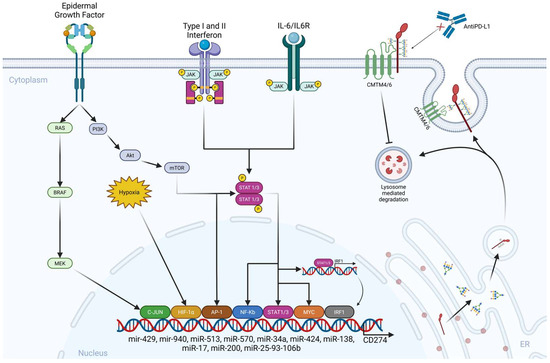
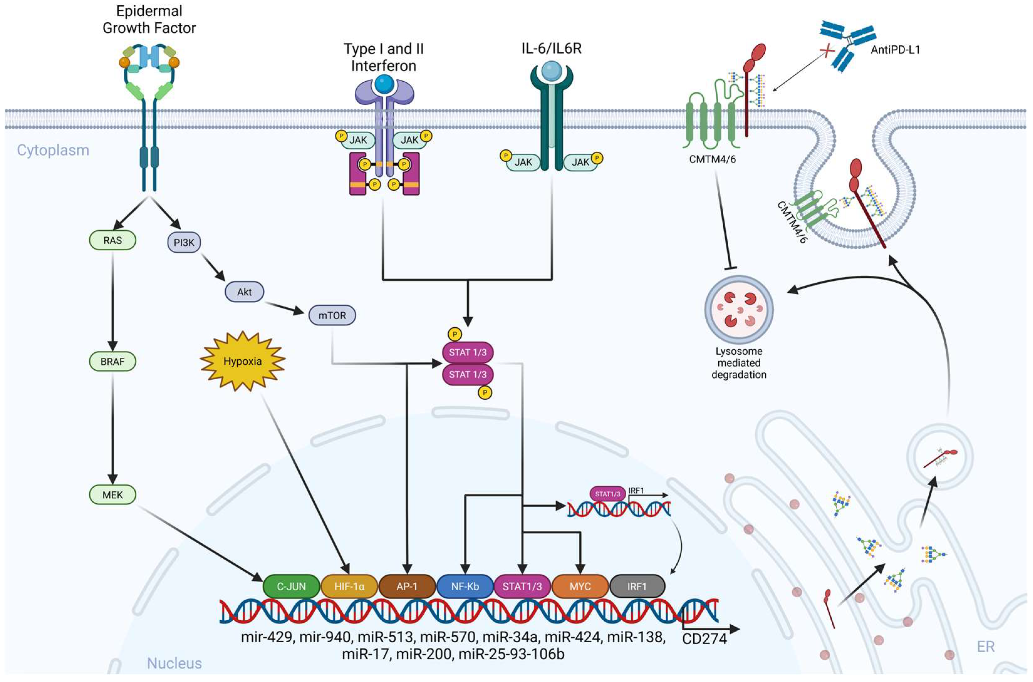
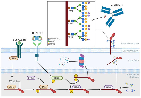
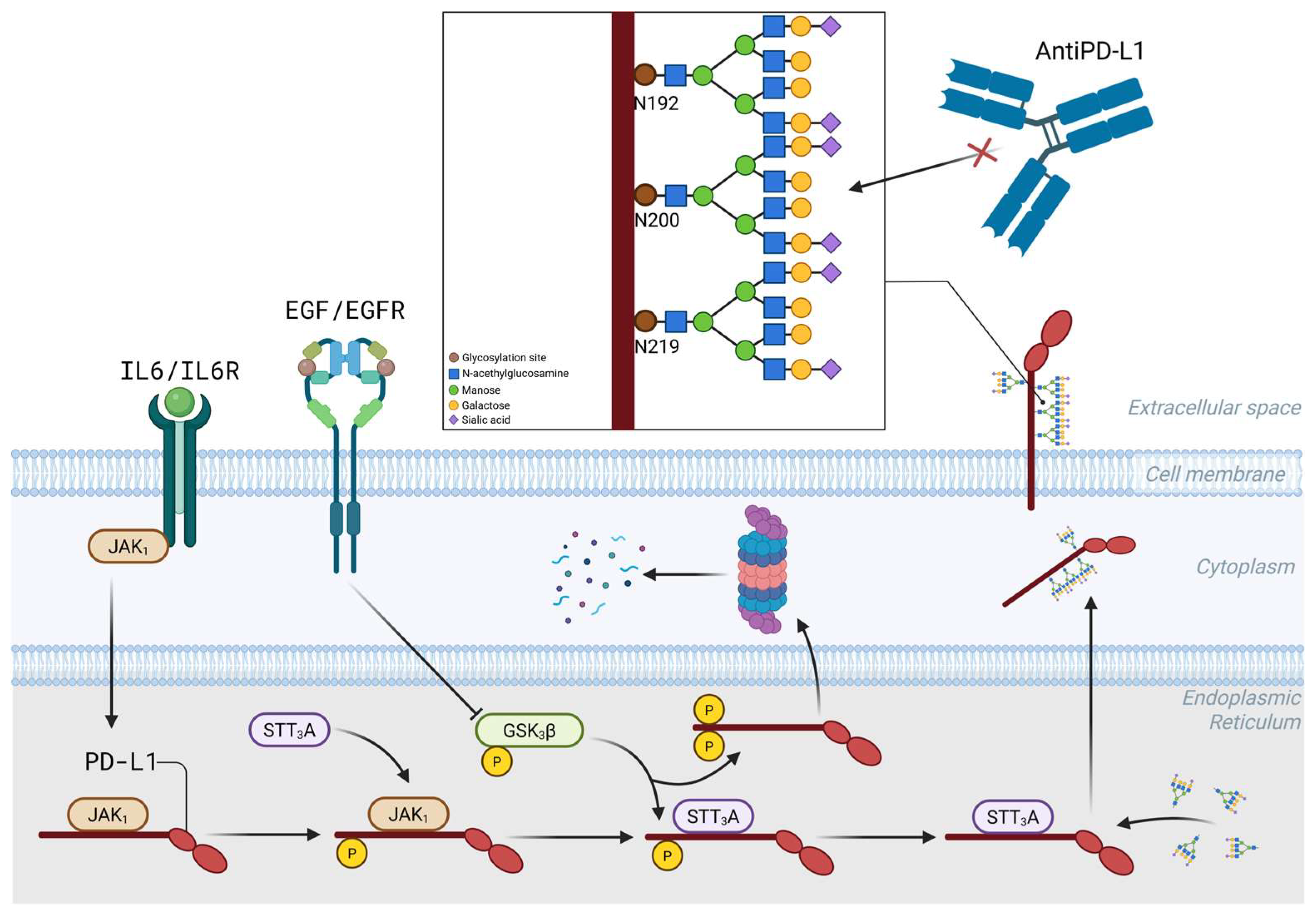
PD-L1 is upregulated in a JAK1/JAK2-/STAT1/STAT2/STAT3-dependent manner by type I and II IFNs through IFN regulatory factor 1 [30,63]. Amplification of the PD-L1 gene (CD274) drives PD-L1 expression in only 33% of solid tumors [64,65]. The transcriptional activation of PD-L1 is influenced by transcription factors such as HIF1-a [66,67], Myc, Stat1/3 [68], NF-kB, and AP-1, C-Jun, and IRF1, which are regulated by pathways involving EGF/PI3K/AKT/MTOR, RTK/Ras/Raf/MEK/ERK, IFNγ/JAKs, TLRs/Myd88/Traf6/IKKs, and a lactate-enriched microenvironment [69,70,71,72]. Micro RNAs, especially miR-513, miR-570, miR-34a, miR-424, miR-138, miR-17, miR-200, and the cluster miR-25-93-106b [73,74], have been implicated in regulating PD-L1 expression [75], along with CMTM4/6, a type I transmembrane protein that prevents PD-L1 from lysosome-mediated degradation and maintains its cell surface expression (Figure 1) [76,77,78,79,80,81]. Methylation of the PD-L1 gene inhibits PD-L1 expression [82]. There are two forms of PD-L1: a 45 kDa protein glycosylated at N35, N192, N200, and N219 aa residues that stabilizes PD-L1 on the cancer cell surface and contributes to immune evasion [83], and a 33 kDa non-glycosylated form (Figure 2) [84]. However, the stability of PD-L1 is significantly influenced by post-transcriptional modifications, including glycosylation, promoted by IL6/IL6R and EGF/EGFR signaling, phosphorylation, and ubiquitination [84,85] (Figure 2). A recently described PD-L1 regulatory mechanism in gastric adenocarcinoma is enhanced glycolysis mediated by claudin 9, which facilitates intracellular lactylation of PD-L1 [86]. Post-translational modifications of PD-L1 modulate its biology and impact receptor–ligand affinity and membrane retention [87]. The addition of a sialic acid residue by the ST3 beta-galactoside alpha-2,3-sialyltransferase 4 (ST3GAL4) to the terminal ends of N-glycan chains facilitates PD-L1 interaction with the cell adhesion molecule Sialoadhesin involved in melanoma lymph node metastatic colonization [88]. PD-L1 has two distinct functions: one as a T cell deactivator, allowing tumors to evade the immune system, and the other as a pro-oncogene. Understanding PD-L1 gene amplification and its regulatory mechanisms is necessary to validate its predictive value as a biomarker [89,90]. For instance, it has been established that circular RNA 0000372, acting as a potential oncogene in gastric cancer [91], upregulates the JAK2/STAT3 signaling pathway [92], positively regulating PD-L1 expression; its silencing suppressed cell invasion and immune escape. The likely wrong assumption related to PD-L1 expression is that it is constantly expressed on tumor cells, and it is highly possible that within each tumor microenvironment, prostatic or gastric, there are at least some sensitive cells with low PD-L1 expression and a pre-existing sub-clone of intrinsically resistant cells with a high capacity for PD-L1 upregulation [93,94].
Figure 1.
PD-L1 inducers and regulatory mechanisms. Created in BioRender. Meléndez, R. (2026) https://BioRender.com/vu2a0mv.
Figure 2.
Post-translational modifications of PD-L1. The PD-L1 extracellular domain modifications occur in the lumen of the endoplasmic reticulum. N-glycosylation of PD-L1, mediated by STT3A, requires JAK1-mediated PD-L1 phosphorylation, but N-glycosylation inhibits phosphorylation mediated by Glycogen Synthase 3 beta (GSK3β). STT3A is the human gene encoding the catalytic subunit in the N-olygosaccharyltransferase complex Created in BioRender. Meléndez, R. (2026) https://BioRender.com/8lf9ijs.
6. Epithelial–Mesenchymal Transition
EMT is a cellular process in which cells lose epithelial characteristics (E-cadherin, a tumor suppressor protein) [95] and acquire mesenchymal features (N-cadherin). Type 1 EMT is essential for embryonic development, Type 2 for wound healing and tissue fibrosis, and Type 3 is closely linked to cancer progression [96,97]. Although EMT is considered a reversible and plastic program with different phenotypic stages of progression [98,99,100], it is also a dynamic and pivotal driver of tumorigenesis [7] and has been implicated in tumor development, cancer stemness, and therapeutic resistance [101]. The EMT program is orchestrated by a core group of EMT-inducing transcription factors (EMT-TFs), including the SNAIL family (SNAI1 [102] and SNAI2 [103]); the ZEB family (ZEB1 [104]); and the TWIST family (TWIST1/2) [8]). These EMT-TFs are essential regulators of, among others, therapeutic resistance and tumor immune evasion.
EMT facilitates tumor metastasis via the ZEB family of transcription factors that represses the scaffolding protein WWC1, leading to impaired Hippo signaling and activation of the transcriptional coactivator YAP which induces, among many others, the transcription of PD-L1 [105,106], thus contributing to immune evasion. YAP, a multi-functional regulator in tumor progression and metastasis [107], also promotes EMT through the activation of SNAIL1, SLUG, ZEB1, and TWIST; it is clearly established that the induction of EMT in the gastric primary tumor is followed by enhanced migration [108,109].
7. Epithelial–Mesenchymal Transition and PD-L1
Bidirectional regulation between EMT and PD-L1 plays a key role in tumor immune escape [12]. PD-L1 expression is increased by IFNγ, especially in the tumor microenvironment [110]. As already mentioned, N-cadherin, ZEB1, SNAIL1, and Vimentin expression correlates with PD-L1 expression, particularly in epithelial–mesenchymal phenotypes [111,112,113]. A positive correlation between PD-L1 expression and the EMT phenotype has been reported but the contribution of tumor-intrinsic PD-L1 signals to the EMT phenotype is slowly emerging [114,115].
The mechanism by which PD-L1 modulates the EMT process remains ambiguous, but it has been established that EMT upregulates PD-L1 expression via the PI3K/AKT pathway in high-EMT-score cancers [116]. PD-L1 expression is regulated at the transcriptional level by the oncogenic transcription factor Yin Yang (YY1) [117] that is significantly overexpressed in gastric cancer [118] and it is implicated in resistance to immune checkpoint therapies [119] through disruption of the PD-1/PD-L1 axis. YY1 contains an miR-200 binding site [120] and upregulates Snail1 [121]. In fact, two distinct mesenchymal states have been defined by PD-L1 expression and miR-200, which regulate the amount of inducing signal required to undergo EMT [122]. As already mentioned, CMTM proteins mediate many mechanisms driving EMT; for instance, TWIST1 is downregulated by CMTM3 in the gastric cancer cell line AGS [123], thereby inhibiting the metastatic potential of cancer cells [124]. Interestingly, CMTM6, the protein that maintains PD-L1 expression on the cell surface [76], has been linked to the induction of T cell tolerance, cytokine synthesis and secretion, and Treg cell differentiation [125], and is now recognized as a master regulator of PD-L1 expression [126], regardless of the presence or absence of IFNγ stimulation [77]. High expression of CMTM6 has been correlated with peritoneal metastasis in gastric cancer [127]. It is important not to set aside the role of the constitutive oncogenic signaling driven by EMT in the upregulation of PD-L1 [12,128]. EMT and PD-L1 upregulation are well-established drivers of tumor progression, as their bidirectional cross-talk facilitates tumor immune evasion. However, the genuine relationship may be hidden at the single-cell level [129].
8. PD-L1 and Cancer Stem Cells
Cancer stem cells (CSCs) are classified into primary CSCs, precancerous stem cells, migratory CSCs, and chemo-radiotherapy-resistant CSCs [130], likely due to metabolic reprogramming that enables them to adapt to their environment and maintain stemness. CSCs acquire a migratory phenotype through EMT [131]. Cancer stem cells, also known as tumor-initiating cells [132], are a distinct subpopulation of tumor cells and key components of the tumor microenvironment, playing roles in immunoregulation and therapy resistance. PD-L1 expression is higher in CSCs than in cancer cells. CSCs possess an efficient redox tolerance system that promotes immunity by selectively inducing PD-L1 expression, which is sometimes mediated by CD44 or by CD44 and CD133 [133,134,135]. For example, CD44+CD24+CD54+EpCAM+ gastric cancer stem cells predict tumor progression and metastasis [136].
CSCs metastasize through a process that involves detachment from the primary tumor, entry into the bloodstream or lymphatic system, survival in circulation, and establishment in distant organs. They accomplish this through EMT mechanisms, which enable them to acquire mesenchymal traits that facilitate detachment. Once in the bloodstream, they survive as they circulate [137,138]. After colonizing a new site, they regain epithelial traits via mesenchymal–epithelial transition mechanisms. These processes require CSCs to maintain their ability to evade immune detection and sustain immune tolerance
Regardless of the CSC markers used to define CSCs across different tumors, most CSCs upregulate immune checkpoint molecules, helping them evade immune surveillance [139]. It is worth noting that environmental factors involved in CSC formation and maintenance, such as hypoxia [140], also contribute to PD-L1 induction. However, it is possible that the overexpression of this crucial molecule, which is essential for allowing tumor cells to evade immune detection and maintain tolerance within the tumor microenvironment, may serve another purpose.
It is well established that PD-1/PD-L1 signaling regulates T cell migration across endothelial cells [141]. PD-L1 is lowly expressed in poorly differentiated and metastatic gastric cancer cells [142]. It is also clear that circulating tumor cells are jettisoned from a single tumor deposit [143,144] composed of tumor cells expressing EMT markers such as CD44 or ICAM-1 [145,146]. PD-L1 expression has been reported on circulating breast cancer cells [147] or on circulating tumor cells isolated from metastatic lung cancer [148], thus enabling evasion of immune cells [149]. Ultimately, understanding the intrinsic properties of circulating tumor cells is an ongoing effort.
9. PD-L1 and the Metastatic Cascade
CSCs metastasize through a process known as the metastatic cascade that involves detachment from the primary tumor, entry into the bloodstream or lymphatic system, survival in circulation, and establishment in distant organs. After colonizing a new site, they regain epithelial traits via mesenchymal–epithelial transition mechanisms. These processes require CSCs to maintain their ability to evade immune detection and sustain immune tolerance [137].
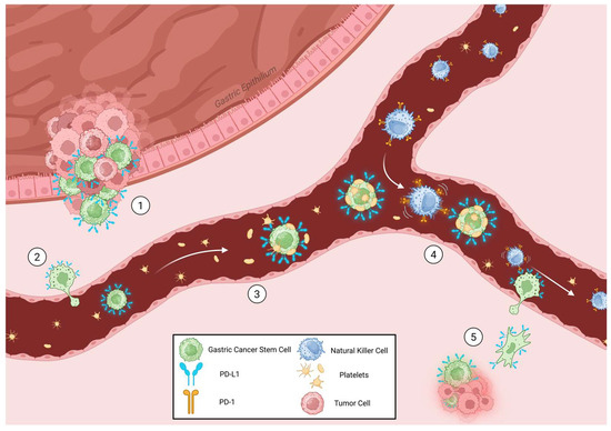
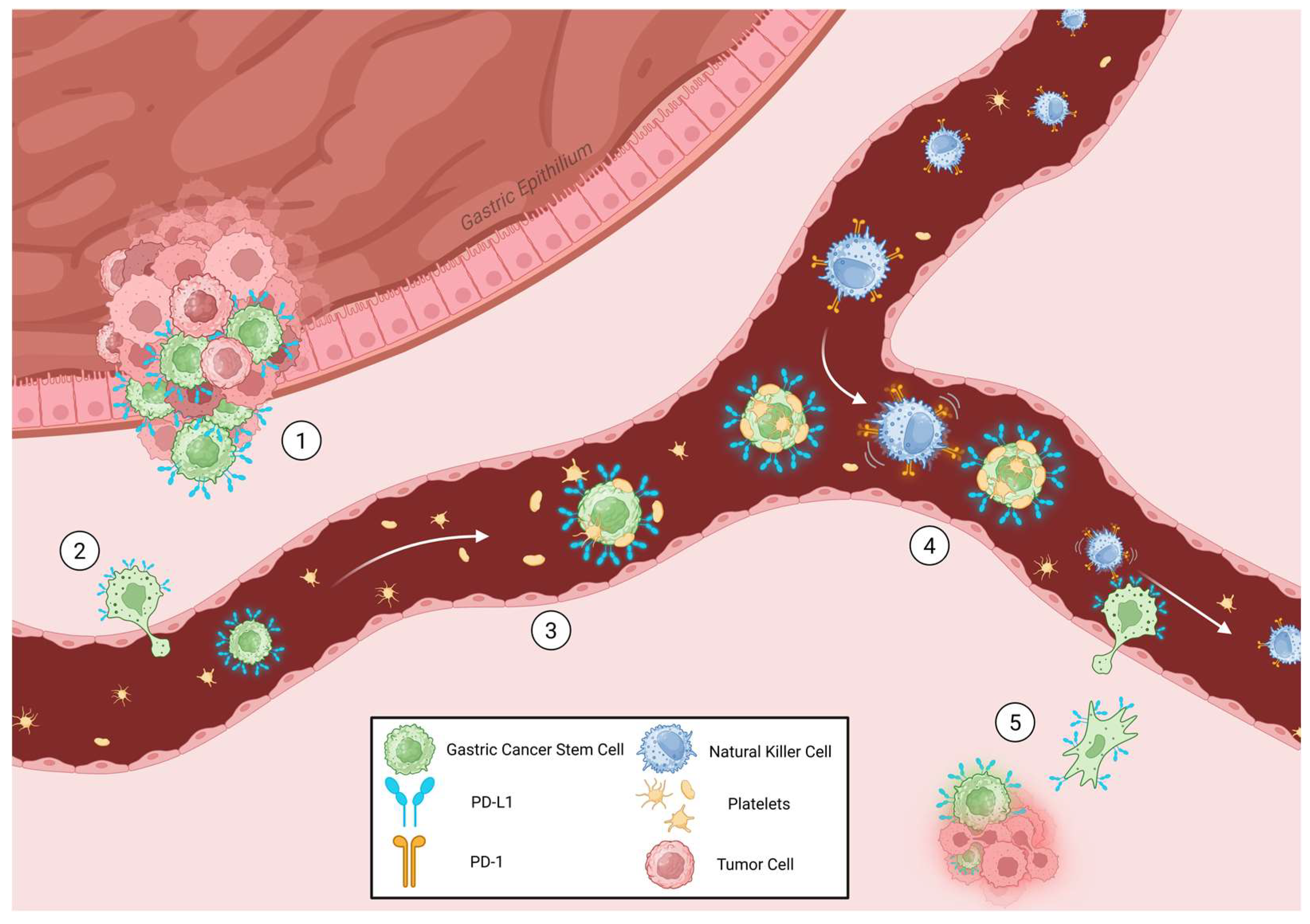
The detachment of cancer cells, presumably cancer stem cells, from the primary tumor is associated with EMT and the downregulation of E-cadherin, followed by degradation of the extracellular matrix and intravasation into blood or lymphatic vessels. Once in circulation, cancer stem cells survive and form aggregates with platelets, which can transfer their MHC-I molecules, tricking the immune system [150]. Platelets also act as armor, preventing the most dangerous enemies, NK cells, from spotting and destroying the abnormal cells [151] (Figure 3).
Figure 3.
Suggested mechanism of metastatic cascade. (1) PD-L1+ gastric CSCs migrate from PD-L1-rich tumoral niche through the extracellular matrix, (2) to the bloodstream by diapedesis. (3) Platelets cover circulating PD-L1+ CSCs to avoid innate immune cell signaling through MHC-1 platelet transfer. (4) Subsequently, the CSC/platelet/NK cell complex is created, and PD-1/PD-L1 axis inhibits cytotoxicity activity, (5) allowing the complex to reach the extravasation site, freeing NK cells and inducing secondary tumor site formation. Created in BioRender. Meléndez, R. (2026) https://BioRender.com/8y5jwo3.
Although the precise origin and phenotype of metastatic gastric cancer cells remain unclear, it is possible that cells that have undergone epithelial–mesenchymal transition [152], thereby replenishing the CSC proportion within the tumor [153], and that express PD-L1 are the source of metastatic cells within tumor niches. Emerging evidence indicates that enhanced PD-L1 expression in tumor-associated neutrophils is the main mechanism suppressing NK cell-mediated antitumor immunity [152] through a STAT3/PD-L1 signaling loop in colon and lung cancer cells [153]. Although there is currently no definitive evidence that our proposal is true, it cannot be ruled out, as PD-L1 expression on the surface of cancer stem cells would protect them from recognition and lytic attack by the tumor environment and circulating NK cells, thereby ensuring the initial steps of the metastatic cascade. NK cells control tumor dissemination, but some circulating tumor cells escape NK cell immunosurveillance by interacting with platelets [154]. Lo et al. showed that NK cells control monoclonal metastasis by eliminating single cancer tumor cells [155]. The relevance of our proposal lies in the idea that metastatic cells arise from PD-L1+ cell niches, creating an “immune-privileged” zone that allows these cells to escape NK cell immunosurveillance within the tumor microenvironment and circulation. Targeting PD-L1 might sensitize these cells to conventional treatment, disrupt the protumor microenvironment, and prevent metastasis and recurrence, underscoring the importance of developing new therapeutic strategies, such as the so-called “niche-busting” strategy using CD133-targeting exosomes, to inhibit PD-L1 expression in CSCs.
Author Contributions
Conceptualization, E.P.R.-H. and L.F.M.; investigation, R.M.-R. and A.-S.A.; data curation, E.P.R.-H.; writing—original draft preparation, L.F.M.; writing—review & editing, L.F.M. and E.P.R.-H. All authors have read and agreed to the published version of the manuscript.
Funding
This research received no external funding.
Data Availability Statement
The original contributions presented in this study are included in the article. Further inquiries can be directed to the corresponding author.
Conflicts of Interest
The authors declare no conflicts of interest.
References
- Gajewski, T.F.; Schreiber, H.; Fu, Y.X. Innate and adaptive immune cells in the tumor microenvironment. Nat. Immunol. 2013, 14, 1014–1022. [Google Scholar] [CrossRef] [Scilit]
- Liu, Y.; Li, C.; Lu, Y.; Liu, C.; Yang, W. Tumor microenvironment-mediated immune tolerance in development and treatment of gastric cancer. Front. Immunol. 2022, 13, 1016817. [Google Scholar] [CrossRef] [Scilit]
- Pitts, S.C.; Schlom, J.; Donahue, R.N. Soluble immune checkpoints: Implications for cancer prognosis and response to immune checkpoint therapy and conventional therapies. J. Exp. Clin. Cancer Res. 2024, 43, 155. [Google Scholar] [CrossRef] [Scilit] [PubMed]
- Cao, Y.; Zhang, L.; Kamimura, Y.; Ritprajak, P.; Hashiguchi, M.; Hirose, S.; Azuma, M. B7-H1 overexpression regulates epithelial-mesenchymal transition and accelerates carcinogenesis in skin. Cancer Res. 2011, 71, 1235–1243. [Google Scholar] [CrossRef] [Scilit] [PubMed]
- Antonangeli, F.; Natalini, A.; Garassino, M.C.; Sica, A.; Santoni, A.; Di Rosa, F. Regulation of PD-L1 Expression by NF-kappaB in Cancer. Front. Immunol. 2020, 11, 584626. [Google Scholar] [CrossRef] [Scilit] [PubMed]
- Peng, Z.; Wang, C.X.; Fang, E.H.; Wang, G.B.; Tong, Q. Role of epithelial-mesenchymal transition in gastric cancer initiation and progression. World J. Gastroenterol. 2014, 20, 5403–5410. [Google Scholar] [CrossRef] [Scilit]
- Brabletz, T.; Kalluri, R.; Nieto, M.A.; Weinberg, R.A. EMT in cancer. Nat. Rev. Cancer 2018, 18, 128–134. [Google Scholar] [CrossRef] [Scilit]
- Goossens, S.; Vandamme, N.; Van Vlierberghe, P.; Berx, G. EMT transcription factors in cancer development re-evaluated: Beyond EMT and MET. Biochim. Biophys. Acta Rev. Cancer 2017, 1868, 584–591. [Google Scholar] [CrossRef] [Scilit]
- Sharif, M.N.; Sosic, D.; Rothlin, C.V.; Kelly, E.; Lemke, G.; Olson, E.N.; Ivashkiv, L.B. Twist mediates suppression of inflammation by type I IFNs and Axl. J. Exp. Med. 2006, 203, 1891–1901. [Google Scholar] [CrossRef] [Scilit]
- Malek, R.; Wang, H.; Taparra, K.; Tran, P.T. Therapeutic Targeting of Epithelial Plasticity Programs: Focus on the Epithelial-Mesenchymal Transition. Cells Tissues Organs 2017, 203, 114–127. [Google Scholar] [CrossRef] [Scilit]
- Terry, S.; Savagner, P.; Ortiz-Cuaran, S.; Mahjoubi, L.; Saintigny, P.; Thiery, J.P.; Chouaib, S. New insights into the role of EMT in tumor immune escape. Mol. Oncol. 2017, 11, 824–846. [Google Scholar] [CrossRef] [Scilit] [PubMed]
- Jiang, Y.; Zhan, H. Communication between EMT and PD-L1 signaling: New insights into tumor immune evasion. Cancer Lett. 2020, 468, 72–81. [Google Scholar] [CrossRef] [Scilit] [PubMed]
- Choi, D.H.; Shin, J.E.; Lee, E.; Kim, S.T.; Lim, S.H. Efficacy of First-Line Nivolumab Plus Chemotherapy in Advanced Gastric Cancer Stratified by PD-L1 Expression: A Real-World Comparison. Cancers 2025, 17, 3716. [Google Scholar] [CrossRef] [Scilit] [PubMed]
- Tian, B.W.; Han, C.L.; Wang, H.C.; Yan, L.J.; Ding, Z.N.; Liu, H.; Mao, X.C.; Tian, J.C.; Xue, J.S.; Yang, L.S.; et al. Effect of liver metastasis on the efficacy of immune checkpoint inhibitors in cancer patients: A systemic review and meta-analysis. Clin. Exp. Metastasis 2023, 40, 255–287. [Google Scholar] [CrossRef] [Scilit]
- Kenison, J.E.; Stevens, N.A.; Quintana, F.J. Therapeutic induction of antigen-specific immune tolerance. Nat. Rev. Immunol. 2024, 24, 338–357. [Google Scholar] [CrossRef] [Scilit]
- Halverson, R.; Torres, R.M.; Pelanda, R. Receptor editing is the main mechanism of B cell tolerance toward membrane antigens. Nat. Immunol. 2004, 5, 645–650. [Google Scholar] [CrossRef] [Scilit]
- Chen, L.; Flies, D.B. Molecular mechanisms of T cell co-stimulation and co-inhibition. Nat. Rev. Immunol. 2013, 13, 227–242, Erratum in Nat. Rev. Immunol. 2013, 13, 542. https://doi.org/10.1038/nri3484. [Google Scholar] [CrossRef] [Scilit]
- Trefzer, A.; Kadam, P.; Wang, S.H.; Pennavaria, S.; Lober, B.; Akcabozan, B.; Kranich, J.; Brocker, T.; Nakano, N.; Irmler, M.; et al. Dynamic adoption of anergy by antigen-exhausted CD4(+) T cells. Cell Rep. 2021, 34, 108748. [Google Scholar] [CrossRef] [Scilit]
- Sharpe, A.H.; Pauken, K.E. The diverse functions of the PD1 inhibitory pathway. Nat. Rev. Immunol. 2018, 18, 153–167. [Google Scholar] [CrossRef] [Scilit]
- Brown, C.C.; Rudensky, A.Y. Spatiotemporal regulation of peripheral T cell tolerance. Science 2023, 380, 472–478. [Google Scholar] [CrossRef] [Scilit]
- Robinson, A.; Han, C.Z.; Glass, C.K.; Pollard, J.W. Monocyte Regulation in Homeostasis and Malignancy. Trends Immunol. 2021, 42, 104–119. [Google Scholar] [CrossRef] [Scilit] [PubMed]
- Xi, J.; Huang, Q.; Wang, L.; Ma, X.; Deng, Q.; Kumar, M.; Zhou, Z.; Li, L.; Zeng, Z.; Young, K.H.; et al. miR-21 depletion in macrophages promotes tumoricidal polarization and enhances PD-1 immunotherapy. Oncogene 2018, 37, 3151–3165. [Google Scholar] [CrossRef] [Scilit] [PubMed]
- Park, B.V.; Freeman, Z.T.; Ghasemzadeh, A.; Chattergoon, M.A.; Rutebemberwa, A.; Steigner, J.; Winter, M.E.; Huynh, T.V.; Sebald, S.M.; Lee, S.J.; et al. TGFbeta1-Mediated SMAD3 Enhances PD-1 Expression on Antigen-Specific T Cells in Cancer. Cancer Discov. 2016, 6, 1366–1381. [Google Scholar] [CrossRef] [Scilit] [PubMed]
- Hanahan, D. Hallmarks of Cancer: New Dimensions. Cancer Discov. 2022, 12, 31–46. [Google Scholar] [CrossRef] [Scilit]
- Baker, K.J.; Houston, A.; Brint, E. IL-1 Family Members in Cancer; Two Sides to Every Story. Front. Immunol. 2019, 10, 1197. [Google Scholar] [CrossRef] [Scilit]
- Hazini, A.; Fisher, K.; Seymour, L. Deregulation of HLA-I in cancer and its central importance for immunotherapy. J. Immunother. Cancer 2021, 9, e002899. [Google Scholar] [CrossRef] [Scilit]
- Galassi, C.; Chan, T.A.; Vitale, I.; Galluzzi, L. The hallmarks of cancer immune evasion. Cancer Cell 2024, 42, 1825–1863. [Google Scholar] [CrossRef] [Scilit]
- Sun, N.; Tian, Y.; Chen, Y.; Guo, W.; Li, C. Metabolic rewiring directs melanoma immunology. Front. Immunol. 2022, 13, 909580. [Google Scholar] [CrossRef] [Scilit]
- Takahashi, H.; Kawabata-Iwakawa, R.; Ida, S.; Mito, I.; Tada, H.; Chikamatsu, K. Upregulated glycolysis correlates with tumor progression and immune evasion in head and neck squamous cell carcinoma. Sci. Rep. 2021, 11, 17789. [Google Scholar] [CrossRef] [Scilit]
- Garcia-Diaz, A.; Shin, D.S.; Moreno, B.H.; Saco, J.; Escuin-Ordinas, H.; Rodriguez, G.A.; Zaretsky, J.M.; Sun, L.; Hugo, W.; Wang, X.; et al. Interferon Receptor Signaling Pathways Regulating PD-L1 and PD-L2 Expression. Cell Rep. 2017, 19, 1189–1201. [Google Scholar] [CrossRef] [Scilit]
- Casey, S.C.; Tong, L.; Li, Y.; Do, R.; Walz, S.; Fitzgerald, K.N.; Gouw, A.M.; Baylot, V.; Gutgemann, I.; Eilers, M.; et al. MYC regulates the antitumor immune response through CD47 and PD-L1. Science 2016, 352, 227–231, Erratum in Science 2016, 352, aaf7984. https://doi.org/10.1126/science.aaf7984. [Google Scholar] [CrossRef] [Scilit] [PubMed]
- Triulzi, T.; Forte, L.; Regondi, V.; Di Modica, M.; Ghirelli, C.; Carcangiu, M.L.; Sfondrini, L.; Balsari, A.; Tagliabue, E. HER2 signaling regulates the tumor immune microenvironment and trastuzumab efficacy. Oncoimmunology 2019, 8, e1512942. [Google Scholar] [CrossRef] [Scilit] [PubMed]
- Ghorani, E.; Swanton, C.; Quezada, S.A. Cancer cell-intrinsic mechanisms driving acquired immune tolerance. Immunity 2023, 56, 2270–2295. [Google Scholar] [CrossRef] [Scilit] [PubMed]
- Dillekas, H.; Rogers, M.S.; Straume, O. Are 90% of deaths from cancer caused by metastases? Cancer Med. 2019, 8, 5574–5576. [Google Scholar] [CrossRef] [Scilit]
- Valastyan, S.; Weinberg, R.A. Tumor metastasis: Molecular insights and evolving paradigms. Cell 2011, 147, 275–292. [Google Scholar] [CrossRef] [Scilit]
- Liu, Y.; Cao, X. Characteristics and Significance of the Pre-metastatic Niche. Cancer Cell 2016, 30, 668–681. [Google Scholar] [CrossRef] [Scilit]
- Lambert, A.W.; Pattabiraman, D.R.; Weinberg, R.A. Emerging Biological Principles of Metastasis. Cell 2017, 168, 670–691. [Google Scholar] [CrossRef] [Scilit]
- Gao, D.; Mittal, V.; Ban, Y.; Lourenco, A.R.; Yomtoubian, S.; Lee, S. Metastatic tumor cells—Genotypes and phenotypes. Front. Biol. 2018, 13, 277–286. [Google Scholar] [CrossRef] [Scilit]
- Chiou, S.H.; Risca, V.I.; Wang, G.X.; Yang, D.; Gruner, B.M.; Kathiria, A.S.; Ma, R.K.; Vaka, D.; Chu, P.; Kozak, M.; et al. BLIMP1 Induces Transient Metastatic Heterogeneity in Pancreatic Cancer. Cancer Discov. 2017, 7, 1184–1199. [Google Scholar] [CrossRef] [Scilit]
- Minn, A.J.; Gupta, G.P.; Siegel, P.M.; Bos, P.D.; Shu, W.; Giri, D.D.; Viale, A.; Olshen, A.B.; Gerald, W.L.; Massague, J. Genes that mediate breast cancer metastasis to lung. Nature 2005, 436, 518–524. [Google Scholar] [CrossRef] [Scilit]
- Albano, F.; Russi, S.; Laurino, S.; Mazzone, P.; Di Paola, G.; Zoppoli, P.; Amendola, E.; Balzamo, C.; Bartolo, O.; Ciuffi, M.; et al. Representing ECM composition and EMT pathways in gastric cancer using a new metastatic gene signature. Front. Cell Dev. Biol. 2024, 12, 1481818. [Google Scholar] [CrossRef] [Scilit] [PubMed]
- Elizazu, J.; Artetxe-Zurutuza, A.; Otaegi-Ugartemendia, M.; Moncho-Amor, V.; Moreno-Valladares, M.; Matheu, A.; Carrasco-Garcia, E. Identification of a novel gene signature related to prognosis and metastasis in gastric cancer. Cell. Oncol. 2024, 47, 1355–1373. [Google Scholar] [CrossRef] [Scilit] [PubMed]
- Jeong, H.; Koh, J.; Kim, S.; Yim, J.; Song, S.G.; Kim, H.; Li, Y.; Lee, S.H.; Chung, Y.K.; Kim, H.; et al. Cell-intrinsic PD-L1 signaling drives immunosuppression by myeloid-derived suppressor cells through IL-6/Jak/Stat3 in PD-L1-high lung cancer. J. Immunother. Cancer 2025, 13, e010612. [Google Scholar] [CrossRef] [Scilit] [PubMed]
- Zhang, Q.; Chai, S.; Wang, W.; Wan, C.; Zhang, F.; Li, Y.; Wang, F. Macrophages activate mesenchymal stem cells to acquire cancer-associated fibroblast-like features resulting in gastric epithelial cell lesions and malignant transformation in vitro. Oncol. Lett. 2019, 17, 747–756. [Google Scholar] [CrossRef] [Scilit]
- Ridge, S.M.; Sullivan, F.J.; Glynn, S.A. Mesenchymal stem cells: Key players in cancer progression. Mol. Cancer 2017, 16, 31. [Google Scholar] [CrossRef] [Scilit]
- Xu, R.; Zhao, X.; Zhao, Y.; Chen, B.; Sun, L.; Xu, C.; Shen, B.; Wang, M.; Xu, W.; Zhu, W. Enhanced gastric cancer growth potential of mesenchymal stem cells derived from gastric cancer tissues educated by CD4(+) T cells. Cell Prolif. 2018, 51, e12399. [Google Scholar] [CrossRef] [Scilit]
- Sun, L.; Wang, Q.; Chen, B.; Zhao, Y.; Shen, B.; Wang, X.; Zhu, M.; Li, Z.; Zhao, X.; Xu, C.; et al. Human Gastric Cancer Mesenchymal Stem Cell-Derived IL15 Contributes to Tumor Cell Epithelial-Mesenchymal Transition via Upregulation Tregs Ratio and PD-1 Expression in CD4(+)T Cell. Stem Cells Dev. 2018, 27, 1203–1214. [Google Scholar] [CrossRef] [Scilit]
- He, X.; Guan, X.Y.; Li, Y. Clinical significance of the tumor microenvironment on immune tolerance in gastric cancer. Front. Immunol. 2025, 16, 1532605. [Google Scholar] [CrossRef] [Scilit]
- Liao, W.; Zhou, X.; Lin, H.; Feng, Z.; Chen, X.; Chen, Y.; Chen, M.; Lin, M.; Yao, G.; Chen, J.; et al. Interplay between tumor mutation burden and the tumor microenvironment predicts the prognosis of pan-cancer anti-PD-1/PD-L1 therapy. Front. Immunol. 2025, 16, 1557461. [Google Scholar] [CrossRef] [Scilit]
- Hill, C.N.; Maita, G.; Cabrolier, C.; Aros, C.; Vega-Letter, A.M.; Gonzalez, P.; Kalergis, A.M.; Luz-Crawford, P.; Owen, G.I. Galectin-9 and Tim-3 in gastric cancer: A checkpoint axis driving T cell exhaustion and Treg-mediated immunosuppression independently of anti-PD-1 blockade. Front. Immunol. 2025, 16, 1600792. [Google Scholar] [CrossRef] [Scilit]
- Li, Z.; Liu, X.; Cai, N.; Zhou, Z.; Huang, H.; Wu, Q.; Xu, L.; Zhu, W.G.; Zhang, C.; Wei, Z.; et al. Immune checkpoint reprogramming via sequential nucleic acid delivery strategy optimizes systemic immune responses for gastrointestinal cancer immunotherapy. Cancer Lett. 2024, 599, 217152. [Google Scholar] [CrossRef] [Scilit]
- Wang, D.W.; Zhang, W.H.; Danil, G.; Yang, K.; Hu, J.K. The role and mechanism of claudins in cancer. Front. Oncol. 2022, 12, 1051497. [Google Scholar] [CrossRef] [Scilit]
- Bretscher, P.A. A two-step, two-signal model for the primary activation of precursor helper T cells. Proc. Natl. Acad. Sci. USA 1999, 96, 185–190. [Google Scholar] [CrossRef] [Scilit]
- Wherry, E.J. T cell exhaustion. Nat. Immunol. 2011, 12, 492–499. [Google Scholar] [CrossRef] [Scilit] [PubMed]
- Rotrosen, D.; Matthews, J.B.; Bluestone, J.A. The immune tolerance network: A new paradigm for developing tolerance-inducing therapies. J. Allergy Clin. Immunol. 2002, 110, 17–23. [Google Scholar] [CrossRef] [Scilit] [PubMed]
- Schietinger, A.; Greenberg, P.D. Tolerance and exhaustion: Defining mechanisms of T cell dysfunction. Trends Immunol. 2014, 35, 51–60. [Google Scholar] [CrossRef] [Scilit] [PubMed]
- Wherry, E.J.; Kurachi, M. Molecular and cellular insights into T cell exhaustion. Nat. Rev. Immunol. 2015, 15, 486–499. [Google Scholar] [CrossRef] [Scilit]
- Rizvi, N.A.; Mazieres, J.; Planchard, D.; Stinchcombe, T.E.; Dy, G.K.; Antonia, S.J.; Horn, L.; Lena, H.; Minenza, E.; Mennecier, B.; et al. Activity and safety of nivolumab, an anti-PD-1 immune checkpoint inhibitor, for patients with advanced, refractory squamous non-small-cell lung cancer (CheckMate 063): A phase 2, single-arm trial. Lancet Oncol. 2015, 16, 257–265. [Google Scholar] [CrossRef] [Scilit]
- Herbst, R.S.; Soria, J.C.; Kowanetz, M.; Fine, G.D.; Hamid, O.; Gordon, M.S.; Sosman, J.A.; McDermott, D.F.; Powderly, J.D.; Gettinger, S.N.; et al. Predictive correlates of response to the anti-PD-L1 antibody MPDL3280A in cancer patients. Nature 2014, 515, 563–567. [Google Scholar] [CrossRef] [Scilit]
- Haile, S.T.; Bosch, J.J.; Agu, N.I.; Zeender, A.M.; Somasundaram, P.; Srivastava, M.K.; Britting, S.; Wolf, J.B.; Ksander, B.R.; Ostrand-Rosenberg, S. Tumor cell programmed death ligand 1-mediated T cell suppression is overcome by coexpression of CD80. J. Immunol. 2011, 186, 6822–6829. [Google Scholar] [CrossRef] [Scilit]
- Ritprajak, P.; Azuma, M. Intrinsic and extrinsic control of expression of the immunoregulatory molecule PD-L1 in epithelial cells and squamous cell carcinoma. Oral Oncol. 2015, 51, 221–228. [Google Scholar] [CrossRef] [Scilit]
- Simon, S.; Labarriere, N. PD-1 expression on tumor-specific T cells: Friend or foe for immunotherapy? Oncoimmunology 2017, 7, e1364828. [Google Scholar] [CrossRef] [Scilit] [PubMed]
- Darnell, J.E., Jr.; Kerr, I.M.; Stark, G.R. Jak-STAT pathways and transcriptional activation in response to IFNs and other extracellular signaling proteins. Science 1994, 264, 1415–1421. [Google Scholar] [CrossRef] [Scilit] [PubMed]
- Clave, S.; Pijuan, L.; Casadevall, D.; Taus, A.; Gimeno, J.; Hernandez-Llodra, S.; Rodriguez-Rivera, M.; Lorenzo, M.; Menendez, S.; Albanell, J.; et al. CD274 (PDL1) and JAK2 genomic amplifications in pulmonary squamous-cell and adenocarcinoma patients. Histopathology 2018, 72, 259–269. [Google Scholar] [CrossRef] [Scilit] [PubMed]
- Nakayama, T.; Takahama, T.; Chiba, Y.; Shiraishi, N.; Kawakami, H.; Yonesaka, K.; Nakagawa, K.; Hayashi, H. Evaluation of immune checkpoint inhibitor efficacy for solid tumors with CD274 (PD-L1 gene) amplification identified by comprehensive genomic profiling: Retrospective study based on a nationwide database. J. Immunother. Cancer 2024, 12, e010130. [Google Scholar] [CrossRef] [Scilit]
- Pollizzi, K.N.; Powell, J.D. Integrating canonical and metabolic signalling programmes in the regulation of T cell responses. Nat. Rev. Immunol. 2014, 14, 435–446. [Google Scholar] [CrossRef] [Scilit]
- Huang, C.; Chen, B.; Wang, X.; Xu, J.; Sun, L.; Wang, D.; Zhao, Y.; Zhou, C.; Gao, Q.; Wang, Q.; et al. Gastric cancer mesenchymal stem cells via the CXCR2/HK2/PD-L1 pathway mediate immunosuppression. Gastric Cancer 2023, 26, 691–707. [Google Scholar] [CrossRef] [Scilit]
- Marzec, M.; Zhang, Q.; Goradia, A.; Raghunath, P.N.; Liu, X.; Paessler, M.; Wang, H.Y.; Wysocka, M.; Cheng, M.; Ruggeri, B.A.; et al. Oncogenic kinase NPM/ALK induces through STAT3 expression of immunosuppressive protein CD274 (PD-L1, B7-H1). Proc. Natl. Acad. Sci. USA 2008, 105, 20852–20857. [Google Scholar] [CrossRef] [Scilit]
- Chan, L.C.; Li, C.W.; Xia, W.; Hsu, J.M.; Lee, H.H.; Cha, J.H.; Wang, H.L.; Yang, W.H.; Yen, E.Y.; Chang, W.C.; et al. IL-6/JAK1 pathway drives PD-L1 Y112 phosphorylation to promote cancer immune evasion. J. Clin. Investig. 2019, 129, 3324–3338. [Google Scholar] [CrossRef] [Scilit]
- Wang, Y.M.; Liu, H.X.; Fang, N.Y. High Glucose Concentration Impairs 5-PAHSA Activity by Inhibiting AMP-Activated Protein Kinase Activation and Promoting Nuclear Factor-Kappa-B-Mediated Inflammation. Front. Pharmacol. 2018, 9, 1491. [Google Scholar] [CrossRef] [Scilit]
- Atefi, M.; Avramis, E.; Lassen, A.; Wong, D.J.; Robert, L.; Foulad, D.; Cerniglia, M.; Titz, B.; Chodon, T.; Graeber, T.G.; et al. Effects of MAPK and PI3K pathways on PD-L1 expression in melanoma. Clin. Cancer Res. 2014, 20, 3446–3457. [Google Scholar] [CrossRef] [Scilit]
- Moon, J.W.; Kong, S.K.; Kim, B.S.; Kim, H.J.; Lim, H.; Noh, K.; Kim, Y.; Choi, J.W.; Lee, J.H.; Kim, Y.S. IFNgamma induces PD-L1 overexpression by JAK2/STAT1/IRF-1 signaling in EBV-positive gastric carcinoma. Sci. Rep. 2017, 7, 17810. [Google Scholar] [CrossRef] [Scilit] [PubMed]
- Cioffi, M.; Trabulo, S.M.; Vallespinos, M.; Raj, D.; Kheir, T.B.; Lin, M.L.; Begum, J.; Baker, A.M.; Amgheib, A.; Saif, J.; et al. The miR-25-93-106b cluster regulates tumor metastasis and immune evasion via modulation of CXCL12 and PD-L1. Oncotarget 2017, 8, 21609–21625. [Google Scholar] [CrossRef] [Scilit] [PubMed]
- Jin, M.; Fang, J.; Peng, J.; Wang, X.; Xing, P.; Jia, K.; Hu, J.; Wang, D.; Ding, Y.; Wang, X.; et al. PD-1/PD-L1 immune checkpoint blockade in breast cancer: Research insights and sensitization strategies. Mol. Cancer 2024, 23, 266. [Google Scholar] [CrossRef] [Scilit] [PubMed]
- Wang, Q.; Lin, W.; Tang, X.; Li, S.; Guo, L.; Lin, Y.; Kwok, H.F. The Roles of microRNAs in Regulating the Expression of PD-1/PD-L1 Immune Checkpoint. Int. J. Mol. Sci. 2017, 18, 2540. [Google Scholar] [CrossRef] [Scilit]
- Mezzadra, R.; Sun, C.; Jae, L.T.; Gomez-Eerland, R.; de Vries, E.; Wu, W.; Logtenberg, M.E.W.; Slagter, M.; Rozeman, E.A.; Hofland, I.; et al. Identification of CMTM6 and CMTM4 as PD-L1 protein regulators. Nature 2017, 549, 106–110. [Google Scholar] [CrossRef] [Scilit]
- Burr, M.L.; Sparbier, C.E.; Chan, Y.C.; Williamson, J.C.; Woods, K.; Beavis, P.A.; Lam, E.Y.N.; Henderson, M.A.; Bell, C.C.; Stolzenburg, S.; et al. CMTM6 maintains the expression of PD-L1 and regulates anti-tumour immunity. Nature 2017, 549, 101–105. [Google Scholar] [CrossRef] [Scilit]
- Mamessier, E.; Birnbaum, D.J.; Finetti, P.; Birnbaum, D.; Bertucci, F. CMTM6 stabilizes PD-L1 expression and refines its prognostic value in tumors. Ann. Transl. Med. 2018, 6, 54. [Google Scholar] [CrossRef] [Scilit]
- Li, X.; Chen, L.; Gu, C.; Sun, Q.; Li, J. CMTM6 significantly relates to PD-L1 and predicts the prognosis of gastric cancer patients. PeerJ 2020, 8, e9536. [Google Scholar] [CrossRef] [Scilit]
- Xiao, M.; Duhem, C.; Chammout, A.; Berchem, G.; Janji, B. CMTM6 and CMTM7: New leads for PD-L1 regulation in breast cancer cells undergoing EMT. J. Cell. Biochem. 2022, 123, 1025–1031. [Google Scholar] [CrossRef] [Scilit]
- Zhang, T.; Yu, T.; Yang, X.; Tian, W.; Wei, J. Editorial: The mechanism and novel strategies of overcoming resistance of hematological malignancies to CAR-T cell killing. Front. Immunol. 2022, 13, 1093339. [Google Scholar] [CrossRef] [Scilit] [PubMed]
- Goltz, D.; Gevensleben, H.; Dietrich, J.; Dietrich, D. PD-L1 (CD274) promoter methylation predicts survival in colorectal cancer patients. Oncoimmunology 2017, 6, e1257454. [Google Scholar] [CrossRef] [Scilit] [PubMed]
- Lee, T.A.; Tsai, E.Y.; Liu, S.H.; Chou, W.C.; Hsu Hung, S.D.; Chang, C.Y.; Chao, C.H.; Yamaguchi, H.; Lai, Y.J.; Chen, H.L.; et al. Regulation of PD-L1 glycosylation and advances in cancer immunotherapy. Cancer Lett. 2025, 612, 217498. [Google Scholar] [CrossRef] [Scilit] [PubMed]
- Li, C.W.; Lim, S.O.; Xia, W.; Lee, H.H.; Chan, L.C.; Kuo, C.W.; Khoo, K.H.; Chang, S.S.; Cha, J.H.; Kim, T.; et al. Glycosylation and stabilization of programmed death ligand-1 suppresses T-cell activity. Nat. Commun. 2016, 7, 12632. [Google Scholar] [CrossRef] [Scilit]
- Horita, H.; Law, A.; Hong, S.; Middleton, K. Identifying Regulatory Posttranslational Modifications of PD-L1: A Focus on Monoubiquitinaton. Neoplasia 2017, 19, 346–353. [Google Scholar] [CrossRef] [Scilit]
- Hu, X.; Ouyang, W.; Chen, H.; Liu, Z.; Lai, Z.; Yao, H. Claudin-9 (CLDN9) promotes gastric cancer progression by enhancing the glycolysis pathway and facilitating PD-L1 lactylation to suppress CD8(+) T cell anti-tumor immunity. Cancer Pathog. Ther. 2025, 3, 253–266. [Google Scholar] [CrossRef] [Scilit]
- Hsieh, H.C.; Ling, L.L.; Wang, Y.C. Post-translational modifications of immune checkpoints: Molecular mechanisms, tumor microenvironment remodeling, and therapeutic implications. J. Biomed. Sci. 2026, 33, 3. [Google Scholar] [CrossRef] [Scilit]
- Singh, R.; Choi, B.K. Siglec1-expressing subcapsular sinus macrophages provide soil for melanoma lymph node metastasis. Elife 2019, 8, e48916. [Google Scholar] [CrossRef] [Scilit]
- Pell, B.; Kalizhanova, A.; Tursynkozha, A.; Dengi, D.; Kashkynbayev, A.; Kuang, Y. Dynamic PD-L1 Regulation Shapes Tumor Immune Escape and Response to Immunotherapy. Cancers 2025, 17, 3803. [Google Scholar] [CrossRef] [Scilit]
- Lin, X.; Kang, K.; Chen, P.; Zeng, Z.; Li, G.; Xiong, W.; Yi, M.; Xiang, B. Regulatory mechanisms of PD-1/PD-L1 in cancers. Mol. Cancer 2024, 23, 108. [Google Scholar] [CrossRef] [Scilit]
- Shen, C.; Li, W.; Shi, J.; Wu, Y.; Yan, Y. Knockdown of circ_0000372 elevates PD-L1 level through sponging miR-488-3p to restrain cell viability, motility and immune escape in gastric cancer. J. Mol. Histol. 2025, 56, 312. [Google Scholar] [CrossRef] [Scilit] [PubMed]
- Liu, X.; Qin, Y.; Tang, X.; Wang, Y.; Bian, C.; Zhong, J. Circular RNA circ_0000372 contributes to the proliferation, migration and invasion of colorectal cancer by elevating IL6 expression via sponging miR-495. Anticancer. Drugs 2021, 32, 296–305. [Google Scholar] [CrossRef] [Scilit] [PubMed]
- Gniadek, T.J.; Li, Q.K.; Tully, E.; Chatterjee, S.; Nimmagadda, S.; Gabrielson, E. Heterogeneous expression of PD-L1 in pulmonary squamous cell carcinoma and adenocarcinoma: Implications for assessment by small biopsy. Mod. Pathol. 2017, 30, 530–538. [Google Scholar] [CrossRef] [Scilit]
- Kume, K.; Iida, M.; Iwaya, T.; Yashima-Abo, A.; Koizumi, Y.; Endo, A.; Wade, K.; Hiraki, H.; Calvert, V.; Wulfkuhle, J.; et al. Targeted Dynamic Phospho-Proteogenomic Analysis of Gastric Cancer Cells Suggests Host Immunity Provides Survival Benefit. Mol. Cell. Proteom. 2024, 23, 100870. [Google Scholar] [CrossRef] [Scilit] [PubMed]
- Na, T.Y.; Schecterson, L.; Mendonsa, A.M.; Gumbiner, B.M. The functional activity of E-cadherin controls tumor cell metastasis at multiple steps. Proc. Natl. Acad. Sci. USA 2020, 117, 5931–5937. [Google Scholar] [CrossRef] [Scilit]
- Nieto, M.A.; Huang, R.Y.; Jackson, R.A.; Thiery, J.P. Emt: 2016. Cell 2016, 166, 21–45. [Google Scholar] [CrossRef] [Scilit]
- Tomecka, P.; Kunachowicz, D.; Gorczynska, J.; Gebuza, M.; Kuznicki, J.; Skinderowicz, K.; Choromanska, A. Factors Determining Epithelial-Mesenchymal Transition in Cancer Progression. Int. J. Mol. Sci. 2024, 25, 8972. [Google Scholar] [CrossRef] [Scilit]
- Chaffer, C.L.; San Juan, B.P.; Lim, E.; Weinberg, R.A. EMT, cell plasticity and metastasis. Cancer Metastasis Rev. 2016, 35, 645–654. [Google Scholar] [CrossRef] [Scilit]
- Zhang, Y.; Weinberg, R.A. Epithelial-to-mesenchymal transition in cancer: Complexity and opportunities. Front. Med. 2018, 12, 361–373. [Google Scholar] [CrossRef] [Scilit]
- Zheng, X.; Yu, C.; Xu, M. Linking Tumor Microenvironment to Plasticity of Cancer Stem Cells: Mechanisms and Application in Cancer Therapy. Front. Oncol. 2021, 11, 678333. [Google Scholar] [CrossRef] [Scilit]
- Lambert, A.W.; Weinberg, R.A. Linking EMT programmes to normal and neoplastic epithelial stem cells. Nat. Rev. Cancer 2021, 21, 325–338. [Google Scholar] [CrossRef] [Scilit] [PubMed]
- Batlle, E.; Sancho, E.; Franci, C.; Dominguez, D.; Monfar, M.; Baulida, J.; Garcia De Herreros, A. The transcription factor snail is a repressor of E-cadherin gene expression in epithelial tumour cells. Nat. Cell Biol. 2000, 2, 84–89. [Google Scholar] [CrossRef] [Scilit] [PubMed]
- Bolos, V.; Peinado, H.; Perez-Moreno, M.A.; Fraga, M.F.; Esteller, M.; Cano, A. The transcription factor Slug represses E-cadherin expression and induces epithelial to mesenchymal transitions: A comparison with Snail and E47 repressors. J. Cell Sci. 2003, 116, 499–511, Erratum in J. Cell Sci. 2016, 129, 1283. https://doi.org/10.1242/jcs.188243. [Google Scholar] [CrossRef] [Scilit] [PubMed]
- Eger, A.; Aigner, K.; Sonderegger, S.; Dampier, B.; Oehler, S.; Schreiber, M.; Berx, G.; Cano, A.; Beug, H.; Foisner, R. DeltaEF1 is a transcriptional repressor of E-cadherin and regulates epithelial plasticity in breast cancer cells. Oncogene 2005, 24, 2375–2385. [Google Scholar] [CrossRef] [Scilit]
- Janse van Rensburg, H.J.; Azad, T.; Ling, M.; Hao, Y.; Snetsinger, B.; Khanal, P.; Minassian, L.M.; Graham, C.H.; Rauh, M.J.; Yang, X. The Hippo Pathway Component TAZ Promotes Immune Evasion in Human Cancer through PD-L1. Cancer Res. 2018, 78, 1457–1470. [Google Scholar] [CrossRef] [Scilit]
- Huang, X.; Zhang, M.; Pearce, A.D.; Gibbons, M.D.; Jin, D.; Li, L.; Hu, D.; Liu, R.; Yu, M.; Tan, M.; et al. Epithelial-Mesenchymal Transition Activates YAP to Drive Malignant Progression and Immune Evasion. Cancers 2025, 17, 2767. [Google Scholar] [CrossRef] [Scilit]
- Thrash, H.L.; Pendergast, A.M. Multi-Functional Regulation by YAP/TAZ Signaling Networks in Tumor Progression and Metastasis. Cancers 2023, 15, 4701. [Google Scholar] [CrossRef] [Scilit]
- Sun, D.; Li, X.; He, Y.; Li, W.; Wang, Y.; Wang, H.; Jiang, S.; Xin, Y. YAP1 enhances cell proliferation, migration, and invasion of gastric cancer in vitro and in vivo. Oncotarget 2016, 7, 81062–81076. [Google Scholar] [CrossRef] [Scilit]
- Liu, Y.; Jin, T.; Chen, R.; Miao, R.; Zhou, Y.; Shao, S. High expression of ABL2 promotes gastric cancer cells migration, invasion and proliferation via the TGF-beta and YAP signaling pathways. J. Cancer 2024, 15, 5719–5728. [Google Scholar] [CrossRef] [Scilit]
- Shiravand, Y.; Khodadadi, F.; Kashani, S.M.A.; Hosseini-Fard, S.R.; Hosseini, S.; Sadeghirad, H.; Ladwa, R.; O’Byrne, K.; Kulasinghe, A. Immune Checkpoint Inhibitors in Cancer Therapy. Curr. Oncol. 2022, 29, 3044–3060. [Google Scholar] [CrossRef] [Scilit]
- Lou, Y.; Diao, L.; Cuentas, E.R.; Denning, W.L.; Chen, L.; Fan, Y.H.; Byers, L.A.; Wang, J.; Papadimitrakopoulou, V.A.; Behrens, C.; et al. Epithelial-Mesenchymal Transition Is Associated with a Distinct Tumor Microenvironment Including Elevation of Inflammatory Signals and Multiple Immune Checkpoints in Lung Adenocarcinoma. Clin. Cancer Res. 2016, 22, 3630–3642. [Google Scholar] [CrossRef] [Scilit]
- Dongre, A.; Rashidian, M.; Reinhardt, F.; Bagnato, A.; Keckesova, Z.; Ploegh, H.L.; Weinberg, R.A. Epithelial-to-Mesenchymal Transition Contributes to Immunosuppression in Breast Carcinomas. Cancer Res. 2017, 77, 3982–3989. [Google Scholar] [CrossRef] [Scilit] [PubMed]
- Tsutsumi, S.; Saeki, H.; Nakashima, Y.; Ito, S.; Oki, E.; Morita, M.; Oda, Y.; Okano, S.; Maehara, Y. Programmed death-ligand 1 expression at tumor invasive front is associated with epithelial-mesenchymal transition and poor prognosis in esophageal squamous cell carcinoma. Cancer Sci. 2017, 108, 1119–1127. [Google Scholar] [CrossRef] [Scilit] [PubMed]
- Clark, C.A.; Gupta, H.B.; Sareddy, G.; Pandeswara, S.; Lao, S.; Yuan, B.; Drerup, J.M.; Padron, A.; Conejo-Garcia, J.; Murthy, K.; et al. Tumor-Intrinsic PD-L1 Signals Regulate Cell Growth, Pathogenesis, and Autophagy in Ovarian Cancer and Melanoma. Cancer Res. 2016, 76, 6964–6974, Erratum in Cancer Res. 2017, 77, 2770. https://doi.org/10.1158/0008-5472.CAN-17-0351. [Google Scholar] [CrossRef] [Scilit] [PubMed]
- Li, Z.; Wang, F.; Dang, J.; Cheng, F.; Zheng, F. Bidirectional regulation between tumor cell-intrinsic PD-L1 and TGF-beta1 in epithelial-to-mesenchymal transition in melanoma. Transl. Cancer Res. 2022, 11, 3698–3710. [Google Scholar] [CrossRef] [Scilit]
- Alsuliman, A.; Colak, D.; Al-Harazi, O.; Fitwi, H.; Tulbah, A.; Al-Tweigeri, T.; Al-Alwan, M.; Ghebeh, H. Bidirectional crosstalk between PD-L1 expression and epithelial to mesenchymal transition: Significance in claudin-low breast cancer cells. Mol. Cancer 2015, 14, 149. [Google Scholar] [CrossRef] [Scilit]
- Hays, E.; Bonavida, B. YY1 regulates cancer cell immune resistance by modulating PD-L1 expression. Drug Resist. Updat. 2019, 43, 10–28. [Google Scholar] [CrossRef] [Scilit]
- Chen, W.; Wang, H.; Achi, N.; Hao, J.; Gong, R.; Zhao, Q. Bioinformatics Analysis of the Expression and Prognostic Significance of Transcription Factor YY1 in Gastric Cancer. Cancer Rep. 2025, 8, e70181. [Google Scholar] [CrossRef] [Scilit]
- Li, M.; Wei, J.; Xue, C.; Zhou, X.; Chen, S.; Zheng, L.; Duan, Y.; Deng, H.; Xiong, W.; Tang, F.; et al. Dissecting the roles and clinical potential of YY1 in the tumor microenvironment. Front. Oncol. 2023, 13, 1122110. [Google Scholar] [CrossRef] [Scilit]
- Jo, H.; Shim, K.; Jeoung, D. Potential of the miR-200 Family as a Target for Developing Anti-Cancer Therapeutics. Int. J. Mol. Sci. 2022, 23, 5881. [Google Scholar] [CrossRef] [Scilit]
- Bonavida, B.; Kaufhold, S. Prognostic significance of YY1 protein expression and mRNA levels by bioinformatics analysis in human cancers: A therapeutic target. Pharmacol. Ther. 2015, 150, 149–168. [Google Scholar] [CrossRef] [Scilit] [PubMed]
- Burger, G.A.; Nesenberend, D.N.; Lems, C.M.; Hille, S.C.; Beltman, J.B. Bidirectional crosstalk between epithelial-mesenchymal plasticity and IFNgamma-induced PD-L1 expression promotes tumour progression. R. Soc. Open Sci. 2022, 9, 220186. [Google Scholar] [CrossRef] [Scilit] [PubMed]
- Yuan, W.; Li, T.; Mo, X.; Wang, X.; Liu, B.; Wang, W.; Su, Y.; Xu, L.; Han, W. Knockdown of CMTM3 promotes metastasis of gastric cancer via the STAT3/Twist1/EMT signaling pathway. Oncotarget 2016, 7, 29507–29519. [Google Scholar] [CrossRef] [Scilit] [PubMed]
- Li, W.; Zhang, S. CKLF-Like MARVEL Transmembrane Domain-Containing Member 3 (CMTM3) Inhibits the Proliferation and Tumorigenisis in Hepatocellular Carcinoma Cells. Oncol. Res. 2017, 25, 285–293. [Google Scholar] [CrossRef] [Scilit]
- Wu, J.; Li, L.; Wu, S.; Xu, B. CMTM family proteins 1-8: Roles in cancer biological processes and potential clinical value. Cancer Biol. Med. 2020, 17, 528–542. [Google Scholar] [CrossRef] [Scilit]
- Yaseen, M.M.; Abuharfeil, N.M.; Darmani, H. CMTM6 as a master regulator of PD-L1. Cancer Immunol. Immunother. 2022, 71, 2325–2340. [Google Scholar] [CrossRef] [Scilit]
- Zhang, C.; Zhao, S.; Wang, X. Co-expression of CMTM6 and PD-L1: A novel prognostic indicator of gastric cancer. Cancer Cell Int. 2021, 21, 78. [Google Scholar] [CrossRef] [Scilit]
- Bouillez, A.; Rajabi, H.; Jin, C.; Samur, M.; Tagde, A.; Alam, M.; Hiraki, M.; Maeda, T.; Hu, X.; Adeegbe, D.; et al. MUC1-C integrates PD-L1 induction with repression of immune effectors in non-small-cell lung cancer. Oncogene 2017, 36, 4037–4046. [Google Scholar] [CrossRef] [Scilit]
- Lems, C.M.; Burger, G.A.; Beltman, J.B. Tumor-mediated immunosuppression and cytokine spreading affects the relation between EMT and PD-L1 status. Front. Immunol. 2023, 14, 1219669. [Google Scholar] [CrossRef] [Scilit]
- Han, H.; He, T.; Wu, Y.; He, T.; Zhou, W. Multidimensional analysis of tumor stem cells: From biological properties, metabolic adaptations to immune escape mechanisms. Front. Cell Dev. Biol. 2024, 12, 1441081. [Google Scholar] [CrossRef] [Scilit]
- Singh, D.; Siddique, H.R. Epithelial-to-mesenchymal transition in cancer progression: Unraveling the immunosuppressive module driving therapy resistance. Cancer Metastasis Rev. 2024, 43, 155–173. [Google Scholar] [CrossRef] [Scilit]
- Hsu, J.M.; Xia, W.; Hsu, Y.H.; Chan, L.C.; Yu, W.H.; Cha, J.H.; Chen, C.T.; Liao, H.W.; Kuo, C.W.; Khoo, K.H.; et al. STT3-dependent PD-L1 accumulation on cancer stem cells promotes immune evasion. Nat. Commun. 2018, 9, 1908. [Google Scholar] [CrossRef] [Scilit] [PubMed]
- Wei, F.; Zhang, T.; Deng, S.C.; Wei, J.C.; Yang, P.; Wang, Q.; Chen, Z.P.; Li, W.L.; Chen, H.C.; Hu, H.; et al. PD-L1 promotes colorectal cancer stem cell expansion by activating HMGA1-dependent signaling pathways. Cancer Lett. 2019, 450, 1–13. [Google Scholar] [CrossRef] [Scilit] [PubMed]
- Fortunato, O.; Belisario, D.C.; Compagno, M.; Giovinazzo, F.; Bracci, C.; Pastorino, U.; Horenstein, A.; Malavasi, F.; Ferracini, R.; Scala, S.; et al. CXCR4 Inhibition Counteracts Immunosuppressive Properties of Metastatic NSCLC Stem Cells. Front. Immunol. 2020, 11, 02168. [Google Scholar] [CrossRef] [Scilit] [PubMed]
- Sarabia-Sanchez, M.A.; Tinajero-Rodriguez, J.M.; Ortiz-Sanchez, E.; Alvarado-Ortiz, E. Cancer Stem Cell markers: Symphonic masters of chemoresistance and immune evasion. Life Sci. 2024, 355, 123015. [Google Scholar] [CrossRef] [Scilit]
- Gomez-Gallegos, A.A.; Ramirez-Vidal, L.; Becerril-Rico, J.; Perez-Islas, E.; Hernandez-Peralta, Z.J.; Toledo-Guzman, M.E.; Garcia-Carranca, A.; Langley, E.; Hernandez-Guerrero, A.; Lopez-Casillas, F.; et al. CD24+CD44+CD54+EpCAM+ gastric cancer stem cells predict tumor progression and metastasis: Clinical and experimental evidence. Stem Cell Res. Ther. 2023, 14, 16. [Google Scholar] [CrossRef] [Scilit]
- Mittal, V. Epithelial Mesenchymal Transition in Tumor Metastasis. Annu. Rev. Pathol. 2018, 13, 395–412. [Google Scholar] [CrossRef] [Scilit]
- Baccelli, I.; Trumpp, A. The evolving concept of cancer and metastasis stem cells. J. Cell Biol. 2012, 198, 281–293. [Google Scholar] [CrossRef] [Scilit]
- Mortezaee, K.; Majidpoor, J. Alternative immune checkpoints in immunoregulatory profile of cancer stem cells. Heliyon 2023, 9, e23171. [Google Scholar] [CrossRef] [Scilit]
- Chu, X.; Tian, W.; Ning, J.; Xiao, G.; Zhou, Y.; Wang, Z.; Zhai, Z.; Tanzhu, G.; Yang, J.; Zhou, R. Cancer stem cells: Advances in knowledge and implications for cancer therapy. Signal Transduct. Target. Ther. 2024, 9, 170. [Google Scholar] [CrossRef] [Scilit]
- Piao, W.; Li, L.; Saxena, V.; Iyyathurai, J.; Lakhan, R.; Zhang, Y.; Lape, I.T.; Paluskievicz, C.; Hippen, K.L.; Lee, Y.; et al. PD-L1 signaling selectively regulates T cell lymphatic transendothelial migration. Nat. Commun. 2022, 13, 2176. [Google Scholar] [CrossRef] [Scilit] [PubMed]
- He, P.X.; Ma, Z.L.; Han, H.; Zhang, X.Y.; Niu, S.H.; Du, L.N.; Zheng, Y.C.; Liu, H.M. Expression of programmed death ligand 1 (PD-L1) is associated with metastasis and differentiation in gastric cancer. Life Sci. 2020, 242, 117247. [Google Scholar] [CrossRef] [Scilit] [PubMed]
- Aceto, N.; Bardia, A.; Miyamoto, D.T.; Donaldson, M.C.; Wittner, B.S.; Spencer, J.A.; Yu, M.; Pely, A.; Engstrom, A.; Zhu, H.; et al. Circulating tumor cell clusters are oligoclonal precursors of breast cancer metastasis. Cell 2014, 158, 1110–1122. [Google Scholar] [CrossRef] [Scilit] [PubMed]
- Dai, C.S.; Mishra, A.; Edd, J.; Toner, M.; Maheswaran, S.; Haber, D.A. Circulating tumor cells: Blood-based detection, molecular biology, and clinical applications. Cancer Cell 2025, 43, 1399–1422. [Google Scholar] [CrossRef] [Scilit]
- Taftaf, R.; Liu, X.; Singh, S.; Jia, Y.; Dashzeveg, N.K.; Hoffmann, A.D.; El-Shennawy, L.; Ramos, E.K.; Adorno-Cruz, V.; Schuster, E.J.; et al. ICAM1 initiates CTC cluster formation and trans-endothelial migration in lung metastasis of breast cancer. Nat. Commun. 2021, 12, 4867. [Google Scholar] [CrossRef] [Scilit]
- Liu, X.; Taftaf, R.; Kawaguchi, M.; Chang, Y.F.; Chen, W.; Entenberg, D.; Zhang, Y.; Gerratana, L.; Huang, S.; Patel, D.B.; et al. Homophilic CD44 Interactions Mediate Tumor Cell Aggregation and Polyclonal Metastasis in Patient-Derived Breast Cancer Models. Cancer Discov. 2019, 9, 96–113. [Google Scholar] [CrossRef] [Scilit]
- Mazel, M.; Jacot, W.; Pantel, K.; Bartkowiak, K.; Topart, D.; Cayrefourcq, L.; Rossille, D.; Maudelonde, T.; Fest, T.; Alix-Panabieres, C. Frequent expression of PD-L1 on circulating breast cancer cells. Mol. Oncol. 2015, 9, 1773–1782. [Google Scholar] [CrossRef] [Scilit]
- Dhar, M.; Wong, J.; Che, J.; Matsumoto, M.; Grogan, T.; Elashoff, D.; Garon, E.B.; Goldman, J.W.; Sollier Christen, E.; Di Carlo, D.; et al. Evaluation of PD-L1 expression on vortex-isolated circulating tumor cells in metastatic lung cancer. Sci. Rep. 2018, 8, 2592. [Google Scholar] [CrossRef] [Scilit]
- Andresen, N.K.; Rossevold, A.H.; Borgen, E.; Schirmer, C.B.; Gilje, B.; Garred, O.; Lomo, J.; Stensland, M.; Nordgard, O.; Falk, R.S.; et al. Circulating tumor cells in metastatic breast cancer patients treated with immune checkpoint inhibitors—A biomarker analysis of the ALICE and ICON trials. Mol. Oncol. 2025, 19, 2092–2108. [Google Scholar] [CrossRef] [Scilit]
- Schaller, J.; Agudo, J. Metastatic Colonization: Escaping Immune Surveillance. Cancers 2020, 12, 3385. [Google Scholar] [CrossRef] [Scilit]
- Wrenn, E.; Huang, Y.; Cheung, K. Collective metastasis: Coordinating the multicellular voyage. Clin. Exp. Metastasis 2021, 38, 373–399. [Google Scholar] [CrossRef] [Scilit]
- Sun, R.; Xiong, Y.; Liu, H.; Gao, C.; Su, L.; Weng, J.; Yuan, X.; Zhang, D.; Feng, J. Tumor-associated neutrophils suppress antitumor immunity of NK cells through the PD-L1/PD-1 axis. Transl. Oncol. 2020, 13, 100825. [Google Scholar] [CrossRef] [Scilit]
- Zhang, S.; Sun, L.; Zuo, J.; Feng, D. Tumor associated neutrophils governs tumor progression through an IL-10/STAT3/PD-L1 feedback signaling loop in lung cancer. Transl. Oncol. 2024, 40, 101866. [Google Scholar] [CrossRef] [Scilit]
- Masmoudi, D.; Villalba, M.; Alix-Panabieres, C. Natural killer cells: The immune frontline against circulating tumor cells. J. Exp. Clin. Cancer Res. 2025, 44, 118. [Google Scholar] [CrossRef] [Scilit]
- Lo, H.C.; Xu, Z.; Kim, I.S.; Pingel, B.; Aguirre, S.; Kodali, S.; Liu, J.; Zhang, W.; Muscarella, A.M.; Hein, S.M.; et al. Resistance to natural killer cell immunosurveillance confers a selective advantage to polyclonal metastasis. Nat. Cancer 2020, 1, 709–722. [Google Scholar] [CrossRef] [Scilit]
Disclaimer/Publisher’s Note: The statements, opinions and data contained in all publications are solely those of the individual author(s) and contributor(s) and not of MDPI and/or the editor(s). MDPI and/or the editor(s) disclaim responsibility for any injury to people or property resulting from any ideas, methods, instructions or products referred to in the content. |
© 2026 by the authors. Licensee MDPI, Basel, Switzerland. This article is an open access article distributed under the terms and conditions of the Creative Commons Attribution (CC BY) license.