


In the original publication [1], there were some mistakes in Figures 10 and 12 as published. The corrected version of the figures appears below.
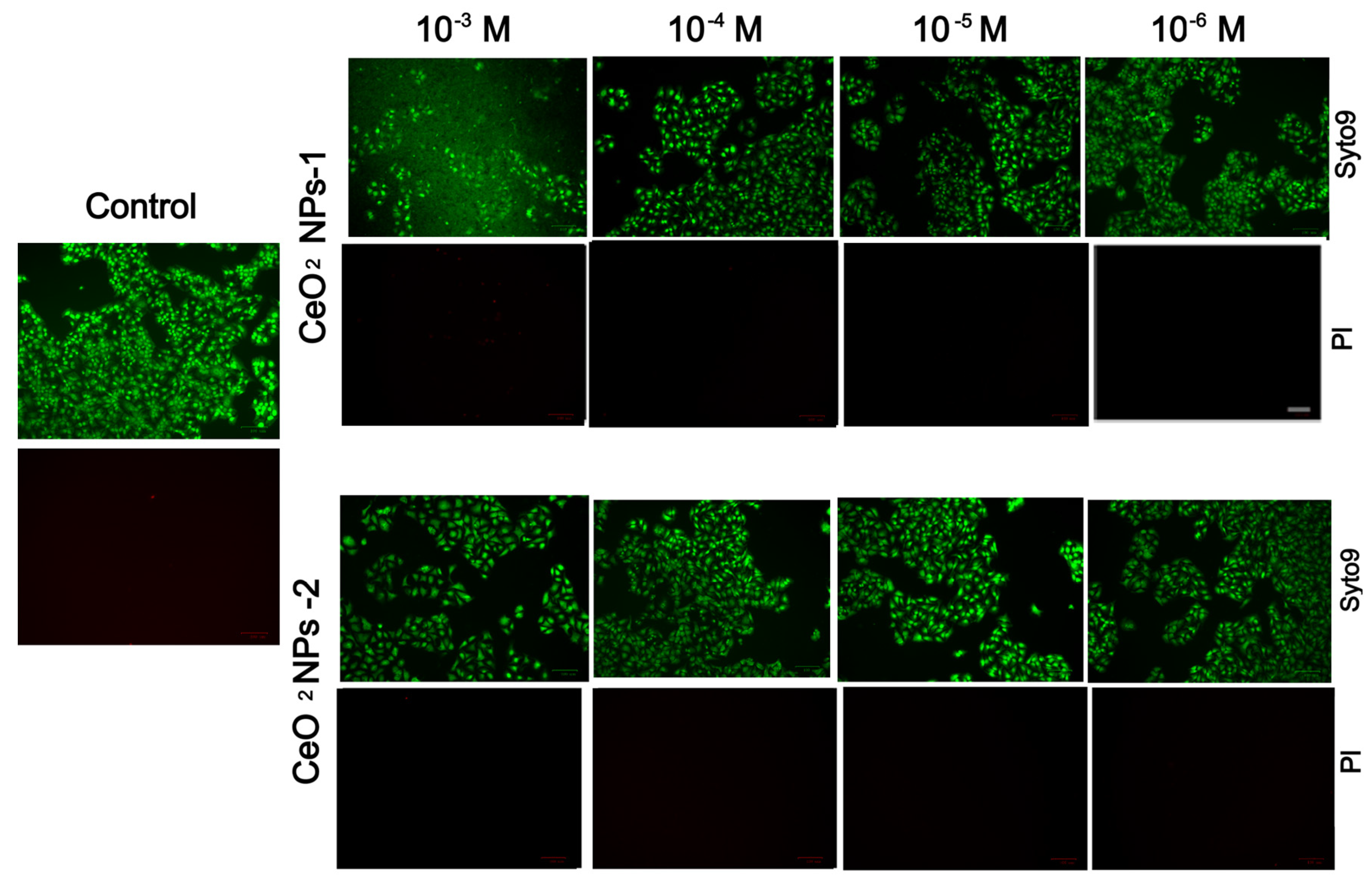
Two co-authors performed the study on HaCaT keratinocytes, and they provided identical photos of dead cells, which may be due to a technical but unprincipled error (red dead cells were almost nowhere to be found and were not visually distinguishable). This technical error showed the authors’ synthesized nanoceria worse than they are in reality. Nevertheless, the conclusion about nontoxicity (biocompatibility) is correct. The corrected Figure 10 appears below.
Figure 10.
Staining of keratinocytes with SYTO9 and propidium iodide fluorescent dyes after cultivation with nanoparticles for 72 h. Quantitative analysis of the number of dead cells in relation to the total number of cells. Scale bar—100 µm.
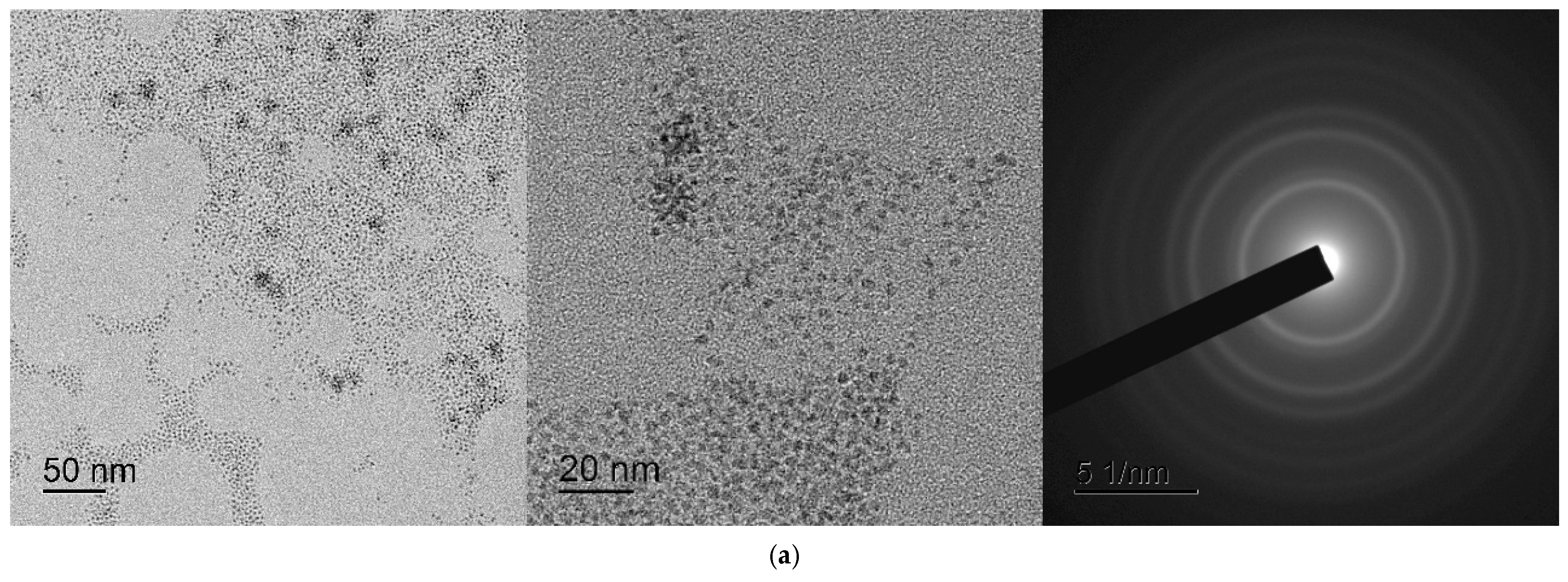
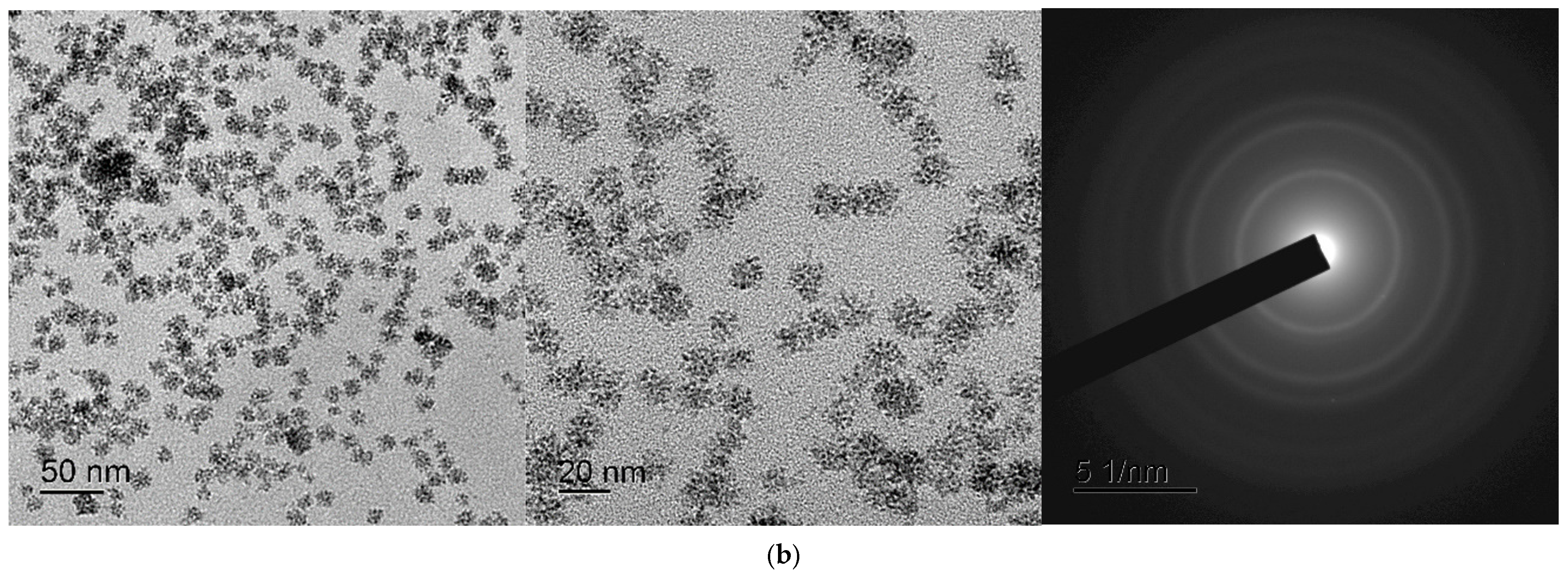
The original electron diffraction patterns are of poor quality. The same nanocerium samples were remade using another better transmission electron microscope (JEOL JEM 2100, Akishima, Japan). Repeated studies of these samples confirmed that the location of the diffraction maxima on the ring electronograms of the samples corresponds to the CeO2 phase, but the quality of the electronograms was significantly improved. The corrected Figure 12 appears below.
Figure 12.
TEM images of cerium nanoparticles: (a) CeO2 precipitate redispersed in water (CeO-NPs-1); (b) CeO2 precipitate redispersed in water (CeO-NPs-2).
The authors state that the scientific conclusions are unaffected. This correction was approved by the Academic Editor. The original publication has also been updated.
Reference
- Silina, E.V.; Stupin, V.A.; Manturova, N.E.; Ivanova, O.S.; Popov, A.L.; Mysina, E.A.; Artyushkova, E.B.; Kryukov, A.A.; Dodonova, S.A.; Kruglova, M.P.; et al. Influence of the Synthesis Scheme of Nanocrystalline Cerium Oxide and Its Concentration on the Biological Activity of Cells Providing Wound Regeneration. Int. J. Mol. Sci. 2023, 24, 14501. [Google Scholar] [CrossRef] [Scilit]
Disclaimer/Publisher’s Note: The statements, opinions and data contained in all publications are solely those of the individual author(s) and contributor(s) and not of MDPI and/or the editor(s). MDPI and/or the editor(s) disclaim responsibility for any injury to people or property resulting from any ideas, methods, instructions or products referred to in the content. |
© 2026 by the authors. Licensee MDPI, Basel, Switzerland. This article is an open access article distributed under the terms and conditions of the Creative Commons Attribution (CC BY) license.