


Abstract
In this study, polysaccharides were isolated from black garlic aged for 0, 10, 17, and 20 days to treat lipopolysaccharide (LPS)-stimulated mouse peritoneal macrophages. The results showed that 20-day-aged black garlic polysaccharides (20dBGPS) significantly inhibited TNF-α secretion and had the strongest anti-inflammatory effects among the isolated polysaccharides. Therefore, 20dBGPS was purified and resolved into three polysaccharide subfractions (coded BPSF1, BPSF2, and BPSF3) using a gel filtration column. High-performance molecular weight size-exclusion chromatography showed that molecular weight distributions of 20dBGPS were 1.65 × 10⁷ Da, 1.88 × 10⁵ Da, and 4.18 × 10³ Da (major peak). High-performance liquid chromatography determined that the monosaccharide composition of 20dBGPS consisted mainly of galactose, galacturonic acid, and glucose. The molecular weight of BPSF1 distributed at 2.23 × 10⁸ Da and 6.92 × 10³ (major peak) and its monosaccharide composition was mainly galactose, xylose, and mannose; BPSF2 distributed at 1.95 × 10⁷ Da and 8.72 × 10³ (major peak) and its monosaccharides were mainly composed of galactose, galacturonic acid, and arabinose; and BPSF3 distributed at 4.27 × 10³ Da and its monosaccharides mainly consisted of glucose, galactose, and mannose, respectively. However, the fructose content in the test samples was not determined in the experiment. BPSF3 significantly (p < 0.05) reduced TNF-α secretion and downregulated toll-like receptor (TLR)-2 and nuclear factor (NF)-κB mRNA expression in LPS-stimulated macrophages, as analyzed by real-time quantitative polymerase chain reaction.
1. Introduction
Inflammatory responses, which result from a large number of soluble inflammatory mediators, including pro-inflammatory cytokines (such as tumor necrosis factor (TNF)-α and interleukin (IL)-1β) and inflammatory mediator molecules (such as nitric oxide (NO) and prostaglandins (PG)) primarily produced by macrophages, are a complicated defense mechanism in the body [1,2,3]. However, excessive and chronic inflammation leads to diverse diseases and damage, including tissue destruction, immune system imbalances, multi-organ disorders, and cancers [4,5]. Persistent inflammation also causes many chronic inflammatory diseases, such as osteoarthritis. Various risk-associated molecular patterns within the joint can activate pattern recognition receptors, primarily overexpressed toll-like receptor (TLR)-2 and TLR-4, leading to chronic activation of the innate immune response [6,7]. They produce pro-inflammatory cytokines and chemokines, including TNF, IL-1, and IL-6, NO, as well as tissue-destructive enzymes, and promote downstream nuclear factor (NF)-κB and mitogen-activated protein kinase (MAPK) dependent pathways [8,9]. High glucose concentrations make cells susceptible to inflammation, and advanced glycation end-products (AGEs) also integrate into the TLR signaling pathway through their own innate immune receptors [10]. Therefore, avoiding excessive and chronic inflammation by daily intake of potential phytochemicals, particularly polysaccharides, may prevent various diseases and tissue damage.
Polysaccharides, such as black garlic polysaccharides, have recently attracted attention for their health potential; they are polymers of monosaccharides primarily linked by glycosidic chains and exhibit diverse biological structures, such as uronic acids, which are acidic sugars found in polysaccharides with both aldehyde and carboxyl groups [11]. Current research has confirmed that polysaccharides possess physiological activity and are considered to be substances that enhance immune responses—biological response modifiers (BRMs) [12,13]. Polysaccharides isolated from Agaricus blazei can directly increase the proliferation, differentiation, and cytokine secretion of B cells and macrophages, reduce the production of pro-inflammatory cytokines, and downregulate the expression of genes in the lipopolysaccharide (LPS)-induced mitogen-activated protein kinase (MAPK) pathway, thereby exerting anti-inflammatory effects [14]. Low molecular weight polysaccharides purified from the leaves of Perilla frutescens, a medicinal vegetable, possess immunomodulatory properties through enhancing and stabilizing the activity of the human complement system [15]. Acidic polysaccharides extracted from ginseng have been found to stimulate immune cells, such as T cells, cytotoxic T lymphocytes, and natural killer cells, and activate macrophages to produce cytokines, achieving anti-tumor effects [16]. However, the functions of black garlic polysaccharides remain limited.
Black garlic, which is produced by aging fresh raw garlic bulbs at high temperature (60–90 °C) and relative humidity (70–90%) for different days, is a health food with a variety of biological activities that has become popular in recent years in the world, particularly in Asia [17,18,19]. It is believed to reduce the unpleasant, pungent, and spicy taste of raw garlic and extend its shelf life [19]. Raw garlic is converted into black garlic under high temperatures and high humidity, primarily involving enzymatic activity in the early stage and the Maillard reaction in the late stage during the transformation (aging) process [20,21]. The increase in black garlic sweetness mainly results from the breakdown of fructans into monosaccharides, while the color change is primarily due to the Maillard reaction between glucose and amino acids [20,22,23]. After aging, black garlic extracts, including black garlic polysaccharides, significantly inhibit the production of inflammatory factors, such as NO, TNF-α, and prostaglandin (PGE2), in LPS-stimulated RAW 264.7 macrophages [18,24]. Additionally, black garlic extracts also enhance DPPH free radical scavenging activity, exhibiting antioxidant activity [25]. Animal studies on pathogen resistance have also found that black garlic improves gastrointestinal function, thereby helping the body fight ingested pathogens, allergens, and toxins [26]. Black garlic possesses lipid-lowering, antioxidant, anti-allergic, anti-liver disease, and anti-diabetic effects [17,27,28,29,30,31]; this has led to the recent popularity of black garlic products and has attracted considerable attention in the market.
Our laboratory previously discovered that aged black garlic has better immunomodulatory potential than raw garlic in vitro, particularly the hot water extract of aged black garlic, showing the greatest anti-inflammatory potential and suggesting a polysaccharide or polyphenol component. However, their effective anti-inflammatory components and mechanisms are not yet fully understood. Therefore, this study aims to investigate changes in black garlic polysaccharides during maturation. The anti-inflammatory characteristics and mechanisms of black garlic polysaccharides and their purified components using peritoneal macrophages of female BALB/c mice were explored.
2. Results and Discussion
2.1. Changes in Appearance and Absorption Spectral Scanning of Crude Polysaccharides Isolated from Black Garlic Aged for Different Days
To unravel the physicochemical characteristics of crude black garlic polysaccharides, 0dRG, 10dBG, 17dBG, and 20dBG were analyzed. The appearance of aged black garlic samples is shown in Figure S1. The results showed that the color of both the garlic skin and pulp turned blackish-brown after ten days of maturation, suggesting that the Maillard reaction that results from the carbonyl-amine condensation may occur during the aging process.
To understand the compositional characteristics of black garlic polysaccharides aged for different days, this study first performed absorption spectral scanning on the extracted samples, including 0dRGPS, 10dBGPS, 17dBGPS, and 20dBGPS. The results showed that black garlic polysaccharides exhibited significant absorption peaks at around 200 nm, 280 nm, 320–360 nm, and 420–450 nm starting from 10 days of aging (Figure 1). This indicates that the composition of black garlic polysaccharides changes continuously as aging time increases. It is speculated that during the aging process, the relative content of protein may increase (280 nm), possibly due to the formation of new glycoproteins or proteopolysaccharides. The increasing absorption peak at 280 nm may be due to the degradation or condensation of polymers after the Maillard reaction. The results of this study are consistent with previous literature, which indicated that raw garlic has a maximum absorbance at around 230 nm, and with increasing aging days, absorption peaks also appear at 280–310 nm and 360 nm. Black garlic exhibits a maximum absorption peak in the 425–450 nm range between days 21 and 28. The color change caused by heat treatment is usually due to the Maillard reaction. Depending on the nature of the reactants involved, the Maillard reaction products are generally associated with increases in absorbance at 280 nm, 320–360 nm, and 420–450 nm. The initial stage of the Maillard reaction produces a colorless intermediate (approximately 280 nm), resulting from the carbonyl-amine condensation and the Amadori rearrangement. The intermediate stage involves sugar dehydration, cleavage, and amino acid degradation (Strecker degradation) to produce a colorless or yellow product (320–360 nm). The final stage involves aldol condensation, with aldehyde-amine condensation forming a dark brown product (420–450 nm) [32].
Figure 1.
UV-visible absorption spectra of black garlic polysaccharides through different aging periods: (A) polysaccharides isolated from 0d raw garlic (0dRGPS), (B) polysaccharides isolated from 10d aged black garlic (10dBGPS), (C) polysaccharides isolated from 17d black garlic (17dBGPS), and (D) polysaccharides isolated from 20d aged black garlic (20dBGPS) using a spectrophotometer scanning from 190 nm to 500 nm.
2.2. Extraction Yields and Composition Characteristics of Crude Black Garlic Polysaccharides Aged for Different Days
Changes in polysaccharide yields, as well as total sugar and total protein contents in the black garlic polysaccharides across different aging periods, are shown in Table 1. The highest extraction yield was obtained from garlic aged for 0 days (16.97 ± 2.50%), which then decreased through the aging process. The next highest yields were obtained from garlic aged for 10 days (10.43 ± 0.10%) and 17 days (6.19 ± 0.48%), but slightly increased at 20 days (7.19 ± 0.12%). The polysaccharide extraction yield decreased through the aging process, suggesting that black garlic polysaccharide degradation occurs under acidic and high-temperature conditions. After 20 days of high-temperature and high-humidity aging, the protein ratio in black garlic polysaccharides increased during the longer aging process (>17 days), while the sugar ratio and total content decreased, suggesting that black garlic polysaccharides might be degraded and polymerized during the aging process. The degradation of cell wall polysaccharides during black garlic aging leads to tissue softening and increased sweetness. Therefore, black garlic has a much softer texture and greater sweetness than raw garlic. Additionally, the significant decrease in pH value (pH < 4.5, a high-acid food) in black garlic and high-temperature heat treatment during the process can further promote the hydrolysis of polysaccharides into monosaccharides [32,33,34]. The increased monosaccharides may further react with amino acids, resulting in the Maillard reaction and blackish-brown color of aged black garlic (Figure S1).
Table 1.
Changes in polysaccharide yields as well as total sugar and total protein contents in the black garlic polysaccharides through different aging periods.
2.3. Effects of Crude Black Garlic Polysaccharides on the Cell Viability of Primary Splenocytes from BALB/c Mice
To achieve the optimal and non-cytotoxic concentrations for anti-inflammatory assessments, the cell viability of primary splenocytes treated with different concentrations of black garlic polysaccharides (0dRGPS, 10dBGPS, 17dBGPS, and 20dBGPS) was measured. The results showed that most black garlic polysaccharides within the indicated concentrations of 1.6–1000 μg/mL had no significant cytotoxicity compared to the control (Figure 2). However, black garlic polysaccharides aged for ten days exhibited a slight (p > 0.05) cytotoxicity at concentrations higher than 500 μg/mL. To avoid unpredictable cytotoxicity for immune cells, concentrations of 1.6, 8, 40, and 200 μg/mL of isolated crude polysaccharides were selected as optimal concentrations to treat macrophages for subsequent cytokine secretion assays. In our preliminary study, we found that primary mouse splenocytes and peritoneal macrophages have the same cytotoxic sensitivity; therefore, only splenocytes were used to perform the MTT assay in the present experiment to select optimal concentrations. LPS is an endotoxin and B-cell mitogen that resulted in a cell viability of 448 ± 91%, suggesting a proliferative effect and indicating that the cell culture model is suitable.
Figure 2.
Effects of black garlic polysaccharides through different aging periods on cell viability of primary mouse splenocytes: (A) polysaccharides isolated from 0d raw garlic (0dRGPS), (B) polysaccharides isolated from 10d aged black garlic (10dBGPS), (C) polysaccharides isolated from 17d aged black garlic (17dBGPS), and (D) polysaccharides isolated from 20d aged black garlic (20dBGPS). Values are means ± SD (n = 6 biological replicates), analyzed using one-way ANOVA, followed by Duncan’s multiple range test. Bars in the same plot not sharing a common small letter were significantly different (p < 0.05) from each other. Cell viability after incubation for 72 h was assayed using the MTT test. The original cultured cell density was 5 × 10⁶ cells/mL. Lipopolysaccharide (LPS) at 2.5 μg/mL in each experiment was selected as a positive control; the cell viability (% of control) was 448 ± 91%.
2.4. Effects of Crude Black Garlic Polysaccharides on Pro- and Anti-Inflammatory Cytokine Secretion by LPS-Stimulated Macrophages
To evaluate the anti-inflammatory potential of crude black garlic polysaccharides, the isolated polysaccharides at the optimal noncytotoxic concentrations were selected to treat primary peritoneal macrophages in the presence of LPS. The profile of pro-inflammatory (IL-1β, IL-6, and TNF-α) and anti-inflammatory (IL-10) cytokine secretions was analyzed. The results showed that LPS alone (control) significantly (p < 0.05) stimulated pro- and anti-inflammatory cytokine secretions as well as the TNF-α/IL-10 secretion ratio compared to those of the negative control (NC), suggesting that this in vitro cell culture model is a suitable model for anti-inflammatory assessments (Table 2). Notably, Dex (treatment control) slightly (p > 0.05) decreased pro-inflammatory cytokine secretion and the TNF-α/IL-10 secretion ratio, but increased anti-inflammatory (IL-10) cytokine secretion by LPS-stimulated macrophages compared to those of the control group, indicating that Dex, which is a glucocorticoid, had mild anti-inflammatory potential. Treatment with 0dRGPS slightly (p > 0.05) decreased pro-inflammatory cytokine secretion and the TNF-α/IL-10 secretion ratio, but could not increase anti-inflammatory (IL-10) cytokine secretion by LPS-stimulated macrophages compared to those of the control group, indicating that 0dRGPS from raw garlic had little anti-inflammatory potential. Importantly, 10dBGPS, 17dBGPS, and 20dBGPS treatments significantly (p < 0.05) decreased TNF-α secretion and the TNF-α/IL-10 secretion ratio by LPS-stimulated macrophages compared to those of the control group, indicating that 10dBGPS, 17dBGPS, and 20dBGPS treatments had significant anti-inflammatory potential, although these polysaccharides could not significantly (p > 0.05) influence anti-inflammatory (IL-10) cytokine secretion. In particular, 20dBGPS treatments at the indicated concentrations showed a significant effect, evidencing that 20dBGPS had the most anti-inflammatory effect against LPS-induced inflammation in macrophages among these black garlic polysaccharides. This indicates that the polysaccharides from black garlic aged for 20 days can achieve anti-inflammatory effects by inhibiting the secretion of the pro-inflammatory TNF-α cytokine and the TNF-α/IL-10 secretion ratio. The chloroform extract of black garlic has been found to reduce TNF-α-induced reactive oxygen species (ROS) production in human umbilical vein endothelial cells and decrease NF-κB activation and monocyte adhesion [35]. This finding is similar to the decrease in TNF-α secretion observed in this experiment. Overall assessment showed that black garlic polysaccharides aged for 20 days (20dBGPS) had significant anti-inflammatory potential; therefore, 20dBGPS were further purified to explore their effective components and anti-inflammatory mechanisms.
Table 2.
Effects of black garlic polysaccharides on pro- and anti-inflammatory cytokine secretions by LPS-stimulated primary peritoneal macrophages from female BALB/c mice.
2.5. Purification of 20dBGPS Using Sepharose 6B Gel Filtration Chromatography
To further purify 20dBGPS and explore its effective components and anti-inflammatory mechanisms, a Sepharose 6B gel filtration chromatography column was used in this study, and chromatographic fractions were collected based on absorbance at 280 nm (protein) and total sugar content using the phenol-sulfuric acid method. The result of the chromatographic pattern is shown in Figure S2. Two obvious peaks and a trace peak were observed in the absorbance analysis at 280 nm, while the total sugar content spectrum showed three obvious peaks: fraction 1 (tubes 13–17, coded as BPSF1), fraction 2 (tubes 21–29, coded as BPSF2), and fraction 3 (tubes 36–42, coded as BPSF3). The three peaks (fractions) based on absorbance at 280 nm (protein) and total sugar content that corresponded to each other were harvested. BPSF1 did not show a significant peak based on the absorbance at 280 nm, indicating a low protein content in this purified black garlic polysaccharide. BPSF2 and BPSF3 showed significant peaks at 280 nm and in total sugar content, suggesting that these purified fractions may be glycoproteins or proteoglycans.
2.6. Effects of Purified Black Garlic Polysaccharide BPSF1, BPSF2 and BPSF3 on TNF-α Secretion by LPS-Stimulated Macrophages
This study used the MTT assay to assess the effects of purified black garlic polysaccharides BPSF1, BPSF2, and BPSF3 on the growth of primary splenocytes to determine the optimal concentrations of the samples with non-cytotoxic effects. The results are shown in Figure 3. The results showed that BPSF3 at concentrations of 1.6–500 μg/mL showed no significant difference compared to the control group. BPSF2 at 500 μg/mL significantly (p < 0.05) reduced the survival rate of splenocytes, while BPSF1 at 200 μg/mL significantly (p < 0.05) reduced cell viability. Therefore, this experiment selected optimal concentrations at 1.6, 8, and 40 μg/mL for BPSF1, BPSF2, and BPSF3 as the treatment doses for subsequent cytokine secretion assays.
Figure 3.
Effects of (A) BPSF1, (B) BPSF2, and (C) BPSF3 on cell viability of primary mouse splenocytes. BPSF1, fraction 1 of polysaccharides isolated from black garlic aged for 20 days; BPSF2, fraction 2 of polysaccharides isolated from black garlic aged for 20 days; BPSF3, fraction 3 of polysaccharides isolated from black garlic aged for 20 days. Values are means ± SD (n = 6 biological replicates), analyzed using one-way ANOVA, followed by Duncan’s multiple range test. Bars not sharing a common small letter were significantly different (p < 0.05) from each other. Cell viability after 72 h of incubation was assayed using the MTT test. The original cultured cell density was 5 × 10⁶ cells/mL.
The present experiment has shown that crude black garlic polysaccharide 20dBGPS significantly reduces the secretion of the pro-inflammatory cytokine TNF-α by LPS-stimulated macrophages (Table 2). Therefore, the optimal concentrations of 1.6, 8, and 40 μg/mL for BPSF1, BPSF2, and BPSF3 were selected to treat LPS-stimulated macrophages to assess anti-inflammatory effects based on the secretion of the pro-inflammatory cytokine TNF-α. Table 3 shows that LPS stimulation of peritoneal macrophages significantly (p < 0.05) stimulated the secretion of the pro-inflammatory cytokine TNF-α compared to the control group (LPS alone). When BPSF1, BPSF2, and BPSF3 were added, TNF-α secretion was significantly reduced at a concentration of 40 μg/mL compared to the control group. All three purified black garlic polysaccharides, BPSF1, BPSF2, and BPSF3, showed good anti-inflammatory effects, but BPSF3 was the most effective because it worked at a lower concentration of 8 μg/mL. Therefore, BPSF3 was further selected to analyze its molecular weight, monosaccharide composition, and anti-inflammatory mechanisms.
Table 3.
Effects of purified fractions of polysaccharides isolated from black garlic aged for 20 days in vitro on TNF-α secretion by LPS-stimulated primary peritoneal macrophages from female BALB/c mice.
2.7. Molecular Weights of Crude Black Garlic Polysaccharides and Purified BPSF1, BPSF2, and BPSF3
To investigate the molecular weight distribution of crude black garlic polysaccharides and purified polysaccharides BPSF1, BPSF2, and BPSF3, water-soluble polysaccharide standards (molecular weight 6.2–805 kDa) were prepared at a concentration of 0.25% in deionized water and analyzed using HPSEC with deionized water at a flow rate of 0.7 mL/min. The logarithmic molecular weight of the standards was plotted against their retention time to obtain the calibration curve (Figure S3). Crude black garlic polysaccharides and purified polysaccharides BPSF1, BPSF2, and BPSF3 were analyzed in the same HPSEC. The HPLC chromatograms for test samples are shown in Figure S4, and the molecular weights corresponding to the retention times of black garlic polysaccharides were estimated. Chromatographic analysis showed that the crude polysaccharide extracts from raw and black garlic contained significantly more peaks, averaging 2–4 peaks, while purified polysaccharides BPSF1, BPSF2, and BPSF3 contained fewer peaks, suggesting their relative purity. The corresponding molecular weights are summarized in Table 4. The molecular weight distribution of 0dRGPS was 4.12 × 10⁶ Da and 1.30 × 10⁴ (major peak);10dBGPS was 1.44 × 10⁷ Da, 6.55 × 10⁵ and 1.11 × 10⁴ (major peak); 17dBGPS was 2.28 × 10⁷ Da, 6.67 × 10⁶, 3.07 × 10⁵, and 4.59 × 10³ (major peak); 20dBGPS was 1.65 × 10⁷ Da, 1.88 × 10⁵, and 4.18 × 10³ (major peak), respectively. There was a low molecular weight (104–103 Da) polysaccharide in each crude garlic polysaccharide, showing a decreasing trend in low molecular weight through the black garlic aging process. The results suggest that low molecular weight polysaccharides in garlic might be hydrolyzed through the black garlic aging process. However, not all chemical modifications reduce the molecular weight; polysaccharides may undergo cross-linking or amination due to chemical modifications and high temperatures. It has been found that polysaccharides can modify a low molecular weight xylan at the reducing end through reductive amination, thus increasing the molecular weight of the coupled product [36]. In the present experiment, the highest molecular weight polysaccharide (106–107 Da) in each crude black garlic polysaccharide slightly increased through the black garlic aging process. The results suggest that a polymerization reaction in polysaccharides may occur during the aging process, leading to changes in the polysaccharide molecular structure and the formation of higher molecular weight polysaccharides. Additionally, 20dBGPS was resolved into three subfractions, including BPSF1, BPSF2, and BPSF3, using gel filtration chromatography (Table 4). The molecular weight distribution of BPSF1 was 2.23 × 10⁸ Da and 6.92 × 10³ Da (major peak); BPSF2 was 1.95 × 10⁷ Da and 8.72 × 10³ Da (major peak), and BPSF3 was 4.27 × 10³ Da (only one peak), respectively. Our results suggest that BPSF3 is a purified polysaccharide fraction, while BPSF1 and BPSF2 may exist as impurities. Based on the results of purified polysaccharide fractions and anti-inflammatory efficacy (Table 3 and Table 4), BPSF3, a low molecular weight purified black garlic polysaccharide, possesses anti-inflammatory effects at lower concentrations (8 μg/mL), suggesting that the smallest molecular weight black garlic polysaccharide may have the greatest anti-inflammatory potential. Furthermore, in vitro studies have found that low molecular weight herbal polysaccharides are more effective than high molecular weight polysaccharides in stimulating spleen cell growth and enhancing immunity [37]. Experiments have also confirmed that polysaccharides below 10 kDa still have immunomodulatory effects [38], confirming that the lower molecular weight BPSF3 may be the most potent anti-inflammatory black garlic polysaccharide fraction. However, the monosaccharide composition of polysaccharides may also influence their anti-inflammatory functions. Therefore, the monosaccharide composition of 20dBGPS, BPSF1, BPSF2, and BPSF3 was further analyzed. In the present experiment, molecular weights determined by HPSEC carry an extrapolation risk, so the absolute values are unreliable. It is suggested that standards should cover the molecular weight range, or HPSEC coupled with multi-angle light scattering (MALS)/multi-detector be used for measurement, so that the reported values may clarify relative molecular weights/extrapolated values and explain whether the 2.23 × 10⁸ peak of BPSF1 is an aggregate or impurity.
Table 4.
Molecular weight distribution of polysaccharides isolated from black garlic aged for 0, 10, 17, and 20 days (0dRGPS, 10dBGPS, 17dBGPS, and 20dBGPS) and subfractions of polysaccharides isolated from black garlic aged for 20 days (BPSF1, BPSF2, BPSF3) detected by HPSEC.
2.8. Monosaccharide Composition of 20dBGPS, BPSF1, BPSF2, and BPSF3
The basic building block of polysaccharides is the monosaccharide. The type and bonding method of monosaccharides determine their chemical structure and properties. The most fundamental information for polysaccharide research is its monosaccharide composition. Therefore, this experiment further investigated the monosaccharide composition ratio in 20dBGPS, BPSF1, BPSF2, and BPSF3. However, most sugars are uncharged and lack absorbing chromophores or fluorophores, making them undetectable by direct HPLC measurement [39]. Many derivatization methods for monosaccharides have been developed to attach ultraviolet or fluorophores to sugars, enabling their detection by ultraviolet light. Currently, derivatization reagents are classified into several categories, including 1-phenyl-3-methyl-5-pyrazolone (PMP) and ABEE. Due to their ease of operation and high sensitivity, this experiment uses ABEE to derivatize reducing sugars. Polysaccharides found in natural plants are primarily composed of acidic and neutral sugars. This experiment ultimately used HPLC with a UV detector to analyze the monosaccharide content and proportion of 20dBGPS and its purified fractions BPSF1, BPSF2, and BPSF3. Figure 4 shows the HPLC chromatograms of ten monosaccharide standards and test samples. The monosaccharide contents of black garlic polysaccharides aged for 20 days and their gel filtration fractions were determined using the standard curve obtained from the mixed standards at different concentrations and the integral peak area, and are shown in Table 5. The results showed significant differences in monosaccharide composition among 20dBGPS, BPSF1, BPSF2, and BPSF3. The monosaccharide composition of 20dBGPS was mainly composed of galactose, galacturonic acid, and glucose. Previous literature indicates that black garlic polysaccharides consist of galactose and galacturonic acid [18]. In the present study, BPSF1 consisted of galactose (32.6 ± 1.0%), xylose (22.6 ± 3.3%), and mannose (13.8 ± 0.8%). However, BPSF2 was rich in galacturonic acid (25.4 ± 0.1%) and galactose (53.6 ± 0.6%), suggesting that BPSF2 is the major fraction in 20dBGPS (Figure S3). Importantly, BPSF3, having the most anti-inflammatory potential, contained high glucose (53.9 ± 1.3%), but was low in glucuronic acid (0.4 ± 0.1%), galacturonic acid (1.8 ± 0.1%), galactose (24.2 ± 0.8%), arabinose (1.0 ± 0.2%), and fucose (0.8 ± 0.1%) compared to the other black garlic polysaccharides. Our results suggest that a low molecular weight purified polysaccharide BPSF3 (4.27 × 10³ Da) in 20dBGPS, rich in glucose but low in fucose, might possess strong anti-inflammatory potential. Monosaccharide composition analysis showed that black garlic polysaccharides are composed of a complex and diverse range of monosaccharides, suggesting that their polysaccharide structure may contain many branches. However, the fructose content in the samples was not determined in the experiment. Further investigation is needed to determine their structure and mechanisms of action.
Figure 4.
HPLC-UV chromatograms for (A) monosaccharide standards, (B) 20dBGPS, (C) BPSF1, (D) BPSF2, and (E) BPSF3. A = Glucuronic acid, B = Galacturonic acid, C = Galactose, D = Mannose, E = Glucose, F = Arabinose, G = Ribose, H = Xylose, I = Rhamnose, J = Fucose.
Table 5.
Monosaccharide contents and their relative composition ratios in 20dBGPS, BPSF1, BPSF2, and BPSF3.
2.9. Anti-Inflammatory Mechanisms of 20dBGPS, BPSF1, BPSF2, and BPSF3
To understand the possible anti-inflammatory signaling pathway of 20dBGPS, BPSF1, BPSF2, and BPSF3 against LPS-induced inflammation, LPS-induced primary peritoneal macrophages were treated with the effective concentration (40 μg/mL) of these samples. Total mRNA was extracted to evaluate changes in the expression levels of the TLR-2, TLR-4, and NF-κB genes using real-time qPCR (Table 6). The results showed that LPS treatment alone (control) slightly (p > 0.05) decreased TLR-2, TLR-4, and NF-κB gene expression compared to the negative control, suggesting that LPS, an endotoxin, may damage macrophages and result in a decrease in these mRNA expression levels. Notably, Dex (treatment control) obviously increased TLR-2, TLR-4, and NF-κB gene expression, suggesting that Dex treatment may repair the damage of LPS and restore the immunostimulatory functions of macrophages, as evidenced by increased anti-inflammatory (IL-10) cytokine secretion by LPS-stimulated macrophages (Table 2). Black garlic polysaccharide (20dBGPS) and its purified fractions BPSF1, BPSF2, and BPSF3 demonstrated different effects on LPS-stimulated macrophages. 20dBGPS and its major fraction BPSF2 exerted the same immunomodulatory effect as Dex, suggesting that 20dBGPS and BPSF2 may repair LPS injury and increase the immunostimulatory functions of macrophages (Table 6). However, 20dBGPS and BPSF2 significantly (p < 0.05) increase in NF-κB gene expression levels may also cause an inflammatory status in macrophages. BPSF1 treatment slightly (p > 0.05) increased NF-κB gene expression levels in LPS-stimulated macrophages, suggesting that BPSF1 may repair LPS injury and slightly restore the immunostimulatory functions of macrophages. Importantly, the BPSF3 treatment decreased TLR-2, TLR-4, and NF-κB gene expression compared to the control, suggesting that BPSF3 may inhibit the inflammatory status of LPS-stimulated macrophages. However, BPSF3 may also decrease the immunostimulatory function of macrophages. Berry polysaccharide has been found to dose-dependently inhibit LPS-induced inflammation via the TLR-4 pathway, reducing the expression of the downstream NF-κB gene and decreasing the secretion of the pro-inflammatory cytokine TNF-α [40]. Lycium barbarum polysaccharides attenuate LPS-induced inflammation in inflamed mouse macrophages by inhibiting the TLR4/NF-κB signaling pathway, resulting in reduced secretion of pro-inflammatory cytokines IL-6 and TNF-α [41]. The present study provides evidence that BPSF3 decreased TLR-2, TLR-4, and NF-κB mRNA expression, possibly via the TLR-2/NF-κB signaling pathway in LPS-stimulated macrophages (Figure 5), suggesting that BPSF3 in black garlic may have strong anti-inflammatory potential.
Table 6.
Effects of black garlic polysaccharides on relative gene expression of molecules involved in the inflammation-related signaling pathway in LPS-stimulated primary peritoneal macrophages from female BALB/c mice.
Figure 5.
Possible anti-inflammatory mechanisms of BPSF3 through down-regulating Toll-like receptor-2 (TLR-2) signaling pathways in primary peritoneal macrophages from female BALB/c mice. BPSF3 inhibited TLR-2 and NF-κB mRNA expression, as well as TNF-α secretion. There are two possible pathways in TLR signaling, including myeloid differentiation primary response-88 (MyD88)- and (TRIF)-dependent pathways. LPS, lipopolysaccharide; TRIF, TIR-domain-containing adapter-inducing interferon-β.
The present experiment provides some evidence to characterize the ingredients and anti-inflammatory mechanisms of black garlic polysaccharides in vitro. However, it still has a few drawbacks that need to be further explored. Primarily, only one variety of raw garlic (Allium sativum L.) was used to prepare black garlic to explore its anti-inflammatory characteristics, limiting robustness, although Allium sativum is a popular variety in many countries. Different garlic varieties may have trace differences in polysaccharides and other compositions in raw garlic, resulting in differences in physicochemical properties and functions in black garlic through the aging process [42]. The aging process under diverse conditions may enhance these differences [43]. Under different thermal processes, black garlic increases total free sugars, amino acids, and pyruvic acid and promotes the Maillard reaction that may further result in changes in polysaccharides [44,45]. It is difficult to totally purify polysaccharides because polysaccharides are characterized by their properties of non-uniform molecular size and molecular weight distribution. The present experiment has provided evidence concerning the impurities in subfractions and the molecular weight distribution of black garlic polysaccharides. In general, garlic carbohydrates are predominantly fructans [46,47]. However, black garlic polysaccharides that contain low fructose content inhibited UM-UC-3 cancer cell growth. In contrast, black garlics with high fructose content enhanced UM-UC-3 cell proliferation, suggesting that fructose content in black garlic polysaccharides may influence their functions [46,48]. Unfortunately, this experiment omitted fructose from the HPLC standard mixtures; it should be further investigated in depth. The manuscript has investigated a food-derived polysaccharide from black garlic; however, the biological evaluation relies predominantly on a conventional in vitro macrophage inflammation model and assessment of established inflammatory markers and signaling pathways. Unfortunately, without digestion/bioaccessibility studies, physiologically relevant validation, and/or greater mechanistic depth, the work still lacks relative novelty compared with the extensive literature on the anti-inflammatory properties of food-derived polysaccharides. Additionally, the glycosidic linkage analysis, repeating-unit determination of the polysaccharide structure, branching patterns, and detailed NMR characterization remain to be further studied. The structure-activity relationship and physicochemical properties of black garlic polysaccharides should be deeply explored to enhance their application in different fields [49,50,51,52]. The health functions of black garlic polysaccharides in preventing chronic diseases should be studied for further development as a health or medicinal food [11,53,54]. Finally, the mechanistic study relied exclusively on mRNA expression without validating actual protein expression; more protein analytical techniques, such as Western blotting, should be performed in the future. Although the results obtained from this experiment have provided some solid scientific data and can be translated to most research, the underlying ingredients and anti-inflammatory mechanisms of black garlic polysaccharides from this experiment may not be fully applicable to different domestic or commercial black garlic. To augment the achievements from the present study, the anti-allergic and anti-cancer effects of black garlic polysaccharides are being investigated in vivo. Some valuable evidence concerning the health function of black garlic polysaccharides has been collected.
3. Materials and Methods
3.1. Material
Raw garlic (Allium sativum L.) originating from Yuanchang Township, Yunlin County, Taiwan, was purchased from a local company, Bonbon Farmer.
3.2. Preparation of Aged Black Garlic
Whole heads of fresh raw garlic (RG) bulbs were weighed and subjected to aging at 70 °C and 80% relative humidity (RH) for 0, 10, 17, and 20 days (coded as 0dRG, 10dBG, 17dBG, and 20dBG) in a constant temperature and humidity oven (BTH80/-20, Firstek, New Taipei City, Taiwan). After aging, the aged black garlic was collected, weighed, and stored at room temperature. In the experiment, fresh raw garlic (0-day aging, 0dRG) was chosen for comparison.
To extend the storage period, slices of raw garlic and aged black garlic were put in a 40 °C oven (ULM-400, Memmert, GmbH, Schwabach, Germany) and dried until thin and crisp. The garlic was then ground into powder using a high-speed grinder (RT-04, Rong Tsong Precision Technology, Co., Taichung, Taiwan). The air-dried powder samples were sealed and stored at room temperature for use.
3.3. Preparation of Crude Raw Garlic and Black Garlic Polysaccharides (RGPS and BGPS)
Raw garlic and black garlic air-dried powder samples were weighed. An aliquot of 10 volumes of deionized water was added, mixed, and stirred slowly in a 95 °C water bath for 1 h. After cooling, the mixture was centrifuged at 4 °C, 5000× g for 30 min. The supernatant was collected and filtered by vacuum filtration; the filtrate was harvested, and the volume was quantified. Three times the volume of 95% ethanol was added to the filtrate, and it was slowly stirred at 4 °C for 12 h to precipitate the polysaccharides. The mixture was centrifuged at 4 °C and 5000× g for 30 min to separate the precipitate and the supernatant. The precipitate was harvested and evaporated to remove residual solvent using a vacuum evaporator (EYELA rotary vacuum evaporator, Tokyo Rikakika Co., N-1200AVF, Tokyo, Japan), assisted by an aspirator (EYELA Aspirator, Tokyo Rikakika Co., A-1000S, Tokyo, Japan). The concentrated precipitate was freeze-dried using a freeze dryer (PANCHUM CT-5000D, Kaohsiung, Taiwan) to obtain crude raw garlic polysaccharides (RGPS), coded as 0dRG, and crude black garlic polysaccharides (BGPS), coded as 10dBGPS, 17dBGPS, and 20dBGPS, respectively. The yields of the harvested polysaccharides were calculated, and the harvested polysaccharides were stored at −30 °C for use. Before the experiment, the harvested polysaccharides were weighed and dissolved in deionized water to prepare a 50 mg/mL stock solution. The stock solution was filtered aseptically through a 0.22 μm filter membrane (Merck Millipore Ltd., Tullagreen, Carrigwohill, Co., Cork, Ireland) and stored at −80 °C until use. Before the experiment, the stock solution was further diluted with different solvents or culture media to prepare working solutions.
3.4. Compositional Characteristics of RGPS and BGPS
3.4.1. Spectral Scanning Analysis
The lyophilized powders of crude and purified polysaccharides of black garlic were dissolved in deionized water at appropriate concentrations (250 μg/mL). A full-wavelength spectral scan was performed using a spectrophotometer (Hitachi-U2900 ultraviolet (UV)-vis spectrophotometer, Tokyo, Japan), with the detection range from 190 nm to 500 nm, to investigate the absorption characteristics of the polysaccharide samples [55].
3.4.2. Analysis of Total Sugar Content in RGPS and BGPS
Using the phenol-sulfuric acid method, the isolated black garlic polysaccharides and purified polysaccharide fraction samples were appropriately diluted with deionized water. Glucose (MP Biomedical, Inc., 152527, Eschwege, Germany) was serially diluted to prepare a standard solution. An aliquot of 60 μL of the test sample solution and the standard solution was mixed with 60 μL of 5% phenol (Sigma, P-4161, St. Louis, MO, USA) solution (w/v), and then 300 μL of concentrated sulfuric acid (Sigma, 30743, St. Louis, MO, USA) was added. After mixing thoroughly, the mixture was allowed to cool to room temperature. The absorbance was measured at 490 nm using an ELISA reader. The obtained absorbance value was then substituted into the glucose standard curve to calculate the total sugar content of the sample [22].
3.4.3. Determination of Total Protein Content in RGPS and BGPS
Using the Bradford protein-binding assay, the isolated black garlic polysaccharides and the purified polysaccharide fraction sample were appropriately diluted with deionized water. Bovine serum albumin (BSA, Thermo, Prod 23209, Rockford, IL, USA) was serially diluted to prepare a standard solution. An aliquot of 10 μL of the test sample solution and the standard solution were added to separate wells in a 96-well plate. Then, 200 μL aliquots of freshly prepared Dye reagent (diluted 4 times with deionized water) (Bio-Rad, Catalog 500-0006, Hercules, CA, USA) were added and mixed thoroughly. The mixture was incubated at room temperature in the dark for 10 min. The absorbance was measured at 595 nm using an ELISA reader. The absorbance was then used to calculate the total protein content of the sample by substituting the results into the BSA standard curve [55].
3.5. Effects of RGPS and BGPS on Pro- and Anti-Inflammatory Cytokine Secretion by LPS-Stimulated Macrophages from Female BALB/c Mice
3.5.1. Isolation of Primary Splenocytes and Peritoneal Macrophages
Briefly, BALB/cBJNarl female mice aged six to eight weeks were bought from the National Institutes of Applied Research, National Center for Biomodels, Taipei, Taiwan, and housed in plastic cages. During the experimental period, experimental mice were provided with a standard lab diet (chow diet) of food, water, and bedding, allowing them free access to food and activity in the animal room. The temperature and humidity were maintained at 25 ± 2 °C and 50–75%, with alternating light and dark periods of 12 h. After one to two weeks of acclimatization, the experimental mice were sacrificed at 10 ± 2 weeks of age, and primary splenocytes and peritoneal macrophages were isolated for experiments. The isolated splenocytes and peritoneal macrophages were adjusted to a fixed density of 1 × 10⁷ cells/mL and 2 × 10⁶ cells/mL in TCM medium, respectively [2,3]. In the experiment, splenocytes and peritoneal macrophages were isolated and adjusted to fixed densities of 1 × 10⁷ and 2 × 10⁶ cells/mL for use, respectively. However, the original cultured cell densities in each experiment were 5 × 10⁶ and 1 × 10⁶ cells/mL, respectively. Therefore, the cell density and concentration are consistent in each experiment. The Institutional Animal Care and Use Committee (IACUC) at National Chung Hsing University, Taiwan, had substantively reviewed the protocol for animal use and approved it (IACUC No.: 110-128R2, Approval date: 17 March 2025).
3.5.2. Optimal Concentrations for Crude RGPS and BGPS Based on Splenocytes’ Cell Viability
To obtain optimal and non-cytotoxic concentrations of crude RGPS and BGPS in vitro, the isolated splenocytes were used for the experiment. Briefly, aliquots of 50 μL/well of primary splenocytes (1 × 10⁷ cells/mL in TCM medium) were added with aliquots of 50 μL/well of black garlic polysaccharides at different concentrations or lipopolysaccharide (LPS, Sigma, L2654, St. Louis, MI, USA; a final concentration of 2.5 μg/mL in the well was selected as a positive control) in a 96-well plate. The plate was incubated at 37 °C in a humidified incubator (Shel Lab TC2323, Cornelius, OR, USA) with 5% CO2 and 95% air for 72 h. The cell viability of treated splenocytes was assessed using the reagent 3-(4,5-dimethylthiazol-2-diphenyl)-2,5-tetrazolium bromide (MTT, Sigma M-5655, St. Louis, MO, USA) [2]. The wavelength of 540 nm was used to measure the absorbance (A) using an enzyme-linked immunosorbent assay (ELISA) reader (Microplate Reader FLUOstar Omega, 415-1103, BMG LABTECH, Ortenberg, Germany). Changes in cell numbers (% of control) were used to calculate cell viability by the following equation: change in cell numbers (% of control) = [(Asample − Ablank)/(Acontrol − Ablank)] × 100 [2]. After comparison with the control, optimal non-cytotoxic concentrations of black garlic polysaccharides were determined for anti-inflammatory evaluations using peritoneal macrophages in vitro.
3.5.3. Effects of Black Garlic Polysaccharides on Pro- and Anti-Inflammatory Cytokines Secreted by LPS-Stimulated Macrophages
To evaluate the anti-inflammatory potential of black garlic polysaccharides, isolated peritoneal macrophages were used for the experiment. Briefly, macrophages (2 × 10⁶ cells/mL, 250 μL/well in TCM medium) were added to the wells in a 24-well plate. Aliquots of 125 μL/well TCM medium (negative control) and test samples at optimal non-cytotoxic concentrations (4×) or dexamethasone (Dex, 400 nM, a treatment control) were pipetted into the wells. Aliquots of 125 μL/well TCM medium or LPS (10.0 μg/mL in TCM medium) were added to the corresponding well in the plate. The plate was incubated at 37 °C in a humidified incubator with 5% CO2 and 95% air for 48 h. Macrophages cultured with TCM medium alone were used as a negative control, while macrophages cultured with LPS alone were used as a control in the experiment. After incubation, the cell culture plate was centrifuged for 10 min at 400× g. The supernatant of the cell culture was collected and stored at −80 °C for later analysis of secretion levels of pro-inflammatory (IL-1β, IL-6, and TNF-α) and anti-inflammatory (IL-10) cytokines [3]. The levels of pro-inflammatory (IL-1β, IL-6, and TNF-α)/anti-inflammatory (IL-10) cytokines secreted by treated macrophages were analyzed using sandwich ELISA kits following the instructions provided by the manufacturers. IL-6 and TNF-α were measured using R&D Systems Duo Set ELISA Development Kits (Minneapolis, MN, USA). IL-1β and IL-10 were measured using BioLegend mouse ELISA MAX Standard Kits (San Diego, CA, USA). The limit of detection (LOD) of the ELISA cytokine kits performed in the experiment was <15.6 pg/mL.
3.6. Purification of Polysaccharides Isolated from 20-Day-Aged Black Garlic
Our previous study has shown that 20-day-aged black garlic has the best immunomodulatory functions, suggesting an optimal condition for aging; therefore, 20-day-aged black garlic polysaccharides (20dBG) were further purified using gel filtration to unravel their characteristics. Briefly, the lyophilized powder of isolated polysaccharides from 20-day-aged black garlic (20dBG) was weighed and dissolved in deionized water to a fixed concentration of 2.5 mg/mL. The solution was filtered through a 0.22 μm filter membrane. An aliquot of 5 mL of the solution was loaded into a Sepharose 6B gel (Sigma-Aldrich Co., St. Louis, MO, USA) filtration column (2.5 × 48 cm). Deionized water was used for elution, and a fixed flow rate was set using a peristaltic pump. The eluate was collected in tubes using a fraction collector (Isco Retriever 500, Teledyne Technologies, Inc., Lincoln, NE, USA), and the conditions were set at 5 mL per tube and a total collection of 50 tubes. Each collected tube was measured for absorbance at 280 nm (for protein content) using a spectrophotometer (Hitachi-U2900 UV-vis spectrophotometer, Tokyo, Japan) [55], and the total sugar content was determined using the phenol-sulfuric acid method to determine changes in total sugar content [22]. Based on changes in the absorbance and sugar content in each collected tube, the chromatograms exhibited three separate fractions (fraction 1, fraction 2, and fraction 3), coded as BPSF1, BPSF2, and BPSF3. Each fraction was harvested, concentrated under reduced pressure to remove the solvent, freeze-dried into powder, and stored at −30 °C for use. Black garlic polysaccharides and their fractions were dissolved separately in deionized water or dimethyl sulfoxide (DMSO, Wako, 040-19492, Osaka, Japan) to prepare 50 mg/mL stock solutions. The stock solution was aseptically filtered through a 0.22 μm filter membrane (Merck Millipore Ltd., Tullagreen, Carrigtwohill, Co. Cork, IRL) and stored at −30 °C for use. Before experiments, the stock solution was diluted with deionized water or cell culture medium to prepare a working solution.
3.7. Effects of Purified Polysaccharide Fractions Isolated from 20-Day-Aged Black Garlic Polysaccharides on Pro-Inflammatory Cytokine TNF-α Secreted by LPS-Stimulated Macrophages
To obtain optimal and non-cytotoxic concentrations of BPSF1, BPSF2, and BPSF3 in vitro, the isolated splenocytes were used for the experiment, and the procedure is as described above. The concentrations from 1.6 to 500 μg/mL of these purified polysaccharides were assessed to achieve optimal and non-cytotoxic concentrations.
To compare the anti-inflammatory effect of the purified polysaccharide fractions isolated from 20-day-aged black garlic, isolated peritoneal macrophages were used for the experiment. The optimal and non-cytotoxic concentrations of BPSF1, BPSF2, and BPSF3 were used in the experiment. Briefly, macrophages (2 × 10⁶ cells/mL, 250 μL/well in TCM medium) were added to the wells in a 24-well plate. Aliquots of 125 μL/well TCM medium (negative control) and test samples (BPSF1, BPSF2, BPSF3, and 20dBPGS) at appropriate concentrations (4×) or dexamethasone (Dex, 400 nM, a treatment control) were pipetted into the wells. Aliquots of 125 μL/well TCM medium or LPS (10.0 μg/mL in TCM medium) were added to the corresponding well in the plate. The plate was incubated in a humidified incubator with 5% CO2 and 95% air at 37 °C for 48 h. Macrophages treated with TCM medium alone were used as a negative control, while macrophages treated with LPS alone were used as a control in the experiment. After incubation, the plate was centrifuged at 400× g for 10 min. The supernatant of the cell culture was harvested and stored at −80 °C for later analysis of secretion levels of the pro-inflammatory cytokine TNF-α according to the instructions provided by the manufacturer [3].
3.8. Determination of Molecular Weights of Crude Polysaccharides and Purified Polysaccharide Fractions Isolated from 20-Day-Aged Black Garlic Polysaccharides
0dRGPS, 10dBGPS, 17dBGPS, 20dBGPS, BPSF1, BPSF2, and BPSF3 were dissolved in deionized water and adjusted to a concentration of 0.025% (w/v), respectively. After filtration through a 0.22 μm filter membrane, high-performance molecular weight size-exclusion chromatography (HPSEC) was performed. Polysaccharide standards with different molecular weights (MW: 6.2–805 kDa) were adjusted to a concentration of 0.025% with deionized water and filtered through a 0.22 μm filter membrane. The instruments included a high-performance liquid chromatography (HPLC) pump (HITACHI pump L-2130, Tokyo, Japan), an autosampler (HITACHI Autosampler L-2200, Tokyo, Japan), an RI detector (HITACHI RI Detector L-2490, Tokyo, Japan), analytical software (HITACHI L-2200 HPLC Software EZChrom Elite, Version 3.1.7), and an analytical column (Shodex OHpak SB-805 HQ column, 300 × 8 mm). The mobile phase was deionized water, and the analysis was performed at a fixed flow rate of 0.7 mL/min. A standard curve was obtained by plotting the logarithm of the molecular weights of the polysaccharide standards against the retention time (RT) in HPSEC. The average molecular weight of the test sample was calculated from the identified peak of black garlic polysaccharides and the peak area [56].
3.9. Determination of Monosaccharide Content of Purified Polysaccharide Fractions Isolated from 20-Day-Aged Black Garlic Polysaccharides
Briefly, 10 mg of polysaccharide powder was weighed into a spiral glass test tube, and 10 mL of 2 M trifluoroacetic acid (TFA, Sigma T6508, St. Louis, MO, USA) was added. The mixture was shaken well and hydrolyzed with acid in a boiling water bath. After removing the solvent by vacuum concentration using a vacuum evaporator (EYELA rotary vacuum evaporator, Tokyo Rikakika Co., N-1200AVF, Tokyo, Japan), assisted by an aspirator (EYELA Aspirator, Tokyo Rikakika Co., A-1000S, Tokyo, Japan), deionized water was added to a final volume of 1 mL, and the solution was filtered through a 0.22 μm filter membrane (CNW, PRECLEAN 13 mm Syringe Filter Nylon membrane, ANPEL Laboratory Technologies, Shanghai, China). An aliquot of 30 μL of the above acid hydrolysis solution was transferred into a 2.0 mL microcentrifuge tube. Then, 120 μL of p-aminobenzoic ethyl ester (ABEE) derivatization labeling reagent (composed of 165 mg benzocaine (ABEE, Sigma, E1501, St. Louis, MO, USA), 35 mg of sodium cyanoborohydride (NaBH3CN, Sigma, 71435, St. Louis, MO, USA), 40 μL glacial acetic acid (Wako, 017-00256, Osaka, Japan), and 0.35 mL warm methanol (Wako, 138-14521, Osaka, Japan) were added, mixed and derivatized in a dry bath at 80 °C for 1 h. After the reaction was complete, 0.6 mL of deionized water was added to terminate the reaction, and 0.6 mL of chloroform (Sigma, C5312, St. Louis, MO, USA) was added to remove excess derivatization reagent. After shaking well, the tube was centrifuged at 25 °C, 2000× g for 5 min to precipitate chloroform. The supernatant was harvested and filtered through a 0.22 μm filter membrane. The filtrate was analyzed for its monosaccharide composition with HPLC. Monosaccharide standards including acidic sugar [D-glucuronic acid (GlcUA, Sigma, G5269, St. Louis, MO, USA) and D-galacturonic acid (GalUA, Sigma, 48280, St. Louis, MO, USA)] and neutral sugars [D-glucose (Glc, Sigma, G8270, St. Louis, MO, USA), D-mannose (Man, Sigma, M6020, St. Louis, MO, USA), D-galactose (Gal, Sigma, G0750, St. Louis, MO, USA), D-xylose (Xyl, Sigma, X1500, St. Louis, MO, USA), L-fucose (Fuc, Sigma, F2252, St. Louis, MO, USA), L-rhamnose (Rha, Sigma, R3875, St. Louis, MO, USA), L-arabinose (Ara, Sigma, A3256, St. Louis, MO, USA), and D-ribose (Rib, Sigma, R7500, St. Louis, MO, USA)] were performed with ABEE derivatization and used in the experiment. The instruments included an HPLC pump (HITACHI pump L-2130, Tokyo, Japan), an autosampler (HITACHI Autosampler L-2200, Tokyo, Japan), an UV detector (HITACHI UV Detector L-2400, Tokyo, Japan), analytical software (HITACHI L-2200 HPLC Software EZChrom Elite), an analytical column (Phenomenex Gemini C18, 5 μm, 250 × 4.6 mm, Torrance, CA, USA), and a column thermostat (HT-230A Column heater, Fuji Electric, Tokyo, Japan). Mobile phase A was pH 8.9, 0.04 M potassium borate buffer (Sigma, P5754, St. Louis, MO, USA), and mobile phase B was 100% acetonitrile (ECHO, AH2301, Miaoli, Taiwan), and the analysis was performed by HPLC at a fixed flow rate of 0.8 mL/min. The oven temperature was set to 50 °C. The mobile phase ratio of A:B = 90:10 was set to equilibrate the column. The detection wavelength was set at 308 nm. An aliquot of 5 μL of a derivatized mixture of ten monosaccharide standards and the test samples was injected for detection. The concentrations of the mixed monosaccharide standards were 0, 5, 10, 50, 100, 500, and 1000 μg/mL. The mobile phase gradient was applied for the first 55 min with a concentration gradient of A:B = 90:10. After all peaks of the monosaccharide samples appeared, a concentration gradient of mobile phase A:B = 80:20 was performed from 56 to 80 min at a flow rate of 1 mL/min to remove excess ABEE derivative reagents. The integral area of the monosaccharide standard was then plotted against the prepared concentration to obtain a standard curve for monosaccharides. The monosaccharide composition of the test samples was then analyzed using the standard curve [57,58].
3.10. Effects of Purified Polysaccharide Fractions Isolated from 20-Day-Aged Black Garlic on Relative Gene Expressions of Molecules Involved in Inflammation-Related Signaling Pathways in LPS-Stimulated Macrophages
Based on in vitro inflammation model experiments, the fractions of polysaccharides from 20-day-aged black garlic (BPSF1, BPSF2, BPSF3) reduced the secretion of the pro-inflammatory cytokine TNF-α, confirming that polysaccharides from 20-day-matured black garlic can influence the protein expression of the TNF-α cytokine. To understand its anti-inflammatory mechanism, the expression of inflammation-related genes in LPS-stimulated macrophages was explored, analyzing the expression of TLR pathway-related genes (TLR-2, TLR-4, and NF-κB) to clarify whether it achieves its anti-inflammatory effect by regulating the TLR-2 and TLR-4/NF-κB signaling pathways [6,7].
3.10.1. Cell Culture and Total RNA Extraction
Briefly, macrophages (2 × 10⁶ cells/mL, 2.0 mL/well in TCM medium) were added to the wells in a 6-well plate. Aliquots of 1.0 mL/well TCM medium (negative control) and test samples (BPSF1, BPSF2, BPSF3, and 20dBPGS) at appropriate concentrations (4×) or dexamethasone (Dex, 400 nM, a treatment control) were pipetted into the wells. Aliquots of 1.0 mL/well TCM medium or LPS (10.0 μg/mL in TCM medium) were added to the corresponding well in the plate. The plate was incubated in a humidified incubator with 5% CO2 and 95% air at 37 °C for 6 h. Macrophages treated with TCM medium alone served as a negative control, while those treated with LPS alone served as a positive control in the experiment. After incubation, the plate was centrifuged at 400× g for 10 min. The cell culture supernatant was removed.
The cell pellet was washed with 1 mL/well of phosphate-buffered saline (composed of 1.5 mM KH2PO4, 8.1 mM Na2HPO4, 137 mM NaCl, and 2.7 mM KCl, pH 7.4, 0.22 μm filtered). The cell pellet was treated with 1 mL/well of TRIzol reagent (Invitrogen, 15596-018, Carlsbad, CA, USA) to extract total RNA from the cells. The cells were repeatedly lysed using a pipette, and the cell culture was transferred to a 1.5 mL microcentrifuge tube. After standing for 15 min, an aliquot of 0.2 mL of chloroform (Sigma, C5312, St. Louis, MO, USA) was added, shaken vigorously for 15 s, and incubated on ice for 5 min. The reaction mixture in the tube was centrifuged at 12,000× g, 4 °C, for 15 min. The cell slurry separated into three layers: a clear aqueous layer on top, a translucent thin layer of trace proteins in the middle, and a red organic layer at the bottom. The upper aqueous layer was carefully aspirated into another clean 1.5 mL microcentrifuge tube. An equal volume of isopropanol (Sigma, I9516, St. Louis, MO, USA) was added to the collected liquid, gently shaken to mix, and incubated on ice for 20 min to precipitate RNA. The tube was centrifuged at 12,000× g, 4 °C for 20 min, and the supernatant was carefully removed. The pellet was carefully washed with 1 mL of 75% ethanol (J. T. Baker 8006, Deventer, The Netherlands) and centrifuged at 7500× g, 4 °C for 5 min. The supernatant (ethanol) was removed, and the RNA pellet was air-dried at room temperature until it became translucent. Finally, 10 μL of nuclease-free water (Promega, P1193, Madison, WI, USA) was added to reconstitute the RNA pellet by thorough mixing. The total RNA was harvested. To evaluate the quality of the extracted RNA, 1 µL of RNA solution was pipetted and diluted 100-fold with 99 µL of 10 mM Tris-HCl/DEPC-H2O (Biotecx Laboratories Inc., BL-5611, Houston, TX, USA). The diluted RNA solution was measured for absorbance at 260 and 280 nm using a spectrophotometer. The RNA purity is represented by the ratio of the absorbance at 260/280 nm. A ratio between 1.5 and 2.0 indicates low protein contamination, which is necessary for subsequent experiments. One unit of absorbance at 260 nm equals 40 µg RNA/mL. The RNA concentration could be converted using the following calculation formula: OD260 × dilution factor × 40 = total RNA (µg RNA/mL). The harvested RNA was stored at −80 °C for later analysis [59,60,61].
3.10.2. Converting RNA into Complementary DNA (cDNA) Using a Reverse Transcription Reaction
Briefly, 2 μg of RNA sample, 1 µL of 10× reaction buffer (Promega, M1984, Madison, WI, USA), and 2 µL of RQ1 RNase-free DNase I (Promega, M6106, Madison, WI, USA) were added to DEPC-H2O to a total of 10 μL solution in a 1.5 mL microcentrifuge tube. After spin-down, the mixture was incubated at room temperature for 30 min to remove the DNA. After the reaction, 1 μL of stop solution (Promega, M199A, Madison, WI, USA) was added to the mixture to bind calcium and magnesium ions, preventing metal ions from catalyzing the hydrolysis of RNA and affecting reverse transcriptase activity. Then, the reaction mixture was heated at 70 °C for 10 min to destroy DNase I activity. The RNA mixture was placed on ice and cooled for 15 min to prevent RNA from reverting to its secondary structure.
After cooling, the 11 μL RNA mixture was thoroughly mixed with 5 µL of M-MLV Reverse transcriptase 5× reaction buffer (Promega, M531A, Madison, WI, USA), 4.75 µL of DEPC-H2O, 1.25 µL of 10 mM dNTP mix (Promega, U151B, Madison, WI, USA), 2 µL of 100 µM Oligo dT (Invitrogen, 17781, Carlsbad, CA, USA), and 1 µL of M-MLV Reverse transcriptase (Promega, M1705, Madison, WI, USA). The reaction mixture (25 µL) was used for reverse transcription polymerase chain reaction (RT-PCR) in a PCR gradient thermal cycler (Pebio Scientific Company, Genesis 96, Taipei, Taiwan). The RT reaction was set as follows: 25 °C for 5 min, 42 °C for 60 min, 70 °C for 15 min, and 4 °C for 60 min. Immediately after the reaction, the tube was cooled on ice. After spin-down, 1 μL of cDNA was diluted 100-fold with 99 μL of 10 mM Tris-HCl/DEPC-H2O. The absorbance at wavelengths of 260 and 280 nm was measured using a spectrophotometer. At 260 nm, 1 unit of absorbance = 33 μg cDNA/mL. The cDNA concentration was calculated using the formula: OD260 × Dilution factor × 33 = Total cDNA (μg DNA/mL). All cDNA samples were adjusted to the same concentration with DEPC-H2O and stored at −30 °C for subsequent quantitative polymerase chain reaction (qPCR) [59,60,61].
3.10.3. Real-Time Quantitative Polymerase Chain Reaction (Real-Time qPCR)
Briefly, the cDNA sample was diluted five-fold with nuclease-free water. An aliquot of 5 μL of the diluted sample was added to 4 μL of nuclease-free water, 10 μL of KAPA SYBR FAST qPCR kit Master Mix (2× Universal, Kapa Biosystems, West Cummings Park, Suite Woburn, MA, USA), 0.5 μL of PCR forward primer (10 μM), and 0.5 μL of PCR reverse primer (10 μM) in a clean PCR tube. The primer sequences designed for target genes, including TLR-2, TLR-4, NF-κB, and β-actin, are provided in Table S1. The reagents in the PCR tube were mixed thoroughly, spun down, and stored in the dark to proceed with real-time qPCR using a real-time rotary analyzer (Corbett Research Rotor-Gene 6000, Sydney, Australia). The real-time qPCR reaction conditions were set as follows: hot-start activation at 95 °C for 15 min, followed by 40–45 cycles of denaturation at 95 °C for 30 s, annealing at 60 °C for 30 s, and extension at 72 °C for 30 s. Ct value, which is the threshold cycle number required for the fluorescence signal in the PCR sample to reach the set threshold, was measured. Higher cDNA concentrations result in earlier curves and lower Ct values. In the experiment, relative gene expression fold is represented by the equation 2−ΔΔCt [59,60,61].
ΔCt = Ct (target) − Ct (reference)
ΔΔCt = ΔCt (group x) − ΔCt (control)
= (Ct, target − Ct, β-actin) group x − (Ct, target − Ct, β-actin) control
Ct (target): threshold for target genes (TLR-2, TLR-4, and NF-κB)
Ct (reference): threshold for a housekeeping gene (β-actin)
ΔΔCt = ΔCt (group x) − ΔCt (control)
= (Ct, target − Ct, β-actin) group x − (Ct, target − Ct, β-actin) control
Ct (target): threshold for target genes (TLR-2, TLR-4, and NF-κB)
Ct (reference): threshold for a housekeeping gene (β-actin)
3.11. Statistical Analyses
All experimental data are expressed as mean ± standard deviation (mean ± SD). Animal experiments were performed with at least 6 biological replicates. Statistical analysis was performed first using one-way ANOVA, followed by Duncan’s Multiple Range Test. The test was used to determine the differences in data between the treatment groups. A p-value lower than 0.05 was considered statistically significant. The analysis was performed using IBM SPSS Statistics 20.
4. Conclusions
This study aimed to investigate the anti-inflammatory effects and mechanisms of BGPS. Polysaccharides were isolated from hot water extracts of black garlic aged for 0, 10, 17, and 20 days, coded as 0dRGPS, 10dBGPS, 17dBGPS, and 20dBGPS, to treat LPS-stimulated primary macrophages. As a result, the protein ratio in BGPS increased through the aging process, while the sugar ratio and total content decreased, suggesting that BGPS might be degraded and/or polymerized during the aging process. 10dBGPS, 17dBGPS, and 20dBGPS significantly (p < 0.05) decreased the TNF-α/IL-10 secretion ratio compared to the control. The 20dBGPS significantly inhibited TNF-α secretion, showing the strongest anti-inflammatory effects among isolated polysaccharides. Therefore, 20dBGPS was further selected for purification and resolved into three polysaccharide subfractions, coded as BPSF1, BPSF2, and BPSF3. The molecular weight distribution of 0dRGPS was 4.12 × 10⁶ Da and 1.30 × 10⁴ (major peak);10dBGPS was 1.44 × 10⁷ Da, 6.55 × 10⁵ and 1.11 × 10⁴ (major peak); 17dBGPS was 2.28 × 10⁷ Da, 6.67 × 10⁶, 3.07 × 10⁵, and 4.59 × 10³ (major peak); 20dBGPS was 1.65 × 10⁷ Da, 1.88 × 10⁵, and 4.18 × 10³ (major peak), respectively. The monosaccharide composition of 20dBGPS was mainly composed of galactose, galacturonic acid, and glucose. The molecular weight distribution of BPSF1 was 2.23 × 10⁸ Da and 6.92 × 10³ (major peak) and its monosaccharide composition was mainly galactose, xylose, and mannose; BPSF2 was 1.95 × 10⁷ Da and 8.72 × 10³ (major peak) and its monosaccharides were mainly composed of galactose, galacturonic acid, and arabinose; and BPSF3 was 4.27 × 10³ Da (only one peak) and its monosaccharides mainly consisted of glucose, galactose, and mannose, respectively. However, the fructose content in the test samples was not determined in the experiment. BPSF3 significantly (p < 0.05) reduced TNF-α secretion and downregulated toll-like receptor (TLR)-2 and nuclear factor (NF)-κB mRNA expression in LPS-stimulated macrophages, suggesting the strongest anti-inflammatory potential among isolated BGPS.
Supplementary Materials
The following supporting information can be downloaded at: https://www.mdpi.com/article/10.3390/ijms272210432/s1.
Author Contributions
Y.-C.T.: Data curation, formal analysis, investigation, writing—original draft preparation. J.-Y.L.: Conceptualization, investigation, methodology, validation, writing—reviewing and editing, funding acquisition. All authors have read and agreed to the published version of the manuscript.
Funding
This study was kindly supported by research grants 106AS-3.5.3-FD-Z1(2) from the Ministry of Agriculture, Taiwan, and NSTC 113-2320-B-005-001 and NSTC 114-2320-B-005-003-MY2 from the National Science and Technology Council, Taiwan.
Institutional Review Board Statement
The animal study protocol was approved by the Institutional Animal Care and Use Committee (IACUC) at National Chung Hsing University (IACUC No.: 110-128R2, Approval date: 17 March 2025).
Informed Consent Statement
Not applicable.
Data Availability Statement
The datasets are available from the corresponding author upon reasonable request.
Conflicts of Interest
The authors declare no conflicts of interest.
References
- Allam, R.; Anders, H.J. The role of innate immunity in autoimmune tissue injury. Curr. Opin. Rheumatol. 2008, 20, 538–544. [Google Scholar] [CrossRef] [Scilit] [PubMed]
- Chang, K.C.; Lin, J.Y. Immunomodulatory evaluation of caffeic acid and its quinic acid derivatives using Th1/Th2 cytokine secretion profiles and Principal Component Analysis. Chem.-Biol. Interact. 2026, 424, 111865. [Google Scholar] [CrossRef] [Scilit] [PubMed]
- Wu, T.Y.; Chen, C.C.; Lin, J.Y. Anti-inflammatory in vitro activities of eleven selected caffeic acid derivatives based on a combination of pro-/anti-inflammatory cytokine secretions and Principal Component Analysis—A comprehensive evaluation. Food Chem. 2024, 458, 140201. [Google Scholar] [CrossRef] [Scilit]
- Aggarwal, B.B.; Vijayalekshmi, R.V.; Sung, B. Targeting inflammatory pathways for prevention and therapy of cancer: Short-term friend, long-term foe. Clin. Cancer Res. 2009, 15, 425. [Google Scholar] [CrossRef] [Scilit] [PubMed]
- Rodier, F.; Coppé, J.P.; Patil, C.K.; Hoeijmakers, W.A.M.; Muñoz, D.P.; Raza, S.R.; Freund, A.; Campeau, E.; Davalos, A.R.; Campisi, J. Persistent DNA damage signalling triggers senescence-associated inflammatory cytokine secretion. Nat. Cell Biol. 2009, 11, 973, Erratum in Nat. Cell Biol. 2009, 11, 1272.. [Google Scholar] [CrossRef] [Scilit] [PubMed]
- Liao, C.H.; Lin, J.Y. Purified active lotus plumule (Nelumbo nucifera Gaertn) polysaccharides exert anti-inflammatory activity through decreasing Toll-like receptor-2 and -4 expressions using mouse primary splenocytes. J. Ethnopharmacol. 2013, 147, 164–173. [Google Scholar] [CrossRef] [Scilit]
- Liao, Y.R.; Lin, J.Y. Quercetin modulates cytokine expression and inhibits TLR2 expression and STAT3 activation in mouse activated inflammatory macrophages. J. Explor. Res. Pharmacol. 2020, 5, 31–41. [Google Scholar] [CrossRef] [Scilit]
- Cohn, E.F.; Nathan, C.; Radzioch, D.; Yu, H.; Xiang, Z.; Ding, A. Abrupt expression of TLR4 in TLR4-deficient macrophages imposes a selective disadvantage: Genetic evidence for TLR4-dependent responses to endogenous, nonmicrobial stimuli. J. Immunol. (Baltim. Md. 1950) 2006, 176, 1185–1194. [Google Scholar] [CrossRef] [Scilit] [PubMed]
- Yeh, T.H.; Lin, J.Y. Acorus gramineusand and Euodia ruticarpa steam distilled essential oils exert anti-inflammatory effects through decreasing Th1/Th2 and pro-/anti-inflammatory cytokine secretion ratios in vitro. Biomolecules 2020, 10, 338. [Google Scholar] [CrossRef] [Scilit]
- Najm, A.; Blanchard, F.; Le Goff, B. Micro-RNAs in inflammatory arthritis: From physiopathology to diagnosis, prognosis and therapeutic opportunities. Biochem. Pharmacol. 2019, 165, 134–144. [Google Scholar] [CrossRef] [Scilit] [PubMed]
- Zhu, R.; Guan, M.; Li, K.; Chen, Y.; Chen, X.; Jiang, Y.; Xia, T.; Xin, H. A food-derived intervention: Black garlic polysaccharides alleviate slow transit constipation with multifaceted improvements in gastrointestinal function and microbiota. J. Food Sci. Technol. 2026. [Google Scholar] [CrossRef] [Scilit]
- Leung, M.Y.; Liu, C.; Koon, J.C.; Fung, K.P. Polysaccharide biological response modifiers. Immunol. Lett. 2006, 105, 101–114. [Google Scholar] [CrossRef] [Scilit] [PubMed]
- Zhang, S.; Li, W.; Smith, C.J.; Musa, H. Cereal-derived arabinoxylans as biological response modifiers: Extraction, molecular features, and immune-stimulating properties. Crit. Rev. Food Sci. Nutr. 2015, 55, 1035–1052. [Google Scholar] [CrossRef] [Scilit] [PubMed]
- Cheng, F.; Yan, X.; Zhang, M.; Chang, M.; Yun, S.; Meng, J.; Liu, J.; Feng, C.P. Regulation of RAW 264.7 cell-mediated immunity by polysaccharides from Agaricus blazei Murill via the MAPK signal transduction pathway. Food Funct. 2017, 8, 1475–1480. [Google Scholar] [CrossRef] [Scilit] [PubMed]
- Zhang, B.; Leung, W.K.; Zou, Y.; Mabusela, W.; Johnson, Q.; Michaelsen, T.E.; Paulsen, B.S. Immunomodulating polysaccharides from Lessertia frutescens leaves: Isolation, characterization and structure activity relationship. J. Ethnopharmacol. 2014, 152, 340–348. [Google Scholar] [CrossRef] [Scilit] [PubMed]
- Son, S.U.; Hong, K.R.; Shin, K.S. Immunostimulating and anticancer activities of the pectic polysaccharide from Panax ginseng leaves treated with high pressure/enzyme process. Curr. Issues Mol. Biol. 2025, 47, 257. [Google Scholar] [CrossRef] [Scilit] [PubMed]
- Ahmed, T.; Wang, C.K. Black garlic and its bioactive compounds on human health diseases: A review. Molecules 2021, 26, 5028. [Google Scholar] [CrossRef] [Scilit] [PubMed]
- Li, M.; Yan, Y.X.; Yu, Q.T.; Deng, Y.; Wu, D.T.; Wang, Y.; Ge, Y.Z.; Li, S.P.; Zhao, J. Comparison of immunomodulatory effects of fresh garlic and black garlic polysaccharides on RAW 264.7 macrophages. J. Food Sci. 2017, 82, 765–771. [Google Scholar] [CrossRef] [Scilit] [PubMed]
- Vecchi, V.; Sabbioni, G.; Altieri, M.T.; Breveglieri, G.; Costa, S.; Marchetti, F.; Gugel, I.; Vertuani, S.; Borgatti, M. Black garlic: Evolution of the chemical composition and broad biological activities. J. Agric. Food Chem. 2025, 73, 27933–27949. [Google Scholar] [CrossRef] [Scilit] [PubMed]
- Jing, H. Black garlic: Processing, composition change, and bioactivity. eFood 2020, 1, 242–246. [Google Scholar] [CrossRef] [Scilit]
- Wu, J.; Jin, Y.; Zhang, M. Evaluation on the physicochemical and digestive properties of melanoidin from black garlic and their antioxidant activities in vitro. Food Chem. 2021, 340, 127934. [Google Scholar] [CrossRef] [Scilit] [PubMed]
- Lei, M.; Xu, M.; Zhang, Z.; Zhang, M.; Gao, Y. The analysis of saccharides in black garlic and its antioxidant activity. Adv. Food Sci. Technol. 2014, 6, 755–760. [Google Scholar] [CrossRef] [Scilit]
- Yuan, H.; Sun, L.; Chen, M.; Wang, J. An analysis of the changes on intermediate products during the thermal processing of black garlic. Food Chem. 2018, 239, 56–61. [Google Scholar] [CrossRef] [Scilit]
- Tak, H.M.; Kang, M.J.; Min Kim, K.; Kang, D.; Han, S.; Shin, J.H. Anti-inflammatory activities of fermented black garlic. J. Korean Soc. Food Sci. Nutr. 2014, 43, 1527–1534. [Google Scholar] [CrossRef] [Scilit]
- Lu, X.; Li, N.; Qiao, X.; Qiu, Z.; Liu, P. Composition analysis and antioxidant properties of black garlic extract. J. Food Drug Anal. 2017, 25, 340–349. [Google Scholar] [CrossRef] [Scilit] [PubMed]
- Chen, Y.A.; Tsai, J.C.; Cheng, K.C.; Liu, K.F.; Chang, C.K.; Hsieh, C.W. Extracts of black garlic exhibits gastrointestinal motility effect. Food Res. Int. 2018, 107, 102–109. [Google Scholar] [CrossRef] [Scilit] [PubMed]
- Afzaal, M.; Saeed, F.; Rasheed, R.; Hussain, M.; Aamir, M.; Hussain, S.; Mohamed, A.A.; Alamri, M.S.; Anjum, F.M. Nutritional, biological, and therapeutic properties of black garlic: A critical review. Int. J. Food Prop. 2021, 24, 1387–1402. [Google Scholar] [CrossRef] [Scilit]
- Ha, A.W.; Ying, T.; Kim, W.K. The effects of black garlic (Allium satvium) extracts on lipid metabolism in rats fed a high fat diet. Nut. Res. Pract. 2015, 9, 30–36. [Google Scholar] [CrossRef] [Scilit] [PubMed]
- Kim, J.H.; Nam, S.H.; Rico, C.W.; Kang, M.Y. A comparative study on the antioxidative and anti-allergic activities of fresh and aged black garlic extracts. Int. J. Food Sci. Technol. 2012, 47, 1176–1182. [Google Scholar] [CrossRef] [Scilit]
- Kim, J.H.; Yu, S.H.; Cho, Y.J.; Pan, J.H.; Cho, H.T.; Kim, J.H.; Bong, H.; Lee, Y.; Chang, M.H.; Jeong, Y.J.; et al. Preparation of S-allylcysteine-enriched black garlic juice and its antidiabetic effects in streptozotocin-induced insulin-deficient mice. J. Agric. Food Chem. 2017, 65, 358–363. [Google Scholar] [CrossRef] [Scilit] [PubMed]
- Sasmaz, H.K.; Uzlasir, T.; Selli, S.; Kelebek, H. Black garlic: Production process, bioactive compounds, and health effects. Food Chem. Int. 2025, 1, 24–30. [Google Scholar] [CrossRef] [Scilit]
- Choi, S.I.; Cha, S.H.; Lee, S.Y. Physicochemical and antioxidant properties of black garlic. Molecules 2014, 19, 16811–16823. [Google Scholar] [CrossRef] [Scilit] [PubMed]
- Kimura, S.; Tung, Y.C.; Pan, M.H.; Su, N.W.; Lai, Y.J.; Cheng, K.C. Black garlic: A critical review of its production, bioactivity, and application. J. Food Drug Anal. 2017, 25, 62–70. [Google Scholar] [CrossRef] [Scilit] [PubMed]
- Zhang, Z.; Lei, M.; Liu, R.; Gao, Y.; Xu, M.; Zhang, M. Evaluation of alliin, saccharide contents and antioxidant activities of black garlic during thermal processing. J. Food Biochem. 2015, 39, 39–47. [Google Scholar] [CrossRef] [Scilit]
- Lee, E.N.; Choi, Y.W.; Kim, H.K.; Park, J.K.; Kim, H.J.; Kim, M.J.; Lee, H.W.; Kim, K.H.; Bae, S.S.; Kim, B.S.; et al. Chloroform extract of aged black garlic attenuates TNF-α-induced ROS generation, VCAM-1 expression, NF-κB activation and adhesiveness for monocytes in human umbilical vein endothelial cells. Phytother. Res. 2011, 25, 92–100. [Google Scholar] [CrossRef] [Scilit] [PubMed]
- Li, S.; Xiong, Q.; Lai, X.; Li, X.; Wan, M.; Zhang, J.; Yan, Y.; Cao, M.; Lu, L.; Guan, J.; et al. Molecular modification of polysaccharides and resulting bioactivities. Compr. Rev. Food. Sci. Food Saf. 2016, 15, 237–250. [Google Scholar] [CrossRef] [Scilit] [PubMed]
- Chen, H.L.; Li, D.F.; Chang, B.Y.; Gong, L.M.; Dai, J.G.; Yi, G.F. Effects of Chinese herbal polysaccharides on the immunity and growth performance of young broilers. Poult. Sci. 2003, 82, 364–370. [Google Scholar] [CrossRef] [Scilit] [PubMed]
- Bi, D.; Yu, B.; Han, Q.; Lu, J.; White, W.L.; Lai, Q.; Cai, N.; Luo, W.; Gu, L.; Li, S.; et al. Immune activation of RAW264.7 macrophages by low molecular weight fucoidan extracted from New Zealand Undaria pinnatifida. J. Agric. Food Chem. 2018, 66, 10721–10728. [Google Scholar] [CrossRef] [Scilit] [PubMed]
- Honda, S.; Akao, E.; Suzuki, S.; Okuda, M.; Kakehi, K.; Nakamura, J. High-performance liquid chromatography of reducing carbohydrates as strongly ultraviolet-absorbing and electrochemically sensitive 1-phenyl-3-methyl-5-pyrazolone derivatives. Anal. Biochem. 1989, 180, 351–357. [Google Scholar] [CrossRef] [Scilit]
- Liu, H.; Zhang, W.; Dong, S.; Song, L.; Zhao, S.; Wu, C.; Wang, X.; Liu, F.; Xie, J.; Wang, J.; et al. Protective effects of sea buckthorn polysaccharide extracts against LPS/d-GalN-induced acute liver failure in mice via suppressing TLR4-NF-kappaB signaling. J. Ethnopharmacol. 2015, 176, 69–78. [Google Scholar] [CrossRef] [Scilit] [PubMed]
- Peng, Q.; Liu, H.; Shi, S.; Li, M. Lycium ruthenicum polysaccharide attenuates inflammation through inhibiting TLR4/NF-kappaB signaling pathway. Int. J. Biol. Macromol. 2014, 67, 330–335. [Google Scholar] [CrossRef] [Scilit] [PubMed]
- Bedrnicek, J.; Vecerova, K.; Triska, J.; Swieca, M.; Petraskova, E.; Lorenc, F.; Jarosova, M.; Oravec, M.; Milkova, E.; Klicnarova, J.; et al. Association between fresh garlic composition and black garlic antioxidant capacity. Food Chem. 2026, 508, 148347. [Google Scholar] [CrossRef] [Scilit] [PubMed]
- Dewi, N.N.A.; Mustika, I.W. Nutrition content and antioxidant activity of black garlic. Int. J. Health Sci. 2018, 2, 11–20. [Google Scholar] [CrossRef] [Scilit]
- Kang, O.J. Physicochemical characteristics of black garlic after different thermal processing steps. Prev. Nutr. Food Sci. 2016, 21, 348–354. [Google Scholar] [CrossRef] [Scilit] [PubMed]
- Lu, X.; Li, N.; Qiao, X.; Qiu, Z.; Liu, P. Effects of thermal treatment on polysaccharide degradation during black garlic processing. LWT-Food Sci. Technol. 2018, 95, 223–229. [Google Scholar] [CrossRef] [Scilit]
- Lu, X.; Li, N.; Zhao, R.; Zhao, M.; Cui, X.; Xu, Y.; Qiao, X. In vitro prebiotic properties of garlic polysaccharides and its oligosaccharide mixtures obtained by acid hydrolysis. Front. Nutr. 2021, 8, 798450. [Google Scholar] [CrossRef] [Scilit] [PubMed]
- Sembiring, N.; Iskandar, Y. A review of component and pharmacology activities of black garlic. Tradit. Med. J. 2019, 24, 178–183. [Google Scholar] [CrossRef] [Scilit]
- Matsuse, K.; Hirata, S.; Abdelrahman, M.; Nakajima, T.; Iuchi, Y.; Kambayashi, S.; Okuda, M.; Kazumura, K.; Manochai, B.; Shigyo, M. Comparative studies of bioactivities and chemical components in fresh and black garlics. Molecules 2024, 29, 2258. [Google Scholar] [CrossRef] [Scilit] [PubMed]
- Jiang, X.Y.; Liang, J.Y.; Jiang, S.Y.; Zhao, P.; Tao, F.; Li, J.; Li, X.X.; Zhao, D.S. Garlic polysaccharides: A review on their extraction, isolation, structural characteristics, and bioactivities. Carbohydr. Res. 2022, 518, 108599. [Google Scholar] [CrossRef] [Scilit] [PubMed]
- Liang, J.; Zhao, Y.; Yang, F.; Zheng, L.; Ma, Y.; Liu, Q.; Cai, L.; Gong, W.; Wang, B. Preparation and structure-activity relationship of highly active black garlic polysaccharides. Int. J. Biol. Macromol. 2022, 220, 601–612. [Google Scholar] [CrossRef] [Scilit] [PubMed]
- Utama, G.L.; Rahmi, Z.; Sari, M.P.; Hanidah, I.I. Physicochemical changes and functional properties of organosulfur and polysaccharide compounds of black garlic (Allium sativum L.). Curr. Res. Food Sci. 2024, 8, 100717. [Google Scholar] [CrossRef] [Scilit] [PubMed]
- Yuan, X.; Wang, Z.; Liu, L.; Mu, D.; Wu, J.; Li, X.; Wu, X. Changes of physicochemical properties in black garlic during fermentation. Fermentation 2022, 8, 653. [Google Scholar] [CrossRef] [Scilit]
- Shao, X.; Sun, C.; Tang, X.; Zhang, X.; Han, D.; Liang, S.; Qu, R.; Hui, X.; Shan, Y.; Hu, L.; et al. Anti-inflammatory and intestinal microbiota modulation properties of Jinxiang galic (Allium sativum L.) polysaccharides toward dextran sodium sulfate-induced colitis. J. Agric. Food Chem. 2020, 68, 12295–12309. [Google Scholar] [CrossRef] [Scilit] [PubMed]
- Zhou, M.; Han, Q.; Li, C. Optimization of extraction process, structure characterization, and immunomodulatory activity of polysaccharides from black garlic. Starch 2025, 77, 2400177. [Google Scholar] [CrossRef] [Scilit]
- Zhang, Y.; Liu, Y.; Chen, Y.; Guo, C.; Ou, F.; Lin, J.; Guo, H.; Ke, T.; Yu, L. Quantifying protein residues in APIs: Bradford assay mechanism and limitations, outperformed by HILIC-MS/MS. J. Pharm. Anal. 2026, 16, 101552. [Google Scholar] [CrossRef] [Scilit] [PubMed]
- White, G.W.; Katona, T.; Zodda, J.P. The use of high-performance size exclusion chromatography (HPSEC) as a molecular weight screening technique for polygalacturonic acid for use in pharmaceutical applications. J. Pharm. Biomed. Anal. 1999, 20, 905–912. [Google Scholar] [CrossRef] [Scilit] [PubMed]
- Chen, Y.; Yao, Q.; Zeng, X.; Hao, C.; Li, X.; Zhang, L.; Zeng, P. Determination of monosaccharide composition in human serum by an improved HPLC method and its application as candidate biomarkers for endometrial cancer. Front. Oncol. 2022, 12, 1014159. [Google Scholar] [CrossRef] [Scilit] [PubMed]
- Wong, T.L.; Li, L.F.; Zhang, J.X.; Bai, S.P.; Zhou, L.S.; Fung, H.Y.; Zhang, Q.W.; Ma, D.L.; Leung, C.H.; Zhao, Z.Z.; et al. Oligosaccharide-marker approach for qualitative and quantitative analysis of specific polysaccharide in herb formula by ultra-high-performance liquid chromatography-quadrupole-time-of-flight mass spectrometry: Dendrobium officinale, a case study. J. Chromatogr. A 2019, 1607, 460388. [Google Scholar] [CrossRef] [Scilit] [PubMed]
- Chang, K.C.; Lin, J.Y. Caffeic acid inhibits degranulation, cytokine secretions, and IP3 receptor 2 gene expression in compound 48/80-stimulated mouse P815 mast cells. Int. J. Mol. Sci. 2025, 26, 10086. [Google Scholar] [CrossRef] [Scilit] [PubMed]
- Lin, H.C.; Lin, J.Y. GSF3, a polysaccharide from guava (Psidium guajava L.) seeds, inhibits MCF-7 breast cancer cell growth via increasing Bax/Bcl-2 ratio or Fas mRNA expression levels. Int. J. Biol. Macromol. 2020, 161, 1261–1271. [Google Scholar] [CrossRef] [Scilit] [PubMed]
- Wu, T.Y.; Chen, C.C.; Lin, J.Y. Caffeic acid and 5-caffeoylquinic acid inhibit HT-29 colorectal cancer cell growth through immunotherapy by inducing the Bax/Bcl-2 apoptotic pathway in vitro. Bioorg. Chem. 2025, 163, 108723. [Google Scholar] [CrossRef] [Scilit] [PubMed]
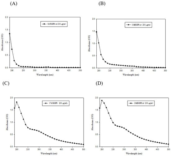
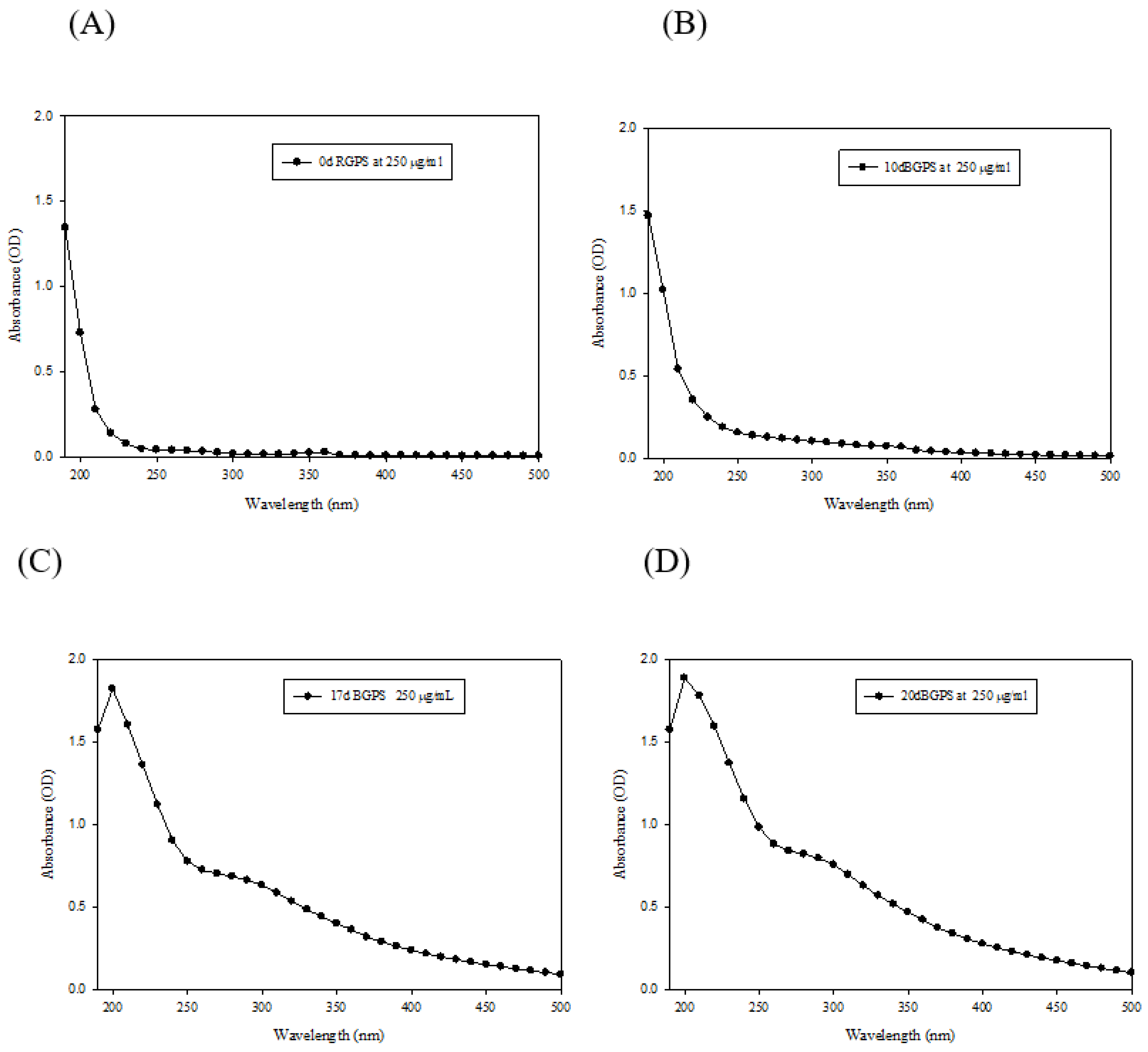
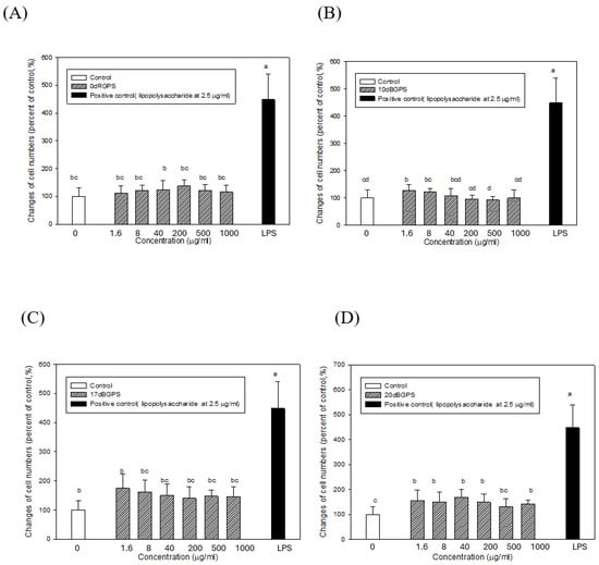
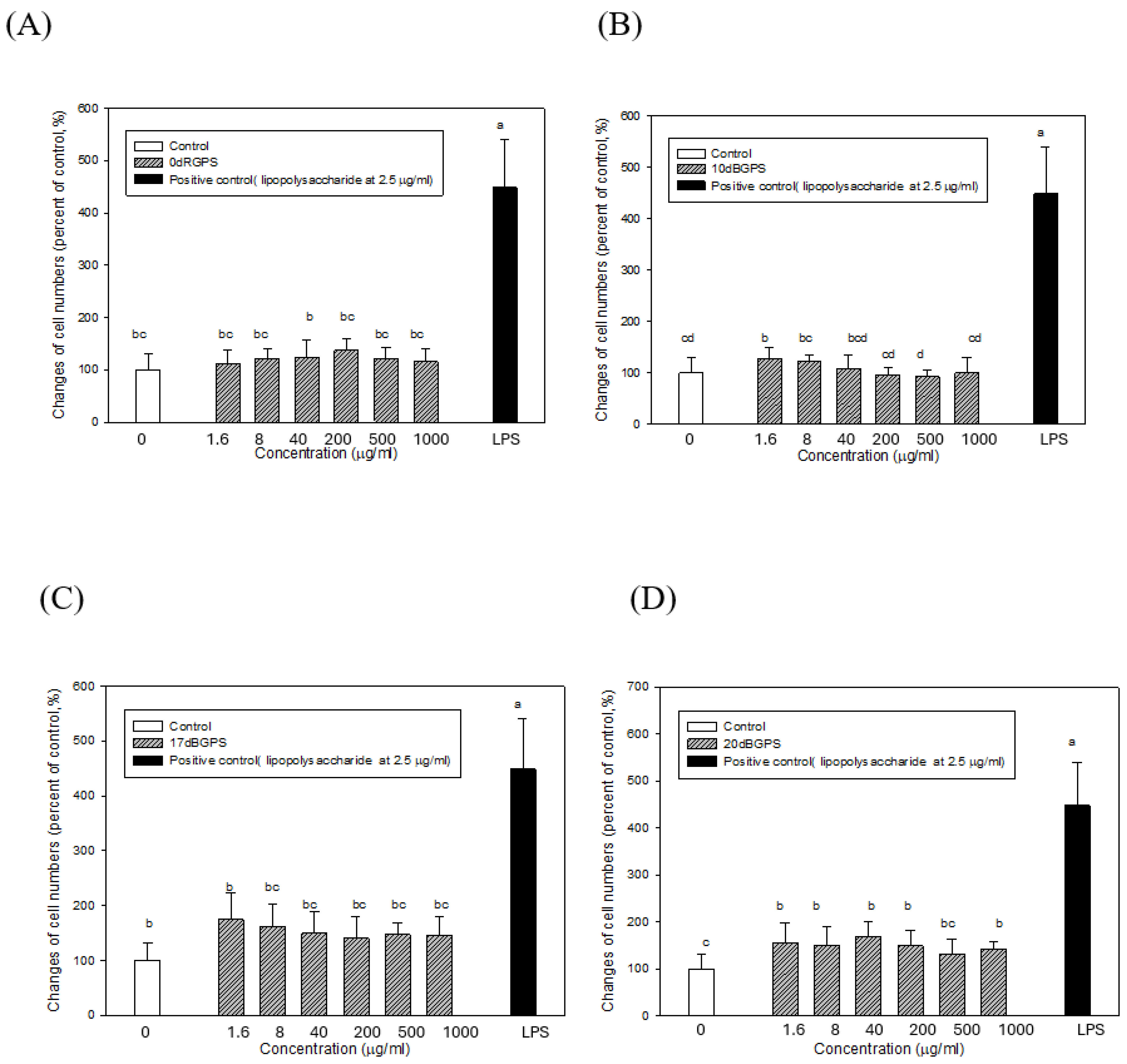
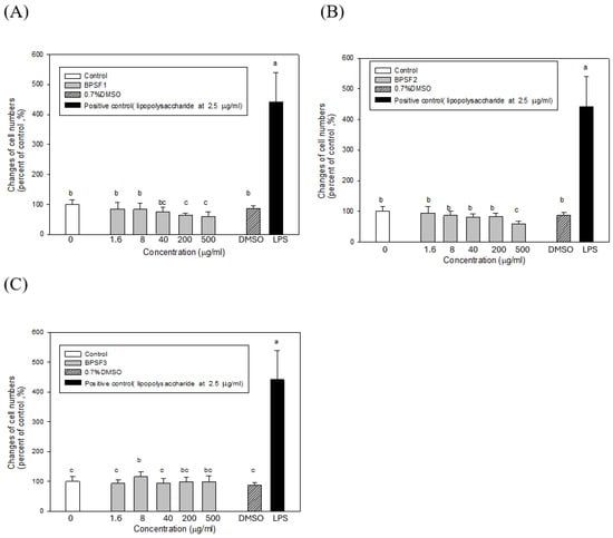
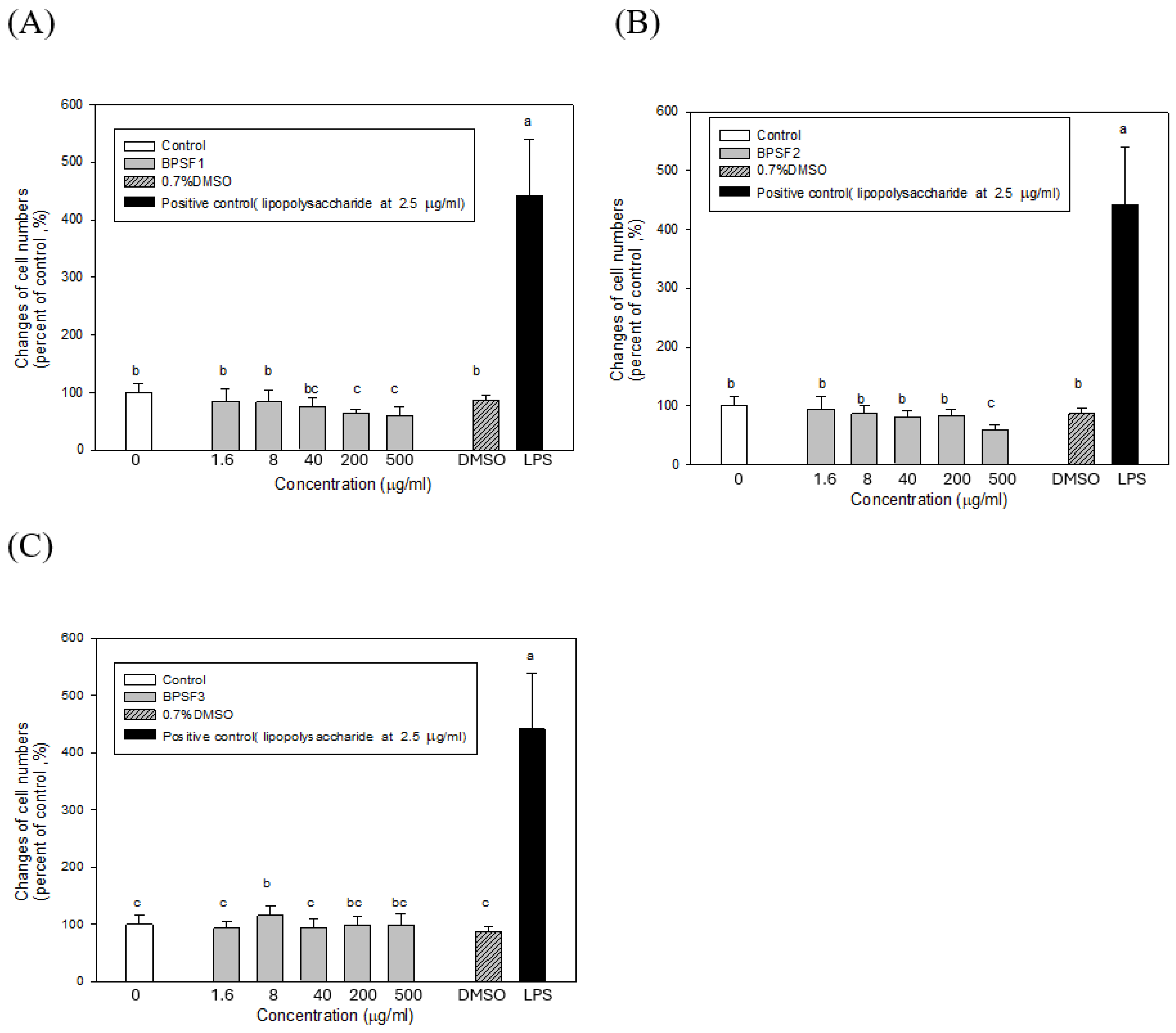
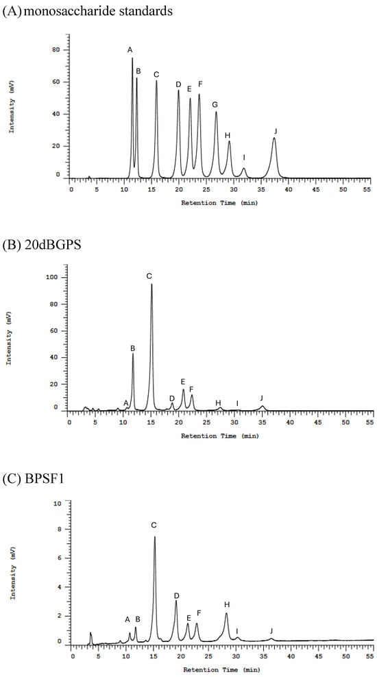
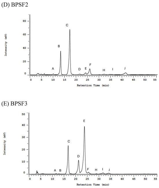
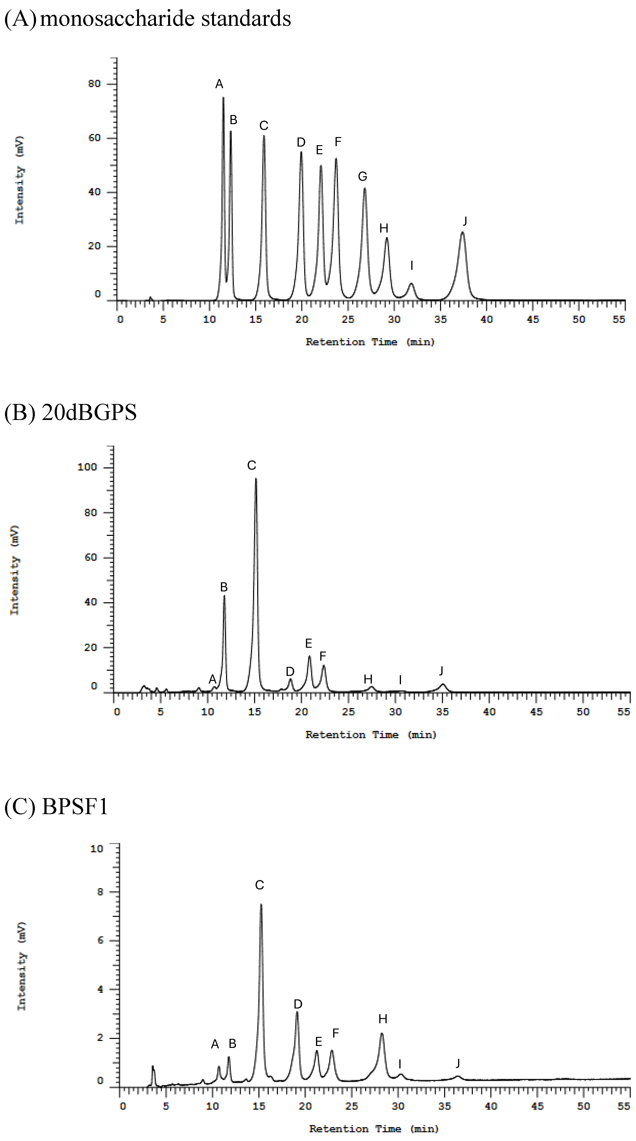
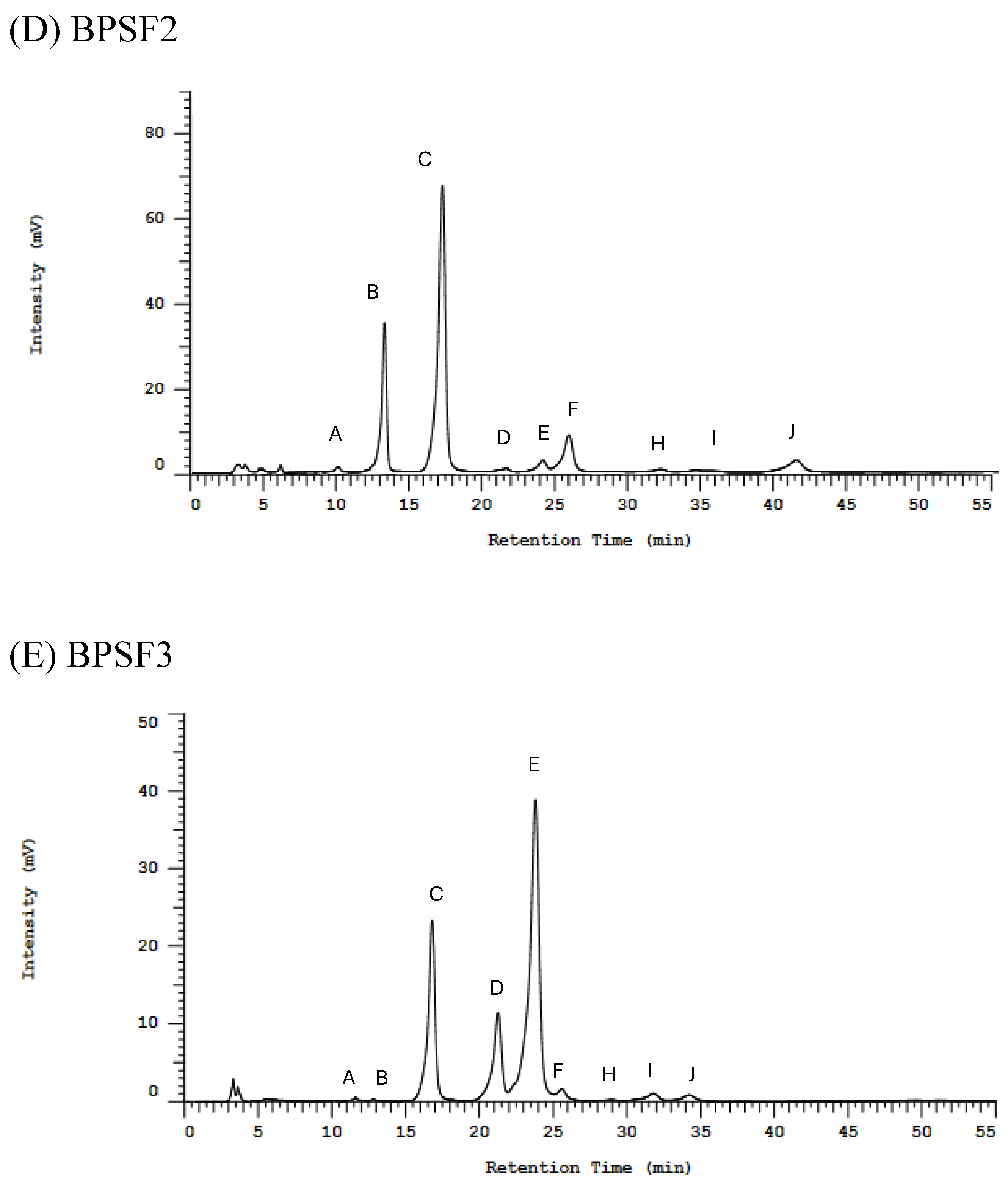
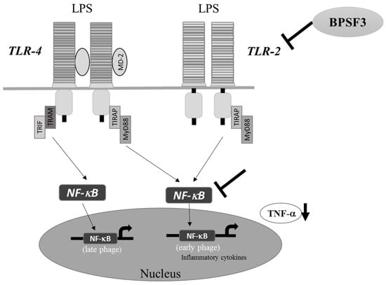
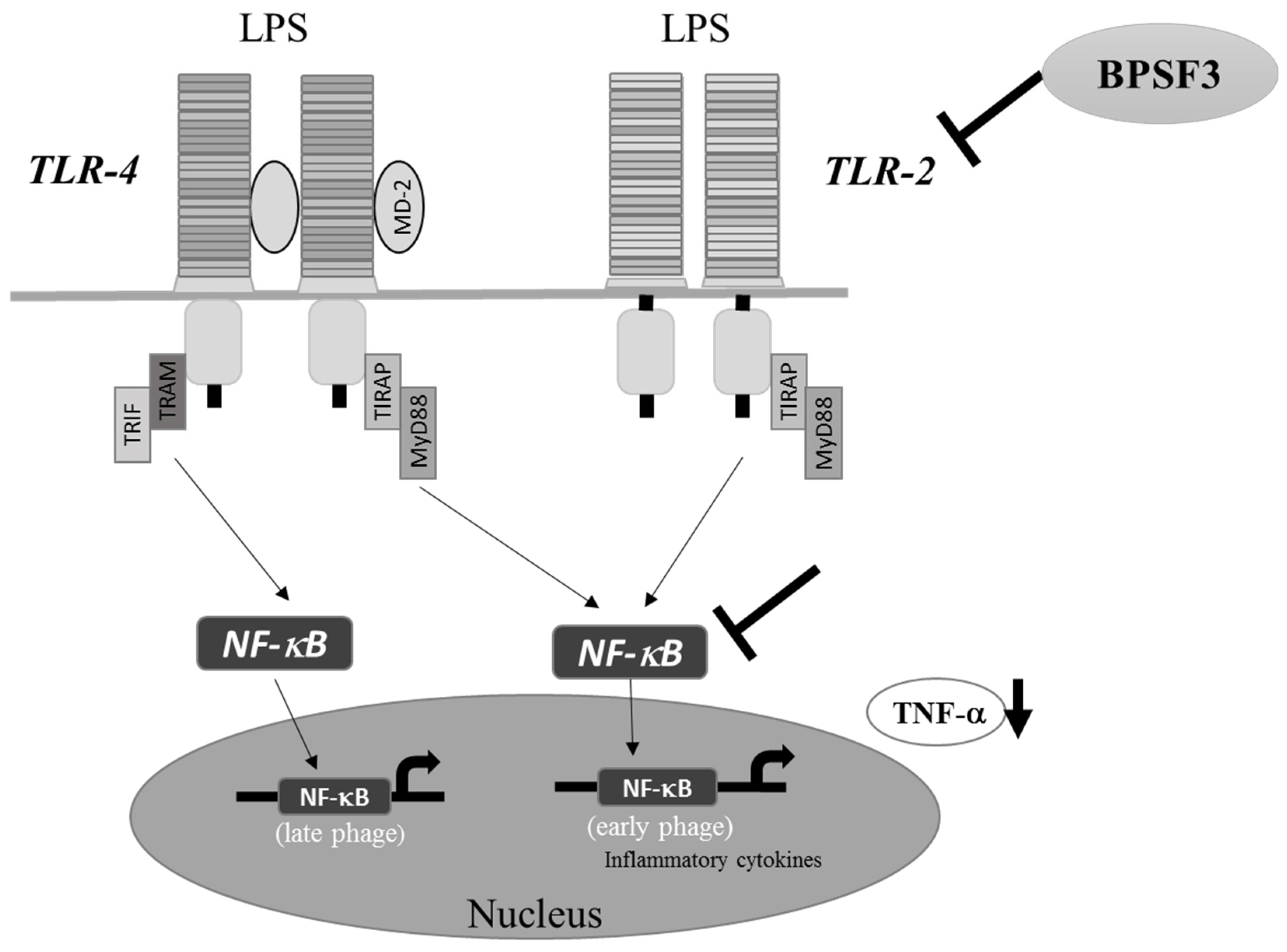

Disclaimer/Publisher’s Note: The statements, opinions and data contained in all publications are solely those of the individual author(s) and contributor(s) and not of MDPI and/or the editor(s). MDPI and/or the editor(s) disclaim responsibility for any injury to people or property resulting from any ideas, methods, instructions or products referred to in the content. |
© 2026 by the authors. Licensee MDPI, Basel, Switzerland. This article is an open access article distributed under the terms and conditions of the Creative Commons Attribution (CC BY) license.