


Abstract
Inflammation is a major pathological process underlying numerous chronic diseases, highlighting the need for anti-inflammatory inhibitors from nature. This study investigated the anti-inflammatory potential of S. praecox leaf extracts and elucidated molecular mechanisms through an integrated experimental and computational approach. As a result, the 75% methanol extract (75M) inhibited tumor necrosis factor-α (TNF-α) and interleukin-6 (IL-6) production in lipopolysaccharide (LPS)-stimulated RAW264.7. Untargeted metabolomic analysis led to the tentative identification of 96 metabolites, predominantly flavonoids, terpenoids, ellagitannins, fatty acids, and phenolic acids. Network pharmacology analysis of overlapping targets suggested TNF and IL6 emerging as crucial hub genes. Functional enrichment analyses further revealed significant involvement of inflammatory pathways. ADMET screening suggested potentially favorable predicted ADMET characteristics. Molecular docking predicted favorable binding energies between constituents and inflammatory targets. Experimental validation showed that the 75M extract significantly inhibited NF-κB activation by suppressing IκBα and p65 phosphorylation. Furthermore, the extract attenuated MAPK signaling by inhibiting ERK1/2 and p38 phosphorylation. These findings suggest that S. praecox exerts potent anti-inflammatory activity through a multi-component, multi-target mechanism involving the coordinated regulation of the NF-κB and MAPK signaling pathways, thereby providing a therapeutic basis for further investigation of S. praecox as a promising candidate for treating inflammation.
1. Introduction
Inflammation is a fundamental host defense response that maintains tissue homeostasis following infection, injury, or other harmful stimuli. Whereas acute inflammation is a tightly regulated and self-resolving process, its persistent activation can lead to chronic inflammation, which contributes to tissue damage and disease progression [1]. At the molecular level, inflammatory responses are mediated by signaling pathways such as the nuclear factor-kappa B (NF-κB) and mitogen-activated protein kinase (MAPK) pathways [2], which induce the expression of pro-inflammatory cytokines (e.g., TNF-α, IL-1β, and IL-6) [3] and inflammatory mediators, including cyclooxygenase-2 (COX-2), inducible nitric oxide synthase (iNOS), prostaglandins, and nitric oxide [4]. The dysregulated production of these cytokines and mediators promotes oxidative stress and chronic inflammatory states that are closely associated with cardiovascular diseases, diabetes, neurodegenerative disorders, cancer, and other chronic conditions [5,6]. Consequently, modulation of inflammatory pathways remains a major therapeutic strategy. Although conventional anti-inflammatory drugs are effective, their long-term use is often limited by adverse effects [7], therefore phytochemical regulation of cellular signaling provides a robust basis for addressing the limitations of conventional non-steroidal anti-inflammatory drugs.
Stachyurus praecox Siebold & Zucc. is a deciduous shrub distributed throughout East Asia, including Japan, China, and Taiwan [8]. Previous studies have demonstrated that S. praecox extracts showed biological activities such as anticancer [8] and antimicrobial [9]. Chemical studies revealed that this plant is a rich source of tannins and phenolics [8,10]. They are known to play a key part in the modulation of inflammatory responses [11,12]. Notably, in South Korea, this plant is distributed primarily in the Wando area of Jeonnam Province and is increasingly being propagated in neighboring regions due to its beautiful blossoms. However, there are still few studies on S. praecox, in quantity and scientific exploration of its pharmacological potential. There is limited investigation of the biological and chemical properties of this plant [13] through advanced technologies and thorough research.
The pharmacology of medicinal plants is frequently a consequence of the synergism of multiple bioactives that act on different biological targets. Understanding these intricate interactions is still a major challenge in natural product research and drug discovery. Network pharmacology has been used to study herbal medicines and to provide a holistic platform for the characterization of compound-target-pathway networks and the discovery of system-level mechanisms of action [14]. Molecular docking provides additional valuable information on the structural basis of ligand–target interactions and helps in the prioritization of possible therapeutic targets identified by network analysis [15]. The combination of these complementary strategies has evolved into a powerful approach for the unveiling of the multitargeted and multi-component nature of medicinal plants. With the ongoing advancement of bioinformatics, omics technologies, and computational modeling, integrated network pharmacology and molecular docking analyses are increasingly facilitating the discovery of phytotherapeutics and their underlying molecular mechanisms.
In the present study, we established an integrated holistic strategy comprising computational methods (network pharmacology integrated with molecular docking) and experimental techniques such as LC-MS/MS analysis, in vitro assays, and Western blotting analysis, including the production of plant extracts, chemical identification, and the evaluation of the anti-inflammatory potential of S. praecox leaves. This research strategy led to a multidimensional view and effective tools to discover the anti-inflammatory effects of S. praecox.
2. Results
2.1. Extraction Efficiency of S. praecox Leaves
We employed an ultrasonic-assisted extraction method [16] to extract S. praecox leaves under nine extraction conditions using different ratios of organic solvent [methanol (M) or ethanol (E)] to water. As a result, nine extracts were obtained and classified into three categories: M, E, and water (W) extracts. Among the methanol-based extracts, extraction efficiency decreased progressively with increasing methanol concentration, from 29.10% at 25% methanol (25M) to 19.95% at 100% methanol (100M), indicating that a lower methanol-to-water ratio was more effective in solubilizing the extractable constituents of the leaves. A similar concentration-dependent trend was observed for the ethanol-based extracts, with efficiency declining from 33.60% at 25% ethanol (25E) to a markedly low 8.88% at 100% ethanol (100E), the lowest yield among all nine conditions tested. Notably, 25% ethanol (25E) produced the highest overall extraction efficiency (33.60%, 336.0 mg), slightly surpassing pure water extraction (100W, 32.56%, 325.6 mg) and outperforming all methanol conditions (Table S1, Supplementary Materials). Extraction efficiency increased progressively and in near-direct proportion to the water content of the solvent. Consequently, water alone or hydroalcoholic mixtures with a low-to-moderate organic fraction (25–50%, v/v) were more effective than high-concentration organic solvents for the ultrasound-assisted extraction of S. praecox leaves under the tested conditions.
2.2. Anti-Inflammatory Effect of S. praecox Leaves
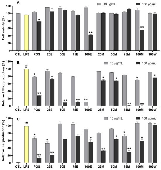
As part of our continuing efforts to find anti-inflammatory candidates from natural sources, the S. praecox extracts were examined for their anti-inflammatory capacity (Figure 1 and Figure S1 in Supplementary Materials). Specifically, the extracts were evaluated for their suppression of TNF-α and IL-6 production in LPS-activated RAW264.7 cells. In the TNF-α assay at 100 μg/mL, the 75M and 100M extracts significantly inhibited TNF-α production, with relative production levels of 1.51 ± 0.13% and 0.63 ± 0.54%, respectively, compared to those of the LPS treatment. The 50E, 75E, and 100E extracts displayed strong inhibition of TNF-α production, with relative amounts of 9.85 ± 1.27%, 11.36 ± 1.28%, and 0.18 ± 0.01%, respectively. At 10 μg/mL, the 100E extract potently suppressed TNF-α secretion by LPS-activated RAW264.7 cells, with a relative amount of 11.58 ± 1.56% compared to that of the LPS treatment without sample addition. The other samples exhibited weak or no inhibition of TNF-α production. The 50E, 75E, and 75M extracts displayed anti-inflammatory effects on LPS-induced TNF-α production in RAW264.7 cells with IC50 values of 69.30 ± 3.66, 44.40 ± 1.07, and 41.60 ± 2.85 μg/mL, respectively (Table S2, Supplementary Materials). In the IL-6 assay at 100 μg/mL, the 25E, 25M, and 75M extracts strongly suppressed IL-6 secretion by LPS-stimulated RAW264.7 cells, with relative amounts of 21.90 ± 1.97%, 33.40 ± 0.47%, and 29.91 ± 2.89%, respectively, compared to the LPS group without sample addition. The 50E, 75E, 100E, 50M, and 100M extracts moderately inhibited IL-6 production, with relative amounts ranging from 59.32 ± 2.42% to 72.83 ± 3.38%. At 10 μg/mL, the 25E and 75M extracts significantly inhibited LPS-induced IL-6 production in RAW264.7 cells. The water extract (100W) did not show significant activity under the experimental conditions. The 50E, 75E, and 75M extracts showed anti-inflammatory effects on LPS-induced IL-6 production in RAW264.7 cells with IC50 values of 19.20 ± 0.23, 53.10 ± 4.11, and 56.70 ± 3.24 μg/mL, respectively (Table S2, Supplementary Materials). We also tested the cytotoxic effects of these samples at concentrations of 10 and 100 μg/mL. The results indicated that LPS treatment did not cause any toxic effects on RAW264.7 cells under the experimental conditions, as shown by comparing the cell viability (%) of the LPS group (96.35 ± 0.12%) with that of the control (100 ± 0.59%, CTL without LPS addition). Among the extracts, 100M and 100E displayed cytotoxic effects on RAW264.7 cells, with cell viabilities of 56.18 ± 1.29% and 42.94 ± 0.30%, respectively, at 100 μg/mL. The other samples, as well as all samples tested at 10 μg/mL, did not significantly affect cell viability (survival rate over 90%).
Figure 1.
Anti-inflammatory effects of S. praecox leaf extract. RAW264.7 cells were pretreated with the tested samples (10 and 100 µg/mL) for 2 h and then stimulated with LPS (100 ng/mL) for 24 h. (A) Cell viability was assessed using the MTT assay. (B) The leaf extracts inhibited TNF-α production using an enzyme-linked immunosorbent assay (ELISA) kit. (C) The leaf extracts inhibited IL-6 production using an ELISA kit test. # p < 0.05 vs. the CTL group (Untreated control: without both LPS and sample addition). Each experiment was performed in triplicate. Differences were significant at * p < 0.05 and ** p < 0.01 compared to the LPS control group without sample addition. Dexamethasone was used as a positive control (POS) under the same experimental conditions.
2.3. Chemical Profiles and Compounds Identified from S. praecox Extract
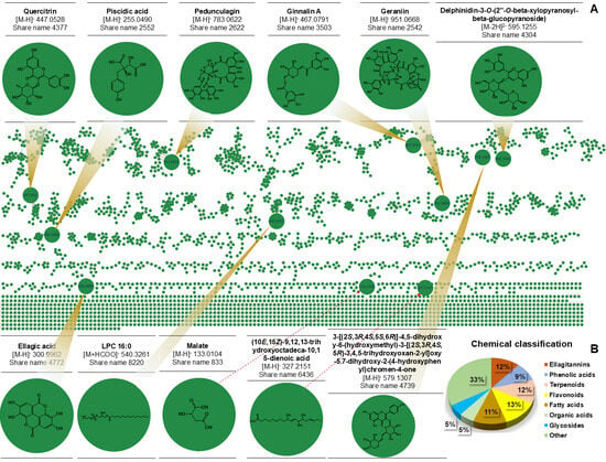
The active extract (75M) was further analyzed to identify the chemical constituents present in the S. praecox extract (Figure 2). As a result, 96 compounds were putatively identified by analyzing the mass data, comprising the precursor ions and fragmentation pathways of the ion peaks detected in the negative ion mode, and by comparing these data with reference data in public mass databases. The annotated compounds are listed in detail in Table S3 (Supplementary Materials). Among them, flavonoids, terpenoids, ellagitannins, fatty acids, and phenolic acids displayed high contents of approximately 9–13%, whereas organic acids and glycosides showed lower contents of about 5% (Figure 2).
Figure 2.
Visualization of feature-based molecular network of S. praecox metabolites. (A) Global molecular network generated from MS/MS spectral data using the GNPS2 feature-based molecular networking workflow, where each node represents a unique MS feature (grouped by spectral similarity into clusters, connected by edges). (B) Chemical composition of compound derivatives was classified.
The peak at tR 9.981 min was tentatively annotated as a kaempferol diglycoside containing glucose and arabinopyranose residues based on its accurate mass and characteristic MS/MS fragmentation profile [17]. The precursor ion detected at m/z 579.1298 in the negative-ion mode was assigned to the deprotonated molecular ion [M−H]−, consistent with the molecular formula C26H28O15. Upon collision-induced dissociation, the precursor ion underwent characteristic glycosidic fragmentation to yield a prominent product ion at m/z 429.0827 [M−2H−C5H9O5]−, corresponding to a mass difference of approximately 149 Da. This fragmentation was attributed to the loss of an O-arabinopyranosyl-derived residue through dehydrative cleavage, providing evidence for the presence of an arabinose (Ara) moiety (Figure S2, Supplementary materials). Further fragmentation of the ion at m/z 429.0785 generated product ions at m/z 285.0369 [M−H−C5H9O5−C6H10O4]− and 284.0302 [M−2H−C5H9O5−C6H10O4]−, corresponding to the kaempferol-3-O-glucoside-derived radical aglycone formed after cleavage of the remaining hexose residue and to neutral hydrogen radical ions, which are associated with homolytic cleavage at the hydroxyl groups and glycosidic bonds [18]. The formation of the aglycone-related ion was further supported by the sequential fragmentation of m/z 284.0302 to m/z 255.076 and m/z 227.0330, attributed to the successive elimination of CHO and CO [17], respectively. In addition, a characteristic ion at m/z 151.0023 was observed and assigned to retro-Diels–Alder (RDA) cleavage of the C-ring [19], providing diagnostic evidence for the kaempferol skeleton. Therefore, the accurate precursor mass, sequential losses of glycoside-derived residues, formation of the kaempferol aglycone ion, and characteristic RDA fragmentation strongly support the annotation of the peak at tR 9.981 min as 5,7-dihydroxy-2-(4-hydroxyphenyl)-3-[(2S,3R,4S,5S,6R)-3,4,5-trihydroxy-6-[[(2S,3R,4S,5S)-3,4,5-trihydroxyoxan-2-yl]oxymethyl]oxan-2-yl]oxychromen-4-one.
The peak at tR 10.083 min displayed a deprotonated ion at m/z 300.9965 [M−H]−, corresponding to the elemental composition C14H5O8− and the neutral molecular formula C14H6O8. Its major fragmentation pathway proceeded through successive neutral losses from the deprotonated molecular ion. In particular, the transition m/z 300.9965 → 283.9936 involved a loss of approximately 18.01 Da, corresponding to H2O, producing the ion C14H4O7−. This dehydration pathway is characteristic of the highly oxygenated phenolic structure of ellagic acid. In particular, the transition m/z 300.9965 → 257.0072 involves a loss of approximately 44 Da, corresponding to CO2 reduction, producing the ion C13H5O6−. The precursor ion underwent characteristic decarboxylation to produce the fragment ion at m/z 257.0072, followed by sequential losses of CO and additional carbonyl-containing moieties, yielding diagnostic fragment ions at m/z 229.0923 (Figure S3, Supplementary materials). Subsequent loss of approximately 44 Da, corresponding to CO2, generates the characteristic fragment at m/z 185.0229 (C11H5O3−) [20]. Additionally, the accurate precursor mass, together with the sequential H2O losses and the formation of the diagnostic product ions at m/z 283.9942, provides strong evidence for the annotation of this metabolite as ellagic acid. The observed fragmentation pattern is in good agreement with that of ellagic acid [20].
2.4. Anti-Inflammatory Capacity-Based In Silico Approaches of Compounds Identified from S. praecox Extract
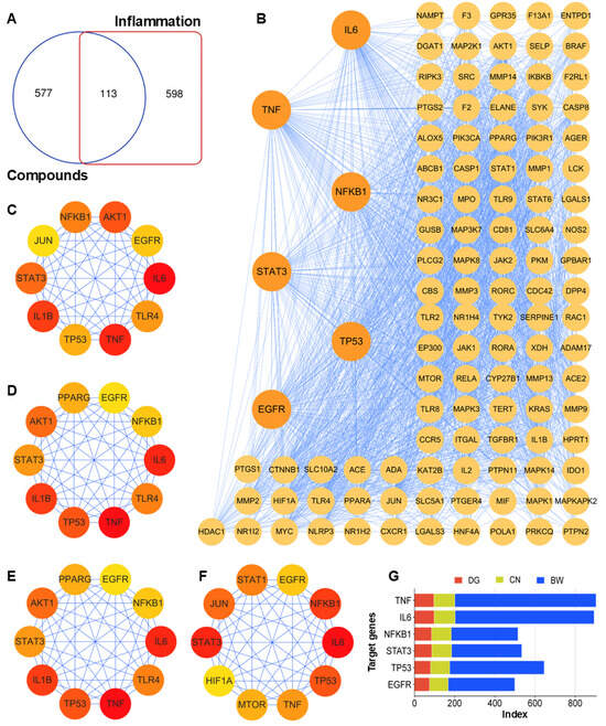
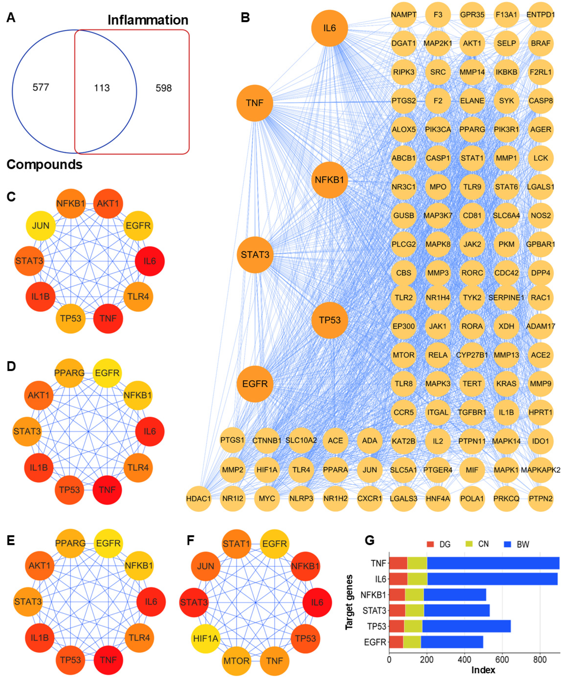
To explore the pharmacological mechanisms of S. praecox constituents in treating inflammation-related diseases, we performed network pharmacology analysis using the 30 primary compounds, which were selected based on peak areas exceeding 5000 arbitrary units estimated by LC-MS/MS analysis. The putative targets of these compounds were collected using the SwissTargetPrediction web platform, resulting in the identification of 690 gene symbols, which were intersected with 711 inflammatory disease-related targets using a Venn diagram (Figure 3A). The resulting 113 overlapping targets were used to construct a protein–protein interaction (PPI) network, illustrating their multiple interacting roles within the network (Figure 3B). The network topology was analyzed based on the degree, closeness, and betweenness centrality scores using the CytoHubba algorithm in the Cytoscape program (version 3.10.4) [21]. The top ten nodes for each topological parameter were identified, reflecting their contribution to interactions with other targets within the PPI network. Among them, IL6, TNF, and NFKB1 displayed high index values, suggesting that they may act as essential bridges in the inflammatory signaling cascade (Figure 3C–G).
Figure 3.
Network Pharmacology analysis of compounds for the treatment of inflammatory diseases. (A) Overlap analysis between compound-associated targets and inflammation-related targets. A total of 113 common targets were identified at the intersection of compound targets (690) and inflammation-associated targets (711), representing potential therapeutic targets. (B) The protein–protein interaction (PPI) network of the overlapping targets. Large orange nodes represent the most significant hub targets (including IL6, TNF, NFKB1, STAT3, TP53, and EGFR). The purple edges indicate interactions between targets. Core targets were identified using four different topological analysis algorithms: degree ((C): DG), closeness ((D): CN), betweenness ((E): BW), and maximal clique centrality ((F): MCC). Key targets are correlated with color intensity expression, ranging from red to yellow. Consistent identification of targets such as TNF, IL6, and NFKB1 across multiple methods suggests their critical role in the anti-inflammatory mechanism of the studied compounds. (G) Comparison of topological centralities for the top six hub targets based on DG, CN, and BW scores.
2.4.1. Biological Mechanisms of Targets
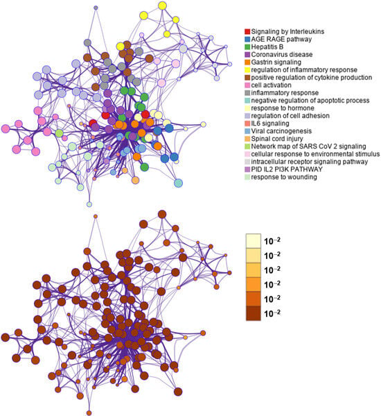
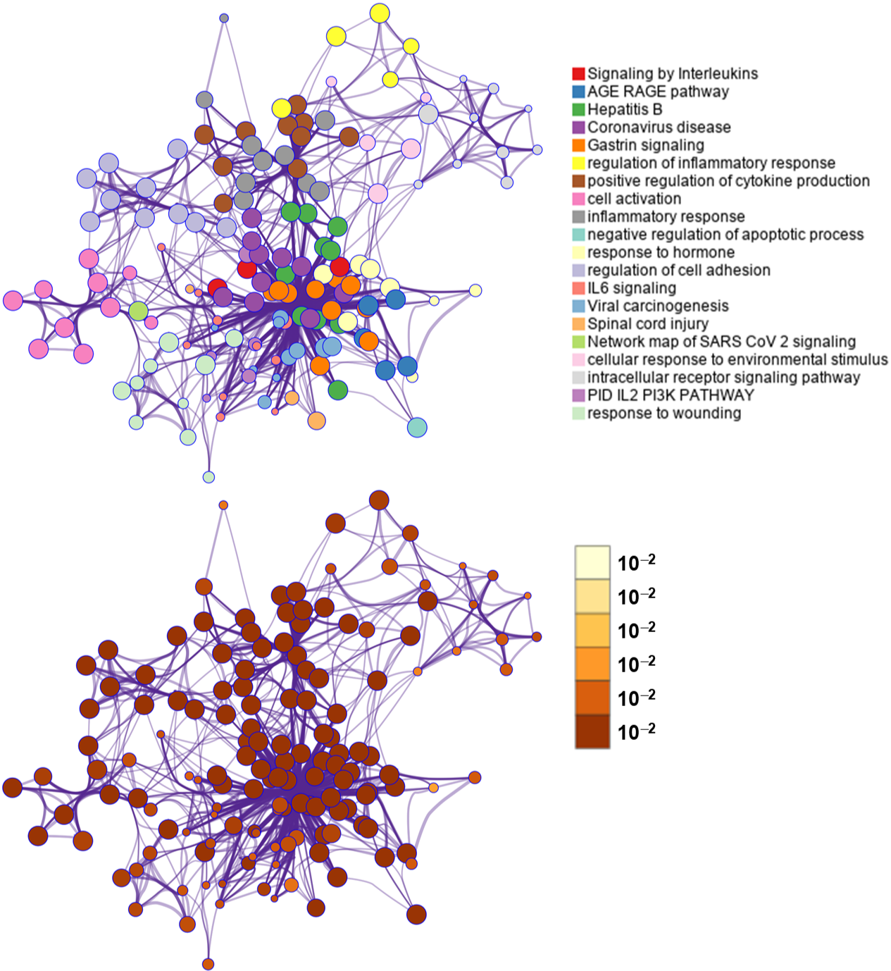
To investigate the molecular mechanisms underlying the potential therapeutic effects of these compounds against inflammatory diseases, the 113 overlapping targets were subjected to functional enrichment and network analyses using the Metascape platform (Figure 4). The enrichment network revealed that these targets were predicted in association with inflammation- and immune-related biological processes and signaling pathways, including signaling by interleukins, inflammatory response, regulation of inflammatory response, positive regulation of cytokine production, IL-6 signaling, and the PID IL-2/PI3K signaling pathway. The significant clustering and extensive interconnection among these functional terms suggested a high degree of functional overlap among the identified targets, suggesting that the compounds may exert their anti-inflammatory effects through the coordinated modulation of multiple molecular targets and signaling networks. In addition, enrichment in pathways associated with cellular responses to environmental stimuli, response to wounding, neurotrophin receptor signaling, and AGE–RAGE signaling further supports the involvement of the target genes in inflammatory processes, cellular stress responses, and tissue injury. Collectively, these findings suggest that the investigated compounds may exert potential anti-inflammatory effects through predicted interactions with multiple molecular targets and associated signaling pathways, particularly those involved in cytokine production and interleukin-mediated signaling (Figure S4, Supplementary Materials). These findings provide preliminary molecular insights into the potential anti-inflammatory properties of the investigated compounds and warrant further experimental validation.
Figure 4.
Metascape enrichment network analysis of overlapping targets involved in the treatment of inflammatory diseases. Network visualization of the top 20 enriched pathways and biological processes associated with the overlapping (shared) target genes, generated using the Metascape database. Each node represents an enriched term (GO biological process, KEGG pathway, or curated pathway database term), with node size proportional to the number of genes annotated to that term, and edges representing shared gene membership between connected terms.
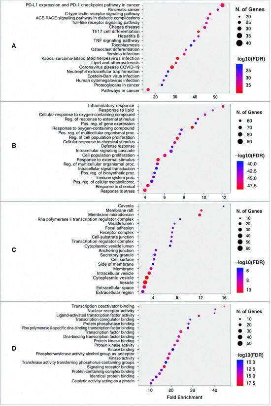
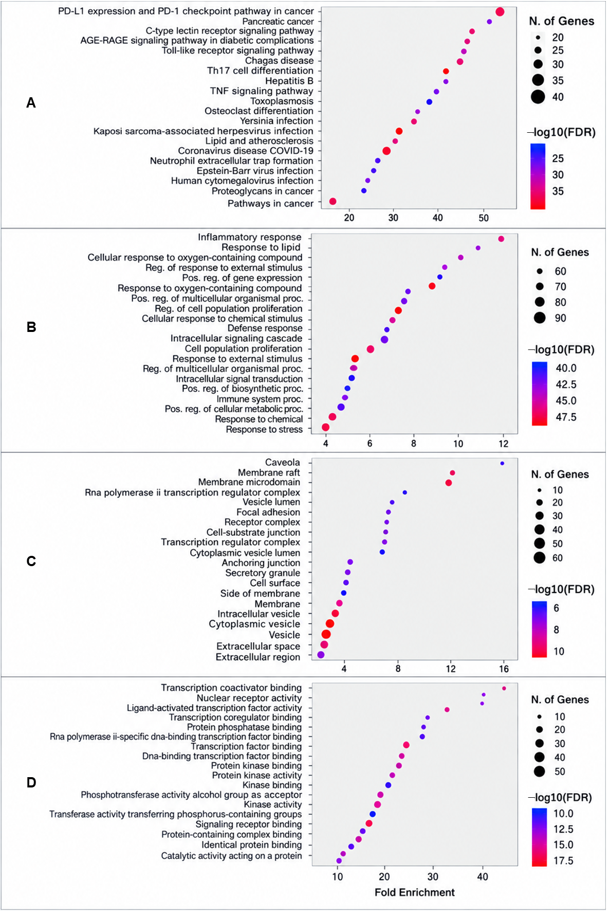
To investigate the potential molecular mechanisms underlying the biological pathways of the overlapping targets, Kyoto Encyclopedia of Genes and Genomes (KEGG) pathway enrichment analysis was performed, and significantly enriched pathways were identified based on the fold enrichment and the number of genes involved in each pathway (Figure 5A). A total of 217 pathways were enriched. Among them, the top 20 enriched terms were closely associated with inflammatory and immune responses, particularly through the regulation of cytokine production, oxidative stress, innate immune activation, and inflammatory cell signaling. The enrichment of the Toll-like receptor signaling pathway, Th17 differentiation pathway, C-type lectin receptor signaling pathway, and TNF signaling pathway suggested potential associations between the predicted targets and key inflammatory and immune-related processes. These findings provide preliminary computational insights into the potential involvement of multiple molecular targets and interconnected signaling pathways in the anti-inflammatory effects of these compounds.
Figure 5.
KEGG pathway and gene ontology (A) enrichment analyses of the potential therapeutic targets. Bubble plots illustrate the top 15 enriched KEGG pathways and GO terms, including biological process (B), cellular component (C), and molecular function (D) categories. The x-axis represents the fold enrichment, while the y-axis lists the significantly enriched pathways or GO terms. Bubble size indicates the number of genes enriched in each term, and bubble color represents the enrichment significance expressed as −log10(FDR), with warmer colors indicating higher statistical significance.
To further characterize the potential biological functions of the predicted targets associated with investigated compounds, gene ontology (GO) enrichment analysis was performed for three categories: biological process (BP), cellular component (CC), and molecular function (MF) (Figure 5B–D). BP analysis revealed that the intersecting targets were predominantly involved in classical inflammatory and immune-related pathways. Notably, the inflammatory response term exhibited the highest fold enrichment, alongside other critical processes such as response to lipid, cellular response to oxygen-containing compounds, and response to stress. Furthermore, significant enrichment was observed in regulatory processes, including positive regulation of gene expression and regulation of cell population proliferation. These results suggest that these compounds are functionally associated with biological processes involved in anti-inflammatory responses, cellular adaptation to external stimuli, and immune regulation. The high significance levels (−log10 FDR > 45) and large gene counts in these categories support the statistical significance of these biological associations. CC analysis showed that the predicted targets were significantly associated with membrane-associated structures. The top 20 enriched terms included membrane raft and membrane microdomain, which showed significantly high fold enrichment values (between 8 and 16). These findings suggest that a proportion of the predicted targets may be associated with specialized plasma membrane regions involved in cellular signal transduction. Additionally, enrichment in vesicle lumen, cytoplasmic vesicle, and extracellular space suggests that these targets may also play roles in cellular secretion and in the modulation of the extracellular inflammatory microenvironment. In terms of MF, the targets were highly enriched in activities related to gene regulation and signal transduction. The highest fold enrichment was found for transcription coactivator binding, nuclear receptor activity, and ligand-activated transcription factor activity. These results suggested that the predicted target proteins are functionally associated with transcription regulatory processes that may contribute to inflammatory gene expression. Furthermore, significant enrichment was noted for protein kinase activity, enzyme binding, and signaling receptor binding, highlighting the potential involvement of these targets in intracellular signaling cascades or kinase-mediated pathways to suppress the production of pro-inflammatory mediators.
2.4.2. Construction of Compound–Target–Disease Interaction (CTDI) Network
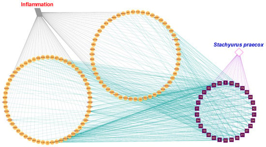
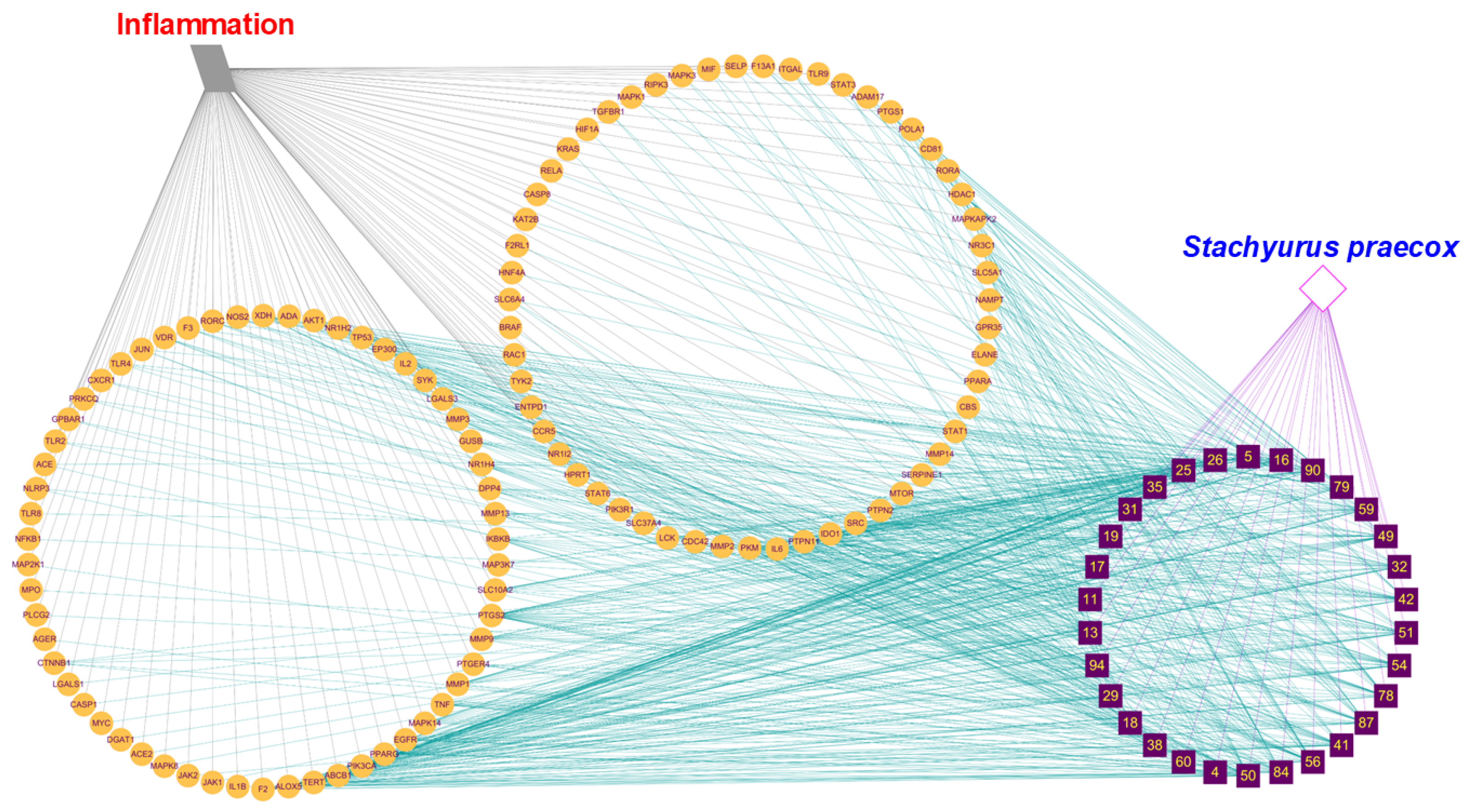
To systematically illustrate the multi-compound, multi-target therapeutic mechanism of S. praecox against inflammation, a comprehensive interaction network was constructed (Figure 6). The network consists of four hierarchical levels: a plant source (S. praecox), its bioactive constituents (compounds 1–30), 113 potential therapeutic targets, and a disease source (Inflammation). A total of 30 bioactive compounds (represented by purple rectangles) were identified from S. praecox (pink diamond). These compounds were found to interact with the 113 common targets (orange circles), which are collectively associated with the pathogenesis of inflammation (gray parallelogram). The network revealed that these proteins are the primary focal point through which the plant exerts its therapeutic effect. The dense interactions between the compound layer and the target layer (teal edges) illustrate the extensive compound–target associations, suggesting the potential pharmacological complexity of this medicinal plant. By simultaneously modulating 113 targets across various biological processes, the compounds in S. praecox are hypothesized to have a synergistic action in suppressing inflammatory conditions. This holistic interaction network supports the traditional use of the plant and provides a molecular basis for its development as a multi-target anti-inflammatory agent.
Figure 6.
Plant-compound–target–disease interaction network. Target (orange circles) interacted with inflammation-related diseases (grey parallelogram) through grey edges. Target nodes connected with compounds (purple rectangles) through teal edges. Compound nodes interacted with the plant (Stachyurus praecox: pink diamond) via magenta edges.
2.5. Pharmacokinetic Guided Selection of Drug Candidates
We employed the PSCKM web server to evaluate the favorable pharmacokinetic and safety properties of the above 30 candidates (Table S4, Supplementary Materials). The absorption, distribution, metabolism, excretion, and toxicity (ADMET) profiles of these compounds revealed 15 promising candidates that were predicted using the web platform. These compounds (4, 5, 13, 17, 18, 29, 42, 54, 56, 60, 78, 79, 84, 87, and 90) showed considerable variability in their ADMET characteristics. Predicted human intestinal absorption ranged widely from 0% (compound 18) to 100% (compound 17), and compounds 60, 79, 84, 87, and 90 also showed high absorption (>85%). Water solubility values were generally low to moderate, ranging from −3.933 (compound 87) to −1.381 (compound 4), consistent with those of moderately hydrophilic to lipophilic natural compounds. Regarding distribution, most compounds displayed low blood–brain barrier (BBB) permeability (logBB values between −4.156 and −0.136), with compound 17 exhibiting the most restricted central nervous system (CNS) penetration (logBB = −4.156, logPS = −6.138), suggesting minimal CNS exposure. In terms of metabolism, most compounds showed no inhibitory activity toward the major cytochrome P450 (CYP) isoforms (CYP1A2, CYP2C9, CYP2C19, CYP2D6, and CYP3A4), except for compound 60, which was predicted to inhibit CYP1A2; in addition, compound 84 was identified as a CYP3A4 substrate. All compounds were predicted to be negative for AMES mutagenicity, hERG I/II inhibition, and hepatotoxicity, indicating a favorable preliminary genotoxic and cardiotoxic safety profile. Notably, only compound 78 was flagged as a potential skin sensitizer. The oral rat acute toxicity (LD50) values of these compounds ranged from 1.245 (compound 78) to 3.135 mol/kg (compound 13), indicating that compound 17 possessed the most favorable long-term safety margin among the series.
2.6. Molecular Docking Verification of Primary Constituents and Hub-Gene Targets
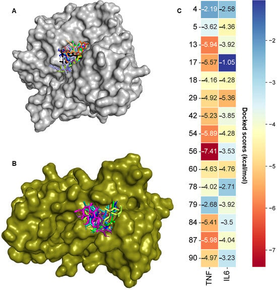
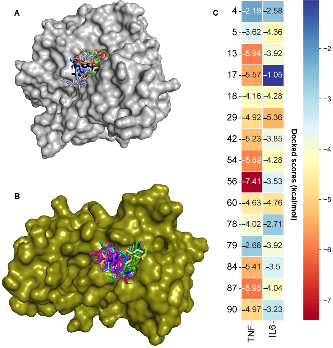
To assess the anti-inflammatory capacity of the above compounds, we performed molecular docking studies using MGLTools with AutoDock 4.2.6. All compounds were docked into the same site as the native ligand of the 2AZ5 protein (Figure 7), indicating that they occupied the binding pocket of the protein. Compound 56 demonstrated the strongest binding to the 2AZ5 protein, with a docking score of −7.41 kcal/mol. Compounds 13, 17, 54, and 87 displayed favorable binding, with docking scores of −5.94, −5.57, −5.89, and −5.98 kcal/mol, respectively. Compounds 18, 29, 42, 60, 78, and 90 showed moderate binding to the protein, with docking scores ranging from −4.02 to 4.97 kcal/mol. The other compounds displayed weak binding to the 2AZ5 protein. Notably, the compounds with favorable binding to the 2AZ5 protein interacted with crucial residues of the protein [22]. In particular, compounds 60, 54, 56, 59, 84, 87, and 90 interacted with a key residue (SER60) of the 2AZ5 protein by forming hydrogen bonds. Additionally, compounds 5, 13, 18, 42, 54, 56, 60, 78, and 84 formed hydrogen bonds with another key residue (LEU120), whereas compounds 4, 17, 18, 29, 54, 60, 78, 87, and 90 interacted with the GLY121 residue through hydrogen bonding. Compounds 4, 13, 18, 29, 78, 79, 84, 87, and 90 formed hydrogen bonds with the TYR151 residue of the 2AZ5 protein (Figure S5, Supplementary Materials).
Figure 7.
Molecular docking analysis of compounds and target proteins. Selected compounds were docked into the binding pockets of the TNF (PDB ID: 2AZ5; A) and IL-6 (PDB ID: 1ALU; B) proteins. (C) These compounds exhibited favorable energies with the two proteins.
Similarly, these compounds were docked into the same active site of the 1ALU protein, interacting with amino acids in the binding pocket of the protein. Among them, compound 29 exhibited the strongest binding, with a docking score of −5.36 kcal/mol. Compounds 5, 13, 18, 42, 54, 56, 60, 79, and 87 showed moderate binding to the 1ALU protein, with binding energies ranging from −4.76 to −3.23 kcal/mol (Figure S6, Supplementary Materials). The other compounds displayed weak binding to the target protein. These compounds interacted with key residues of the proteins through hydrogen bonds and hydrophobic interactions [23].
2.7. S. praecox Extract Suppressed Activation of NF-κB and MAPK Signaling Pathways
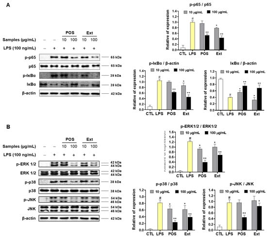
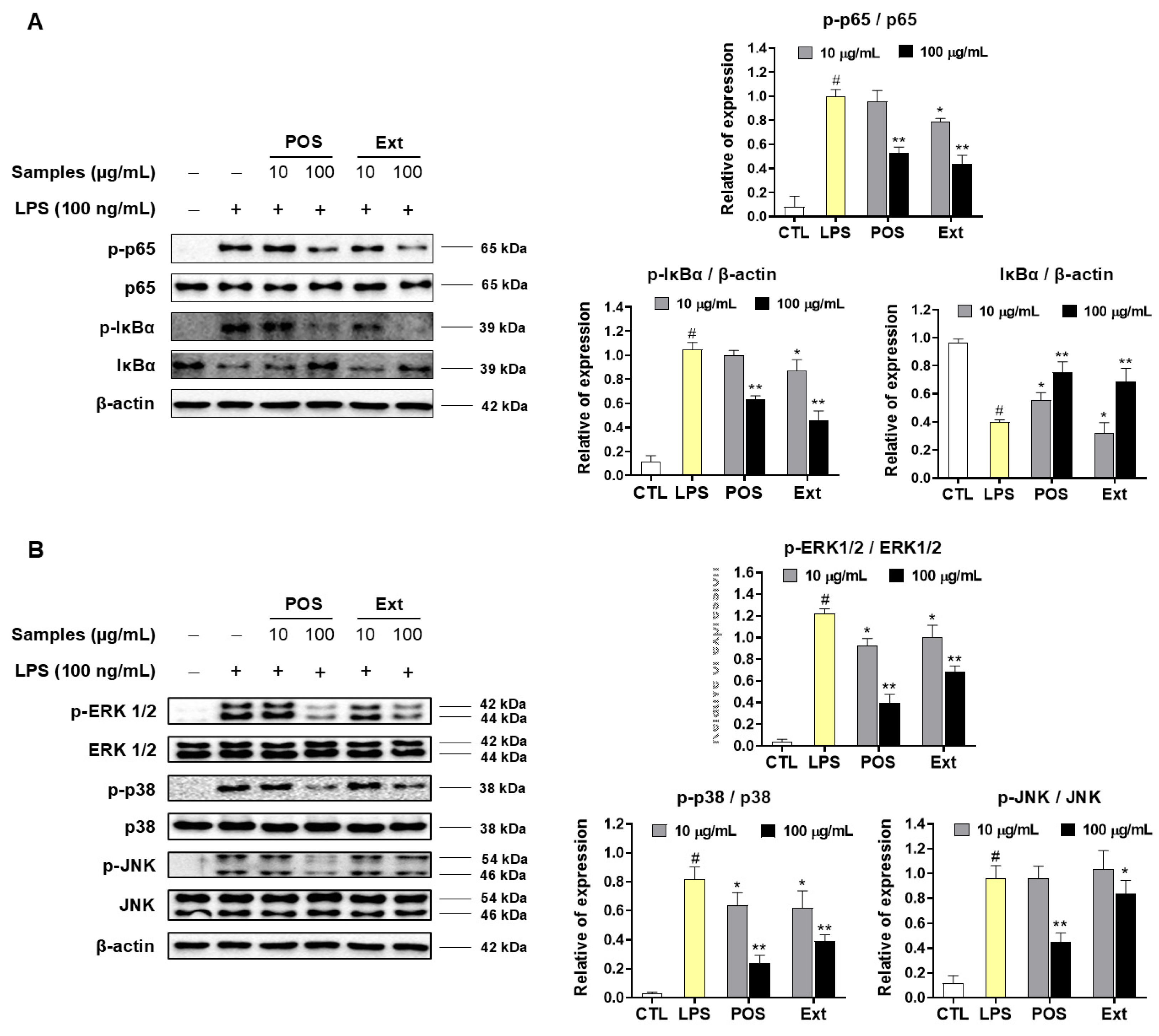
Network pharmacological analysis suggested that S. praecox extract has anti-inflammatory capacity by interacting with inflammatory cytokines through the NF-κB and MAPK metabolic pathways (Figure 8). Therefore, Western blotting assays were further employed to evaluate the impact of S. praecox extract on NF-κB and MAPK signaling pathways. To investigate the molecular mechanisms underlying the anti-inflammatory effects of the S. praecox extract (Ext), we analyzed the activation of the mitogen-activated protein kinase (MAPK) signaling pathways, including ERK, p38, and JNK. We found that LPS stimulation significantly increased the phosphorylation levels of ERK, p38, and JNK compared to the untreated LPS group. After Ext treatment, the expression levels of MAPK proteins were decreased. Particularly, Ext significantly attenuated the phosphorylation of ERK1/2 (p < 0.01) and p38 (p < 0.05) compared to those of the LPS band without Ext addition. In contrast, Ext did not significantly regulate the JNK MAPK expression levels at both tested concentrations of 10 and 100 μg/mL. Dexamethasone (Dex) effectively suppressed the phosphorylation of three MAPK proteins under the same experimental conditions. The total protein levels of ERK1/2, p38, and JNK remained constant across all groups, indicating that the extract selectively modulates the activation (phosphorylation) of these kinases rather than their total protein expression.
Figure 8.
The inhibitory effects of dexamethasone (Dex) and extraction (Ext) from S. praecox leaf extract on the LPS-induced NF-κB (A) and MAPK (B) signaling pathways in RAW 264.7 cells. After pretreating cells with these samples (10, 100 µg/mL) for 2 h, cells were stimulated with LPS (100 ng/mL) for 15 min. Total proteins were then isolated, separated by SDS-PAGE, and immunoblotted using specific antibodies for p-p65, p65, p-IκBα, IκBα, p-ERK, p-p38, p-JNK, ERK, p38, and JNK. β-actin served as an internal control. # p < 0.01 vs. non-treated group (CTL). Differences were significant at * p < 0.05 and ** p < 0.01, compared with the LPS control group that was induced only by LPS stimulation. Dexamethasone was used as a positive control (POS) under the same experimental conditions.
The Western blot results comprehensively demonstrate that LPS stimulation robustly activated the NF-κB signaling pathway, as evidenced by concordant changes across all three key markers: p-p65/p65, p-IκBα/β-actin, and total IκBα/β-actin. LPS treatment markedly increased phosphorylated p65 (p-p65/p65 ratio rising from about 0.18 to 1.10, p < 0.05) and phosphorylated IκBα (p-IκBα/β-actin ratio increasing from about 0.15 to 1.10, p < 0.05), while simultaneously depleting total IκBα protein (about 0.95 to 0.40, p < 0.05). This coordinated pattern is mechanistically consistent, since IKK-mediated phosphorylation of IκBα targets it for ubiquitin–proteasomal degradation, which in turn permits phosphorylation and nuclear translocation of p65, the transcriptionally active NF-κB subunit driving pro-inflammatory gene expression.
The Ext effectively altered the expression levels of NF-κB proteins under experimental conditions. At 100 μg/mL, Ext significantly decreased p-p65/p65 to approximately 0.52 (p < 0.01) with an effect comparable to that of Dex at the same concentration. Ext also strongly reduced p-IκBα/β-actin to approximately 0.18 (p < 0.01) with the lowest value among all treatment groups, suggesting strong suppression of IκBα phosphorylation. Correspondingly, total IκBα/β-actin was significantly restored to approximately 0.62 (p < 0.01 vs. LPS). However, this recovery was lower than that achieved by Dex at the equivalent dose. At the lower concentration (10 μg/mL), Ext also showed significant effects on all three markers (p < 0.05), indicating that its inhibitory activity on NF-κB signaling is evident even at lower concentrations. In contrast, treatment with Dexamethasone (Dex) significantly reversed LPS-induced NF-κB activation in a concentration-dependent manner across all three proteins. At 100 μg/mL, Dex markedly reduced p-p65/p65 to approximately 0.58 (p < 0.01 vs. LPS) and suppressed p-IκBα/β-actin to approximately 0.52 (p < 0.01), while correspondingly restoring total IκBα/β-actin to approximately 0.78 (p < 0.01). Even at the lower dose (10 μg/mL), Dex produced significant, albeit more modest, effects (p < 0.05 for p-p65 and IκBα). These findings confirm that Dexamethasone exerts its well-characterized anti-inflammatory action by inhibiting upstream IκBα phosphorylation/degradation, thereby preventing p65 activation and nuclear translocation. Ext and Dex did not cause any effect on the internal control (β-actin) protein compared to those of either negative control (no LPS addition) or LPS group.
Taken together, these results indicate that S. praecox extract attenuates LPS-induced NF-κB pathway activation through a consistent upstream-to-downstream mechanism: suppression of IκBα phosphorylation in order to prevent IκBα degradation, and suppression of p65 phosphorylation and, presumably, its subsequent nuclear translocation. Notably, the S. praecox extract effect on p-p65 suppression and p-IκBα inhibition at 100 μg/mL was comparable to or exceeded that of the positive control (Dex), highlighting its strong anti-inflammatory potential acting through modulation of the IκBα/NF-κB signaling axis. This mechanistic consistency across all three markers strengthens the conclusion that the extract’s anti-inflammatory activity, like that of Dex, is mediated substantially through inhibition of the canonical NF-κB activation cascade, supporting its candidacy for further mechanistic and in vivo validation, assessment of NF-κB nuclear translocation by immunofluorescence, or measurement of downstream inflammatory mediators such as TNF-α and IL-6.
3. Discussion
To further elucidate the anti-inflammatory potential of the extract, a systematic screening across multiple extraction conditions (25–100%, ethanol [E], methanol [M], and water [W]) was performed using MTT, TNF-α, and IL-6 assays in LPS-stimulated cells. Among these, the 75% methanol extract (75M) emerged as a particularly promising candidate: it markedly suppressed both TNF-α and IL-6 production at both tested concentrations (10 and 100 μg/mL), reducing relative TNF-α production to near-baseline levels (less than about 5% at 100 μg/mL, ** p < 0.01) and significantly attenuating IL-6 production (about 30% at 100 μg/mL, ** p < 0.01), while displaying no toxic effect on cell viability compared to the control at both tested concentrations (10 and 100 μg/mL) (Figure 1). These results suggest that anti-inflammatory activity is attributable to extracts that inhibit inflammatory cytokines without damaging host cells. Mass spectrometric analysis of the 75M extract enabled the putative identification of its constituents by interpreting precursor masses and fragmentation patterns against mass databases (GNPS2, American, HMDB). The untargeted metabolomics analysis achieved high annotation accuracy (mass errors less than 5 ppm), resulting in the identification of 96 compounds characterized by high LC-MS signal intensities, including phenolic derivatives such as praecoxins (A-C), stachyurin, and casuarinin [24,25]. Among the annotated compounds, 30 primary compounds selected based on the integrated peak areas in the mass chromatograms were subsequently used to evaluate the anti-inflammatory potential of the extract. We employed network pharmacology, a powerful computational technology that systematically screens and predicts how compounds interact with complex biological networks to treat inflammation-related diseases [26]. Network pharmacology analysis was performed to elucidate the potential molecular targets and mechanisms underlying the anti-inflammatory activity of the 30 compounds identified from S. praecox. Analysis of the PPI network revealed that TNF and IL6 were the hub targets, and both are known to play important roles in inflammation [3]. These results suggested that the 75M extract has an anti-inflammatory effect on LPS-induced TNF and IL-6 production in RAW267.4 cells because it contains these active compounds. In addition, the Metascape functional clustering of the overlapping targets further revealed strong enrichment in terms directly relevant to inflammatory signaling, including “regulation of inflammatory response,” “positive regulation of cytokine production,” “inflammatory response,” and “IL6 signaling” (Figure 4), reinforcing that the identified targets converge on canonical inflammatory and cytokine-signaling machinery rather than on unrelated biological processes. This convergence was corroborated by the GO and KEGG enrichment analyses (Figure 5), which showed that the overlapping targets were significantly associated with inflammatory response as the top-ranked biological process; membrane raft/caveola as the predominant cellular component, consistent with the membrane-proximal localization of cytokine receptor signaling complexes; and transcription factor binding and kinase binding as the leading molecular functions, reflecting the transcriptional and kinase-mediated regulatory nature of the hub genes (NFKB1 and STAT3). KEGG pathway enrichment highlighted the TNF signaling pathway, Toll-like receptor signaling pathway, AGE-RAGE signaling pathway in diabetic complications, and Th17 cell differentiation among the most significantly enriched pathways, further supporting a mechanistic basis centered on NF-κB and cytokine-driven inflammatory cascades [5,27]. Finally, the plant–compound–target–disease interaction network (Figure 6) visually integrates these findings, illustrating that a subset of the 30 primary constituents simultaneously engages multiple hub and peripheral inflammation-associated targets, consistent with a multi-compound, multi-target mode of action rather than a single ligand–single target interaction.
Network pharmacology and molecular docking integration suggested anti-inflammatory potential of the key constituents presented in the 75M extract. These computational results may support the anti-inflammatory effects of the 75M extract through the above predicted inflammatory pathways. To clarify these findings, Western blot analysis was employed to evaluate the anti-inflammatory effect of the 75M extract, focusing on the NF-κB and MAPK signaling cascades. Indeed, the 75M extract significantly suppressed LPS-induced phosphorylation of p65 and IκBα while restoring total IκBα protein levels under experimental conditions, indicating effective modulation of NF-κB pathway activation. In parallel, 75M substantially attenuated the phosphorylation of all three major MAPK subfamilies (ERK1/2, p38, and JNK), with the most pronounced suppression observed for p-ERK1/2 and p-p38 at 100 μg/mL; these effects closely paralleled, and for some markers (p-p65, p-IκBα) even exceeded, those achieved by the Dex positive control at the equivalent dose. Given that the NF-κB and MAPK pathways converge to drive the transcriptional activation of the TNF-α and IL-6 genes, the concordant suppression observed across p-p65/p-IκBα and p-ERK1/2/p-p38/p-JNK provides strong mechanistic support for the cytokine-suppressive effects observed in the functional assays. Further studies, including in vivo experiments and clinical trials, should be conducted to verify these anti-inflammatory effects and to provide a full overview of the potential of S. praecox in treating inflammatory disorders.
Pharmacokinetic and ADMET evaluations of these primary compounds were then conducted to predict their metabolic stability and toxicity profiles. Such analyses facilitate the early identification of promising candidates with favorable ADMET characteristics, thereby supporting promising candidates for further investigation [28]. The ADMET predictions collectively suggest that several of the evaluated natural compounds, particularly compounds 17, 60, and 90, possess pharmacokinetic and toxicological profiles compatible with further development as orally administered anti-inflammatory candidates. Compound 17 stands out due to its complete predicted intestinal absorption, restricted BBB penetration, absence of cytochrome P450 inhibition, and the highest chronic toxicity threshold (lowest observed adverse effect level among the tested set, which together indicate both good oral bioavailability and a wide safety margin for peripheral, non-CNS-targeted anti-inflammatory applications. In contrast, compound 18, despite an acceptable toxicity profile, exhibited essentially no predicted intestinal absorption, implying that conventional oral formulation would be unsuitable and that alternative delivery strategies, such as nanoencapsulation, topical application, or prodrug design, would be necessary to optimize its therapeutic potential. The absence of AMES mutagenicity, hERG inhibition, and hepatotoxicity signals across nearly all compounds is encouraging from a translational safety standpoint, as these endpoints represent common causes of attrition in natural-product-derived drug development. However, the skin sensitization predicted for compound 56 warrants caution if topical formulations are considered. The general lack of CYP450 inhibitory activity across the series, with the exception of compound 42 (CYP1A2) and compound 79 (CYP3A4 substrate), further suggests a low likelihood of clinically significant drug–drug interactions, an important consideration given that anti-inflammatory therapies are frequently co-administered with other medications in chronic disease management. Nonetheless, these findings are derived exclusively from computational prediction models, and although such approaches are valuable for early-stage compound prioritization and resource allocation, they cannot substitute for experimental validation. Future work should therefore focus on in vitro assays (e.g., hepatocyte viability, Caco-2 permeability, CYP450 inhibition assays) and in vivo pharmacokinetic studies to confirm the predicted ADMET behavior of the top-ranked candidates, particularly compound 17, prior to advancing toward formulation development and efficacy testing in relevant inflammatory disease models.
Besides some results achieved in this study, there are several issues that warrant further investigation in future research. Specifically, S. praecox leaves should be collected with sufficient biomass to isolate the pure compounds identified in this study. Subsequently, the biological effects of isolated compounds should be evaluated to validate the anti-inflammatory mechanisms. Furthermore, the ADMET profiles of these isolated compounds should be assessed through relevant experiments. Moreover, the anti-inflammatory effects of S. praecox or pure compounds should be evaluated in vivo using animal models.
4. Materials and Methods
4.1. Plant Materials and Extraction
The S. praecox leaves were collected at Jeollanamdo Forest Research Institute in October of 2025. Its taxonomy was identified by Prof. Mina Lee, College of Pharmacy, Sunchon National University. A voucher specimen (SCNUP 43) is preserved at the Pharmacognosy Laboratory, College of Pharmacy, Sunchon National University, Suncheon, Jeonnam-Gwangju 57922, Republic of Korea.
The leaves of S. praecox were dried using a dryer at 35 °C for 20 h. The dried material was pulverized into a fine powder using a mechanical grinder. To ensure a uniform particle size and maximize the extraction efficiency, the resulting powder was subjected to a sieving process. We employed a standard test sieve (Chung Gye, Republic of Korea) with an aperture size of 250 μm (60 mesh) and a wire diameter of 160 μm. The fraction passing through the 250 μm mesh was collected to obtain a homogeneous powder, while the oversized particles were re-ground. The standardized powder was then stored in an airtight container at room temperature until further extraction. Each extraction batch was created by accurately weighing 1 g of dried powder into 100 mL of a solvent mixture that included varying concentrations of organic solvents, specifically methanol (M) or ethanol (E) in water, with combinations of 25%, 50%, 75%, and 100% M; 25%, 50%, 75%, and 100% E; and 100% W (water without any organic solvents). Each sample was prepared by sonicating for a duration of 120 min at room temperature, with three repetitions for each sample. The extracts were strained using Whatman No. 2 filter paper. The extracts were subjected to drying using a vacuum rotary evaporator set at 40 °C, incorporating solvent recovery cooling. The raw extracts were kept in the refrigerator for subsequent utilization.
4.2. Sample Preparation and Data Acquisition Using LC-MS/MS Conditions
We weighed 5 mg of each sample accurately, followed by dissolving in 50% high-grade methanol. They were further filtered through a polytetrafluoroethylene membrane (size of 0.22 μm, manufactured by Agilent Technologies, Santa Clara, CA, USA) before being inserted intothe samples were analyzed using high-sensitivity nano flow rate liquid chromatograph (Vanquish flex systems, ThermoFisher Scientific, Waltham, MA, USA, AI Bio Autonomous Research Core Facility Center, NFEC-2025-03-304724) and were analyzed using ultra-high resolution liquid chromatography mass spectrometer (Orbitrap Exploris120, Thermo Fisher Scientific, Waltham, MA, USA, AI Bio Autonomous Research Core Facility Center, NFEC-2025-03-304722).. Data acquisition and compound annotation were performed using our previous method [29].
4.3. Cell Culture
Macrophage RAW 264.7 cells, purchased from the Korean Cell Line Bank (Seoul, Republic of Korea), were cultured at 37 °C in a humidified incubator containing 5% CO2. Complete growth medium consisted of Dulbecco’s modified Eagle’s medium (DMEM) supplemented with 10% heat-inactivated fetal bovine serum (FBS) and 100 IU/mL penicillin solution (HyClone, Logan, UT, USA).
4.4. Cell Viability Assay
For the cell viability assay, macrophages were seeded into a 96-well plate at a density of 2 × 10⁵ cells/well and maintained in DMEM for 24 h. The cells were analyzed using cell preprocessing-automation linkage system (LM-1500BS, Labconco, Kansas City, Missouri, USA, AI Bio Autonomous Research Core Facility Center, NFEC-2026-10-319158). Cells were treated with test samples at 10, 100 µg/mL for 2 h, followed by LPS treatment (100 ng/mL) for 24 h. Afterward, MTT solution (5 mg/mL; Sigma-Aldrich, St. Louis, MO, USA) was added and incubated at 37 °C for 4 h. The culture supernatant was carefully aspirated, and the insoluble formazan product was dissolved in 100 µL of DMSO. Absorbance at 570 nm was measured using a microplate spectrophotometer (BioTek Instruments, Winooski, VT, USA).
4.5. Measurement of TNF-α and IL-6 Production
RAW264.7 cells were seeded into 96-well plates at a density of 2 × 10⁵ cells/well. After pre-incubating with the test samples or dexamethasone at 10 and 100 µg/mL for 2 h, the cells were stimulated with LPS for 24 h. The secretion of TNF-α and IL-6 into the culture supernatant was quantified using a commercial mouse TNF-α and IL-6 ELISA kit (BD OptEIA™, San Jose, CA, USA) according to the manufacturer’s protocol.
4.6. Western Blot Assays
Macrophages were seeded into 6-well plates at a density of 2 × 10⁵ cells/well in Dulbecco’s modified Eagle’s medium (DMEM) and incubated for 24 h. The cells were then pretreated with test samples for 2 h before stimulation with LPS (100 ng/mL). To extract total cellular proteins, cells were lysed using a commercial protein extraction solution (PRO-PREP, iNtRON Biotechnology, Daejeon, Republic of Korea) on ice for 30 min. Protein concentrations were quantified using the Bradford method with a Pierce™ Bradford Protein Assay Kit (Thermo Fisher Scientific, Waltham, MA, USA). Equal amounts of protein (20 µg per lane) were resolved by sodium dodecyl sulfate–polyacrylamide gel electrophoresis (SDS-PAGE) on 10% acrylamide gels and subsequently transferred onto polyvinylidene difluoride (PVDF) membranes. To prevent non-specific binding, the membranes were blocked with 5% non-fat dry milk in Tris-buffered saline containing 0.1% Tween-20 at room temperature for 2 h. The membranes were then incubated overnight at 4 °C (18 h) with primary antibodies against phospho-p44/42 MAPK (#9101), p44/42 MAPK (#4695), phospho-p38 (#9211), p38 (#9212), JNK (#4668), phospho-JNK (#9252), phospho-p65 (#3033), p65 (#8242), and phospho-IκBα (#9246) (Cell Signaling Technology, Danvers, MA, USA), as well as IκBα (SC-1643; Santa Cruz Biotechnology, Santa Cruz, CA, USA) and β-actin (AB_2289199; BD Biosciences, San Jose, CA, USA), all diluted 1:1000 in 2.5% skim milk. Following primary antibody incubation, the membranes were washed three times with TBST and incubated with horseradish peroxidase (HRP)-conjugated secondary antibodies (1:2000 in 5% skim milk) for 2 h at room temperature. After five additional TBST washes, target protein bands were visualized using SuperSignal™ West Femto Maximum Sensitivity Substrate (Thermo Fisher Scientific) and captured using a MicroChemi 4.2 Chemiluminescence System (DNR Bio-Imaging Systems, Neve Yamin, Israel).
4.7. Determination of Compound Targets and Inflammation-Related Targets
The primary compounds annotated from S. praecox extracts were inserted into the Pubchem web platform (https://pubchem.ncbi.nlm.nih.gov/, accessed on 11 August 2026). The obtained SMILES were then input into the SwissTargetPrediction platform (https://www.swisstargetprediction.ch/predict.php, accessed on 15 August 2026) to retrieve the compound targets. Additionally, inflammation-related targets were obtained from the Gencard database (https://www.genecards.org/, accessed on 18 August 2026) [30] by setting a threshold cutoff of 5 and “Homo sapiens” as the species. These targets were further standardized by using the UniProt database. The intersection targets between compound and inflammation-related targets were identified by employing the Venn diagram web platform at https://jvenn.toulouse.inrae.fr/app/example.html (accessed on 11 August 2026).
4.8. Gene Topology and KEGG Enrichment Analysis
The overlapping targets were further analyzed for enrichment analysis using the Metascape and SHINYGO 0.86 databases for the human species set [31]. To systematically investigate the biological significance of these targets, functional enrichment analysis was performed on both Metascape and ShinyGO. The analysis included gene ontology (GO) enrichment analysis, including biological processes (BP), molecular functions (MF), and cellular components (CC), as well as KEGG pathway enrichment analysis. The aim was to find significantly enriched signaling pathways and functional terms. The enrichment analysis showed the statistical robustness of the integration of multiple authoritative biological databases enabled by the Metascape platform. Functional annotations were performed by ShinyGO, and the results of the enrichment analysis were intuitively validated and visualized. Then, significantly enriched GO terms and KEGG pathways were identified at a threshold of p < 0.01 after multiple testing correction.
4.9. Construction of PPI and CTDI Networks
The overlapping targets were imported into the STRING database (version 12.0; https://string-db.org) to construct the PPI network, in which the minimum interaction confidence score was set to 0.7 for Homo sapiens species. Data visualization was conducted using Cytoscape. The Cytoscape Cytohubba plugin was utilized to determine network parameters based on the degree, betweenness, closeness, and centrality, and maximal clique centrality scores. Concurrently, a compound–target–disease network was constructed to visualize the relationships between the selected compounds of plant, their protein targets, and inflammation-related genes [30].
4.10. Calculation of Binding Affinities and Interactions
The protein structures (PDB ID: 2AZ5) and IL-6 (PDB ID: 1ALU) were collected from the protein data bank (https://www.rcsb.org/, accessed on 1 September 2026). The 3D structures (sdf format) of the selected compounds were obtained from the Pubchem database. Receptors and ligands were prepared using the MGL tool (version 1.5.7) by following our previous study [32,33]. Briefly, receptors were processed by removing water molecules, followed by removing the heteroatoms and original native ligands. Then, hydrogen atoms were added, followed by adding Kollman charges before checking for missing loops to ensure all loops are modeled properly. Finally, receptors were saved in the pdbqt format. The Open Babel application was used to convert the SDF formats of selected compounds to the PDBQT formats. Once optimized, the ligand was prepared by following the step of adding Compute Gasteiger charges before saving as pdbqt format. Each grid box dimension was followed by our previous studies or by selecting the binding sites surrounding the original native ligand of the protein, which were defined for 2AZ5 (x, y, z: 40, 40, 40; coordinates of −19.573, 74.852, and 33.838) and for 1ALU (x, y, z: 40, 40, 40; coordinates of −7.272, −11.997, and 3.767). Autodock4.2.6 was used to estimate the docked scores between ligands and target proteins, with the receptor set to rigid. Docking calculations were performed using the Lamarckian genetic algorithm with ten runs. Each docking result was ordered according to the function of the binding energy. The RMSD value was determined to be 1.04 Å between the original and redocked native ligands of the 6,7-dimethyl-3-[(methyl{2-[methyl({1-[3-(trifluoromethyl)phenyl]-1H-indol-3-yl}methyl)amino]ethyl}amino)methyl]-4H-chromen-4-one. The interactions of the ligand at the binding pocket of the protein were visualized by Discovery studio 2021 and Pymol(version 3.1.4.1) software.
4.11. Screening Pharmacokinetics and ADMET Profiles
The smiles of each selected compound were inserted into the pkCSM web databases to screen the various variations [34] including absorption, distribution, metabolism, elimination, and toxicity.
4.12. Statistical Analysis
Data is expressed as mean ± S.D. of three independent biological experiments (n = 3). The observed differences were statistically analyzed using one-way ANOVA and Dunnett’s post hoc test in GraphPad Prism software version 8.0 (GraphPad Software, Inc., San Diego, CA, USA). Comparisons versus the control group were analyzed with Bonferroni adjustment. * p < 0.05 and ** p < 0.01 were declared statistically significant.
5. Conclusions
This study has successfully integrated advanced research technologies to explore the chemical composition and biological functions of S. praecox leaves by combining computational methods with biological experiments. Our findings propose the therapeutic potential of S. praecox as a natural anti-inflammatory resource, but they also present challenges for future research that aims to validate these results, particularly in studies focused on the separation of bioactive constituents, in vivo efficacy evaluation, pharmacokinetic validation, and clinical translation.
Supplementary Materials
The following supporting information can be downloaded at: https://www.mdpi.com/article/10.3390/ijms272210430/s1.
Author Contributions
Methodology, Writing—original draft, Writing—review and editing, Conceptualization, Formal analysis, Investigation, Visualization, D.L.; Data curation, Methodology, Formal analysis, T.D. (Thientam Dinh); Methodology, Investigation, T.D. (Thinhulinh Dang) Conceptualization, Validation, Investigation, S.J.; Formal analysis, Methodology, S.T.; Investigation, Validation, H.L.; Project administration, S.Y.; Conceptualization, Project administration, Funding acquisition, Resource, Supervisor, Writing—review and editing, M.L. All authors have read and agreed to the published version of the manuscript.
Funding
This research was supported by the National Research Foundation of Korea (NRF) grant funded by the Ministry of Science and ICT (MIST), Government of Korea (Grant No. RS-2022-NR070862, RS-2026-25594694) and by Korea Basic Science Institute(National research Facilities and Equipment Center) grant funded by the Korea government(MOE)(RS-2026-25533557). This research was also supported by the BK21 FOUR (Fostering Outstanding Universities for Research, No.2120260515565) funded by the Ministry of Education (MOE, Korea) and National Research Foundation of Korea (NRF).
Institutional Review Board Statement
Not applicable.
Informed Consent Statement
Not applicable.
Data Availability Statement
All data are contained within the article.
Acknowledgments
The authors sincerely thank Wando County, Jeonnam-Gwangju, Republic of Korea.
Conflicts of Interest
The authors declare no conflicts of interest.
References
- Chen, L.; Deng, H.; Cui, H.; Fang, J.; Zuo, Z.; Deng, J.; Li, Y.; Wang, X.; Zhao, L. Inflammatory responses and inflammation-associated diseases in organs. Oncotarget 2017, 9, 7204–7218. [Google Scholar] [CrossRef] [Scilit] [PubMed]
- Satapathy, T.; Patel, N.; Sahu, P.; Satapathy, A. Decoding inflammatory signaling networks: From molecular mechanisms to therapeutic targets. Adv. Biomark. Sci. Technol. 2025, 7, 204–221, Corrigendum in Adv. Biomark. Sci. Technol. 2025, 7, 395. Erratum in Adv. Biomark. Sci. Technol. 2027, 9, 131–135. [Google Scholar] [CrossRef] [Scilit]
- Kany, S.; Vollrath, J.T.; Relja, B. Cytokines in inflammatory disease. Int. J. Mol. Sci. 2019, 20, 6008. [Google Scholar] [CrossRef] [Scilit] [PubMed]
- Kim, M.E.; Lee, J.S. Advances in the regulation of inflammatory mediators in nitric oxide synthase: Implications for disease modulation and therapeutic approaches. Int. J. Mol. Sci. 2025, 26, 1204. [Google Scholar] [CrossRef] [Scilit] [PubMed]
- Mao, H.; Zhao, X.; Sun, S.-C. NF-κB in inflammation and cancer. Cell. Mol. Immunol. 2025, 22, 811–839. [Google Scholar] [CrossRef] [Scilit] [PubMed]
- Pervin, M.; de Haan, J.B. Dysregulated redox signaling and its impact on inflammatory pathways, mitochondrial dysfunction, autophagy and cardiovascular diseases. Antioxidants 2025, 14, 1278. [Google Scholar] [CrossRef] [Scilit] [PubMed]
- Ho, K.Y.; Gwee, K.A.; Cheng, Y.K.; Yoon, K.H.; Hee, H.T.; Omar, A.R. Nonsteroidal anti-inflammatory drugs in chronic pain: Implications of new data for clinical practice. J. Pain Res. 2018, 11, 1937–1948. [Google Scholar] [CrossRef] [Scilit] [PubMed]
- Orejola, J.; Luz, M.A.; Matsuo, Y.; Saito, Y.; Morita, K.; Tanaka, T. Characterization and cytotoxicity of ellagitannins from Stachyurus praecox fruit. Tetrahedron 2019, 75, 4042–4052. [Google Scholar] [CrossRef] [Scilit]
- Taguri, T.; Tanaka, T.; Kouno, I. Antibacterial spectrum of plant polyphenols and extracts depending upon hydroxyphenyl structure. Biol. Pharm. Bull. 2006, 29, 2226–2235. [Google Scholar] [CrossRef] [Scilit] [PubMed]
- Okuda, T.; Hatano, T.; Yazaki, K. Praecoxin B, C, D and E, novel ellagitannins from Stachyurus praecox. Chem. Pharm. Bull. 1983, 31, 333–336. [Google Scholar] [CrossRef] [Scilit]
- Hollebeeck, S.; Winand, J.; Hérent, M.-F.; During, A.; Leclercq, J.; Larondelle, Y.; Schneider, Y.-J. Anti-inflammatory effects of pomegranate (Punica granatum L.) husk ellagitannins in Caco-2 cells, an in vitro model of human intestine. Food Funct. 2012, 3, 875–885. [Google Scholar] [CrossRef] [Scilit] [PubMed]
- Granica, S.; Piwowarski, J.P.; Kiss, A.K. Ellagitannins modulate the inflammatory response of human neutrophils ex vivo. Phytomedicine 2015, 22, 1215–1222. [Google Scholar] [CrossRef] [Scilit] [PubMed]
- Oh, C.-J.; Shin, S.-H.; Cho, J.-W.; Kim, H.-W.; Han, S.-S.; Kim, J.-W.; Chung, J.-M. Stachyurus praecox (Stachyuraceae), first report in Korea. Korean J. Plant Taxon. 2021, 51, 372–377. [Google Scholar] [CrossRef] [Scilit]
- Lee, W.-Y.; Park, K.-I.; Bak, S.-B.; Lee, S.; Bae, S.-J.; Kim, M.-J.; Park, S.-D.; Kim, C.O.; Kim, J.-H.; Kim, Y.W.; et al. Evaluating current status of network pharmacology for herbal medicine focusing on identifying mechanisms and therapeutic effects. J. Adv. Res. 2025, 76, 799–815. [Google Scholar] [CrossRef] [Scilit] [PubMed]
- Xuan-Yu, M.; Hong-Xing, Z.; Mihaly, M.; Meng, C. Molecular docking: A powerful approach for structure-based drug discovery. Curr. Comput.-Aided Drug Des. 2011, 7, 146–157. [Google Scholar] [CrossRef] [Scilit] [PubMed]
- Le, D.; Dang, T.; Dinh, T.; Yu, S.; Truong, V.; Kim, M.; Lyu, S.-Y.; Ahn, K.S.; Lee, M. Integrating molecular analysis and the pharmacology network to discover the antioxidative effects of Zanthoxylum piperitum fruits. Plants 2026, 15, 148. [Google Scholar] [CrossRef] [Scilit] [PubMed]
- March, R.E.; Miao, X.-S. A fragmentation study of kaempferol using electrospray quadrupole time-of-flight mass spectrometry at high mass resolution. Int. J. Mass Spectrom. 2004, 231, 157–167. [Google Scholar] [CrossRef] [Scilit]
- Yamagaki, T.; Watanabe, T.; Tanaka, M.; Sugahara, K. Laser-induced hydrogen radical removal in UV MALDI-MS allows for the differentiation of flavonoid monoglycoside isomers. J. Am. Soc. Mass Spectrom. 2013, 25, 88–94. [Google Scholar] [CrossRef] [Scilit] [PubMed]
- March, R.E.; Lewars, E.G.; Stadey, C.J.; Miao, X.-S.; Zhao, X.; Metcalfe, C.D. A comparison of flavonoid glycosides by electrospray tandem mass spectrometry. Int. J. Mass Spectrom. 2006, 248, 61–85. [Google Scholar] [CrossRef] [Scilit]
- Wu, Z.; Wu, X.; Chen, P.; Zou, Z.; Liu, Y. Identification of tannin from Phyllanthus emblica and its mechanism of astringency caused by interaction with saliva protein in vitro. Food Bioprocess Technol. 2025, 18, 4025–4034. [Google Scholar] [CrossRef] [Scilit]
- Shannon, P.; Markiel, A.; Ozier, O.; Baliga, N.S.; Wang, J.T.; Ramage, D.; Amin, N.; Schwikowski, B.; Ideker, T. Cytoscape: A software environment for integrated models of biomolecular interaction networks. Genome Res. 2003, 13, 2498–2504. [Google Scholar] [CrossRef] [Scilit] [PubMed]
- Gorgi, S.; Kiani-anbouhi, R.; Doosti, R.; Asadian, M. Extraction of secondary metabolites and computational prediction of their anti-inflammatory potential. RSC Adv. 2025, 15, 33601–33609. [Google Scholar] [CrossRef] [Scilit] [PubMed]
- Akey, K.S.; Sanapalli, B.K.; Sigalapalli, D.K.; Tokala, R.; Sanapalli, V. Advancing drug repurposing for rheumatoid arthritis: Integrating protein–protein interaction, molecular docking, and dynamics simulations for targeted therapeutic approaches. Curr. Issues Mol. Biol. 2025, 47, 1039. [Google Scholar] [CrossRef] [Scilit] [PubMed]
- Han, L.; Hatano, T.; Okuda, T.; Yoshida, T. Tannins of Stachyurus species. III. Stachyuranins A, B and C, three new complex tannins from Stachyurus praecox leaves. Chem. Pharm. Bull. 1995, 43, 2109–2114. [Google Scholar] [CrossRef] [Scilit]
- Hatano, T.; Yazaki, K.; Okonogi, A.; Okuda, T. Tannins of Stachyurus species. II. Praecoxins A, B, C and D, four new hydrolyzable Tannins from Stachyurus praecox leaves. Chem. Pharm. Bull. 1991, 39, 1689–1693. [Google Scholar] [CrossRef] [Scilit]
- Liu, Y.; Zhang, S.; Liu, K.; Hu, X.; Gu, X. Advances in drug discovery based on network pharmacology and omics technology. Curr. Pharm. Anal. 2024, 21, 33–43. [Google Scholar] [CrossRef] [Scilit]
- Jiménez-Dalmaroni, M.J.; Gerswhin, M.E.; Adamopoulos, I.E. The critical role of toll-like receptors—From microbial recognition to autoimmunity: A comprehensive review. Autoimmun. Rev. 2016, 15, 1–8. [Google Scholar] [CrossRef] [Scilit] [PubMed]
- Ancuceanu, R.; Lascu, B.E.; Drăgănescu, D.; Dinu, M. In silico ADME methods used in the evaluation of natural products. Pharmaceutics 2025, 17, 1002. [Google Scholar] [CrossRef] [Scilit] [PubMed]
- Subedi, L.; Le, D.D.; Kim, E.; Phuyal, S.; Bamjan, A.D.; Truong, V.; Kim, N.A.; Shim, J.-H.; Seo, J.B.; Oh, S.-J.; et al. Sulforaphane-rich Broccoli sprout extract promotes hair regrowth in an androgenetic alopecia mouse model via enhanced dihydrotestosterone metabolism. Int. J. Mol. Sci. 2025, 26, 7467. [Google Scholar] [CrossRef] [Scilit] [PubMed]
- Le, D.D.; Dang, T.; Truong, V.; Yu, S.; Yang, S.-H.; Choi, M.-H.; Lee, M. Integrative discovery through network pharmacology and molecular docking approaches of phenolic compounds isolated from Torreya nucifera to treat rheumatoid arthritis. Int. J. Mol. Sci. 2025, 26, 11629. [Google Scholar] [CrossRef] [Scilit] [PubMed]
- Wang, X.; Wang, Y.; Yuan, T.; Wang, H.; Zeng, Z.; Tian, L.; Cui, L.; Guo, J.; Chen, Y. Network pharmacology provides new insights into the mechanism of traditional Chinese medicine and natural products used to treat pulmonary hypertension. Phytomedicine 2024, 135, 156062. [Google Scholar] [CrossRef] [Scilit] [PubMed]
- Song, S.-Y.; Le, D.D.; Lee, M.; Cho, S.-S.; Park, D.-H. Camellia japonica hyperoside exhibits anti-age-related macular degeneration effects in an ARPE-19 cell model by inhibiting apoptosis via JNK-Nrf2/HO-1 activation. J. Ethnopharmacol. 2026, 362, 121305. [Google Scholar] [CrossRef] [Scilit] [PubMed]
- Choi, J.; Le, D.D.; Roh, N.; Lee, J.; Shrestha, D.; Dinh, T.; Truong, V.; Bazarragchaa, B.; Kim, S.-Y.; Suh, S.-S.; et al. Chemical composition and biological activities of Lagopsis supina extract: Antioxidant, adipogenic, and ani-inflammatory effects. Pharmaceuticals 2025, 18, 150. [Google Scholar] [CrossRef] [Scilit] [PubMed]
- Pires, D.E.V.; Blundell, T.L.; Ascher, D.B. pkCSM: Predicting small-molecule pharmacokinetic and toxicity properties using graph-based signatures. J. Med. Chem. 2015, 58, 4066–4072. [Google Scholar] [CrossRef] [Scilit] [PubMed]
Disclaimer/Publisher’s Note: The statements, opinions and data contained in all publications are solely those of the individual author(s) and contributor(s) and not of MDPI and/or the editor(s). MDPI and/or the editor(s) disclaim responsibility for any injury to people or property resulting from any ideas, methods, instructions or products referred to in the content. |
© 2026 by the authors. Licensee MDPI, Basel, Switzerland. This article is an open access article distributed under the terms and conditions of the Creative Commons Attribution (CC BY) license.