


Abstract
The placental extracellular matrix undergoes continuous remodeling during gestation; however, collagen-related changes in spontaneous preterm birth (sPTB), preeclampsia (PE), and HELLP syndrome remain incompletely characterized. To this end, we compared placental COL1A2 and COL11A1 mRNA expression by RT-qPCR, collagen type I distribution by immunohistochemistry, and cytokine release from placental explants in pregnancies with sPTB (n = 13), PE (n = 10), HELLP syndrome (n = 10), and uncomplicated term deliveries (n = 8). COL11A1 mRNA expression levels were elevated in sPTB compared to the other study groups, while COL1A2 mRNA expression exhibited significant differences between sPTB and HELLP. Furthermore, placental explants from sPTB pregnancies exhibited higher levels of IL-6 and IL-8 release compared to those from PE and HELLP pregnancies, though not significantly higher than those from term controls. Type I collagen staining was reduced in the stem and intermediate villi of patients with preeclampsia (PE), while staining of terminal villi was observed more often in patients with HELLP and sPTB. Exploratory analyses revealed no significant COL1A2 or COL11A1 mRNA disparities between infection-associated (n = 6) and other sPTB cases (n = 7). These findings identify distinct associations between placental collagen-related measurements and clinical pregnancy outcomes. The limitations of the study include the modest sample size, gestational-age differences relative to term controls, and the absence of target-specific protein validation. These limitations preclude causal or biomarker-specific conclusions.
Keywords:
COL1A2; COL11A1; placenta; preterm delivery; preeclampsia; HELLP; collagen type I; extracellular matrix 1. Introduction
Preterm birth (PTB), which is defined as delivery before the completion of 37 weeks of gestation, affects around 10% of pregnancies worldwide. It remains the leading cause of neonatal morbidity and mortality. The obstetric precursors that ultimately result in preterm birth can be categorized into two groups: delivery for maternal or fetal indications and spontaneous preterm birth (sPTB) [1]. sPTB encompasses cases associated with infection, as well as those occurring in the absence of identifiable infection and cases of preterm premature rupture of membranes (PPROM). sPTB is structurally driven by the premature activation of the physiological pathways of normal labor. At the maternal–fetal interface, pathogens or sterile danger signals bind to Toll-like receptors (TLRs). This, in turn, activates nuclear factor kappa B (NF-κB), which results in the release of pro-inflammatory cytokines such as IL-1β, IL-6, and TNF-α [2,3,4]. Inflammation has been demonstrated to directly stimulate the expression of cyclooxygenase-2 (COX-2), resulting in an exponential surge in prostaglandins. Prostaglandins have been demonstrated to induce myometrial contractions, thereby initiating the onset of labor. sPTB accounts for nearly two-thirds of all preterm deliveries. Conversely, medically indicated cases of PTB include preeclampsia (PE) and HELLP (Hemolysis, Elevated Liver enzymes, Low platelets). The manifestation of PE is characterized by the occurrence of hypertension and proteinuria after the 20th week of gestation. The condition can be further complicated by hemolysis, liver damage and thrombocytopenia, which can result in the emergence of HELLP syndrome [5]. In healthy pregnancies, decidual spiral artery remodeling necessitates a coordinated dialogue between fetal extravillous trophoblasts (EVTs) and maternal decidual immune cells, including uterine natural killer (NK) cells, lymphocytes, and others [6,7]. This collaborative process involves the conversion of narrow, high-resistance vessels into wide, low-resistance channels. A hallmark of PE is an angiogenic imbalance secondary to placental ischemia, resulting from defective extravillous trophoblast invasion and poor spiral artery remodeling. The hypoxic placenta has been found to overproduce and secrete sFlt-1 (soluble FMS-like tyrosine kinase 1) and sEng (soluble endoglin). These proteins enter the maternal circulation and bind to pro-angiogenic factors such as PlGF (placental growth factor) and VEGF. This results in a deficiency of essential survival signals for maternal endothelial cells, which consequently leads to systemic endothelial dysfunction, vasoconstriction, and proteinuria. Despite the multifactorial etiology of sPTB, mounting evidence indicates that these factors ultimately converge on shared biological pathways involving inflammation, extracellular matrix (ECM) remodeling and tissue repair, resulting in the premature initiation of parturition [8,9].
The placenta is a highly dynamic organ, the structural integrity of which depends on continuous remodeling of the ECM throughout gestation. This process is subject to stringent regulation in order to maintain normal trophoblast function, placental vascular architecture, and maternal–fetal exchange. Physiological ECM turnover is mediated by a balance between collagen synthesis and degradation, which is largely controlled by matrix metalloproteinases (MMPs) and their endogenous inhibitors [10]. Dysregulation of these pathways has been implicated in pregnancy complications, particularly sPTB, where excessive ECM remodeling contributes to tissue weakening and inflammatory activation [11,12]. Although these mechanisms have been extensively studied in fetal membranes and the cervix, comparatively little is known about collagen remodeling within the placenta itself.
Fibrillar collagens represent a significant structural component of the placental ECM. The COL1A2 gene encodes the α2 chain of type I collagen, the predominant fibrillar collagen that provides tensile strength and structural support, whereas the COL11A1 gene encodes a minor fibrillar collagen that regulates collagen fibril assembly and organization. In addition to its developmental functions, COL11A1 has been identified as a marker of pathological ECM remodeling and activated mesenchymal cells in fibrotic and remodeling tissues. Despite established associations between altered collagen metabolism, connective tissue integrity, and susceptibility to PTB [13], there remains a paucity of research investigating the individual contribution of placental collagen genes, particularly COL1A2 and COL11A1, to this phenomenon.
Recent single-cell transcriptomic studies have provided new insights into the cellular heterogeneity of the human placenta and the contribution of distinct cell populations to placental development and function. In addition to trophoblast populations, placental villi contain stromal cells, fibroblasts, perivascular cells, endothelial cells, and immune cells, which contribute to tissue organization and homeostasis [14,15]. It is important to note that COL1A2 has been identified as a gene associated with placental stromal cells. However, it should be noted that its expression is not exclusively limited to this specific population. In addition, its presence has also been detected in perivascular cells [15]. Recent spatially resolved single-cell analyses have further demonstrated the cellular and molecular heterogeneity of the maternal–fetal interface across gestation, highlighting the importance of anatomical location and cellular composition when interpreting placental gene expression [16]. These findings establish a cellular framework for investigating collagen-related gene expression in placental disorders. However, the cellular origin and functional significance of COL11A1 expression in spontaneous preterm birth remain insufficiently characterized.
Placental dysfunction is also a hallmark of PE and HELLP. However, the etiology of these disorders is primarily attributed to defective placentation, impaired spiral artery remodeling, placental ischemia, oxidative stress, and maternal endothelial dysfunction. While ECM dysregulation has been described in these conditions, it remains unclear whether alterations in placental collagen expression represent a common response to placental injury or distinguish sPTB from other gestational pathologies.
Therefore, the present study investigated the placental expression of COL1A2 and COL11A1 in sPTB, including both infectious and non-infectious cases, and compared these findings with placentae from pregnancies complicated by PE, HELLP, and uncomplicated term deliveries. We hypothesized that placental COL1A2 and COL11A1 expression and collagen type I distribution would differ among sPTB, PE, HELLP syndrome, and uncomplicated term pregnancies. Given the variation in gestational age among the groups, the analyses were regarded as descriptive and exploratory, rather than as evidence of disease mechanisms that are independent of gestational age.
2. Results
2.1. Patient Characteristics
No statistically significant differences were observed in maternal age or number of pregnancies or deliveries (Table 1). The gestational age at delivery was found to be higher in patients without pregnancy complications delivering at term (“control”) compared with those with PE (p < 0.0001), HELLP (p = 0.0026) and sPTB (p = 0.0083). However, no significant differences were observed between the patients with PE, HELLP and sPTB. In maternal blood, no differences were detected in leukocyte and erythrocyte counts, but the thrombocyte count was lower in HELLP than in control (p = 0.0015), PE (p = 0.0001) and PTB patients (p = 0.0005). The soluble FMS-like tyrosine kinase-1/placental growth factor (sFLT/PlGF) ratio was not measured in the control patients and was measured in only one of the 13 patients with sPTB, so these groups were excluded from the analysis. The sFLT/PlGF ratio was increased in both PE and HELLP, but there was no difference between the two patient groups. Furthermore, aspartate aminotransaminase (ASAT), alanine transaminase (ALAT) and C-reactive protein (CRP) levels were not determined in control patients. Since elevated liver enzyme levels are a hallmark of HELLP, we detected increased ALAT (compared to PE: p = 0.0051; compared to sPTB patients: p = 0.0002) and ASAT levels (compared to PE: p = 0.0077; compared to sPTB: p < 0.0001) in HELLP patients. CRP levels did not differ between the groups.
Table 1.
Patient characteristics.
The newborns exhibited no disparities in terms of cord blood pH value or base excess. The mean birth weight was higher in newborns of control patients than in those of patients with PE (p < 0.0001), HELLP (p = 0.0005) and sPTB (p < 0.0001). The mean birth length was higher in newborns of control patients compared with those of PE (p = 0.0123) and PTB patients (p = 0.0078). Although the APGAR scores at 1 min and 5 min did not differ significantly, the score at 10 min was lower in newborns of PE patients than in newborns of control women (p = 0.0092) and PTB patients (p = 0.0152).
2.2. Placental Expression of COL1A2 and COL11A1 Is Altered in Spontaneous Preterm Birth
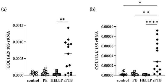
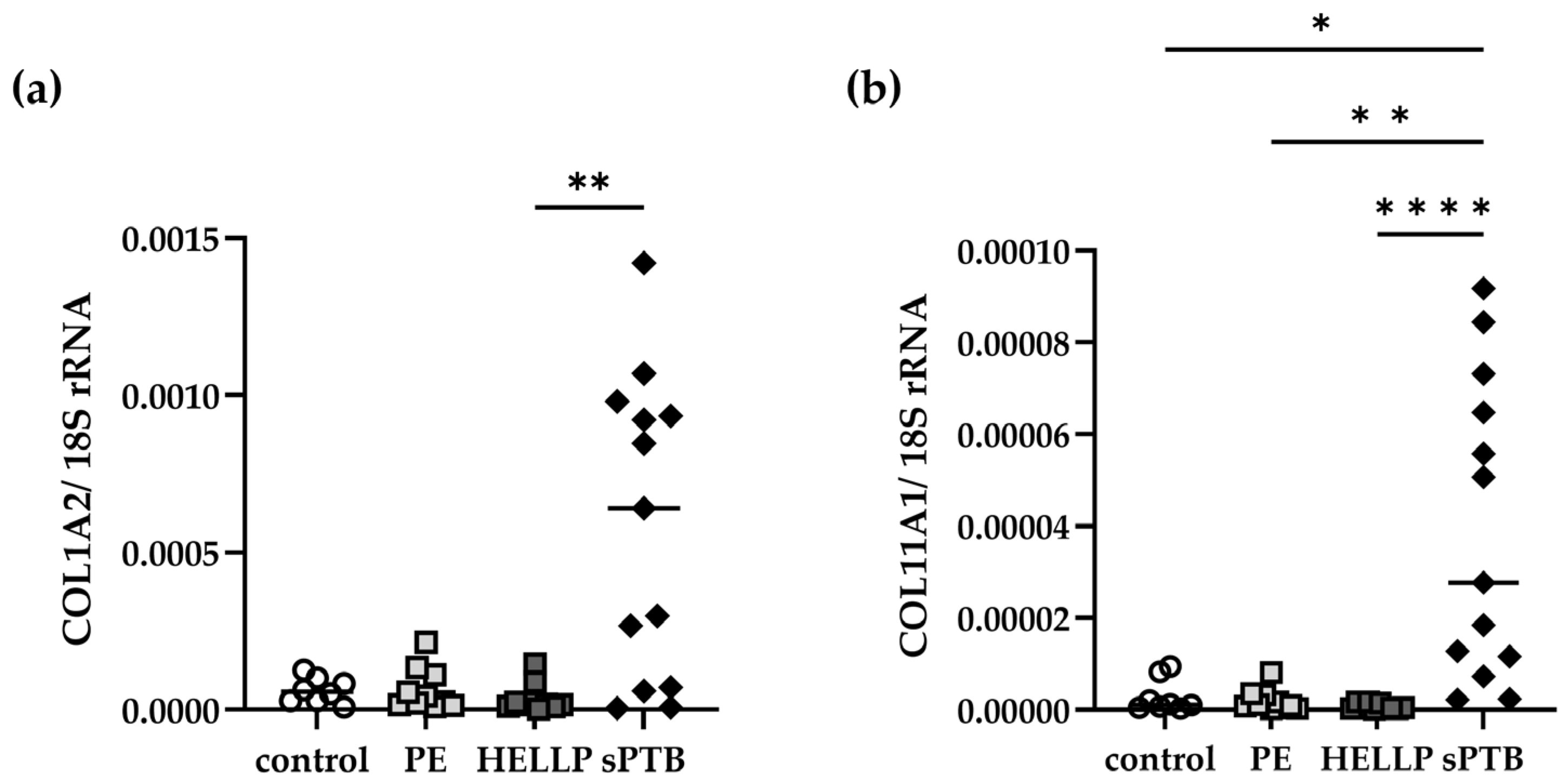
Placental COL1A2 expression differed significantly among the four study groups (Kruskal–Wallis H = 11.48, p = 0.0094; Figure 1a). COL1A2 expression was significantly higher in sPTB (mean: 5.79 × 10⁻⁴) than in HELLP placentae (mean: 3.48 × 10⁻⁵; Dunn-adjusted p = 0.0033), whereas no significant differences were observed between sPTB and either PE (mean: 6.41 × 10⁻⁵) or control placentae (mean: 3.25 × 10⁻⁵).
Figure 1.
Alterations in placental COL1A2 and COL11A1 expression in spontaneous preterm birth. The placental expression of COL1A2 and COL11A1 was examined in healthy pregnancies at term (control), medically indicated PTB (PE and HELLP), and spontaneous PTB (sPTB). (a) Expression of COL1A2 in the placentae of the control, PE, HELLP, and sPTB groups. (b) Expression of COL11A1 in the placentae of the control, PE, HELLP and sPTB groups. p < 0.05 (*), p < 0.005 (**), p < 0.0001 (****).
Placental COL11A1 expression also differed significantly among the groups (Kruskal–Wallis H = 24.78, p < 0.0001; Figure 1b). COL11A1 expression was significantly higher in sPTB (mean: 3.87 × 10⁻⁵) than in control (mean: 2.96 × 10⁻⁶; adjusted p = 0.0120), PE (mean: 2.03 × 10⁻⁶; adjusted p = 0.0018), and HELLP placentae (mean: 7.03 × 10⁻⁷; adjusted p < 0.0001).
In order to complement the hypothesis tests, effect sizes were calculated for placental COL1A2 and COL11A1 expression. The Kruskal–Wallis effect size, expressed as epsilon squared, was 0.229 for COL1A2 and 0.589 for COL11A1, indicating more pronounced between-group differences in COL11A1 expression. For pairwise comparisons, the non-parametric effect size, known as Cliff’s delta, was utilized, with 95% confidence intervals estimated using stratified bootstrap resampling. COL11A1 demonstrated substantial effects when comparing sPTB with term controls (δ = 0.885), PE (δ = 0.892), and HELLP (δ = 1.000). In contrast, COL1A2 demonstrated more modest effects, ranging from δ = 0.558 to δ = 0.662. In the comparison between sPTB and HELLP, COL11A1 expression showed complete separation of the observed values, resulting in a Cliff’s delta of 1.000. This finding indicates a pronounced difference in the observed samples, although the small sample sizes limit the precision and generalizability of the estimated effect.
2.3. Cytokine Release in Placenta Explant Supernatants Following Spontaneous or Medically Indicated PTB
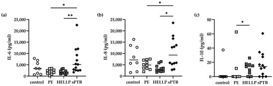
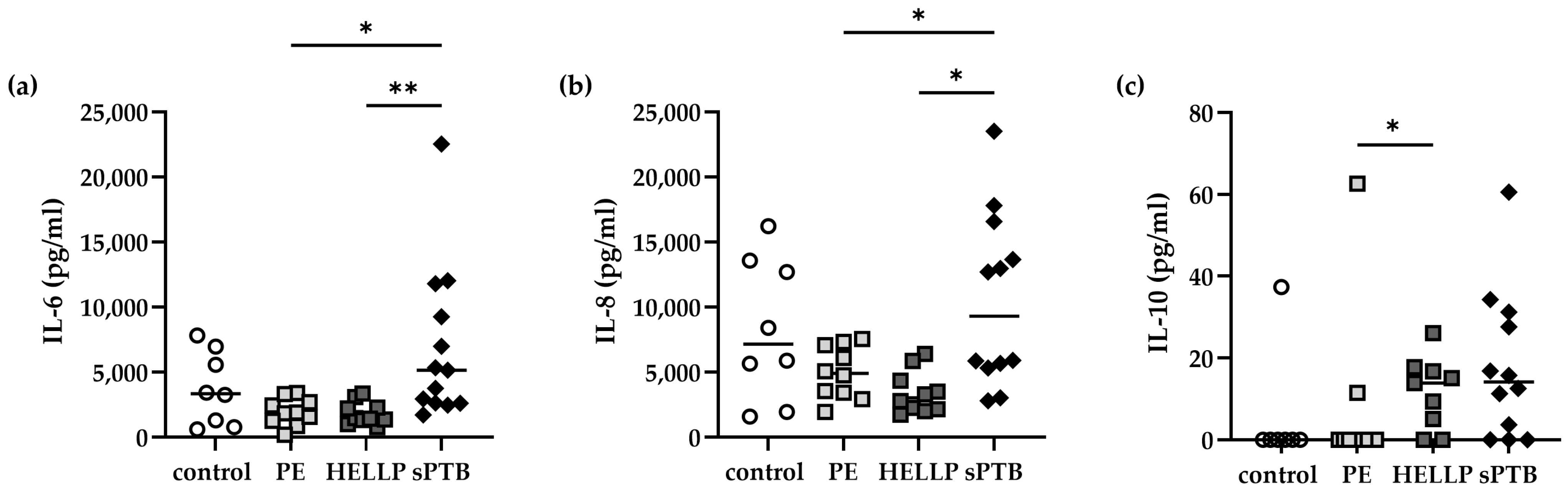
Since inflammation can result in collagen alterations, we measured cytokine release in placental explant supernatants. In sPTB, IL-6 secretion was higher than in HELLP (p = 0.0041; Figure 2a) and PE (p = 0.0114) but did not differ from that observed in healthy pregnancies at term. Additionally, the concentrations of IL-8 released by placentae from sPTB patients were higher than those released by HELLP (p = 0.0105; Figure 2b) or PE (p = 0.0492) patients, but not higher than those released by the control group. HELLP placentae released higher levels of IL-10 compared to those from PE patients (p = 0.0329), and there was a trend towards higher levels than in the control group (p = 0.0647). There was also a trend towards higher IL-12p70 and IL-17A secretion by HELLP placentae, though this was not statistically significant.
Figure 2.
Differential expression of pro- and anti-inflammatory cytokines in spontaneous and medically indicated preterm birth. Detection of IL-6, IL-8, and IL-10 in placenta explant supernatants in healthy pregnancies at term (control), medically indicated PTB (PE and HELLP) and spontaneous PTB (sPTB). (a) Concentration of IL-6, (b) IL-8, and (c) IL-10 in placental supernatant from healthy term controls and PE, HELLP and sPTB patients. p < 0.05 (*), p < 0.005 (**).
In order to investigate the relationship between the expression of the two collagen-related genes, Spearman correlation analyses were performed. Across the pooled cohort, COL1A2 and COL11A1 expression demonstrated a strong positive correlation (r = 0.825, p < 0.0001). A similarly robust positive correlation was observed within the sPTB subgroup (r = 0.797, p = 0.0018). However, in order to further explore the potential contribution of infection to altered placental collagen gene expression, a comparison was made between cases of sPTB of an infectious nature (n = 6, one case of chorioamnionitis and five cases of urinary tract infection; individual diagnoses are shown in Table S2) and cases of sPTB of a non-infectious nature (n = 7). No significant differences were observed in the expression levels of either COL1A2 (p = 0.5338) or COL11A1 (p = 1.000) between the two distinct subgroups. Moreover, within the overall sPTB cohort, COL1A2 and COL11A1 expression exhibited a strong correlation with each other and with maternal leukocyte counts (Table 2). Furthermore, COL1A2 expression demonstrated a positive correlation with placental IL-8 secretion, while COL11A1 expression exhibited no significant association with IL-8 or IL-6 (Table 2). The Benjamini–Hochberg procedure was used to adjust the p-values for the correlations examined within the sPTB cohort, controlling the false discovery rate at 5%. Four of the nine correlations remained significant after adjustment: COL1A2–COL11A1, COL1A2–maternal leukocyte count, COL11A1–maternal leukocyte count, and COL1A2–placental IL-8. The nominal association between COL11A1 and CRP did not remain significant after adjustment (Table 2).
Table 2.
Spearman correlation analysis and Benjamini–Hochberg (BH) correction (FDR 5%) of placental COL1A2 and COL11A1 expression, peripheral maternal leukocyte count, C-reactive protein (CRP) level, and interleukin (IL)-6 and IL-8 concentrations in placental supernatant from patients with spontaneous preterm birth (sPTB).
2.4. Differences in Collagen Type I Expression in Placentae
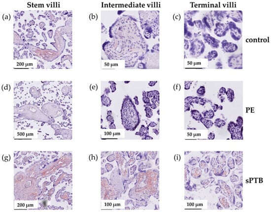
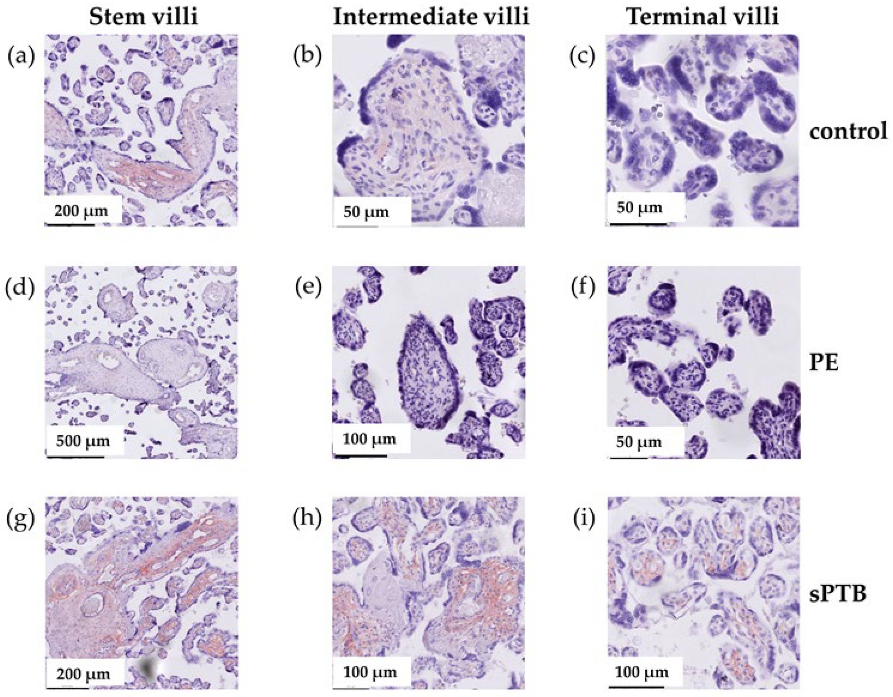
The staining of placental collagen type I was analyzed separately in stem villi, intermediate villi and terminal villi and was classified as negative (staining score = 0), weakly positive (score = 0.5) or positive (score = 1; Figure 3). In the control group and in patients diagnosed with HELLP or sPTB, stem villi tested positive for collagen type I, whereas in PE placentae, only one-third tested positive, one-third tested weakly positive, and one-third tested negative (see Table 3). This resulted in a significant difference compared to all other groups (Table S3). Similarly, intermediate villi were positive or weakly positive in healthy term, HELLP and sPTB placentae. However, none of the intermediate villi in the PE group were positive for collagen type I staining. Half of the PE placentae presented intermediate villi that were weakly positive for collagen type I; the other half were negative (Table 3). This led to a significant difference in PE compared to all other groups (Table S4). Conversely, terminal villi in term placentae and the majority of PE placentae expressed no collagen type I, whereas the majority of terminal villi in HELLP and sPTB placentae were positive for collagen type I staining (Table 3). Despite the significant differences observed between the control group and both the HELLP and sPTB groups, as well as between the PE and sPTB groups, no significant differences were identified between the PE and HELLP groups (Table S4). Further analysis did not reveal a correlation between the histopathological results and gestational age or birth weight (Table S3). These findings indicate a loss of proximal villous staining in PE and an extension of staining into terminal villi in HELLP and sPTB.
Figure 3.
Examples of type I collagen staining of stem villi, intermediate villi, and terminal villi. The examples illustrate the application of type I collagen staining to stem villi (a,d,g), intermediate villi (b,e,h), and terminal villi (c,f,i). Control term birth placentae stained positive in the stem and intermediate villi (a,b) and negative in the terminal villi (c). PE placentae stained negative (d–f), while sPTB placentae stained positive (g–i) in all investigated compartments of the placenta.
Table 3.
Collagen type I staining.
3. Discussion
Our findings suggest that the biological significance of COL11A1 and COL1A2 differs in placental pathology. Further, our findings indicate a close association between placental COL1A2 and COL11A1 mRNA expression.
Although it is best studied in fibrosis and cancer, COL11A1 is increasingly recognized as a marker of activated mesenchymal cells and pathological tissue remodeling. Placental COL11A1 mRNA expression levels were elevated in cases of sPTB compared with those observed in PE, HELLP syndrome, and uncomplicated term controls. A notable limitation of the study is the absence of gestational-age-matched uncomplicated preterm controls. This limitation is important because placental ECM composition and collagen gene expression may vary across gestation. Despite the absence of statistically significant differences in gestational age among the three clinical preterm groups, the possibility of confounding by developmental stage remains a concern. Consequently, the observed COL11A1 expression pattern should be interpreted as an association with the studied clinical groups rather than as a demonstrated sPTB-specific remodeling program. The current limitations of our bulk-tissue measurements preclude identifying the cellular source of the transcript or establishing its functional role.
Recent single-cell transcriptomic studies provide additional context for interpreting the observed alterations in placental collagen gene expression. Derisoud et al. [15] identified COL1A2 among genes associated with placental stromal cells while also demonstrating its expression in perivascular populations. Furthermore, Garcia-Flores et al. [17] demonstrated that parturition is accompanied by alterations in maternal–fetal cellular communication involving collagen-related, CXC, TNF, and IL-6 signaling pathways. These findings suggest that extracellular matrix-related processes are part of the complex cellular responses associated with labor. Our observation of increased COL11A1 expression in sPTB may therefore reflect alterations in placental extracellular matrix-associated processes. However, the present study does not establish whether this increase results from enhanced transcription within specific cell populations, changes in their relative abundance, or a combination of both. This distinction is of particular relevance in view of recent spatially resolved single-cell analyses, which have demonstrated pronounced cellular and molecular heterogeneity across gestational and anatomical compartments of the maternal–fetal interface [16]. Since our analyses were performed on bulk placental tissue, the cellular source of COL11A1 expression could not be determined. Future studies combining spatial transcriptomics with protein-level localization will be required to clarify the contribution of individual placental cell populations to the observed expression patterns.
Although we have demonstrated that sPTB, PE and HELLP are characterized by immune imbalance and increased release of inflammatory mediators in peripheral blood [18,19,20], we did not observe increased pro-inflammatory cytokine release in placental explants in PE and HELLP. Instead, we detected significantly higher levels of IL-6 and IL-8 in sPTB placental supernatants than in those from PE and HELLP. Thus, despite exhibiting placental dysfunction, PE and HELLP appear to lack this labor-associated cytokine profile, which is consistent with their distinct pathophysiological mechanisms. Furthermore, HELLP placentae released more of the anti-inflammatory cytokine IL-10 into the supernatant, distinguishing the two disorders from each other.
There was no difference in placental cytokine secretion between sPTB and healthy term deliveries. This finding supports the idea that sPTB and physiological term labor involve the activation of similar inflammatory pathways associated with parturition. However, only sPTB demonstrated increased placental COL11A1 expression. This suggests that extracellular matrix remodeling is an additional feature that distinguishes pathological premature labor from physiological term labor. As the sPTB group includes both infectious and non-infectious cases, COL11A1 may represent a shared downstream pathway activated regardless of the triggering event. Infection, sterile inflammation, oxidative stress or mechanical stretch may all converge on ECM remodeling, with COL11A1 induction reflecting this common response.
The COL1A2 gene encodes the α2 chain of type I collagen, which is the major fibrillar collagen responsible for tissue strength and fibrosis. The expression of the COL1A2 gene is antagonistically regulated by pro-inflammatory cytokines (such as TNF-α and IL-1β) and pro-fibrotic cytokines (TGF-β) [21,22]. There is evidence to suggest a link between collagen genes, such as COL1A2, and connective tissue integrity, as well as susceptibility to PTB and preterm premature rupture of membranes (PPROM) [13]. For fetal membranes, it has been shown that collagen type I mRNA levels are increased in PTB compared to term birth [23]. The increased expression that we detected in sPTB may indicate enhanced ECM deposition and repair responses, such as collagen synthesis, as well as collagen degradation via MMPs. This could be the result of inflammation, activation of fibroblast- or mesenchymal-like stromal cells, and aberrant placental remodeling. MMP-9 is a highly inducible enzyme that is heavily regulated by inflammatory cytokines, growth factors, and stress signals. These signals are often linked to intrauterine infection or sterile inflammation, and they drive the rapid degradation of the amnion and chorion. Levels of MMP-9 and proteolytic activity exhibit a marked increase in fetal membranes, amniotic fluid, and the placenta during sPTB and PPROM [24,25].
The analysis of correlations between cases of sPTB that are infectious and non-infectious indicates that the observations made are consistent with the hypothesis that placental collagen gene expression is associated with inflammatory processes. However, there may also be a difference in the relationships of COL1A2 and COL11A1 with individual inflammatory mediators. Nevertheless, it is important to note that these observed correlations do not necessarily imply causality. The limited sample size, the heterogeneity of the clinical presentations, and the potential presence of confounding factors all serve to preclude drawing definitive mechanistic conclusions. Furthermore, COL1A2 and COL11A1 mRNA expression exhibited a positive correlation in both the pooled cohort and sPTB. Pooled correlations may, in part, reflect between-group differences and should not be interpreted as evidence of common regulation without group-adjusted analysis. In the sPTB group, COL1A2 exhibited a correlation with placental IL-8 and maternal leukocytes, while COL11A1 demonstrated a correlation with leukocyte count, though not with IL-6 or IL-8. The results of the cytokine analysis demonstrate that COL11A1 expression is not simply reflected by IL-6/IL-8 release. This is evidenced by the fact that sPTB did not differ significantly from term controls for these cytokines.
Furthermore, COL1A2 is not increased in PTB overall; rather, there is a difference between sPTB and HELLP. In fact, HELLP placentae have the lowest expression of COL1A2, approximately half the level seen in controls and PE. Although PE and HELLP overlap clinically, HELLP has distinct pathological features. HELLP is associated with greater placental tissue injury, including extensive trophoblast injury, intervillous fibrin deposition, villous infarction and syncytiotrophoblast degeneration. If COL1A2 is produced primarily by placental stromal or mesenchymal cells, severe tissue damage could reduce its expression. COL1A2 mRNA expression may reflect transcriptional regulation of a collagen type I component; protein synthesis and turnover were not measured. Conversely, HELLP is characterized by endothelial injury, oxidative stress, apoptosis and ischemia. These processes may suppress ECM synthesis while favoring tissue destruction. Additionally, HELLP may exhibit relatively greater MMP activity, proteolysis and inflammatory injury than compensatory collagen synthesis [26]. Thus, HELLP may represent a state in which matrix degradation predominates over repair.
The processes leading to HELLP may suppress structural collagen synthesis and alter ECM homeostasis differently from the inflammatory remodeling observed in sPTB. Thus, COL1A2 may distinguish between different modes of placental remodeling, rather than serving as a marker of PTB itself. We hypothesize that infectious or sterile signals initiate inflammation, while cytokines (e.g., IL-1β, TNF-α, and IL-6) and pro-fibrotic cytokines (particularly TGF-β) activate placental stromal cells. These cells then increase the expression of ECM remodeling genes, including COL11A1. This remodeling represents an attempt to maintain tissue integrity while matrix degradation by MMPs is simultaneously increased. HELLP syndrome follows a different pathological pathway, with less activation of this reparative collagen remodeling program. Therefore, increased COL1A2 in sPTB could reflect an ongoing attempt to repair ECM, whereas HELLP placentae may have lost this reparative response.
It is noteworthy that the expression pattern of COL11A1 did not fully correspond to the secretion of inflammatory cytokines. Despite the elevated concentrations of IL-6 and IL-8 observed in placental explant extracts from sPTB cases compared to those from PE and HELLP pregnancies, no statistically significant disparities were identified between sPTB and uncomplicated term deliveries. In contrast, COL11A1 expression was significantly increased in sPTB compared with that in all three groups. These findings imply that elevated COL11A1 expression cannot be solely explained by differences in IL-6 and IL-8 secretion. It is important to note that additional factors, which may include other inflammatory mediators, mechanical signals or differences in placental cellular composition, may also contribute to its regulation. However, the present study did not directly investigate these mechanisms, and further studies are required to determine their relative contributions.
While COL6A1 and COL1A1 have been identified as potential targets for PE diagnosis and therapy [27], our findings revealed no alterations in COL1A2 and COL11A1 expression in PE compared to healthy term placentae.
We analyzed collagen type I expression in stem villi, intermediate villi and terminal villi. Stem villi are characterized by a condensed fibrous stroma that consists of large vessels and microvessels, which support the villous tree structures. Intermediate villi are peripheral ramifications that lack fetal vessels in the stroma and produce terminal villi. Terminal villi are the functional units of the placenta in a normal, mature placenta. They comprise nearly 40% of the total volume of the placenta [28]. Our findings demonstrate distinct patterns of placental extracellular matrix remodeling. PE was associated with reduced collagen staining in stem and intermediate villi, suggesting impaired matrix organization in the structural villous compartments. Previous research showed that placentae from PE patients exhibited a significantly higher degree of fibrosis in stem villi and terminal villi than healthy term placentae and were characterized by collagen type I deposition [29]. Furthermore, placental ischemia in PE may lead to abnormal vascular remodeling via TNF-α secretion, which alters collagen deposition, causing inadequate tissue remodeling [30]. In contrast, HELLP and sPTB showed an abnormal extension of collagen staining into terminal villi, potentially indicating distal stromal remodeling that may compromise placental exchange function. A decrease in the expression of the propeptide of type I procollagen has also been reported in fetal membranes of patients with PPROM, which is often associated with infection [31].
Alterations in placental collagen deposition have previously been described in several pregnancy complications. According to Shi et al. [30], there is a growing body of evidence indicating increased collagen accumulation and stromal fibrosis in preeclamptic placentae. These changes are potentially associated with hypoxia, altered fibroblast activity, and dysregulated extracellular matrix turnover. Conversely, our immunohistochemical analysis revealed a decrease in collagen type I staining in stem and intermediate villi from PE placentae. This observed discrepancy may be indicative of variations in the investigated placental compartments, the methodologies employed to assess collagen, or the clinical characteristics of the study populations. Additionally, the deposition of collagen is not solely governed by gene transcription; it is also subject to regulation by post-transcriptional processes, extracellular processing, and enzymatic degradation. Consequently, COL1A2 mRNA expression and collagen type I immunostaining do not necessarily exhibit parallel changes. It has been previously established through a range of studies that collagen remodeling plays a pivotal role in the maintenance of fetal membrane integrity and the prevention of rupture. Specifically, altered expression of COL1A1 and COL1A2, along with changes in matrix metalloproteinase activity, has been reported in association with membrane rupture and parturition [30]. However, these findings predominantly concern fetal membranes rather than placental villous tissue, and their relevance to the altered placental COL11A1 expression observed in our sPTB cohort remains to be established.
Our study is subject to several limitations that must be taken into consideration when interpreting the results. First, the study included a relatively small number of participants, and no uncomplicated gestational-age-matched preterm control group was available. The sPTB infection-status subgroups were defined using available clinical and microbiological documentation. The absence of documented infection does not establish sterile inflammation. The present study is exploratory in nature and highlights the need for validation in larger independent cohorts. Second, bulk-tissue RT-qPCR does not identify the cell types expressing COL1A2 or COL11A1, and collagen type I immunohistochemistry is not a direct validation of either target at the protein level. Although RNA purity was assessed using absorbance ratios, RNA integrity was not quantified using RNA integrity numbers. In addition, dedicated positive and negative technical immunohistochemical controls were not included, and interobserver agreement was not quantified statistically; instead, the pathologists used independent scoring followed by consensus. Next, the study did not measure MMP activity, collagen turnover or cellular function, so the proposed remodeling pathways remain hypotheses. Finally, the findings from the effect size analysis support the observation that COL11A1 expression differs more consistently between sPTB and the other investigated pregnancy groups than COL1A2 expression. However, as the number of secondary analyses increases, the risk of false-positive results increases; these findings require confirmation in independent, larger cohorts with standardized sampling and cell-resolved protein measurements.
In summary, we observed differences in the mRNA expression of COL1A2 and COL11A1, as well as in the protein expression of type I collagen, depending on the type of pregnancy-related condition. Spontaneous preterm births, marked by heightened placental inflammation, exhibited augmented COL1A2 and COL11A1 expression. However, the type I collagen staining profile in these cases was comparable to that observed in HELLP syndrome. Although medically indicated preterm births due to PE and HELLP share a similar pathophysiology, significant differences in type I collagen expression were observed in these cases. A more thorough examination of the underlying causes is necessary to gain a comprehensive understanding of this phenomenon.
4. Materials and Methods
4.1. Human Subjects
The Ethics Committee of the Otto-von-Guericke University Medical Faculty approved the study (EK28/08, EK175/21, EK19/22). All patients were properly informed about the purpose of the study and gave written consent before participating. Samples of tissue were obtained from the central part of the placenta by biopsy. Patients included in this study were recruited between April 2016 and April 2024. The exclusion criteria encompassed maternal immunological or coagulation disorders, other pregnancy-related disorders (e.g., gestational diabetes), fetal congenital/chromosomal anomalies, and maternal connective tissue disorders. The placentae were processed in accordance with the methodology outlined in [32]. Briefly, placentae were snap-frozen for RNA isolation or cultured in RPMI 1640 supplemented with 3% charcoal-treated fetal bovine serum and 1% penicillin/streptomycin for 24 h. Afterwards, the supernatant was harvested, centrifuged (3000 rpm, 10 min, RT), snap-frozen and stored at −80 °C.
4.2. RNA Isolation and RT-qPCR
Total RNA from frozen tissues was isolated using TRIzol (Ambion, Thermo Fisher Scientific, Dreieich, Germany) as previously described [4]. In summary, 1 g of placental tissue was homogenized in 1 mL of TRIzol, after which chloroform and isopropanol were added. The tissue was then washed with ethanol, and the RNA pellet was resuspended in RNase-free dH2O at a concentration of 1 µg/µL. The A260/280 ratios for all samples were measured and found to be consistently within the optimal range of 1.8 to 2.1.
cDNA was synthesized using the RevertAid First Strand cDNA Synthesis Kit (Thermo Fisher Scientific, San Diego, CA, USA) with 1 µg of RNA per sample. For qPCR, SYBR® Select Master Mix (Thermo Fisher Scientific, San Diego, CA, USA) was used according to the manufacturer’s instructions with gene-specific primers (see Table 4, Merck, Darmstadt, Germany).
Table 4.
Primer sequences.
qPCR was performed on the StepOnePlus Real-time PCR System (Applied Biosystems, Waltham, MA, USA) as follows: holding stage (2 min at 50 °C, then 2 min at 95 °C), 40 cycles: cycling stage (15 s at 95 °C, 15 s at 60 °C, 1 min at 72 °C), and melt curve stage (15 s at 95 °C, 1 min at 60 °C, 15 s at 95 °C). PCRs were carried out in duplicate. The data were analyzed using the LinRegPCR program (version 2021.2, [33]).
4.3. Measurement of Cytokines in Placenta Supernatants
The measurement of cytokines was conducted using the LEGENDplex Human Inflammation Panel 1 from BioLegend (San Diego, CA, USA) in accordance with the supplier’s recommendations. The measurements were conducted using an Attune NxT flow cytometer (Thermo Fisher Scientific, Waltham, MA, USA) and subsequently analyzed with FlowJo software 10.10.1 (Ashland, OR, USA).
4.4. Histology
Subsequent to delivery, the placentae were meticulously divided into approximately 1cm3 fragments and subjected to repeated washes with PBS. Thereafter, the placentae were fixed in 4% (w/v) paraformaldehyde (PFA) with 0.1 M sucrose (pH 7.4) for 6 h, after which a dehydration treatment was conducted using 100% ethanol and xylene. The fragments were embedded in paraffin and cut into slides measuring 4 µm. The staining procedure was initiated with a deparaffinization step, which was then followed by a hydration process.
The samples were boiled in a citrate buffer solution for 10 min. All further steps were performed at room temperature (RT). The slides were washed once with PBS for 5 min and treated with 3% H2O2 for 30 min, followed by washing with PBS for 5 min and blocking with 10% FBS/PBS for 40 min. After rinsing with PBS, the slides were incubated with mouse anti-human collagen type I monoclonal IgG1 antibody (Proteintech, Planegg-Martinsried, Germany; #67288-1-Ig; 1:5000) for 1 h. Following washing with PBS, slides were incubated with the secondary antibody, goat anti-mouse HRP (Invitrogen, Darmstadt, Germany; #31430; 1:500), for 1 h. After washing, an AEC substrate was added for 5 min, and the slides were rinsed with distilled water and then incubated with hematoxylin for 5 min. After washing, the slides were mounted with Aquatex. The staining was performed at the Department of Experimental Obstetrics and Gynecology, while the scoring was carried out at the Institute of Pathology. Two experienced pathologists (A.H. and E.T.T.) evaluated collagen type I immunostaining independently and in a blinded manner. Beforehand, they agreed upon a standardized semi-quantitative scoring system, categorizing staining intensity as negative (score 0), weakly positive (score 0.5) or positive (score 1). Stem villi, intermediate villi and terminal villi were assessed separately. Following the independent evaluations, the two observers reviewed their assessments together and resolved any discrepancies by discussion. A final consensus score was established for each sample and villous compartment, which was then used for statistical analysis. Interobserver agreement was not quantified statistically. The results were matched to the patients at the Department of Experimental Obstetrics and Gynecology.
4.5. Data Analysis and Statistics
Statistical analysis was performed using GraphPad Prism 10. The normality of distribution was determined by means of the Shapiro–Wilk test. For non-normally distributed variables, differences among the four study groups were assessed using the Kruskal–Wallis test, followed by Dunn’s multiple-comparisons test. The Dunn-adjusted p-values refer to pairwise comparisons within each analyzed variable. For the two collagen-related genes, the overall effect sizes were expressed as epsilon squared (ε2 = (H − k + 1)/(N − k)), where H is the Kruskal–Wallis test statistic, k is the number of groups, and N is the total sample size. Cliff’s delta (δ) was calculated for pairwise gene-expression comparisons as the proportion of intergroup pairs in which the value in the first group exceeded that in the second group, minus the proportion showing the reverse order; ties contributed zero. Exploratory 95% confidence intervals were obtained from 30,000 non-parametric bootstrap resamples stratified by group. The bootstrap intervals were not recalibrated in accordance with the multiple testing procedure. For COL11A1 in sPTB versus HELLP, complete separation in the observed sample yielded δ = 1 and a degenerate percentile-bootstrap interval; this interval does not imply an absence of population uncertainty. Immunohistochemical scores were analyzed as categorical data using Fisher’s exact test for the overall comparison (see Table 3). Separate exploratory rank-based comparisons are reported in Table S3, which is located in the Supplementary Materials. Spearman rank correlation coefficients (r) and unadjusted p-values were calculated for the purpose of establishing associations between collagen gene expression and inflammatory parameters. For the nine exploratory correlations in the sPTB cohort, p-values were additionally adjusted using the Benjamini–Hochberg procedure to control the false discovery rate at 5%. The exploratory analyses conducted across various outcome domains were not subject to a single global multiplicity adjustment. Consequently, nominally significant findings were interpreted with a degree of caution. The significance of the results was determined by p-values, which were categorized as follows: p < 0.05 (*), p < 0.005 (**), p < 0.001 (***) or p < 0.0001 (****).
Supplementary Materials
The following supporting information can be downloaded at: https://www.mdpi.com/article/10.3390/ijms272210064/s1.
Author Contributions
Conceptualization, M.B.; methodology, M.B., P.G., C.W.-R., G.M.U., L.T., A.H. and E.T.T.; validation, M.B., V.G., J.M., C.W.-R., G.M.U., A.H., E.T.T. and A.I.; formal analysis, M.B., L.T., C.W.-R., G.M.U., A.H. and E.T.T.; investigation, C.W.-R., G.M.U., A.H., E.T.T. and M.B.; resources, P.G., J.M. and A.I.; data curation, M.B., L.T., C.W.-R., G.M.U., A.H. and E.T.T.; writing—original draft preparation, M.B.; writing—review and editing, M.B., P.G., J.M., L.T., C.W.-R., G.M.U., A.H., E.T.T. and A.I.; visualization, M.B.; supervision, M.B. All authors have read and agreed to the published version of the manuscript.
Funding
This research received no external funding.
Institutional Review Board Statement
The study was conducted in accordance with the Declaration of Helsinki and approved by the Ethics Committee of the Otto-von-Guericke University medical faculty (EK28/08: 16.05.2008; EK175/21: 19.11.2021; EK19/22: 04.02.2022).
Informed Consent Statement
Informed consent was obtained from all subjects involved in the study.
Data Availability Statement
The raw data supporting the conclusions of this article will be made available by the authors, without undue reservation.
Acknowledgments
We thank Ruby Siebach and Markus Scharm for technical assistance.
Conflicts of Interest
The authors declare no conflicts of interest.
Abbreviations
The following abbreviations are used in this manuscript:
| COL | collagen |
| ECM | extracellular matrix |
| HELLP | Hemolysis, Elevated Liver enzymes, Low platelets |
| IL | interleukin |
| PE | preeclampsia |
| PlGF | placental growth factor |
| PPROM | preterm premature rupture of membranes |
| sFlt-1 | soluble FMS-like tyrosine kinase-1 |
| sPTB | spontaneous preterm birth |
References
- Goldenberg, R.L.; Culhane, J.F.; Iams, J.D.; Romero, R. Epidemiology and causes of preterm birth. Lancet 2008, 371, 75–84. [Google Scholar] [CrossRef] [Scilit] [PubMed]
- Wang, Y.; Leung, H.H.Y.; Hui, A.S.Y.; Wong, L.; Leung, T.Y. Molecular Mechanism and Pathways of Spontaneous Preterm Birth in Different Gestational Tissues: A Systematic Review of Transcriptome Studies. Int. J. Mol. Sci. 2026, 27, 6006. [Google Scholar] [CrossRef] [Scilit] [PubMed]
- Busse, M.; Plenagl, S.; Campe, N.K.J.; Muller, A.J.; Tedford, K.; Schumacher, A.; Zenclussen, A.C. Maternal B Cell-Intrinsic MyD88 Signaling Mediates LPS-Driven Intrauterine Fetal Death. Cells 2021, 10, 2693. [Google Scholar] [CrossRef] [Scilit] [PubMed]
- Uehre, G.M.; Tchaikovski, S.; Ignatov, A.; Zenclussen, A.C.; Busse, M. B Cells Induce Early-Onset Maternal Inflammation to Protect against LPS-Induced Fetal Rejection. Int. J. Mol. Sci. 2023, 24, 16091. [Google Scholar] [CrossRef] [Scilit] [PubMed]
- Gardikioti, A.; Venou, T.M.; Gavriilaki, E.; Vetsiou, E.; Mavrikou, I.; Dinas, K.; Daniilidis, A.; Vlachaki, E. Molecular Advances in Preeclampsia and HELLP Syndrome. Int. J. Mol. Sci. 2022, 23, 3851. [Google Scholar] [CrossRef] [Scilit] [PubMed]
- Busse, M.; Langwisch, S.; Tedford, K.; Fischer, K.D.; Zenclussen, A.C. Maternal B cell signaling orchestrates fetal development in mice. Development 2022, 149, dev199783. [Google Scholar] [CrossRef] [Scilit] [PubMed]
- Ma, Y.; Fang, X.; Wang, S.; Yu, X.; Wang, Y.L.; Shao, X. Immunoregulation and cellular crosstalk in uterine spiral artery remodeling during human pregnancy. J. Reprod. Immunol. 2026, 175, 104907. [Google Scholar] [CrossRef] [Scilit] [PubMed]
- Strauss, J.F., 3rd. Extracellular matrix dynamics and fetal membrane rupture. Reprod. Sci. 2013, 20, 140–153. [Google Scholar] [CrossRef] [Scilit] [PubMed]
- Vink, J.; Mourad, M. The pathophysiology of human premature cervical remodeling resulting in spontaneous preterm birth: Where are we now? Semin. Perinatol. 2017, 41, 427–437. [Google Scholar] [CrossRef] [Scilit] [PubMed]
- Maymon, E.; Romero, R.; Pacora, P.; Gervasi, M.T.; Gomez, R.; Edwin, S.S.; Yoon, B.H. Evidence of in vivo differential bioavailability of the active forms of matrix metalloproteinases 9 and 2 in parturition, spontaneous rupture of membranes, and intra-amniotic infection. Am. J. Obstet. Gynecol. 2000, 183, 887–894. [Google Scholar] [CrossRef] [Scilit] [PubMed]
- Vidal, M.S., Jr.; Lintao, R.C.V.; Severino, M.E.L.; Tantengco, O.A.G.; Menon, R. Spontaneous preterm birth: Involvement of multiple feto-maternal tissues and organ systems, differing mechanisms, and pathways. Front. Endocrinol. 2022, 13, 1015622. [Google Scholar] [CrossRef] [Scilit] [PubMed]
- Menon, R.; Behnia, F.; Polettini, J.; Richardson, L.S. Novel pathways of inflammation in human fetal membranes associated with preterm birth and preterm pre-labor rupture of the membranes. Semin. Immunopathol. 2020, 42, 431–450. [Google Scholar] [CrossRef] [Scilit] [PubMed]
- Anum, E.A.; Hill, L.D.; Pandya, A.; Strauss, J.F., 3rd. Connective tissue and related disorders and preterm birth: Clues to genes contributing to prematurity. Placenta 2009, 30, 207–215. [Google Scholar] [CrossRef] [Scilit] [PubMed]
- Li, H.; Huang, Q.; Liu, Y.; Garmire, L.X. Single cell transcriptome research in human placenta. Reproduction 2020, 160, R155–R167. [Google Scholar] [CrossRef] [Scilit] [PubMed]
- Derisoud, E.; Jiang, H.; Zhao, A.; Chavatte-Palmer, P.; Deng, Q. Revealing the molecular landscape of human placenta: A systematic review and meta-analysis of single-cell RNA sequencing studies. Hum. Reprod. Update 2024, 30, 410–441. [Google Scholar] [CrossRef] [Scilit] [PubMed]
- Wang, C.; Zhou, Y.; Wang, Y.; Guha, T.K.; Luo, Z.; Mustafaraj, A.; McIntyre, T.I.; Schwab, M.E.; Davidson, B.R.; Reeder, G.C.; et al. Single-cell spatiotemporal dissection of the human maternal-fetal interface. Nature 2026, 653, 167–179. [Google Scholar] [CrossRef] [Scilit] [PubMed]
- Garcia-Flores, V.; Romero, R.; Tarca, A.L.; Peyvandipour, A.; Xu, Y.; Galaz, J.; Miller, D.; Chaiworapongsa, T.; Chaemsaithong, P.; Berry, S.M.; et al. Deciphering maternal-fetal cross-talk in the human placenta during parturition using single-cell RNA sequencing. Sci. Transl. Med. 2024, 16, eadh8335. [Google Scholar] [CrossRef] [Scilit] [PubMed]
- Busse, M.; Scharm, M.; Oettel, A.; Redlich, A.; Costa, S.D.; Zenclussen, A.C. Enhanced S100B expression in T and B lymphocytes in spontaneous preterm birth and preeclampsia. J. Perinat. Med. 2022, 50, 157–166. [Google Scholar] [CrossRef] [Scilit] [PubMed]
- Campe, K.J.; Redlich, A.; Zenclussen, A.C.; Busse, M. An increased proportion of progesterone receptor A in peripheral B cells from women who ultimately underwent spontaneous preterm birth. J. Reprod. Immunol. 2022, 154, 103756. [Google Scholar] [CrossRef] [Scilit] [PubMed]
- Busse, M.; Campe, K.J.; Redlich, A.; Oettel, A.; Hartig, R.; Costa, S.D.; Zenclussen, A.C. Regulatory B Cells Are Decreased and Impaired in Their Function in Peripheral Maternal Blood in Pre-term Birth. Front. Immunol. 2020, 11, 386. [Google Scholar] [CrossRef] [Scilit] [PubMed]
- Greenwel, P.; Tanaka, S.; Penkov, D.; Zhang, W.; Olive, M.; Moll, J.; Vinson, C.; Di Liberto, M.; Ramirez, F. Tumor necrosis factor alpha inhibits type I collagen synthesis through repressive CCAAT/enhancer-binding proteins. Mol. Cell Biol. 2000, 20, 912–918. [Google Scholar] [CrossRef] [Scilit] [PubMed]
- Sawamura, S.; Makino, K.; Ide, M.; Shimada, S.; Kajihara, I.; Makino, T.; Jinnin, M.; Fukushima, S. Elevated Alpha 1(I) to Alpha 2(I) Collagen Ratio in Dermal Fibroblasts Possibly Contributes to Fibrosis in Systemic Sclerosis. Int. J. Mol. Sci. 2022, 23, 6811. [Google Scholar] [CrossRef] [Scilit] [PubMed]
- Meirowitz, N.B.; Smulian, J.C.; Hahn, R.A.; Zhou, P.; Shen-Schwarz, S.; Lambert, G.H.; Gerecke, D.R.; Gordon, M.K. Collagen messenger RNA expression in the human amniochorion in premature rupture of membranes. Am. J. Obstet. Gynecol. 2002, 187, 1679–1685. [Google Scholar] [CrossRef] [Scilit] [PubMed]
- Ulrich, C.C.; Arinze, V.; Wandscheer, C.B.; Copley Salem, C.; Nabati, C.; Etezadi-Amoli, N.; Burkin, H.R. Matrix metalloproteinases 2 and 9 are elevated in human preterm laboring uterine myometrium and exacerbate uterine contractilitydagger. Biol. Reprod. 2019, 100, 1597–1604. [Google Scholar] [CrossRef] [Scilit] [PubMed]
- Duran-Chavez, J.; Grandi, C.; Dos, S.R.L.; de Freitas, S.F.; Cardoso, V.C.; Carvalho Cavalli, R. Relationship between metalloproteinase-2 and -9 levels in plasma and vaginal secretion with preterm birth. Eur. J. Obstet. Gynecol. Reprod. Biol. 2021, 261, 217–221. [Google Scholar] [CrossRef] [Scilit] [PubMed]
- Petca, A.; Miron, B.C.; Pacu, I.; Dumitrascu, M.C.; Mehedintu, C.; Sandru, F.; Petca, R.C.; Rotar, I.C. HELLP Syndrome-Holistic Insight into Pathophysiology. Medicina 2022, 58, 326. [Google Scholar] [CrossRef] [Scilit] [PubMed]
- He, P.; Shao, D.; Ye, M.; Zhang, G. Analysis of gene expression identifies candidate markers and pathways in pre-eclampsia. J. Obstet. Gynaecol. 2015, 35, 578–584. [Google Scholar] [CrossRef] [Scilit] [PubMed]
- Wang, Y.; Zhao, S. Vascular Biology of the Placenta. In Vascular Biology of the Placenta; Integrated Systems Physiology: From Molecules to Function to Disease; Morgan & Claypool Life Sciences: San Rafael, CA, USA, 2010. [Google Scholar]
- Feng, Y.; Chen, X.; Wang, H.; Chen, X.; Lan, Z.; Li, P.; Cao, Y.; Liu, M.; Lv, J.; Chen, Y.; et al. Collagen I Induces Preeclampsia-Like Symptoms by Suppressing Proliferation and Invasion of Trophoblasts. Front. Endocrinol. 2021, 12, 664766. [Google Scholar] [CrossRef] [Scilit] [PubMed]
- Shi, J.W.; Lai, Z.Z.; Yang, H.L.; Yang, S.L.; Wang, C.J.; Ao, D.; Ruan, L.Y.; Shen, H.H.; Zhou, W.J.; Mei, J.; et al. Collagen at the maternal-fetal interface in human pregnancy. Int. J. Biol. Sci. 2020, 16, 2220–2234. [Google Scholar] [CrossRef] [Scilit] [PubMed]
- Remneva, O.B.; Kolyado, K.O.; Pesotskaya, A.V.; Starodubtsev, E.G.; Gumenyuk, I.S. Pathomorphological features of placenta in patients with different clinical phenotypes of spontaneous preterm births. Akusherstvo Ginekol./Obstet. Gynecol. 2021, 8, 111–118. [Google Scholar] [CrossRef] [Scilit]
- Hoymann, N.; Scholz, L.; Alboradi, S.; Grabar, V.; Uehre, G.M.; Khuankhunsathid, J.T.; Taube, E.T.; Toth, G.; Meszaros, J.; Gennari, P.; et al. Alterations in the Placenta Following Vaccination and Infection with SARS-CoV-2 During Pregnancy. Int. J. Mol. Sci. 2026, 27, 5473. [Google Scholar] [CrossRef] [Scilit] [PubMed]
- Ruijter, J.M.; Ramakers, C.; Hoogaars, W.M.; Karlen, Y.; Bakker, O.; van den Hoff, M.J.; Moorman, A.F. Amplification efficiency: Linking baseline and bias in the analysis of quantitative PCR data. Nucleic Acids Res. 2009, 37, e45. [Google Scholar] [CrossRef] [Scilit] [PubMed]

Disclaimer/Publisher’s Note: The statements, opinions and data contained in all publications are solely those of the individual author(s) and contributor(s) and not of MDPI and/or the editor(s). MDPI and/or the editor(s) disclaim responsibility for any injury to people or property resulting from any ideas, methods, instructions or products referred to in the content. |
© 2026 by the authors. Licensee MDPI, Basel, Switzerland. This article is an open access article distributed under the terms and conditions of the Creative Commons Attribution (CC BY) license.