


Abstract
Hyaluronan (HA) is increasingly implicated as a contributor to metabolic disease; however, the strongest causal evidence still derives from preclinical models, and no HA-directed treatment has reached the metabolic clinic. That gap points to a weakness in the underlying therapeutic logic. Clinical results for systemic removal of HA have been mixed rather than uniformly negative. In the phase II HALO-202 trial, PEGPH20 improved progression-free survival in patients with hyaluronan-high pancreatic tumors, whereas the phase III HALO-301 trial, which enrolled only hyaluronan-high patients, showed no overall survival benefit; depletion alone was therefore insufficient, and this outcome cannot be attributed to unselected treatment or to molecular size. Its biological activity is not determined by molecular size alone. Concentration, preparation purity, receptor context, tissue location, and the balance between high-molecular-weight HA (HMW-HA, generally above 1000 kDa) and smaller fragments, which range from oligosaccharides of about ten saccharides or fewer to low-molecular-weight HA in between, jointly influence whether the matrix mainly protects or injures metabolic tissues. The argument here is that the field should move away from bulk depletion and toward precision delivery and biomarker-guided patient selection. The therapeutic landscape is organized around four strategies: pharmacological inhibition of HA synthesis, enzymatic degradation, HA-based biomaterials, and defined-molecular-weight HA fragments. Throughout, we distinguish interventions that modify endogenous HA biology from platforms that use HA as a carrier or scaffold, because improved drug delivery or wound healing does not by itself demonstrate correction of pathological HA signaling. Their translational obstacles are then assessed, including patient stratification by HA size and CD44 signature, standardization of size-resolved HA quantification, thromboembolic risk, manufacturing and regulatory hurdles for drug–device combinations, and the design of trials with composite metabolic endpoints. HA is best viewed as an amplifier of metabolic stress rather than a bystander, and the route to clinical translation lies in restoring the equilibrium between HA synthesis and degradation rather than eliminating the molecule, a goal that remains a translational hypothesis rather than a demonstrated clinical outcome.
1. Introduction
Two of the most prevalent chronic metabolic diseases, obesity and type 2 diabetes (T2D), carry much of their burden through low-grade inflammation that remodels the extracellular matrix (ECM) in metabolically active tissues [1,2]. The ECM is not an inert scaffold but a dynamic structure whose composition and mechanical properties govern nutrient sensing, immune-cell recruitment, and the transduction of insulin signals [1,3]. Hyaluronan (HA) stands out among matrix macromolecules because its synthesis responds to nutrient supply, its fragmentation tracks tissue stress, and its receptors are present on nearly every metabolic and immune cell [4,5]. These observations fit a single framework in which HA amplifies metabolic stress without initiating it. Nutrient excess and tissue injury raise HA levels, and the resulting matrix remodeling spreads inflammation and insulin resistance, positioning the HA axis as both a biomarker and a node for intervention [6,7]. The World Health Organization estimates that more than 650 million adults are living with obesity and over half a billion have diabetes, which points to an urgent need for therapies directed at pathological matrix remodeling. The review examines HA metabolism and receptor signaling, organ-specific pathophysiology, biomarker value, and treatment options, and it closes with a forward-looking view of the main obstacles to clinical implementation.
Among the glycosaminoglycans, HA is an unsulfated linear polymer assembled from repeating D-glucuronic acid and N-acetyl-D-glucosamine units and produced at the cell surface through the action of three hyaluronan synthases (HAS1-3) [8,9,10,11,12]. In healthy tissue, HA exists largely as high-molecular-weight HA (HMW-HA), which sustains hydration, keeps the tissue compliant, and holds the microenvironment in an immunosuppressive state [4,8]. Under metabolic stress, HA synthesis rises while hyaluronidases and reactive oxygen species cleave the polymer into low-molecular-weight HA (LMW-HA) and oligosaccharides. In specific experimental and pathological contexts, depending on fragment size, local concentration, and preparation purity, these fragments can behave as damage-associated molecular patterns that activate CD44 and Toll-like receptors (TLRs) to promote inflammatory signaling cascades [8,13,14,15,16,17].
HA has served clinical medicine for decades as a viscosupplement, a dermal filler, and a biomaterial [18,19]. Its contribution to metabolic disease, however, has become clear only recently, through genetic and pharmacological approaches [20,21,22]. The strength of that evidence differs by context. Causality is best supported in preclinical models, in which genetic deletion or pharmacological inhibition of HA synthesis or of CD44 changes insulin sensitivity, β-cell survival, and tissue inflammation [20,21,22,23,24,25]. In humans the evidence remains largely associative, because circulating HA and endothelial glycocalyx markers track obesity, insulin resistance, and renal or vascular complications without establishing the direction of the effect or its mechanism [26,27,28]. We therefore use causal language only where genetic or pharmacological manipulation supports it and describe the remaining findings as associations. In the review we bring together what is now known about HA metabolism and receptor signaling, and the HA-directed therapeutic strategies now emerging, from inhibiting HA synthesis to building HA-based delivery systems. We end by setting out the uncertainties that must be resolved for HA-based interventions to reach metabolic medicine. A comprehensive literature search was conducted in PubMed (search strategy detailed in Supplementary Table S1), from which records were screened and selected for inclusion (Supplementary Table S2). The sharp increase in retrieved publications over recent decades underscores the accelerating interest in this field (Supplementary Figure S1).
2. Hyaluronan Biology: Synthesis, Degradation, and Receptor Signaling
2.1. Biosynthesis and Catabolism
Three plasma-membrane enzymes, HAS1, HAS2, and HAS3, polymerize HA from the nucleotide-sugar donors UDP-glucuronic acid and UDP-N-acetylglucosamine. Because UDP-glucose dehydrogenase and glutamine:fructose-6-phosphate aminotransferase lie upstream, HA output is coupled to glucose and glutamine metabolism [8,12]. HAS2 is the most dynamically regulated isoform and is controlled after translation by O-GlcNAcylation, which links hexosamine flux directly to HA synthesis [12]. The three isoforms are not interchangeable. They differ in enzymatic properties and in the polymer sizes they generate, and their expression is governed at the transcriptional, post-transcriptional, and post-translational levels, with a natural antisense transcript regulating HAS2 [29,30]. Inflammatory cytokines and growth factors induce HAS activity, PPAR-γ ligands repress it in certain cell types, and oxidized lipoproteins modulate it [31,32,33]. Energy status also influences HA synthesis. AMP-activated protein kinase (AMPK) suppresses HAS2 activity and HA generation in human aortic smooth muscle cells, tying energy sensing directly to matrix synthesis [34]. The isoforms also produce different-sized polymers. HAS2 tends to generate large HA, HAS3 generates shorter chains, and HAS1 has been linked to HMW-HA and tissue-protective responses [35,36]. Consistently with this context-dependent behavior, enforced HAS1 expression dampens inflammation in experimental wounds, whereas LMW-HA generated by HAS3 is associated with worsened acute lung injury in preclinical ventilated rat models [35,36].
HA breakdown is equally dynamic. Hyaluronidases, including HYAL1, HYAL2, and the secreted PH20 among them, cut the polymer apart, while cell-surface receptors take up the resulting fragments for intracellular degradation [8,9,14,37]. The human genome contains six hyaluronidase-like genes, and the encoded enzymes differ in tissue distribution, subcellular location, substrate preference, and the fragments they produce [37,38]. HYAL1 is the principal lysosomal hyaluronidase and degrades oligosaccharides to tetrasaccharides, whereas HYAL2 is anchored at the cell surface, associates with CD44, and initiates cleavage of HMW-HA in the extracellular space [37,38]. HYAL3 shows little or no catalytic activity toward HA and may instead regulate the other enzymes, whereas HYAL4 acts on chondroitin sulfate and is not a hyaluronidase in the strict sense [37,38]. PH20 (SPAM1) is expressed in the testis and provides the enzyme used therapeutically as PEGPH20 [38,39]. Two further players, the cell-surface hyaluronidase TMEM2 (CEMIP2) and the secreted protein CEMIP (KIAA1199), depolymerize extracellular HA and expand the range of fragment sizes that cells encounter [40,41]. Reactive oxygen species and transition-metal-driven oxidation also fragment HA without enzymatic activity—a route particularly relevant in inflamed, metabolically stressed tissues as well as in radiation-injured skin [14,42,43]. The kinetics of HA turnover in tissues and circulation have been mapped in detail, with intravenously injected HA being cleared rapidly from the bloodstream [44,45]. Furthermore, differentiated macrophages synthesize and secrete a complex ECM, showing that infiltrating leukocytes both shape and respond to HA remodeling in metabolic tissues [46]. Supplementary Table S3 summarizes these enzymatic and non-enzymatic degradation routes.
2.2. Molecular-Weight-Dependent Signaling
Molecular size strongly influences the biological behavior of HA, but its effects depend on tissue, inflammatory state, concentration, purity, and receptor context. Fragments are now regarded as an information-rich signaling system whose output is shaped by these variables [8,9,47,48,49,50]. Measurements in biological fluids show that HA content and its size distribution vary among tissues, providing a frame of reference for interpreting size-dependent signaling [51]. In most settings, HMW-HA is anti-inflammatory and immunosuppressive. It calms allergic airway inflammation, improves hemodynamics in preclinical abdominal sepsis, reduces M1 macrophage polarization and IL-1β-driven synovial inflammation, and dampens neuroinflammatory responses after nerve injury [52,53,54,55]. Conversely, LMW-HA and HA oligosaccharides have frequently been linked to pro-inflammatory signaling, although whether they elicit an inflammatory response depends critically on fragment concentration, preparation purity, endotoxin status, and cellular context [16,17,56]. Where pro-inflammatory signaling occurs, extracellular superoxide dismutase 3 can reduce HA-fragment-mediated skin inflammation, marking oxidative fragmentation as a context-dependent regulatory checkpoint [43]. Cross-linking of HA chains by inter-alpha-inhibitor heavy chains may be protective, stabilizing the HA matrix and keeping pro-inflammatory fragments out of reach [57].
Operational definitions are needed because the same labels are applied to different size ranges in the literature, and this inconsistency is one reason why reports of HMW-HA and LMW-HA activity appear to conflict [16]. In this review, HMW-HA denotes polymers above about 1000 kDa, which corresponds to the size of the native polymer; HA oligosaccharides denote fragments of about ten saccharides or fewer, the size below which monovalent rather than multivalent CD44 engagement predominates; and LMW-HA denotes everything between these limits, a range that in practice spans roughly 10–1000 kDa and is often divided further into small fragments of about 10–100 kDa and intermediate polymers of 100–1000 kDa [16,48]. These boundaries are conventions rather than biological thresholds, and the reported activity of a preparation depends on its size distribution, its concentration, bound proteins, receptor density, and the tissue in which it is tested [16,56,58], as is shown in Table 1.
Table 1.
Molecular-weight classes of hyaluronan: operational definitions, origin, and reported activity.
Preparation purity is an equally important variable that is not always reported. Pharmaceutical-grade HA and HA fragments free of endotoxin did not stimulate TNF-α, interleukin-12, or co-stimulatory molecules in macrophages and dendritic cells, whereas preparations contaminated with endotoxin did so even after polymyxin B treatment [17]. Reported pro-inflammatory effects of HA fragments should therefore be interpreted together with the purity and endotoxin content of the preparation, and studies that omit these parameters deserve caution.
2.3. Receptors: CD44, RHAMM, and Toll-like Receptors
CD44 is the dominant HA receptor carried by leukocytes and stromal cells, and it sits at the center of HA-driven inflammation and its resolution, where CD44-hyaluronan interactions guide leukocyte trafficking in acute inflammation [59,60,61]. Early studies identified CD44 as the principal surface receptor that binds hyaluronate, and its roles have since grown beyond cell adhesion into signal transduction [62,63]. CD44-dependent adhesion to HA matrices controls T-lymphocyte migration, and inflammatory cytokines, tumor necrosis factor-alpha (TNF-α) among them, induce the ability of monocytes to bind HA [64,65]. Binding HA to CD44 is tightly controlled. A labile disulfide bond in the binding groove makes it redox-sensitive, chondroitin sulfate plus the sialidase Neu1 influence it, and inflammatory cytokines, especially IL-1β, modulate it [66,67,68,69]. What CD44 engagement ultimately does depends on context. In models where HA fragments trigger inflammation, this signaling requires CD44, which also drives T-helper type 1 differentiation via an Annexin A1/Akt/mTOR pathway [15,70]. Conversely, CD44 deficiency worsens LPS-triggered pulmonary inflammation and lung injury evoked by hyperoxia, and giving HA beforehand suppresses TLR4 signaling through CD44 [60,71,72,73,74]. The receptor can be exploited therapeutically too, since it strengthens induction of regulatory T cells in allergen-specific immunotherapy [75]. A second HA receptor, the receptor for hyaluronan-mediated motility (RHAMM/HMMR), connects to obesity-related tissue injury [76], while TLR2 and TLR4 have been reported in several experimental systems to participate in sensing small fragments of HA, an interaction whose output remains sensitive to preparation purity and biological context [13,15,77] (Table 2).
Table 2.
Hyaluronan metabolic nodes as therapeutic targets: druggability and intervention status.
2.4. Hyaluronan-Binding Proteins: Regulators of HA-Receptor Interactions
HA does not work through its receptors alone. A set of hyaluronan-binding proteins also organizes the pericellular matrix and modulates receptor engagement [81]. TSG-6 moves inter-alpha-inhibitor heavy chains onto HA, an enzymatic transfer that stabilizes HA-rich matrices and keeps pro-inflammatory fragments from being easily accessed. When this protective mechanism goes awry, metabolic tissue injury may result [57]. Versican and aggrecan, two further HA-binding proteoglycans, cross-link HA networks and affect leukocyte adhesion and the compliance of tissues [81].
2.5. Hyaluronan Synthesis as a Metabolic Node
Beyond consuming matrix precursors, HA synthesis feeds directly into core carbon metabolism. UDP-glucuronic acid is generated from glucose-6-phosphate, and UDP-N-acetylglucosamine is produced through the hexosamine pathway, which means HA production draws directly on glycolytic intermediates and glutamine-derived amino groups [8,12]. Because HA synthesis depends heavily on these nucleotide-sugar donors, changes in HA metabolism perturb cellular bioenergetics. In brown adipose tissue, inhibiting HA synthesis with 4-MU diverts glucose flux toward glycolysis and supports mitochondrial respiration [21], whereas remodeling of the ECM can govern glucose uptake by destabilizing thioredoxin-interacting protein (TXNIP) [3]. Treatment with hyaluronidase also induces metabolic shifts during acute inflammation [82]. The hexosamine route that supplies UDP-N-acetylglucosamine for HA synthesis has long been linked to insulin resistance and diabetic complications, which underscores the metabolic significance of HA precursor flux [83]. Taken together, these observations indicate that whether HA synthesis outpaces its degradation shapes both the extracellular HA pool and intracellular glucose handling, a connection that now links matrix biology with metabolic regulation [3,9,21] (Figure 1). When insulin resistance or hyperglycemia is present, excess HA synthesis may place further load on the hexosamine pathway and increase intracellular metabolic pressure. This coupling appears most pronounced in tissues with high matrix turnover, adipose tissue and liver among them, although its quantitative contribution to metabolic dysfunction has not been established.
Figure 1.
Biosynthesis, enzymatic and oxidative degradation, and signaling pathways of hyaluronan in metabolic regulation. Hyaluronan (HA) biosynthesis is coupled to intracellular nutrient flux. Under metabolic stress, elevated levels of glucose and free fatty acids (FFAs) upregulate membrane-bound HAS2 to synthesize high-molecular-weight hyaluronan (HMW-HA). In the extracellular space, non-enzymatic cleavage by reactive oxygen species (ROS) alongside depolymerization by hyaluronidases breaks down HMW-HA into low-molecular-weight fragments (LMW-HA). Depending on local concentration, preparation purity, and receptor context, these LMW-HA fragments have been reported to act as pro-inflammatory ligands that engage CD44 and Toll-like receptors 2 and 4 (TLR2/4), sequentially activating TAK-1 and p38-MAPK cascades to promote the nuclear translocation of NF-κB. Once inside the nucleus, NF-κB can drive the transcription and release of pro-inflammatory cytokines (TNF-α, IL-6, and IL-1β). Concurrently, activated NF-κB is proposed to form a positive feed-forward loop (red arrows) that transcriptionally upregulates HAS2 and hyaluronidases, potentially sustaining pathological HA fragmentation, chronic matrix remodeling, and metabolic inflammation. Size classes in this figure are operational: HMW-HA generally exceeds 1000 kDa, HA oligosaccharides comprise roughly ten saccharides or fewer, and LMW-HA covers the range between. Fragment-induced inflammatory responses require careful interpretation because concentration, preparation purity, and endotoxin contamination can determine whether a response is observed [16,17].
2.6. Molecular-Weight-Dependent Actions of HA: HMW-HA Versus LMW-HA
CD44 carries a conformationally regulated HA-binding site, and biophysical work in artificial membrane systems shows that low- and high-molecular-weight fragments engage CD44 with binding properties that differ, stemming from differences in avidity and receptor clustering [84,85]. Force measurements further indicate that the mechanical interaction between HA and its receptor scales with molecular weight. These observations define a graded rather than a binary response, because the number of CD44-binding sites engaged by a single chain, and therefore the signaling outcome, depends on chain length, receptor density, and the presence of HA-binding proteins such as TSG-6 and inter-alpha-inhibitor [57,81]. The relative contribution of size, concentration, and receptor context to metabolic outcomes in vivo has not been quantified, and the size-based statements in this review should be read against that limitation.
3. Therapeutic Targeting of Hyaluronan in Metabolic Disease
Strategies that involve hyaluronan are not equivalent, and the distinction runs through the rest of this review. One group modifies endogenous HA biology: inhibition of HA synthesis, enzymatic degradation of the matrix, and supplementation with defined-size fragments or HMW-HA all change the amount, the size, or the signaling behavior of endogenous HA. A second group uses HA as a material. Hydrogels, microneedles, and HA-coated nanoparticles exploit the biocompatibility, CD44 affinity, and enzyme-responsive chemistry of the polymer to deliver another agent, and in these systems the therapeutic effect may come from the payload, from the carrier, or from the combination. Improved insulin delivery or faster wound closure therefore does not by itself demonstrate that pathological HA signaling has been corrected. Attributing an effect to HA biology requires an HA-relevant readout, such as HA content, fragment size, or receptor engagement, together with controls that separate the carrier from the payload. The tables below indicate the category to which each strategy belongs. Therapeutic strategies and their translational characteristics are organized in Table 3, and representative preclinical and clinical studies detailing specific disease models, dosing regimens, primary outcomes, and study limitations are compiled in Supplementary Table S4.
Table 3.
Hyaluronan-based therapeutic strategies in metabolic disease: designs, applications, advantages, and limitations, with the development stage and evidence level as reported in the studies.
3.1. Pharmacological Inhibition of Hyaluronan Synthesis
The orally bioavailable drug 4-MU (hymecromone) is already approved clinically for biliary spasm. Its best-characterized mechanism is substrate depletion: 4-MU is conjugated to 4-methylumbelliferyl glucuronide (4-MUG), which consumes UDP-glucuronic acid and thereby limits the precursor pool available to all three HAS isoforms. This mechanism is not isoform-selective, and 4-MUG itself also inhibits HA synthesis, which explains why the drug is active despite rapid glucuronidation [86]. Reduced HAS2 expression has been reported in several 4-MU-treated models, including pancreatic islets, brown adipose tissue, and kidney, but the effect is tissue- and context-dependent and should not be assumed to occur uniformly or to explain all of the reported metabolic benefits. Across metabolic models, 4-MU preserves glycemic control and β-cell survival in obesity-linked T2D and autoimmune insulitis [20,22], raises thermogenesis in BAT and insulin sensitivity [21,87], slows diabetic kidney disease [88], guards the kidney against ischemia–reperfusion injury [89], and dampens acute inflammation in lung and brain [90,91]. Transcriptomic and public-data analyses point to additional targets beyond HA, which complicates mechanistic attribution but widens the drug’s therapeutic reach [92]. Isoform-selective inhibitors would allow more selective suppression of pathological HA synthesis, but no HAS2- or HAS3-selective compound has been validated in humans, and 4-MU remains the only inhibitor with clinical experience [88,94]. Because 4-MU also affects UDP-glucuronosyltransferases and the pool of glycosaminoglycan precursors, part of its metabolic effect may be independent of HA synthesis, which complicates attribution in single-agent studies [92]. Importantly, increased HA production is not uniformly detrimental. Adipose-specific Has2 overexpression improved systemic glucose homeostasis in a mouse study [130], indicating that the consequences of HA accumulation depend on its tissue source and biological context. These findings caution against extrapolating the benefits of systemic 4-MU treatment to a general requirement for suppressing HA synthesis in all metabolic tissues.
3.2. Enzymatic Degradation of Hyaluronan
PEGPH20, a pegylated hyaluronidase that depletes HA, offers an alternative. In DIO mice it reduces fat mass and restores muscle insulin sensitivity [23,25]. When highly purified, recombinant PH20 does not provoke inflammation [39], and in sepsis models, hyaluronidase therapy changes acute inflammation and metabolism [82]. Removing the parent polymer is not equivalent to removing HA-derived biological activity. Enzymatic cleavage generates fragments whose size determines whether they are cleared or whether they engage CD44, TLR2/4, or RHAMM; small fragments are taken up and degraded in the lysosome, whereas larger fragments can persist in the matrix and retain signaling activity [9,14,37,38,40,41,131]. The therapeutic rationale for enzymatic degradation therefore rests on the balance between removing pathological matrix and generating fragments that may themselves be bioactive, together with the clearance capacity of the tissue. This is one reason why responses to PEGPH20 differ between tissues and why size-resolved analysis of HA fragments are needed to interpret them. Breaking HA down may also serve fibrotic disease, since a micellar hyaluronidase formulation reduced pulmonary fibrosis in a preclinical setting [94]. Oncology provides a cautionary lesson. In the HALO-202 phase II trial, PEGPH20 met its primary endpoints: progression-free survival improved overall (hazard ratio: 0.73; p = 0.049) and in the subgroup with hyaluronan-high tumors (hazard ratio: 0.51; p = 0.048), and the objective response rate in that subgroup was higher (45% versus 31%) without a significant difference in overall survival (11.5 versus 8.5 months) [80]. However, the subsequent HALO-301 phase III trial showed no benefit in overall survival (11.2 versus 11.5 months; hazard ratio: 1.00; p = 0.97) or progression-free survival (7.1 versus 7.1 months), although the objective response rate was higher with PEGPH20 (47% versus 36%) in this hyaluronan-high population [79]. Selection for hyaluronan-high tumors was therefore applied and was not sufficient on its own, and these trials do not establish molecular size as the reason for the clinical failure. Gene therapy built on hyaluronidase could provide sustained enzymatic HA depletion, although it remains at an early stage. The HALO-301 failure highlights the central challenges for HA-directed therapy: the limits of stratification by HA level alone, careful management of clot-related risk, and the recognition that removing HA alone may not suffice unless the underlying tumor or metabolic microenvironment is also addressed.
3.3. Hyaluronan-Based Biomaterials and Delivery Systems
HA-based biomaterials have grown into a leading platform for metabolic-disease applications, thanks to HA’s biocompatibility, its affinity for CD44, and its enzyme-responsive chemistry [9,95,131,132]. HA’s central role in wound healing has long been recognized and has shaped how HA-based dressings are designed [96]. For diabetic wound care, HA hydrogels with self-healing, reactive-oxygen-scavenging, glucose-responsive, antimicrobial, or drug- and exosome-loaded properties accelerate healing through better modulation of inflammation, promotion of angiogenesis, and restoration of the wound microenvironment [97,98,99,100,101,102,103]. Related glucose-responsive designs have also been explored in chitosan/PVA composite nanohydrogels that combine glucose depletion with photothermal and NO/H2S-mediated therapy to promote diabetic wound healing [133]. In diabetic foot ulcers, a pooled review of trials on HA and its derivatives found better healing than with standard dressings [104], and ointments combining HA with collagenase, along with hydrogen-enriched HA dressings, have proved useful for chronic ulcers and diabetic ulcers [105,106].
HA microneedle arrays dissolve quickly in the interstitial fluid of skin and have been built for glucose-responsive insulin delivery, giving closed-loop glycemic regulation in preclinical models [108,109,110,111]. Boronate-modified HA conjugates add glucose sensitivity to insulin formulations [112], and glucose-responsive microneedle patches releasing glucagon help prevent hypoglycemia [113]. HA microneedle patches also promote diabetic wound healing by carrying antimicrobial and immunomodulatory agents into the skin [134,135]. In obesity, microneedles made of HA and loaded with liraglutide, oleanolic acid, semaglutide, hesperidin, or photothermal agents give sustained transdermal delivery and, in certain designs, trigger browning of white adipose tissue [114,115,116,117,118,119], whereas oxidized HA hydrogels release thermogenic agents steadily against high-fat-diet-induced obesity [120]. For DKD, HA microneedle patches combine blood glucose control with reactive oxygen species scavenging [136]. Separately, in a clinical bariatric setting, endoscopic submucosal injection of cross-linked HA at the gastroesophageal junction has been evaluated alongside intragastric balloons as a mechanical bulking strategy for obesity [137].
CD44-targeted HA nanoparticles and nanoconjugates form a third route. In diet-induced obese and T2D mouse models, self-assembled HA nanoparticles reduced lipid accumulation and improved glucose handling [121,122]. HA nanoparticles home to inflamed intestinal mucosa in colitis [123]. HA-based nanogels carry miR-133a to lessen sepsis-induced cardiomyopathy [124]. HA-bilirubin nanomedicine corrects dysregulated intestinal barrier, gut microbiota, and mucosal immunity [125]. In this class of studies HA functions as a carrier and targeting ligand rather than as the pharmacological target, so the reported benefits do not by themselves demonstrate correction of pathological HA signaling; controls with an empty carrier and with the free drug are required to attribute the effect. These combined HA nanobioconjugates are being tested preclinically for CD44-directed obesity therapy [1]. Exogenous HMW-HA is itself anti-inflammatory in sepsis, allergy, and nerve injury models [52,53,55], which suggests that restoring the HMW-HA-to-LMW-HA ratio, rather than removing HA altogether, may be a sound therapeutic principle [4,9]. CD44-mediated uptake in these studies was shown by carrier accumulation and receptor-dependent internalization in cultured cells and in mouse models, which supports target engagement in those models but does not establish it in patients. Figure 2 depicts advanced hyaluronan-based therapeutic biomaterials and delivery modalities.
Figure 2.
Advanced hyaluronan-based therapeutic biomaterials for metabolic disease management. (A) Glucose-responsive dissolving HA-based microneedles penetrate the skin and enable on-demand insulin delivery. In appropriately engineered formulations, glucose-responsive components trigger changes in hydrogel swelling or network integrity under hyperglycemic conditions, promoting insulin release. (B) CD44-targeted self-assembled HA-coated nanoparticles selectively bind CD44 on inflamed macrophages, undergoing CD44-mediated endocytosis into intracellular endosomes followed by endosomal escape to achieve efficient intracellular cargo delivery and promote M2 macrophage reprogramming (suppressing inflammatory activation). (C) Three-dimensional (3D) HA hydrogel matrices applied to diabetic wound beds actively scavenge reactive oxygen species (ROS); progressive hydrogel degradation over time promotes accelerated angiogenesis and facilitates complete dermal tissue repair. Glucose responsiveness, enhanced ROS-scavenging activity, and specific immunomodulatory effects are formulation-dependent.
From a design standpoint, modulators of the CD44-HA axis built from small molecules, 4-MU, metformin, and CD44 antagonists among them, win on synthesis, bioavailability, and cost but struggle with limited specificity and possible systemic toxicity [1,92]. Nanoconjugates built on HA, by contrast, deliver CD44-targeted drug release with prolonged duration and fewer off-target effects, although scale-up, immunogenicity, and regulatory clearance remain open questions [1,9,95]. Pairing HA synthesis inhibition with CD44-targeted nanocarriers may thus exploit complementary mechanisms and improve metabolic outcomes [1]. The hypothetical outcomes of such a platform are summarized in Figure 3.
Figure 3.
Conceptual design for an HA-based nanoplatform in metabolic disease. Mesoporous silica nanoparticles coated with HMW-HA and co-loaded with 4-MU and GLP-1 receptor agonist peptides are proposed for delivery to adipose tissue. (A) The platform could potentially normalize the adipose tissue microenvironment by reducing HA deposition and matrix remodeling. (B) The combination might affect UCP1 expression in brown adipocytes, enhancing thermogenesis and energy expenditure. The schematic represents a conceptual framework; the combined formulation remains to be tested experimentally.
3.4. Defined-Molecular-Weight Hyaluronan Fragments
Defined-size HA fragments make up a distinct therapeutic category. After oral administration, the 35 kDa fragment HA35 protects the gut, dampens inflammation, and reduces liver injury in diet-induced models [127,128]. A pilot study in healthy adults reported that oral HA35 was well tolerated [129], which is currently the only human safety information for this fragment and does not establish efficacy. Specific HA oligosaccharides also alter fibroblast migration and inflammatory responses depending on their size [58]. For these fragments to reach clinical use, production of defined-molecular-weight preparations that are standardized and endotoxin-free will be essential, and purity should be documented so that endotoxin-related effects are not mistaken for HA fragment activity [17]. Table 3 shows the current clinical development landscape for HA-directed therapies, distinguishing approved agents, repurposed drugs, and preclinical platforms, together with the principal barriers to translation.
3.5. Combination Regimens and Repurposing of Approved Drugs
No single HA-directed agent is likely to control a disease as multifactorial as obesity-linked type 2 diabetes, and the fastest route to clinical testing runs through combinations with drugs that already anchor metabolic care. Hymecromone is a candidate for repurposing because it is already prescribed in humans. It primarily limits HA synthesis through UDP-glucuronic acid depletion; changes in HAS expression have also been reported in selected experimental models [86]. Its metabolic actions complement those of incretin-based therapy, since hymecromone raises the thermogenic capacity of brown adipose tissue and improves glycemic control in obese and autoimmune diabetes models [21], whereas GLP-1 receptor agonists act mainly through appetite and insulin secretion [138]. Delivering the peptide with an HA carrier places both mechanisms in one formulation, and HA-modified mesoporous silica nanoparticles have been used for the oral delivery of liraglutide [138]. Metformin provides a second partner: HA-modified carriers concentrate it in CD44-expressing cells in cancer models [139], and injectable HA hydrogels release it in a microenvironment-responsive manner in diabetic wounds [140]. Computational analysis of HA conjugates with anti-diabetic small molecules supports the feasibility of such prodrug designs [141].
Sequencing deserves as much attention as the combination itself. A matrix dense with HA restricts the access of macromolecular drugs, so partial matrix normalization before delivery may improve exposure to the second agent, whereas the reverse order concentrates a drug in CD44-rich tissue before the matrix is remodeled [126]. Early trials will need factorial or sequential designs to separate additive effects from synergistic effects, and the endpoints must capture matrix remodeling alongside glycemic control. Repurposing lowers the regulatory burden because safety data for hymecromone, metformin, and incretin mimetics are extensive [142,143]. Two cautions follow. Combination regimens multiply off-target effects, and the HA-independent actions of hymecromone remain incompletely mapped [93,144]. Benefit observed in one model should also not be projected onto patients without evidence that the target tissue is engaged. Table 4 shows the combinations that are closest to clinical testing. Figure 3 illustrates a proposed nanoplatform that would combine 4-MU and a GLP-1 receptor agonist in a single HMW-HA-coated carrier. This formulation has not been tested experimentally: matrix normalization and UCP1-dependent thermogenesis are outcomes predicted from the separate activities of the two agents [21,138], and should be treated as hypothetical until the co-loaded carrier is characterized in vitro and in vivo.
Table 4.
Combination and repurpose strategies for HA-directed therapy, with the mechanistic rationale, development stage, evidence that the combination itself has been tested, and principal risks of each approach.
3.6. Patient Selection and Biomarker-Guided Stratification
Whether the right patient receives an agent will matter more than the potency of that agent. HA can be measured in blood, and its concentration and the size distribution of its fragments differ between healthy and metabolically compromised states [26,151]. In obesity-related kidney disease, HA deposition and the abundance of CD44 and RHAMM track tissue injury and predict progression [76,152]. Endothelial glycocalyx markers signal vascular risk in diabetes and critical illness [26,27,153]. A practical stratification strategy therefore pairs a systemic marker with a tissue-specific one, using circulating HA or glycocalyx fragments as the screening layer and imaging- or biopsy-based matrix assessment as the confirmatory layer. Receptor-level information, especially Layilin expression in immune and stromal cells, may complement matrix assessment but has not been standardized for stratification [154]. The framework is proposed rather than validated. Circulating HA is a marker of disease association, and no measurement of HA, of its size distribution, or of glycocalyx fragments has yet been shown prospectively to predict the response to an HA-directed therapy [26,151].
Several requirements follow from that logic. Assays must be calibrated across platforms, because absolute HA values differ between analytical methods and depend on sample handling [26]. Molecular-weight resolution matters when the therapeutic decision depends on whether the axis is dominated by pro-inflammatory fragments or by intact polymer, and size-resolved methods have been validated for surface-attached and soluble HA in methodological settings [155,156]. Early trials should enrich patients with a measurable matrix burden, and endpoints should combine glycemic measures with matrix readouts so that target engagement can be distinguished from symptomatic benefit. Several constraints apply to this framework. Serum HA reflects the balance between production in peripheral tissues and hepatic clearance, so concentrations rise with hepatic dysfunction, systemic inflammation, and age independently of the target tissue, and measured values depend on fasting status, sample handling, and the assay platform [28]. Tissue-level confirmation therefore remains necessary, decision thresholds have not been established for any of these readouts, and distinguishing patients who will respond to a given intervention from those who will not remains an open research question. Table 5 lists candidate markers together with the evidence that supports each of them and the level of that evidence.
Table 5.
Candidate biomarkers and stratification tools for HA-directed therapy, with the biological meaning, evidence, and practical limitations of each readout, distinguishing markers associated with disease from markers shown to predict treatment response.
3.7. Emerging Modalities and Next-Generation Delivery Platforms
Three modalities are advancing faster than the rest. The first is receptor-directed nanomedicine, in which HA-coated carriers accumulate in CD44-rich tissue and deliver anti-inflammatory or metabolic cargo in obesity models [145,159,160]. The second is transdermal delivery, where dissolving microneedles made of HA release insulin, glucagon, or incretin mimetics in response to glucose, and where glucose-responsive and pH-responsive matrices have improved dosing control [146,161,162,163,164]. The third is local therapy for the endocrine pancreas, where islets encapsulated in HA-based matrices retain function and oxygen-releasing hydrogels address the diffusion limit that constrains transplanted islets [165,166]. Local matrices formulated for wound repair follow the same principle and are already being tested in diabetic wounds [147,167,168].
Every modality raises a specific question. For nanomedicine, the issue is whether accumulation reflects target engagement rather than sequestration by the mononuclear phagocyte system [169]. For microneedles, the issue is whether the dose precision and skin tolerability hold up in patients with neuropathy and impaired wound healing [170,171,172,173]. For encapsulation and injectable matrices, the issue is whether biocompatibility persists for months and whether the HA component can be produced at clinical grade without batch-to-batch variation [174]. The same materials are under study for obesity-associated complications, where a hydrogel platform was designed to combine drug delivery with local immunomodulation [175]. A fourth approach, oral delivery of peptides with HA-modified carriers, sits between these categories and has shown favorable pharmacokinetics for liraglutide [138]. Experience with HA products in other indications shows that HA-based materials can clear regulatory review when the indication and the endpoint are defined sharply [107,176]. Beyond metabolic disease, HA-based microneedle–nanocarrier platforms are also being explored for tolerogenic immunotherapy in autoimmune skin disease such as psoriasis [177].
4. Challenges and Future Perspectives
The clinical trajectory of PEGPH20 underscores the translational challenges ahead: despite promising preclinical and phase II results, its failure to improve survival in the biomarker-selected phase III HALO-301 trial demonstrates the limits of stratifying patients by total HA abundance alone and highlights the need to manage thromboembolic risk [79,80]. For HA-based biomaterials, scale-up, consistency between batches, immunogenicity, and regulatory paths for drug–device combinations remain open questions, although progress in HA hydrogel engineering has eased some of these barriers [178,179]. HA-directed therapies also face well-defined translational obstacles. Hymecromone already holds clinical approval and is rapidly converted to 4-methylumbelliferyl glucuronide, which itself inhibits HA synthesis, but questions remain about metabolic stability, dose windows, and chronic-use safety for metabolic indications, and liver-related effects, together with several off-target actions, complicate dose selection [93,144]. Countermeasures include patient selection by biomarker, size-specific HA quantification, combining regimens, and adopting rigorous quality-by-design manufacturing.
The HA axis offers a chance to intervene where matrix biology meets metabolism. Several questions come first. Which HAS isoforms and which tissues warrant targeting in T2D and obesity? Does normalization of islet HA restore functional β-cell mass in humans? Is metabolic outcome predicted by HA size distribution instead of total concentration? Can HA-based delivery systems be paired with HA synthesis inhibition to normalize the matrix while releasing drugs in a controlled way? Biomarker-guided patient selection, built on HA profiling by size and matrix signatures tied to CD44, could flag patients most likely to respond to HA-directed therapy [28,50,180]. RNA therapeutics carried by HA, siRNA, microRNA, and shRNA among them, are already being tested in models of metabolic and inflammatory disease [102,115,124], and HA-built platforms that touch immune receptors are being honed for tolerogenic use [75]. Beyond metabolic disease models, sodium hyaluronate-based hydrogels have been used to deliver siRNA for sustained SMAD3 silencing in experimental intrauterine adhesions, providing a further example of HA-based RNA delivery [181]. Further out, gene-editing strategies that adjust HAS or CD44 expression, together with computational drug design, may allow finer targeting of the HA axis, though these approaches remain early-stage. These questions can only be answered through coordinated genetic, pharmacological, and clinical work, yet the emerging evidence casts hyaluronan in the dual role of biomarkers and actionable targets in metabolic medicine.
4.1. Positioning Relative to Previous Reviews
Extensive reviews of hyaluronan already exist, so a new synthesis must justify its scope. Strong accounts already cover HA metabolism and function [182], the biology of the polymer in its structural and contextual complexity [183,184], the regulation of HA biosynthesis [185], the coupling of HA synthesis to cellular energy metabolism [186], the role of HA in adipose biology and systemic glucose homeostasis [7,130], and HA in kidney physiology and disease [187]. Therapeutic modulation of HA signaling has also been surveyed [142]. This review differs in three respects. It treats molecular size as an organizing variable and gives the size classes operational definitions, it separates interventions that modify endogenous HA biology from platforms that use HA as a carrier or scaffold, and it concentrates on the translational evidence that links target engagement, stratification, manufacturing, and trial design.
That final point merits emphasis. Most earlier reviews stop at mechanisms, or they catalog delivery systems without tying them to trial design. Section 3 and Section 4 combine the therapeutic landscape, the stratification tools, the manufacturing constraints, and the regulatory questions into a single translational framework, and they spell out the evidence that is still missing. The framework is deliberately falsifiable. If HA accumulation were only a marker of metabolic stress, size-resolved depletion should fail to change insulin sensitivity in adequately powered studies. If HA acts as an amplifier, partial normalization of the matrix should produce measurable benefit even while the primary metabolic insult persists.
4.2. Target Engagement and Pharmacodynamic Readouts
A readout showing that the drug reached its target is essential for trials of HA-directed therapy. Circulating HA is an attractive candidate, but it combines production, fragmentation, and hepatic clearance, so a change in concentration cannot be assigned to one tissue [188]. Tissue-level matrix imaging, size-resolved fragment analysis, and receptor-occupancy measurement each answer part of the question [151,155,156]. A practical pharmacodynamic package would pair one systemic marker with one tissue-specific measure and sample at fixed intervals after dosing, because hymecromone is rapidly glucuronidated while its active metabolite contributes to the effect [86,144].
Preclinical studies should report the matrix phenotype that accompanies a metabolic response. Work that shows improved glucose tolerance after HA depletion is most informative when it quantifies HA content, fragment size, and receptor expression in the tissue of interest, and when it separates effects on inflammation from effects on insulin signaling [24,82,88]. Without such measurements, positive and negative results cannot be compared across laboratories, and the field cannot establish which model best predicts human biology. Receptor diversity adds a further layer, because HA receptors beyond CD44 drive distinct pathological processes, with RHAMM mediating metabolic tissue injury [158], and layilin governing immune and cell-motility signaling [154,189].
Figure 4 integrates these pharmacodynamic considerations into a patient-stratification and decision framework, linking serum HA molecular-weight profiling to a clinical decision tree and a staged timeline for target engagement and composite metabolic endpoints. Lipid droplet-targeted fluorescent probes could complement HA-specific measurements by providing exploratory readouts of lipid microenvironment changes in preclinical liver models [190,191]. Their value for evaluating HA-directed interventions remains to be established.
Figure 4.
Proposed biomarker-guided stratification and clinical translation framework for HA-directed therapy. (Left) Patient cohort screening. Blood sampling followed by SEC-HPLC analysis of serum HA molecular-weight distribution distinguishes patients with fragmented LMW-HA profiles from those with homeostatic HMW-HA profiles. Markers of endothelial glycocalyx shedding are measured in parallel as a vascular risk indicator. (Middle) Clinical decision tree. Patients with fragmented HA profiles would be directed to HAS inhibitors, hyaluronidase de-escalation, or target-directed biomaterials, depending on the dominant HA phenotype and tissue involvement. Hyaluronidase de-escalation refers to reducing or stopping enzymatic depletion when size-resolved profiling indicates that the high-molecular-weight pool has been restored; this strategy is hypothetical and would require prospective validation. (Right) Clinical timeline. The proposed translational pathway proceeds from target mapping to target tissue engagement, stage-gate progression, and composite metabolic endpoints that combine glycemic control with matrix remodeling readouts. Assay validation, decision thresholds, and the relationship between circulating HA profiles and tissue-specific pathology have not been established.
4.3. Safety, Immunogenicity, and Manufacturing Considerations
Safety questions cluster around three points. The first concerns the consequences of removing a molecule that carries homeostatic functions, since HMW-HA supports barrier integrity and immune quiescence whereas lower-molecular-weight fragments can promote inflammation depending on local concentration, purity, and tissue context [17,58,192]. While the HALO-301 oncology trial failed despite enriching for hyaluronan-high tumors [79], whether partial, localized HA depletion can achieve a safer and more effective balance in chronic metabolic disease remains untested. The second concerns the immunogenicity of the carrier rather than of the drug, because HA itself is well tolerated while residual protein, cross-linker, or endotoxin can provoke reactions [39,179].
The third point is manufacturing. HA hydrogels and microneedles must be produced with defined molecular weight, defined cross-linking density, and defined limits for residual solvents, because these variables determine both release kinetics and biological response [169,178]. Batch-to-batch consistency is a regulatory requirement for drug–device combinations, and the analytical toolkit resembles the one used for biologics more than the one used for small molecules. Scale-up also has an economic dimension, since the cost of goods for a hydrogel dressing differs by orders of magnitude from that of an oral tablet, and the price must remain defensible against existing standard care [107,193].
4.4. Regulatory Pathways and Trial Endpoints
Repurposed small molecules and HA-based devices follow different regulatory routes. Hymecromone already has a clinical dossier, so an additional metabolic indication can be pursued if dose, exposure, and the safety database are adequate for chronic use [93,144]. HA devices enter through the device or combination-product route, where the principal evidence requirements concern biocompatibility, degradation, and reproducibility of manufacturing [169,178]. Biomarker claims sit between the two, because a marker used for stratification is not the same as a marker used for monitoring, and the analytical validation package differs accordingly [26,151].
Endpoint selection will decide whether trials can succeed. Glycated hemoglobin and fasting glucose remain the regulatory currency, but they change slowly and they do not report matrix biology. Composite endpoints that combine a glycemic measure with a matrix readout, a wound-closure measure, or a vascular function measure are more informative, provided that each component is prespecified, is clinically meaningful, is measured with a validated assay, and is analyzed independently; combining glycemic and matrix measures does not by itself improve the interpretation of a trial [151,193]. Early-phase studies should also preselect patients with a measurable HA burden, because a trial that enrolls unstratified participants dilutes any real effect and cannot distinguish a failed mechanism from an insensitive design [7,76]. Randomized experience in diabetic foot ulcers illustrates both the feasibility of such designs and the difficulty of demonstrating benefit beyond optimized standard care [107].
4.5. Priority Research Agenda
A small set of unresolved questions defines the near-term agenda. Target selection remains open, namely which HAS isoform should be engaged in each metabolic tissue and whether isoform-selective inhibitors can achieve an acceptable safety margin [92,185]. Measurement remains open, namely whether size-resolved HA quantification predicts therapeutic response better than total concentration [155,156]. Combination remains open, namely whether hymecromone and incretin-based therapy act additively or synergistically in patients [21,138]. Delivery remains open, namely whether receptor-directed platforms achieve functional target engagement in adipose tissue and islets rather than passive accumulation [145,165]. Manufacturing remains open, namely which analytical and process controls are sufficient to guarantee batch consistency for HA devices [178,179].
5. Conclusions
Once viewed as a passive matrix component, hyaluronan has become a therapeutic target in metabolic disease, but that recognition has not yet produced clinical success. The evidence reviewed here points in one direction. Molecular size is one important determinant of whether HA protects or harms metabolic tissue, although concentration, preparation purity, receptor context, and tissue location modify the response. Systemic depletion has not yet produced a survival benefit, most clearly in the phase III HALO-301 trial, in which patients were selected for hyaluronan-high tumors; that result shows the limits of depletion in oncology and does not establish molecular size as the explanation. Restoring the balance between HA synthesis and degradation, not eliminating the molecule, is the therapeutic objective. Reaching it will require size-resolved quantification, validated stratification markers, and trial designs that measure matrix remodeling alongside glycemic control. The near-term opportunity lies in combining approved agents, such as hymecromone, with incretin-based therapy, while CD44-targeted nanocarriers, glucose-responsive microneedles, and encapsulated islet platforms mature in parallel. Three categories of evidence should be separated. Demonstrated in preclinical models: HA accumulates in metabolically injured tissue; genetic or pharmacological manipulation of HA synthesis or CD44 changes insulin sensitivity and inflammation and defined-size fragments such as HA35 modify gut and liver injury. Established in humans: HA is measurable in blood and associates with hepatic, renal, and vascular disease; hymecromone is an approved drug with an acceptable safety profile for its current indication, and PEGPH20 does not improve survival in hyaluronan-high pancreatic cancer. Still hypothetical: Size-resolved profiling as a predictive biomarker of treatment response, combination regimens such as hymecromone plus incretin therapy, receptor-directed nanocarriers, and glucose-responsive microneedles can be treatments for metabolic disease. HA is best understood as an amplifier of metabolic stress. Whether that amplifier can be turned down in patients rather than silenced in model systems is the question that will define the next phase of this field.
Supplementary Materials
The following supporting information can be downloaded at https://www.mdpi.com/article/10.3390/ijms27219712/s1.
Author Contributions
M.F. contributed to the conceptualization, methodology, formal analysis, data curation, visualization, and writing of the original draft. X.H. contributed to the conceptualization, methodology, formal analysis, data curation, visualization, and writing of the original draft. H.Z. was responsible for methodology, software analysis, data curation, conceptualization, supervision, project administration, funding acquisition, writing of the original draft and critical writing—review and editing. All authors have read and agreed to the published version of the manuscript.
Funding
This research received no external funding.
Institutional Review Board Statement
Not applicable.
Informed Consent Statement
Not applicable.
Data Availability Statement
No new data were created or analyzed during this study. Data sharing is not applicable to the review.
Conflicts of Interest
The authors declare no conflicts of interest.
Abbreviations
| HA | hyaluronic acid (hyaluronan) |
| HMW-HA | high-molecular-weight hyaluronan |
| LMW-HA | low-molecular-weight hyaluronan |
| HAS2 | hyaluronan synthase 2 |
| HYAL1/2 | hyaluronidases 1 and 2 |
| 4-MU | 4-methylumbelliferone |
| 4-MUG | 4-methylumbelliferyl glucuronide |
| CD44 | cluster of differentiation 44 |
| RHAMM | receptor for hyaluronan-mediated motility |
| TLR | Toll-like receptor |
| ECM | extracellular matrix |
| T2D | type 2 diabetes |
| DIO | diet-induced obesity |
| BAT | brown adipose tissue |
| UCP1 | uncoupling protein 1 |
| PEGPH20 | pegylated recombinant human hyaluronidase PH20 |
| DKD | diabetic kidney disease |
| IR | insulin resistance |
| ROS | reactive oxygen species |
| NF-κB | nuclear factor kappa B |
| LPS | lipopolysaccharide |
| GLP-1 | glucagon-like peptide-1 |
| DAMPs | damage-associated molecular patterns |
| TAK-1 | transforming growth factor beta-activated kinase 1 |
| p38-MAPK | p38 mitogen-activated protein kinase |
| TNF-α | tumor necrosis factor-alpha |
| UDP | uridine diphosphate |
| PPAR-γ | peroxisome proliferator-activated receptor gamma |
| AMPK | AMP-activated protein kinase |
References
- Uti, D.E.; Omang, W.A.; Alum, E.U.; Ugwu, O.P.; Wokoma, M.A.; Oplekwu, R.I.; Atangwho, I.J.; Egbung, G.E. Combined Hyaluronic Acid Nanobioconjugates Impair CD44-Signaling for Effective Treatment Against Obesity: A Review of Comparison with Other Actors. Int. J. Nanomed. 2025, 20, 10101–10126. [Google Scholar] [CrossRef] [Scilit] [PubMed]
- Gowd, V.; Gurukar, A.; Chilkunda, N.D. Glycosaminoglycan remodeling during diabetes and the role of dietary factors in their modulation. World J. Diabetes 2016, 7, 67–73. [Google Scholar] [CrossRef] [Scilit] [PubMed]
- Sullivan, W.J.; Mullen, P.J.; Schmid, E.W.; Flores, A.; Momcilovic, M.; Sharpley, M.S.; Jelinek, D.; Whiteley, A.E.; Maxwell, M.B.; Wilde, B.R.; et al. Extracellular Matrix Remodeling Regulates Glucose Metabolism through TXNIP Destabilization. Cell 2018, 175, 117–132.e21. [Google Scholar] [CrossRef] [Scilit] [PubMed]
- Chrostek, L.; Cylwik, B. Hyaluronic Acid in Immune Response. Biomolecules 2025, 15, 1008. [Google Scholar] [CrossRef] [Scilit] [PubMed]
- Ugwoke, C.K.; Cvetko, E.; Umek, N. Pathophysiological and Therapeutic Roles of Fascial Hyaluronan in Obesity-Related Myofascial Disease. Int. J. Mol. Sci. 2022, 23, 11843. [Google Scholar] [CrossRef] [Scilit] [PubMed]
- Musale, V.; Wasserman, D.H.; Kang, L. Extracellular matrix remodelling in obesity and metabolic disorders. Life Metab. 2023, 2, load021. [Google Scholar] [CrossRef] [Scilit] [PubMed]
- Drygalski, K.; Lecoutre, S.; Clément, K.; Dugail, I. Hyaluronan in Adipose Tissue, Metabolic Inflammation, and Diabetes: Innocent Bystander or Guilty Party? Diabetes 2023, 72, 159–169. [Google Scholar] [CrossRef] [Scilit] [PubMed]
- Kavasi, R.M.; Berdiaki, A.; Spyridaki, I.; Corsini, E.; Tsatsakis, A.; Tzanakakis, G.; Nikitovic, D. HA metabolism in skin homeostasis and inflammatory disease. Food Chem. Toxicol. 2017, 101, 128–138. [Google Scholar] [CrossRef] [Scilit] [PubMed]
- Kotla, N.G.; Bonam, S.R.; Rasala, S.; Wankar, J.; Bohara, R.A.; Bayry, J.; Rochev, Y.; Pandit, A. Recent advances and prospects of hyaluronan as a multifunctional therapeutic system. J. Control. Release 2021, 336, 598–620. [Google Scholar] [CrossRef] [Scilit] [PubMed]
- Laurent, T.C.; Laurent, U.B.; Fraser, J.R. The structure and function of hyaluronan: An overview. Immunol. Cell Biol. 1996, 74, A1–A7. [Google Scholar] [CrossRef] [Scilit] [PubMed]
- Toole, B.P. Hyaluronan: From extracellular glue to pericellular cue. Nat. Rev. Cancer 2004, 4, 528–539. [Google Scholar] [CrossRef] [Scilit] [PubMed]
- Vigetti, D.; Deleonibus, S.; Moretto, P.; Karousou, E.; Viola, M.; Bartolini, B.; Hascall, V.C.; Tammi, M.; De Luca, G.; Passi, A. Role of UDP-N-acetylglucosamine (GlcNAc) and O-GlcNAcylation of hyaluronan synthase 2 in the control of chondroitin sulfate and hyaluronan synthesis. J. Biol. Chem. 2012, 287, 35544–35555. [Google Scholar] [CrossRef] [Scilit] [PubMed]
- Campo, G.M.; Avenoso, A.; D’Ascola, A.; Prestipino, V.; Scuruchi, M.; Nastasi, G.; Calatroni, A.; Campo, S. 4-mer hyaluronan oligosaccharides stimulate inflammation response in synovial fibroblasts in part via TAK-1 and in part via p38-MAPK. Curr. Med. Chem. 2013, 20, 1162–1172. [Google Scholar] [CrossRef] [Scilit] [PubMed]
- Soltés, L.; Kogan, G. Catabolism of hyaluronan: Involvement of transition metals. Interdiscip. Toxicol. 2009, 2, 229–238. [Google Scholar] [CrossRef] [Scilit] [PubMed]
- Campo, G.M.; Avenoso, A.; Campo, S.; D’Ascola, A.; Nastasi, G.; Calatroni, A. Small hyaluronan oligosaccharides induce inflammation by engaging both toll-like-4 and CD44 receptors in human chondrocytes. Biochem. Pharmacol. 2010, 80, 480–490. [Google Scholar] [CrossRef] [Scilit] [PubMed]
- Cyphert, J.M.; Trempus, C.S.; Garantziotis, S. Size Matters: Molecular Weight Specificity of Hyaluronan Effects in Cell Biology. Int. J. Cell Biol. 2015, 2015, 563818. [Google Scholar] [CrossRef] [Scilit] [PubMed]
- Dong, Y.; Arif, A.; Olsson, M.; Cali, V.; Hardman, B.; Dosanjh, M.; Lauer, M.; Midura, R.J.; Hascall, V.C.; Brown, K.L.; et al. Endotoxin free hyaluronan and hyaluronan fragments do not stimulate TNF-α, interleukin-12 or upregulate co-stimulatory molecules in dendritic cells or macrophages. Sci. Rep. 2016, 6, 36928. [Google Scholar] [CrossRef] [Scilit] [PubMed]
- Marinho, A.; Nunes, C.; Reis, S. Hyaluronic Acid: A Key Ingredient in the Therapy of Inflammation. Biomolecules 2021, 11, 1518. [Google Scholar] [CrossRef] [Scilit] [PubMed]
- Moreland, L.W. Intra-articular hyaluronan (hyaluronic acid) and hylans for the treatment of osteoarthritis: Mechanisms of action. Arthritis Res. Ther. 2003, 5, 54–67. [Google Scholar] [CrossRef] [Scilit] [PubMed]
- Nagy, N.; Kaber, G.; Sunkari, V.G.; Marshall, P.L.; Hargil, A.; Kuipers, H.F.; Ishak, H.D.; Bogdani, M.; Hull, R.L.; Grandoch, M.; et al. Inhibition of hyaluronan synthesis prevents β-cell loss in obesity-associated type 2 diabetes. Matrix Biol. 2023, 123, 34–47. [Google Scholar] [CrossRef] [Scilit] [PubMed]
- Grandoch, M.; Flögel, U.; Virtue, S.; Maier, J.K.; Jelenik, T.; Kohlmorgen, C.; Feldmann, K.; Ostendorf, Y.; Castañeda, T.R.; Zhou, Z.; et al. 4-Methylumbelliferone improves the thermogenic capacity of brown adipose tissue. Nat. Metab. 2019, 1, 546–559. [Google Scholar] [CrossRef] [Scilit] [PubMed]
- Nagy, N.; Kaber, G.; Johnson, P.Y.; Gebe, J.A.; Preisinger, A.; Falk, B.A.; Sunkari, V.G.; Gooden, M.D.; Vernon, R.B.; Bogdani, M.; et al. Inhibition of hyaluronan synthesis restores immune tolerance during autoimmune insulitis. J. Clin. Invest. 2015, 125, 3928–3940. [Google Scholar] [CrossRef] [Scilit] [PubMed]
- Kang, L.; Lantier, L.; Kennedy, A.; Bonner, J.S.; Mayes, W.H.; Bracy, D.P.; Bookbinder, L.H.; Hasty, A.H.; Thompson, C.B.; Wasserman, D.H. Hyaluronan accumulates with high-fat feeding and contributes to insulin resistance. Diabetes 2013, 62, 1888–1896. [Google Scholar] [CrossRef] [Scilit] [PubMed]
- Hasib, A.; Hennayake, C.K.; Bracy, D.P.; Bugler-Lamb, A.R.; Lantier, L.; Khan, F.; Ashford, M.L.J.; McCrimmon, R.J.; Wasserman, D.H.; Kang, L. CD44 contributes to hyaluronan-mediated insulin resistance in skeletal muscle of high-fat-fed C57BL/6 mice. Am. J. Physiol. Endocrinol. Metab. 2019, 317, E973–E983. [Google Scholar] [CrossRef] [Scilit] [PubMed]
- Fogelstrand, P.; Borén, J. Treatment of hyaluronan accumulation ameliorates high-fat diet-induced insulin resistance in mice. Diabetes 2013, 62, 1816–1817. [Google Scholar] [CrossRef] [Scilit] [PubMed]
- Becker, B.F.; Jacob, M.; Leipert, S.; Salmon, A.H.; Chappell, D. Degradation of the endothelial glycocalyx in clinical settings: Searching for the sheddases. Br. J. Clin. Pharmacol. 2015, 80, 389–402. [Google Scholar] [CrossRef] [Scilit] [PubMed]
- Dogné, S.; Flamion, B.; Caron, N. Endothelial Glycocalyx as a Shield Against Diabetic Vascular Complications: Involvement of Hyaluronan and Hyaluronidases. Arterioscler. Thromb. Vasc. Biol. 2018, 38, 1427–1439. [Google Scholar] [CrossRef] [Scilit] [PubMed]
- Yagmur, E.; Koch, A.; Haumann, M.; Kramann, R.; Trautwein, C.; Tacke, F. Hyaluronan serum concentrations are elevated in critically ill patients and associated with disease severity. Clin. Biochem. 2012, 45, 82–87. [Google Scholar] [CrossRef] [Scilit] [PubMed]
- Itano, N.; Kimata, K. Mammalian hyaluronan synthases. IUBMB Life 2002, 54, 195–199. [Google Scholar] [CrossRef] [Scilit] [PubMed]
- Vigetti, D.; Deleonibus, S.; Moretto, P.; Bowen, T.; Fischer, J.W.; Grandoch, M.; Oberhuber, A.; Love, D.C.; Hanover, J.A.; Cinquetti, R.; et al. Natural antisense transcript for hyaluronan synthase 2 (HAS2-AS1) induces transcription of HAS2 via protein O-GlcNAcylation. J. Biol. Chem. 2014, 289, 28816–28826. [Google Scholar] [CrossRef] [Scilit] [PubMed]
- Campo, G.M.; Avenoso, A.; Campo, S.; D’Ascola, A.; Traina, P.; Calatroni, A. Effect of cytokines on hyaluronan synthase activity and response to oxidative stress by fibroblasts. Br. J. Biomed. Sci. 2009, 66, 28–36. [Google Scholar] [CrossRef] [Scilit] [PubMed]
- Viola, M.; Bartolini, B.; Vigetti, D.; Karousou, E.; Moretto, P.; Deleonibus, S.; Sawamura, T.; Wight, T.N.; Hascall, V.C.; De Luca, G.; et al. Oxidized low density lipoprotein (LDL) affects hyaluronan synthesis in human aortic smooth muscle cells. J. Biol. Chem. 2013, 288, 29595–29603. [Google Scholar] [CrossRef] [Scilit] [PubMed]
- Guo, N.; Woeller, C.F.; Feldon, S.E.; Phipps, R.P. Peroxisome proliferator-activated receptor gamma ligands inhibit transforming growth factor-beta-induced, hyaluronan-dependent, T cell adhesion to orbital fibroblasts. J. Biol. Chem. 2011, 286, 18856–18867. [Google Scholar] [CrossRef] [Scilit] [PubMed]
- Vigetti, D.; Clerici, M.; Deleonibus, S.; Karousou, E.; Viola, M.; Moretto, P.; Heldin, P.; Hascall, V.C.; De Luca, G.; Passi, A. Hyaluronan synthesis is inhibited by adenosine monophosphate-activated protein kinase through the regulation of HAS2 activity in human aortic smooth muscle cells. J. Biol. Chem. 2011, 286, 7917–7924. [Google Scholar] [CrossRef] [Scilit] [PubMed]
- Caskey, R.C.; Allukian, M.; Lind, R.C.; Herdrich, B.J.; Xu, J.; Radu, A.; Mitchell, M.E.; Liechty, K.W. Lentiviral-mediated over-expression of hyaluronan synthase-1 (HAS-1) decreases the cellular inflammatory response and results in regenerative wound repair. Cell Tissue Res. 2013, 351, 117–125. [Google Scholar] [CrossRef] [Scilit] [PubMed]
- Mrabat, H.; Beagle, J.; Hang, Z.; Garg, H.G.; Hales, C.A.; Quinn, D.A. Inhibition of HA synthase 3 mRNA expression, with a phosphodiesterase 3 inhibitor, blocks lung injury in a septic ventilated rat model. Lung 2009, 187, 233–239. [Google Scholar] [CrossRef] [Scilit] [PubMed]
- Stern, R.; Kogan, G.; Jedrzejas, M.J.; Soltés, L. The many ways to cleave hyaluronan. Biotechnol. Adv. 2007, 25, 537–557. [Google Scholar] [CrossRef] [Scilit] [PubMed]
- Csoka, A.B.; Frost, G.I.; Stern, R. The six hyaluronidase-like genes in the human and mouse genomes. Matrix Biol. 2001, 20, 499–508. [Google Scholar] [CrossRef] [Scilit] [PubMed]
- Huang, Z.; Zhao, C.; Chen, Y.; Cowell, J.A.; Wei, G.; Kultti, A.; Huang, L.; Thompson, C.B.; Rosengren, S.; Frost, G.I.; et al. Recombinant human hyaluronidase PH20 does not stimulate an acute inflammatory response and inhibits lipopolysaccharide-induced neutrophil recruitment in the air pouch model of inflammation. J. Immunol. 2014, 192, 5285–5295. [Google Scholar] [CrossRef] [Scilit] [PubMed]
- Yoshida, H.; Nagaoka, A.; Kusaka-Kikushima, A.; Tobiishi, M.; Kawabata, K.; Sayo, T.; Sakai, S.; Sugiyama, Y.; Enomoto, H.; Okada, Y.; et al. KIAA1199, a deafness gene of unknown function, is a new hyaluronan binding protein involved in hyaluronan depolymerization. Proc. Natl. Acad. Sci. USA 2013, 110, 5612–5617. [Google Scholar] [CrossRef] [Scilit] [PubMed]
- Yamamoto, H.; Tobisawa, Y.; Inubushi, T.; Irie, F.; Ohyama, C.; Yamaguchi, Y. A mammalian homolog of the zebrafish transmembrane protein 2 (TMEM2) is the long-sought-after cell-surface hyaluronidase. J. Biol. Chem. 2017, 292, 7304–7313. [Google Scholar] [CrossRef] [Scilit] [PubMed]
- Dong, J.; Ren, B.; Tian, Y.; Peng, G.; Zhai, H.; Meng, Z.; Gu, R.; Gan, H.; Wu, Z.; Sun, Y.; et al. Effects of Radiation-Induced Skin Injury on Hyaluronan Degradation and Its Underlying Mechanisms. Molecules 2023, 28, 7449. [Google Scholar] [CrossRef] [Scilit] [PubMed]
- Kwon, M.J.; Han, J.; Kim, B.H.; Lee, Y.S.; Kim, T.Y. Superoxide dismutase 3 suppresses hyaluronic acid fragments mediated skin inflammation by inhibition of toll-like receptor 4 signaling pathway: Superoxide dismutase 3 inhibits reactive oxygen species-induced trafficking of toll-like receptor 4 to lipid rafts. Antioxid. Redox Signal. 2012, 16, 297–313. [Google Scholar] [CrossRef] [Scilit] [PubMed]
- Fraser, J.R.; Laurent, T.C. Turnover and metabolism of hyaluronan. Ciba Found. Symp. 1989, 143, 41–53; discussion 53–49, 281–285. [Google Scholar]
- Fraser, J.R.; Laurent, T.C.; Engström-Laurent, A.; Laurent, U.G. Elimination of hyaluronic acid from the blood stream in the human. Clin. Exp. Pharmacol. Physiol. 1984, 11, 17–25. [Google Scholar] [CrossRef] [Scilit] [PubMed]
- Chang, M.Y.; Chan, C.K.; Braun, K.R.; Green, P.S.; O’Brien, K.D.; Chait, A.; Day, A.J.; Wight, T.N. Monocyte-to-macrophage differentiation: Synthesis and secretion of a complex extracellular matrix. J. Biol. Chem. 2012, 287, 14122–14135. [Google Scholar] [CrossRef] [Scilit] [PubMed]
- Tavianatou, A.G.; Caon, I.; Franchi, M.; Piperigkou, Z.; Galesso, D.; Karamanos, N.K. Hyaluronan: Molecular size-dependent signaling and biological functions in inflammation and cancer. FEBS J. 2019, 286, 2883–2908. [Google Scholar] [CrossRef] [Scilit] [PubMed]
- Stern, R.; Asari, A.A.; Sugahara, K.N. Hyaluronan fragments: An information-rich system. Eur. J. Cell Biol. 2006, 85, 699–715. [Google Scholar] [CrossRef] [Scilit] [PubMed]
- Zheng, B.W.; Wang, B.Y.; Xiao, W.L.; Sun, Y.J.; Yang, C.; Zhao, B.T. Different molecular weight hyaluronic acid alleviates inflammation response in DNFB-induced mice atopic dermatitis and LPS-induced RAW 264.7 cells. Life Sci. 2022, 301, 120591. [Google Scholar] [CrossRef] [Scilit] [PubMed]
- D’Agostino, A.; Stellavato, A.; Corsuto, L.; Diana, P.; Filosa, R.; La Gatta, A.; De Rosa, M.; Schiraldi, C. Is molecular size a discriminating factor in hyaluronan interaction with human cells? Carbohydr. Polym. 2017, 157, 21–30. [Google Scholar] [CrossRef] [Scilit] [PubMed]
- Cowman, M.K.; Lee, H.G.; Schwertfeger, K.L.; McCarthy, J.B.; Turley, E.A. The Content and Size of Hyaluronan in Biological Fluids and Tissues. Front. Immunol. 2015, 6, 261. [Google Scholar] [CrossRef] [Scilit] [PubMed]
- Barrueta Tenhunen, A.; van der Heijden, J.; Dogné, S.; Flamion, B.; Weigl, W.; Frithiof, R.; Skorup, P.; Larsson, A.; Larsson, A.; Tenhunen, J. High-Molecular-Weight Hyaluronan-A Potential Adjuvant to Fluid Resuscitation in Abdominal Sepsis? Shock 2023, 59, 763–770. [Google Scholar] [CrossRef] [Scilit] [PubMed]
- Johnson, C.G.; Stober, V.P.; Cyphert-Daly, J.M.; Trempus, C.S.; Flake, G.P.; Cali, V.; Ahmad, I.; Midura, R.J.; Aronica, M.A.; Matalon, S.; et al. High molecular weight hyaluronan ameliorates allergic inflammation and airway hyperresponsiveness in the mouse. Am. J. Physiol. Lung Cell. Mol. Physiol. 2018, 315, L787–L798. [Google Scholar] [CrossRef] [Scilit] [PubMed]
- Lee, C.H.; Chiang, C.F.; Kuo, F.C.; Su, S.C.; Huang, C.L.; Liu, J.S.; Lu, C.H.; Hsieh, C.H.; Wang, C.C.; Lee, C.H.; et al. High-Molecular-Weight Hyaluronic Acid Inhibits IL-1β-Induced Synovial Inflammation and Macrophage Polarization through the GRP78-NF-κB Signaling Pathway. Int. J. Mol. Sci. 2021, 22, 11917. [Google Scholar] [CrossRef] [Scilit] [PubMed]
- Jou, I.M.; Wu, T.T.; Hsu, C.C.; Yang, C.C.; Huang, J.S.; Tu, Y.K.; Lee, J.S.; Su, F.C.; Kuo, Y.L. High molecular weight form of hyaluronic acid reduces neuroinflammatory response in injured sciatic nerve via the intracellular domain of CD44. J. Biomed. Mater. Res. B Appl. Biomater. 2021, 109, 673–680. [Google Scholar] [CrossRef] [Scilit] [PubMed]
- Baeva, L.F.; Lyle, D.B.; Rios, M.; Langone, J.J.; Lightfoote, M.M. Different molecular weight hyaluronic acid effects on human macrophage interleukin 1β production. J. Biomed. Mater. Res. A 2014, 102, 305–314. [Google Scholar] [CrossRef] [Scilit] [PubMed]
- Day, A.J.; de la Motte, C.A. Hyaluronan cross-linking: A protective mechanism in inflammation? Trends Immunol. 2005, 26, 637–643. [Google Scholar] [CrossRef] [Scilit] [PubMed]
- Rayahin, J.E.; Buhrman, J.S.; Zhang, Y.; Koh, T.J.; Gemeinhart, R.A. High and low molecular weight hyaluronic acid differentially influence macrophage activation. ACS Biomater. Sci. Eng. 2015, 1, 481–493. [Google Scholar] [CrossRef] [Scilit] [PubMed]
- Puré, E.; Cuff, C.A. A crucial role for CD44 in inflammation. Trends Mol. Med. 2001, 7, 213–221. [Google Scholar] [CrossRef] [Scilit] [PubMed]
- Liang, J.; Jiang, D.; Griffith, J.; Yu, S.; Fan, J.; Zhao, X.; Bucala, R.; Noble, P.W. CD44 is a negative regulator of acute pulmonary inflammation and lipopolysaccharide-TLR signaling in mouse macrophages. J. Immunol. 2007, 178, 2469–2475. [Google Scholar] [CrossRef] [Scilit] [PubMed]
- McDonald, B.; Kubes, P. Interactions between CD44 and Hyaluronan in Leukocyte Trafficking. Front. Immunol. 2015, 6, 68. [Google Scholar] [CrossRef] [Scilit] [PubMed]
- Aruffo, A.; Stamenkovic, I.; Melnick, M.; Underhill, C.B.; Seed, B. CD44 is the principal cell surface receptor for hyaluronate. Cell 1990, 61, 1303–1313. [Google Scholar] [CrossRef] [Scilit] [PubMed]
- Ponta, H.; Sherman, L.; Herrlich, P.A. CD44: From adhesion molecules to signalling regulators. Nat. Rev. Mol. Cell Biol. 2003, 4, 33–45. [Google Scholar] [CrossRef] [Scilit] [PubMed]
- Evanko, S.P.; Potter-Perigo, S.; Bollyky, P.L.; Nepom, G.T.; Wight, T.N. Hyaluronan and versican in the control of human T-lymphocyte adhesion and migration. Matrix Biol. 2012, 31, 90–100. [Google Scholar] [CrossRef] [Scilit] [PubMed]
- Levesque, M.C.; Haynes, B.F. Cytokine induction of the ability of human monocyte CD44 to bind hyaluronan is mediated primarily by TNF-alpha and is inhibited by IL-4 and IL-13. J. Immunol. 1997, 159, 6184–6194. [Google Scholar] [CrossRef] [Scilit]
- Kellett-Clarke, H.; Stegmann, M.; Barclay, A.N.; Metcalfe, C. CD44 Binding to Hyaluronic Acid Is Redox Regulated by a Labile Disulfide Bond in the Hyaluronic Acid Binding Site. PLoS ONE 2015, 10, e0138137. [Google Scholar] [CrossRef] [Scilit] [PubMed]
- Ruffell, B.; Poon, G.F.; Lee, S.S.; Brown, K.L.; Tjew, S.L.; Cooper, J.; Johnson, P. Differential use of chondroitin sulfate to regulate hyaluronan binding by receptor CD44 in Inflammatory and Interleukin 4-activated Macrophages. J. Biol. Chem. 2011, 286, 19179–19190. [Google Scholar] [CrossRef] [Scilit] [PubMed]
- Katoh, S.; Maeda, S.; Fukuoka, H.; Wada, T.; Moriya, S.; Mori, A.; Yamaguchi, K.; Senda, S.; Miyagi, T. A crucial role of sialidase Neu1 in hyaluronan receptor function of CD44 in T helper type 2-mediated airway inflammation of murine acute asthmatic model. Clin. Exp. Immunol. 2010, 161, 233–241. [Google Scholar] [CrossRef] [Scilit] [PubMed]
- Jokela, T.; Oikari, S.; Takabe, P.; Rilla, K.; Kärnä, R.; Tammi, M.; Tammi, R. Interleukin-1β-induced Reduction of CD44 Ser-325 Phosphorylation in Human Epidermal Keratinocytes Promotes CD44 Homomeric Complexes, Binding to Ezrin, and Extended, Monocyte-adhesive Hyaluronan Coats. J. Biol. Chem. 2015, 290, 12379–12393. [Google Scholar] [CrossRef] [Scilit] [PubMed]
- Chen, J.; Meng, J.; Li, X.; Li, X.; Liu, Y.; Jin, C.; Zhang, L.; Hao, Z.; Chen, X.; Zhang, M.; et al. HA/CD44 Regulates the T Helper 1 Cells Differentiation by Activating Annexin A1/Akt/mTOR Signaling to Drive the Pathogenesis of EAP. Front. Immunol. 2022, 13, 875412. [Google Scholar] [CrossRef] [Scilit] [PubMed]
- van der Windt, G.J.; Schouten, M.; Zeerleder, S.; Florquin, S.; van der Poll, T. CD44 is protective during hyperoxia-induced lung injury. Am. J. Respir. Cell Mol. Biol. 2011, 44, 377–383. [Google Scholar] [CrossRef] [Scilit] [PubMed]
- Muto, J.; Yamasaki, K.; Taylor, K.R.; Gallo, R.L. Engagement of CD44 by hyaluronan suppresses TLR4 signaling and the septic response to LPS. Mol. Immunol. 2009, 47, 449–456. [Google Scholar] [CrossRef] [Scilit] [PubMed]
- Lim, H.W.; Pak, K.; Kurabi, A.; Ryan, A.F. Lack of the hyaluronan receptor CD44 affects the course of bacterial otitis media and reduces leukocyte recruitment to the middle ear. BMC Immunol. 2019, 20, 20. [Google Scholar] [CrossRef] [Scilit] [PubMed]
- Hutás, G.; Bajnok, E.; Gál, I.; Finnegan, A.; Glant, T.T.; Mikecz, K. CD44-specific antibody treatment and CD44 deficiency exert distinct effects on leukocyte recruitment in experimental arthritis. Blood 2008, 112, 4999–5006. [Google Scholar] [CrossRef] [Scilit] [PubMed]
- Katoh, S.; Uesaka, T.; Tanaka, H.; Matsuhara, H.; Ohashi-Doi, K.; Oga, T. CD44 is critical for the enhancing effect of hyaluronan in allergen-specific sublingual immunotherapy in a murine model of chronic asthma. Clin. Exp. Immunol. 2022, 208, 202–211. [Google Scholar] [CrossRef] [Scilit] [PubMed]
- Qi, B.; Musale, V.; Weng, X.; Banah, A.K.; Lawlor, A.; Murdoch, C.E.; Lay, A.C.; Heesom, K.J.; Coward, R.J.M.; O’Connor, C.L.; et al. A Novel Role of Hyaluronan and Its Membrane Receptors, CD44 and RHAMM, in Obesity-Related Kidney Pathology. Biomolecules 2025, 15, 1598. [Google Scholar] [CrossRef] [Scilit] [PubMed]
- Campo, G.M.; Avenoso, A.; Campo, S.; Traina, P.; D’Ascola, A.; Calatroni, A. Glycosaminoglycans reduced inflammatory response by modulating toll-like receptor-4 in LPS-stimulated chondrocytes. Arch. Biochem. Biophys. 2009, 491, 7–15. [Google Scholar] [CrossRef] [Scilit] [PubMed]
- Kim, S.M.; Song, G.Y.; Shim, A.; Lee, J.H.; Eom, C.B.; Liu, C.; Yang, Y.M.; Seki, E. Hyaluronan synthase 2, a target of miR-200c, promotes carbon tetrachloride-induced acute and chronic liver inflammation via regulation of CCL3 and CCL4. Exp. Mol. Med. 2022, 54, 739–752. [Google Scholar] [CrossRef] [Scilit] [PubMed]
- Van Cutsem, E.; Tempero, M.A.; Sigal, D.; Oh, D.Y.; Fazio, N.; Macarulla, T.; Hitre, E.; Hammel, P.; Hendifar, A.E.; Bates, S.E.; et al. Randomized Phase III Trial of Pegvorhyaluronidase Alfa With Nab-Paclitaxel Plus Gemcitabine for Patients With Hyaluronan-High Metastatic Pancreatic Adenocarcinoma. J. Clin. Oncol. 2020, 38, 3185–3194. [Google Scholar] [CrossRef] [Scilit] [PubMed]
- Hingorani, S.R.; Zheng, L.; Bullock, A.J.; Seery, T.E.; Harris, W.P.; Sigal, D.S.; Braiteh, F.; Ritch, P.S.; Zalupski, M.M.; Bahary, N.; et al. HALO 202: Randomized Phase II Study of PEGPH20 Plus Nab-Paclitaxel/Gemcitabine Versus Nab-Paclitaxel/Gemcitabine in Patients With Untreated, Metastatic Pancreatic Ductal Adenocarcinoma. J. Clin. Oncol. 2018, 36, 359–366. [Google Scholar] [CrossRef] [Scilit] [PubMed]
- Day, A.J.; Prestwich, G.D. Hyaluronan-binding proteins: Tying up the giant. J. Biol. Chem. 2002, 277, 4585–4588. [Google Scholar] [CrossRef] [Scilit] [PubMed]
- Pereira, P.A.T.; Bitencourt, C.S.; Reis, M.B.; Frantz, F.G.; Sorgi, C.A.; Souza, C.O.S.; Silva, C.L.; Gardinassi, L.G.; Faccioli, L.H. Immunomodulatory activity of hyaluronidase is associated with metabolic adaptations during acute inflammation. Inflamm. Res. 2020, 69, 105–113. [Google Scholar] [CrossRef] [Scilit] [PubMed]
- Buse, M.G. Hexosamines, insulin resistance, and the complications of diabetes: Current status. Am. J. Physiol. Endocrinol. Metab. 2006, 290, E1–E8. [Google Scholar] [CrossRef] [Scilit] [PubMed]
- Wolny, P.M.; Banerji, S.; Gounou, C.; Brisson, A.R.; Day, A.J.; Jackson, D.G.; Richter, R.P. Analysis of CD44-hyaluronan interactions in an artificial membrane system: Insights into the distinct binding properties of high and low molecular weight hyaluronan. J. Biol. Chem. 2010, 285, 30170–30180. [Google Scholar] [CrossRef] [Scilit] [PubMed]
- Lesley, J.; Hascall, V.C.; Tammi, M.; Hyman, R. Hyaluronan binding by cell surface CD44. J. Biol. Chem. 2000, 275, 26967–26975. [Google Scholar] [CrossRef] [Scilit] [PubMed]
- Nagy, N.; Gurevich, I.; Kuipers, H.F.; Ruppert, S.M.; Marshall, P.L.; Xie, B.J.; Sun, W.; Malkovskiy, A.V.; Rajadas, J.; Grandoch, M.; et al. 4-Methylumbelliferyl glucuronide contributes to hyaluronan synthesis inhibition. J. Biol. Chem. 2019, 294, 7864–7877. [Google Scholar] [CrossRef] [Scilit] [PubMed]
- Nagy, N.; Czepiel, K.S.; Kaber, G.; Stefanovski, D.; Hargil, A.; Pennetzdorfer, N.; Targ, R.; Reghupaty, S.C.; Wight, T.N.; Vernon, R.B.; et al. Hymecromone Promotes Longevity and Insulin Sensitivity in Mice. Cells 2024, 13, 1727. [Google Scholar] [CrossRef] [Scilit] [PubMed]
- Selman, G.; Martinez, L.; Lightle, A.; Aguilar, A.; Woltmann, D.; Xiao, Y.; Vazquez-Padron, R.I.; Salman, L.H. A hyaluronan synthesis inhibitor delays the progression of diabetic kidney disease in a mouse experimental model. Kidney360 2021, 2, 809–818. [Google Scholar] [CrossRef] [Scilit] [PubMed]
- Colombaro, V.; Declèves, A.E.; Jadot, I.; Voisin, V.; Giordano, L.; Habsch, I.; Nonclercq, D.; Flamion, B.; Caron, N. Inhibition of hyaluronan is protective against renal ischaemia-reperfusion injury. Nephrol. Dial. Transplant. 2013, 28, 2484–2493. [Google Scholar] [CrossRef] [Scilit] [PubMed]
- McKallip, R.J.; Ban, H.; Uchakina, O.N. Treatment with the hyaluronic Acid synthesis inhibitor 4-methylumbelliferone suppresses LPS-induced lung inflammation. Inflammation 2015, 38, 1250–1259. [Google Scholar] [CrossRef] [Scilit] [PubMed]
- Chistyakov, D.V.; Nikolskaya, A.I.; Gorbatenko, V.O.; Goriainov, S.V.; Silachev, D.N.; Sergeeva, M.G. Inhibitor of hyaluronic acid synthesis 4-methylumbelliferone (4-MU) as a potential anti-inflammatory substance in acute neuroinflammation model in vivo. Inflammopharmacology 2026, 34, 485–494. [Google Scholar] [CrossRef] [Scilit] [PubMed]
- Tsitrina, A.A.; Halimani, N.; Andreichenko, I.N.; Sabirov, M.; Nesterchuk, M.; Dashenkova, N.O.; Romanov, R.; Bulgakova, E.V.; Mikaelyan, A.; Kotelevtsev, Y. 4-Methylumbelliferone Targets Revealed by Public Data Analysis and Liver Transcriptome Sequencing. Int. J. Mol. Sci. 2023, 24, 2129. [Google Scholar] [CrossRef] [Scilit] [PubMed]
- Chen, X.; Li, H.; Deng, Y.; Meng, J.; Zhao, S.; Wooton-Kee, C.R.; Gao, X.; Dong, B.; Guan, D.; Wu, C.; et al. The potential of 4-Methylumbelliferone to be repurposed for treating liver fibrosis. Biomed. Pharmacother. 2025, 190, 118427. [Google Scholar] [CrossRef] [Scilit] [PubMed]
- Skurikhin, E.; Madonov, P.; Pershina, O.; Ermakova, N.; Pakhomova, A.; Widera, D.; Pan, E.; Zhukova, M.; Sandrikina, L.; Artamonov, A.; et al. Micellar Hyaluronidase and Spiperone as a Potential Treatment for Pulmonary Fibrosis. Int. J. Mol. Sci. 2021, 22, 5599. [Google Scholar] [CrossRef] [Scilit] [PubMed]
- Luo, Y.; Tan, J.; Zhou, Y.; Guo, Y.; Liao, X.; He, L.; Li, D.; Li, X.; Liu, Y. From crosslinking strategies to biomedical applications of hyaluronic acid-based hydrogels: A review. Int. J. Biol. Macromol. 2023, 231, 123308. [Google Scholar] [CrossRef] [Scilit] [PubMed]
- Aya, K.L.; Stern, R. Hyaluronan in wound healing: Rediscovering a major player. Wound Repair Regen. 2014, 22, 579–593. [Google Scholar] [CrossRef] [Scilit] [PubMed]
- Astaneh, M.E.; Fereydouni, N. A focused review on hyaluronic acid contained nanofiber formulations for diabetic wound healing. Int. J. Biol. Macromol. 2023, 253, 127607. [Google Scholar] [CrossRef] [Scilit] [PubMed]
- Yang, J.; Chu, Z.; Jiang, Y.; Zheng, W.; Sun, J.; Xu, L.; Ma, Y.; Wang, W.; Shao, M.; Qian, H. Multifunctional Hyaluronic Acid Microneedle Patch Embedded by Cerium/Zinc-Based Composites for Accelerating Diabetes Wound Healing. Adv. Healthc. Mater. 2023, 12, e2300725. [Google Scholar] [CrossRef] [Scilit] [PubMed]
- Wei, S.; Xu, P.; Yao, Z.; Cui, X.; Lei, X.; Li, L.; Dong, Y.; Zhu, W.; Guo, R.; Cheng, B. A composite hydrogel with co-delivery of antimicrobial peptides and platelet-rich plasma to enhance healing of infected wounds in diabetes. Acta Biomater. 2021, 124, 205–218, Erratum in Acta Biomater. 2021, 193, 632–636. https://doi.org/10.1016/j.actbio.2024.11.030. [Google Scholar] [CrossRef] [Scilit] [PubMed]
- Hong, J.Y.; Kim, M.J.; Min, C.H.; Kim, Y.S.; Kim, M.J.; Lee, S.E.; Chun, Y.; Choy, Y.B. Hyaluronic Acid-Based Hydrogel with a Glucose-Responsive ROS Scavenging Ability for Diabetic Wound Healing. ACS Appl. Bio Mater. 2026, 9, 7130–7142. [Google Scholar] [CrossRef] [Scilit] [PubMed]
- Zhou, J.; Liu, Y.; Liu, X.; Wan, J.; Zuo, S.; Pan, T.; Liu, Y.; Sun, F.; Gao, M.; Yu, X.; et al. Hyaluronic acid-based dual network hydrogel with sustained release of platelet-rich plasma as a diabetic wound dressing. Carbohydr. Polym. 2023, 314, 120924. [Google Scholar] [CrossRef] [Scilit] [PubMed]
- Wang, J.; Cao, B.; Su, H.; Feng, R.; Xie, L.; Han, P.; Huang, Y.; Wang, Z. ROS-responsive phenylboronic acid-modified hyaluronic acid-loaded TA-siRNA nanogels accelerate diabetic wound healing. Int. J. Biol. Macromol. 2026, 365, 152314. [Google Scholar] [CrossRef] [Scilit] [PubMed]
- Liu, H.; Ai, R.; Liu, B.Z.; He, L. Recent advances in hyaluronic acid-based hydrogels for diabetic wound healing. Int. J. Biol. Macromol. 2025, 304, 140797. [Google Scholar] [CrossRef] [Scilit] [PubMed]
- Yao, L.; Xie, Q.; Dai, J.; Huang, G. Effectiveness of hyaluronic acid and its derivatives on diabetic foot ulcer: A systematic review and meta-analysis. Front. Endocrinol. 2025, 16, 1627558. [Google Scholar] [CrossRef] [Scilit] [PubMed]
- Calavul, A. Efficacy of Hyaluronic Acid/Collagenase Ointment in the Treatment of Chronic Wounds: A Retrospective Observational Study. Int. J. Low. Extrem. Wounds 2026, 25, 534–542. [Google Scholar] [CrossRef] [Scilit] [PubMed]
- Xu, Z.; Cui, X.; Chu, H.; Wan, H.; Xie, Y.; Wang, Y.; Ni, Q.; Ding, X.; Song, G. Hydrogen-Enriched Hyaluronic Acid Dressing Ameliorates Diabetic Foot Ulcer via Promoting Mitophagy. J. Diabetes 2026, 18, e70209. [Google Scholar] [CrossRef] [Scilit] [PubMed]
- Razmandeh, R.; Abbaspour, F.; Yazdanpanah, L.; Shahbazi, S.; Heidari, M.; Tabatabaei-Malazy, O.; Amini, M. Evaluation of the Effectiveness of Hyodine on Healing of Diabetic Foot Ulcer: A Randomized Single-Blind Controlled Trial. Endocr. Metab. Immune Disord. Drug Targets 2026, 26, e18715303426166. [Google Scholar] [CrossRef] [Scilit] [PubMed]
- Hua, J.; Huang, R.; Yu, M.; You, R.; Wang, L.; Yan, S.; Huang, Y.; Zhang, Q. High-performance silk fibroin/hyaluronic acid interpenetrating network hydrogel microneedles for diabetes management. Int. J. Biol. Macromol. 2025, 298, 140357. [Google Scholar] [CrossRef] [Scilit] [PubMed]
- Liu, J.F.; GhavamiNejad, A.; Lu, B.; Mirzaie, S.; Samarikhalaj, M.; Giacca, A.; Wu, X.Y. “Smart” Matrix Microneedle Patch Made of Self-Crosslinkable and Multifunctional Polymers for Delivering Insulin On-Demand. Adv. Sci. 2023, 10, e2303665. [Google Scholar] [CrossRef] [Scilit] [PubMed]
- Yu, J.; Zhang, Y.; Ye, Y.; DiSanto, R.; Sun, W.; Ranson, D.; Ligler, F.S.; Buse, J.B.; Gu, Z. Microneedle-array patches loaded with hypoxia-sensitive vesicles provide fast glucose-responsive insulin delivery. Proc. Natl. Acad. Sci. USA 2015, 112, 8260–8265. [Google Scholar] [CrossRef] [Scilit] [PubMed]
- Liu, S.; Jin, M.N.; Quan, Y.S.; Kamiyama, F.; Katsumi, H.; Sakane, T.; Yamamoto, A. The development and characteristics of novel microneedle arrays fabricated from hyaluronic acid, and their application in the transdermal delivery of insulin. J. Control. Release 2012, 161, 933–941. [Google Scholar] [CrossRef] [Scilit] [PubMed]
- Xian, S.; Xiang, Y.; Mitrová, K.; Jiráček, J.; Webber, M.J. Diboronate-Modified Hyaluronic Acid for Glucose-Responsive Insulin Delivery. Biomacromolecules 2025, 26, 2700–2707. [Google Scholar] [CrossRef] [Scilit] [PubMed]
- GhavamiNejad, A.; Li, J.; Lu, B.; Zhou, L.; Lam, L.; Giacca, A.; Wu, X.Y. Glucose-Responsive Composite Microneedle Patch for Hypoglycemia-Triggered Delivery of Native Glucagon. Adv. Mater. 2019, 31, e1901051. [Google Scholar] [CrossRef] [Scilit] [PubMed]
- Juhng, S.; Song, J.; You, J.; Park, J.; Yang, H.; Jang, M.; Kang, G.; Shin, J.; Ko, H.W.; Jung, H. Fabrication of liraglutide-encapsulated triple layer hyaluronic acid microneedles (TLMs) for the treatment of obesity. Lab Chip 2023, 23, 2378–2388. [Google Scholar] [CrossRef] [Scilit] [PubMed]
- Choi, H.; Hong, J.; Seo, Y.; Joo, S.H.; Lim, H.; Lahiji, S.F.; Kim, Y.H. Self-Assembled Oligopeptoplex-Loaded Dissolving Microneedles for Adipocyte-Targeted Anti-Obesity Gene Therapy. Adv. Mater. 2024, 36, e2309920. [Google Scholar] [CrossRef] [Scilit] [PubMed]
- Chen, S.; Wang, J.; Sun, L.; Xia, F.; Li, W.; Yuan, L.; Liu, C.; Li, P.; Bao, C.; Wang, M.; et al. A quick paster type of soluble nanoparticle microneedle patch for the treatment of obesity. Biomaterials 2024, 311, 122687. [Google Scholar] [CrossRef] [Scilit] [PubMed]
- Gao, Z.; Liu, Y.; Lin, W.; Lian, H.; Meng, Z. A microneedle patch realizes weight loss through photothermal induction of fat browning. Biomater. Sci. 2024, 12, 1726–1737. [Google Scholar] [CrossRef] [Scilit] [PubMed]
- Gopan, G.; Jose, J.; Khot, K.B.; Bandiwadekar, A.; Deshpande, N.S. Hyaluronic acid-based hesperidin nanostructured lipid carriers loaded dissolving microneedles: A localized delivery approach loaded for the management of obesity. Int. J. Biol. Macromol. 2025, 305, 140948. [Google Scholar] [CrossRef] [Scilit] [PubMed]
- Woo, C.R.; Kim, G.L.; Han, H.K. Fabrication and Preclinical Evaluation of Hyaluronic Acid/Aminoclay Nanocomposite Microneedles for Noninvasive Delivery of Semaglutide in Anti-Obesity Therapy. Int. J. Nanomed. 2025, 20, 11843–11858. [Google Scholar] [CrossRef] [Scilit] [PubMed]
- Chen, W.Y.; Lin, F.H. Oxidized Hyaluronic Acid Hydrogels as a Carrier for Constant-Release Clenbuterol Against High-Fat Diet-Induced Obesity in Mice. Front. Endocrinol. 2021, 12, 572690. [Google Scholar] [CrossRef] [Scilit] [PubMed]
- Rho, J.G.; Han, H.S.; Han, J.H.; Lee, H.; Nguyen, V.Q.; Lee, W.H.; Kwon, S.; Heo, S.; Yoon, J.; Shin, H.H.; et al. Self-assembled hyaluronic acid nanoparticles: Implications as a nanomedicine for treatment of type 2 diabetes. J. Control. Release 2018, 279, 89–98. [Google Scholar] [CrossRef] [Scilit] [PubMed]
- Lee, W.H.; Rho, J.G.; Han, H.S.; Kweon, S.; Nguyen, V.Q.; Park, J.H.; Kim, W. Self-assembled hyaluronic acid nanoparticle suppresses fat accumulation via CD44 in diet-induced obese mice. Carbohydr. Polym. 2020, 237, 116161. [Google Scholar] [CrossRef] [Scilit] [PubMed]
- Vafaei, S.Y.; Esmaeili, M.; Amini, M.; Atyabi, F.; Ostad, S.N.; Dinarvand, R. Self assembled hyaluronic acid nanoparticles as a potential carrier for targeting the inflamed intestinal mucosa. Carbohydr. Polym. 2016, 144, 371–381. [Google Scholar] [CrossRef] [Scilit] [PubMed]
- Zheng, C.; Gao, Y.; Wei, X.; Huang, X.; Wu, D.; Tian, J. MiR-133a encapsulated in hyaluronic acid nanoparticles alleviates sepsis-induced cardiomyopathy by decreasing M1 macrophage polarization via the SOCS3/JAK/STAT3 pathway. Arch. Biochem. Biophys. 2026, 775, 110673. [Google Scholar] [CrossRef] [Scilit] [PubMed]
- Lee, Y.; Sugihara, K.; Gillilland, M.G., 3rd; Jon, S.; Kamada, N.; Moon, J.J. Hyaluronic acid-bilirubin nanomedicine for targeted modulation of dysregulated intestinal barrier, microbiome and immune responses in colitis. Nat. Mater. 2020, 19, 118–126. [Google Scholar] [CrossRef] [Scilit] [PubMed]
- Uti, D.E.; Omang, W.A.; Alum, E.U.; Ugwu, O.P.; Wokoma, M.A.; Oplekwu, R.I.; Atangwho, I.J.; Egbung, G.E. Targeting CD44-Hyaluronic Acid Signalling in Obesity Treatment: Insights from Small Molecules and Nanobioconjugates. Nutr. Metab. Insights 2026, 19, 11786388251408961. [Google Scholar] [CrossRef] [Scilit] [PubMed]
- Ray, S.; Huang, E.; McMullen, M.R.; Jatana, S.; de la Motte, C.; Nagy, L.E. 35k Da specific-sized hyaluronan ameliorates high-fat diet-induced liver injury in murine model of moderate obesity. Matrix Biol. 2025, 136, 1–8. [Google Scholar] [CrossRef] [Scilit] [PubMed]
- Burge, K.; Eckert, J.; Wilson, A.; Trammell, M.; Lueschow, S.R.; McElroy, S.J.; Dyer, D.; Chaaban, H. Hyaluronic Acid 35 kDa Protects against a Hyperosmotic, Formula Feeding Model of Necrotizing Enterocolitis. Nutrients 2022, 14, 1779. [Google Scholar] [CrossRef] [Scilit] [PubMed]
- Bellar, A.; Kessler, S.P.; Obery, D.R.; Sangwan, N.; Welch, N.; Nagy, L.E.; Dasarathy, S.; de la Motte, C. Safety of Hyaluronan 35 in Healthy Human Subjects: A Pilot Study. Nutrients 2019, 11, 1135. [Google Scholar] [CrossRef] [Scilit] [PubMed]
- Zhu, Y.; Li, N.; Huang, M.; Bartels, M.; Dogné, S.; Zhao, S.; Chen, X.; Crewe, C.; Straub, L.; Vishvanath, L.; et al. Adipose tissue hyaluronan production improves systemic glucose homeostasis and primes adipocytes for CL 316,243-stimulated lipolysis. Nat. Commun. 2021, 12, 4829. [Google Scholar] [CrossRef] [Scilit] [PubMed]
- Chen, L.H.; Xue, J.F.; Zheng, Z.Y.; Shuhaidi, M.; Thu, H.E.; Hussain, Z. Hyaluronic acid, an efficient biomacromolecule for treatment of inflammatory skin and joint diseases: A review of recent developments and critical appraisal of preclinical and clinical investigations. Int. J. Biol. Macromol. 2018, 116, 572–584. [Google Scholar] [CrossRef] [Scilit] [PubMed]
- Ouyang, Q.; Zhao, Y.; Xu, K.; He, Y.; Qin, M. Hyaluronic Acid Receptor-Mediated Nanomedicines and Targeted Therapy. Small Methods 2024, 8, e2400513. [Google Scholar] [CrossRef] [Scilit] [PubMed]
- Yang, R.; Sun, J.; Peng, G.; Wu, M.; Guan, C.; Yang, J.; Wang, Z.; Li, W.; Zhu, L.; Wang, J.; et al. Synergistic promotion of diabetic wound healing by glucose-responsive functional chitosan-based composite nanohydrogels. Carbohydr. Polym. 2026, 377, 124835. [Google Scholar] [CrossRef] [Scilit] [PubMed]
- Lu, Q.; Tang, X.; Tao, B.; Huang, K.; Li, K.; Liu, C.; Gao, B.; Xu, M.; Geng, W.; Li, K.; et al. Multifunctional hyaluronic acid microneedle patch enhances diabetic wound healing in diabetic infections. Int. J. Biol. Macromol. 2025, 296, 139685. [Google Scholar] [CrossRef] [Scilit] [PubMed]
- He, J.; Wang, X.; Jiang, J.; Sun, D.; Zhang, J.; Ouyang, X.K.; Wang, N.; Yang, G. Engineered Yeast-Derived Hyaluronic Acid Microneedles with Immunomodulatory and Synergistic Antibacterial Activities for Diabetic Wound Healing. ACS Appl. Mater. Interfaces 2025, 17, 70535–70549. [Google Scholar] [CrossRef] [Scilit] [PubMed]
- Zheng, Y.; Ma, Y.; He, P.; Yan, L.; Cao, H.; Shao, F. Phycocyanin/Hyaluronic Acid Microneedle Patches Loaded with Celastrol Nanoparticles for Synergistic Treatment of Diabetic Nephropathy. ACS Biomater. Sci. Eng. 2025, 11, 371–382. [Google Scholar] [CrossRef] [Scilit] [PubMed]
- Dargent, J.; Mion, F.; Costil, V.; Ecochard, R.; Pontette, F.; Mion, V.; Angella, S. Multicenter Randomized Study of Obesity Treatment with Minimally Invasive Injection of Hyaluronic Acid Versus and Combined with Intragastric Balloon. Obes. Surg. 2015, 25, 1842–1847. [Google Scholar] [CrossRef] [Scilit] [PubMed]
- Shams, N.; Esfandyari-Manesh, M.; Sharifzadeh, M.; Amini, M.; Fatahi, Y.; Dinarvand, R. Oral delivery of liraglutide using CPP/hyaluronic acid-modified mesoporous silica nanoparticles: A virus-inspired strategy for intestinal absorption. Biomater. Adv. 2026, 182, 214714. [Google Scholar] [CrossRef] [Scilit] [PubMed]
- Zhang, F.; Liu, W.; Long, Y.; Peng, H. Targeted Delivery of Metformin Against Lung Cancer Cells Via Hyaluronan-Modified Mesoporous Silica Nanoparticles. Appl. Biochem. Biotechnol. 2023, 195, 4067–4083. [Google Scholar] [CrossRef] [Scilit] [PubMed]
- Zhang, W.; Yao, W.; Ma, X.; Cheng, H.; Li, X.; Bai, Y.; Yang, T.; Zhao, B.; Xu, Y.; Li, Z.; et al. Injectable adhesive self-healing hyaluronic acid-based hydrogel with microenvironment-responsive metformin delivery and photothermal antibacterial activity for diabetic infected wound healing. Int. J. Biol. Macromol. 2026, 337, 149560. [Google Scholar] [CrossRef] [Scilit] [PubMed]
- Jayabal, D.; Jayanthi, S.; Thirumalaisamy, R.; Shimu, M.S.S. Molecular insights of anti-diabetic compounds and its hyaluronic acid conjugates against aldose reductase enzyme through molecular modeling and simulations study-a novel treatment option for inflammatory diabetes. J. Mol. Model. 2023, 29, 238. [Google Scholar] [CrossRef] [Scilit] [PubMed]
- Garantziotis, S. Modulation of hyaluronan signaling as a therapeutic target in human disease. Pharmacol. Ther. 2022, 232, 107993. [Google Scholar] [CrossRef] [Scilit] [PubMed]
- Weiss, L.; Slavin, S.; Reich, S.; Cohen, P.; Shuster, S.; Stern, R.; Kaganovsky, E.; Okon, E.; Rubinstein, A.M.; Naor, D. Induction of resistance to diabetes in non-obese diabetic mice by targeting CD44 with a specific monoclonal antibody. Proc. Natl. Acad. Sci. USA 2000, 97, 285–290. [Google Scholar] [CrossRef] [Scilit] [PubMed]
- Yang, S.; Ling, Y.; Zhao, F.; Li, W.; Song, Z.; Wang, L.; Li, Q.; Liu, M.; Tong, Y.; Chen, L.; et al. Hymecromone: A clinical prescription hyaluronan inhibitor for efficiently blocking COVID-19 progression. Signal Transduct. Target. Ther. 2022, 7, 91. [Google Scholar] [CrossRef] [Scilit] [PubMed]
- Rao, N.V.; Rho, J.G.; Um, W.; Ek, P.K.; Nguyen, V.Q.; Oh, B.H.; Kim, W.; Park, J.H. Hyaluronic Acid Nanoparticles as Nanomedicine for Treatment of Inflammatory Diseases. Pharmaceutics 2020, 12, 931. [Google Scholar] [CrossRef] [Scilit] [PubMed]
- Yehia, R.M.; Ossama, M.; Elosaily, G.H.; Fayez, A.M.; Amer, R.I.; Attia, D.A. Biodegradable microneedles for transdermal sitagliptin delivery: In vivo insights into enhanced diabetes management. Drug Deliv. Transl. Res. 2026, 16, 1912–1924. [Google Scholar] [CrossRef] [Scilit] [PubMed]
- Chen, J.; Zhang, W.; Zhang, X.; Zhang, Y.; Yang, G.; Yang, D.; Gao, Y. Fabrication and characterization of a multifunctional hyaluronic acid-based microneedle system for diabetic wound healing. Eur. J. Pharm. Biopharm. 2025, 211, 114704. [Google Scholar] [CrossRef] [Scilit] [PubMed]
- Xu, X.; Shen, Y.; Wu, J.; Hong, F.; Xia, H.; Chen, X.; Lin, G.; Liu, L.; Xu, R.A. Methacrylated sericin/methacrylated hyaluronic acid composite microneedles with exosomes for diabetic wound healing. Int. J. Biol. Macromol. 2025, 318, 144957. [Google Scholar] [CrossRef] [Scilit] [PubMed]
- Wan, R.; Lin, Z.; Xu, M.; Luo, W.; Jia, H.; Hu, Z.; Fang, Z.; Sun, J.; Chen, Y.; Chen, S.; et al. An injectable hyaluronic acid-silanol hydrogel containing arginine and puerarin for immune modulation and enhanced diabetic wound healing. Bioact. Mater. 2025, 54, 850–870. [Google Scholar] [CrossRef] [Scilit] [PubMed]
- Lu, W.; Zhao, Y.; Wen, H.; Liu, J.; Niu, Y.; Yang, Z.; Su, Y.; Li, S.; Xia, N.; Chen, Q. ROS-responsive multifunctional hyaluronic acid/gelatin based adhesive hydrogel encapsulating baicalin and cerium dioxide nanozymes accelerates infected diabetic wounds healing. Int. J. Biol. Macromol. 2026, 369, 152689. [Google Scholar] [CrossRef] [Scilit] [PubMed]
- Richter, R.P.; Payne, G.A.; Ambalavanan, N.; Gaggar, A.; Richter, J.R. The endothelial glycocalyx in critical illness: A pediatric perspective. Matrix Biol. Plus 2022, 14, 100106. [Google Scholar] [CrossRef] [Scilit] [PubMed]
- Wheeler-Jones, C.P.; Farrar, C.E.; Pitsillides, A.A. Targeting hyaluronan of the endothelial glycocalyx for therapeutic intervention. Curr. Opin. Investig. Drugs 2010, 11, 997–1006. [Google Scholar] [PubMed]
- Wang, G.; Tiemeier, G.L.; van den Berg, B.M.; Rabelink, T.J. Endothelial Glycocalyx Hyaluronan: Regulation and Role in Prevention of Diabetic Complications. Am. J. Pathol. 2020, 190, 781–790. [Google Scholar] [CrossRef] [Scilit] [PubMed]
- Mellema, R.A.; Petrey, A.C. Layilin at the crossroads of immunity and motility: A C-type lectin receptor in Hyaluronan Signaling. Glycobiology 2025, 35, cwaf071. [Google Scholar] [CrossRef] [Scilit] [PubMed]
- Srimasorn, S.; Souter, L.; Green, D.E.; Djerbal, L.; Goodenough, A.; Duncan, J.A.; Roberts, A.R.E.; Zhang, X.; Débarre, D.; DeAngelis, P.L.; et al. A quartz crystal microbalance method to quantify the size of hyaluronan and other glycosaminoglycans on surfaces. Sci. Rep. 2022, 12, 10980. [Google Scholar] [CrossRef] [Scilit] [PubMed]
- Nguyen, L.; Lin, X.; Verma, S.; Puri, S.; Hascall, V.; Gesteira, T.F.; Coulson-Thomas, V.J. Characterization of the Molecular Weight of Hyaluronan in Eye Products Using a Novel Method of Size Exclusion High-Pressure Liquid Chromatography. Transl. Vis. Sci. Technol. 2023, 12, 13. [Google Scholar] [CrossRef] [Scilit] [PubMed]
- Weng, X.; Maxwell-Warburton, S.; Hasib, A.; Ma, L.; Kang, L. The membrane receptor CD44: Novel insights into metabolism. Trends Endocrinol. Metab. 2022, 33, 318–332. [Google Scholar] [CrossRef] [Scilit] [PubMed]
- Wang, T.; Sun, H.; Banah, A.K.; Weng, X.; Dayalan Naidu, S.; Kisielewski, D.; Ang, A.; Hayes, J.D.; Dinkova-Kostova, A.T.; Kang, L. Genetic Deletion of RHAMM Alleviates Hepatic Oxidative Stress, Reversing Thyroid Stimulating Hormone Elevation in Male Obese Mice. Cells 2025, 14, 1448. [Google Scholar] [CrossRef] [Scilit] [PubMed]
- Ebrahimnejad, P.; Rezaeiroshan, A.; Babaei, A.; Khanali, A.; Aghajanshakeri, S.; Farmoudeh, A.; Nokhodchi, A. Hyaluronic Acid-Coated Chitosan/Gelatin Nanoparticles as a New Strategy for Topical Delivery of Metformin in Melanoma. Biomed. Res. Int. 2023, 2023, 3304105. [Google Scholar] [CrossRef] [Scilit] [PubMed]
- Tao, X.; Liu, Y.; Ding, Z.; Xie, S.; Cao, W.; Li, X. Injectable cell-targeting fiber rods to promote lipolysis and regulate inflammation for obesity treatment. Biomater. Sci. 2023, 11, 5663–5673. [Google Scholar] [CrossRef] [Scilit] [PubMed]
- Zhao, E.; Tang, X.; Zhao, M.; Yang, L. Biodegradable multifunctional hyaluronic acid hydrogel microneedle band-aids for accelerating skin wound healing. Drug Deliv. Transl. Res. 2026, 16, 316–329. [Google Scholar] [CrossRef] [Scilit] [PubMed]
- Lin, Y.; Wu, J.; Zhuang, Z.; Gong, X.; Jin, Z.; Lin, X.; Zhang, C.; Zhao, K. A pH-responsive microneedle patch for the transdermal delivery of biomineralized insulin nanoparticles to diabetes treatment. Int. J. Biol. Macromol. 2025, 284, 137955. [Google Scholar] [CrossRef] [Scilit] [PubMed]
- Li, X.; Huang, Y.; Du, Y.; Song, M.; Wang, L.; Li, H.; Zhao, Y.Q.; Li, C.; Yan, H.; Shang, P.; et al. Glucose-responsive hyaluronic acid/carboxymethyl chitosan-based hydrogel with on-demand berberine release for diabetic wound healing. Int. J. Biol. Macromol. 2026, 339, 149772. [Google Scholar] [CrossRef] [Scilit] [PubMed]
- Dangol, M.; Kim, S.; Li, C.G.; Fakhraei Lahiji, S.; Jang, M.; Ma, Y.; Huh, I.; Jung, H. Anti-obesity effect of a novel caffeine-loaded dissolving microneedle patch in high-fat diet-induced obese C57BL/6J mice. J. Control. Release 2017, 265, 41–47. [Google Scholar] [CrossRef] [Scilit] [PubMed]
- Li, H.; Shang, Y.; Feng, Q.; Liu, Y.; Chen, J.; Dong, H. A novel bioartificial pancreas fabricated via islets microencapsulation in anti-adhesive core-shell microgels and macroencapsulation in a hydrogel scaffold prevascularized in vivo. Bioact. Mater. 2023, 27, 362–376. [Google Scholar] [CrossRef] [Scilit] [PubMed]
- Toftdal, M.S.; Taebnia, N.; Kadumudi, F.B.; Andresen, T.L.; Frogne, T.; Winkel, L.; Grunnet, L.G.; Dolatshahi-Pirouz, A. Oxygen releasing hydrogels for beta cell assisted therapy. Int. J. Pharm. 2021, 602, 120595. [Google Scholar] [CrossRef] [Scilit] [PubMed]
- Ge, X.; Hu, J.; Qi, X.; Shi, Y.; Chen, X.; Xiang, Y.; Xu, H.; Li, Y.; Zhang, Y.; Shen, J.; et al. An Immunomodulatory Hydrogel Featuring Antibacterial and Reactive Oxygen Species Scavenging Properties for Treating Periodontitis in Diabetes. Adv. Mater. 2025, 37, e2412240. [Google Scholar] [CrossRef] [Scilit] [PubMed]
- Liu, H.; He, L.; Liu, X.; Tang, Y.; Cheng, Y.Y.; Li, X.; Wang, H.; Gao, H.; Song, K. Optimization Strategies of Polymeric Hydrogels and Decellularized Extracellular Matrices for Diabetic Chronic Wound Healing. Biotechnol. J. 2026, 21, e70218. [Google Scholar] [CrossRef] [Scilit] [PubMed]
- Wang, L.; Zhou, F.; Xie, W. Advances in hyaluronic acid-based biomaterials: Applications in cancer therapy, wound healing, and disease management. J. Mater. Sci. Mater. Med. 2025, 36, 91. [Google Scholar] [CrossRef] [Scilit] [PubMed]
- P, L.P.; Ghosh, T.; Sri, R.; B, V.B. Advancements in polymer-based approaches in diabetic wound healing: A comprehensive review. J. Biomater. Sci. Polym. Ed. 2025, 36, 2333–2347. [Google Scholar] [CrossRef] [Scilit] [PubMed]
- Kenchegowda, M.; Hani, U.; Al Fatease, A.; Haider, N.; Ramesh, K.; Talath, S.; Gangadharappa, H.V.; Kiran Raj, G.; Padmanabha, S.H.; Osmani, R.A.M. Tiny titans- unravelling the potential of polysaccharides and proteins based dissolving microneedles in drug delivery and theranostics: A comprehensive review. Int. J. Biol. Macromol. 2023, 253, 127172. [Google Scholar] [CrossRef] [Scilit] [PubMed]
- Saleem, M.; Syed Khaja, A.S.; Moursi, S.; Altamimi, T.A.; Alharbi, M.S.; Usman, K.; Khan, M.S.; Alaskar, A.; Alam, M.J. Narrative review on nanoparticles based on current evidence: Therapeutic agents for diabetic foot infection. Naunyn-Schmiedeberg’s Arch. Pharmacol. 2024, 397, 6275–6297. [Google Scholar] [CrossRef] [Scilit] [PubMed]
- Qin, W.; Wu, Y.; Liu, J.; Yuan, X.; Gao, J. A Comprehensive Review of the Application of Nanoparticles in Diabetic Wound Healing: Therapeutic Potential and Future Perspectives. Int. J. Nanomed. 2022, 17, 6007–6029. [Google Scholar] [CrossRef] [Scilit] [PubMed]
- Ghazwani, M.; Hani, U.; Kyada, A.; Ballal, S.; Issa, B.S.; Abosaoda, M.K.; Singh, A.; Sabarivani, A.; Ray, S. Advancements in insulin delivery: The potential of natural polymers for improved diabetes management. Front. Bioeng. Biotechnol. 2025, 13, 1566743. [Google Scholar] [CrossRef] [Scilit] [PubMed]
- Zhao, H.; Meng, D.; Wang, Y.; Wang, Y.; Li, Q.; Guo, Y.; Song, Q.; Wang, L. A multifunctional hydrogel for obesity-associated tumor immunotherapy and postsurgical wound healing promotion. Bioact. Mater. 2026, 57, 710–725. [Google Scholar] [CrossRef] [Scilit] [PubMed]
- Monfort, J.; Henrotin, Y.; Chevalier, X.; Migliore, A.; Baron, D.; Jerosch, J.; Richette, P.; Bard, H.; Brittberg, M.; Diraçoglù, D.; et al. EUROVISCO Good Medical Practice Recommendations for the Use of Viscosupplementation with Hyaluronic Acid in the Management of Knee Osteoarthritis. Cartilage 2026, 17, 322–330. [Google Scholar] [CrossRef] [Scilit] [PubMed]
- Feng, M.; Zhou, H. Can Microneedle-Nanocarrier Platforms Deliver True Tolerogenic Immunotherapy for Psoriasis? Vaccines 2026, 14, 784. [Google Scholar] [CrossRef] [Scilit] [PubMed]
- Burdick, J.A.; Prestwich, G.D. Hyaluronic acid hydrogels for biomedical applications. Adv. Mater. 2011, 23, H41–H56. [Google Scholar] [CrossRef] [Scilit] [PubMed]
- Highley, C.B.; Prestwich, G.D.; Burdick, J.A. Recent advances in hyaluronic acid hydrogels for biomedical applications. Curr. Opin. Biotechnol. 2016, 40, 35–40. [Google Scholar] [CrossRef] [Scilit] [PubMed]
- Thalén, D.; Hellman, U.; Wennerlund, J.; Gunnarsson, U.; Sundbom, M.; Strigård, K. Hyaluronan and Associated Biomarkers: A Longitudinal Cohort Study in Patients with Obesity Following Gastric Bypass Surgery. Obes. Surg. 2026, 36, 1696–1704. [Google Scholar] [CrossRef] [Scilit] [PubMed]
- Chen, K.; Wang, J.; Liu, Q.; Wang, H.; Ning, X.; Wang, J.; Hu, X.; Zong, L.; Wu, L.; Luo, T.; et al. Sodium hyaluronate based multifunctional hydrogels delivering SiRNA for sustained silencing of SMAD3: A novel strategy to treat intrauterine adhesions. Carbohydr. Polym. 2026, 380, 125123. [Google Scholar] [CrossRef] [Scilit] [PubMed]
- Kobayashi, T.; Chanmee, T.; Itano, N. Hyaluronan: Metabolism and Function. Biomolecules 2020, 10, 1525. [Google Scholar] [CrossRef] [Scilit] [PubMed]
- Garantziotis, S.; Savani, R.C. Hyaluronan biology: A complex balancing act of structure, function, location and context. Matrix Biol. 2019, 78–79, 1–10. [Google Scholar] [CrossRef] [Scilit] [PubMed]
- Simpson, M.A.; de la Motte, C.; Sherman, L.S.; Weigel, P.H. Advances in Hyaluronan Biology: Signaling, Regulation, and Disease Mechanisms. Int. J. Cell Biol. 2015, 2015, 690572. [Google Scholar] [CrossRef] [Scilit] [PubMed]
- Heldin, P.; Lin, C.Y.; Kolliopoulos, C.; Chen, Y.H.; Skandalis, S.S. Regulation of hyaluronan biosynthesis and clinical impact of excessive hyaluronan production. Matrix Biol. 2019, 78–79, 100–117. [Google Scholar] [CrossRef] [Scilit] [PubMed]
- Caon, I.; Parnigoni, A.; Viola, M.; Karousou, E.; Passi, A.; Vigetti, D. Cell Energy Metabolism and Hyaluronan Synthesis. J. Histochem. Cytochem. 2021, 69, 35–47. [Google Scholar] [CrossRef] [Scilit] [PubMed]
- Rabelink, T.J.; Wang, G.; van der Vlag, J.; van den Berg, B.M. The roles of hyaluronan in kidney development, physiology and disease. Nat. Rev. Nephrol. 2024, 20, 822–832. [Google Scholar] [CrossRef] [Scilit] [PubMed]
- Fraser, J.R.; Laurent, T.C.; Pertoft, H.; Baxter, E. Plasma clearance, tissue distribution and metabolism of hyaluronic acid injected intravenously in the rabbit. Biochem. J. 1981, 200, 415–424. [Google Scholar] [CrossRef] [Scilit] [PubMed]
- Downes, G.M.; Bechi Genzano, C.; Silva, C.R.; Apley, K.D.; Creusot, R.J.; Berkland, C.J. Soluble Antigen Arrays Alter the Response of Epitope-Specific Diabetogenic T Cells. ACS Appl. Bio Mater. 2026, 9, 1939–1950. [Google Scholar] [CrossRef] [Scilit] [PubMed]
- Yang, J.; Wang, Y.; Wang, Z.; Wang, J.; Zhang, C.; Gu, X.; Hu, L.; Wang, H. Multifunctional lipid droplet probes for observing the polarity change of ferroptosis, inflammation, fatty liver and evaluating the efficacy of drugs. Anal. Chim. Acta 2024, 1312, 342747. [Google Scholar] [CrossRef] [Scilit] [PubMed]
- Yu, K.; Wang, Z.; Wang, Y.; Yang, J.; Wang, J.; Wang, C.; Hu, L.; Wang, H. Turn-on near-infrared fluorescent probes for lipid droplets-specific imaging in metabolic liver diseases, drug-induced hepatotoxicity, acute liver injury, and tumors. Microchem. J. 2025, 218, 115248. [Google Scholar] [CrossRef] [Scilit]
- Yeop Han, C.; Kargi, A.Y.; Omer, M.; Chan, C.K.; Wabitsch, M.; O’Brien, K.D.; Wight, T.N.; Chait, A. Differential effect of saturated and unsaturated free fatty acids on the generation of monocyte adhesion and chemotactic factors by adipocytes: Dissociation of adipocyte hypertrophy from inflammation. Diabetes 2010, 59, 386–396. [Google Scholar] [CrossRef] [Scilit] [PubMed]
- Qiao, L.; Chen, J.; Liang, Y.; Huang, Y.; Li, M.; Yang, Y.; Guo, B. Intelligent drug delivery-wound healing integrated hydrogel dressing for Type 2 Diabetes Mellitus wounds with wound microenvironment modulation. Biomaterials 2026, 329, 123991. [Google Scholar] [CrossRef] [Scilit] [PubMed]
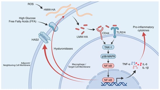
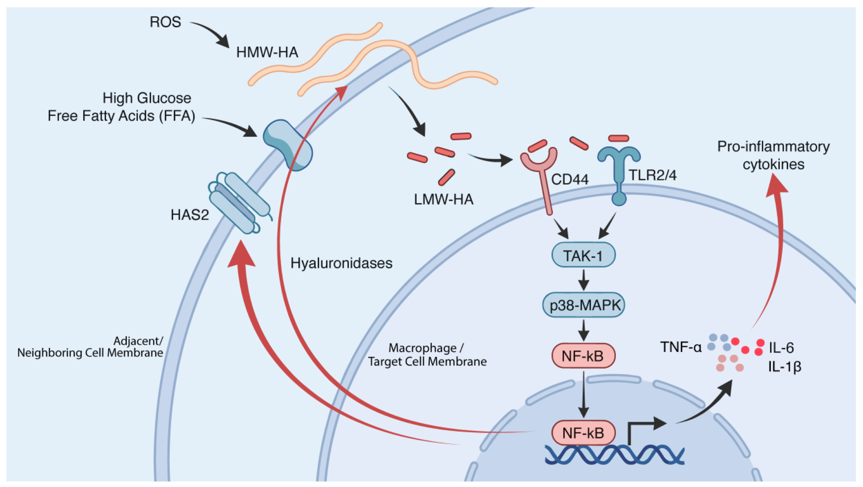
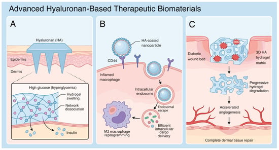
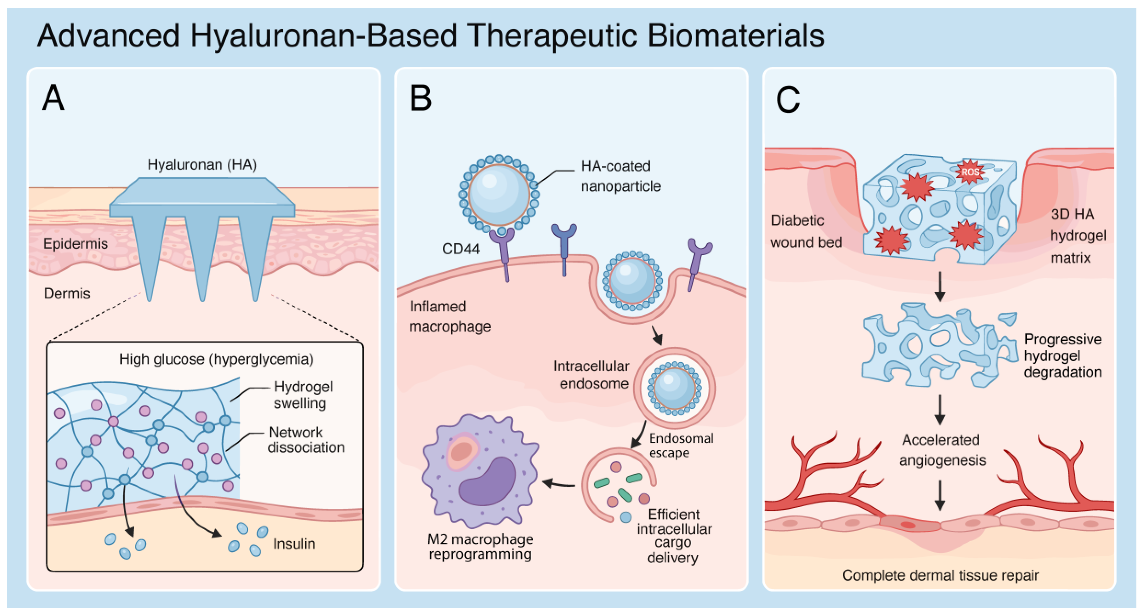
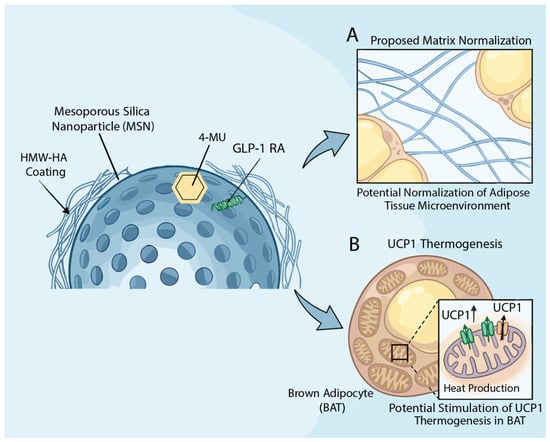
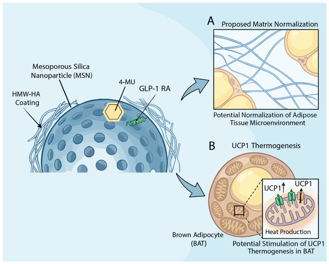
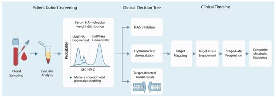
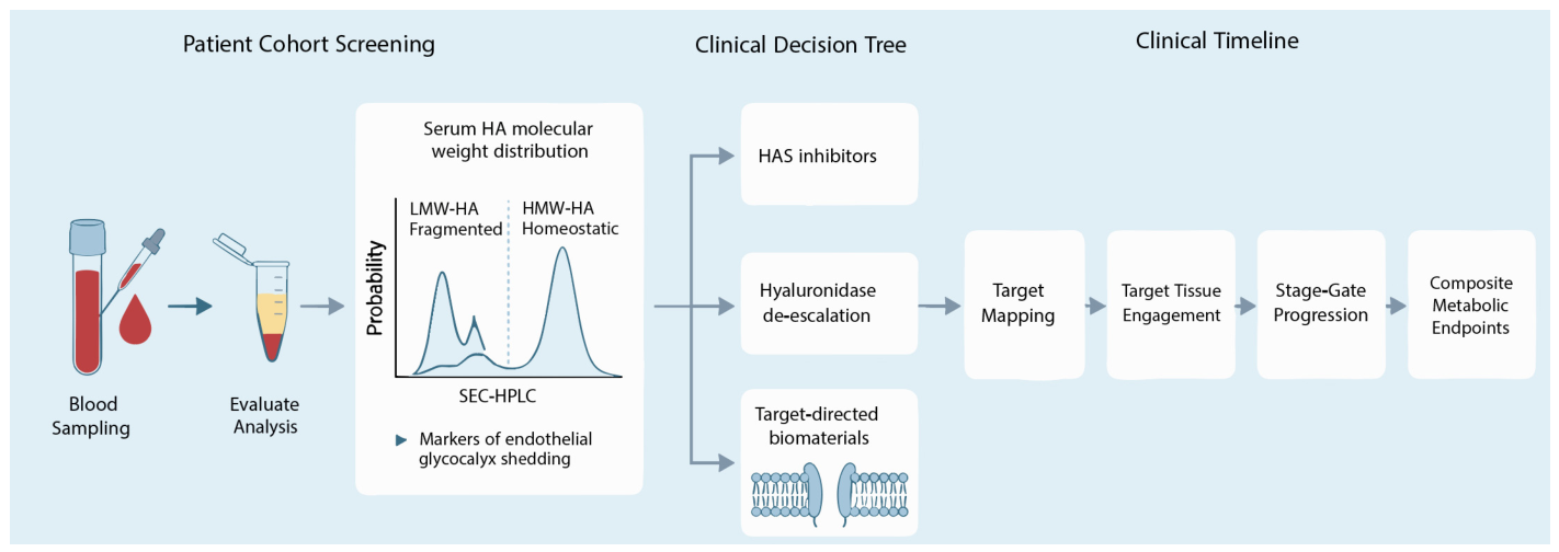

Disclaimer/Publisher’s Note: The statements, opinions and data contained in all publications are solely those of the individual author(s) and contributor(s) and not of MDPI and/or the editor(s). MDPI and/or the editor(s) disclaim responsibility for any injury to people or property resulting from any ideas, methods, instructions or products referred to in the content. |
© 2026 by the authors. Licensee MDPI, Basel, Switzerland. This article is an open access article distributed under the terms and conditions of the Creative Commons Attribution (CC BY) license.