


Abstract
KRAS-mutant pancreatic ductal adenocarcinoma (PDAC) is characterized by a high mortality that still lacks efficient therapies. Monoketone-based bisarylidene curcuminoid derivatives have shown promising activities against various PDAC models. Based on our previous findings on anticancer-active N-acryloyl-modified piperidin-4-one derivatives, we replaced the reactive acryloyl moiety by a series of N-arylsulfonyl bis-difluoro/trifluoromethylbenzylidene piperidin-4-one derivatives and tested them for antiproliferative activity against KRAS-mutant PDAC cells. Furthermore, apoptosis induction based on caspase-3/7 activation and STAT3 inhibition were investigated for the most promising compound. Notably, the N-tosyl derivative 3a with 4-trifluoromethyl substituents at the benzylidene rings exhibited the highest antiproliferative activities against PDAC cells (IC50 = 0.22–0.26 µM, 48 h). Derivative 3a also strongly induced apoptosis in PDAC cells and suppressed STAT3 activation/phosphorylation, which was supported by molecular docking of this curcuminoid into the SH2 domain of STAT3 and by 3a-STAT3 binding confirmed using CETSA in MiaPaCa-2 cells. These findings identify compound 3a as a promising lead for further preclinical evaluation in KRAS-mutant PDAC models and in other STAT3-dependent cancer types.
1. Introduction
The curative therapy of pancreatic cancer is still an unmet health issue, and thus pancreatic cancer remains one of the deadliest human tumor diseases. Clinically applied treatment options such as surgery, chemotherapy (e.g., gemcitabine, erlotinib, FOLFIRINOX), and radiotherapy can prolong patient survival but usually do not protect from tumor relapse and recurrence [1,2]. Pancreatic ductal adenocarcinoma (PDAC) is an especially perilous cancer type, and the development of new efficient drugs against PDAC has been complicated [3]. Notably, approximately 90% of all PDAC cases are KRAS-mutant, which has an impact on the overall survival of PDAC patients [4]. Thus, recent therapeutic advances include daraxonrasib, a multi-selective RAS inhibitor approved by the U.S. Food and Drug Administration, for certain adults with metastatic pancreatic adenocarcinoma after prior systemic therapy or for those who are not candidates for multi-agent systemic therapy [5].
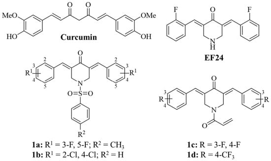
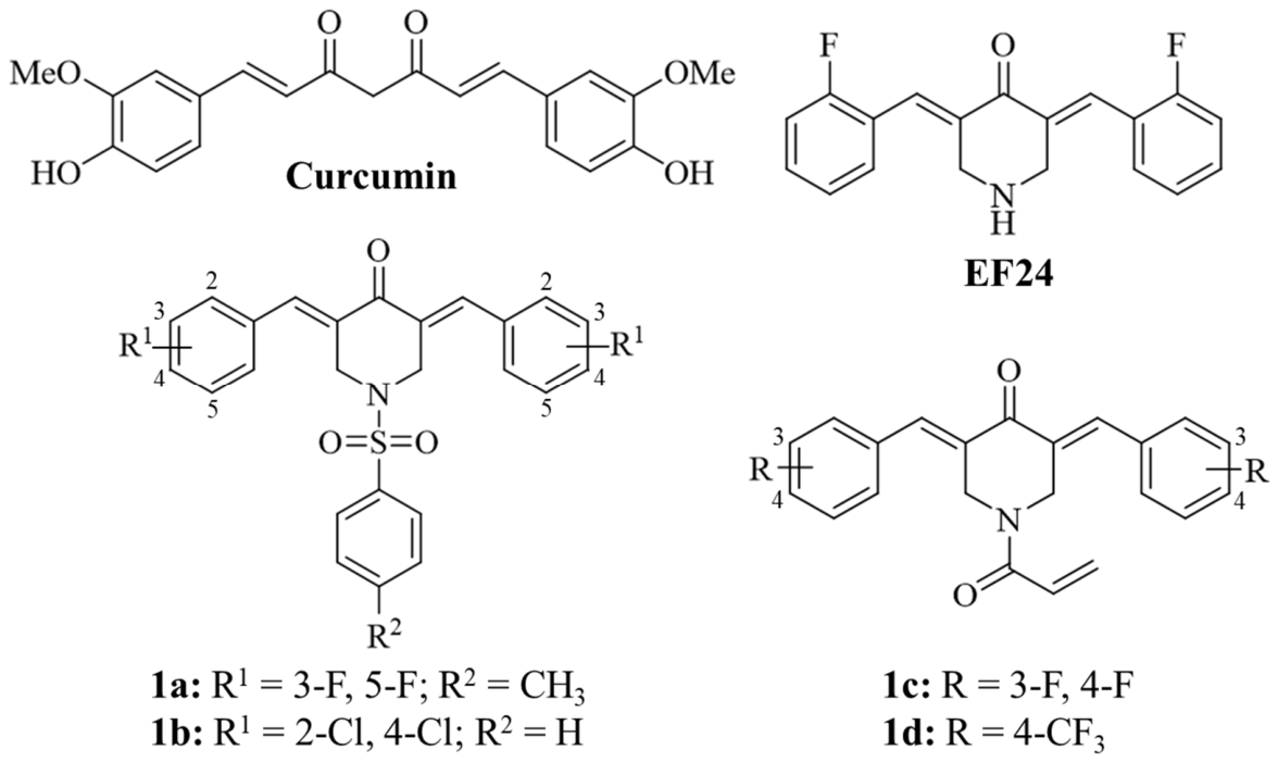
However, along with the increasing relevance of RAS inhibitors to KRAS-mutant PDAC therapy, the problem of RAS inhibitor resistance is also rising [6]. The EGFR/HER2-4 inhibitor afatinib and the STAT3 suppressor SD36 were recommended as combination partners of daraxonrasib to prevent adaptive RAS inhibitor resistance in PDAC models [7]. In particular, natural STAT3 inhibitors have shown promising activities against cancers and associated inflammatory processes [8]. Curcumin (diferuloylmethane), a biologically active constituent of Curcuma longa root powder used as a spice in Indian cuisine, is a prominent example since it has shown both STAT3- and EGFR-suppressing effects in cancer (Figure 1) [9,10]. In addition, curcumin was shown to be a potent inhibitor of drug-resistant cancers and exhibited promising activities against PDACs, including suppression of HRAS, AKT1, and EGFR [11,12]. However, curcumin has poor bioavailability, limiting its clinical application as an anticancer drug [13].
Figure 1.
Structures of curcumin and of the anticancer active piperidin-4-one-based curcuminoids EF24, arylsulfonamides 1a and 1b, and N-acryloyl derivatives 1c and 1d.
To improve activity and pharmacological properties in cancer, researchers developed various monocarbonyl-based curcuminoids, including the most prominent example, the piperidin-4-one derivative EF24 (Figure 1) [14,15]. Meanwhile, numerous curcuminoid derivatives, such as modified curcumins and monoketone-based EF24-type curcuminoids, have been described as active compounds against PDAC [16]. Notably, a curcumin-sulfonamide hybrid was reported to be active against lung and gastric cancer cells [17]. In terms of N-sulfonyl-modified EF24 derivatives, the monoketone-based N-arylsulfonamides 1a and 1b were identified as having considerable anticancer activity against the NCI panel of 60 cancer cell lines (Figure 1) [18]. However, PDAC cell lines were not included in these tests. Notably, analogous aryl sulfonamides showed high activities against human pathogenic protozoal parasites such as Leishmania major and Toxoplasma gondii [19]. Various other sulfonamide derivatives have been reported as pro-apoptotic agents (Bcl-2 inhibitors) [20]. Moreover, anticancer-active sulfonamides have been described as inhibitors of relevant tumor targets such as protein kinases, carbonic anhydrases, matrix metalloproteinases, and histone deacetylases [21]. To target STAT3 in KRAS-mutant PDAC cells, our group recently designed and identified N-acryloyl derivatives of EF24, such as compounds 1c and 1d, with improved activity against PDAC (Figure 1) [22]. However, reactive N-acryloyl moieties can have detrimental pharmacological properties. Aryl sulfonyl fragments can be suitable, more stable alternatives to acryl derivatives and can lead to new promising drug candidates for PDAC treatment.
Since there are no anticancer data for the N-arylsulfonyl curcuminoids 1a and 1b from PDAC cells available yet, we decided to fill this gap, prepared a series of known and new N-arylsulfonamide-based monocarbonyl curcuminoids with difluoro- or trifluoromethylbenzylidene moieties, and tested them for anticancer activity against PDACs together with suitable control compounds.
2. Results
2.1. Curcuminoid Test Compounds
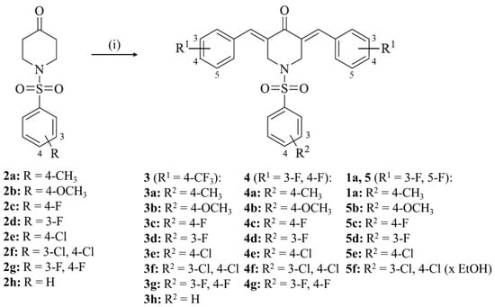
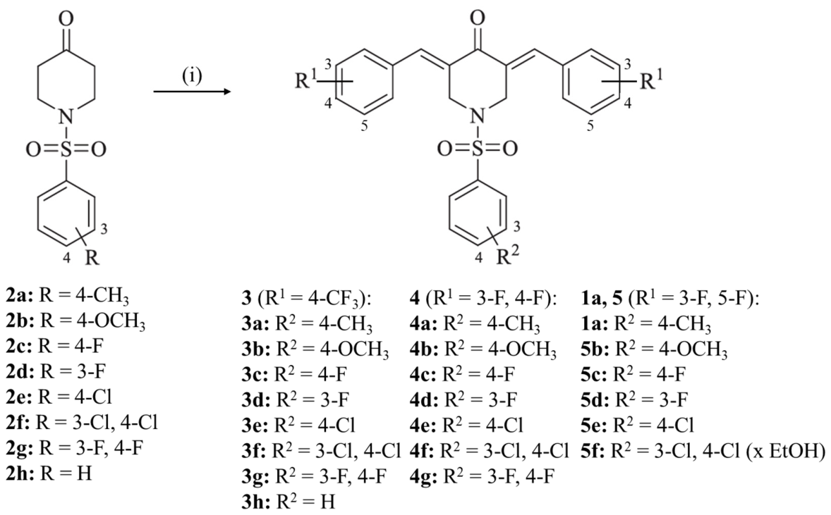
The N-arylsulfonyl-modified bis-3,5-(4-trifluoromethylbenzylidene)-piperidin-4-ones 3a–h were prepared from the condensation reaction of the N-aryl sulfonyl piperidin-4-ones 2a–h with 4-trifluoromethylbenzaldehyde under acidic conditions in refluxing EtOH, which was described previously (Scheme 1) [18,19].
Scheme 1.
Reagents and conditions: (i) 4-trifluoromethylbenzaldehyde or 3,4-/3,5-difluorobenzaldehyde, conc. HCl, EtOH, reflux, 24 h, 30–61%.
Analogously to compounds 3a–h, the 3,4-difluorobenzylidenes 4a–g and 3,5-difluorobenzylidenes 1a and 5b–f were obtained (Scheme 1). Among these compound series, only the N-tosyl derivatives 1a and 4a have already been published, whereas 4b–g and 5b–f are new sulfonamide-based curcuminoids (Scheme 1, Figure 1) [18]. The new sulfonamides were obtained as yellow solids in accordance with the color and aggregation state of the known sulfonamide analogs.
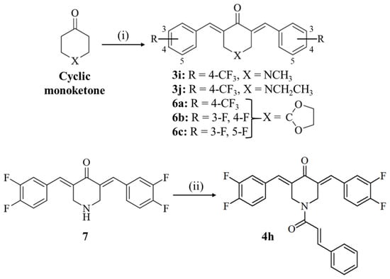
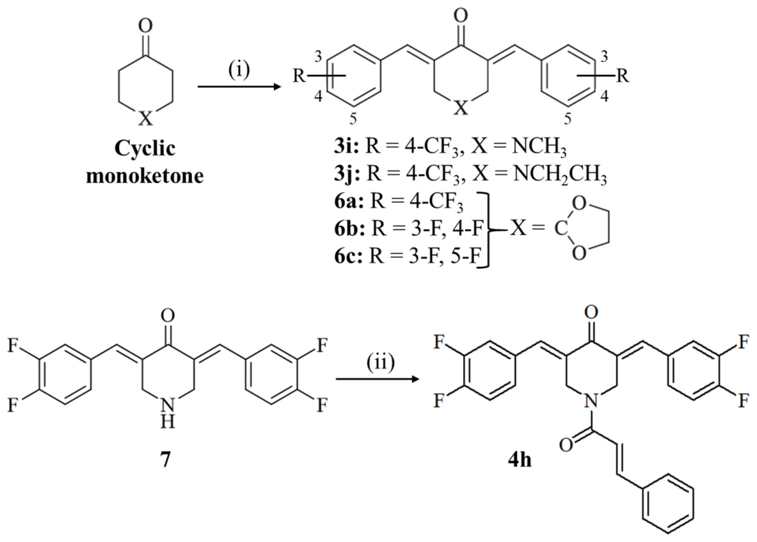
The known N-(m)ethyl curcuminoids 3i and 3j were applied for comparison purposes and were prepared under basic conditions in MeOH at room temperature (Scheme 2) [19]. The monoethylene acetals 6a–c were also prepared under basic conditions in MeOH at room temperature (Scheme 2). The ethylene acetal moiety has two oxygen atoms that can mimic, in theory, the sulfonyl oxygen atoms of the sulfonamides. Compound 6a was published in a previous study, whereas 6b and 6c are new derivatives [19]. Moreover, a new cinnamide 4h was prepared from the reaction of the known piperidin-4-one-based curcuminoid and EF24 analog 7 with cinnamoyl chloride for comparison with the sulfonamide analogs (Scheme 2) [19]. The new acetals 6b and 6c, as well as the cinnamide 4h, were also obtained as yellow solids. NMR spectra of the target compounds are provided in the Supplementary Materials (Figures S1–S42).
Scheme 2.
Reagents and conditions: (i) 4-trifluoromethylbenzaldehyde or 3,4-/3,5-difluorobenzaldehyde, aq. NaOH, MeOH, r.t., 1 h, 40–44%; (ii) cinnamoyl chloride, aq. K2CO3, acetone, r.t., 24 h, 75%.
2.2. Anticancer Activity
2.2.1. Antiproliferative Activity
The curcuminoids described in Section 2.1. were tested for their antiproliferative activities against two KRAS-mutant PDAC cell lines, MiaPaCa-2 and S2-007 (Table 1). The N-tosyl derivative 3a of the 4-trifluoromethylphenyl series was the most active compound with IC50 values of 0.22–0.26 µM in both PDAC cell lines. Compound 3a was more active than the N-alkyl analogs (3i and 3j) and the approved anticancer drug 5-fluorouracil (5-FU, a component of the FOLFIRINOX regimen). The replacement of the methyl group of the tosyl fragment by a chloro substituent (3e) led to slightly lower activities (IC50 = 0.47–0.55 µM), while analogs with fluoro (3c) and methoxy (3b) substituents, as well as with 3,4-difluoro (3g) and 3,4-dichloro (3f) substituents, were distinctly less active. The besylate 3h was also less active, underscoring the importance of the aryl sulfonyl substituent for activity against PDAC cells. Only the 3-fluorophenylsulfonamide 3d showed IC50 values below 1 µM and was thus more active than its 4-fluorophenyl congener 3c. Furthermore, 3b and 3h showed activity only against S2-007 cells (IC50 = 1.18 µM and 3.03 µM, respectively) but were inactive against MiaPaCa-2 cells (IC50 >10 µM).
Table 1.
Antiproliferative activity of test compounds against MiaPaCa-2 and S2-007 pancreatic cancer cells, expressed as IC50 (µM) 1 after 48 h of treatment, as determined by a hexosaminidase assay. 5-Fluorouracil (5-FU) served as a positive control.
Notably, the tosyl fragment was also present in the most active compounds 1a (IC50 = 0.67–0.70 µM) and 4a (IC50 = 0.55–0.83 µM) in the 3,5-difluorophenyl and 3,4-difluorophenyl curcuminoid series, respectively, and these were only slightly less active than 3a (Table 1). The 3,4-difluorophenyl analogs 4b, 4e, and 4f exhibited virtually the same or slightly better activities than 4a against both PDAC cell lines (IC50 = 0.44–0.73 µM). Compounds 4c, 4d, and 4g showed slightly lower activity, with IC50 values still below or around 1 µM. Cinnamide 4h was only slightly less active than the sulfonamide analogs.
Among the 3,5-difluorophenyl compounds, only tosylate 1a was active against PDAC cell lines at concentrations below 1 µM (Table 1). Derivative 5e reached the activity of 1a only against S2-007 cells. The other compounds were distinctly less active, with 5d being the least active analog.
Notably, the monoethylene acetals 6a–c were much less active than the sulfonamide derivatives (Table 1). All three compounds were inactive against MiaPaCa-2 cells (IC50 > 10 µM), whereas only the 4-trifluoromethylphenyl analog 6a exhibited moderate activity against S2-007 cells (IC50 = 4.68 µM).
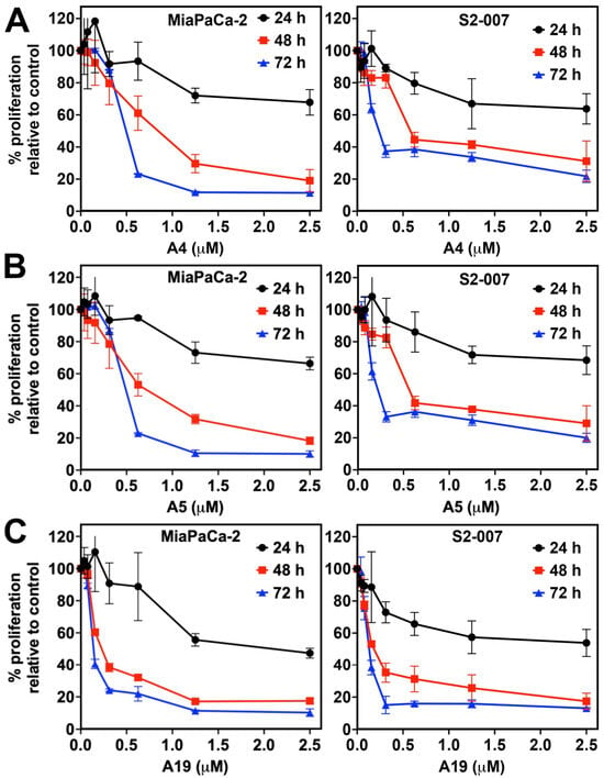
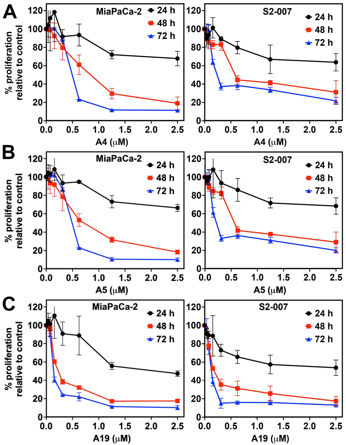
We also investigated the time- and dose-dependent antiproliferative activities of 3a (A19), 4a (A5), and 4b (A4) against MiaPaCa-2 and S2-007 cells (Figure 2). All three curcuminoids showed their highest activities after 72 h (IC50 = 0.13–0.14 µM for 3a, 0.21–0.49 µM for 4a, 0.24–0.49 µM for 4b). In addition, considerable antiproliferative activity (approximately 60% proliferation in comparison with control) of these curcuminoids was already achieved after 24 h at higher concentrations (between 1 and 2.5 µM). But only 3a exhibited a 24 h–IC50 value below 2.5 µM (2.09 µM) in the MiaPaCa-2 cells, indicating a particularly early sensitivity of these PDAC cells to this curcuminoid.
Figure 2.
Dose- and time-dependent antiproliferative activity of 4b (A4, panel (A)), 4a (A5, panel (B)), and 3a (A19, panel (C)) against MiaPaCa-2 and S2-007 PDAC cells. Cells were treated with 0–2.5 µM of each compound for 24–72 h, and proliferation was measured by hexosaminidase assay and expressed as a percentage of untreated control. Data are presented as mean ± SD of three independent experiments.
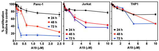
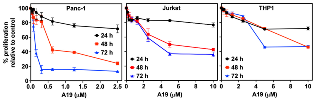
To assess the selectivity, compound 3a was also tested against Panc-1 PDAC cells and two human hematopoietic cell lines, Jurkat (an immortalized line of human T lymphocyte cells) and THP-1 monocytes (Figure 3). The results were compared with its activities against MiaPaCa-2 and S2-007 cells (Table 2). At shorter incubation times (24 h and 48 h), 3a was somewhat less effective against Panc-1 cells than against MiaPaCa-2 and S2-007 cells. Its 48 h IC50 value in Panc-1 cells (0.56 µM) was about twice that obtained in MiaPaCa-2 and S2-007 cells (0.22–0.26 µM). After 72 h, however, 3a inhibited Panc-1 proliferation as potently as in the other two PDAC cell lines, with an identical IC50 value of 0.13 µM (Figure 3).
Figure 3.
Dose- and time-dependent antiproliferative activity of 3a (A19) against Panc-1 PDAC cells (0–2.5 µM), Jurkat, and THP-1 cells (0–10 µM) after 24–72 h of compound treatment. Proliferation was measured by hexosaminidase assay (Panc-1) or CCK-8 assay (Jurkat and THP-1) and expressed as a percentage of untreated control. Data are presented as mean ± SD of three independent experiments.
Table 2.
Time-dependent antiproliferative activity of 3a against KRAS-mutant PDAC cells (MiaPaCa-2, S2-007, Panc-1) and the human leukemia cell lines Jurkat and THP-1 cells, expressed as IC50 (µM) 1, as determined by a hexosaminidase assay (PDAC lines) or CCK-8 assay (Jurkat and THP-1 cells).
In contrast, 3a was distinctly less active against Jurkat and THP-1 cells, which required considerably higher concentrations (up to 10 µM) for growth inhibition (Figure 3). Neither cell line reached 50% inhibition after 24 h (IC50 > 10 µM). After 48 h and 72 h, the IC50 values were 5.59 µM and 3.44 µM for Jurkat cells and 9.23 µM and 4.75 µM for THP-1 cells, respectively (Table 2). Based on the 72 h IC50 values, 3a was approximately 25- to 26-fold more active against the three PDAC cell lines than against Jurkat cells and approximately 34- to 37-fold more active than against THP-1 cells (Supplementary Table S1). This indicates a considerable preference for PDAC cells after 72 h incubation across all three PDAC cell lines.
2.2.2. Apoptosis Induction
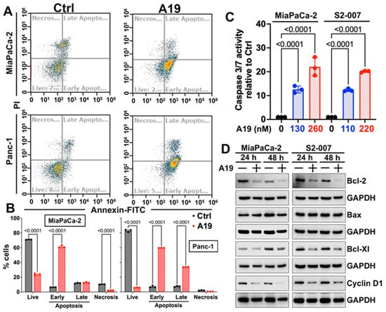
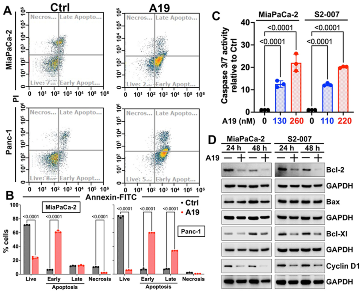
To directly confirm apoptosis induction by 3a (A19), apoptosis was quantified by Annexin V-FITC/PI flow cytometry in MiaPaCa-2 and Panc-1 cells following treatment with 3a at its IC50 concentration for 48 h (Figure 4A,B). Treatment with 3a markedly reduced the live cell population and increased both early and late apoptotic fractions in both cell lines relative to vehicle-treated controls. In MiaPaCa-2 cells, the combined early and late apoptotic population increased from 18.5% to 74.1% (p < 0.0001). An even more pronounced shift was observed in Panc-1 cells, where the combined apoptotic fraction rose from 14.7% to 93.7% (p < 0.0001). Necrotic cell fractions remained low and were not increased by 3a treatment in either cell line, consistent with an apoptotic rather than necrotic mechanism of cell death.
Figure 4.
Compound 3a induces apoptosis in PDAC cells. (A) Representative Annexin V-FITC/PI flow cytometry dot plots of MiaPaCa-2 (top) and Panc-1 (bottom) cells treated with vehicle (Ctrl) or 3a (A19) at IC50 (260 nM MiaPaCa-2; 560 nM Panc-1) for 48 h. Quadrants show live (Annexin V−/PI−), early apoptotic (Annexin V+/PI−), late apoptotic (Annexin V+/PI+), and necrotic (Annexin V−/PI+) cells. (B) Quantification of the percentage of cells in each quadrant for MiaPaCa-2 (left) and Panc-1 (right) cells. Dots represent independent experiments (n = 3); p-values were determined by an unpaired two-tailed Student t-test for each quadrant. (C) Caspase-3/7 activity in MiaPaCa-2 and S2-007 cells after 48 h of treatment with 3a (A19) at ½ IC50 (130 nM MiaPaCa-2; 110 nM S2-007) or IC50 (260 nM MiaPaCa-2; 220 nM S2-007), expressed relative to untreated control (Ctrl, 0 nM). Data are presented as mean ± SD; dots represent independent experiments (n = 3). p-values were determined by one-way ANOVA with Bonferroni–Šídák correction for multiple comparisons versus control. (D) Effect of 3a (A19) on apoptosis-related protein expression in MiaPaCa-2 and S2-007 cells. Western blots show reduced Bcl-2, Bcl-Xl, and cyclin D1 levels in cells treated with 3a at its IC50 concentration (+) for 24 h or 48 h compared with vehicle (−), while Bax expression remained unchanged. GAPDH was used as a loading control. Densitometric quantification from three independent experiments is shown in Figure S43.
The ability of 3a to induce apoptosis via caspase-3/7 activation was further assessed in MiaPaCa-2 and S2-007 cells (Figure 4C). Cells were treated for 48 h at the ½IC50 (130 nM MiaPaCa-2; 110 nM S2-007) and IC50 (260 nM MiaPaCa-2; 220 nM S2-007) concentrations of 3a, and caspase-3/7 activity was measured relative to untreated controls. At ½IC50, 3a produced a 12- to 13-fold increase in caspase-3/7 activity in both cell lines relative to untreated controls (p < 0.0001). At the IC50, this increased further to 20- to 22-fold, indicating clear dose-dependent induction of apoptosis by 3a in both PDAC lines. The pro-apoptotic effects of 3a (A19) on MiaPaCa-2 and S2-007 cells were further examined by monitoring the expression of pro- and anti-apoptotic proteins, including Bcl-2, Bcl-Xl, and Bax (Figure 4D and Figure S43). Treatment with 3a (A19) at its IC50 concentration markedly suppressed the anti-apoptotic proteins Bcl-2 and Bcl-Xl in both cell lines at 24 h and 48 h. The expression of pro-apoptotic Bax remained unaffected by 3a (A19) at either time point in either cell line. Cyclin D1 was also suppressed by 3a in both cell lines at both time points.
2.2.3. PDAC Spheroid Growth and Colony Formation
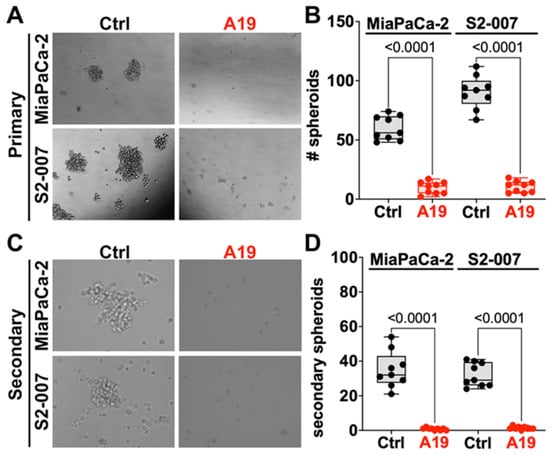
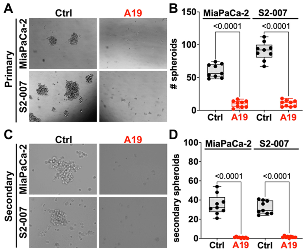
The growth of PDAC spheroids derived from MiaPaCa-2 and S2-007 cells was investigated in the absence and presence of 3a (Figure 5). Indeed, 3a treatment at its IC50 concentration produced a strong, significant inhibition of primary spheroid formation in both cell lines (p < 0.0001) (Figure 5A,B). To assess whether 3a also affects the self-renewal capacity of spheroid-forming cells, primary spheroids were dissociated and re-plated in drug-free medium to form secondary spheroids (Figure 5C,D). Cells derived from control spheroids readily formed secondary spheroids. In contrast, cells derived from 3a-treated spheroids almost completely failed to form secondary spheroids, with counts reduced in both cell lines (p < 0.0001). Secondary spheroid formation remained suppressed after drug removal, indicating that 3a impairs the self-renewal of PDAC cells with stem-like properties.
Figure 5.
Compound 3a inhibits primary and secondary spheroid formation in PDAC cells. (A) Representative brightfield images of primary MiaPaCa-2 (top) and S2-007 (bottom) spheroids after 7 days in the absence (Ctrl) or presence of 3a (A19) at its IC50 concentration. (B) Quantification of primary spheroid number per well for MiaPaCa-2 and S2-007 cells. (C) Representative brightfield images of secondary MiaPaCa-2 (top) and S2-007 (bottom) spheroids formed after primary spheroids from control and 3a-treated wells were dissociated and re-plated in drug-free spheroid medium for 5 days. (D) Quantification of secondary spheroid number per well for MiaPaCa-2 and S2-007 cells. Data in (B) and (D) are shown as box-and-whisker plots (median, interquartile range, and min–max) with individual data points; p-values were determined by unpaired two-tailed Student’s t-test. Dots represent individual wells (n = 9 per group, 3 wells from each of 3 independent experiments).
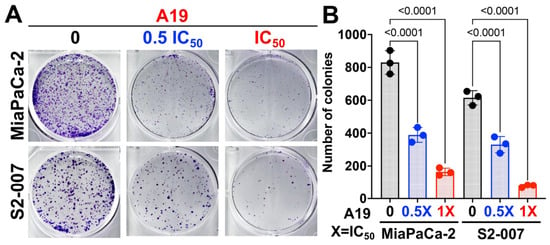
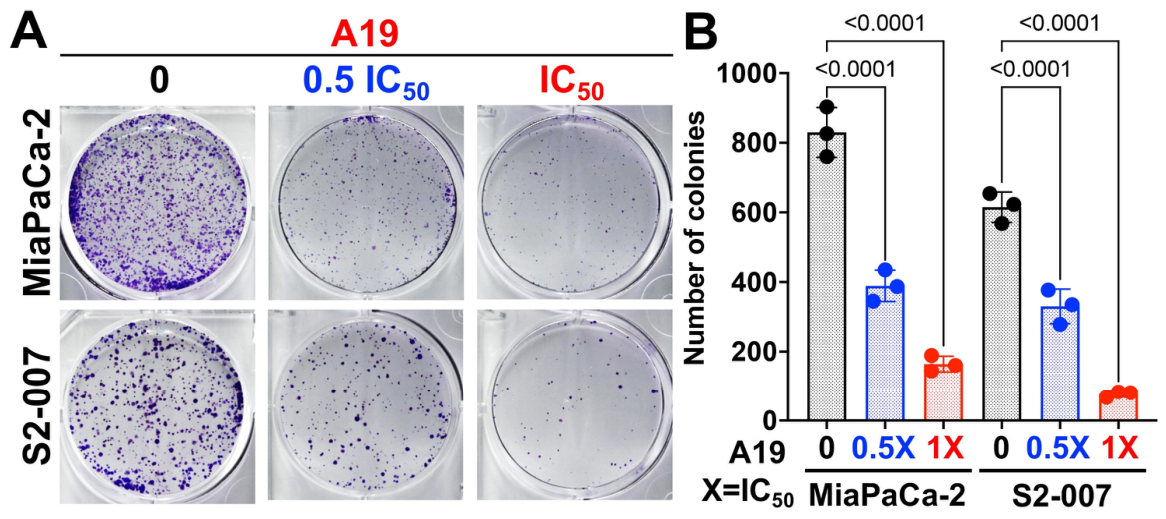
Moreover, the inhibition of colony formation by 3a was evaluated in MiaPaCa-2 and S2-007 cells (Figure 6A,B). At its ½IC50 concentration, 3a reduced colony formation by approximately 50% in both cell lines compared with untreated cells. At the IC50 concentration, colony formation was strongly abolished in MiaPaCa-2 and S2-007 cells.
Figure 6.
Compound 3a inhibited colony formation in PDAC cells. (A) Representative crystal-violet-stained wells of MiaPaCa-2 (top) and S2-007 (bottom) cells 10–12 days after a 48 h treatment with 0, ½ IC50 (0.5X), or IC50 (1X) concentrations of 3a (A19) followed by drug-free recovery. (B) Quantification of colony number for MiaPaCa-2 and S2-007 cells under the same conditions. Data are presented as mean ± SD; dots represent independent experiments (n = 3). p-values were determined by one-way ANOVA with Bonferroni–Šídák correction for multiple comparisons versus untreated control.
2.2.4. STAT3 Inhibition
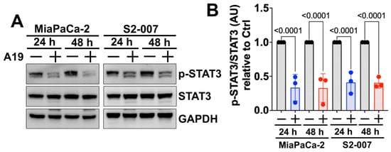
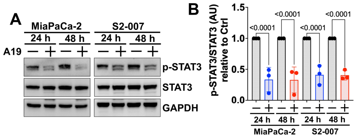
The inhibition of STAT3 phosphorylation (Tyr705) by 3a in PDAC cells (MiaPaCa-2 and S2-007) was investigated by Western blot (Figure 7A) and quantified relative to total STAT3 (Figure 7B). While 3a did not affect total STAT3 expression, it markedly reduced levels of activated/phosphorylated p-STAT3 in both cell lines as early as 24 h, with suppression sustained through 48 h (p < 0.0001 at both time points in both cell lines).
Figure 7.
Compound 3a inhibited STAT3 phosphorylation (Tyr705) in PDAC cells. (A) Western blots of p-STAT3 in MiaPaCa-2 and S2-007 cells treated with vehicle or 3a (A19) at its IC50 concentration for 24 h or 48 h; GAPDH served as a loading control. (B) Densitometric quantification of p-STAT3 (Tyr705) normalized to total STAT3, relative to the respective untreated control, for each cell line and time point. Data are presented as mean ± SD; dots represent independent experiments (n = 3). p-values were determined by an unpaired two-tailed Student t-test versus the time-matched vehicle control.
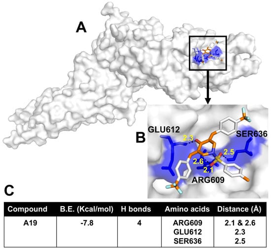
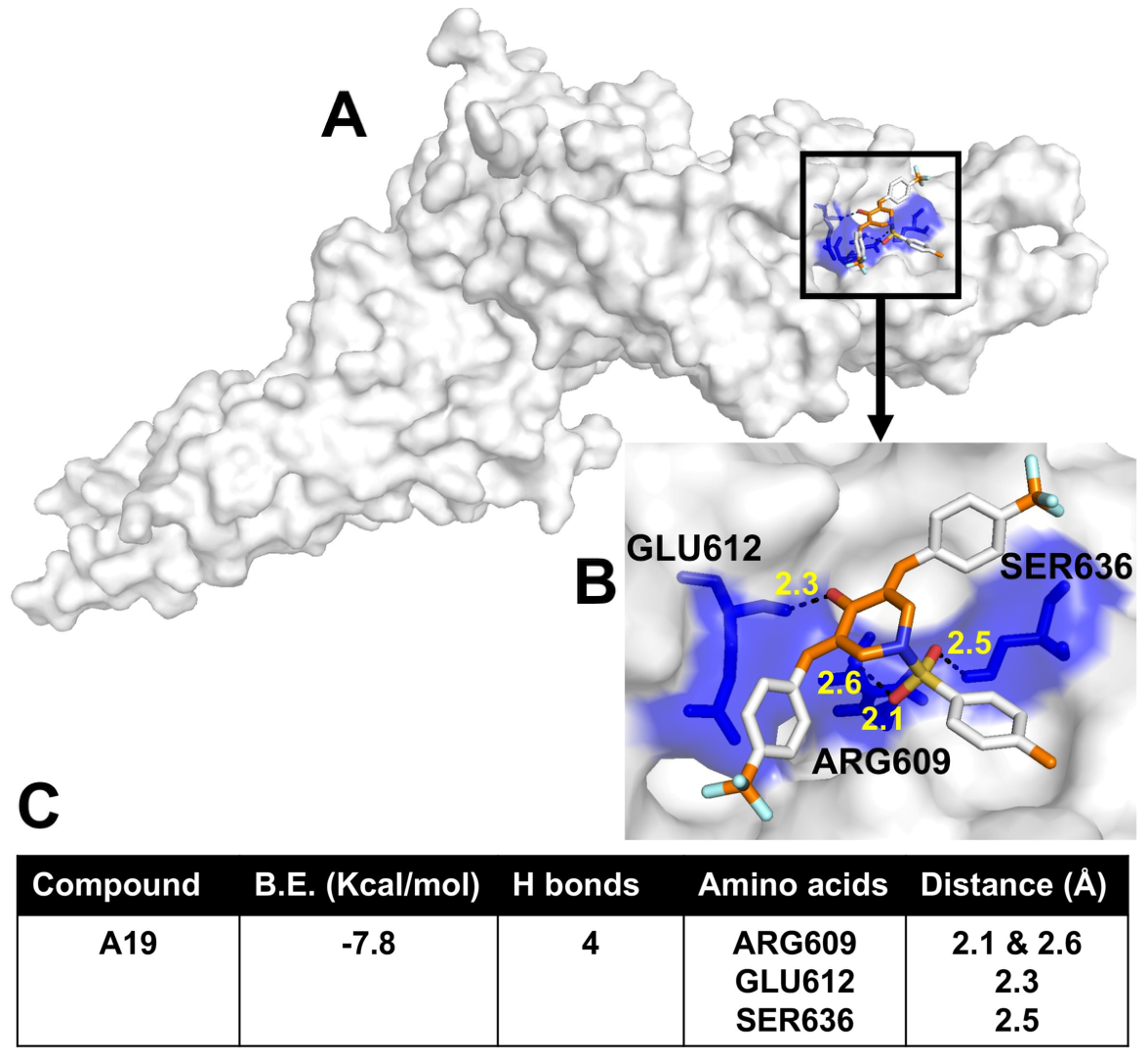
The inhibition of STAT3 phosphorylation by 3a suggests an interaction with the protein. A possible binding of 3a to STAT3 was evaluated by molecular docking using a suitable STAT3 structure (PDB ID: 6NJS). Molecular docking of 3a into the STAT3 SH2 domain revealed a considerable binding energy (B.E. = −7.8 kcal/mol). The formation of four H-bonds with the STAT3 amino acids ARG609 (two H-bonds via a sulfonyl oxygen of 3a; 2.1 and 2.6 Å), GLU612 (one H-bond via the piperidin-4-one carbonyl oxygen of 3a; 2.3 Å), and SER636 (one H-bond via a sulfonyl oxygen of 3a; 2.5 Å) was determined, indicating a prominent role of the central N-sulfonyl-4-piperidone moiety in the interaction with the SH2 domain (Figure 8A–C).
Figure 8.
Docking of 3a into the SH2 domain of STAT3. (A) Overview of the STAT3 surface (PDB ID: 6NJS; grey-white), with the predicted binding site of 3a boxed. (B) Close-up of the boxed region. 3a is shown as sticks with carbon atoms in orange (aromatic rings in white), oxygen in red, nitrogen in blue, sulfur in yellow and fluorine in cyan. Binding-site residues are shown as blue sticks, and the surface they contribute to is shaded blue; the three residues that hydrogen-bond to 3a are labeled. Hydrogen bonds are drawn as black dashed lines, with the corresponding donor–acceptor distances given in Å (yellow). (C) Summary of the docking result for 3a (A19): binding energy, number of hydrogen bonds, interacting residues, and hydrogen-bond distances.
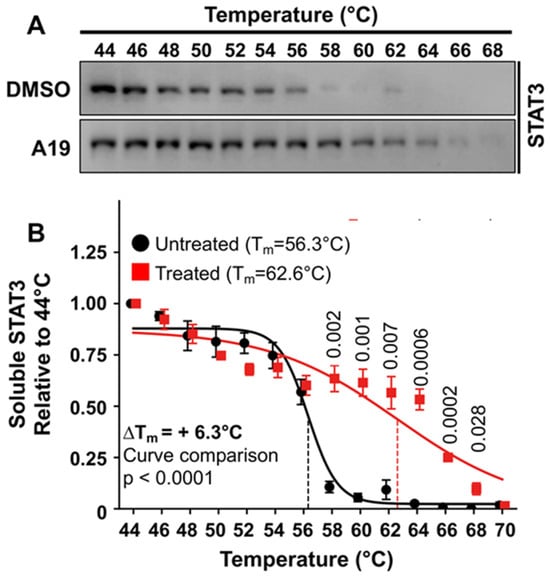
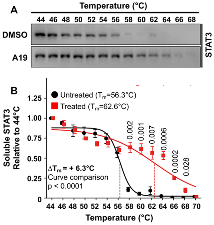
To confirm direct engagement of STAT3 by 3a, a cellular thermal shift assay (CETSA) was performed in MiaPaCa-2 cell lysates (Figure 9). In vehicle-treated cell lysates, soluble STAT3 levels declined sharply above 54 °C, and the protein was largely precipitated at 58 °C and higher (Figure 9A). In cell lysates treated with 3a at 20 µM for 4 h, soluble STAT3 remained detectable up to 64–66 °C. Quantification of the melting curves showed that 3a shifted the melting temperature (Tm) of STAT3 from 56.3 °C to 62.6 °C (ΔTm = +6.3 °C; curve comparison p < 0.0001) (Figure 9B). Soluble STAT3 levels were significantly higher in 3a-treated cell lysates at all temperatures between 58 °C and 68 °C (p-value = 0.0002–0.028). This thermal stabilization of STAT3 supports a direct interaction of 3a with STAT3 in PDAC cells, consistent with the binding mode predicted by molecular docking.
Figure 9.
Cellular thermal shift assay (CETSA) showing that 3a binds STAT3. (A) Representative Western blots of soluble STAT3 in MiaPaCa-2 cell lysates incubated with vehicle (DMSO) or 3a (A19) at 20 µM for 4 h, then heated at the indicated temperatures (44–68 °C) for 3 min. (B) Melting curves of soluble STAT3, quantified by densitometry and normalized to the 44 °C sample of each group. Curves were fitted by nonlinear (Boltzmann sigmoidal) regression. Derivative 3a raised the melting temperature (Tm) of STAT3 from 56.3 °C (vehicle) to 62.6 °C (ΔTm = +6.3 °C), indicating thermal stabilization of STAT3 by direct ligand binding. Data are presented as mean ± SD (n = 3). p-values above individual temperatures compare vehicle with 3a at each temperature; the overall curve comparison (extra sum-of-squares F-test) gave p < 0.0001.
3. Discussion
PDAC is a highly aggressive and lethal form of cancer with limited treatment options [3]. Research efforts to develop new potent drugs against PDAC include therapies targeting RAS, p53, CDKN2A mutants, NRG1 fusions, and TGF-β, as well as immune and CAR-T-cell therapies [23]. In this study, several N-aryl sulfonyl-modified monoketone curcuminoids were identified with high antiproliferative activity (i.e., nanomolar IC50 values) against PDAC cells. Except for a few examples (3b, 3h, 5d), the N-aryl sulfonamides were generally superior to the acetal analogs 6a–c. The N-tosyl derivative 3a, which is substituted with para-CF3 groups at both benzylidene rings, turned out to be the most active compound with a reasonable selectivity for PDAC cells. It was equally potent at nanomolar concentrations against all three PDAC cell lines in a dose- and time-dependent manner and was about 26- to 37-fold less active against the Jurkat and THP-1 cell lines. It should be noted, however, that Jurkat and THP-1 are leukemia-derived cell lines. These data therefore indicate preferential activity towards PDAC cells relative to cells of a different lineage, rather than selectivity over non-malignant cells. Evaluation in non-transformed pancreatic cells, such as hTERT-HPNE or HPDE, and in normal fibroblasts will be necessary to establish a therapeutic window for 3a. The CF3 group is frequently applied in drug design and development because of its beneficial pharmacological properties [24]. However, in the present study, the CF3 group also conveyed higher antiproliferative activity in the N-tosylate 3a in comparison with the analogous 3,4- and 3,5-difluorobenzylidene compounds 1a and 4a. Nevertheless, the tosyl group likely plays an important role in anticancer activity since N-tosylates 1a and 4a were also the best (3,5-difluoro analogs) or among the best sulfonamides (3,4-difluoro analogs) of the two difluorophenyl curcuminoid series. Thus, the tosylates 1a, 3a, and 4a add well to the current arsenal of sulfonamide-based anticancer drugs and drug candidates [20,21]. Moreover, their activities against PDAC cells fill the gap left by a study from 2014, which reported the anticancer activity of 1a against the NCI 60 cell line panel excluding PDAC cells [18]. The antiproliferative activities of 3a were also in the activity range of the N-acryl derivatives 1c and 1d against MiaPaCa-2 and Panc-1 cells, while 3a was distinctly more active than EF24 and definitely superior to the N-(m)ethyl analogs 3i and 3j [22]. Thus, the tosyl moiety is a promising N-attachment for 4-piperidone-based curcuminoids and a suitable surrogate for the N-acryl moiety of 1c and 1d.
Taken together, further experimental results support 3a as a promising anticancer agent against PDAC. Notably, the compound inhibited PDAC spheroid growth and colony formation, which indicates that 3a can suppress clonogenic survival and tumor sphere formation. Moreover, cells derived from 3a-treated spheroids almost completely lost their ability to form secondary spheroids, even after replating them in drug-free medium. Secondary sphere formation is a measure of self-renewal, a defining property of cancer stem cells. Confirmation with established PDAC stem cell markers, such as CD44, CD133 and ALDH1, will be required to define the effect of 3a on the cancer stem cell compartment more precisely. These data therefore suggest that 3a does not merely delay sphere growth but durably impairs the stem-like cell population that drives tumor relapse and drug resistance in PDAC. These effects are in line with reports that curcumin and its derivatives can target cancer stem cells and suppress or prevent tumor growth and regrowth in various cancers, including PDAC [16,25]. Annexin V/PI flow cytometry and activation of caspases 3 and 7 required for apoptosis induction underscored the excellent cell-killing properties of 3a without causing necrosis. This shows that 3a kills PDAC cells mainly by apoptosis rather than by nonspecific necrotic toxicity. Consistent with these findings, 3a downregulated the anti-apoptotic proteins Bcl-2 and Bcl-Xl in PDAC cells. The inhibition of apoptosis, or programmed cell death, is a hallmark of cancer; therefore, potent apoptosis inducers such as 3a are needed as new effective anticancer agents [26]. The development and clinical approval of the Bcl-2 inhibitor venetoclax underscored the importance of apoptosis in cancer therapy; however, resistance limited its application [27]. Curcumin was reported to downregulate cyclin D1 [28]. Cyclin D1 can promote cell cycle progression in pancreatic cancer cells and maintain levels of the anti-apoptotic proteins Bcl-2 and Bcl-Xl [29]. Notably, 3a also suppressed cyclin D1 expression. Since cyclin D1 has been reported to maintain the levels of the anti-apoptotic proteins Bcl-2 and Bcl-Xl in pancreatic cancer cells [29], its downregulation may contribute to the pro-apoptotic activity of 3a, rather than reflecting cell-cycle arrest.
In addition to these promising cell death-associated mechanisms, 3a also suppressed STAT3 activation in PDAC cells. Docking of 3a into the SH2 domain (a protein–protein interaction domain required for STAT3 dimerization) revealed a considerable binding energy and the formation of four H-bonds between the N-sulfonyl-4-piperidone core fragment and crucial STAT3 amino acids. This predicted interaction was supported experimentally by CETSA. Derivative 3a increased the thermal stability of STAT3 in MiaPaCa-2 cell lysates by 6.3 °C, a shift that indicates direct binding of the compound to the protein [30,31]. Together, docking and CETSA suggest that the reduced STAT3 phosphorylation in 3a-treated PDAC cells results, at least in part, from direct engagement of STAT3 rather than only from effects on upstream kinases. Future studies should directly confirm binding of 3a to recombinant STAT3 by surface plasmon resonance and determine whether this interaction disrupts STAT3 dimerization, a process mediated by reciprocal SH2-domain engagement between STAT3 monomers that would be expected to be impaired by occupancy of this domain by the N-sulfonyl-4-piperidone core fragment of 3a, as predicted by molecular docking. In addition, because the thermal shift assay was performed in cell lysates, it demonstrates a direct interaction between 3a and STAT3 but does not exclude additional contributions from upstream signaling in intact cells. Analysis of upstream kinases such as JAK2 and Src will therefore be required to define the mechanism of p-STAT3 suppression fully. These findings add to the current knowledge of other STAT3-inhibitory curcumin derivatives, since the compound class of curcuminoids is a treasure trove of potent STAT3 inhibitors [16]. Actually, STAT3 is a valuable drug target in KRAS-mutant PDAC because STAT3 expression and the hyper-activation of associated signaling pathways (e.g., JAK/STAT signaling, as well as STAT3 activation by upstream tyrosine kinases and mTOR) were correlated with vital mechanisms such as tumor cell survival, angiogenesis, metastasis formation, and immune escape [32]. Moreover, several STAT3 inhibitors have entered clinical trials for PDAC, underscoring STAT3 as an important drug target in PDAC [33]. STAT3-targeting drugs may be beneficial in combination with RAS and RTK inhibitors for treating PDAC [7]. STAT3 inhibitors may also prevent immune evasion and enhance the activity of immune therapies, which are particularly challenging in PDAC because it is classified as an immunologically “cold” tumor [34,35].
Hence, the tosylamide-based curcuminoid 3a has the potential to become a valuable anticancer drug for the treatment of PDAC. While its activity as a single agent is already pronounced, studies with suitable combination partners, such as currently used PDAC therapies (e.g., FOLFIRINOX, gemcitabine, erlotinib) or emerging drug candidates such as RAS inhibitors, appear promising for future development [23,36]. The anti-PDAC activity of 3a also warrants further studies in suitable in vivo models to determine its suitability as a new anticancer drug. In addition to PDACs, the previously reported broad-spectrum anticancer activity of 1a against the NCI panel of cancer cell lines may suggest further cancer types, especially cancers dependent on oncogenic STAT3 (e.g., breast cancers, head-and-neck cancers, leukemias, lymphomas, and myelomas), as possible therapeutic targets of 3a [18,37].
4. Materials and Methods
4.1. General Procedures
Column chromatography: silica gel 60 (230–400 mesh). Melting points (uncorrected), Electrothermal 9100 (ThermoFisher Scientific, Geel, Belgium); IR spectra, Perkin-Elmer Spectrum One FT-IR spectrophotometer with ATR sampling unit (Perkin Elmer, Rodgau, Germany); NMR spectra, Bruker Avance 300/500 spectrometer (Bruker, Billerica, MA, USA); chemical shifts (δ) are given in parts per million (ppm) downfield from tetramethylsilane as internal standard; Mass spectra, Thermo Finnigan MAT 8500 (EI) (ThermoFisher Scientific, Geel, Belgium); elemental analyses: Perkin-Elmer 2400 CHN elemental analyzer (Perkin Elmer, Rodgau, Germany).
4.2. Materials
Starting compounds and reagents were obtained from Alfa Aesar (Karlsruhe, Germany), Sigma-Aldrich (Darmstadt, Germany), and TCI (Zwijndrecht, Belgium). The curcuminoids 1a, 3a–j, and 4a were prepared and published in previous studies [18,19]. The syntheses of the N-sulfonylpiperidin-4-one starting compounds 2a–h and of curcuminoid 7 were also reported in previous studies [18,19].
4.3. Synthesis of New Curcuminoids
(3E,5E)-3,5-Bis-(3,4-difluorobenzylidene)-1-(1-methoxybenzene-4-sulfonyl)-piperidone (4b)—Typical procedure
1-(4-Methoxybenzenesulfonyl)-4-piperidinone (213 mg, 0.79 mmol) and 3,4-difluorobenzaldehyde (213 mg, 1.5 mmol) were dissolved in EtOH (10 mL) and conc. HCl (1 mL) was added. The reaction mixture was refluxed for 24 h. After cooling down to room temperature, the formed precipitate was collected, washed with EtOH and dried in vacuum. Yield: 125 mg (0.24 mmol, 31%); yellow solid of m.p. 238 °C; υmax(ATR)/cm−1 3964, 2846, 1677, 1596, 1518, 1496, 1440, 1422, 1342, 1300, 1274, 1261, 1233, 1176, 1151, 1116, 1092, 1042, 1019, 997, 972, 950, 927, 876, 849, 830, 773, 721, 694, 675; 1H NMR (300 MHz, DMSO-d6) δ 3.86 (3 H. s), 4.61 (4 H, s), 7.05 (2 H, d, J = 9.0 Hz), 7.3–7.4 (2 H, m), 7.45 (2 H, d, J = 9.0 Hz), 7.52 (2 H, s), 7.6–7.7 (4 H, m); 13C NMR (75.5 MHz, DMSO-d6) δ 46.4, 55.8, 114.5, 118.0, 118.2, 119.3, 119.6, 127.6, 127.7, 128.9, 129.5, 131.5, 131.6, 135.1, 147.7, 147.8, 148.2, 148.4, 150.9, 151.1, 162.9, 183.9; m/z (%) 517 (7) [M+], 346 (100), 317 (6), 194 (7), 180 (16), 171 (15), 151 (23); anal. calcd. for C26H19F4NO4S: C, 60.35; H, 3.70. Found: C, 60.25; H, 3.65.
(3E,5E)-3,5-Bis-(3,4-difluorobenzylidene)-1-(1-fluorobenzene-4-sulfonyl)-piperidone (4c)
Analogously to the synthesis of 4b, compound 4c was obtained from 1-(4-fluorobenzenesulfonyl)-4-piperidinone (203 mg, 0.79 mmol) and 3,4-difluorobenzaldehyde (213 mg, 1.5 mmol) in EtOH (10 mL) and conc. HCl (1 mL). Yield: 244 mg (0.48 mmol, 61%); yellow solid of m.p. 202 °C; υmax(ATR)/cm−1 3059, 2935, 2823, 1668, 1615, 1591, 1574, 1517, 1493, 1420, 1362, 1300, 1281, 1251, 1236, 1207, 1172, 1149, 1117, 1089, 1041, 1000, 974, 954, 936, 895, 848, 825, 791, 774, 741, 719, 698, 666, 602, 574; 1H NMR (500 MHz, DMSO-d6) δ4.67 (4 H, s), 7.3–7.4 (4 H, m), 7.51 (2 H, s), 7.6–7.9 (6 H, m); 13C NMR (125 MHz, DMSO-d6) δ 46.5, 116.6, 116.8, 118.1, 119.5, 119.7, 127.9, 130.5, 131.1, 131.6, 133.9, 135.4, 148.4, 148.5, 149.1, 150.4, 151.1, 163.8, 165.8, 183.9; m/z (%) 505 (28) [M+], 346 (100), 317 (37), 180 (50), 151 (92), 127 (26), 95 (39); anal. calcd. for C25H16F5NO3S: C, 59.41; H, 3.19. Found: C, 59.28; H, 3.14.
(3E,5E)-3,5-Bis-(3,4-difluorobenzylidene)-1-(1-fluorobenzene-3-sulfonyl)-piperidone (4d)
Analogously to the synthesis of 4b, compound 4c was obtained from 1-(3-fluorobenzenesulfonyl)-4-piperidinone (203 mg, 0.79 mmol) and 3,4-difluorobenzaldehyde (213 mg, 1.5 mmol) in EtOH (10 mL) and conc. HCl (1 mL). Yield: 160 mg (0.32 mmol, 40%); yellow solid of m.p. 203–204 °C; υmax(ATR)/cm−1 3060, 1676, 1599, 1513, 1479, 1432, 1350, 1301, 1280, 1265, 1232, 1200, 1160, 1118, 1086, 1072, 1044, 994, 959, 944, 923, 881, 829, 812, 791, 777, 750, 718, 699, 688, 678, 638, 607; 1H NMR (300 MHz, DMSO-d6) δ 4.70 (4 H, s), 7.3–7.4 (4 H, m), 7.51 (2 H, s), 7.6–7.7 (6 H, m); 13C NMR (75.5 MHz, DMSO-d6) δ 46.3, 114.0–114.4 (m), 118.0–119.6 (m), 120.6–120.9 (m), 123.4, 127.7–127.9 (m), 131.4, 131.5–132.0 (m), 139.7, 147.7–151.8 (m), 159.8–163.3 (m), 184.0; m/z (%) 505 (22) [M+], 346 (100), 317 (23), 180 (30), 151 (74), 95 (36); anal. calcd. for C25H16F5NO3S: C, 59.41; H, 3.19. Found: C, 59.31; H, 3.13.
(3E,5E)-3,5-Bis-(3,4-difluorobenzylidene)-1-(1-chlorobenzene-4-sulfonyl)-piperidone (4e)
Analogously to the synthesis of 4b, compound 4e was obtained from 1-(4-chlorobenzenesulfonyl)-4-piperidinone (216 mg, 0.79 mmol) and 3,4-difluorobenzaldehyde (213 mg, 1.5 mmol) in EtOH (10 mL) and conc. HCl (1 mL). Yield: 123 mg (0.24 mmol, 30%); yellow solid of m.p. 232–233 °C; υmax(ATR)/cm−1 3095, 2910, 1664, 1593, 1569, 1517, 1476, 1420, 1360, 1301, 1276, 1235, 1207, 1163, 1116, 1094, 1046, 1012, 996, 977, 950, 877, 848, 818, 775, 754, 717, 694, 649, 608; 1H NMR (300 MHz, DMSO-d6) δ 4.67 (4 H, s), 7.3–7.4 (2 H, m), 7.5–7.7 (10 H, m); 13C NMR (75.5 MHz, DMSO-d6) δ 46.4, 118.0, 118.2, 119.4, 119.7, 127.7, 127.8, 129.1, 129.6, 131.1, 131.5, 131.6, 135.4, 136.4, 138.5, 147.7, 147.9, 148.3, 148.5, 151.0, 151.1, 151.6, 151.8, 183.9; m/z (%) 523 (12) [M+], 521 (32) [M+], 346 (100), 317 (46), 180 (55), 152 (90), 151 (92), 111 (33); anal. calcd. for C25H16ClF4NO3S: C, 57.53; H, 3.09. Found: C, 57.39; H, 3.05.
(3E,5E)-3,5-Bis-(3,4-difluorobenzylidene)-1-(1,2-dichlorobenzene-4-sulfonyl)-piperidone (4f)
Analogously to the synthesis of 4b, compound 4f was obtained from 1-(3,4-dichlorobenzenesulfonyl)-4-piperidinone (243 mg, 0.79 mmol) and 3,4-difluorobenzaldehyde (213 mg, 1.5 mmol) in EtOH (10 mL) and conc. HCl (1 mL). Yield: 156 mg (0.28 mmol, 35%); yellow solid of m.p. 188–190 °C; 1H NMR (300 MHz, DMSO-d6) δ 4.74 (4 H, s), 7.3–7.4 (2 H, m), 7.4–7.7 (8 H, m), 7.84 (1 H, d, J = 8.5 Hz); 13C NMR (75.5 MHz, DMSO-d6) δ 46.4, 118.0–118.2 (m), 119.4–119.7 (m), 127.2, 127.9, 128.9, 130.9, 131.3–131.5 (m), 131.8, 132.3, 135.4, 136.7, 138.0, 147.7–151.8 (m), 183.9; m/z (%) 557 (11) [M+], 555 (13) [M+], 378 (2), 376 (3), 346 (100), 317 (17), 180 (24), 151 (42); anal. calcd. for C25H15Cl2F4NO3S: C, 53.97; H, 2.72. Found: C, 53.89; H, 2.67.
(3E,5E)-3,5-Bis-(3,4-difluorobenzylidene)-1-(1,2-difluorobenzene-4-sulfonyl)-piperidone (4g)
Analogously to the synthesis of 4b, compound 4g was obtained from 1-(3,4-difluorobenzenesulfonyl)-4-piperidinone (217 mg, 0.79 mmol) and 3,4-difluorobenzaldehyde (213 mg, 1.5 mmol) in EtOH (10 mL) and conc. HCl (1 mL). Yield: 128 mg (0.24 mmol, 32%); pale yellow solid of m.p. 193–194 °C; υmax(ATR)/cm−1 3073, 2835, 1680, 1611, 1597, 1516, 1507, 1455, 1420, 1354, 1303, 1280, 1230, 1205, 1160, 1146, 1124, 1113, 1070, 1054, 993, 972, 952, 936, 914, 883, 871, 860, 839, 822, 790, 775, 735, 716, 694, 674, 638, 608; 1H NMR (300 MHz, DMSO-d6) δ 4.70 (4 H, s), 7.3–7.5 (3 H, m), 7.53 (2 H, s), 7.6–7.8 (6 H, m); 13C NMR (75.5 MHz, DMSO-d6) δ 46.4, 117.0–117.4 (m), 118.0–118.2 (m), 118.8–119.6 (m), 125.2–125.3 (m), 125.3, 127.8–127.9 (m), 131.1, 131.5, 134.8, 135.3, 147.7–151.8 (m), 184.0; m/z (%) 523 (37) [M+], 346 (100), 317 (28), 180 (35), 151 (67); anal. calcd. for C25H15F6NO3S: C, 57.37; H, 2.89. Found: C, 57.29; H, 2.84.
(3E,5E)-3,5-Bis-(3,4-difluorobenzylidene)-1-cinnamoylpiperidone (4h)
Compound 7 (112 mg, 0.32 mmol) was suspended in acetone and treated with cinnamoyl chloride (82 mg, 0.49 mmol). K2CO3 (223 mg, 1.61 mmol, dissolved in 2 mL H2O) was added and the reaction mixture was stirred at room temperature for 24 h. Water (20 mL) was added and the formed precipitate was collected, washed with water and water/methanol and dried in vacuum. Yield: 115 mg (0.24 mmol, 75%); yellow solid of m.p. 169–170 °C; υmax(ATR)/cm−1 3062, 1679, 1645, 1602, 1513, 1454, 1431, 1326, 1301, 1265, 1234, 1212, 1195, 1168, 1150, 1115, 997, 958, 940, 927, 879, 853, 826, 794, 762, 744, 706, 681; 1H NMR (300 MHz, CDCl3) δ 4.8–4.9 (4 H, br s), 6.41 (1 H, d, J = 15.4 Hz), 7.1–7.3 (11 H, m), 7.52 (1 H, d, J = 15.4 Hz), 7.72 (2 H, s); 13C NMR (75.5 MHz, CDCl3) δ 46.1, 115.8, 117.9, 118.1, 119.0, 119.2, 126.9, 127.6, 128.8, 130.1, 131.5, 132.7, 134.6, 135.9, 144.0, 148.7, 148.9, 149.3, 149.4, 152.0, 152.6, 152.8, 165.8, 186.4; m/z (%) 477 (96) [M+], 346 (46), 151 (14), 131 (100), 103 (54). 77 (16); anal. calcd. for C28H19F4NO2: C, 70.44; H, 4.01. Found: C, 70.31; H, 3.97.
(3E,5E)-3,5-Bis-(3,5-difluorobenzylidene)-1-(1-methoxybenzene-4-sulfonyl)-piperidone (5b)
Analogously to the synthesis of 4b, compound 5b was obtained from 1-(4-methoxybenzenesulfonyl)-4-piperidinone (213 mg, 0.79 mmol) and 3,5-difluorobenzaldehyde (213 mg, 1.5 mmol) in EtOH (10 mL) and conc. HCl (1 mL). Yield: 130 mg (0.25 mmol, 32%); yellow solid of m.p. 241–242 °C; υmax(ATR)/cm−1 3088, 2964, 2841, 1672, 1615, 1592, 1497, 1471, 1434, 1383, 1350, 1315, 1253, 1236, 1220, 1188, 1165, 1152, 1122, 1094, 1051, 1034, 1024, 997, 963, 944, 887, 845, 803, 773, 750, 719, 671, 647, 630; 1H NMR (300 MHz, DMSO-d6) δ 3.86 (3 H, s), 4.63 (4 H, s), 7.04 (2 H, d, J = 9.0 Hz), 7.2–7.3 (4 H, m), 7.3–7.5 (4 H, m), 7.52 (2 H, s); 13C NMR (75.5 MHz, DMSO-d6) δ 46.3, 55.8, 104.7–105.4 (m), 113.1–113.3 (m), 114.6, 128.9, 129.5, 132.6, 135.0, 137.2, 137.4, 160.6–160.8 (m), 163.0, 163.9–164.1 (m), 183.9; m/z (%) 517 (31) [M+], 346 (100), 171 (33), 151 (51), 107 (23), 77 (20); anal. calcd. for C26H19F4NO4S: C, 60.35; H, 3.70. Found: C, 60.28; H, 3.63.
(3E,5E)-3,5-Bis-(3,5-difluorobenzylidene)-1-(1-fluorobenzene-4-sulfonyl)-piperidone (5c)
Analogously to the synthesis of 4b, compound 5c was obtained from 1-(4-fluorobenzenesulfonyl)-4-piperidinone (203 mg, 0.79 mmol) and 3,5-difluorobenzaldehyde (213 mg, 1.5 mmol) in EtOH (10 mL) and conc. HCl (1 mL). Yield: 120 mg (0.24 mmol, 30%); yellow solid of m.p. 264–265 °C; υmax(ATR)/cm−1 3077, 1677, 1617, 1590, 1493, 1468, 1433, 1380, 1352, 1314, 1289, 1238, 1219, 1168, 1157, 1121, 1088, 1051, 1031, 999, 959, 949, 930, 880, 867, 851, 832, 809, 757, 739, 716, 678, 665, 646, 624; 1H NMR (300 MHz, DMSO-d6) δ 4.70 (4 H, s), 7.2–7.3 (4 H, m), 7.3–7.4 (4 H, m), 7.51 (2 H, s), 7.5–7.6 (2 H, m); 13C NMR (75.5 MHz, DMSO-d6) δ 46.4, 104.8–105.5 (m), 113.2–113.5 (m), 116.5–116.8 (m), 130.3, 132.3, 134.0, 135.2, 137.0, 137.3, 160.6–160.8 (m), 163.1, 163.9–164.1 (m), 166.4, 183.8; m/z (%) 505 (24) [M+], 346 (100), 180 (13), 151 (46), 95 (15); anal. calcd. for C25H16F5NO3S: C, 59.41; H, 3.19. Found: C, 59.27; H, 3.11.
(3E,5E)-3,5-Bis-(3,5-difluorobenzylidene)-1-(1-fluorobenzene-3-sulfonyl)-piperidone (5d)
Analogously to the synthesis of 4b, compound 5d was obtained from 1-(3-fluorobenzenesulfonyl)-4-piperidinone (166 mg, 0.65 mmol) and 3,5-difluorobenzaldehyde (174 mg, 1.23 mmol) in EtOH (10 mL) and conc. HCl (1 mL). Yield: 107 mg (0.21 mmol, 32%); yellow solid of m.p. 185–187 °C; υmax(ATR)/cm−1 3080, 1676, 1616, 1607, 1591, 1477, 1435, 1351, 1313, 1273, 1234, 1217, 1162, 1120, 1082, 1051, 1028, 998, 959, 948, 897, 866, 841, 815, 787, 756, 739, 714, 695, 676, 667, 636; 1H NMR (300 MHz, DMSO-d6) δ 4.72 (4 H, s), 7.2–7.4 (8 H, m), 7.51 (2 H, s), 7.6–7.7 (2 H, m); 13C NMR (75.5 MHz, DMSO-d6) δ 46.3, 104.9–105.5 (m), 113.2–113.5 (m), 114.1–114.4 (m), 120.7–121.0 (m), 123.3, 131.9, 132.3, 135.1, 137.0–137.3 (m), 139.8, 160.0–164.1 (m), 184.0; m/z (%) 505 (60) [M+], 346 (100), 180 (33), 151 (96), 95 (41); anal. calcd. for C25H16F5NO3S: C, 59.41; H, 3.19. Found: C, 59.33; H, 3.16.
(3E,5E)-3,5-Bis-(3,5-difluorobenzylidene)-1-(1-chlorobenzene-4-sulfonyl)-piperidone (5e)
Analogously to the synthesis of 4b, compound 5e was obtained from 1-(4-chlorobenzenesulfonyl)-4-piperidinone (216 mg, 0.79 mmol) and 3,5-difluorobenzaldehyde (213 mg, 1.5 mmol) in EtOH (10 mL) and conc. HCl (1 mL). Yield: 130 mg (0.25 mmol, 32%); yellow solid of m.p. 261 °C; υmax(ATR)/cm−1 3087, 1679, 1617, 1591, 1475, 1433, 1396, 1352, 1319, 1280, 1237, 1220, 1168, 1120, 1090, 1048, 1032, 1011, 997, 956, 862, 848, 835, 808, 755, 714, 669, 655, 647, 601; 1H NMR (300 MHz, DMSO-d6) δ 4.70 (4 H, s), 7.2–7.3 (4 H, m), 7.3–7.4 (2 H, m), 7.5–7.6 (4 H, m), 7.6–7.7 (2 H, m); 13C NMR (75.5 MHz, DMSO-d6) δ 46.4, 104.8–105.5 (m), 113.2–113.5 (m), 129.1, 129.6, 132.3, 136.5, 137.0, 138.5, 160.6–160.8 (m), 163.9–164.1 (m), 183.9; m/z (%) 523 (9) [M+], 521 (23) [M+], 346 (100), 180 (15), 151 (51), 111 (15); anal. calcd. for C25H16ClF4NO3S: C, 57.53; H, 3.09. Found: C, 57.44; H, 3.06.
(3E,5E)-3,5-Bis-(3,5-difluorobenzylidene)-1-(1,2-dichlorobenzene-4-sulfonyl)-piperidone x EtOH (5f)
Analogously to the synthesis of 4b, compound 5f was obtained from 1-(3,4-dichlorobenzenesulfonyl)-4-piperidinone (243 mg, 0.79 mmol) and 3,5-difluorobenzaldehyde (213 mg, 1.5 mmol) in EtOH (10 mL) and conc. HCl (1 mL). Yield: 142 mg 602.43 (0.24 mmol, 30%); yellow solid of m.p. 196–197 °C; υmax(ATR)/cm−1 3550, 3101, 3049, 1004, 2974, 2921, 2864, 1675, 1612, 1598, 1511, 1474, 1432, 1371, 1351, 1319, 1240, 1222, 1170, 1146, 1122, 1101, 1069, 1049, 1031, 998, 956, 930, 876, 858, 826, 805, 758, 739, 714, 701, 666, 657, 613; 1H NMR (300 MHz, DMSO-d6) δ 1.05 (3 H, t, J = 6.2 Hz), 3.3–3.5 (2 H, m), 4.34 (1 H, t, J = 5.1 Hz), 4.75 (4 H, s), 7.2–7.3 (4 H, m), 7.4–7.6 (5 H, m), 7.68 (1 H, s), 7.86 (1 H, d, J = 8.5 Hz); 13C NMR (75.5 MHz, DMSO-d6) δ 18.5, 46.4, 56.0, 104.9–105.6 (m), 113.3–113.6 (m), 127.2, 128.9, 131.8, 132.1–132.3 (m), 135.2, 136.8–137.2 (m), 138.1, 160.6–164.1 (m), 183.9; m/z (%) 557 (17) [M+ − EtOH], 555 (25) [M+ − EtOH], 346 (97), 180 (43), 151 (100), 145 (40), 127 (34); anal. calcd. for C27H21Cl2F4NO4S: C, 53.83; H, 3.51. Found: C, 53.71; H, 3.48.
(7E,9E)-7,9-Bis-(3,4-difluorobenzylidene)-1,4-dioxaspiro[4.5]decan-8-one (6b)—Typical procedure
1,4-Cyclohexanedione monoethylene acetal (156 mg, 1 mmol) and 3,4-difluorobenzaldehyde (284 mg, 2.0 mmol) were dissolved in methanol (10 mL) and NaOH (200 mg, dissolved in 2 mL H2O) was added. The reaction mixture was stirred at room temperature for 1 h. The formed precipitate was collected and dried in vacuum. Yield: 177 mg (0.44 mmol, 44%); yellow solid of m.p. 186–188 °C; νmax (ATR)/cm−1: 3049, 2970, 2946, 2879, 1677, 1611, 1597, 1515, 1422, 1341, 1306, 1264, 1227, 1216, 1200, 1150, 1116, 1101, 1045, 997, 968, 946, 929, 895, 876, 834, 820, 787, 775, 751, 712, 693, 620; 1H NMR (300 MHz, DMSO-d6): δ 3.1–3.2 (4 H, m), 3.8–3.9 (4 H, m), 7.4–7.6 (4 H, m), 7.6–7.7 (4 H, m); 13C NMR (75 MHz, DMSO-d6): δ 37.2, 64.3, 105.8, 117.6–119.2 (m), 127.5–127.6 (m), 132.4–132.6 (m), 133.8, 134.3, 135.3, 147.5–148.0 (m), 150.8–151.3 (m), 187.1; m/z (%) 404 (96) [M+], 319 (23), 280 (30), 223 (22), 151 (72), 141 (32), 99 (100); anal. calcd. for C22H16F4O3: C, 65.35; H, 3.99. Found: C, 65.27; H, 3.94.
(7E,9E)-7,9-Bis-(3,5-difluorobenzylidene)-1,4-dioxaspiro[4.5]decan-8-one (6c)
Analogously to the synthesis of 6b, compound 6c was obtained from 1,4-cyclohexanedione monoethylene acetal (156 mg, 1 mmol) and 3,5-difluorobenzaldehyde (284 mg, 2.0 mmol) in methanol (10 mL) and NaOH (200 mg, dissolved in 2 mL H2O). Yield: 163 mg (0.40 mmol, 40%); yellow solid of m.p. 186–188 °C; νmax (ATR)/cm−1: 3079, 2994, 2952, 2880, 1677, 1619, 1593, 1471, 1434, 1345, 1322, 1269, 1235, 1214, 1151, 1120, 1075, 1048, 1030, 1011, 990, 976, 949, 932, 920, 864, 848, 827, 772, 753, 708, 676, 645, 609; 1H NMR (300 MHz, DMSO-d6): δ 3.1–3.2 (4 H, m), 3.8–3.9 (4 H, m), 7.2–7.3 (6 H, m), 7.63 (2 H, s); 13C NMR (75 MHz, DMSO-d6): δ 37.0, 64.3, 104.1–104.7 (m), 105.7, 112.9–113.3 (m), 135.1, 138.1–138.4 (m), 160.5–164.0 (m), 187.1; m/z (%) 404 (52) [M+], 280 (46), 151 (41), 141 (46), 99 (100), 86 (19), 55 (24); anal. calcd. for C22H16F4O3: C, 65.35; H, 3.99. Found: C, 65.22; H, 3.91.
4.4. Anticancer Activity
4.4.1. Cell Lines and Culture Conditions
The pancreatic cancer cell lines MiaPaCa-2 (ATCC CRL-1420), S2-007 (RRID: CVCL_B279; a clone of the SUIT-2 line originally established by Iwamura et al.) [38], and Panc-1 (ATCC CRL-1469), and the human leukemia cell lines THP-1 (ATCC TIB-202) and Jurkat (clone E6-1, ATCC TIB-152) were provided as a gift by Dr. Shrikant Anant’s laboratory. MiaPaCa-2, S2-007, and Panc-1 were cultured in complete Dulbecco’s Modified Eagle Medium (DMEM; Corning, Tewksbury, MA, USA), supplemented with 4.5 g/L glucose, L-glutamine, and sodium pyruvate; 10% heat-inactivated fetal bovine serum (FBS; Sigma-Aldrich, St. Louis, MO, USA); and 1% antibiotic–antimycotic solution (Corning, Tewksbury, MA, USA). THP-1 and Jurkat cells were cultured in complete RPMI-1640 medium prepared the same way. All cell lines were maintained at 37 °C in a humidified incubator with 5% CO2 and used within 15 passages.
4.4.2. Antiproliferation Assay
A total of 5 × 10³ MiaPaCa-2, S2-007, and Panc-1 cells/well were added to 96-well plates. After 24 h of plating, the cells were treated with increasing concentrations of curcuminoids (0–10 µM). An enzymatic hexosaminidase assay was used to record the viability of pancreatic cancer cells at different time points [39]. For THP-1 and Jurkat cells, a CCK-8 assay (Abcam #ab228554, Waltham, MA, USA) was used [40]. Briefly, 1 × 10⁴ THP-1 and Jurkat cells/well were plated and treated with curcuminoids after 24 h of plating. The CCK-8 reagent was incubated with THP-1 and Jurkat cells for 60 min at 37 °C, and the absorbance (OD value) was recorded at 450 nm. Percent inhibition of proliferation was estimated by comparing cell viability in the treated groups with that of untreated cells.
4.4.3. Spheroid Growth Inhibition Assay
In ultra-low-attachment plates (Corning, Tewksbury, MA, USA), 500 MiaPaCa-2 or S2-007 cells were seeded in each well with spheroid medium [DMEM supplemented with EGF (20 ng/mL), FGF (20 ng/mL), heparin (4 µg/mL), B27 supplement (50×; 10 mL per 500 mL medium; Invitrogen, Carlsbad, CA, USA), and 1% penicillin/streptomycin]. After 24 h, spheroids were treated with 3a at its respective IC50 concentration. Spheroids were counted and imaged after 7 days [41]. To evaluate self-renewal, primary spheroids from vehicle- and 3a-treated wells were collected, dissociated into single cells with trypsin, counted, and re-plated at 500 cells per well in fresh spheroid medium without 3a. Secondary spheroids were counted and imaged after 5 days [30,31].
4.4.4. Colony Formation Assay
In total, 500 S2-007 cells and MiaPaCa-2 cells were seeded in each well of 6-well plates. Cells were allowed to attach for 24 h before treatment with 3a at its IC50 and ½IC50 concentrations. After 48 h, the drug-containing medium was replaced with drug-free medium to terminate drug exposure, and cells were allowed to grow and form colonies for 10–12 days. At the end of the experiment, colonies were washed with PBS three times, fixed with 10% formalin solution (Fisher Scientific #23-305510, Pittsburgh, PA, USA), and stained with 1% crystal violet (Sigma-Aldrich#C6158, St. Louis, MO, USA) in 10% ethanol for 15 min. Colonies were then washed with water, dried at room temperature, counted, and imaged. The number of colonies in treated cells was compared with untreated cells [41].
4.4.5. Apoptosis Assay
For Annexin V/PI staining, 1×10⁵ MiaPaCa-2 or Panc-1 cells were seeded in 6-well plates and allowed to attach for 24 h. The cells were then treated with vehicle (DMSO) or 3a at IC50 (260 nM MiaPaCa-2, 560 nM Panc-1) for 48 h. Floating and adherent cells were trypsinized, collected, washed twice with cold PBS, and resuspended in Annexin V binding buffer. The cells were stained with Annexin V-FITC and propidium iodide (PI) using the Dead Cell Apoptosis Kit with Annexin V for Flow Cytometry (ThermoFisher, catalog number V13242), according to the manufacturer’s instructions and incubated for 15 min at room temperature in the dark. Samples were analyzed on a Attune NxT flow cytometer (ThermoFisher Scientific, Waltham, MA, USA), acquiring 10,000 events per sample. Data were analyzed with FlowJo Version 10.10.2 (BD Biosciences, Ashland, OR, USA). Cells were classified as live (Annexin V−/PI−), early apoptotic (Annexin V+/PI−), late apoptotic (Annexin+/PI+), or necrotic (Annexin V−/PI+) [30].
To study the caspase3/7 activity in pancreatic cancer cells (MiaPaCa-2 and S2-007) after 3a treatment, we used the Apo-ONE Homogeneous Caspase-3/7 Assay kit (Promega Corporation, Madison, WI, USA). We followed the manufacturer’s instructions to perform this assay.
4.4.6. Western Blot
A total of 5 × 10⁵ MiaPaCa-2 and S2-007 cells were seeded in 10 cm dishes for Western blot analysis. Twenty-four hours after plating, cells were treated with vehicle or 3a at its IC50 concentration. After 24 h and 48 h of treatment, media were aspirated, and cells were washed with PBS, lysed, and sonicated in lysis buffer containing a protease inhibitor cocktail (Thermo Scientific, Rockford, IL, USA). Cell lysates were centrifuged at 6000 rpm for 10 min at 4 °C, and total protein concentration was determined using the BCA method (Thermo Scientific, Rockford, IL, USA). Equal amounts of protein were separated by SDS-PAGE and transferred onto PVDF membranes (Immobilon, Millipore, Bedford, MA, USA) for 2 h at 90 V. Membranes were blocked in 5% skimmed milk for 1 h and washed three times with TBST for 5 min each. Membranes were then incubated with primary antibodies overnight at 4 °C on a shaker, washed the next day, probed with the appropriate secondary antibodies for 1 h, and washed again before chemiluminescent detection. Blots were developed using chemiluminescence reagents (GE Healthcare, Piscataway, NJ, USA) and imaged using a ChemiDoc-XRS+ instrument (Bio-Rad Laboratories, Hercules, CA, USA); protein levels were quantified using Image Lab software (Image Lab 6.1, Bio-Rad Laboratories, Inc.). Bcl-2 (CST#4223), Bax (CST#2772), Bcl-Xl (CST #2762), p-STAT3 (CST#4113S), total STAT3 (CST#9139), and cyclin D1 (CST#55506) antibodies were obtained from Cell Signaling Technology (CST; Beverly, MA, USA); GAPDH (sc-365062) antibody was obtained from Santa Cruz Biotechnology (Santa Cruz, CA, USA). All antibodies were used at a 1:1000 dilution in 5% BSA in TBST.
4.4.7. Cellular Thermal Shift Assay (CETSA)
Cellular thermal shift assay (CETSA) was used to assess how compound 3a affects the stability of STAT3 in PDAC cells, specifically MiaPaCa-2 cells. Initially, cells were cultured until they reached 70–80% confluency. Following growth, cells were prepared using a lysis buffer. Further, cells were centrifuged at 4 °C for 10 min at 6000 rpm. The lysates were then treated with either DMSO (as a control, <1% concentration) or compound 3a at 20 µM for 4 h on a rotating shaker. Post-treatment, the lysates were transferred to polymerase chain reaction (PCR) tubes and heated for 3 min at various temperature gradients (40–58 °C, 2 °C intervals) to denature the proteins. This was followed by centrifugation for 20 min to separate the soluble fractions from the precipitated proteins. The supernatant was carefully collected, mixed with 2X Laemmli buffer, and then boiled at 70 °C for 10 min. A Western blot was performed using an STAT3 antibody (CST #9139,1:1000 dilution) [30,31,42]. Band intensities were quantified with Image Lab software and normalized to the 44 °C sample of the respective group.
4.5. Molecular Docking
Molecular docking was performed using AutoDock Vina 1.1.2 to study the interaction of active curcuminoids with the STAT3 protein (PDB ID: 6NJS) [43,44]. This PDB structure has a resolution of 2.70 Å and contains a co-crystallized ligand, making it well suited for studying STAT3 inhibitors [45]. The 3D grid was designed around the SH2 domain, encompassing the amino acids interacting with the co-crystallized ligand, with a grid spacing of 1.0 Å and dimensions of 56 × 82 × 68 points (Å). Preparation of the curcuminoid ligand and the STAT3 protein was conducted using the default parameters of AutoDock Tools 1.5.7 (MGLTools); Kollman united-atom charges were added to the STAT3 protein and Gasteiger charges to the curcuminoid ligand. Docking was performed using AutoDock Vina’s default search algorithm. Ten predicted conformations were generated, and the most favorable conformation located within the SH2 domain was selected on the basis of binding energy and hydrogen bond formation. The 3a (A19)–STAT3 complexes were visualized using the educational PyMOL program (https://pymol.org/2/, accessed 28 July 2026) [46]. The docking protocol was validated by redocking the co-crystallized ligand of 6NJS under identical conditions; a near-native pose was recovered among the generated solutions (heavy-atom RMSD 1.83 Å; 0.29 Å for the anchoring phosphonate group).
4.6. Statistical Analysis
All quantitative data are expressed as mean ± standard deviation (SD) unless otherwise noted. Statistical analyses were performed using GraphPad Prism (version 10.6.1; GraphPad Software, Boston, MA, USA). For comparisons between two groups (e.g., vehicle-treated vs. 3a-treated), an unpaired two-tailed Student’s t-test was used. For comparisons among three or more groups, a one-way analysis of variance (ANOVA) was performed followed by Bonferroni–Šídák correction for multiple comparisons. CETSA melting curves were fitted by nonlinear regression (Boltzmann sigmoidal equation) to determine the melting temperature (Tm). Curves for vehicle- and 3a-treated cells were compared with the extra sum-of-squares F-test, and individual temperatures were compared by unpaired two-tailed Student’s t-test. Densitometric data were normalized to the respective loading control (GAPDH or total STAT3) and expressed relative to the vehicle control. Unless otherwise stated, experiments were performed in three independent replicates; the number of replicates (n) and what each data point represents are given in the figure legends. p-value < 0.05 was considered to represent statistical significance; exact p-values are reported in the figures.
5. Conclusions
This study identified the N-tosyl-substituted curcuminoid 3a as a promising agent against KRAS-mutant PDAC based on its strong antiproliferative and cytotoxic activities, and on its notable STAT3-inhibitory mechanism. Thus, this work supports two options. On the one hand, 3a could be developed as a new treatment for PDAC, which would require additional pre-clinical experiments, including in vivo models. On the other hand, the structure of 3a can be further chemically optimized or formulated to meet criteria associated with general toxicity, pharmacokinetics, and/or bioavailability, if needed. Both options can pave the way to a promising new therapy for PDACs.
Supplementary Materials
The following supporting information can be downloaded at https://www.mdpi.com/article/10.3390/ijms272110013/s1.
Author Contributions
Conceptualization, B.B. and P.D.; methodology, B.B. and P.D.; software, P.D.; validation, formal analysis, H.G., D.Q.A., A.S., N.B., B.B. and P.D.; investigation, H.G., D.Q.A., A.S., N.B., B.B. and P.D.; resources, R.S. and P.D.; data curation, H.G., B.B. and P.D.; writing—original draft preparation, B.B. and P.D.; writing—review and editing, H.G., D.Q.A., A.S., N.B. and R.S.; supervision, R.S., B.B. and P.D.; project administration, B.B. and P.D.; funding acquisition, R.S. and P.D. All authors have read and agreed to the published version of the manuscript.
Funding
P.D. thanks the University of Kansas Cancer Center and the Cancer Biology Department for providing start-up funding (Dandawate). Flow Cytometry Core Laboratory is partially sponsored by the NIH COBRE program of the NCRR P20 RR016443 and the University of Kansas Cancer Center P30CA168524–01 grants. The integrative imaging core was supported by the KIDDRC (NIH U54 HD 090216) at the University of Kansas Medical Center, Kansas City, KS 66160.
Institutional Review Board Statement
Not applicable.
Informed Consent Statement
Not applicable.
Data Availability Statement
Original data associated with this article can be obtained from the authors upon reasonable request.
Acknowledgments
P.D. thanks the University of Kansas Cancer Center and the Cancer Biology Department for providing facilities.
Conflicts of Interest
The authors declare no conflicts of interest.
Abbreviations
The following abbreviations are used in this manuscript:
| Bax | Bcl-2-associated X protein |
| Bcl-2 | B-cell lymphoma 2 |
| EGFR | Epidermal growth factor receptor |
| FOLFIRINOX | Folinic acid plus 5-FU plus irinotecan plus oxaliplatin |
| 5-FU | 5-Fluorouracil |
| HER2 | Human epidermal growth factor receptor 2 |
| JAK | Janus kinase |
| PDAC | Pancreatic ductal adenocarcinoma |
| KRAS | Kirsten rat sarcoma |
| SH2 | Src homology 2 |
| STAT3 | Signal transducer and activator of transcription 3 |
References
- McGuigan, A.; Kelly, P.; Turkington, R.C.; Jones, C.; Coleman, H.G.; McCain, R.S. Pancreatic cancer: A review of clinical diagnosis, epidemiology, treatment and outcomes. World J. Gastroenterol. 2018, 24, 4846–4861. [Google Scholar] [CrossRef] [Scilit] [PubMed]
- Pereira, N.P.; Correa, J.R. Pancreatic cancer: Treatment approaches and trends. J. Cancer Metastasis Treat. 2018, 4, 30. [Google Scholar] [CrossRef] [Scilit]
- Orth, M.; Metzger, P.; Gerum, S.; Mayerle, J.; Schneider, G.; Belka, C.; Schnurr, M.; Lauber, K. Pancreatic ductal adenocarcinoma: Biological hallmarks, current status, and future perspectives of combined modality treatment approaches. Radiat. Oncol. 2019, 13, 141. [Google Scholar] [CrossRef] [Scilit] [PubMed]
- Yousef, A.; Yousef, M.; Chowdhury, S.; Abdilleh, K.; Knafl, M.; Edelkamp, P.; Alfaro-Munoz, K.; Chacko, R.; Peterson, J.; Smaglo, B.G.; et al. Impact of KRAS mutations and co-mutations on clinical outcomes in pancreatic ductal adenocarcinoma. npj Precis. Oncol. 2024, 8, 27. [Google Scholar] [CrossRef] [Scilit] [PubMed]
- Killock, D. Pan-RAS inhibitor daraxonrasib shows promise in pancreatic cancer. Nat. Rev. Clin. Oncol. 2026, 23, 475. [Google Scholar] [CrossRef] [Scilit] [PubMed]
- Mariano, M.P.; Sgarilli, E.; Mijnlieff, D.; Visuvasam, B.; Blesio, A.; Digiacomo, G.; Cavazzoni, A.; Ferracin, M.; Giovannetti, E. Bracing for the storm: Emerging resistance mechanisms to KRAS inhibitors in pancreatic cancer and strategies to overcome them. Cancer Drug Resist. 2026, 9, 31. [Google Scholar] [CrossRef] [Scilit] [PubMed]
- Liaki, V.; Barrambana, S.; Kostopoulou, M.; Lechuga, C.G.; Zamorano-Dominguez, E.; Acosta-Gallego, D.; Morales-Cacho, L.; Álvarez, R.; Sun, P.; Rosas-Perez, B.; et al. A targeted combination therapy achieves effective pancreatic cancer regression and prevents tumor resistance. Proc. Natl. Acad. Sci. USA 2026, 123, e2610708123. [Google Scholar] [CrossRef] [Scilit] [PubMed]
- Wang, Z.; Hui, C.; Xie, Y. Natural STAT3 inhibitors: A mini perspective. Bioorganic Chem. 2021, 115, 105169. [Google Scholar] [CrossRef] [Scilit] [PubMed]
- Akter, K.; Gul, K.; Mumtaz, S. Revisiting curcumin in cancer therapy: Recent insights into molecular mechanisms, nanoformulations, and synergistic combinations. Curr. Issues Mol. Biol. 2025, 47, 716. [Google Scholar] [CrossRef] [Scilit] [PubMed]
- Biersack, B.; Tahtamouni, L.; Höpfner, M. Role and function of receptor tyrosine kinases in BRAF mutant cancers. Receptors 2024, 3, 58–106. [Google Scholar] [CrossRef] [Scilit]
- Shaikh, S.; Shaikh, J.; Naba, Y.S.; Doke, K.; Ahmed, K.; Yusufi, M. Curcumin: Reclaiming the lost ground against cancer resistance. Cancer Drug Resist. 2021, 4, 298–320. [Google Scholar] [CrossRef] [Scilit] [PubMed]
- Cao, J.-F.; Zhang, X.; Xia, Q.; Hang, K.; Men, J.; Tian, J.; Liao, D.; Xia, Z.; Li, K. Insights into curcumin’s anticancer activity in pancreatic ductal adenocarcinoma: Experimental and computational evidence targeting HRAS, CCND1, EGFR and AKT1. Bioorganic Chem. 2025, 157, 108264. [Google Scholar] [CrossRef] [Scilit] [PubMed]
- Shoba, G.; Joy, D.; Joseph, T.; Majeed, M.; Rajendran, R.; Srinivas, P.S. Influence of piperine on the pharmacokinetics of curcumin in animals and human volunteers. Planta Med. 1998, 64, 353–356. [Google Scholar] [CrossRef] [Scilit] [PubMed]
- Adams, B.K.; Cai, J.; Armstrong, J.; Herold, M.; Lu, Y.J.; Sun, A.; Snyder, J.P.; Liotta, D.C.; Jones, D.P.; Shoji, M. EF24, a novel synthetic curcumin analog, induces apoptosis in cancer cells via a redox-dependent mechanism. Anti-Cancer Drugs 2005, 16, 263–275. [Google Scholar] [CrossRef] [Scilit] [PubMed]
- Sazdova, I.; Keremidarska Markova, M.; Dimitrova, D.; Mitrokhin, V.; Kamkin, A.; Hadzi Petrushev, N.; Bogdanov, J.; Schubert, R.; Gagov, H.; Avtanski, D.; et al. Anticarcinogenic potency of EF24: An overview of its pharmacokinetics, efficacy, mechanism of action, and nanoformulation for drug delivery. Cancers 2023, 15, 5478. [Google Scholar] [CrossRef] [Scilit] [PubMed]
- Jha, M.; Jha, A. From spice to scaffold: Design and development of curcumin analogs to combat pancreatic cancer. Organics 2026, 7, 30. [Google Scholar] [CrossRef] [Scilit]
- Banuppriya, G.; Sribalan, R.; Padmini, V. Synthesis and characterization of curcumin-sulfonamide hybrids: Biological evaluation and molecular docking studies. J. Mol. Struct. 2018, 1155, 90–100. [Google Scholar] [CrossRef] [Scilit]
- Thakur, A.; Manohar, S.; Gerena, C.E.V.; Zayas, B.; Kumar, V.; Malhotra, S.V.; Rawat, D.S. Novel 3,5-bis(arylidene)-4-piperidone based monocarbonyl analogs of curcumin: Anticancer activity evaluation and mode of action study. Med. Chem. Commun. 2014, 5, 576–586. [Google Scholar] [CrossRef] [Scilit]
- Al Nasr, I.S.; Hanachi, R.; Ben Said, R.; Rahali, S.; Tangour, B.; Abdelwahab, S.I.; Farasani, A.; Taha, M.M.E.; Bidwai, A.; Koko, W.S.; et al. p-Trifluoromethyl- and p-pentafluorothio-substituted curcuminoids of the 2,6-di[(E)-benzylidene)]cycloalkanone type: Syntheses and activites against Leishmania major and Toxoplasma gondii parasites. Bioorganic Chem. 2021, 114, 105099. [Google Scholar] [CrossRef] [Scilit] [PubMed]
- Vogler, M.; Braun, Y.; Smith, V.M.; Westhoff, M.-A.; Pereira, R.S.; Pieper, N.M.; Anders, M.; Callens, M.; Vervliet, T.; Abbas, M.; et al. The BCL2 family: From apoptosis mechanisms to new advances in targeted therapy. Signal Transduct. Target. Ther. 2025, 10, 91. [Google Scholar] [CrossRef] [Scilit] [PubMed]
- Elsayad, K.A.; Elmasry, G.F.; Mahmoud, S.T.; Awadallah, F.M. Sulfonamides as anticancer agents: A brief review on sulfonamide derivatives as inhibitors of various proteins overexpressed in cancer. Bioorganic Chem. 2024, 147, 107409, Erratum in Bioorg Chem. 2024, 150, 107583. https://doi.org/10.1016/j.bioorg.2024.107583. [Google Scholar] [CrossRef] [Scilit] [PubMed]
- Ghosh, H.; Bhattacharyya, S.; Schobert, R.; Dandawate, P.; Biersack, B. Fluorinated and N-acryloyl-modified 3,5-di[(E)benzylidene]piperidin-4-one curcuminoids for the treatment of pancreatic carcinoma. Pharmaceutics 2023, 15, 1921. [Google Scholar] [CrossRef] [Scilit] [PubMed]
- Malla, M.; Sakr, Y.; Gupta, G.; Akce, M.; El-Rayes, B. Emerging therapeutic advancements in pancreatic cancer: A contemporary review. Discov. Oncol. 2025, 16, 1885. [Google Scholar] [CrossRef] [Scilit] [PubMed]
- Novás, M.; Matos, M.J. The role of trifluoromethyl and trifluoromethoxy groups in medicinal chemistry: Implications for drug design. Molecules 2025, 30, 3009. [Google Scholar] [CrossRef] [Scilit] [PubMed]
- Rana, J.N.; Ghosh, J.; Mumtaz, S. Curcumin and cancer stem cells: Epigenetic mechanisms underlying therapeutic resistance and tumor relapse. Int. J. Mol. Sci. 2026, 27, 6945. [Google Scholar] [CrossRef] [Scilit] [PubMed]
- Hanahan, D. Hallmarks of cancer—Then and now, and beyond. Cell 2026, 189, 2254–2277. [Google Scholar] [CrossRef] [Scilit] [PubMed]
- Ong, F.; Kim, K.; Konopleva, M.Y. Venetoclax resistance, mechanistic insights and future strategies. Cancer Drug Resist. 2022, 5, 380–400. [Google Scholar] [CrossRef] [Scilit] [PubMed]
- Goel, A.; Kunnumakkara, A.B.; Aggarwal, B.B. Curcumin as “curecumin”: From kitchen to clinic. Biochem. Pharmacol. 2008, 75, 787–809. [Google Scholar] [CrossRef] [Scilit] [PubMed]
- Zalatnai, A.; Molnár, J. Molecular background of chemoresistance in pancreatic cancer. In Vivo 2007, 21, 339–348. [Google Scholar] [PubMed]
- Dandawate, P.; Subramaniam, D.; Panovich, P.; Standing, D.; Krishnamachary, B.; Kaushik, G.; Thomas, S.M.; Dhar, A.; Weir, S.J.; Jensen, R.A.; et al. Cucurbitacin B and I inhibits colon cancer growth by targeting the Notch signaling pathway. Sci. Rep. 2020, 10, 1290. [Google Scholar] [CrossRef] [Scilit] [PubMed]
- Dandawate, P.; Kaushik, G.; Ghosh, C.; Standing, D.; Sayed, A.A.A.; Choudhury, S.; Subramaniam, D.; Manzardo, A.; Banerjee, T.; Santra, S.; et al. Diphenylbutylpiperidine antipsychotic drugs inhibit prolactin receptor signaling to reduce growth of pancreatic ductal adenocarcinoma in mice. Gastroenterology 2020, 158, 1433–1449.e27. [Google Scholar] [CrossRef] [Scilit] [PubMed]
- Hamel, Z.; Sanchez, S.; Standing, D.; Anant, S. Role of STAT3 in pancreatic cancer. Explor. Target. Anti-Tumor Ther. 2024, 5, 20–33. [Google Scholar] [CrossRef] [Scilit] [PubMed]
- Peisl, S.; Mellenthin, C.; Vignot, L.; Gonelle-Gispert, C.; Bühler, L.; Egger, B. Therapeutic targeting of STAT3 pathways in pancreatic adenocarcinoma: A systematic review of clinical and preclinical literature. PLoS ONE 2021, 16, e0252397. [Google Scholar] [CrossRef] [Scilit] [PubMed]
- Akhuba, L.; Tigai, Z.; Shek, D. Major hurdles of immune-checkpoint inhibitors in pancreatic ductal adenocarcinoma. Cancer Drug Resist. 2023, 6, 327–331. [Google Scholar] [CrossRef] [Scilit] [PubMed]
- Wang, J.; Tan, J.S.Y.; Shelat, V.G.; Lim, J. IL-6 as a central driver of immune evasion in PDAC: From IDO-mediated tolerance to multi-pathway immunosuppression. Front. Immunol. 2026, 17, 1885999. [Google Scholar] [CrossRef] [Scilit] [PubMed]
- Hayat, U.; Croce, P.S.; Saadeh, A.; Desai, K.; Appiah, J.; Khan, S.; Khan, Y.I.; Kumar, K.; Hanif, A. Current and emerging treatment options for pancreatic cancer: A comprehensive review. J. Clin. Med. 2025, 14, 1129. [Google Scholar] [CrossRef] [Scilit] [PubMed]
- Adesoye, T.; Tripathy, D.; Hunt, K.K.; Keyomarsi, K. Exploring novel frontiers: Leveraging STAT3 signaling for advanced cancer therapeutics. Cancers 2024, 16, 492. [Google Scholar] [CrossRef] [Scilit] [PubMed]
- Iwamura, T.; Taniguchi, S.; Kitamura, N.; Yamanari, H.; Kojima, A.; Hidaka, K.; Setoguchi, T.; Katsuki, T. Correlation between CA19-9 production in vitro and histological grades of differentiation in vivo in clones isolated from a human pancreatic cancer cell line (SUIT-2). J. Gastroenterol. Hepatol. 1992, 7, 512–519. [Google Scholar] [CrossRef] [Scilit] [PubMed]
- Landegren, U. Measurement of cell numbers by means of the endogenous enzyme hexosaminidase. Applications to detection of lymphokines and cell surface antigens. J. Immunol. Methods 1984, 67, 379–388. [Google Scholar] [CrossRef] [Scilit] [PubMed]
- Wu, F.; Yin, Y.-Y.; Fan, W.-H.; Zhai, Y.; Yu, M.-C.; Wang, D.; Pan, C.-Q.; Zhao, Z.; Li, G.-Z.; Zhang, W. Immunological profiles of human oligodendrogliomas define two distinct molecular subtypes. eBioMedicine 2023, 87, 104410. [Google Scholar] [CrossRef] [Scilit] [PubMed]
- Bhattacharyya, S.; Ghosh, H.; Covarrubias-Zambrano, O.; Jain, K.; Swamy, K.V.; Kasi, A.; Hamza, A.; Anant, S.; VanSaun, M.; Weir, S.J.; et al. Anticancer activity of novel difluorinated curcumin analog and its inclusion complex with 2-hydroxypropyl-β-cyclodextrin against pancreatic cancer. Int. J. Mol. Sci. 2023, 24, 6336. [Google Scholar] [CrossRef] [Scilit] [PubMed]
- Jafari, R.; Almqvist, H.; Axelsson, H.; Ignatushchenko, M.; Lundbäck, T.; Nordlund, P.; Molina, D.M. The cellular thermal shift assay for evaluating drug target interactions in cells. Nat. Protoc. 2014, 9, 2100–2122. [Google Scholar] [CrossRef] [Scilit] [PubMed]
- Trott, O.; Olson, A.J. AutoDock Vina: Improving the speed and accuracy of docking with a new scoring function, efficient optimization, and multithreading. J. Comput. Chem. 2010, 31, 455–461. [Google Scholar] [CrossRef] [Scilit] [PubMed]
- Bai, L.; Zhou, H.; Xu, R.; Zhao, Y.; Chinnaswamy, K.; McEachern, D.; Chen, J.; Yang, C.-Y.; Liu, Z.; Wang, M.; et al. A potent and selective small-molecule degrader of STAT3 achieves complete tumor regression in vivo. Cancer Cell 2019, 36, 498–511.E17. [Google Scholar] [CrossRef] [Scilit] [PubMed]
- Jaradat, N.J.; Alshaer, W.; Hatmal, M.; Taha, M.O. Discovery of new STAT3 inhibitors as anticancer agents using ligand-receptor contact fingerprints and docking-augmented machine learning. RSC Adv. 2023, 13, 4623–4640. [Google Scholar] [CrossRef] [Scilit] [PubMed]
- Alexander, N.; Woetzel, N.; Meiler, J. bcl::Cluster: A method for clustering biological molecules coupled with visualization in the Pymol Molecular Graphics System. In Proceedings of the 2011 IEEE 1st International Conference on Computational Advances in Bio and Medical Sciences (ICCABS), Orlando, FL, USA, 3–5 February 2011; IEEE: Piscataway, NJ, USA, 2011; pp. 13–18. [Google Scholar]

Disclaimer/Publisher’s Note: The statements, opinions and data contained in all publications are solely those of the individual author(s) and contributor(s) and not of MDPI and/or the editor(s). MDPI and/or the editor(s) disclaim responsibility for any injury to people or property resulting from any ideas, methods, instructions or products referred to in the content. |
© 2026 by the authors. Licensee MDPI, Basel, Switzerland. This article is an open access article distributed under the terms and conditions of the Creative Commons Attribution (CC BY) license.