


Abstract
This work aims to characterize the bioactive metabolite profile and in vitro multifunctional properties of postbiotic cell-free supernatants (Postbiotic-CFSs) derived from three lactic acid bacteria (Levilactobacillus brevis S27, Lactiplantibacillus pentosus S42, and Lactiplantibacillus plantarum S62) isolated from fermented green olives. The postbiotic-CFSs were analyzed for their bioactive metabolites, emulsification index E24%, and antimicrobial, antioxidant, anti-inflammatory, and antidiabetic activities. The bioactive metabolites identified in all postbiotics included organic acids (lactate, citrate, and oxalate), SCFAs (acetate), hydrogen peroxide, diacetyl, exopolysaccharides (EPSs), proteins, biosurfactants, polyphenols, and flavonoids. The postbiotic-CFSs exhibited emulsification index (E24%) values ranging from 21.5% to 38.2%. Furthermore, they demonstrated antimicrobial properties with inhibition diameters ranging from 14.10 to 16.9 mm against pathogenic bacteria and from 15.8 to 23.50 mm against yeast, while fungal mycelial growth was inhibited by 35.77 to 45.09%. All postbiotics demonstrated antioxidant activities of DPPH from 65.91 to 68.55% and FRAP values from 1707.31 to 1765 µg AAE/mL, as well as anti-inflammatory activity (11.83–12.88%) and antidiabetic activity (34.38–93.75%). These findings highlight the significant potential of postbiotic-CFSs as natural bio-preservative agents in food products and as a source of bioactive metabolites with multifunctional health benefits for consumers.
1. Introduction
Lactic acid bacteria (LAB) are a group of Gram-positive cocci or rods, a type of non-spore-forming, non-respiring bacteria responsible for producing lactic acid through the fermentation of carbohydrates [1], and they demonstrate diverse roles and applications in food, gut health, biomedicine, agriculture, and the environment [1,2]. Thus, LAB are generally recognized as safe (GRAS) and show beneficial health properties, leading them to be considered probiotics [3]. The FAO and WHO define probiotics as “live microorganisms which, when administered in adequate amounts, confer a health benefit on the host”. According to the current definition, probiotics are required to be living microorganisms, excluding non-viable bacterial cells or their components. Nevertheless, research has shown that inactivated probiotic bacteria can still exert biological effects on the host and are considered postbiotics.
Postbiotics are inanimate microbial cells, cell fragments, or metabolites produced during fermentation [4]. They are also known as metabolites, cell-free supernatants (CFSs), or bioactive compounds generated by live probiotics or liberated following bacterial lysis [5]. Moreover, a CFS is an inanimate liquid preparation derived from bacterial fermentation that contains active metabolites, provides beneficial effects, and is considered a postbiotic [6]. The precise definition of postbiotics, according to the International Scientific Association for Probiotics and Prebiotics (ISAPP), is defined as a “preparation of inanimate microorganisms and/or their components that confers a health benefit on the host” [7], and they are a promising, stable, and effective alternative to traditional probiotics and prebiotics [4]. Furthermore, several recent studies classify sterilized CFSs containing microbial metabolites and cellular components as postbiotics when evaluating functional properties [8,9]. In this study, the postbiotic preparation comprises a bioactive cell-free supernatant (CFS) containing secreted cellular components and metabolites produced during fermentation.
Postbiotics exhibit various functional properties and innovative applications across multiple sectors, including human nutrition, animal feed, and the food, pharmaceutical, and cosmetic industries [10,11,12]. Moreover, postbiotics present a collection of compounds including probiotics, such as polymeric macromolecules (exopolysaccharides (EPSs) and peptides), low-molecular-weight metabolites (short-chain fatty acids (SCFAs), organic acids, and volatile compounds), cell-free supernatants (CFSs) (peptides, reuterin, hydrogen peroxide, phenolic compounds, cyclic dipeptides, biosurfactants, and flavonoids), and structural cell-wall components (peptidoglycan, teichoic acids, and lipoteichoic acids) [13]. Furthermore, postbiotics derived from bacteria demonstrate various therapeutic properties, including antibacterial, antioxidant, anti-inflammatory, antidiabetic, anticancer, and immunomodulatory activities [14,15,16]. They also provide natural antimicrobial compounds against pathogenic fungi [17,18], applied in the development of food antimicrobial packaging to extend food shelf life [19,20], and are used as functional ingredients in food products [12].
In recent years, research on postbiotics has increased and garnered significant attention due to their many advantages. In this context, our previous study focused exclusively on isolating antifungal LAB and characterizing the probiotic properties (e.g., acid/bile tolerance, hydrophobicity, auto-aggregation, and direct antifungal activity) of the viable LAB strains from fermented green olives. In this study, we aimed to demonstrate various bioactive compounds in postbiotic cell-free supernatants derived from lactic acid bacteria strains and to investigate in vitro multifunctional properties (antimicrobial, antioxidant, anti-inflammatory, antidiabetic, and emulsifying activities) of these novel postbiotic cell-free supernatants.
2. Results
2.1. Profiling of Bioactive Metabolites from Postbiotic-CFS
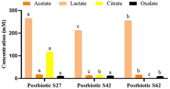
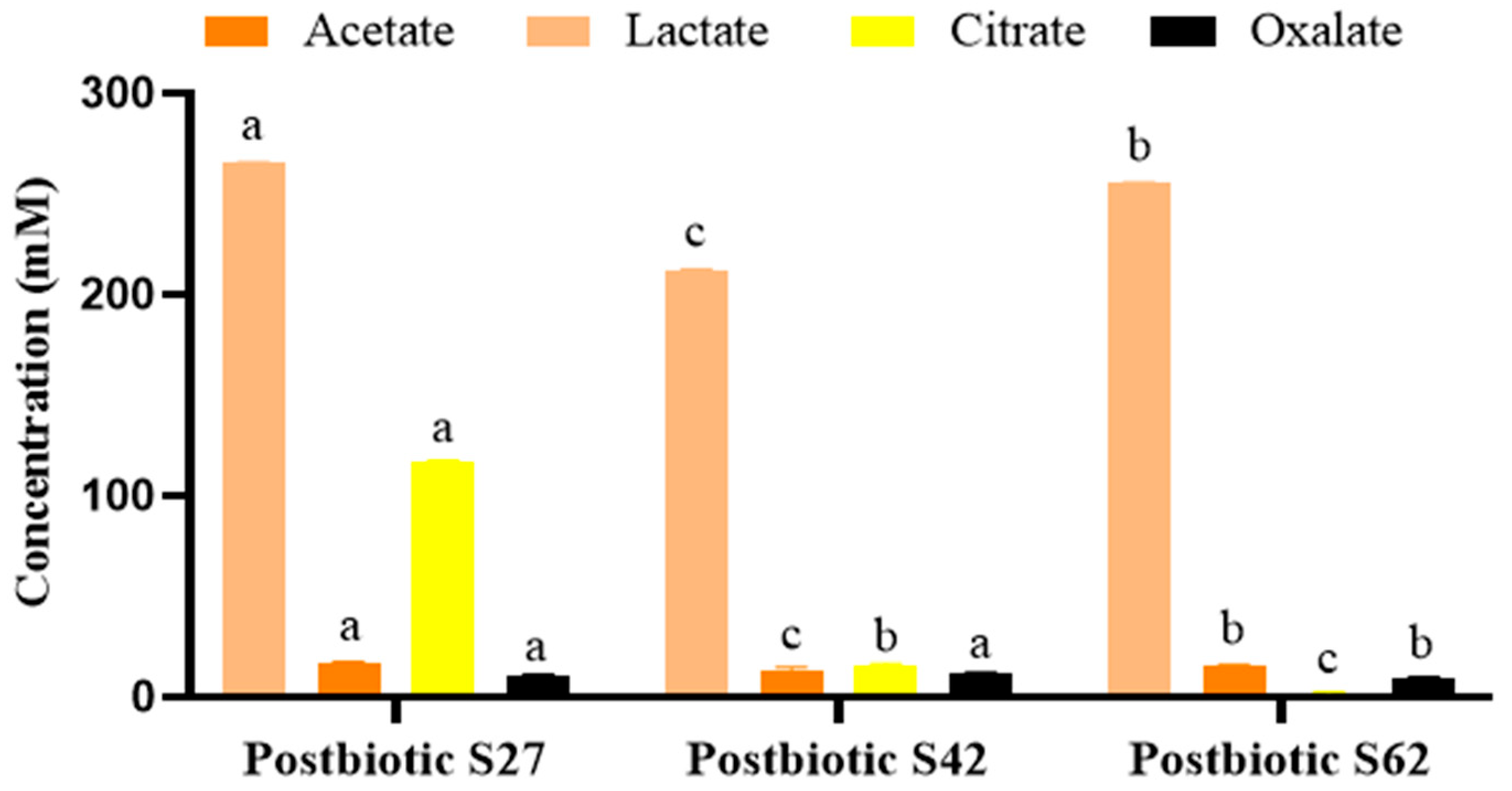
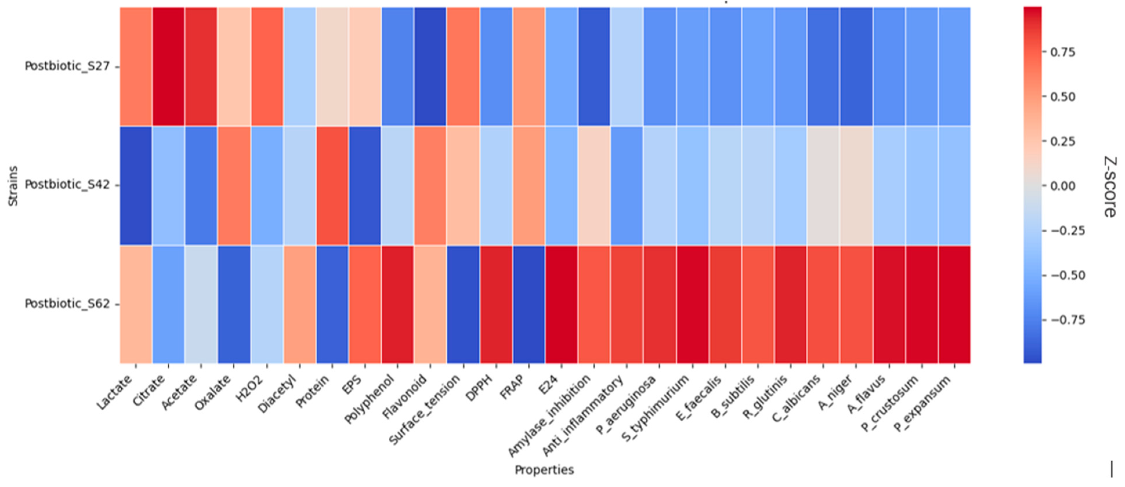
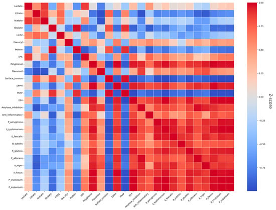
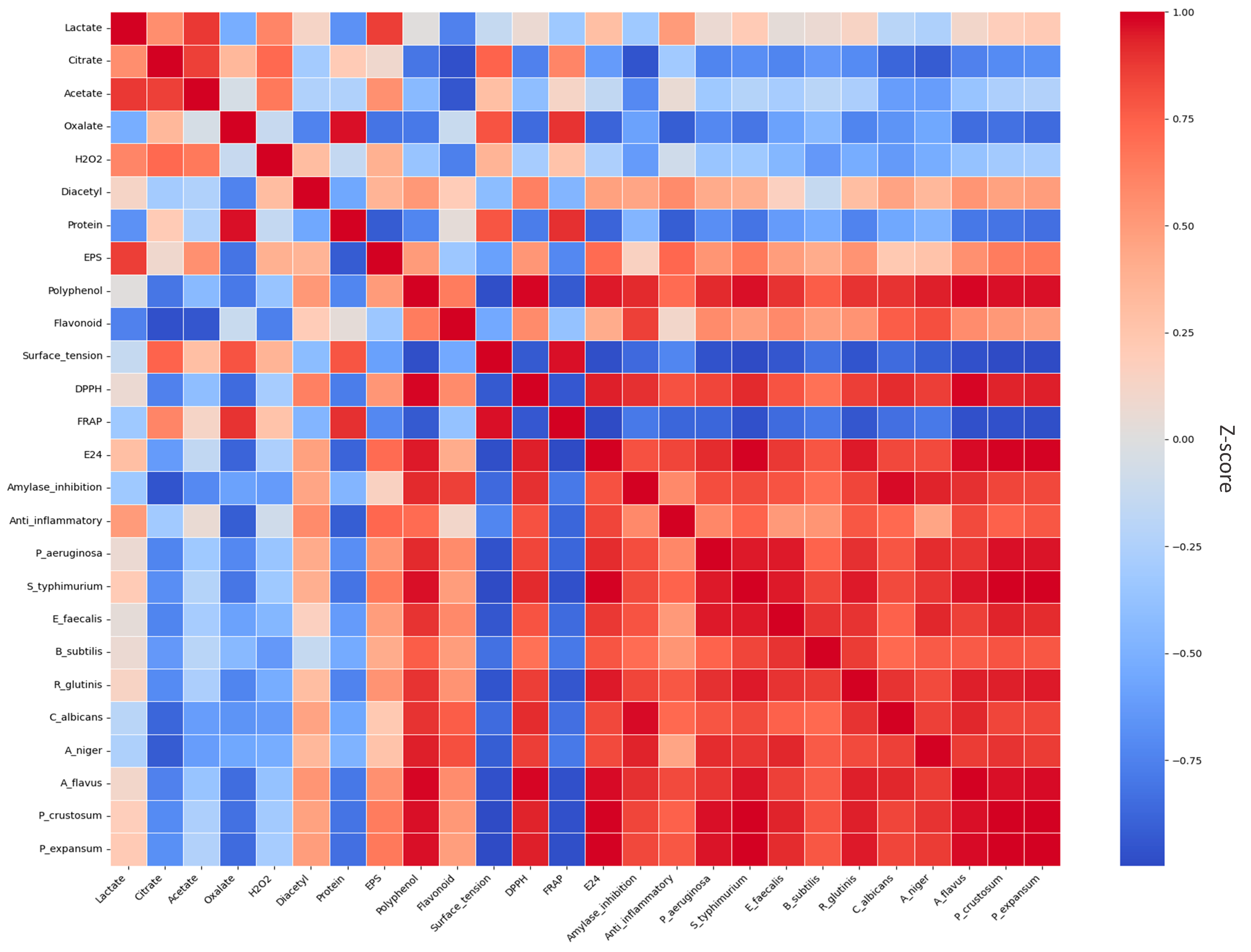
The characterization of the metabolite profile of postbiotics was performed using modern instruments and colorimetric methods. The results of the organic acid and short-chain fatty acid (SCFA) quantification by HPLC in the postbiotics from Levilactobacillus and Lactiplantibacillus strains are indicated in Figure 1. Lactate, citrate, and oxalate are organic acids, while acetate is a short-chain fatty acid (SCFA). Organic acids and short-chain fatty acid (SCFA) profiles across all postbiotics were significantly (p < 0.05) dominated by lactate, accompanied by lower concentrations of citrate, acetate, and oxalate. Lactate was the predominant organic acid produced in postbiotic-CFS S27, postbiotic-CFS S42, and postbiotic-CFS S62, with concentrations of 265.46 mM, 211.82 mM, and 255.28 mM, respectively. Citrate was produced at a high concentration (116.92 mM) in postbiotic S27. Moreover, low concentrations of acetate and oxalate were produced in all postbiotics, ranging between 13.73–17.49 mM and 9.30–11.53 mM, respectively. Acetate concentrations detected in postbiotic-CFSs were lower than the initial concentration in uninoculated MRS broth, indicating metabolic consumption of acetate by L. brevis S27, L. pentosus S42, and L. plantarum S62 during fermentation. Conversely, postbiotic-CFS S27 exhibited a high citrate concentration, which may be due to citrate synthesis and accumulation or co-elution of polar compounds. The results of other metabolites produced are presented in Table 1. The protein contents of postbiotic-CFSs S27, S42, and S62 were 177.16 µg/mL, 192.42 µg/mL, and 154.65 µg/mL, respectively. Moreover, the exopolysaccharide (EPS) concentrations were 535.27 mg/L, 244 mg/L, and 600.62 mg/L in postbiotic-CFSs S27, S42, and S62, respectively. The hydrogen peroxide and diacetyl concentrations were detected in low concentrations, 0.0070–0.0074 µg/mL and 0.43–0.59 µg/mL, respectively (Table 1), and no significant difference (p > 0.05) was observed among all postbiotic-CFSs. On the other hand, Polyphenol and flavonoid concentrations were 386.00 µg GAE/mL, 407.60 µg GAE/mL, and 451.93 µg GAE/mL; and 83.66 µg QuE/mL, 114.51 µg QuE/mL, and 109.73 µg QuE/mL in postbiotic-CFSs S27, S42, and S62, respectively (Table 1). Furthermore, an investigation into biosurfactant production was conducted using several methods, and the results are summarized in Table 2. The results showed the ability of all strains to produce biosurfactants in all postbiotics, based on an oil displacement test with diameters between 7.1 mm and 10.3 mm, a Parafilm M Test, a drop collapse test (microliter plates), and a glass slide test (Table 2). In addition, the surface tensions of postbiotic-CFS S27, S42, S62, and uninoculated MRS are 38.3 mN/m, 37.45 mN/m, 34.35 mN/m, and 40.65 mN/m, respectively, indicating a higher biosurfactant concentration in postbiotic S62 compared to postbiotic-CFSs S27 and S42 (Table 2). Reuterin was not detected in the postbiotics from any of these LAB strains (Table 1). The heatmap presents the correlation analysis between postbiotics and their bioactive profiling (Figure 2). Postbiotic S27 showed significantly higher concentrations of organic acids, particularly lactate and citrate, indicating a metabolically active profile. In contrast, postbiotic S42 exhibited an intermediate profile, combining high EPS production and protein content, suggesting interest for applications involving cell adhesion, mucosal protection, and some technological properties. However, the bioactive profile of postbiotic S62 suggests that it is particularly rich in proteins, polyphenols, and antimicrobial molecules, which is consistent with strong biotechnological potential.
Figure 1.
Organic acids and short-chain fatty acid production in postbiotic-CFSs derived from Levilactobacillus and Lactiplantibacillus strains. a,b,c Means in the same postbiotic-CFS at each parameter with different lowercase letters differed significantly (p < 0.05).
Table 1.
Profiling of bioactive metabolites in postbiotic-CFSs derived from Levilactobacillus and Lactiplantibacillus strains.
Table 2.
Biosurfactant production in postbiotic-CFSs derived from Levilactobacillus and Lactiplantibacillus strains.
Figure 2.
Heatmap of the association between postbiotic-CFSs derived from Levilactobacillus and Lactiplantibacillus strains and their bioactive profiling and functional properties. Color intensity reflects the normalized feature value (z-score) for each parameter across strains.
2.2. Antimicrobial Activity of Postbiotic-CFS
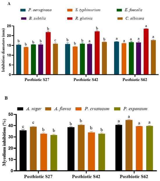
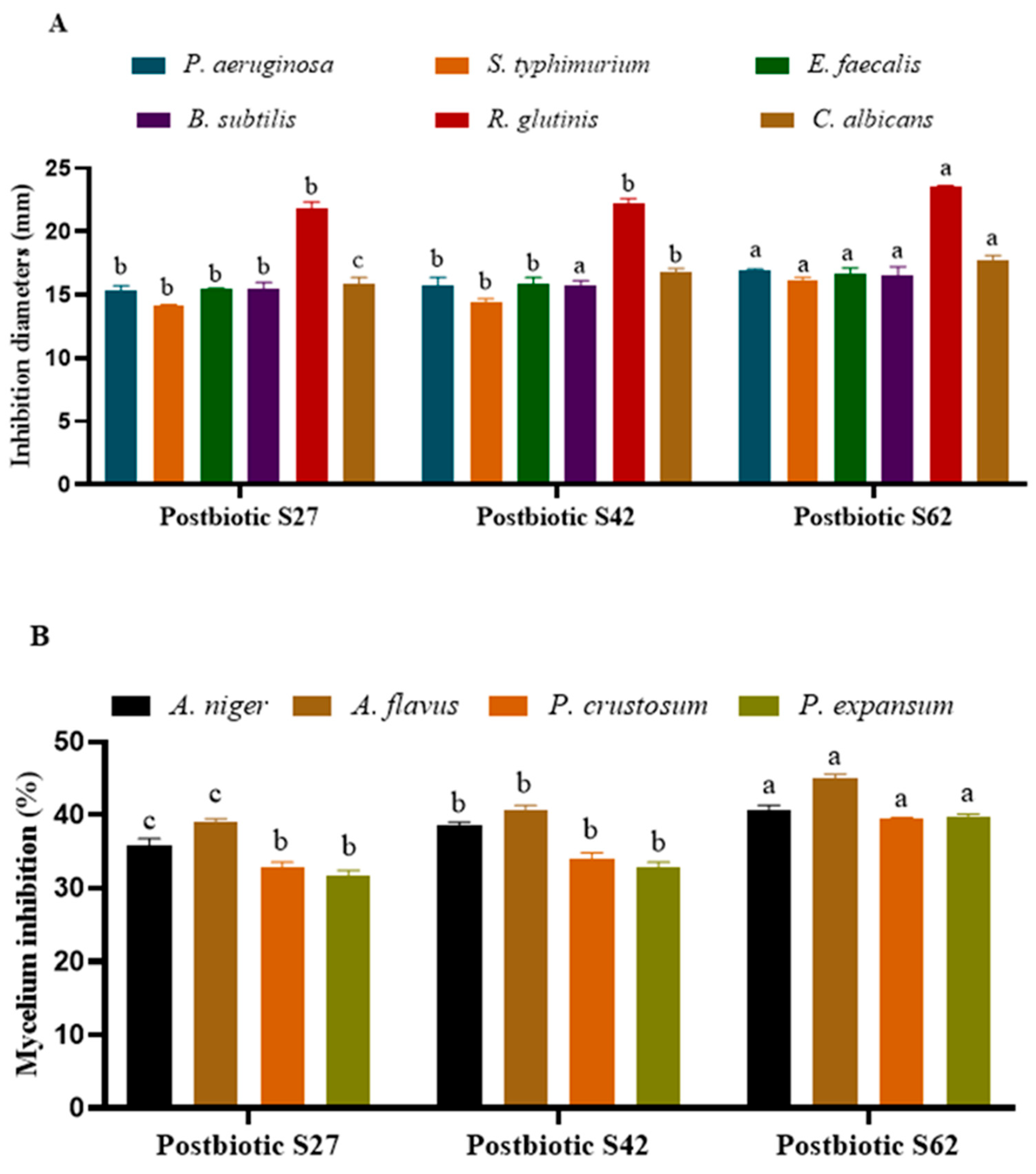
The results of the antimicrobial activity of postbiotics against pathogenic and spoilage microorganisms are presented in Figure 3. All of the postbiotic-CFSs exhibited antibacterial activity against foodborne pathogenic bacteria, displaying inhibition zone diameters of 15.4–16.5 mm against B. subtilis, 15.4–16.7 mm against E. faecalis, 14.10–16.15 mm against S. typhimurium, and 15.30–16.9 mm against P. aeruginosa (Figure 3A). On the other hand, all postbiotics demonstrated high antifungal activity against R. glutinis and C. albicans, with the inhibition diameter ranging between 21.85 and 23.50 mm and 15.8 and 17.7 mm, respectively (Figure 3A). Moreover, the postbiotic-CFSs presented mycelium inhibition against A. niger, A. flavus, P. crustosum, and P. expansum, with the percentage ranging between 35.77 and 40.56%, 39.10 and 45.09%, 32.78 and 39.57%, and 31.67 and 39.70%, respectively (Figure 3B). Furthermore, the postbiotic-CFS S62 produced by L. plantarum S62 demonstrated the highest (p < 0.05) antimicrobial activity against pathogenic and spoilage microorganisms. In addition, postbiotic-CFS S62 exhibited significantly higher antimicrobial activity compared to S27 and S42 (Figure 2). These antimicrobial activities were positively associated with certain metabolites, highlighting their potential contribution to inhibitory effects against pathogenic and spoilage microorganisms (foodborne pathogenic bacteria, yeast, and molds) (Figure 4).
Figure 3.
Antimicrobial activities of postbiotic-CFSs derived from Levilactobacillus and Lactiplantibacillus strains; Inhibition diameter (mm) (A); Mycelium inhibition (%) (B). a,b,c Means in the same postbiotic-CFS at each parameter with different lowercase letters differed significantly (p < 0.05).
Figure 4.
Pearson correlation matrix of bioactive profiling and functional properties of postbiotic-CFSs derived from three Levilactobacillus and Lactiplantibacillus strains.
2.3. Antioxidant Activity of Postbiotic-CFS
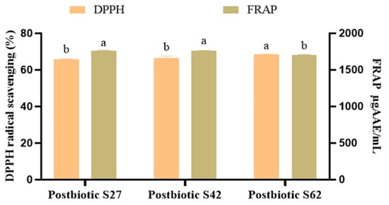
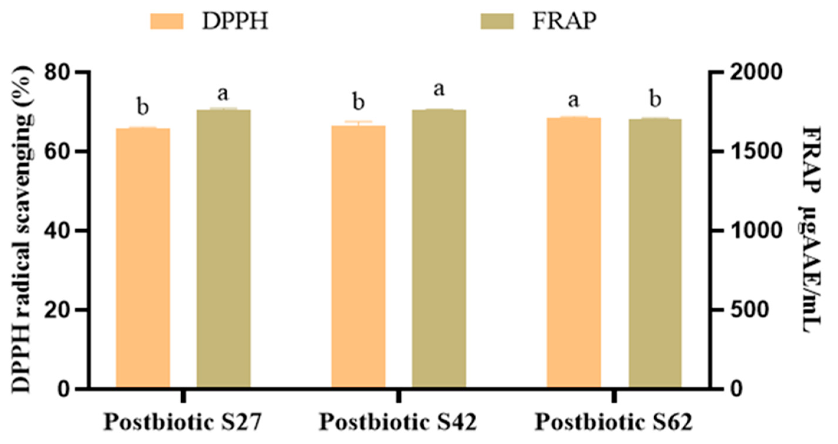
The results of DPPH and FRAP assays of the postbiotic-CFSs are presented in Figure 5. The DPPH radical scavenging activities were 65.91%, 66.63%, and 68.55% for postbiotic-CFSs S27, S42, and S62, respectively. The FRAP values of postbiotic-CFSs S27, S42, and S62 were 1765 µgAAE/mL, 1764.23 µgAAE/mL, and 1707.31 µgAAE/mL, respectively. Moreover, postbiotic-CFS S62 exhibited strong DPPH radical scavenging activity (68.55%) and was significantly higher (p < 0.05) compared to S27 and S42. However, postbiotic-CFSs S27 and S42 were significantly higher (p < 0.05) compared to S62 in terms of the FRAP assay. Furthermore, postbiotic-CFSs S27 and S62 exhibited strong antioxidant activities (DPPH and FRAP) (Figure 2) and positively correlated with polyphenol and flavonoid contents (Figure 4), suggesting a potential contribution of these metabolites to the antioxidant properties.
Figure 5.
Antioxidant activities of postbiotic-CFSs derived from Levilactobacillus and Lactiplantibacillus strains. a,b Means in the same postbiotic-CFS at each parameter with different lowercase letters differed significantly (p < 0.05).
2.4. Anti-Inflammatory Activity of Postbiotic-CFSs
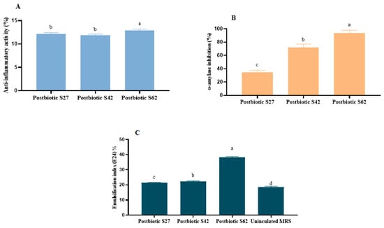
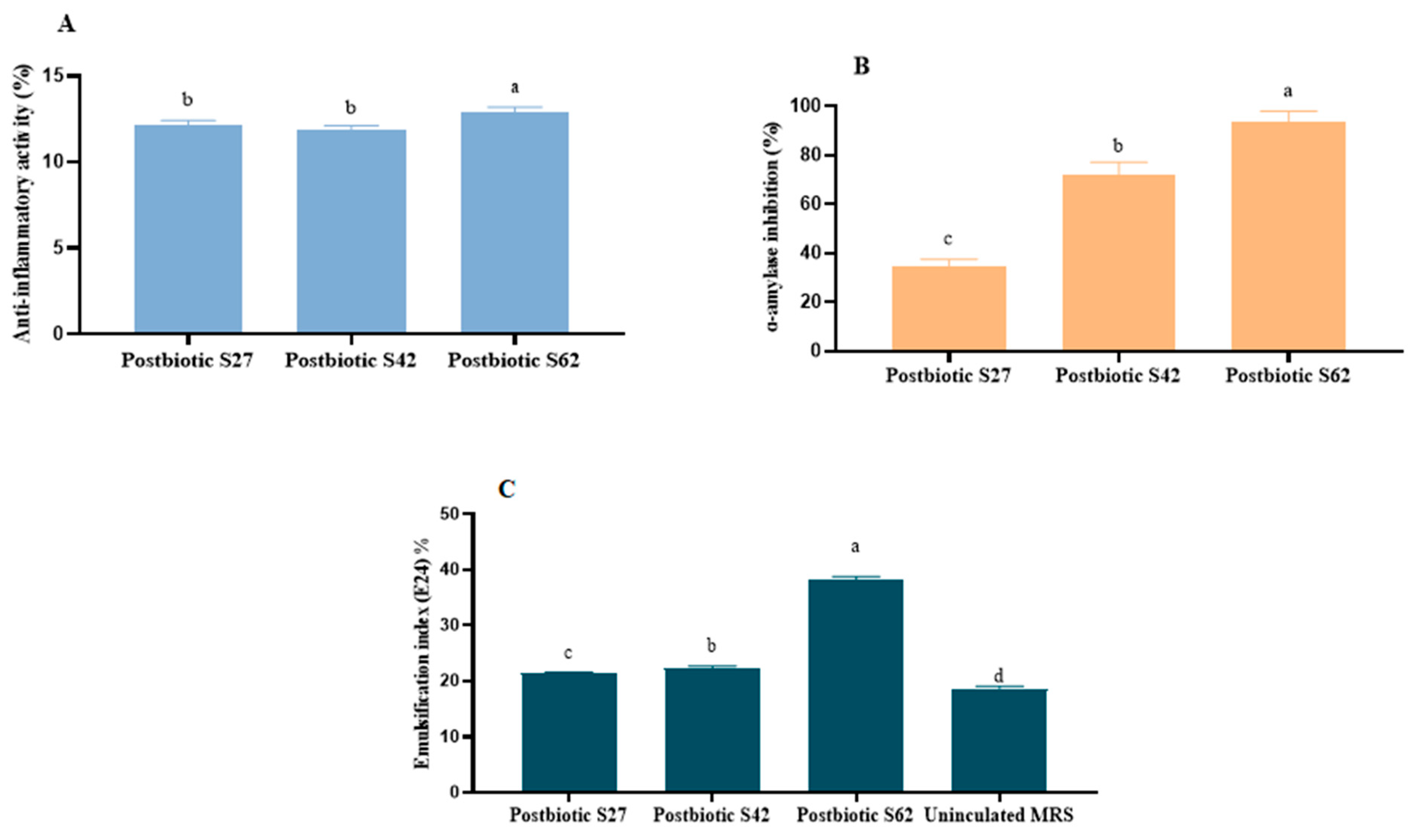
The anti-inflammatory activity of postbiotic-CFSs was evaluated by the inhibitory effect on protein (albumin) denaturation in vitro, and the results are summarized in Figure 6A. All of the postbiotic-CFSs from the LAB strains showed anti-inflammatory activity, with a percentage around 12%, and the anti-inflammatory activity of postbiotic-CFS S62 was significantly higher (p < 0.05) than that of postbiotics S27 and S42. Moreover, Figure 2 indicates that the molecules associated with the antioxidant effect could also contribute to the modulation of the inflammatory response.
Figure 6.
Functional properties of postbiotic-CFSs derived from Levilactobacillus and Lactiplantibacillus strains. Anti-inflammatory inhibition (A); α amylase inhibition (B); and emulsification index (E24%) (C). a–d Means in the same postbiotic-CFS at each parameter with different lowercase letters differed significantly (p < 0.05).
2.5. Antidiabetic Activity of Postbiotic-CFSs
The in vitro antidiabetic potential of postbiotic-CFSs was evaluated using the α-amylase inhibition assay, and the results are presented in Figure 6B. All of the postbiotic-CFSs demonstrated significant α-amylase inhibition, with percentages of 34.38%, 71.88%, and 93.75% for postbiotic-CFSs S27, S42, and S62, respectively. Moreover, postbiotic-CFS S62 had significantly higher (p < 0.05) inhibition, with a high α-amylase inhibition of more than 93%.
2.6. Emulsification Index (E24%) Activity of Postbiotic-CFSs
The results of the emulsification index (E24%) activity of all of the postbiotic-CFSs are presented in Figure 6C. The E24% showed percentages of 21.5%, 22.3%, and 38.2% for postbiotic-CFSs S27, S42, and S62, respectively. Uninoculated MRS broth displayed an E24% of 18.75% attributable to Tween 80 content. Moreover, the emulsification index (E24%) of postbiotic-CFS S62 was significantly higher (p < 0.05) compared with S27 and S42 (Figure 6C). In addition, the emulsification index (E24%) showed a strong positive correlation with exopolysaccharide production, indicating its role in emulsification capacity. Conversely, surface tension showed a negative correlation with E24%, confirming biosurfactant activity (Figure 4).
2.7. Principal Component Analysis (PCA)
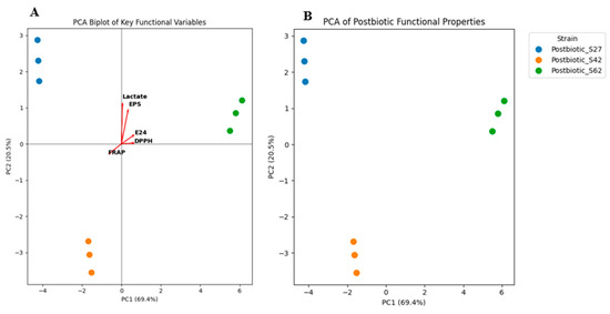
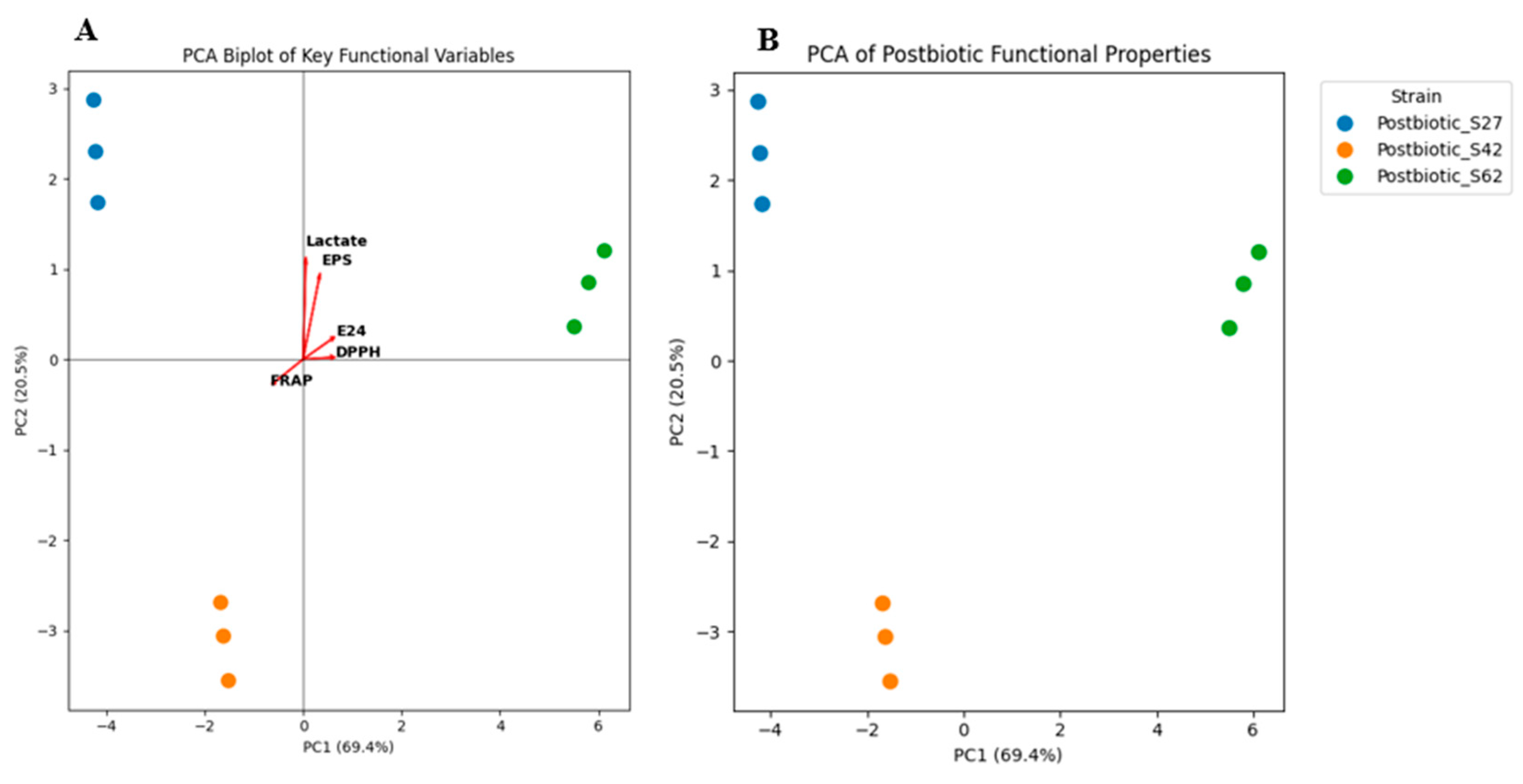
Principal component analysis (PCA) was performed to explore the overall variation and the relationships between the different functional properties of postbiotics and to highlight the similarities or differences between the three strains studied (postbiotic-CFS S27, postbiotic-CFS S42 and postbiotic-CFS S62) (Figure 7). While PCA explains most of the total variance, these multivariate statistical analyses were performed on a sample size of three strains. Consequently, these models serve primarily as exploratory comparative tools to highlight strain-specific profile clustering. PCA explains 89.9% of the total variance, with the first principal component (PC1) of 69.4% and the second component (PC2) accounting for 20.5%, indicating that these two components effectively summarize the variability of the data.
Figure 7.
Principal component analysis (PCA) biplot illustrating the contribution of key functional variables (A) and the distribution of postbiotics based on their bioactive profiling and functional properties (B).
The biplot (Figure 7A) simultaneously represents the samples and the functional variables, thus providing a better understanding of the variables contributing to the observed separation among the postbiotic-CFSs. Postbiotic-CFS S62 was associated with lactate, DPPH, EPSs, and E24, which exhibited high values of these properties, while S42 showed higher FRAP activity. However, postbiotic-CFS S42 had lower overall organic acid levels, and showed a distinct profile enriched in proteinaceous components and total flavonoids, which are important in terms of overall functional properties. This finding highlights that postbiotic-CFS S27 is linked with fermentation metabolites and postbiotic-CFS S62 with EPSs, antioxidant and emulsifying properties, while S42 presents an intermediate functional profile.
The PCA score plot revealed a clear separation of the three postbiotic-CFSs along PC1 and PC2 (Figure 7B). Postbiotic-CFS S27 clustered in the positive PC2 region and was located on the opposite side of postbiotic-CFS S62, indicating that this postbiotic-CFS is primarily characterized by a relatively high amount of lactate and EPSs, with a lower contribution to DPPH and E24%, suggesting a focus on fermentative rather than antioxidant properties. Furthermore, postbiotic-CFS S62 clustered on the positive side for both PC1 and PC2 and was distinctly separated from S27 and S42, indicating substantial differences in its functional properties. This postbiotic is strongly associated with EPSs, lactate, antioxidant activities, and bioactive compounds. Thus, postbiotic-CFS S62 appears to have the most important functional profile. Conversely, postbiotic-CFS S42 occupied the negative region of both PC1 and PC2 and an intermediate position between the two strains, and this position reflects a less favorable functional profile, making it the least robust of the three studied. Consequently, these results indicate that the separation of strains in the PCA space is driven by differences in both bioactive profiling and functional properties.
3. Discussion
Postbiotic-CFSs derived from Levilactobacillus and Lactiplantibacillus strains showed the ability to produce organic acids and SCFAs, and this suggests that they are produced simultaneously during fermentative metabolism. Our findings were consistent with previous studies, which demonstrate that lactic acid was the predominant organic acid in postbiotic-CFSs, and the other organic acids and SCFAs were present in variable quantities [8,9,21]. In addition, SCFAs and organic acids exhibit various benefits in food products and have therapeutic potential, such as by contributing to the inhibition of pathogen proliferation, enhancing the aroma and flavor of fermented food products, and modulating systemic immune responses [7,21]. Moreover, our results show the presence of various bioactive metabolites with different concentrations. Previous studies demonstrate the presence of several bioactive compounds in postbiotics, including organic acids, fatty acids, protein compounds, vitamins, hydrogen peroxide alcohols, phenolic acids, and flavonoids [8,9,22], and the studies conducted by Ghasemi et al. [23] and Lara et al. [24] showed the ability of LAB to produce biosurfactants with a reduction of the surface tension mN/m. Overall, the presence of various metabolites in postbiotic-CFSs facilitates their application in several food products, enhancing their functional, sensorial, and bio-preservative properties, as well as in vitro therapeutic potential, while offering numerous health benefits for human consumers.
Postbiotic-CFSs exhibited high antimicrobial activities against foodborne pathogenic bacteria, yeast, and molds. These results agree with those of previous studies, which present the ability of postbiotic-CFSs to inhibit the growth of Gram-negative and Gram-positive pathogenic bacteria and biofilm formation [25,26,27]. Also, the postbiotic-CFS from L. plantarum exhibited antibacterial activity in vitro and in vivo against foodborne pathogens [28]. These findings demonstrate the possibility of applying postbiotic-CFSs as a therapeutic approach against infections and as an alternative to antibiotics, reducing the transmission of resistance genes to humans. Postbiotics were used to control opportunistic fungal spoilage and infections, promote anti-aflatoxigenic effects [17,29,30], and modulate the gut microbiota [31]. In our previous study focused on the antimicrobial activity of Levilactobacillus and Lactiplantibacillus strains, we demonstrate that the neutralization of CFS showed a reduction in antimicrobial activity, while the activity completely disappeared after treatment with proteolytic enzymes, which confirms the contribution of bioactive proteins to this antimicrobial activity [32]. Moreover, the antimicrobial activities of all of the postbiotic-CFSs were positively associated with several metabolites. Additionally, the antimicrobial activity of postbiotics (CFSs) is due to proteinaceous compounds acting in synergy with organic acids and other compounds [33]. Based on our previous findings regarding using CFSs against R. glutinis and P. aeruginosa [32], the inhibition diameters were aligned with the values reported in our previous study, and the antimicrobial spectrum was expanded in this study to include foodborne pathogens and spoilage microorganisms. In fact, the synergistic potential of all bioactive molecules present in postbiotic-CFSs contributes to inhibitory activity against various microorganisms, leading to the development of natural bio-preservatives and active food packaging to extend food shelf life.
All of the postbiotic-CFSs showed good antioxidant activities. The postbiotics demonstrated varied and strong antioxidant activities, including DPPH, FRAP, ABTS, and others [9,25,34]. Moreover, the antioxidant properties are related to the various bioactive metabolites, including total phenolic acid and total soluble sugar [34], short-chain fatty acids [35], exopolysaccharides [36], and bioactive peptides [37]. In the present study, the antioxidant activities of postbiotic-CFS S27 and S62 were linked to polyphenol and flavonoid contents (electron-donating bioactive metabolites), suggesting their contribution in vitro to radical-scavenging and reducing activities.
The inhibition of protein denaturation represents a standard preliminary screening for anti-inflammatory potential in vitro. Postbiotic-CFSs showed inflammatory properties through the inhibition of protein denaturation. Postbiotics exhibit in vitro functional potential by reducing inflammation in tissues and organs [38], and are used in the management of chronic inflammation [39]. Moreover, previous studies demonstrate good anti-inflammatory activity of postbiotics, and the inhibition of albumin denaturation has been related to a dose-dependent anti-inflammatory response [9,34]. Consequently, our postbiotic-CFSs exhibited a modest protective effect and were able to influence the immune response by reducing inflammation.
Postbiotics demonstrate antidiabetic potential through the inhibition of α-amylase activity. The α-amylase enzyme is associated with carbohydrate hydrolysis, releasing glucose into the bloodstream, and inhibiting its function is necessary to regulate blood sugar levels. Postbiotics present the greatest inhibition of α-amylase activity, and this activity is due to bioactive peptides [40,41]. In the present study, the correlation between different bioactive molecules and α-amylase inhibition suggests an interaction between enzymatic activities and bioactive metabolite production. These findings demonstrate in vitro inhibition of α-amylase, suggesting potential for delaying carbohydrate digestion and supporting the use of postbiotic-CFSs as a natural alternative in vitro for controlling diabetes and as an antidiabetic agent in the formulation of pharmaceutical products.
The results showed that all postbiotic-CFSs presented emulsification activity, and the uninoculated MRS showed an E24% of 18.75% and a surface tension of 40.65 mN/m. Various studies demonstrate that emulsification activity, measured by the emulsification index (E24%), is linked to biosurfactant and exopolysaccharide production [24,42]. These findings exhibited the potential uses of these postbiotic-CFSs rich in EPSs and biosurfactants as emulsifying agents in the formulation of food products.
4. Materials and Methods
4.1. Probiotic Lactic Acid Bacteria Strains
Three lactic acid bacteria (Levilactobacillus brevis S27, Lactiplantibacillus pentosus S42, and Lactiplantibacillus plantarum S62) were used to produce postbiotic-CFSs. These strains were previously isolated from fermented green olives and characterized for their probiotic properties [43] and maintained in De Man, Rogosa, and Sharpe (MRS) medium (Biokar, France) at −20 °C, supplemented with 20% (v/v) glycerol. They were cultured in MRS medium at 37 °C for 48 h before use.
4.2. Production and Preparation of Postbiotic Cell-Free Supernatants (Postbiotic-CFSs)
Postbiotic cell-free supernatants (postbiotic-CFSs) were obtained from LAB fermentations in MRS broth after 48 h at 37 °C. Following the incubation period, postbiotics were prepared and obtained by centrifugation of the culture at 10,000× g for 10 min at 4 °C, and then filter sterilization was performed using 0.22 μm pore size membrane filters (MF-Millipore, Merck, Darmstadt, Germany). The final pH values recorded were 3.95 for postbiotic-CFS S27, 3.9 for S42, and 3.8 for S62.
4.3. Profiling of Bioactive Metabolites from Postbiotic-CFSs
4.3.1. Organic Acids and Short-Chain Fatty Acid (SCFA) Quantification by HPLC
The short-chain fatty acids were determined in the sterilized postbiotics from LAB strains using an HPLC system (Waters, Milford, MA, USA) equipped with a 1525 binary pump, 2489 refractometer, and Rezex-ROA-Organic acid H+ column (7.8 mm × 30 cm, Phenomenex, Torrance, CA, USA). Twenty microliters of the CFS was injected into the HPLC, and the elution was carried out at 0.6 mL/min using 0.005 N sulfuric acid at 40 °C. The calibration curves for lactate, acetate, citrate, butyrate, malate, and oxalate were used as standards, and the analysis was conducted in triplicate.
4.3.2. Hydrogen Peroxide Quantification
Analysis of hydrogen peroxide in postbiotics was determined using the method of the AOAC [44]. The mixture of postbiotics with diluted sulphuric acid in equal volumes of 25 mL was titrated with potassium permanganate (0.1 M) until discoloration. Each milliliter of potassium permanganate was equivalent to 1.070 mg of hydrogen peroxide production, and uninoculated MRS broth was used as a control. The test was carried out in triplicate.
4.3.3. Diacetyl Quantification
The production of diacetyl in postbiotics was determined according to the method of the AOAC [44]. First, 25 mL of postbiotics was mixed with 7.5 mL of hydroxylamine solution (1 M). The mixture was titrated with 0.1 M HCl, using bromophenol blue as an indicator, to a greenish-yellow endpoint. The equivalence factor of HCl to diacetyl is 21.52 mg, and uninoculated MRS broth was used as a control. The experiment was conducted in triplicate.
4.3.4. Protein Contents
The protein content was estimated using the Bradford method [45]. A mixture of 200 µL of Bradford reagent (kit of Bio-Rad, Hercules, CA, USA) and 800 µL of each postbiotic sample was used to determine the protein concentrations at 595 nm, and uninoculated MRS broth was used as a control. Bovine serum albumin (Sigma-Aldrich, St. Louis, MO, USA) was used as a standard to determine the protein concentration in the samples. The experiment was conducted in triplicate.
4.3.5. Reuterin Quantification
Reuterin production in postbiotics was determined using the colorimetric method of Circle, et al. [46]. The mixture of 1 mL of each sample of postbiotics and 0.75 mL of a 10 mM tryptophan solution (dissolved in 0.05 M HCl) was added to 3 mL of 37% HCl and incubated at 37 °C for 20 min. Acrolein (Sigma-Aldrich) was used as a standard solution and prepared in MRS broth medium with concentrations ranging from 0.05 to 6 mM. The absorbance of all samples was recorded at 560 nm, and the measurement was evaluated in triplicate.
4.3.6. Exopolysaccharide (EPS) Quantification
Quantification of exopolysaccharide production in postbiotics was conducted using the protocol of Wu, et al. [47], with some modifications. The postbiotics were treated with a 4% (v/v) solution of 85% trichloroacetic acid (TCA) to remove the proteins. After centrifugation at 4 °C at 10,000 rpm for 30 min at 4 °C, EPSs were precipitated by adding twice the amount of absolute cold ethanol and maintaining it at 4 °C for 24 h, followed by 30 min of centrifugation at 4 °C, 10,000 rpm. The pellet was then redissolved in distilled water. EPS production was quantified by measuring the mass of lyophilized powder obtained per liter of culture medium (mg/L). Uninoculated MRS broth was used as a control, and all tests were conducted in triplicate.
4.3.7. Total Phenolic and Flavonoid Contents
The total phenolic content of postbiotics was determined in samples using the Folin-Ciocalteu method [48] and was expressed in Gallic acid equivalents, using a Gallic acid (20–500 µg/mL) standard calibration curve. The total flavonoid content was determined using the aluminum chloride colorimetric method using Quercetin (5–250 µg/mL) as a standard, and the results were expressed as µg QuE/mL [49]; uninoculated MRS broth was used as a control. All tests were performed in triplicate.
4.3.8. Biosurfactants Analysis
The biosurfactant production of postbiotics was evaluated using different qualitative tests. The tests included drop collapse, oil dispersion, Parafilm M, and microliter plates [23,50]. Uninoculated MRS broth without Tween 80 was used as a control, and all tests were performed in triplicate.
4.4. Antimicrobial Activity of Postbiotic-CFSs
4.4.1. Antimicrobial Activity Using the Well Diffusion Assay
The antimicrobial activity of postbiotics derived from Levilactobacillus and Lactiplantibacillus strains was determined using a well diffusion assay. For this assay, 105 CFU/mL of pathogenic bacteria (Salmonella typhimurium, P. aeruginosa, Enterococcus faecalis, or Bacillus subtilis) or yeast (Rhodotorula glutinis UPM22 or Candida albicans) was evenly spread on Mueller–Hinton (MH) broth (Biokar, France) or YEGP broth. The wells, cut in the agar using a sterile filter pipette tip, were charged with 100 μL of the postbiotic CFS, and the 1 mg/mL each of gentamicin and cycloheximide were used as controls against bacteria and yeast respectively. After incubation for 24 h at 37 °C/25 °C for bacteria and yeast, respectively; the inhibition zones were determined in mm. All tests were conducted in triplicate.
4.4.2. Antifungal Activity
The antifungal activity of postbiotics was evaluated by the mycelium inhibition of fungi (Aspergillus niger, Aspergillus flavus, Penicillium crustosum, and Penicillium expansum). For this purpose, 10% (v/v) of each postbiotic-CFS derived from the LAB strains was added to the PDA medium and inoculated with 105 spores/mL of fungi, and the medium with 10% (v/v) of uninoculated MRS broth was used as a control. All tests were incubated at 25 °C for 5 days, and the mycelium inhibition was calculated in triplicate using the following formula:
Mycelium inhibition (%) = (1 − (Fungal colony diameter (mm) in assay)/(Fungal colony diameter (mm) in control)) × 100
4.5. Antioxidant Activity of Postbiotic-CFSs
4.5.1. DPPH Radical Scavenging Assay
The free radical-scavenging capacity of the postbiotic was evaluated using the 2,2-diphenyl-1-picrylhydrazyl (Sigma-Aldrich, USA) assay with gallic acid as the standard. Briefly, 100 µL of postbiotic was mixed with 1.0 mL of a 0.2 mM DPPH solution prepared in methanol. The mixtures were incubated at room temperature in the dark for 30 min, and the absorbance was measured at 517 nm. Uninoculated MRS broth was used as a control, using ascorbic acid (25–1000 µg/mL) as a standard. DPPH radical scavenging activity was performed in triplicate and calculated as the percentage inhibition.
DPPH radical scavenging (%) = (1 − (Absorbance of sample − Absorbance of blank)/(Absorbance of control)) × 100
4.5.2. Ferric Reducing Antioxidant Power (FRAP) Assay
The ferric reducing antioxidant power (FRAP) assay of all postbiotics was performed following the method described by Ghabbour et al. [51], with some modifications. First, 1 mL of each postbiotic was mixed with 3 mL FRAP reagent, incubated in the dark for 4 min, and the absorbance was measured at 700 nm. The results of the FRAP assay were expressed as µg of ascorbic acid equivalents per milliliter of solution (µgAAE/mL) using ascorbic acid (25–1000 µg/mL) as a standard, and uninoculated MRS broth was used as a control. All experiments were performed in triplicate.
4.6. Anti-Inflammatory Activity of Postbiotic-CFSs
The anti-inflammatory activity of the postbiotics was performed in vitro using the protein (albumin) denaturation inhibitory activity described by Tarannum et al. [52], with some modifications. First, 1 mL of each postbiotic was mixed with 1 mL of BSA (1% in Tris-HCl 0.05 M, pH 6.6) and incubated in a 37 °C water bath for 15 min, followed by an additional incubation at 70 °C for 5 min. After cooling on ice for 5 min, the absorbance of the mixture was recorded at 660 nm. The calibration curve of diclofenac sodium (25–400 µg/mL) was used as a standard, and uninoculated MRS broth was used as a control. The percentage of protein denaturation inhibition was determined in triplicate and calculated using the following formula:
Anti-inflammatory activity (%) = (1 − (Absorbance of sample − Absorbance of blank)/(Absorbance of control)) × 100
4.7. Antidiabetic Activity of Postbiotic-CFSs
The antidiabetic activity of the postbiotics was evaluated using α amylase inhibitory activity, with minor modifications [40]. Briefly, 250 μL of postbiotics was added to 250 μL of 0.1 M PBS (containing 0.5 mg/mL of α-amylase solution, pH 6.4) and incubated at 25 °C for 10 min. Then, 250 μL of starch solution (1% w/v in 0.02 M sodium phosphate buffer) was added to the reaction mixture and incubated at 25 °C for 10 min. The reaction mixture was stopped using 500 μL of the DNS reagent incubated in a boiling water bath for 5 min. The reaction mixture was then cooled, and 10 mL of distilled water was added. The calibration curve of Acarbose (25–500 µg/mL) was used as a standard and uninoculated MRS broth was used as a control. The absorbance was measured at 540 nm, and the percent inhibition was calculated as follows:
α-amylase Inhibitory Activity (%) = (1 − (Absorbance of sample − Absorbance of blank)/(Absorbance of control)) × 100
4.8. Emulsification Index (E24%) of Postbiotic-CFSs
The Emulsification activity of postbiotics was detected by the addition of 2 mL of motor oil to an equal volume of each postbiotic, mixed with a vortex for 2 min, and allowed to stand for 24 h. Distilled water was used as a negative control and the uninoculated MRS as a positive control. The E24% was calculated by measuring the height of the emulsified layer (mm) using the following formula:
Emulsification index (E24%) = ((Height of the emulsion layer)/(Total height)) × 100
4.9. Statistical Analysis
Statistical analyses were conducted via Python (v3.12) followed by one-way ANOVA, and the GraphPad Prism 8 software (San Diego, CA, USA) was used to generate the figures. The data were displayed as means ± standard deviation, and Tukey’s HSD post hoc test was applied, which compared the means with a significant difference (p < 0.05).
5. Conclusions
This study investigates the bioactive profiling and in vitro multifunctional properties of postbiotic cell-free supernatants produced by three LAB strains. The postbiotic-CFSs obtained from three different probiotics Levilactobacillus brevis S27, Lactiplantibacillus pentosus S42, and Lactiplantibacillus plantarum S62, demonstrate various bioactive molecules. The evidence of antimicrobial, antioxidant, anti-inflammatory, antidiabetic, and emulsifying activities of these postbiotics highlights their multipurpose applications across various domains. Therefore, these postbiotic-CFSs are suitable for use as a natural biopreservative agent in food products and could be used in functional foods to improve consumer health and as next-generation therapeutics. However, further studies (e.g., in vivo) are needed to confirm the beneficial effects and overall properties of postbiotic-CFSs for food and therapeutic applications.
Author Contributions
Conceptualization, Methodology, Investigation, Formal Analysis, Data Curation, Writing—Original Draft, Writing—Review and Editing, H.A.; Formal Analysis, Data Curation, I.M.; Methodology, A.B.; Methodology, N.M.; Methodology, M.B.; Formal Analysis, Data Curation, N.E.B.; Writing—Review and Editing, N.G.; Writing—review & editing, F.M.; Writing—Review and Editing, S.K. All authors have read and agreed to the published version of the manuscript.
Funding
This research received no external funding.
Institutional Review Board Statement
Not applicable.
Informed Consent Statement
Not applicable.
Data Availability Statement
Data will be made available upon request.
Acknowledgments
The authors would like to thank all those who contributed indirectly to the development of this work.
Conflicts of Interest
The authors declare no conflicts of interest.
Abbreviations
| CFS | Cell-free supernatant |
| DPPH | 2,2-diphényl-1-picrylhydrazyle |
| EPS | Exopolysaccharide |
| FRAP | Ferric reducing antioxidant power |
| GRAS | Generally recognized as safe |
| HPLC | High-performance liquid chromatography |
| ISAPP | International scientific association for probiotics and prebiotics |
| LAB | Lactic acid bacteria |
| SCFA | Short-chain fatty acids |
References
- Akpoghelie, P.O.; Edo, G.I.; Ali, A.B.M.; Yousif, E.; Zainulabdeen, K.; Owheruo, J.O.; Isoje, E.F.; Igbuku, U.A.; Essaghah, A.E.A.; Makia, R.S.; et al. Lactic acid bacteria: Nature, characterization, mode of action, products and applications. Process Biochem. 2025, 152, 1–28. [Google Scholar] [CrossRef] [Scilit]
- Shi, X.; Li, Y.; Cheng, Y.; Liu, W.; Li, Z.; Jin, H.; Kwok, L.-Y.; Sun, Z. Emerging roles of lactic acid bacteria in health management: Insights from fermented foods to microbiota. Food Biosci. 2026, 75, 108088. [Google Scholar] [CrossRef] [Scilit]
- Abouloifa, H.; Gaamouche, S.; Ghabbour, N.; El Guerrouj, B.; Karboune, S.; Saalaoui, E.; Asehraou, A. Lactic acid bacteria from Moroccan traditional foods: Techno-functional, health-promoting, nutraceutical value and application as a starter and bio-preservative agent in the food products. Bioresour. Technol. Rep. 2024, 27, 101941. [Google Scholar] [CrossRef] [Scilit]
- Mafe, A.N.; Sharifi-Rad, J.; Calina, D.; Ogunyemi, A.J.; Tubi, A.O. Postbiotics in Functional Foods: Microbial Derivatives Shaping Health, Immunity and Next-Generation Nutrition. Food Front. 2026, 7, e70205. [Google Scholar] [CrossRef] [Scilit]
- Pattapulavar, V.; Ramanujam, S.; Kini, B.; Christopher, J.G. Probiotic-derived postbiotics: A perspective on next-generation therapeutics. Front. Nutr. 2025, 12, 1624539. [Google Scholar] [CrossRef] [Scilit] [PubMed]
- Gurunathan, S.; Thangaraj, P.; Kim, J.-H. Postbiotics: Functional Food Materials and Therapeutic Agents for Cancer, Diabetes, and Inflammatory Diseases. Foods 2024, 13, 89. [Google Scholar] [CrossRef] [Scilit] [PubMed]
- Salminen, S.; Collado, M.C.; Endo, A.; Hill, C.; Lebeer, S.; Quigley, E.M.M.; Sanders, M.E.; Shamir, R.; Swann, J.R.; Szajewska, H.; et al. The International Scientific Association of Probiotics and Prebiotics (ISAPP) consensus statement on the definition and scope of postbiotics. Nat. Rev. Gastroenterol. Hepatol. 2022, 19, 649–667. [Google Scholar] [CrossRef] [Scilit] [PubMed]
- Jalali, S.; Mojgani, N.; Sanjabi, M.R.; Saremnezhad, S.; Haghighat, S. Functional properties and safety traits of L. rhamnosus and L. reuteri postbiotic extracts. AMB Express 2024, 14, 114. [Google Scholar] [CrossRef] [Scilit] [PubMed]
- Davarzani, S.; Mojgani, N.; Sanjabi, M.; Mirdamadi, S.; Soltani, M.; Ashique, S. Functional Properties, Metabolic Profile and Safety Attributes of Postbiotics from Lacticaseibacillus acidophilus and Lactiplantibacillus plantarum. Appl. Food Res. 2025, 5, 101412. [Google Scholar] [CrossRef] [Scilit]
- Amobonye, A.; Pillay, B.; Hlope, F.; Asong, S.T.; Pillai, S. Postbiotics: An insightful review of the latest category in functional biotics. World J. Microbiol. Biotechnol. 2025, 41, 293. [Google Scholar] [CrossRef] [Scilit] [PubMed]
- Chen, X.; Yuan, C.; He, J.; Li, W.; Liao, C. Current research status and trends in the bioactivity of postbiotics. Front. Food Sci. Technol. 2025, 5, 1692683. [Google Scholar] [CrossRef] [Scilit]
- Meena, K.K.; Joshi, M.; Gupta, L.; Meena, S. Comprehensive insights into postbiotics: Bridging the gap to real-world application. Food Nutr. 2025, 1, 100024. [Google Scholar] [CrossRef] [Scilit]
- Pawar, S.V.; Saldinger, S.S.; Shemesh, M. Postbiotics for improving food quality and safety: From mechanistic insights to industrial applications. LWT-Food Sci. Technol. 2026, 241, 119066. [Google Scholar] [CrossRef] [Scilit]
- Erfanian, N.; Nasseri, S.; Miraki Feriz, A.; Safarpour, H.; Namaei, M.H. Characterization of Wnt signaling pathway under treatment of Lactobacillus acidophilus postbiotic in colorectal cancer using an integrated in silico and in vitro analysis. Sci. Rep. 2023, 14, 22988. [Google Scholar] [CrossRef] [Scilit] [PubMed]
- Heniedy, A.M.; Mahdy, D.M.; Abo Elenien, W.I.; Mourad, S.; El-Kadi, R.A. Postbiotics as a health-promoting technique: A review article on scientific and commercial interest. Process Biochem. 2024, 144, 6–19. [Google Scholar] [CrossRef] [Scilit]
- Kavita; Om, H.; Chand, U.; Kushawaha, P.K. Postbiotics: An alternative and innovative intervention for the therapy of inflammatory bowel disease. Microbiol. Res. 2024, 279, 127550. [Google Scholar] [CrossRef] [Scilit] [PubMed]
- Divyashree, S.; Shruthi, B.; Vanitha, P.R.; Sreenivasa, M.Y. Probiotics and their postbiotics for the control of opportunistic fungal pathogens: A review. Biotechnol. Rep. 2023, 38, e00800. [Google Scholar] [CrossRef] [Scilit] [PubMed]
- Moradi, M.; Mardani, K.; Tajik, H. Characterization and application of postbiotics of Lactobacillus spp. on Listeria monocytogenes in vitro and in food models. LWT-Food Sci. Technol. 2019, 111, 457–464. [Google Scholar] [CrossRef] [Scilit]
- Awad, N.M.H.; Capozzi, V.; Guillard, V.; Goksen, G. An innovative approach for probiotics and postbiotics-based antimicrobial packaging systems: Design, characterization, and benefit for food application. Curr. Opin. Food Sci. 2025, 64, 101315. [Google Scholar] [CrossRef] [Scilit]
- Ghamry, M.; Ibrahim, F.; Al-Maqtari, Q.A.; Siddiquy, M.; Zhang, H.; Zhao, W.; Li, L.; Li, J. Development of postbiotic-enriched chitosan films as a novel approach for sustainable food packaging. Food Hydrocoll. 2025, 168, 111515. [Google Scholar] [CrossRef] [Scilit]
- Chang, H.M.; Foo, H.L.; Loh, T.C.; Lim, E.T.C.; Abdul Mutalib, N.E. Comparative Studies of Inhibitory and Antioxidant Activities, and Organic Acids Compositions of Postbiotics Produced by Probiotic Lactiplantibacillus plantarum Strains Isolated From Malaysian Foods. Front. Vet. Sci. 2020, 7, 602280. [Google Scholar] [CrossRef] [Scilit] [PubMed]
- Rahman, M.M.; Sazili, A.Q.; Ahmad, S.A.; Khalil, K.A.; Ismail-Fitry, M.R.; Afsana, A.S.; Sarker, M.S.K. Fatty acid profile and bio-preservative efficacy of lactic acid bacteria postbiotics for improving microbiological safety in broiler meat. Food Control 2026, 181, 111769. [Google Scholar] [CrossRef] [Scilit]
- Ghasemi, A.; Moosavi-Nasab, M.; Setoodeh, P.; Mesbahi, G.; Yousefi, G. Biosurfactant Production by Lactic Acid Bacterium Pediococcus dextrinicus SHU1593 Grown on Different Carbon Sources: Strain Screening Followed by Product Characterization. Sci. Rep. 2020, 10, 5287. [Google Scholar] [CrossRef] [Scilit] [PubMed]
- Lara, V.M.; Vallejo, M.; Parada, R.; Henao Ossa, J.S.; Gliemmo, M.F.; Campos, C.A. Characterization of the emulsifying activity of biosurfactants produced by lactic acid bacteria isolated from the Argentinian Patagonia. J. Dispers. Sci. Technol. 2020, 43, 902–909. [Google Scholar] [CrossRef] [Scilit]
- Jalali, S.; Mojgani, N.; Haghighat, S.; Sanjabi, M.R.; Sarem-Nezhad, S. Investigation of antimicrobial and antioxidant properties of postbiotics produced by Lactobacillus rhamnosus and Limosilactobacillus reuteri and their potential application in surface decontamination of red meat. LWT-Food Sci. Technol. 2024, 209, 116758. [Google Scholar] [CrossRef] [Scilit]
- Kim, H.-J.; Youn, H.-Y.; Moon, J.-S.; Kim, H.; Seo, K.-H. Comparative anti-microbial and anti-biofilm activities of postbiotics derived from kefir and normal raw milk lactic acid bacteria against bovine mastitis pathogens. LWT-Food Sci. Technol. 2024, 191, 115699. [Google Scholar] [CrossRef] [Scilit]
- Rahman, M.M.; Sazili, A.Q.; Ahmad, S.A.; Khalil, K.A.; Ismail-Fitry, M.R.; Sarker, M.S.K. Inhibitory efficacy, production dynamics, and characterization of postbiotics of lactic acid bacteria. BMC Microbiol. 2025, 25, 485. [Google Scholar] [CrossRef] [Scilit] [PubMed]
- Khorshidi, S.; Mehdizadeh, T.; Tajik, H.; Hamishekar, H.; Reale, A. Characterization of Lactiplantibacillus plantarum subsp. plantarum and Bifidobacterium animalis spp. lactis BB-12 postbiotics: In vivo and in vitro experiments against foodborne pathogens. LWT-Food Sci. Technol. 2025, 224, 117823. [Google Scholar] [CrossRef] [Scilit]
- Tutuk, Z.; Karatepe, P.; Akgöl, M.; Bayrak, S.; Vurmaz, M.; Fidan, Ş.; Hayaloğlu, A.A.; İncili, G.K. Inhibition of Aspergillus parasiticus and detoxification of aflatoxin derivatives in tomato paste by adding freeze-dried postbiotic from Lactiplantibacillus plantarum. Food Control 2026, 180, 111664. [Google Scholar] [CrossRef] [Scilit]
- Vega-Vasconez, A.; Castillo-Patino, D.L.; Garza-Cervantes, J.A.; Santacruz, A.; Morones-Ramirez, J.R. Probiotic and Postbiotic Interactions of Lactobacillus Strains with Candida albicans: Antifungal Effects Through Microbial Competition. Antibiotics 2026, 15, 279. [Google Scholar] [CrossRef] [Scilit] [PubMed]
- Garcia-Gamboa, R.; Perfecto-Avalos, Y.; Gonzalez-Garcia, J.; Alvarez-Calderon, M.J.; Gutierrez-Vilchis, A.; Garcia-Gonzalez, A. In vitro analysis of postbiotic antimicrobial activity against Candida Species in a minimal synthetic model simulating the gut mycobiota in obesity. Sci. Rep. 2024, 14, 16760. [Google Scholar] [CrossRef] [Scilit] [PubMed]
- Abouloifa, H.; Gaamouche, S.; Ghabbour, N.; Hasnaoui, I.; Bentouhami, N.E.; Rokni, Y.; Karboune, S.; Asehraou, A. Characterization, Production, and Application of Antifungal Metabolites from Probiotic Levilactobacillus and Lactiplantibacillus Strains Isolated from Fermented Olives. Fermentation 2025, 11, 661. [Google Scholar] [CrossRef] [Scilit]
- Abouloifa, H.; Rokni, Y.; Hasnaoui, I.; Bellaouchi, R.; Gaamouche, S.; Ghabbour, N.; Karboune, S.; Ben Salah, R.; Brasca, M.; D’Hallewin, G.; et al. Characterization of antimicrobial compounds obtained from the potential probiotic Lactiplantibacillus plantarum S61 and their application as a biopreservative agent. Braz. J. Microbiol. 2022, 53, 1501–1513. [Google Scholar] [CrossRef] [Scilit] [PubMed]
- Tong, Y.; Guo, H.; Abbas, Z.; Zhang, J.; Wang, J.; Cheng, Q.; Peng, S.; Yang, T.; Bai, T.; Zhou, Y.; et al. Optimizing postbiotic production through solid-state fermentation with Bacillus amyloliquefaciens J and Lactiplantibacillus plantarum SN4 enhances antibacterial, antioxidant, and anti-inflammatory activities. Front. Microbiol. 2023, 14, 1229952. [Google Scholar] [CrossRef] [Scilit] [PubMed]
- Vera-Santander, V.E.; Mani-López, E.; López-Malo, A.; Jiménez-Munguía, M.T. Use of whey for a sustainable production of postbiotics with potential bioactive metabolites. Sustain. Food Technol. 2024, 2, 1101–1112. [Google Scholar] [CrossRef] [Scilit]
- Pandey, K.; Prakash, T.V.G.; Gosai, H.; Bhatia, D.; Patel, R. Microbial exopolysaccharides as postbiotics: Structure, function, and translational potential. Int. J. Biol. Macromol. 2025, 330, 147956. [Google Scholar] [CrossRef] [Scilit] [PubMed]
- Grigore-Gurgu, L.; Cotârleț, M.; Pihurov, M.; Păcularu-Burada, B.; Vasile, A.M.; Enachi, E.; Chițescu, C.L.; Petre, B.A.; Dumitrașcu, L.; Borda, D.; et al. Lactiplantibacillus plantarum and Lactiplantibacillus paraplantarum postbiotics: Assessment of the biotic-derived metabolites with cytocompatibility and antitumoral potential. Food Biosci. 2024, 59, 103863. [Google Scholar] [CrossRef] [Scilit]
- Bodjrenou, D.M.; Dhital, S.; Li, W.; Zeng, H.; Huang, Q.; Wang, Q.; Mackay, C.R.; Zhang, B. Promises and Challenges of Postbiotics: From Bioactive Mechanisms to Functional Food Applications. Food Wellness 2026, 2, 100041. [Google Scholar] [CrossRef] [Scilit]
- Abbas, A.; Weiskirchen, R.; Bilal, M.; Afzal, M.K.; Malik, A.; Akhtar, S.; Khan, M.; Rashid, I.; Khalid, F.; Akram, S. Postbiotics as Next Generation Biotherapeutics Targeting the Gut–Immune–Metabolic Axis: An Integrative Review. Pharmaceuticals 2026, 19, 1184. [Google Scholar] [CrossRef] [Scilit] [PubMed]
- Azadikhah, F.; Khalesi, M.; Shad, E.; Majdinasab, M.; Razmjooei, M.; Eskandari, M.H. Inhibition of α-amylase and lipase by probiotic fermented milk-derived bioactive peptides: A comprehensive in vitro investigation. LWT-Food Sci. Technol. 2025, 215, 117208. [Google Scholar] [CrossRef] [Scilit]
- Kinariwala, D.; Panchal, G.; Sakure, A.; Hati, S. Exploring the Potentiality of Lactobacillus Cultures on the Production of Milk-Derived Bioactive Peptides with Antidiabetic Activity. Int. J. Pept. Res. Ther. 2019, 26, 1613–1627. [Google Scholar] [CrossRef] [Scilit]
- Gayathiri, E.; Prakash, P.; Pratheep, T.; Ramasubburayan, R.; Thirumalaivasan, N.; Gaur, A.; Govindasamy, R.; Rengasamy, K.R.R. Bio surfactants from lactic acid bacteria: An in-depth analysis of therapeutic properties and food formulation. Crit. Rev. Food Sci. Nutr. 2024, 64, 10925–10949. [Google Scholar] [CrossRef] [Scilit] [PubMed]
- Abouloifa, H.; Rokni, Y.; Bellaouchi, R.; Ghabbour, N.; Karboune, S.; Brasca, M.; Ben Salah, R.; Chihib, N.E.; Saalaoui, E.; Asehraou, A. Characterization of Probiotic Properties of Antifungal Lactobacillus Strains Isolated from Traditional Fermenting Green Olives. Probiotics Antimicrob. Proteins 2020, 12, 683–696. [Google Scholar] [CrossRef] [Scilit] [PubMed]
- AOAC. Official Methods of Analysis; Methods; The Association of Official Analytical Chemists: Gaithersburg, MD, USA, 2000. [Google Scholar]
- Bradford, M.M. A rapid and sensitive method for the quantitation of microgram quantities of protein utilizing the principle of protein-dye binding. Anal. Biochem. 1976, 72, 248–254. [Google Scholar] [CrossRef] [PubMed]
- Circle, S.J.; Stone, L.; Boruff, C. Acrolein determination by means of tryptophane. A colorimetric micromethod. Ind. Eng. Chem. Anal. Ed. 1945, 17, 259–262. [Google Scholar] [CrossRef] [Scilit]
- Wu, J.; Li, Z.; Zhang, Z.; Zhang, J.; Hu, H.; Lan, H.; Hong, W.; Yang, Z. Characterization of a postbiotic exopolysaccharide produced by Lacticaseibacillus paracasei ET-22 with antioxidant and anti-inflammatory efficacy. Int. J. Biol. Macromol. 2025, 306, 141608. [Google Scholar] [CrossRef] [Scilit] [PubMed]
- Kocabey, N.; Yilmaztekin, M.; Hayaloglu, A.A. Effect of maceration duration on physicochemical characteristics, organic acid, phenolic compounds and antioxidant activity of red wine from Vitis vinifera L. Karaoglan. J. Food Sci. Technol. 2016, 53, 3557–3565. [Google Scholar] [CrossRef] [Scilit] [PubMed]
- Syed Salleh, S.N.A.; Mohd Hanapiah, N.A.; Ahmad, H.; Wan Johari, W.L.; Osman, N.H.; Mamat, M.R. Determination of Total phenolics, flavonoids, and antioxidant activity and GC-MS analysis of Malaysian stingless bee propolis water extracts. Scientifica 2021, 2021, 3789351. [Google Scholar] [CrossRef] [Scilit] [PubMed]
- Uyar, E.; Saglam, O. Isolation, screening and molecular characterization of biosurfactant producing bacteria from soil samples of auto repair shops. Arch. Microbiol. 2021, 203, 4929–4939. [Google Scholar] [CrossRef] [Scilit] [PubMed]
- Ghabbour, I.; El Aboui, F.Z.; Bentouhami, N.E.; Ghabbour, N.; Khabbach, A.; Abouloifa, H.; Asehraou, A.; Louahlia, S.; Hammani, K. Sustainable use of cultivated and wild capers in Morocco: A comparative study of physicochemical, microbiological, and functional properties during the natural fermentation process. Sustainability 2026, 18, 4371. [Google Scholar] [CrossRef] [Scilit]
- Tarannum, N.; Hossain, T.J.; Ali, F.; Das, T.; Dhar, K.; Nafiz, I.H. Antioxidant, antimicrobial and emulsification properties of exopolysaccharides from lactic acid bacteria of bovine milk: Insights from biochemical and genomic analysis. LWT-Food Sci. Technol. 2023, 186, 115263. [Google Scholar] [CrossRef] [Scilit]

Disclaimer/Publisher’s Note: The statements, opinions and data contained in all publications are solely those of the individual author(s) and contributor(s) and not of MDPI and/or the editor(s). MDPI and/or the editor(s) disclaim responsibility for any injury to people or property resulting from any ideas, methods, instructions or products referred to in the content. |
© 2026 by the authors. Licensee MDPI, Basel, Switzerland. This article is an open access article distributed under the terms and conditions of the Creative Commons Attribution (CC BY) license.