


Abstract
Cellular senescence is characterized by stable cell-cycle arrest and a senescence-associated secretory phenotype (SASP). Recurrent desiccating, oxidative, ultraviolet, microbial, and immune-mediated insults may induce stress-induced premature senescence (SIPS) in ocular surface tissues. PubMed and Web of Science were searched from database inception to 30 June 2026. Primary human, animal, and in vitro studies were prioritized, and direct evidence of senescence was distinguished from indirect stress-related or SASP-like associations. The p53–p21CIP1–pRB and p16INK4a–pRB pathways mediate senescence-associated cell-cycle arrest. Reactive oxygen species (ROS)–p38 mitogen-activated protein kinase (p38 MAPK) and cyclic GMP–AMP synthase–stimulator of interferon genes (cGAS–STING) signaling act as stress-sensing and inflammatory pathways, whereas mechanistic target of rapamycin (mTOR) and AMP-activated protein kinase (AMPK) contribute to SASP and metabolic regulation. Transient senescence may support wound repair and immune clearance, whereas persistent senescence may contribute to epithelial and endothelial dysfunction, tear-film instability, immune dysregulation, fibrosis, and extracellular matrix remodeling. Mechanistic support is strongest in selected experimental models; evidence in several human ocular surface disorders remains associative. Cellular senescence is a context-dependent contributor to ocular surface pathology. Senolytic and senomorphic strategies remain predominantly preclinical and require disease-, cell-, and stage-specific validation.
1. Introduction
The ocular surface is an integrated functional unit comprising the tear film, cornea, conjunctiva, limbus, lacrimal and meibomian glands, eyelids, and associated neural and immune components [1,2]. Its direct interface with the external environment exposes it to recurrent desiccating, oxidative, ultraviolet, microbial, mechanical, and immune-mediated stressors [3]. Disruption of this homeostasis contributes to ocular surface diseases (OSDs), including dry eye disease, meibomian gland dysfunction, blepharitis, keratoconjunctival disorders, ocular graft-versus-host disease, conjunctivochalasis, and pterygium [3,4]. Because senescence-associated changes may affect different corneal compartments, this review also includes keratoconus, Fuchs endothelial corneal dystrophy, and corneal allograft rejection as selected conditions with direct, supportive, or indirect evidence [5]. This broader cornea–ocular surface framework permits comparison across epithelial, stromal, endothelial, glandular, and immune compartments.
Cellular senescence is generally defined as a stable state of proliferative arrest accompanied by distinct molecular and secretory changes [6,7]. Under certain conditions, external stimuli such as oxidative stress, deoxyribonucleic acid (DNA) damage, and radiation can prematurely activate intracellular senescence cascades, forcing cells to cease division and enter a senescent state; this process is referred to as stress-induced premature senescence (SIPS) [8,9].
Senescent cells develop a senescence-associated secretory phenotype (SASP), characterized by secretion of cytokines, chemokines, growth factors, and extracellular matrix (ECM)-degrading proteases. The SASP can maintain the senescent state and induce senescence in neighboring healthy cells through paracrine signaling [10,11,12]. Although ocular surface tissues possess features of immune privilege, chronic exposure to environmental and immune stressors can trigger SIPS, allowing senescent epithelial, stromal, endothelial, glandular, and immune cells to propagate bystander senescence and reshape the local microenvironment [13]. Previous reviews examined cellular senescence in ocular aging or across a broad range of ocular diseases [13,14]. This review focuses on SIPS in corneal and ocular surface homeostasis and disease. It integrates cell-type-specific mechanisms, SASP-mediated immune and stromal remodeling, and disease-specific evidence grading to assess the rationale and limitations of senescence-targeted therapy.
2. Literature Search and Evidence Evaluation
This narrative review was informed by a structured search of PubMed and Web of Science from database inception to 30 June 2026. Search terms combined “cellular senescence”, “stress-induced premature senescence”, “senescence-associated secretory phenotype” or “SASP” with terms related to the cornea and ocular surface, including “cornea”, “ocular surface”, “dry eye”, “Fuchs endothelial corneal dystrophy”, “ocular graft-versus-host disease”, “corneal transplantation”, “keratoconus”, “pterygium”, and “conjunctivochalasis”.
Titles and abstracts were screened for relevance, potentially eligible articles were assessed in full text, and reference lists of relevant articles were checked for additional studies. Disease-specific evidence evaluation focused on primary human, animal, and in vitro studies examining cellular senescence or potentially related stress responses in corneal or ocular surface disorders. Selected reviews and meta-analyses were included only to provide epidemiological or conceptual context and were not considered direct evidence of cellular senescence. Non-ocular studies were cited selectively to discuss general senescence mechanisms or candidate therapeutic pathways but were excluded from disease-specific evidence grading.
Evidence was qualitatively categorized as direct, supportive, or indirect. Direct evidence was based on complementary features of cellular senescence, particularly stable cell-cycle arrest or reduced proliferative capacity together with established senescence markers and, when available, SASP characterization or functional manipulation. Supportive evidence included senescence-associated markers or pathways without full phenotypic confirmation, whereas indirect evidence comprised oxidative stress, inflammatory or SASP-like changes, or tissue remodeling without direct demonstration of senescent-cell accumulation or stable cell-cycle arrest.
3. Mechanisms of Cellular Senescence
3.1. Senescence-Inducing Stressors
Cellular senescence can be induced by a variety of intrinsic and extrinsic stressors, including oxidative stress, DNA damage, telomere dysfunction, mitochondrial dysfunction, ultraviolet radiation, desiccation, and persistent inflammatory or immune-mediated injury. These stressors are particularly relevant to stress-induced premature senescence (SIPS), in which sustained cellular damage activates stress-response pathways that ultimately promote stable cell-cycle arrest and broader senescence-associated phenotypic changes. Among these upstream responses, reactive oxygen species (ROS), the DNA damage response (DDR), and stress-responsive kinases such as p38 mitogen-activated protein kinase (p38 MAPK) serve as important links between cellular injury and downstream senescence programs [8,15,16,17,18,19].
Oxidative stress is particularly relevant to ocular tissues because of their continuous exposure to environmental stress and their high metabolic demand. Excessive ROS can induce DNA damage, impair mitochondrial function, destabilize telomeres, and activate p38 MAPK and p53-related stress responses [9,16,17,19]. Persistent DNA damage activates the DDR, which can promote cell-cycle arrest while also engaging inflammatory signaling pathways associated with senescence [18,19]. In dry eye disease (DED), desiccating and oxidative stress are associated with epithelial dysfunction, inflammatory signaling, and p38 MAPK-dependent activation of matrix metalloproteinase-9 (MMP-9) [20,21]. Although increased p38 MAPK activity and MMP-9 expression alone do not establish cellular senescence, they provide a mechanistic link between chronic ocular-surface stress and pathways capable of promoting a senescence-associated state. In conjunctivochalasis (CCH), more direct evidence supports oxidative stress-induced senescence of conjunctival fibroblasts through the ROS–p38 MAPK–p53–p21CIP1–pRB pathway [22]. Ultraviolet exposure and chronic oxidative stress are also prominent features of pterygium, whereas persistent inflammation and immune-mediated tissue injury may contribute to senescence-associated changes in ocular graft-versus-host disease (oGVHD). In Fuchs endothelial corneal dystrophy (FECD), oxidative stress, mitochondrial dysfunction, and DNA damage have likewise been implicated in corneal endothelial cell dysfunction and senescence-related phenotypes [22,23,24,25,26].
Persistent DNA damage and mitochondrial dysfunction may additionally promote the accumulation of cytosolic DNA. Nuclear DNA released from damaged nuclei or micronuclei, as well as mitochondrial DNA released from dysfunctional mitochondria, can activate cyclic GMP–AMP synthase–stimulator of interferon genes (cGAS–STING) signaling. Activation of this pathway promotes type I interferon and NF-κB-associated inflammatory responses and therefore provides an additional mechanistic connection between intracellular damage sensing, inflammation, and cellular senescence [17,27,28]. However, current evidence for cGAS–STING-mediated senescence is derived predominantly from general or non-ocular models, and its direct contribution to ocular-surface senescence remains to be established.
3.2. Cell-Cycle Arrest Effector Axes
Stable cell-cycle arrest is a defining feature of cellular senescence and is mediated primarily by the p53–p21CIP1–pRB and p16INK4a–pRB pathways [6,29]. DNA damage, oxidative stress, telomere dysfunction, and replication stress can activate the DNA damage response (DDR) and downstream p53 signaling, thereby increasing p21CIP1 expression [9,19,30,31,32,33]. p21CIP1 inhibits cyclin-dependent kinase (CDK) activity, reduces pRB phosphorylation, and maintains pRB in a hypophosphorylated state that suppresses E2F-dependent transcription and blocks G1/S progression. Persistent oxidative or genotoxic stress can further reinforce this response through p38 MAPK signaling.
The p16INK4a–pRB pathway is another major effector of senescence-associated cell-cycle arrest. p16INK4a inhibits CDK4/6, maintains pRB in its growth-suppressive hypophosphorylated form, and prevents E2F-mediated entry into S phase. Although the p53–p21CIP1–pRB and p16INK4a–pRB pathways converge on pRB-dependent cell-cycle control, their relative contributions and temporal dynamics vary by cell type, senescence inducer, and disease context [34,35].
In ocular tissues, these arrest pathways link chronic stress to functional decline [13,14]. Increased CDKN1A expression has been reported in Fuchs endothelial corneal dystrophy (FECD)-related corneal endothelial models, increased p16INK4a protein expression occurs in chronic ocular graft-versus-host disease (oGVHD) tissues, and p53–p21CIP1–pRB signaling has been implicated in oxidative stress-induced senescence of conjunctivochalasis (CCH) fibroblasts [22,23,24,25,26]. These findings indicate that the dominant effector pathway may differ across ocular cell types and disease conditions.
3.3. SASP Regulation and Immune Interactions
In addition to stable cell-cycle arrest, senescent cells undergo extensive changes in secretion. The SASP is regulated by interconnected signaling networks, among which p38 MAPK and nuclear factor kappa B (NF-κB) are major transcriptional regulators. Persistent stress and DNA damage can activate NF-κB-dependent programs and promote expression of SASP-associated factors [8,15,16,18]. Increased inflammatory or SASP-associated signaling alone, however, is insufficient to establish cellular senescence without stable cell-cycle arrest and complementary senescence markers.
The SASP is highly heterogeneous and varies according to cell type, senescence inducer, tissue microenvironment, and duration of senescence. Its components include pro-inflammatory cytokines such as interleukin-1 alpha (IL-1α), interleukin-1 beta (IL-1β), IL-6, IL-8, and tumor necrosis factor-alpha (TNF-α); chemokines such as C-C motif chemokine ligand 2 (CCL2), C-C motif chemokine ligand 5 (CCL5), and C-X-C motif chemokine ligand 8 (CXCL8); growth factors such as transforming growth factor-β (TGF-β), vascular endothelial growth factor (VEGF), and granulocyte-macrophage colony-stimulating factor (GM-CSF); and matrix-remodeling enzymes such as MMP-1, MMP-3, and MMP-9 [12,36]. Through autocrine signaling, these factors can reinforce the senescent phenotype of the producing cell, whereas paracrine signaling can alter the behavior of neighboring epithelial cells, fibroblasts, endothelial cells, and immune cells.
SASP-associated signaling can also propagate senescence to neighboring cells through IL-1α/NF-κB, signal transducer and activator of transcription 3 (STAT3), and TGF-β-related pathways, thereby contributing to the spatial expansion of senescence within tissues [10]. At the same time, SASP-derived chemokines can recruit monocytes/macrophages, neutrophils, and T cells and thereby reshape the local immune microenvironment. This immune recruitment may initially promote the recognition and clearance of senescent cells, whereas persistent SASP signaling can sustain chronic inflammation, promote secondary senescence, and contribute to a self-perpetuating cycle of immune activation and tissue injury [37].
Senescent cells may also acquire immune-evasive features that limit their clearance. One proposed mechanism involves upregulation of programmed death-ligand 1 (PD-L1), which can interact with programmed cell death protein 1 (PD-1) on immune cells and attenuate immune effector responses [38,39,40]. More broadly, impaired immune surveillance and immune-evasion mechanisms may facilitate the persistence and accumulation of senescent cells within tissues, thereby prolonging SASP-associated inflammatory and tissue-remodeling effects [37,41,42,43]. However, the extent to which these mechanisms operate in ocular-surface tissues remains incompletely defined.
3.4. Metabolic and Mitochondrial Modulators
Mitochondrial dysfunction, altered redox homeostasis, and metabolic reprogramming influence both the establishment and persistence of cellular senescence. Dysfunctional mitochondria can generate excessive mitochondrial ROS, which in turn reinforces DNA damage, telomere instability, p38 MAPK activation, and p53–p21CIP1–pRB-mediated cell-cycle arrest [9,16,17]. Mitochondrial damage may therefore participate in a positive feedback loop in which metabolic dysfunction promotes senescence, while the senescent state further compromises mitochondrial homeostasis.
The mechanistic target of rapamycin (mTOR) and AMP-activated protein kinase (AMPK) are additional metabolic regulators implicated in senescence. mTOR integrates nutrient and growth-factor signals and can promote the translation of selected SASP components, whereas AMPK senses cellular energy status and may counterbalance metabolic stress and restrain senescence progression [44,45,46,47]. Through their effects on protein synthesis, autophagy, mitochondrial function, and cellular energy balance, these pathways may influence both the magnitude and persistence of the senescent phenotype. However, evidence supporting their roles in ocular-surface senescence is derived largely from general or non-ocular studies, and their cell-type-specific functions in ocular tissues remain to be directly established.
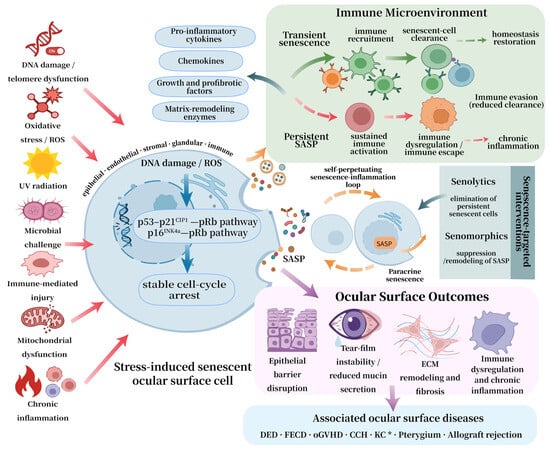
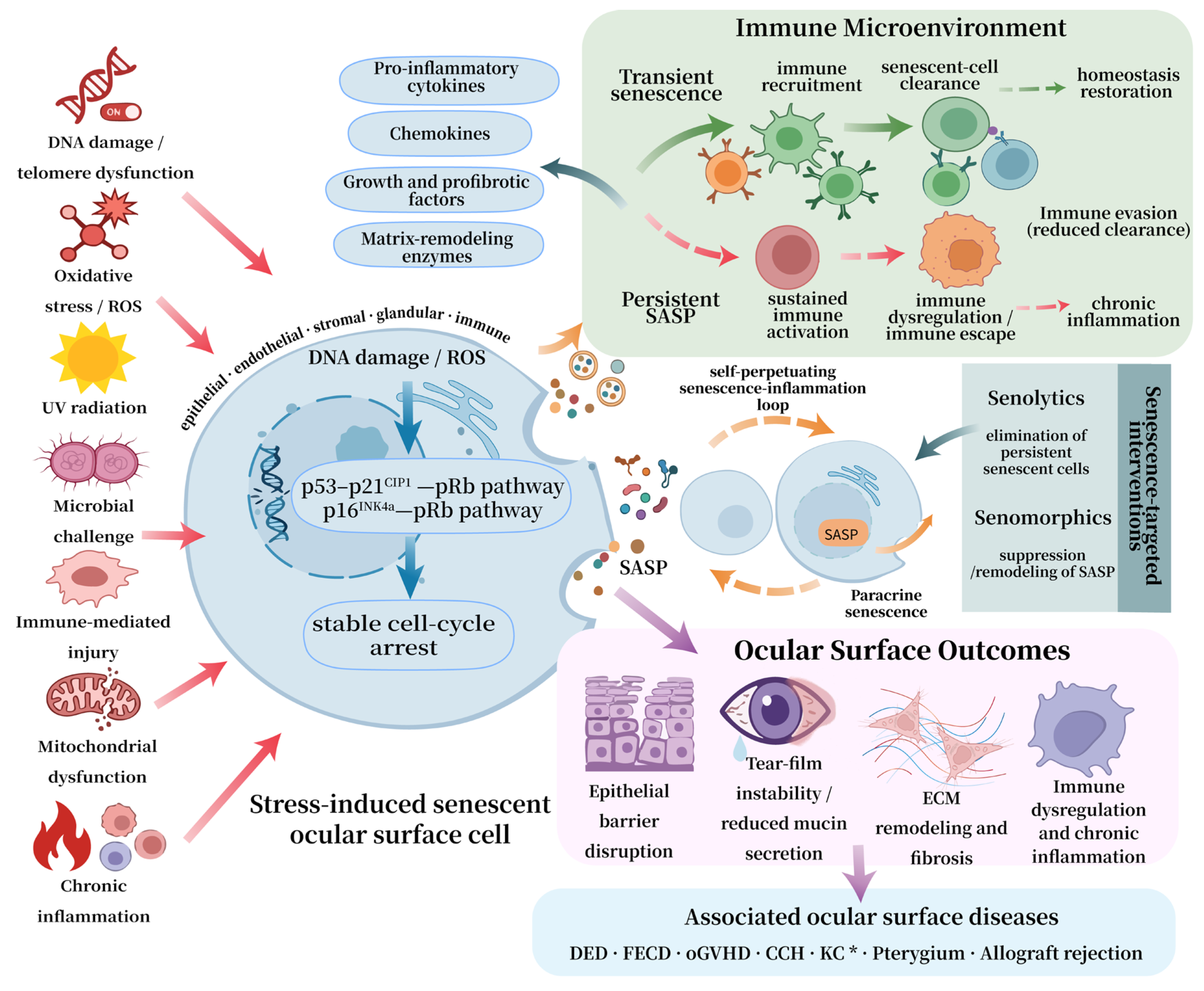
Taken together, these findings support an integrated stress–senescence–SASP–immune remodeling axis that links ocular surface injury to tissue dysfunction and provides the biological rationale for senescence-targeted interventions, as summarized in Figure 1.
Figure 1.
Integrated framework of stress-induced cellular senescence and SASP-mediated ocular surface pathology. Ocular surface stressors, including DNA damage, telomere dysfunction, oxidative stress, ultraviolet radiation, microbial challenge, immune-mediated injury, mitochondrial dysfunction, and chronic inflammation, promote cellular senescence through canonical cell-cycle-arrest pathways. Transient senescence may facilitate immune recruitment, senescent-cell clearance, and restoration of homeostasis, whereas persistent SASP signaling promotes paracrine senescence, sustained immune activation, immune evasion, epithelial barrier disruption, tear-film instability, extracellular-matrix remodeling, fibrosis, and chronic inflammation. Senolytics and senomorphics are shown as experimental strategies for reducing persistent senescent-cell burden or modifying SASP activity. Solid arrows indicate relationships supported by ocular experimental or clinical evidence, whereas dashed arrows indicate proposed mechanisms or relationships supported mainly by indirect evidence. * KC is included on the basis of indirect molecular associations; direct evidence of cellular senescence remains limited. Abbreviations: CCH, conjunctivochalasis; DED, dry eye disease; DNA, deoxyribonucleic acid; ECM, extracellular matrix; FECD, Fuchs endothelial corneal dystrophy; KC, keratoconus; oGVHD, ocular graft-versus-host disease; p16INK4a, cyclin-dependent kinase inhibitor p16INK4a; p21CIP1, cyclin-dependent kinase inhibitor p21CIP1; p53, tumor protein p53; pRB, retinoblastoma protein; ROS, reactive oxygen species; SASP, senescence-associated secretory phenotype.
4. Cellular Senescence in Ocular Surface Diseases
4.1. Dry Eye Disease
DED is a multifactorial chronic OSD characterized by loss of tear-film homeostasis, ocular discomfort, visual disturbance, and ocular surface inflammation. Increasing evidence suggests that DED is not merely a tear-deficient disorder but a chronic inflammatory condition shaped by aging-related immune dysregulation, environmental stress, and ocular surface dysfunction [48,49].
Aging is an independent risk factor for DED [50,51]. Age-related changes in lacrimal gland function, meibomian gland structure, goblet-cell support, and ocular surface immunity create a susceptible background for disease [50,51]. Senescence-associated beta-galactosidase (SA-β-gal) positivity and activation of the p53–p21CIP1–pRB and p16INK4a–pRB pathways have been observed in aged lacrimal and meibomian glands, suggesting that cellular senescence may contribute to glandular remodeling and functional decline [24,52,53,54]. Desiccating and oxidative stress activate inflammatory mediators and matrix metalloproteinase-9 (MMP-9), disrupt epithelial barrier integrity, and amplify tear-film instability [20,55,56]. SIPS may therefore link environmental injury to chronic ocular surface inflammation, but whether it independently drives DED onset or progression remains uncertain. Future studies should identify the affected cell populations, determine whether senescent-cell accumulation precedes disease progression, and test whether targeted clearance restores ocular surface homeostasis.
4.2. Fuchs Endothelial Corneal Dystrophy
FECD is a genetically heterogeneous, progressive corneal endothelial disorder characterized by loss of corneal endothelial cells (CEnCs), Descemet membrane thickening, guttae formation, excessive ECM deposition, stromal edema, and progressive visual decline [26]. Reported senescence-associated features include oxidative stress, impaired DNA repair, increased p21CIP1 and p16INK4a protein expression, SASP activation, and fibrotic matrix remodeling. Transcriptomic profiling of human FECD endothelium found 31 of 89 senescence-associated genes to be differentially expressed, including increased CDKN2A and NOX4 expression [23,57]. These findings suggest a potential link between oxidative stress and endothelial senescence.
In a Col8a2 mutant mouse model, Matthaei et al. observed increased Cdkn1a expression without a clear change in Cdkn2a expression, suggesting that a p53–p21CIP1–pRB-dominant stress response may predominate in this model [23,58]. Human late-onset FECD endothelium showed increased CDKN2A expression [57], but this finding alone does not establish irreversible senescence in a tissue with intrinsically limited proliferative capacity. In vitro, p16INK4a-positive senescent CEnCs promoted guttae-like structures and abnormal ECM deposition, whereas CDKN2A knockdown suppressed these changes [26]. Senescent CEnCs may also promote endothelial–mesenchymal transition and fibrosis through SASP-associated mediators, including IL-8 and IL-17 [11,59,60]. Ultraviolet A exposure can induce cell-cycle re-entry followed by G2/M arrest and generate senescence markers and fibrotic phenotypes, including alpha-smooth muscle actin expression [61]. Chronic stress may instead drive irreversible G0/G1 arrest through sustained ataxia telangiectasia mutated (ATM) activation; ATM inhibition restored repair capacity and reduced senescence burden in experimental models [62]. Overall, current evidence supports a role for cellular senescence in FECD-related endothelial dysfunction, guttae formation, and matrix remodeling, although its contribution to disease progression requires further clarification.
4.3. Ocular Graft-Versus-Host Disease
Ocular graft-versus-host disease (oGVHD) is a common and severe complication of allogeneic hematopoietic stem cell transplantation, with a reported prevalence of up to 60% among patients with graft-versus-host disease (GVHD). Aberrant activation of donor-derived immune cells leads to chronic inflammation and progressive fibrosis in multiple organs [63,64]. Cellular senescence has been implicated in chronic oGVHD. Ocular tissues from patients with oGVHD show DDR activation, increased p16INK4a and p21CIP1 protein expression, mitochondrial dysfunction, and enhanced SASP activity [24,63]. IL-6 is among the SASP-associated mediators implicated in inflammatory and fibrotic remodeling in oGVHD [24,63].
In oGVHD-associated keratopathy, oxidative damage markers such as 8-hydroxy-2′-deoxyguanosine (8-OHdG) and 4-hydroxynonenal (4-HNE) are increased, whereas antioxidant defenses, including nuclear factor erythroid 2-related factor 2 (Nrf2) and catalase, are suppressed. p16INK4a and p38 MAPK signaling are also activated, promoting NF-κB-mediated release of IL-6 and IL-8 [21]. Desiccating stress may reinforce this pathology by intensifying local alloimmune responses [63,65,66]. p16INK4a-positive senescent cells and mitochondrial dysfunction have been observed in lacrimal gland and conjunctival tissues [24,63]. SASP-associated mediators may contribute to fibroblast activation and fibrosis, and cellular senescence has also been implicated in chronic GVHD-associated meibomian gland dysfunction [53,63]. Thus, current evidence supports a role for senescence in chronic oGVHD, although some links remain supportive or indirect.
4.4. Corneal Allograft Rejection
Corneal transplantation remains the principal treatment for end-stage corneal blindness, but immune rejection is a leading cause of graft failure. Transplantation-related stress, endothelial vulnerability, inflammation, and oxidative injury may create a permissive environment for SIPS-like phenotypes [67,68]. In a murine penetrating keratoplasty model, graft endothelial cells showed DNA damage, ROS accumulation, activation of the p16INK4a–pRB pathway, SA-β-gal positivity, increased CDKN2A expression, and reduced proliferative capacity [67]. These findings support a senescence-associated endothelial response after transplantation, but they do not independently establish a stable senescent state in cells with intrinsically limited proliferative capacity. Stromal fibroblast senescence may limit proliferation early after injury but promote chronic inflammation and remodeling through the SASP at later stages [69].
As rejection progresses toward chronic graft failure, cytokines may cooperate with oxidative stress to promote senescence. In human corneal endothelial B4G12 cells, transforming growth factor beta 1 (TGF-β1) increased mitochondrial ROS and induced increased p16INK4a and p21CIP1 protein expression, G1 arrest, and elevated SA-β-gal activity; N-acetyl-L-cysteine attenuated these changes [70]. Increased p16INK4a and p27KIP1 protein expression has also been reported in selected renal allografts with rejection [71], and increased biliary p16INK4a, p21CIP1, and p53 protein expression has been observed in liver allograft rejection [72]. In experimental corneal transplantation, clearance of senescent cells restored angiotensin-converting enzyme 2 (ACE2), reduced SASP activity, and improved immune homeostasis [73]. Direct causal evidence in human corneal graft rejection nevertheless remains limited.
4.5. Other Ocular Surface Diseases
Pterygium is an ocular surface degenerative disorder associated with ultraviolet exposure and chronic oxidative stress. Increased IL-6, IL-8, matrix metalloproteinases, transforming growth factor beta, and vascular endothelial growth factor resemble a SASP-like profile and may contribute to epithelial hyperplasia, stromal remodeling, fibrovascular proliferation, and angiogenesis [74]. These mediators are not specific to senescence and may also reflect chronic inflammation or tissue remodeling. Current findings therefore provide indirect rather than definitive evidence. Confirmation requires cell-type-specific localization with complementary markers, evidence of stable cell-cycle arrest, and functional manipulation of senescent cells.
Keratoconus (KC) is increasingly recognized as a multifactorial corneal ectatic disorder involving oxidative stress, inflammation, ECM remodeling, and metabolic dysregulation [75]. Tear proteomic analysis by Kannan et al. identified reduced expression of antioxidant enzymes, including GSS and SOD1, together with alterations in inflammation-related and ECM-associated proteins. Pathway enrichment further implicated neutrophil degranulation, autophagy, and metabolic reprogramming [76]. However, canonical senescence markers and senescent-cell accumulation were not directly assessed. Thus, these findings suggest biological overlap between KC and senescence-related stress responses but do not establish cellular senescence or SASP signaling as causal mechanisms in KC.
CCH is common in older adults and is characterized by redundant conjunctival folds between the globe and lower eyelid. In conjunctival fibroblasts, increased intracellular ROS activates p38 MAPK and promotes SIPS through the p53–p21CIP1–pRB pathway [22]. These findings provide direct mechanistic evidence linking ROS–p38 MAPK signaling to fibroblast senescence and conjunctival remodeling. Larger clinical studies are needed to distinguish the contributions of senescence, age-related degeneration, and chronic inflammation.
Representative studies linking cellular senescence and stress-related mechanisms to ocular surface diseases are summarized in Table 1.
Table 1.
Representative studies linking cellular senescence and stress-related mechanisms to ocular surface diseases.
5. Role of Senescence in Ocular Surface Homeostasis
5.1. Dual Roles of Senescence in Ocular Surface Physiology and Pathology
Under physiological conditions, ocular surface homeostasis depends on the coordinated regulation of barrier, secretory, immune, neural, stromal, and local microecological functions. Corneal and conjunctival epithelial cells maintain surface integrity; goblet cells stabilize the tear film through mucin secretion; corneal endothelial cells (CEnCs) preserve stromal deturgescence; sensory nerves regulate blinking and lacrimal secretion; and resident immune cells, including dendritic cells, macrophages, γδ T cells, natural killer (NK)/natural killer T (NKT) cells, CD8+ T cells, and B cells, together with regulatory cytokines such as TGF-β and IL-10, maintain immune surveillance without excessive inflammation [48,49,79]. This integrated view provides the basis for understanding why senescence can be either adaptive or harmful depending on its duration and clearance.
Cellular senescence is not necessarily pathological. A transient response may limit aberrant proliferation of damaged cells and coordinate tissue repair. Senescent fibroblasts have been observed during mouse corneal wound healing [69], although proposed benefits of senescence-associated platelet-derived growth factor AA secretion derive from non-ocular wound-healing models and require direct validation in the cornea [5,80]. Effective immune clearance prevents excessive senescent-cell accumulation and allows repair-associated SASP signals to resolve [48].
When stress persists or immune clearance fails, the reparative response may become maladaptive. Persistent SASP signaling can impair epithelial renewal, destabilize the tear film, activate fibroblasts, disrupt immune tolerance, and reinforce paracrine senescence [11,37,39,81,82]. The biological effect of senescence therefore depends on its duration, cellular context, and efficiency of immune clearance.
5.2. Senescence-Driven Cell-Type-Specific Pathologies
Cellular senescence exerts cell-type-specific effects on ocular surface homeostasis by altering the functional properties of epithelial, endothelial, stromal, secretory, and immune cells. In corneal epithelial cells, stress-induced senescence reduces proliferative reserve, delays re-epithelialization, weakens barrier integrity, and impairs epithelial stem cell function and cell migration [77,83]. SASP-mediated inflammatory signaling further disrupts epithelial homeostasis, and TGF-β can induce corneal epithelial senescence and canonical SASP production; inhibition of this pathway partially reverses the associated phenotype [78]. CEnCs are particularly susceptible to senescence because of their limited regenerative capacity in vivo. Senescence reduces endothelial cell density and pump reserve, increases cellular pleomorphism and polymegathism, and compromises stromal deturgescence, thereby predisposing the cornea to edema and endothelial decompensation [23]. Upregulation of p21CIP1 further supports the involvement of premature senescence in endothelial functional decline, although its causal contribution remains to be fully validated.
In corneal fibroblasts, persistent senescence can promote inflammatory and matrix-remodeling phenotypes, although senescent fibroblasts may also limit fibrosis in specific wound-healing contexts [24,25,69]. Direct evidence for senescence in conjunctival goblet cells remains limited; chronic inflammation and oxidative stress may impair mucin secretion and tear-film stability. Immune-cell senescence may further reduce immune regulation and senescent-cell clearance. Senescent T cells can exhibit functional exhaustion and impaired immunoregulatory activity, potentially weakening local immune tolerance [10,38,48].
Under pathological stress, cellular senescence is not merely a passive marker of tissue injury. Rather, it alters the core functions of ocular surface cells, including epithelial renewal, barrier maintenance, endothelial pump activity, stromal remodeling, mucin secretion, and immune surveillance. This stress–senescence–functional decline axis provides a useful framework for understanding how ocular surface homeostasis becomes progressively destabilized [11,13,14].
6. Therapeutic Strategies Targeting Senescence
6.1. Senolytics
Senolytics are agents designed to selectively eliminate senescent cells by inducing apoptosis, thereby reducing senescent-cell burden and attenuating detrimental SASP signaling [84,85]. Representative senolytics include B-cell lymphoma 2 (BCL-2) family inhibitors such as navitoclax (ABT-263), tyrosine kinase inhibitors such as dasatinib, natural compounds such as quercetin and fisetin, and newer investigational agents such as UBX-1325 [84,86]. However, most evidence for these compounds derives from systemic aging models, non-ocular diseases, or ocular diseases outside the ocular surface, rather than from OSD models [84,85,86].
Direct preclinical evidence for senolytics in ocular surface and adnexal models remains limited but is emerging. In the p16-3MR mouse model, ganciclovir-mediated ablation of p16INK4a-positive cells reduced senescence- and SASP-related gene expression, corneal opacity, and angiogenesis after lacrimal gland excision [13]. Because ablation depends on the engineered p16-3MR/HSV-TK system, this result is proof of concept rather than evidence for pharmacological treatment of DED. In chronic oGVHD models, systemic navitoclax (ABT-263) reduced senescence-associated markers, inflammation, and fibrosis in lacrimal and meibomian glands [24,53]. These studies support further evaluation of senescent-cell clearance, but efficacy and safety in human disease remain unestablished [85,87,88].
Senolytics may also be relevant to corneal transplantation. In a murine model, systemic ABT-263 reduced alloimmune responses, and its protective effect was closely linked to ACE2 signaling [73]. Whether these findings translate to human transplantation is unknown. Translation also requires caution because transient senescence may support wound repair, ocular cell types may differ in senolytic sensitivity, and systemic BCL-2 family inhibitors can cause hematologic toxicity [84,85]. Future studies should define timing, dose, delivery route, cell-type specificity, and long-term ocular safety. In another ocular tissue, BST2-targeted senotherapy restored visual function in a preclinical retinal model [89], but its relevance to OSDs is unknown.
6.2. Senomorphics
Unlike senolytics, which eliminate senescent cells, senomorphics suppress or reshape the SASP. This approach may be relevant to OSDs because epithelial, glandular, endothelial, and stromal integrity is essential for tear-film stability, barrier function, corneal transparency, and immune homeostasis.
Several pathways are candidates for SASP modulation. mTOR regulates SASP production [44], but the value of mTOR-directed therapy in OSDs has not been established [84,90]. Janus kinase (JAK)/signal transducer and activator of transcription (STAT) inhibition have shown senomorphic effects in non-ocular models, including suppression of JAK2/STAT3-associated senescence [91]. In a murine chronic GVHD model, an anti-IL-6 receptor antibody reduced lacrimal gland fibrosis [24], supporting the potential of targeting this SASP-associated cytokine pathway. These findings do not distinguish selective senomorphic effects from broader anti-inflammatory actions [63].
NF-κB integrates signals from the DDR, cGAS–STING, oxidative stress, and inflammatory pathways to drive SASP gene expression. Bromodomain-containing protein 4 (BRD4) and other epigenetic regulators can cooperate with NF-κB to sustain this transcriptional program [92,93]. Their therapeutic relevance in OSDs remains unproven. Delivery is an additional constraint: systemic treatment can increase off-target toxicity, whereas topical delivery is limited by tear turnover, blinking, nasolacrimal drainage, tissue barriers, and short precorneal residence time [94]. Periocular or local injection may increase exposure but introduces risks of injury, inflammation, clearance, and tissue toxicity [95]. Future ocular senotherapy must therefore optimize delivery, retention, tissue specificity, and safety without disrupting physiological repair [13,24,53,73,84].
7. Conclusions and Perspectives
Current evidence supports cellular senescence as a context-dependent link between ocular surface stress, inflammation, cellular dysfunction, and tissue remodeling. Direct mechanistic contributions have been demonstrated in selected experimental models, whereas evidence in several human OSDs remains associative. Senolytics and senomorphics require further validation of ocular safety, cell-type specificity, timing, and delivery. Future studies should define disease- and cell-specific markers, use cell-resolved profiling to identify senescent populations, and test whether selective depletion or functional modulation improves disease outcomes.
Author Contributions
Conceptualization, L.Z. and S.W.; literature search and investigation, L.Z. and S.L.; evidence evaluation and verification, L.Z., S.L. and Q.G.; writing—original draft preparation, L.Z.; writing—review and editing, S.L. and S.W.; visualization, L.Z., S.L. and H.Z. (Hongkun Zhang); supervision, S.W. and H.Z. (Hong Zhang); project administration, S.W. All authors have read and agreed to the published version of the manuscript.
Funding
This research was funded by the National Natural Science Foundation of China (grant no. 82301164), Outstanding Young Scholar Fund of the First Affiliated Hospital of Harbin Medical University (grant no. 2024YQ05).
Data Availability Statement
No new data were created or analyzed in this study. Data sharing is not applicable to this article.
Conflicts of Interest
The authors declare no conflicts of interest.
References
- Pflugfelder, S.C.; Stern, M.E. Biological Functions of Tear Film. Exp. Eye Res. 2020, 197, 108115. [Google Scholar] [CrossRef] [Scilit] [PubMed]
- de Paiva, C.S.; St Leger, A.J.; Caspi, R.R. Mucosal Immunology of the Ocular Surface. Mucosal Immunol. 2022, 15, 1143–1157. [Google Scholar] [CrossRef] [Scilit] [PubMed]
- Alves, M.; Asbell, P.; Dogru, M.; Giannaccare, G.; Grau, A.; Gregory, D.; Kim, D.H.; Marini, M.C.; Ngo, W.; Nowinska, A.; et al. TFOS Lifestyle Report: Impact of Environmental Conditions on the Ocular Surface. Ocul. Surf. 2023, 29, 1–52. [Google Scholar] [CrossRef] [Scilit] [PubMed]
- Soifer, M.; Azar, N.S.; Mousa, H.M.; Perez, V.L. Ocular Surface Inflammatory Disorders (OSID): A Collective of Systemic Etiologies Which Cause or Amplify Dry Eye Syndrome. Front. Med. 2022, 9, 949202. [Google Scholar] [CrossRef] [Scilit] [PubMed]
- Zheng, X.; Wu, Y.; Feng, Y.; Cai, M.; Zhang, M.; Zhang, L.; Yang, D.; Mao, T.; Gu, H.; Ou, S. The aging cornea: From mechanisms to clinical applications. Ageing Res. Rev. 2026, 118, 103103. [Google Scholar] [CrossRef] [Scilit] [PubMed]
- Gorgoulis, V.; Adams, P.D.; Alimonti, A.; Bennett, D.C.; Bischof, O.; Bishop, C.; Campisi, J.; Collado, M.; Evangelou, K.; Ferbeyre, G.; et al. Cellular Senescence: Defining a Path Forward. Cell 2019, 179, 813–827. [Google Scholar] [CrossRef] [Scilit] [PubMed]
- López-Otín, C.; Blasco, M.A.; Partridge, L.; Serrano, M.; Kroemer, G. The Hallmarks of Aging. Cell 2013, 153, 1194–1217. [Google Scholar] [CrossRef] [Scilit] [PubMed]
- Rodier, F.; Coppé, J.-P.; Patil, C.K.; Hoeijmakers, W.A.M.; Muñoz, D.P.; Raza, S.R.; Freund, A.; Campeau, E.; Davalos, A.R.; Campisi, J. Persistent DNA damage signalling triggers senescence-associated inflammatory cytokine secretion. Nat. Cell Biol. 2009, 11, 973–979, Erratum in Nat. Cell Biol. 2009, 11, 1272. https://doi.org/10.1038/ncb1009-1272a. [Google Scholar] [CrossRef] [Scilit] [PubMed]
- von Zglinicki, T. Oxidative stress shortens telomeres. Trends Biochem. Sci. 2002, 27, 339–344. [Google Scholar] [CrossRef] [Scilit] [PubMed]
- Admasu, T.D.; Rae, M.J.; Stolzing, A. Dissecting primary and secondary senescence to enable new senotherapeutic strategies. Ageing Res. Rev. 2021, 70, 101412. [Google Scholar] [CrossRef] [Scilit] [PubMed]
- Wang, B.; Han, J.; Elisseeff, J.H.; Demaria, M. The senescence-associated secretory phenotype and its physiological and pathological implications. Nat. Rev. Mol. Cell Biol. 2024, 25, 958–978. [Google Scholar] [CrossRef] [Scilit] [PubMed]
- Coppé, J.-P.; Patil, C.K.; Rodier, F.; Sun, Y.; Muñoz, D.P.; Goldstein, J.; Nelson, P.S.; Desprez, P.-Y.; Campisi, J. Senescence-Associated Secretory Phenotypes Reveal Cell-Nonautonomous Functions of Oncogenic RAS and the p53 Tumor Suppressor. PLoS Biol. 2008, 6, e301. [Google Scholar] [CrossRef] [Scilit] [PubMed]
- Kitazawa, K.; Campisi, J.; Numa, K.; Patel, S.K.; King, C.D.; Matsumoto, A.; Sotozono, C.; Desprez, P.-Y.; Schilling, B. Cellular Senescence Exacerbates Features of Aging in the Eyes. Aging Biol. 2023, 1, 20230014. [Google Scholar] [CrossRef] [Scilit] [PubMed]
- Wu, J.; Liu, X.; Liu, Y.; Su, W.; Zhuo, Y. New Insights into the Role of Cellular Senescence and Its Therapeutic Implications in Ocular Diseases. Bioengineering 2025, 12, 563. [Google Scholar] [CrossRef] [Scilit] [PubMed]
- Freund, A.; Patil, C.K.; Campisi, J. p38MAPK is a novel DNA damage response-independent regulator of the senescence-associated secretory phenotype. EMBO J. 2011, 30, 1536–1548. [Google Scholar] [CrossRef] [Scilit] [PubMed]
- Ya, J.; Bayraktutan, U. Senolytics and Senomorphics Targeting p38MAPK/NF-κB Pathway Protect Endothelial Cells from Oxidative Stress-Mediated Premature Senescence. Cells 2024, 13, 1292. [Google Scholar] [CrossRef] [Scilit] [PubMed]
- Kim, J.; Kim, H.-S.; Chung, J.H. Molecular mechanisms of mitochondrial DNA release and activation of the cGAS-STING pathway. Exp. Mol. Med. 2023, 55, 510–519. [Google Scholar] [CrossRef] [Scilit] [PubMed]
- Kang, C.; Xu, Q.; Martin, T.D.; Li, M.Z.; Demaria, M.; Aron, L.; Lu, T.; Yankner, B.A.; Campisi, J.; Elledge, S.J. The DNA damage response induces inflammation and senescence by inhibiting autophagy of GATA4. Science 2015, 349, aaa5612. [Google Scholar] [CrossRef] [Scilit] [PubMed]
- Shreeya, T.; Ansari, M.S.; Kumar, P.; Saifi, M.; Shati, A.A.; Alfaifi, M.Y.; Elbehairi, S.E.I. Senescence: A DNA damage response and its role in aging and Neurodegenerative Diseases. Front. Aging 2024, 4, 1292053. [Google Scholar] [CrossRef] [Scilit] [PubMed]
- Seen, S.; Tong, L. Dry eye disease and oxidative stress. Acta Ophthalmol. 2018, 96, 412–420. [Google Scholar] [CrossRef] [Scilit] [PubMed]
- Wu, R.; Liu, S.; Shen, Z.; Zhao, Y.; Hong, J.; Ma, J. Corneal oxidative stress, inflammation, and senescence as key drivers of ocular pathology in a chronic graft-versus-host disease model. BMC Ophthalmol. 2025, 25, 379. [Google Scholar] [CrossRef] [Scilit] [PubMed]
- Xiang, M.; Mo, L.; Zhan, Y.; Wen, H.; Zhou, H.; Miao, W. P38-Mediated Cellular Senescence in Conjunctivochalasis Fibroblasts. Investig. Ophthalmol. Vis. Sci. 2019, 60, 4643. [Google Scholar] [CrossRef] [Scilit] [PubMed]
- Matthaei, M.; Meng, H.; Meeker, A.K.; Eberhart, C.G.; Jun, A.S. EndothelialCdkn1a(p21) Overexpression and Accelerated Senescence in a Mouse Model of Fuchs Endothelial Corneal Dystrophy. Investig. Ophthalmol. Vis. Sci. 2012, 53, 6718. [Google Scholar] [CrossRef] [Scilit] [PubMed]
- Yamane, M.; Sato, S.; Shimizu, E.; Shibata, S.; Hayano, M.; Yaguchi, T.; Kamijuku, H.; Ogawa, M.; Suzuki, T.; Mukai, S.; et al. Senescence-associated secretory phenotype promotes chronic ocular graft-vs-host disease in mice and humans. FASEB J. 2020, 34, 10778–10800. [Google Scholar] [CrossRef] [Scilit] [PubMed]
- Ajoolabady, A.; Pratico, D.; Bahijri, S.; Eldakhakhny, B.; Tuomilehto, J.; Wu, F.; Ren, J. Hallmarks and mechanisms of cellular senescence in aging and disease. Cell Death Discov. 2025, 11, 364. [Google Scholar] [CrossRef] [Scilit] [PubMed]
- Parekh, M.; Price, M.O.; Price, F.W.; Jurkunas, U.V. Permanent Senescence Via p16 Leads to Guttae Formation in an In Vitro Human Corneal Endothelial Cell Model. Investig. Ophthalmol. Vis. Sci. 2025, 66, 1. [Google Scholar] [CrossRef] [Scilit] [PubMed]
- Cancado de Faria, R.; Silva, L.N.D.; Teodoro-Castro, B.; McCommis, K.S.; Shashkova, E.V.; Gonzalo, S. A Noncanonical cGAS–STING Pathway Drives Cellular and Organismal Aging. Proc. Natl. Acad. Sci. USA 2025, 122, e2424666122. [Google Scholar] [CrossRef] [Scilit] [PubMed]
- Gulen, M.F.; Samson, N.; Keller, A.; Schwabenland, M.; Liu, C.; Glück, S.; Thacker, V.V.; Favre, L.; Mangeat, B.; Kroese, L.J.; et al. cGAS–STING drives ageing-related inflammation and neurodegeneration. Nature 2023, 620, 374–380. [Google Scholar] [CrossRef] [Scilit] [PubMed]
- Sherr, C.J.; McCormick, F. The RB and p53 pathways in cancer. Cancer Cell 2002, 2, 103–112. [Google Scholar] [CrossRef] [Scilit] [PubMed]
- Wade Harper, J. The p21 Cdk-interacting protein Cip1 is a potent inhibitor of G1 cyclin-dependent kinases. Cell 1993, 75, 805–816. [Google Scholar] [CrossRef] [Scilit] [PubMed]
- El-Deiry, W.S.; Tokino, T.; Velculescu, V.E.; Levy, D.B.; Parsons, R.; Trent, J.M.; Lin, D.; Mercer, W.E.; Kinzler, K.W.; Vogelstein, B. WAF1, a potential mediator of p53 tumor suppression. Cell 1993, 75, 817–825. [Google Scholar] [CrossRef] [Scilit] [PubMed]
- Correia-Melo, C.; Hewitt, G.; Passos, J.F. Telomeres, oxidative stress and inflammatory factors: Partners in cellular senescence? Longev. Healthspan 2014, 3, 1. [Google Scholar] [CrossRef] [Scilit] [PubMed]
- Abbas, T.; Dutta, A. p21 in cancer: Intricate networks and multiple activities. Nat. Rev. Cancer 2009, 9, 400–414. [Google Scholar] [CrossRef] [Scilit] [PubMed]
- Serrano, M.; Lee, H.-W.; Chin, L.; Cordon-Cardo, C.; Beach, D.; DePinho, R.A. Role of the INK4a Locus in Tumor Suppression and Cell Mortality. Cell 1996, 85, 27–37. [Google Scholar] [CrossRef] [Scilit] [PubMed]
- Narita, M.; Nuñez, S.; Heard, E.; Narita, M.; Lin, A.W.; Hearn, S.A.; Spector, D.L.; Hannon, G.J.; Lowe, S.W. Rb-Mediated Heterochromatin Formation and Silencing of E2F Target Genes during Cellular Senescence. Cell 2003, 113, 703–716. [Google Scholar] [CrossRef] [Scilit] [PubMed]
- Cao, L.; Li, K.; Li, Q.; Tong, Q.; Wang, Y.; Huang, L. The controversial role of senescence-associated secretory phenotype (SASP) in cancer therapy. Mol. Cancer 2025, 24, 283. [Google Scholar] [CrossRef] [Scilit] [PubMed]
- Abo Qoura, L.; Churov, A.V.; Maltseva, O.N.; Eruslanova, K.A.; Zeng, G. Senescent cells in systemic aging: SASP heterogeneity, immune escape, and endocrine modulation. Biogerontology 2026, 27, 103. [Google Scholar] [CrossRef] [Scilit] [PubMed]
- Wang, T.-W.; Nakanishi, M. Immune surveillance of senescence: Potential application to age-related diseases. Trends Cell Biol. 2025, 35, 248–257. [Google Scholar] [CrossRef] [Scilit] [PubMed]
- Salminen, A. Cooperation between inhibitory immune checkpoints of senescent cells with immunosuppressive network to promote immunosenescence and the aging process. Ageing Res. Rev. 2025, 106, 102694. [Google Scholar] [CrossRef] [Scilit] [PubMed]
- Das, S.; Ahmad, Z.; Suryawanshi, A.; Kumar, A. Innate immunity dysregulation in aging eye and therapeutic interventions. Ageing Res. Rev. 2022, 82, 101768. [Google Scholar] [CrossRef] [Scilit] [PubMed]
- Yang, H.; Liu, D.; Qiu, L.; Wang, R.; Zhang, C.; Yu, D.; Zhong, Q.; Yuki, N.; Song, Z.; Zhu, T.; et al. Reprogramming cellular senescence and aging clocks for advanced cancer immunotherapy. Mol. Cancer 2025, 24, 237. [Google Scholar] [CrossRef] [Scilit] [PubMed]
- Wang, H.; Yu, Y.; Li, R.; Zhang, H.; Chen, Z.-S.; Sun, C.; Zhuang, J. Immunoregulatory mechanisms in the aging microenvironment: Targeting the senescence-associated secretory phenotype for cancer immunotherapy. Acta Pharm. Sin. B 2025, 15, 4476–4496. [Google Scholar] [CrossRef] [Scilit] [PubMed]
- Mogilenko, D.A.; Shchukina, I.; Artyomov, M.N. Immune ageing at single-cell resolution. Nat. Rev. Immunol. 2022, 22, 484–498. [Google Scholar] [CrossRef] [Scilit] [PubMed]
- Laberge, R.-M.; Sun, Y.; Orjalo, A.V.; Patil, C.K.; Freund, A.; Zhou, L.; Curran, S.C.; Davalos, A.R.; Wilson-Edell, K.A.; Liu, S.; et al. MTOR regulates the pro-tumorigenic senescence-associated secretory phenotype by promoting IL1A translation. Nat. Cell Biol. 2015, 17, 1049–1061, Correction in Nat. Cell Biol. 2021, 23, 564–565. https://doi.org/10.1038/s41556-021-00655-4. [Google Scholar] [CrossRef] [Scilit] [PubMed]
- Zhang, Z.; Zhang, C. Regulation of cGAS–STING signalling and its diversity of cellular outcomes. Nat. Rev. Immunol. 2025, 25, 425–444. [Google Scholar] [CrossRef] [Scilit] [PubMed]
- Dou, Z.; Ghosh, K.; Vizioli, M.G.; Zhu, J.; Sen, P.; Wangensteen, K.J.; Simithy, J.; Lan, Y.; Lin, Y.; Zhou, Z.; et al. Cytoplasmic chromatin triggers inflammation in senescence and cancer. Nature 2017, 550, 402–406. [Google Scholar] [CrossRef] [Scilit] [PubMed]
- Liu, G.Y.; Sabatini, D.M. mTOR at the nexus of nutrition, growth, ageing and disease. Nat. Rev. Mol. Cell Biol. 2020, 21, 183–203, Correction in Nat. Rev. Mol. Cell Biol. 2020, 21, 246. https://doi.org/10.1038/s41580-020-0219-y. [Google Scholar] [CrossRef] [Scilit] [PubMed]
- Galletti, J.G.; de Paiva, C.S. The ocular surface immune system through the eyes of aging. Ocul. Surf. 2021, 20, 139–162. [Google Scholar] [CrossRef] [Scilit] [PubMed]
- de Paiva, C.S. Effects of Aging in Dry Eye. Int. Ophthalmol. Clin. 2017, 57, 47–64. [Google Scholar] [CrossRef] [Scilit] [PubMed]
- Kitazawa, K.; Inomata, T.; Shih, K.; Hughes, J.-W.B.; Bozza, N.; Tomioka, Y.; Numa, K.; Yokoi, N.; Campisi, J.; Dana, R.; et al. Impact of aging on the pathophysiology of dry eye disease: A systematic review and meta-analysis. Ocul. Surf. 2022, 25, 108–118. [Google Scholar] [CrossRef] [Scilit] [PubMed]
- Zhang, Z.; Yuan, Y.; He, Y.; Chi, K.; Zeng, H.; Deng, Y.; Wang, B.; Yu, H.; Calder, V.L.; Yuan, J.; et al. Age-related dry eye disease: Redefining a distinct subtype driven by aging. Ocul. Surf. 2026, 41, 171–184. [Google Scholar] [CrossRef] [Scilit] [PubMed]
- Wang, K.; Xie, Y.; Lin, Y.; Zhu, R.; Gao, T.; Han, Z.; Yang, Z.; Jiang, X.; Chen, H.; Wu, Z.; et al. Unveiling the link: Aging and its impact on ocular diseases. Exp. Eye Res. 2025, 259, 110551. [Google Scholar] [CrossRef] [Scilit] [PubMed]
- Sato, S.; Ogawa, Y.; Shimizu, E.; Asai, K.; Okazaki, T.; Rusch, R.; Hirayama, M.; Shimmura, S.; Negishi, K.; Tsubota, K. Cellular senescence promotes meibomian gland dysfunction in a chronic graft-versus-host disease mouse model. Ocul. Surf. 2024, 32, 198–210. [Google Scholar] [CrossRef] [Scilit] [PubMed]
- Zhu, X.; Xu, M.; Portal, C.; Lin, Y.; Ferdinand, A.; Peng, T.; Morrisey, E.E.; Dlugosz, A.A.; Castellano, J.M.; Lee, V.; et al. Identification of Meibomian gland stem cell populations and mechanisms of aging. Nat. Commun. 2025, 16, 1663. [Google Scholar] [CrossRef] [Scilit] [PubMed]
- Hu, R.; Shi, J.; Xie, C.M.; Yao, X.L. Dry Eye Disease: Oxidative Stress on Ocular Surface and Cutting-Edge Antioxidants. Glob. Chall. 2025, 9, e00068. [Google Scholar] [CrossRef] [Scilit] [PubMed]
- López-Miguel, A.; Tesón, M.; Martín-Montañez, V.; Enríquez-de-Salamanca, A.; Stern, M.E.; Calonge, M.; González-García, M.J. Dry Eye Exacerbation in Patients Exposed to Desiccating Stress under Controlled Environmental Conditions. Am. J. Ophthalmol. 2014, 157, 788–798.e2. [Google Scholar] [CrossRef] [Scilit] [PubMed]
- Matthaei, M.; Zhu, A.Y.; Kallay, L.; Eberhart, C.G.; Cursiefen, C.; Jun, A.S. Transcript profile of cellular senescence-related genes in Fuchs endothelial corneal dystrophy. Exp. Eye Res. 2014, 129, 13–17. [Google Scholar] [CrossRef] [Scilit] [PubMed]
- Matthaei, M.; Hu, J.; Meng, H.; Lackner, E.-M.; Eberhart, C.G.; Qian, J.; Hao, H.; Jun, A.S. Endothelial Cell Whole Genome Expression Analysis in a Mouse Model of Early-Onset Fuchs’ Endothelial Corneal Dystrophy. Investig. Ophthalmol. Vis. Sci. 2013, 54, 1931. [Google Scholar] [CrossRef] [Scilit] [PubMed][Green Version]
- Jurkunas, U.V. Fuchs Endothelial Corneal Dystrophy Through the Prism of Oxidative Stress. Cornea 2018, 37, S50–S54. [Google Scholar] [CrossRef] [Scilit] [PubMed]
- Parekh, M.; Adhikari, Y.; Deshpande, N.; Wong, R.; Price, M.O.; Price, F.W.; Jurkunas, U.V. p16-mediated G0/G1 cell cycle arrest leads to SASP and fibrosis in Fuchs endothelial corneal dystrophy. Cell Death Dis. 2026, 17, 197. [Google Scholar] [CrossRef] [Scilit] [PubMed]
- White, T.L.; Deshpande, N.; Kumar, V.; Gauthier, A.G.; Jurkunas, U.V. Cell cycle re-entry and arrest in G2/M phase induces senescence and fibrosis in Fuchs Endothelial Corneal Dystrophy. Free Radic. Biol. Med. 2021, 164, 34–43. [Google Scholar] [CrossRef] [Scilit] [PubMed]
- Ashraf, S.; Deshpande, N.; Cheung, Q.; Asabere, J.B.; Wong, R.J.; Gauthier, A.G.; Parekh, M.; Adhikari, Y.; Melangath, G.; Jurkunas, U.V. Modulation of ATM enhances DNA repair in G2/M phase of cell cycle and averts senescence in Fuchs endothelial corneal dystrophy. Commun. Biol. 2024, 7, 1482. [Google Scholar] [CrossRef] [Scilit] [PubMed]
- Ogawa, Y.; Kawakami, Y.; Tsubota, K. Cascade of Inflammatory, Fibrotic Processes, and Stress-Induced Senescence in Chronic GVHD-Related Dry Eye Disease. Int. J. Mol. Sci. 2021, 22, 6114. [Google Scholar] [CrossRef] [Scilit] [PubMed]
- He, J.; Zheng, F.; Zhang, L.; Cai, J.; Ogawa, Y.; Tsubota, K.; Liu, S.; Jin, X. Single-cell RNA-sequencing reveals the transcriptional landscape of lacrimal gland in GVHD mouse model. Ocul. Surf. 2024, 33, 50–63. [Google Scholar] [CrossRef] [Scilit] [PubMed]
- Gehlsen, U.; Stern, M.E.; Franklin, J.; Tahmaz, V.; Hallek, M.; Holtick, U.; Scheid, C.; Steven, P. Desiccating Stress Significantly Increases the Risk for Chronic Ocular Graft-versus-Host-Disease. Transplant. Cell. Ther. 2022, 28, 782.e1–782.e7. [Google Scholar] [CrossRef] [Scilit] [PubMed]
- Gehlsen, U.; Maass, M.; Stary, D.; Wagener-Ryczek, S.; Musial, G.; Pasparakis, M.; de Paiva, C.S.; Stern, M.E.; Steven, P. Dessicating stress triggers and exacerbates experimental ocular Graft-versus-host-disease. Ocul. Surf. 2025, 37, 236–246. [Google Scholar] [CrossRef] [Scilit] [PubMed]
- Zhao, X.; Wang, Y.; Wang, Y.; Li, S.; Chen, P. Oxidative stress and premature senescence in corneal endothelium following penetrating keratoplasty in an animal model. BMC Ophthalmol. 2016, 16, 16. [Google Scholar] [CrossRef] [Scilit] [PubMed]
- Achiron, A.; Nahum, Y.; Tiosano, A.; Shehadeh Mashor, R.; Tuuminen, R.; Elbaz, U.; Bahar, I.; Livny, E. Descemet Membrane Endothelial Keratoplasty Outcomes between Young and Old Graft Recipients. Curr. Eye Res. 2021, 46, 1460–1466. [Google Scholar] [CrossRef] [Scilit] [PubMed]
- Wang, X.; Qu, M.; Li, J.; Danielson, P.; Yang, L.; Zhou, Q. Induction of Fibroblast Senescence During Mouse Corneal Wound Healing. Investig. Ophthalmol. Vis. Sci. 2019, 60, 3669. [Google Scholar] [CrossRef] [Scilit] [PubMed]
- Li, Z.; Liu, T.; Ma, J.; Guo, Q.; Ma, L.; Lv, Q.; Jiang, Y.; Wei, C.; Zhang, J. TGF-β induces corneal endothelial senescence via increase of mitochondrial reactive oxygen species in chronic corneal allograft failure. Aging 2018, 10, 3474–3485. [Google Scholar] [CrossRef] [Scilit] [PubMed]
- Vavrinec, P.; Krivý, J.; Sýkorová, S.; Bandžuchová, H.; Žilinská, Z.; Vavrincová-Yaghi, D. Association of Senescence Markers with Age and Allograft Rejection in Renal Transplant Recipients. Biomedicines 2024, 12, 2338. [Google Scholar] [CrossRef] [Scilit] [PubMed]
- Rastogi, A.; Nigam, N.; Gayatri, R.; Bihari, C.; Pamecha, V. Biliary Epithelial Senescence in Cellular Rejection Following Live Donor Liver Transplantation. J. Clin. Exp. Hepatol. 2022, 12, 1420–1427. [Google Scholar] [CrossRef] [Scilit] [PubMed]
- Chi, H.; Ma, L.; Zeng, F.; Wang, X.; Peng, P.; Bai, X.; Zhang, T.; Yin, W.; Yu, Y.; Yang, L.; et al. Senolytic Treatment Alleviates Corneal Allograft Rejection Through Upregulation of Angiotensin-Converting Enzyme 2 (ACE2). Investig. Ophthalmol. Vis. Sci. 2025, 66, 15. [Google Scholar] [CrossRef] [Scilit] [PubMed]
- Di Girolamo, N.; Chui, J.; Coroneo, M.T.; Wakefield, D. Pathogenesis of pterygia: Role of cytokines, growth factors, and matrix metalloproteinases. Prog. Retin. Eye Res. 2004, 23, 195–228. [Google Scholar] [CrossRef] [Scilit] [PubMed]
- Singh, R.B.; Koh, S.; Sharma, N.; Woreta, F.A.; Hafezi, F.; Dua, H.S.; Jhanji, V. Keratoconus. Nat. Rev. Dis. Primers 2024, 10, 81. [Google Scholar] [CrossRef] [Scilit] [PubMed]
- Kannan, R.; Shetty, R.; Panigrahi, T.; Koh, S.K.; Khamar, P.; Deshpande, V.; Nuijts, R.M.M.A.; Gijs, M.; Nishtala, K.; Zhou, L.; et al. Untargeted Tear Proteomics in a Large South-Asian Cohort Reveals Inflammatory Signaling, ECM Remodeling, and Altered Metabolism in Keratoconus. Investig. Ophthalmol. Vis. Sci. 2025, 66, 60. [Google Scholar] [CrossRef] [Scilit] [PubMed]
- Kitazawa, K.; Matsumoto, A.; Numa, K.; Tomioka, Y.; Zhang, Z.A.; Yamashita, Y.; Sotozono, C.; Desprez, P.-Y.; Campisi, J. Gene expression signatures of human senescent corneal and conjunctival epithelial cells. Aging 2023, 15, 9238–9249. [Google Scholar] [CrossRef] [Scilit] [PubMed]
- Li, Z.-Y.; Chen, Z.-L.; Zhang, T.; Wei, C.; Shi, W.-Y. TGF-β and NF-κB signaling pathway crosstalk potentiates corneal epithelial senescence through an RNA stress response. Aging 2016, 8, 2337–2354, Erratum in Aging 2021, 13, 20853. https://doi.org/10.18632/aging.203516. [Google Scholar] [CrossRef] [Scilit] [PubMed] [PubMed Central]
- Bolaños-Jiménez, R.; Navas, A.; López-Lizárraga, E.P.; Ribot, F.M.D.; Peña, A.; Graue-Hernández, E.O.; Garfias, Y. Ocular Surface as Barrier of Innate Immunity. Open Ophthalmol. J. 2015, 9, 49–55. [Google Scholar] [CrossRef] [Scilit] [PubMed]
- Demaria, M.; Ohtani, N.; Youssef, S.A.; Rodier, F.; Toussaint, W.; Mitchell, J.R.; Laberge, R.-M.; Vijg, J.; Van Steeg, H.; Dollé, M.E.T.; et al. An Essential Role for Senescent Cells in Optimal Wound Healing through Secretion of PDGF-AA. Dev. Cell 2014, 31, 722–733. [Google Scholar] [CrossRef] [Scilit] [PubMed]
- He, S.; Sharpless, N.E. Senescence in Health and Disease. Cell 2017, 169, 1000–1011. [Google Scholar] [CrossRef] [Scilit] [PubMed]
- Shvedova, M.; Thanapaul, R.J.R.S.; Wang, Q.; Shin, G.H.; Dhillon, J.; Roh, D.S. Diminished and altered cellular senescence response in delayed wound healing of aging. Aging Cell 2026, 25, e70493. [Google Scholar] [CrossRef] [Scilit] [PubMed]
- Thanapaul, R. An Insight into Aging, Senescence, and Their Impacts on Wound Healing. Adv. Geriatr. Med. Res. 2021, 3, e210017. [Google Scholar] [CrossRef] [Scilit] [PubMed]
- Zhang, L.; Pitcher, L.E.; Prahalad, V.; Niedernhofer, L.J.; Robbins, P.D. Targeting cellular senescence with senotherapeutics: Senolytics and senomorphics. FEBS J. 2023, 290, 1362–1383. [Google Scholar] [CrossRef] [Scilit] [PubMed]
- Childs, B.G.; Gluscevic, M.; Baker, D.J.; Laberge, R.-M.; Marquess, D.; Dananberg, J.; van Deursen, J.M. Senescent cells: An emerging target for diseases of ageing. Nat. Rev. Drug Discov. 2017, 16, 718–735. [Google Scholar] [CrossRef] [Scilit] [PubMed]
- Hassan, J.W.; Bhatwadekar, A.D. Senolytics in the treatment of diabetic retinopathy. Front. Pharmacol. 2022, 13, 896907. [Google Scholar] [CrossRef] [Scilit] [PubMed]
- Baker, D.J.; Wijshake, T.; Tchkonia, T.; LeBrasseur, N.K.; Childs, B.G.; van de Sluis, B.; Kirkland, J.L.; van Deursen, J.M. Clearance of p16Ink4a-positive senescent cells delays ageing-associated disorders. Nature 2011, 479, 232–236. [Google Scholar] [CrossRef] [Scilit] [PubMed]
- Baker, D.J.; Childs, B.G.; Durik, M.; Wijers, M.E.; Sieben, C.J.; Zhong, J.; Saltness, R.A.; Jeganathan, K.B.; Verzosa, G.C.; Pezeshki, A.; et al. Naturally occurring p16Ink4a-positive cells shorten healthy lifespan. Nature 2016, 530, 184–189. [Google Scholar] [CrossRef] [Scilit] [PubMed]
- Oh, J.Y.; Chae, J.-B.; Lee, H.K.; Park, C.-W.; Bae, M.; Kim, G.; Oh, Y.; Yang, G.; Kim, S.; Ok, H.W.; et al. Bst2-targeted senotherapy restores visual function by eliminating senescent retinal cells. Nat. Commun. 2026, 17, 4135. [Google Scholar] [CrossRef] [Scilit] [PubMed]
- Di Micco, R.; Krizhanovsky, V.; Baker, D.; d’Adda di Fagagna, F. Cellular senescence in ageing: From mechanisms to therapeutic opportunities. Nat. Rev. Mol. Cell Biol. 2021, 22, 75–95. [Google Scholar] [CrossRef] [Scilit] [PubMed]
- Yang, B.; Li, T.; Wang, Z.; Zhu, Y.; Niu, K.; Hu, S.; Lin, Z.; Zheng, X.; Jin, X.; Shen, C. Ruxolitinib-based senomorphic therapy mitigates cardiomyocyte senescence in septic cardiomyopathy by inhibiting the JAK2/STAT3 signaling pathway. Int. J. Biol. Sci. 2024, 20, 4314–4340. [Google Scholar] [CrossRef] [Scilit] [PubMed]
- Tasdemir, N.; Banito, A.; Roe, J.-S.; Alonso-Curbelo, D.; Camiolo, M.; Tschaharganeh, D.F.; Huang, C.-H.; Aksoy, O.; Bolden, J.E.; Chen, C.-C.; et al. BRD4 Connects Enhancer Remodeling to Senescence Immune Surveillance. Cancer Discov. 2016, 6, 612–629. [Google Scholar] [CrossRef] [Scilit] [PubMed]
- Dasgupta, N.; Arnold, R.; Equey, A.; Gandhi, A.; Adams, P.D. The role of the dynamic epigenetic landscape in senescence: Orchestrating SASP expression. npj Aging 2024, 10, 48. [Google Scholar] [CrossRef] [Scilit] [PubMed]
- Bachu, R.D.; Chowdhury, P.; Al-Saedi, Z.H.F.; Karla, P.K.; Boddu, S.H.S. Ocular Drug Delivery Barriers—Role of Nanocarriers in the Treatment of Anterior Segment Ocular Diseases. Pharmaceutics 2018, 10, 28. [Google Scholar] [CrossRef] [Scilit] [PubMed]
- Dosmar, E.; Walsh, J.; Doyel, M.; Bussett, K.; Oladipupo, A.; Amer, S.; Goebel, K. Targeting Ocular Drug Delivery: An Examination of Local Anatomy and Current Approaches. Bioengineering 2022, 9, 41. [Google Scholar] [CrossRef] [Scilit] [PubMed]

Disclaimer/Publisher’s Note: The statements, opinions and data contained in all publications are solely those of the individual author(s) and contributor(s) and not of MDPI and/or the editor(s). MDPI and/or the editor(s) disclaim responsibility for any injury to people or property resulting from any ideas, methods, instructions or products referred to in the content. |
© 2026 by the authors. Licensee MDPI, Basel, Switzerland. This article is an open access article distributed under the terms and conditions of the Creative Commons Attribution (CC BY) license.