


Abstract
Coxsackieviruses possess two proteases that are engaged in cleaving viral polyprotein and hijacking host cell processes such as RNA biosynthesis. Integrator subunit 10 (INTS10), a subunit of the integrator complex, facilitates the processing of small nuclear RNAs (U1 and U2 snRNAs) to regulate cellular transcription. We found that INST10 can be cleaved by Coxsackievirus B (CVB). Hence, we hypothesized that INST10 may play a role in CVB infection. In this study, INTS10 is identified as the substrate of CVB3 protease 3C (3Cpro). The cleavage occurs at the residue Q221 and yields a fragment. Depletion of INTS10 enhanced CVB3 replication and blocked snRNA processing. Overexpression of U1 snRNA inhibited CVB3 infection, whereas its knockdown conversely enhanced it. Similarly, knockdown of U2 snRNA was found to promote CVB3 replication. Taken together, the 3Cpro-mediated cleavage of INTS10 disrupts U snRNA processing, which in turn counteracts the inhibitory effect of snRNA U1 and U2 on virus replication and subverts host defenses.
1. Introduction
CVB, a member of the Picornaviridae family, is a positive-sense single-stranded RNA virus that causes acute myocarditis and pancreatitis in humans [1,2,3,4]. The elusive pathogenic mechanisms of CVB impede the development of effective interventions and preventive measures. Therefore, understanding these mechanisms is of paramount importance.
The CVB genome is approximately 7.4 kb in length, with a single open reading frame that encodes a large precursor protein [5]. This precursor protein is processed into four structural proteins (VP1–VP4) and seven nonstructural proteins (2A–2C, 3A–3D) [6,7]. The viral cysteine proteases 2Apro and 3Cpro are involved in biological processes in both CVB and the host [8,9]. CVB 3Cpro cleaves MAVS and TRIF to suppress host innate immune responses [10]. 3Cpro inhibits RIG-I-mediated antiviral signaling by cleaving 14-3-3ε [11]. Similarly, EV-A71 promotes viral replication by cleaving the host protein double-stranded RNA-binding protein Staufen homolog 2 (Stau2) [12]. Enterovirus 3Cpro is transported to the nucleus via its precursor 3CD, where it cleaves transcription and regulatory factors to modulate the host’s RNA synthesis [13]. However, the precise role of CVB in this process remains unclear.
The integrator complex, comprising 12–15 subunits, regulates RNA polymerase II-transcribed RNAs and is essential for the biogenesis of small nuclear RNAs (snRNAs) and enhancer RNAs [14,15,16]. Disrupting the integrator function results in severe genetic diseases [17,18,19]. INTS10, a subunit of this complex, interacts with RNA polymerase II to mediate the 3’ end processing of U1 and U2 snRNAs, the core components of the spliceosome [20,21]. Overexpression of U1 snRNA protects host cells against RNA viruses (e.g., IAV, SeV, VSV, EMCV, RSV), while its knockdown significantly increases viral burden in vitro and in vivo [22]. U1 snRNA interacts with Tripartite motif (TRIM) 25 in the cytoplasm, promoting TRIM25-mediated RIG-I activation to enhance host defense against RNA viruses [22]. U2 snRNA interacts with viral RNA in the cytoplasm, suppressing CVB replication [23]. Notably, INTS10 suppresses HBV replication via Interferon regulatory factor 3 (IRF3) in liver cells [24], but its relationship with CVB has not been explored.
Recently, we found that INST10 was cleaved in the cells infected with CVB, suggesting INST10 may play a role in CVB replication. This study elucidated how INST10 is cleaved in the CVB-infected cells and identified the significance of INST10 cleavage for CVB infection. Our study seeks to uncover a novel mechanism by which CVB hijacks a central component of the host’s RNA processing machinery and adds novel insights into viral pathogenesis for developing host-directed antiviral therapeutics.
2. Results
2.1. CVB3 Infection Induces the Cleavage of INTS10
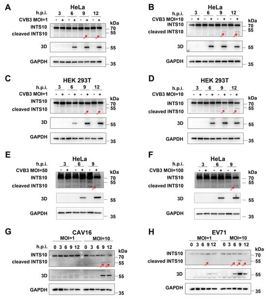
To investigate the impact of CVB3 infection on INTS10, we analyzed its expression in HeLa and HEK 293T cells infected with CVB3 (MOI = 1 or 10). At different time points post-infection, cell lysates were subjected to Western blotting analysis. We detected the effects of CVB3 infection on the expression of endogenous INTS10. The full-length INTS10 was observed with an apparent molecular mass of ~82 kDa in uninfected controls, while a truncated form of INTS10 (less than 55 kDa, red arrows) was detected in CVB3-infected HeLa and HEK 293T cells (Figure 1A–D), suggesting that INTS10 undergoes cleavage during CVB3 replication. To determine whether INTS10 cleavage is dependent on CVB3 concentration, HeLa cells were infected with CVB3 at MOI = 50 and 100 for 3, 6, and 9 h. We observed that the quality of full-length INTS10 reduced, and a small fragment with a molecular weight (MW) less than 55 kDa emerged at 9 h post-infection (Figure 1E,F, red arrows). Consistent with this observation, cleavage bands of INTS10 were also evident at 9 h post-infection with CVB3 at MOI = 50 and MOI = 100 in HEK 293T cells, as shown in Supplementary Figure S1.
Figure 1.
INTS10 is cleaved by CVB3, EV-A71, and CAV16. (A,B) HeLa cells were infected with CVB3 (MOI = 1 or 10). INTS10 expression was determined by Western blotting. (C,D) HEK 293T cells were infected with CVB3 (MOI = 1 or 10). INTS10 expression was determined by Western blotting. (E,F) HeLa cells were infected with CVB3 (MOI = 50 or 100). INTS10 expression was determined by Western blotting. (G,H) HeLa cells were infected with EV-A71 (G) and CAV16 (H). Red arrows indicate INTS10 cleavage fragments.
To determine whether this cleavage is specific to CVB3 or whether it represents a common mechanism utilized by enteroviruses, we assessed the status of INTS10 in HeLa cells infected with EV-A71 or CAV16. Similarly to that observed in CVB3-infected cells, a cleavage fragment of INTS10 appeared in both EV-A71- and CAV16-infected cells (Figure 1G,H, red arrows), indicating that INTS10 cleavage is a shared feature of enterovirus infections.
2.2. CVB3 3Cpro Is Responsible for the Cleavage of INTS10
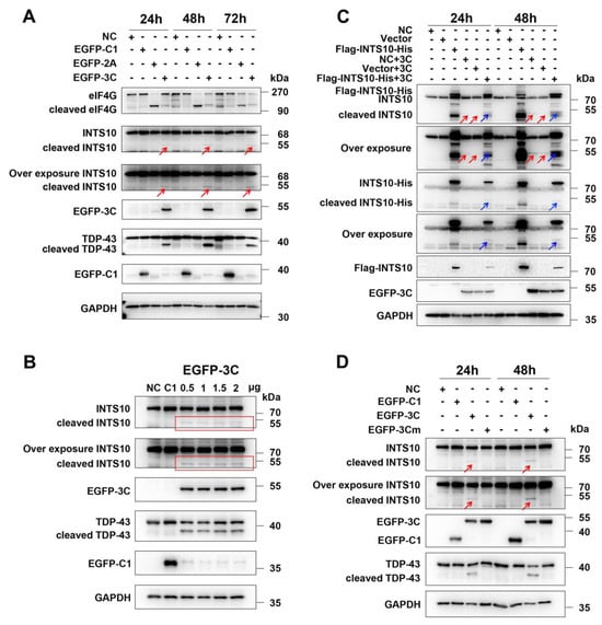
To clarify the precise mechanism by which INTS10 is cleaved, we first investigated whether CVB3-encoded viral proteases 2Apro and 3Cpro participate in this process. Plasmids encoding CVB3 2Apro and 3Cpro (GFP-2A and GFP-3C) were transfected into HEK 293T cells. The results showed that a cleavage fragment was only detected in cells expressing CVB3 3Cpro, but not in GFP control or CVB3 2Apro-expressing cells, indicating that CVB3 3Cpro was involved in the cleavage of INTS10 (Figure 2A, red arrows). We also observed that INTS10 cleavage occurred when co-transfected with different amounts of 3C (Figure 2B, red frame). To further investigate whether CVB3 3Cpro targets INTS10, HEK 293T cells were transfected with GFP-3C plasmid, along with a plasmid expressing INTS10, with a Flag and HA tag fused at its N- and C-termini, respectively. At 48 h after transfection, cell lysates were subjected to detect 3Cpro-mediated cleavage of INTS10 by Western blotting. We found that protein bands (~ 55 kDa of a C-terminal fragment detected by the anti-His antibody) appeared when EGFP-3C was co-transfected with INTS10 but not with the vector plasmid alone (Figure 2C, blue arrows).
Figure 2.
The 3Cpro of CVB3 induces INTS10 cleavage. (A) HEK 293T cells were transfected with the constructs expressing the protease 2A or 3C. INTS10 cleavage was detected by Western blotting. (B) HEK 293T cells were transfected with different concentrations of 3C-expressing vectors for 24 h, and INTS10 cleavage was analyzed by Western blotting. (C) HEK 293T cells were cotransfected with Flag-INTS10-His and 3C-expressing vectors. INTS10 cleavage was detected by Western blotting. (D) HEK 293T cells were transfected with constructs expressing wild-type or mutant 3C. INTS10 cleavage was detected by Western blotting. Red arrows and red frame indicate endogenous INTS10 cleavage fragments, and blue arrows indicate exogenous INTS10 cleavage fragments.
To investigate whether the proteolytic activity of 3Cpro is involved in INTS10 cleavage, we generated a construct expressing 3C enzyme-defective mutant (C147S). The constructs of wild-type (EGFP-3C) and mutant 3C (EGFP-3Cmut) were co-transfected into HEK 293T cells. It is obvious that the 3C enzyme-defective mutant failed to induce INTS10 cleavage compared with the wild-type 3C (Figure 2D, red arrows). These results confirmed that CVB3 3Cpro cleaves INTS10 protein through its proteolytic activity.
2.3. The CVB3 3Cpro Cleaves the INTS10 Protein at Residue Q221
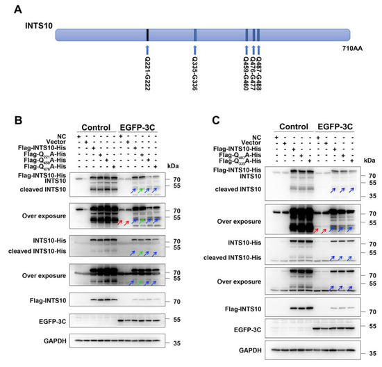
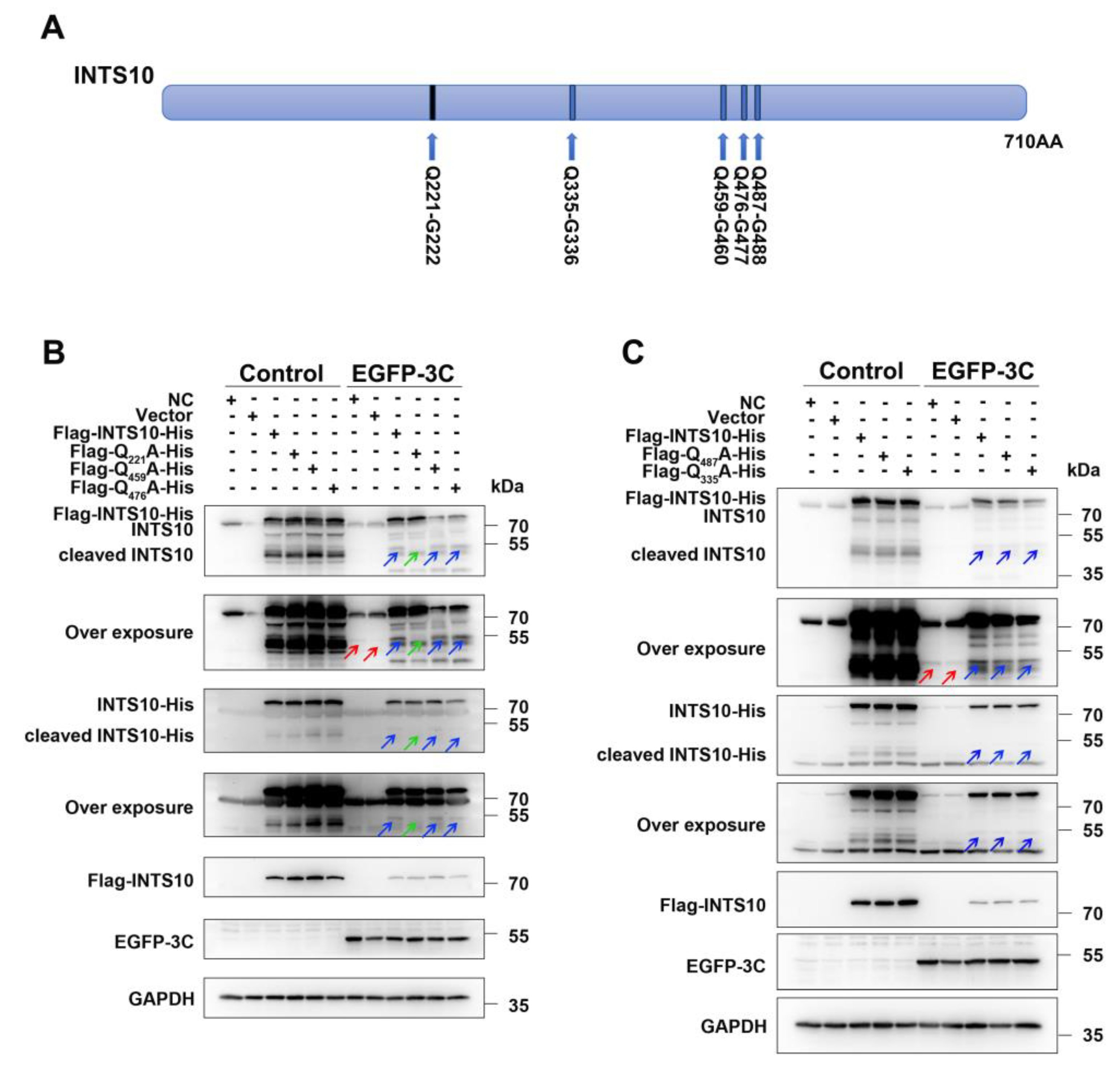
To identify the cleavage site within INTS10, we conducted mutational analysis. Certain glutamine (Q) residues like the Q-G/Q-S motif in the INTS10 sequence are recognized by the CVB3 3Cpro. Based on the molecular weights of the cleavage products, the Q221-G222, Q335-G336, Q459-G460, Q476-G477, and Q487-G488 pairs were identified as candidate cleavage sites (Figure 3A). We generated the five following INTS10 mutants, in which Q residues were substituted with alanine (A): Q221A, Q335A, Q459A, Q476A, and Q487A. As shown in Figure 3B,C, wild-type INTS10 was cleaved by GFP-3C, producing a ~ 55kDa C-terminal fragment (Figure 3B, lane 9, blue arrows). Similarly, the Q335A, Q459A, Q476A, and Q487A mutants were also cleaved by GFP-3C (Figure 3B, lanes 11 and 12; Figure 3C, lanes 8, 9, and 10, blue arrows). In contrast, no cleavage products were detectable in the Q221A mutant co-expressed with 3C (Figure 3B, lane 10, green arrows) with anti-INTS10 or anti-His antibodies. These results demonstrate that the Q221A mutant is resistant to proteolytic cleavage, confirming that CVB3 3Cpro cleaves INTS10 at the Q221-G222 site.
Figure 3.
CVB3 3Cpro cleaves INTS10 at Q221-G222. (A) Schematic diagram of the INTS10 cleavage site. (B,C) HEK 293T cells were co-transfected with Flag-INTS10-His wild-type or mutant variants (Q221A, Q335A, Q459A, Q476A, and Q487A) and 3C-expressing vectors for 48 h. INTS10 cleavage was analyzed by Western blotting. Red arrows indicate endogenous INTS10 cleavage fragments. Blue arrows indicate exogenous INTS10 cleavage fragments. Green arrows indicate the absence of the INTS10 cleavage fragment at its expected migration position.
2.4. Silencing Endogenous INTS10 Expression Enhances CVB3 Replication
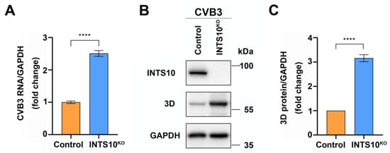
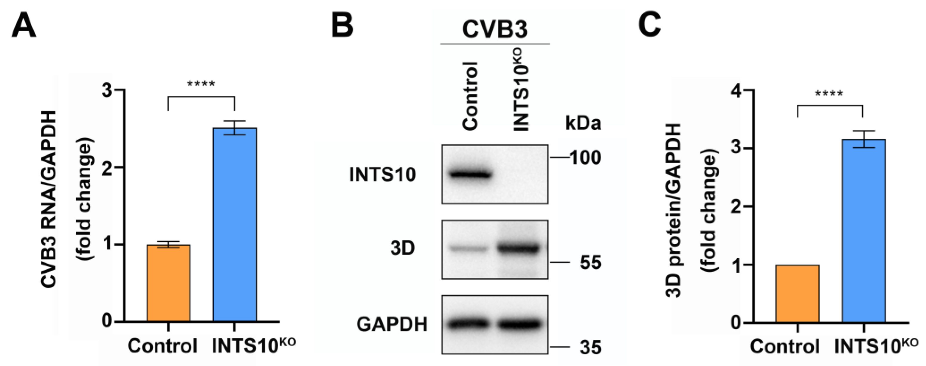
To determine whether INTS10 regulates CVB3 infection, we infected INTS10-KO HEK 293T cells with CVB3. Total RNA and proteins were extracted and analyzed via RT-qPCR and Western blotting. We observed that CVB3 RNA levels increased in the INTS10-silenced cells compared to that of control cells (Figure 4A). Similarly, viral protein 3D levels were elevated in INTS10-KO cells (Figure 4B,C). These results demonstrate that INTS10 exerts an inhibitory effect on CVB3 replication.
Figure 4.
Knockout of INTS10 enhances CVB3 replication. INTS10-KO HEK 293T cells and control cells were infected with CVB3 (MOI = 0.1) for 24 h. (A) The level of CVB3 RNA was determined by RT-qPCR. (B) The expression of CVB3 3D protein was determined via Western blotting. (C) Gray-scale densitometry of CVB3 3D protein bands. Error bars represent the standard deviation (SD). n = 3. **** p < 0.0001.
2.5. 3Cpro-Mediated INTS10 Cleavage Impairs snRNA Processing and Host Defense
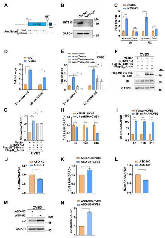
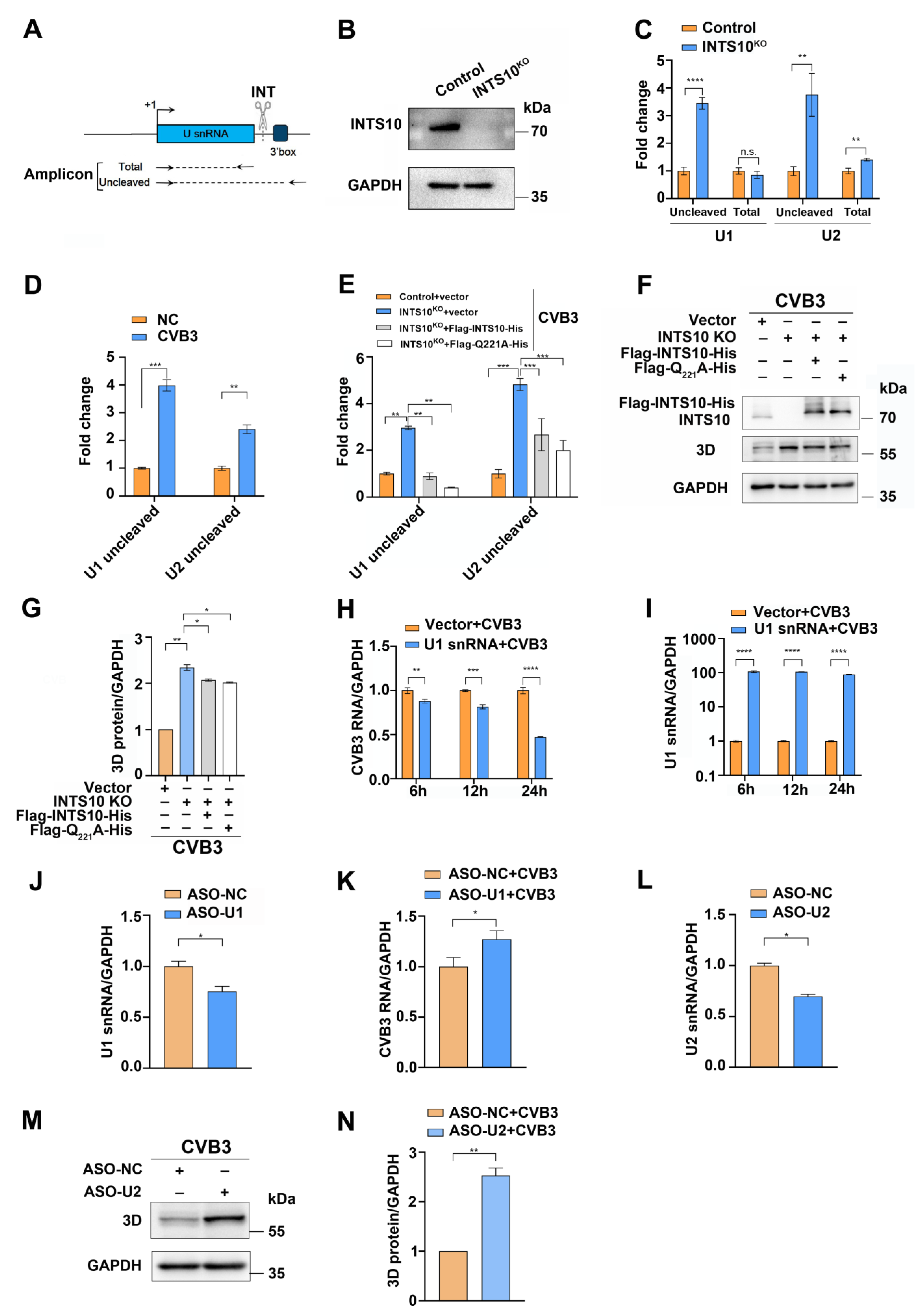
INTS10, a subunit of the integrator complex, plays a critical role in the 3′-end processing of U1 and U2 snRNAs [14]. We hypothesized that 3Cpro-mediated cleavage of INTS10 disrupts snRNA maturation. To test this, total and unprocessed 3′-box regions of U1 and U2 snRNAs were determined by RT-qPCR in INTS10-KO HEK 293T cells (Figure 5A). Notably, unprocessed U1 and U2 snRNA levels increased significantly following INTS10 knockout (Figure 5B,C).
Figure 5.
CVB3-mediated INTS10 cleavage impairs snRNA processing. (A) The schematic diagram of the human U snRNA gene showing the positions of the primers used in RT-qPCR to amplify total and uncleaved U snRNA. (B,C) U1 and U2 snRNA levels in INTS10-KO HEK 293T cells were determined by RT-qPCR. (D) HEK 293T cells were infected with CVB3 (MOI = 0.1) for 24 h. Unprocessed U1 and U2 snRNA levels were subjected to RT-qPCR analysis. (E–G) INTS10-KO HEK 293T cells transfected with Flag-INTS10-His (wild-type or Q221A mutant) were infected with CVB3 (MOI = 0.1). (E) Unprocessed U1 and U2 snRNA levels were determined by RT-qPCR. (F,G) The level of CVB3 3D protein was assessed by Western blotting. (H,I) HEK 293T cells transfected with EGFP-U1 snRNA were infected with CVB3 (MOI = 0.1) for 6, 12, and 24 h. The level of CVB3 RNA and U1 snRNA was determined by RT-qPCR. (J,K) HEK 293T cells transfected with ASO-U1 were infected with CVB3 at MOI of 0.1 for 24 h. The level of U1 snRNA and CVB3 RNA was analyzed by RT-qPCR. (L–N) HEK 293T cells transfected with ASO-U2 were infected with CVB3 (MOI = 0.1) for 24 h. (L) The level of U2 snRNA was determined by RT-qPCR. (M,N) The expression of CVB3 3D protein was detected by Western blotting. Error bars: SD, n = 3. n.s. no significance, * p < 0.05, ** p < 0.01, *** p < 0.001, **** p < 0.0001.
To determine the effect of CVB3 infection on snRNA maturation, HEK 293T cells were infected with CVB3, which resulted in elevated levels of unprocessed U1 and U2 snRNA (Figure 5D). To further assess the impact of CVB3 infection on snRNA maturation, INTS10-KO HEK 293T cells were transfected with wild-type or cleavage-resistant Q221A mutant INTS10 plasmids prior to CVB3 infection. Unprocessed U1 and U2 snRNA levels increased in the CVB3-infected INTS10-KO cells compared to the control cells. However, overexpression of either wild-type or mutant INTS10 reduced unprocessed snRNA levels compared to that of CVB3-infected cells transfected with an empty vector (Figure 5E). These observations demonstrate that INTS10 cleavage disrupts snRNA processing, leading to an accumulation of unprocessed transcripts. Moreover, both the overexpression of wild-type and mutant INTS10 inhibited the expression of the CVB3 3D protein (Figure 5F,G).
Accumulated unprocessed snRNAs correlated with reduced mature U1 and U2 snRNA levels. Given U1 and U2 snRNA’s role in restricting RNA viruses [22,23], we investigated their impact on CVB3 infection. Overexpression of U1 snRNA significantly reduced CVB3 RNA levels (Figure 5H,I), and its knockdown elevated CVB3 RNA levels (Figure 5J,K). This indicates that U1 snRNA inhibits CVB3 infection. Moreover, silencing U2 snRNA significantly increased CVB3 3D protein expression (Figure 5L–N), indicating their antiviral activity. We propose that CVB3 3Cpro cleaves INTS10 to evade U1 and U2 snRNA-dependent host defenses.
3. Discussion
CVB3-encoded 3Cpro targets and disrupts host physiological activities by cleaving cellular proteins that are engaged in gene expression [9,25]. Host proteins with the consensus sequence of AXXQ/GPXX (X denotes any amino acid) could be cleaved between Q and G residues by 3Cpro [26,27]. In this study, we identified that INTS10, a subunit of the integrator complex, is cleaved at Q221-G222 by CVB3 3Cpro. Silencing the expression of INTS10 promoted CVB3 replication, highlighting its critical role in restricting CVB3 infection. Furthermore, silencing the expression of INTS10 induced the accumulation of unprocessed U1 and U2 snRNA, while INTS10 overexpression alleviated this accumulation. We also demonstrated that up-regulating the U1 snRNA inhibits CVB3 replication, while silencing the U1 snRNA promotes CVB3 replication, suggesting that U1 snRNA acts as an intrinsic cellular restriction factor against CVB3 infection. Furthermore, the depletion of U2 snRNA also resulted in enhanced viral replication, suggesting that U2 snRNA also functions as an important host restriction factor for CVB3 infection. We propose that the targeted cleavage of INTS10 by CVB3 3Cpro serves to disrupt the cellular antiviral responses triggered during infection.
Enteroviruses have evolved diverse strategies to disrupt host cell transcription. For example, enterovirus-encoded 3Cpro terminates host transcription during infection [28]. Poliovirus (PV) 2Apro alters pre-mRNA splicing by regulating protein shuttling between the nucleus and the cytoplasm [29]. 3D Polymerase (3Dpol) impairs pre-mRNA splicing by disrupting pre-mRNA processing factor 8 (Prp8), reducing mRNA levels [30]. INTS10, a subunit of the integrator complex, interacts with RNA polymerase II to mediate the 3′ end processing of U1 and U2 snRNAs—core spliceosome components critical for transcription [20,21]. We identified that a lack of INTS10 obstructs U1 and U2 snRNA processing. Moreover, U1 snRNA is essential for host defense against multiple RNA virus infections [22], while the U2 small nuclear ribonucleoprotein (snRNP) suppresses CVB3 replication by exploiting U2 snRNA to interact with viral genomes [23,31]. Here, our results also confirmed that U1 and U2 snRNA have restrictive activity to CVB3 replication, thus providing a mechanistic explanation for how INTS10 cleavage by 3Cpro strategically blocks host transcription to facilitate CVB3 infection.
Previous studies link INTS10 to HBV suppression via IRF3-dependent pathways, which correlates with the persistence of HBV infection [24]. Enteroviruses have developed various countermeasures against the host interferon response [32,33]. In this study, we demonstrated that silencing INTS10 enhances CVB3 RNA and viral proteins, underscoring its role in antiviral defense. Whether INTS10’s inhibition on CVB3 is related to IFN signaling remains to be investigated.
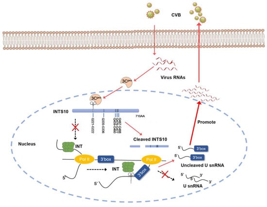
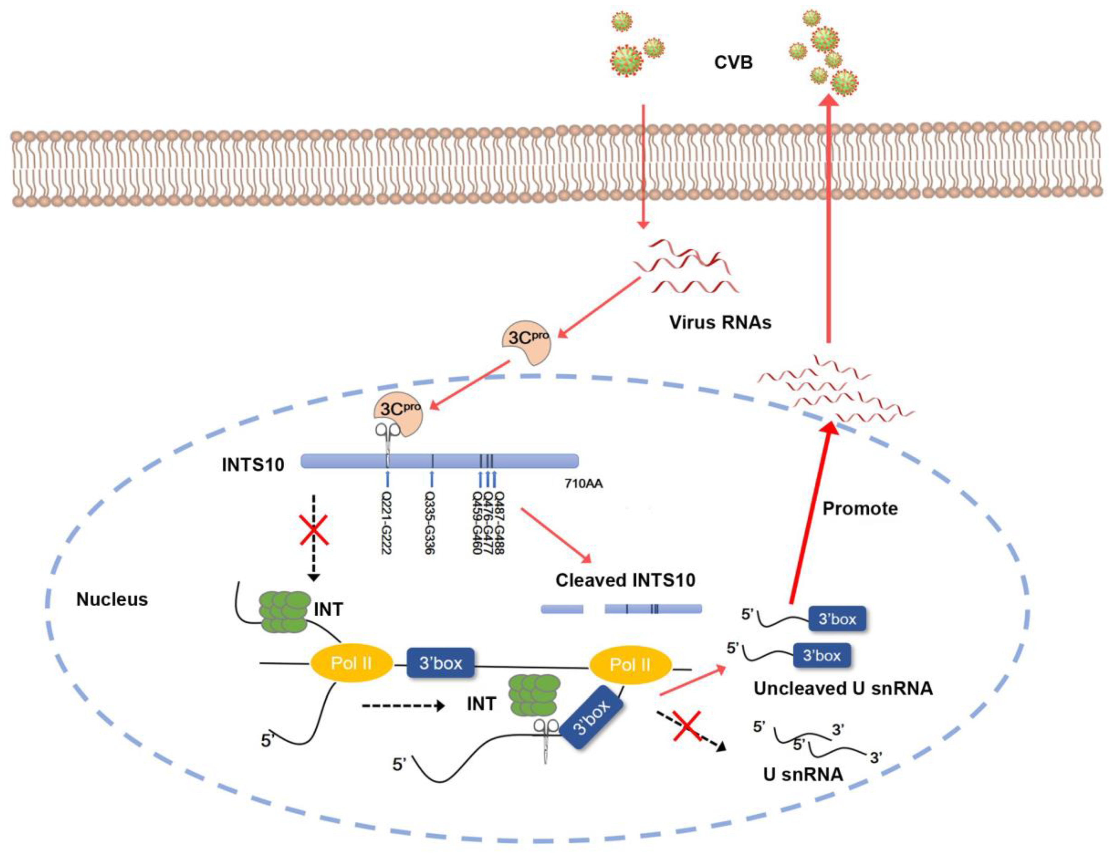
We demonstrated that U1 and U2 snRNA regulate CVB3 replication by reducing levels (MOI = 0.1, 24 h) (Figure 5). Under these conditions, endogenous INTS10 is still present and subject to cleavage, leading to aberrant U1 and U2 snRNA processing and enhanced viral replication. Thus, it is likely that the accumulating misprocessing of U1 and U2 snRNA after multiple infection cycles leads to increased CVB3 replication (Figure 6). A key detail in this schematic diagram that requires further investigation is the subcellular localization of CVB3 3Cpro-mediated cleavage of INTS10. Specifically, it remains unclear whether 3Cpro enters the nucleus to cleave INTS10 or whether 2Apro promotes the nuclear export of INTS10, enabling its cleavage by 3Cpro in the cytoplasm. Previous studies have shown that in PV-infected cells, 3Cpro can localize to the nucleus, where it cleaves cellular transcription factors to suppress host RNA synthesis [13]. Furthermore, enterovirus 2Apro can alter nuclear pore permeability and promote the nuclear export of proteins such as TDP-43, which can be cleaved by 3Cpro [34,35,36]. Based on these findings, we performed cytoplasmic and nuclear fractionation experiments, which revealed that during CVB infection, 3Cpro is present in both the cytoplasm and the nucleus (see Supplementary Figure S2). This observation supports the hypothesis that during CVB3 infection, 3Cpro may enter the nucleus to cleave INTS10. However, this conclusion still requires further experimental validation.
Figure 6.
3Cpro-mediated cleavage of INTS10 increases the U1 and U2 snRNA misprocessing to promote CVB3 replication.
The limitation of this study is that we did not explore the role of cleaved INTS10 fragments in virus infection. Previous studies showed that the proteolytic products of the cellular proteins might interfere with the functions of their normal counterparts [11,37]. Additionally, the molecular mechanism by which U1 snRNA exerts its inhibitory effect on CVB3 replication remains to be studied. It was reported that U1 snRNA interacts with TRIM25 to activate RIG-I and IFN-I signaling, suggesting its role in multiple RNA viruses [22]. Future studies are worth conducting to elucidate the role of U1 snRNA in CVB3 infection.
In summary, our findings reveal how CVB3 overcomes the restriction of snRNA on virus replication by cleaving INTS10. The cleavage of INTS10 leads to disrupted host transcription and innate immune responses, which in turn promote virus infection (Figure 6). Thus, enhancing INTS10 resistance to 3Cpro-mediated cleavage or upregulating U1 and U2 snRNA could offer effective antiviral strategies.
4. Materials and Methods
4.1. Cells and Viruses
HEK 293T and HeLa cells were cultured in Dulbecco’s Modified Eagle Medium (DMEM, Gibco, Shanghai, China) supplemented with 10% fetal bovine serum (FBS, Biological Industries, Israel), and 1% penicillin/streptomycin at 37 °C. CVB3 woodruff was kindly provided by the Scrips Institute (San Diego, CA, USA). CVB3 stocks were propagated and titered in HeLa cells.
4.2. Plasmids and DNA Transfections
Empty vector pcDNA3.4(+)-3 × Flag-MCS-6 × His and wild-type plasmid-expressing INTS10 named pcDNA3.4(+)-3 × Flag-INTS10-6 × His were purchased from Miaoling Biology (Wuhan, China). The expression plasmids for CVB3 2A, and 3C with a GFP tag in pEGFP-C1 were generated as described previously. The CVB3 3C (C147S) and Flag-INTS10-His mutants (Q221A, Q335A, Q459A, Q476A, and Q487A) were constructed by site-specific mutagenesis. The plasmid-expressing U1 snRNA, designated as pEGFP-U1 snRNA, was constructed based on pcDNA3.1-EGFP. The following primers were used:
For Q221A:
- Forward primer: 5′-TGGGCGCCTGCATGCTGATTTTC-3′
- Reverse primer: 5′-AATCAGCATGCAGGCGCCCAGG-3′
For Q335A:
- Forward primer: 5′-GGACCTGCGAAGAGAGATGGCTGTATGC-3′
- Reverse primer: 5′-TCTCTTCGCAGGTCCTAATGCCCCG-3′
For Q459A:
- Forward primer: 5′-CGCCTTTTTATATTGACCCGCATAGATGATC-3′
- Reverse primer: 5′-ATATGATCATCTATGCGGGTCAATATAAAAAGGC-3′
For Q476A:
- Forward primer: 5′-AAATGGATCCCGCGAGAGCTGCTAAG-3′
- Reverse primer: 5′-AGCAGCTCTCGCGGGATCCATTTCT-3′
For Q487A:
- Forward primer: 5′-CAGGGTCCCCGCCCCTGTGATC-3′
- Reverse primer: 5′-ACAGGGGCGGGGACCCTGGAG-3′
For pEGFP-U1 snRNA
- Forward primer: 5′-GCGCGGAAGCTTTGATTTCATACTTACCT-3′
- Reverse primer: 5′-TATATAGAATTCCAGGGGAAAGCGCGAAC-3′
All DNA transfections were performed using Lipofectamine 2000 according to the manufacturer’s protocol (Themo Fisher Scientific, Waltham, MA, USA). Briefly, cells were seeded 24 h before transfection to allow for adherence. Transfection took place in antibiotic-free DMEM supplemented with 10% FBS. All transfections were conducted for 24 h prior to experimentation.
4.3. ASO Transfection
HEK 293T cells were seeded in a 12-well plate for 24 h. Cells were transfected with ASOs (GenePharma, Suzhou, China) at a final concentration of 20 nM, using siRNA-mate plus transfection reagent according to the manufacturer’s instructions (GenePharma, Suzhou, China) and incubated at 37 °C with 5% CO2 for 48 h. U1 snRNA-ASO: mG*mA*mG*mA*mT*A*C*C*A*T*G*A*T*C*mA*mC*mG*mA*mA; U2 snRNA-ASO: mA*mG*mA*mA*mC*A*G*A*T*A*C*T*A*C*A*mC*mT*mT*mG*mA.
4.4. Virus Infections
CVB3 viruses, EV-A71, and CAV16 were incubated with cells at the indicated MOI for 1 h in DMEM + 1% penicillin/streptomycin at 37 °C. After adsorption, the media was replaced with complete DMEM (1× DMEM, 10% FBS, 1% penicillin/streptomycin) and incubated for the designated time.
4.5. Western Blotting
Cells were harvested using RIPA lysis buffer (Thermo Fisher Scientific, Waltham, MA, USA) containing a protease inhibitor cocktail and 1% phenylmethylsulfonyl fluoride (PMSF, Beyotime, Shanghai, China) and were lysed for 30 min. Equal amounts of protein were separated on 10% or 12.5% SDS-PAGE gel and subsequently transferred onto a polyvinylidene difluoride (PVDF, Millipore, Billerica, MA, USA) membrane. PVDF membranes were blocked with skimmed milk for 1 h and incubated with the primary antibody overnight at 4 °C. The primary antibodies used were as follows: 1:2000 INTS10 (Proteintech, Wuhan, China); 1:1000 eIF4G (Proteintech, Wuhan, China); 1:5000 TDP-43 (Proteintech, Wuhan, China); 1:3000 GFP (ABclonal, Wuhan, China); 1:25,000 Flag (Proteintech, Wuhan, China); 1:10,000 His (Proteintech, Wuhan, China); and 1:20,000 GAPDH (ABclonal, Wuhan, China). Membranes were washed with TBST and incubated with anti-rabbit or anti-mouse IgG for 1 h at room temperature. Finally, the membrane was imaged with a FluorChem M CCD camera (ProteinSimple, Santa Clara, CA, USA) and analyzed by ImageJ software, version 1.54g.
4.6. Reverse Transcription Quantitative PCR (RT-qPCR)
RT-qPCR was performed using total cellular cDNA. Briefly, cells were harvested in 1 mL Trizol (Yeasen, Shanghai, China), and total cellular RNA was isolated according to the manufacturer’s protocol. A total of 1 μg of total RNA was used as a template with PrimeScript RT Enzyme Mix I (TaKaRa, Dalian, China) in a reverse transcription system. RT-qPCR was performed on a LightCycler 96 (Roche, Basel, Switzerland) using SYBR Premix Ex Taq II (TaKaRa, Dalian, China) mixed with 1 μL of the synthesized cDNA and sense/antisense primers to reach a final volume of 20 μL. Relative RNA quantity was calculated using the 2−ΔΔCT method and normalized to the quantity of GAPDH. The following primers were used for analysis:
For GAPDH:
- Forward primer: 5′-CTGGGCTACACTGAGCACC-3′
- Reverse primer: 5′-AAGTGGTCGTTGAGGGCAATG-3′
For CVB3:
- Forward primer: 5′-GCACACACCCTCAAACCAGA-3′
- Reverse primer: 5′-ATGAAACACGGACACCCAAAG-3′
For INTS10:
- Forward primer: 5′-GCACACACCCTCAAACCAGA-3′
- Reverse primer: 5′-ATGAAACACGGACACCCAAAG-3′
For uncleaved U1:
- Forward primer: 5′-TACCTGGCAGGGGAGATACC-3′
- Reverse primer: 5′-GCGTACGGTCTGTTTTTGAAACTC-3′
For total U1:
- Forward primer: 5′-ATACCATGATCACGAAGGTGGTT-3′
- Reverse primer: 5′-CAGTCCCCCACTACCACAAATTA-3′
For uncleaved U2:
- Forward primer: 5’-AACATAGGTACACGTGTGCCACGG-3’
- Reverse primer: 5’-ACAAATAGCCAACGCATGCGGGGC-3’
For total U2:
- Forward primer: 5’-CTTCTCGGCCTTTTGGCTAAGAT-3’
- Reverse primer: 5’-GTACTGCAATACCAGGTCGATGC-3’
For U1 snRNA:
- Forward primer: 5’-CTTACCTGGCAGGGGAGATA-3’
- Reverse primer: 5’-GCAGTCGAGTTTCCCACATT-3’
For U2 snRNA:
- Forward primer: 5’-CTTCTCGGCCTTTTGGCTAAGAT-3’
- Reverse primer: 5’-GTACTGCAATACCAGGTCGATGC-3’
4.7. Statistical Analyses
Statistical data were analyzed using Graphpad Prism 8. Quantitative data were expressed as mean ± SD. Differences among test groups were analyzed by an analysis of variance (ANOVA) test or Student’s t-test. A value of p < 0.05 was considered statistically significant. All experiments were repeated three times.
Supplementary Materials
The following supporting information can be downloaded at https://www.mdpi.com/article/10.3390/ijms27020996/s1.
Author Contributions
Conceptualization, W.Z., L.L. (Lexun Lin) and Z.Z.; methodology, L.Y.; software, C.B.; validation, L.L. (Liling Lin) and X.N.; formal analysis, L.L. (Liling Lin) and Y.C.; investigation, Y.F.; resources, G.W.; data curation, H.W. and Y.W.; writing—original draft preparation, L.L. (Lexun Lin); writing—review and editing, W.Z. and Z.Z.; visualization, L.Y.; supervision, L.L. (Lexun Lin); project administration, Z.Z.; funding acquisition, L.L. (Lexun Lin), Z.Z., W.Z., Y.F. and Y.W. All authors have read and agreed to the published version of the manuscript.
Funding
The study was funded by the National Natural Science Foundation of China 82202493, 82172247, 82072278, 82402598, and the Natural Science Foundation of Heilongjiang Province grant LH2022H049.
Institutional Review Board Statement
Not applicable.
Informed Consent Statement
Not applicable.
Data Availability Statement
The original contributions presented in this study are included in the article/Supplementary Material. Further inquiries can be directed to the corresponding authors.
Acknowledgments
We thank Wu Lien-Teh Biomedical Innovation Institute, Harbin Medical University, for technical assistance.
Conflicts of Interest
The authors declare no conflicts of interest.
Abbreviations
The following abbreviations are used in this manuscript:
| INST10 | Integrator subunit 10 |
| CVB | Coxsackievirus B |
| 3Cpro | 3C protease |
| 2Apro | 2A protease |
| Stau2 | Staufen homolog 2 |
| snRNAs | Small nuclear RNAs |
| TRIM | Tripartite motif |
| IRF3 | Interferon regulatory factor 3 |
| MW | Molecular weight |
| PV | Poliovirus |
| 3Dpol | 3D Polymerase |
| Prp8 | Pre-mRNA processing factor 8 |
| snRNP | Small nuclear ribonucleoprotein |
References
- Bouin, A.; Gretteau, P.A.; Wehbe, M.; Renois, F.; N’Guyen, Y.; Lévêque, N.; Vu, M.N.; Tracy, S.; Chapman, N.M.; Bruneval, P.; et al. Enterovirus Persistence in Cardiac Cells of Patients with Idiopathic Dilated Cardiomyopathy Is Linked to 5’ Terminal Genomic RNA-Deleted Viral Populations with Viral-Encoded Proteinase Activities. Circulation 2019, 139, 2326–2338. [Google Scholar] [CrossRef] [Scilit]
- Garmaroudi, F.S.; Marchant, D.; Hendry, R.; Luo, H.; Yang, D.; Ye, X.; Shi, J.; Mcmanus, B.M. Coxsackievirus B3 replication and pathogenesis. Future Microbiol. 2015, 10, 629–653. [Google Scholar] [CrossRef] [Scilit]
- Yajima, T. Viral myocarditis: Potential defense mechanisms within the cardiomyocyte against virus infection. Future Microbiol. 2011, 6, 551–566. [Google Scholar] [CrossRef] [Scilit]
- Knowlton, K.U. CVB infection and mechanisms of viral cardiomyopathy. Curr. Top. Microbiol. Immunol. 2008, 323, 315–335. [Google Scholar]
- Fairweather, D.; Stafford, K.A.; Sung, Y.K. Update on coxsackievirus B3 myocarditis. Curr. Opin. Rheumatol. 2012, 24, 401–407. [Google Scholar] [CrossRef] [Scilit] [PubMed]
- Kemball, C.C.; Alirezaei, M.; Whitton, J.L. Type B coxsackieviruses and their interactions with the innate and adaptive immune systems. Future Microbiol. 2010, 5, 1329–1347. [Google Scholar] [CrossRef] [Scilit] [PubMed]
- Tong, L.; Lin, L.; Zhao, W.; Wang, B.; Wu, S.; Liu, H.; Zhong, X.; Cui, Y.; Gu, H.; Zhang, F.; et al. Destabilization of coxsackievirus b3 genome integrated with enhanced green fluorescent protein gene. Intervirology 2011, 54, 268–275. [Google Scholar] [CrossRef] [Scilit] [PubMed]
- Hanson, P.J.; Hossain, A.R.; Qiu, Y.; Zhang, H.M.; Zhao, G.; Li, C.; Lin, V.; Sulaimon, S.; Vlok, M.; Fung, G.; et al. Cleavage and Sub-Cellular Redistribution of Nuclear Pore Protein 98 by Coxsackievirus B3 Protease 2A Impairs Cardioprotection. Front. Cell. Infect. Microbiol. 2019, 9, 265. [Google Scholar] [CrossRef] [Scilit]
- Lei, X.; Han, N.; Xiao, X.; Jin, Q.; He, B.; Wang, J. Enterovirus 71 3C Inhibits Cytokine Expression through Cleavage of the TAK1/TAB1/TAB2/TAB3 Complex. J. Virol. 2014, 88, 9830–9841. [Google Scholar] [CrossRef] [Scilit]
- Mukherjee, A.; Morosky, S.A.; Delorme-Axford, E.; Dybdahl-Sissoko, N.; Oberste, M.S.; Wang, T.; Coyne, C.B. The coxsackievirus B 3C protease cleaves MAVS and TRIF to attenuate host type I interferon and apoptotic signaling. PLoS Pathog. 2011, 7, e1001311. [Google Scholar] [CrossRef] [Scilit]
- Andrews, D.D.T.; Vlok, M.; Akbari Bani, D.; Hay, B.N.; Mohamud, Y.; Foster, L.J.; Luo, H.; Overall, C.M.; Jan, E. Cleavage of 14-3-3ε by the enteroviral 3C protease dampens RIG-I-mediated antiviral signaling. J. Virol. 2023, 97, e0060423. [Google Scholar] [CrossRef] [Scilit]
- Li, H.; Song, J.; Deng, Z.; Yao, Y.; Qiao, W.; Tan, J. Cleavage of Stau2 by 3C protease promotes EV-A71 replication. Virol. J. 2024, 21, 216. [Google Scholar] [CrossRef] [Scilit]
- De Jesús-González, L.A.; Palacios-Rápalo, S.; Reyes-Ruiz, J.M.; Osuna-Ramos, J.F.; Cordero-Rivera, C.D.; Farfan-Morales, C.N.; Gutiérrez-Escolano, A.L.; Del Ángel, R.M. The Nuclear Pore Complex Is a Key Target of Viral Proteases to Promote Viral Replication. Viruses 2021, 13, 706. [Google Scholar] [CrossRef] [Scilit]
- Mendoza-Figueroa, M.S.; Tatomer, D.C.; Wilusz, J.E. The Integrator Complex in Transcription and Development. Trends Biochem. Sci. 2020, 45, 923–934. [Google Scholar] [CrossRef] [Scilit] [PubMed]
- Razew, M.; Fraudeau, A.; Pfleiderer, M.M.; Linares, R.; Galej, W.P. Structural basis of the Integrator complex assembly and association with transcription factors. Mol. Cell 2024, 84, 2542–2552.e5. [Google Scholar] [CrossRef] [Scilit] [PubMed]
- Lai, F.; Gardini, A.; Zhang, A.; Shiekhattar, R. Integrator mediates the biogenesis of enhancer RNAs. Nature 2015, 525, 399–403. [Google Scholar] [CrossRef] [Scilit]
- Tepe, B.; Macke, E.L.; Niceta, M.; Weisz Hubshman, M.; Kanca, O.; Schultz-Rogers, L.; Zarate, Y.A.; Schaefer, G.B.; Granadillo De Luque, J.L.; Wegner, D.J.; et al. Bi-allelic variants in INTS11 are associated with a complex neurological disorder. Am. J. Hum. Genet. 2023, 110, 774–789. [Google Scholar] [CrossRef] [Scilit]
- Oegema, R.; Baillat, D.; Schot, R.; van Unen, L.M.; Brooks, A.; Kia, S.K.; Hoogeboom, A.J.M.; Xia, Z.; Li, W.; Cesaroni, M.; et al. Human mutations in integrator complex subunits link transcriptome integrity to brain development. PLoS Genet. 2017, 13, e1006809, Erratum in PLoS Genet. 2017, 13, e1006923.. [Google Scholar] [CrossRef] [Scilit] [PubMed]
- Tilley, F.C.; Arrondel, C.; Chhuon, C.; Boisson, M.; Cagnard, N.; Parisot, M.; Menara, G.; Lefort, N.; Guerrera, I.C.; Bole-Feysot, C.; et al. Disruption of pathways regulated by Integrator complex in Galloway-Mowat syndrome due to WDR73 mutations. Sci. Rep. 2021, 11, 5388. [Google Scholar] [CrossRef] [Scilit]
- Yamamoto, J.; Hagiwara, Y.; Chiba, K.; Isobe, T.; Narita, T.; Handa, H.; Yamaguchi, Y. DSIF and NELF interact with Integrator to specify the correct post-transcriptional fate of snRNA genes. Nat. Commun. 2014, 5, 4263. [Google Scholar] [CrossRef] [Scilit]
- Sabath, K.; Stäubli, M.L.; Marti, S.; Leitner, A.; Moes, M.; Jonas, S. INTS10-INTS13-INTS14 form a functional module of Integrator that binds nucleic acids and the cleavage module. Nat. Commun. 2020, 11, 3422. [Google Scholar] [CrossRef] [Scilit]
- Zhang, F.; Liu, S.; Qiao, Z.; Li, L.; Han, Y.; Sun, J.; Ge, C.; Zhu, J.; Li, D.; Yao, H.; et al. Housekeeping U1 snRNA facilitates antiviral innate immunity by promoting TRIM25-mediated RIG-I activation. Cell Rep. 2024, 43, 113945. [Google Scholar] [CrossRef] [Scilit] [PubMed]
- Kamel, W.; Ruscica, V.; Embarc-Buh, A.; Laurent, Z.R.D.; Garcia-Moreno, M.; Demyanenko, Y.; Orton, R.J.; Noerenberg, M.; Madhusudhan, M.; Iselin, L. Alphavirus infection triggers selective cytoplasmic translocation of nuclear RBPs with moonlighting antiviral roles. Mol. Cell 2024, 84, 4896–4911.e7. [Google Scholar] [CrossRef] [Scilit] [PubMed]
- Li, Y.F.; Si, L.L.; Zhai, Y.; Hu, Y.L.; Hu, Z.B.; Bei, J.X.; Xie, B.B.; Ren, Q.; Cao, P.B.; Yang, F.; et al. Genome-wide association study identifies 8p21.3 associated with persistent hepatitis B virus infection among Chinese. Nat. Commun. 2016, 7, 11664. [Google Scholar] [CrossRef] [Scilit]
- Rui, Y.; Su, J.; Wang, H.; Chang, J.; Wang, S.; Zheng, W.; Cai, Y.; Wei, W.; Gordy, J.T.; Markham, R. Disruption of MDA5-Mediated Innate Immune Responses by the 3C Proteins of Coxsackievirus A16, Coxsackievirus A6, and Enterovirus D68. J. Virol. 2017, 91, e00546-17. [Google Scholar] [CrossRef] [Scilit]
- Fan, W.; McDougal, M.B.; Schoggins, J.W.; Schultz-Cherry, S. Enterovirus 3C Protease Cleaves TRIM7 To Dampen Its Antiviral Activity. J. Virol. 2022, 96, e0133222. [Google Scholar] [CrossRef] [Scilit] [PubMed]
- Zhang, D.; Xie, Y.; Cao, J.; Huang, L.; Fan, W.; Dutch, R.E. Enteroviral 3C protease cleaves N4BP1 to impair the host inflammatory response. J. Virol. 2025, 99, e0175824. [Google Scholar] [CrossRef] [Scilit]
- Sharma, R.; Raychaudhuri, S.; Dasgupta, A. Nuclear entry of poliovirus protease-polymerase precursor 3CD: Implications for host cell transcription shut-off. Virology 2004, 320, 195–205. [Google Scholar] [CrossRef] [Scilit]
- Álvarez, E.; Castelló, A.; Carrasco, L.; Izquierdo, J.M. Poliovirus 2A protease triggers a selective nucleo-cytoplasmic redistribution of splicing factors to regulate alternative pre-mRNA splicing. PLoS ONE 2013, 8, e73723. [Google Scholar] [CrossRef] [Scilit]
- Liu, Y.C.; Kuo, R.L.; Lin, J.Y.; Huang, P.N.; Huang, Y.; Liu, H.; Arnold, J.J.; Chen, S.J.; Wang, R.Y.; Cameron, C.E.; et al. Cytoplasmic viral RNA-dependent RNA polymerase disrupts the intracellular splicing machinery by entering the nucleus and interfering with Prp8. PLoS Pathog. 2014, 10, e1004199. [Google Scholar] [CrossRef] [Scilit]
- Castello, A.; Kamel, W. Nuclear RNA-binding proteins meet cytoplasmic viruses. RNA 2025, 31, 444–451. [Google Scholar] [CrossRef] [Scilit]
- Liang, T.; Zhang, Z.; Bai, Z.; Xu, L.; Xu, W. STAT3 Increases CVB3 Replication and Acute Pancreatitis and Myocarditis Pathology via Impeding Nuclear Translocation of STAT1 and Interferon-Stimulated Gene Expression. Int. J. Mol. Sci. 2024, 25, 9007. [Google Scholar] [CrossRef] [Scilit]
- Feng, Q.; Langereis, M.A.; Lork, M.; Nguyen, M.; Hato, S.V.; Lanke, K.; Emdad, L.; Bhoopathi, P.; Fisher, P.B.; Lloyd, R.E.; et al. Enterovirus 2Apro targets MDA5 and MAVS in infected cells. J. Virol. 2014, 88, 3369–3378. [Google Scholar] [CrossRef] [Scilit] [PubMed]
- Park, N.; Schweers, N.J.; Gustin, K.E. Selective Removal of FG Repeat Domains from the Nuclear Pore Complex by Enterovirus 2A(pro). J. Virol. 2015, 89, 11069–11079. [Google Scholar] [CrossRef] [Scilit]
- Wo, X.; Yuan, Y.; Xu, Y.; Chen, Y.; Wang, Y.; Zhao, S.; Lin, L.; Zhong, X.; Wang, Y.; Zhong, Z.; et al. TAR DNA-Binding Protein 43 is Cleaved by the Protease 3C of Enterovirus A71. Virol. Sin. 2020, 36, 95–103. [Google Scholar] [CrossRef] [Scilit] [PubMed]
- Zhang, L.; Yang, J.; Li, H.; Zhang, Z.; Ji, Z.; Zhao, L.; Wei, W. Enterovirus D68 Infection Induces TDP-43 Cleavage, Aggregation, and Neurotoxicity. J. Virol. 2023, 97, e0042523. [Google Scholar] [CrossRef] [Scilit]
- Li, X.; Guo, H.; Yang, J.; Liu, X.; Li, H.; Yang, W.; Zhang, L.; Li, Y.; Wei, W. Enterovirus D68 3C protease antagonizes type I interferon signaling by cleaving signal transducer and activator of transcription 1. J. Virol. 2024, 98, e0199423. [Google Scholar] [CrossRef] [Scilit] [PubMed]
Disclaimer/Publisher’s Note: The statements, opinions and data contained in all publications are solely those of the individual author(s) and contributor(s) and not of MDPI and/or the editor(s). MDPI and/or the editor(s) disclaim responsibility for any injury to people or property resulting from any ideas, methods, instructions or products referred to in the content. |
© 2026 by the authors. Licensee MDPI, Basel, Switzerland. This article is an open access article distributed under the terms and conditions of the Creative Commons Attribution (CC BY) license.