


Abstract
Mesenchymal neoplasms driven by protein kinases represent an increasing group with a broad clinicopathologic and genetic spectrum. Recently, tumors harboring a GAB1::ABL1 fusion have been reported, resulting in constitutive activation of the non-receptor tyrosine kinase ABL1. We describe six cases (one previously reported) investigated by fusion gene analysis and DNA-methylation profiling. Tumors were from three males and three females aged 7–71 years and arose in the neck, axilla, lower back, thigh, and digits. Lesions were excised. Available follow-up was uneventful. Neoplasms were circumscribed but unencapsulated, with infiltration of the surrounding tissue, and characterized by haphazardly arranged monomorphous spindle cells. There was a fibromyxoid stroma and staghorn-like vessels were present. Immunohistochemically, expression of CD34 (3/6), S100 (1/6), EMA (2/6), and GLUT1 (3/3) was observed. All cases harbored a GAB1::ABL1 fusion. RNA expression profile (n = 2) showed high-confidence similarity to dermatofibrosarcoma protuberans (DFSP). DNA-methylation profiling demonstrated that all cases clustered together in close proximity to DFSP. Application of the latest Heidelberg methylation sarcoma classifier did not confidentially classify these neoplasms. However, four showed low-confidence matches to NTRK-rearranged spindle cell neoplasms. GAB1::ABL1 spindle cell neoplasms represent a distinct member of the family of kinase-driven mesenchymal tumors with an indolent clinical course, while the activating ABL1 fusion raises the possibility of targeted therapy in selected cases.
1. Introduction
With the increased use of advanced molecular techniques, the detection of genetic alterations of tumors is rapidly expanding including a group of mesenchymal lesions with changes in genes encoding for protein kinases (PKs). The involved PKs are mainly receptor tyrosine kinases (RTKs), most notably NTRK1, 2, 3, and others, e.g., ALK, RET, MET and EGFR or intracellular non-receptor tyrosine and serine/threonine kinases such as ABL1 and the RAF proteins, respectively [1,2,3,4]. Fusion genes are the most common abnormality, followed by tandem repeat duplications, point mutations, deletions, and genomic amplifications or overexpression, all resulting in an autoactivation of the kinase domain [4,5,6,7]. Mesenchymal neoplasms driven by activated PKs have a variable histomorphology, but common findings are haphazardly distributed (myo)fibroblastic cells—primitive (more oval) and/or maturated (more elongated)—with monomorphic nuclei, staghorn-like vessels with hyalinization, and the presence of thick collagen bundles. An immunohistochemical hallmark is the co-expression of S100 and CD34, without SOX10 positivity. However, the immunophenotype is also variable [6,8].
Lately, several spindle cell neoplasms with a GAB1::ABL1 gene fusion as the oncogenic driver have been reported [9,10,11,12,13]. The fusion gene includes the tyrosine kinase domain of ABL1, which becomes constitutively active, thereby influencing downstream signaling pathways including MAPK and AKT. Therefore, it seems that these neoplasms are a member of the emerging group of PK-related mesenchymal tumors [2,3,6,10].
In this study, we describe the clinicopathological, genetic, and epigenetic characteristics of five new cases and include a previously reported case [14], adding to our understanding, classification, and identification of these tumors.
2. Results
2.1. Clinical Characteristics (Table 1)
The six tumors were from three males and three females, aged 7–71 years (median, 17 years; mean, 26 years). They arose in the soft tissue of the neck, axilla, lower back, thigh, and digits (hand and foot). All lesions were excised, with R0 status in five cases, including re-excision in case 2. Case 1 showed microscopically positive margins. Follow-up, available for four patients (cases 2, 4, 5 and 6), was uneventful (1–8 years, median 3 years). The other two lesions (cases 1 and 3) were diagnosed recently.
Table 1.
Clinical, immunohistochemical, and molecular characteristics of our cases.
2.2. Pathological Findings
Grossly, the tumors were homogeneously tan-white and circumscribed. The size ranged from 1.1 to 13.5 cm (median, 3.25 cm; mean, 5.43 cm) (Table 1).
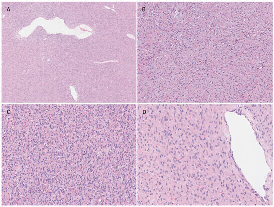
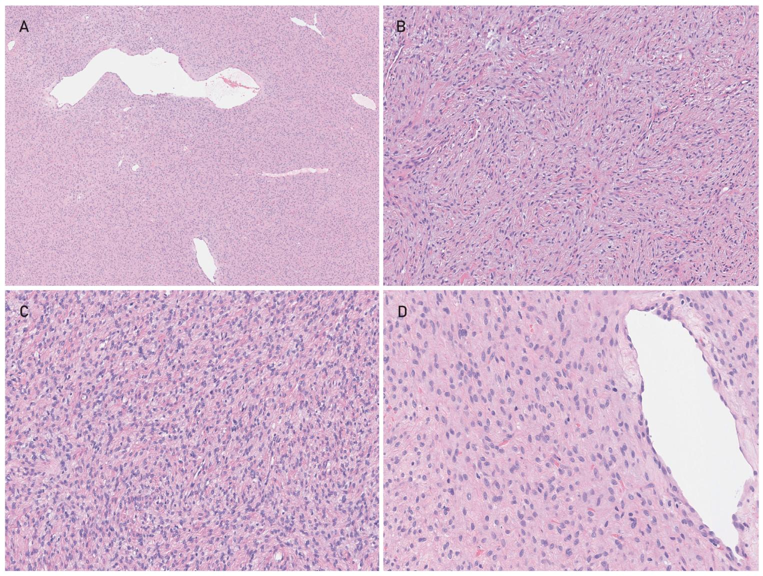
Histologically, all tumors were circumscribed but unencapsulated, with infiltration of the surrounding soft tissue. They were composed of mainly haphazardly arranged monomorphous spindle cells, with some areas showing vague bundles, whorls, or a storiform growth pattern. The nuclei were oval and tapered and had a smooth contour and a homogeneous open chromatin. There was an inconspicuous cytoplasm. Mitotic activity was negligible. The stroma was fibromyxoid with variable wispy or thick collagen bands. The latter were observed in cases 1, 3, 5, and 6. Staghorn-like vessels and hyalinization, also of smaller vessels, was variably observed in all but case 5. None of the lesions showed necrosis (Figure 1).
Figure 1.
Morphological features (H&E stain). (A) Haphazardly arrangend monomorphous spindle cells set in a fibromyxoid stroma with staghorn-like vessels (case 3). Magnification 5×. (B) A whorled and storiform growth pattern was seen in some areas (case 2). Magnification 15×. (C) The spindle cells possess monomorphic oval and tapered nuclei. Note the collagen bundles and small vessels in the background (case 1). Magnification 15×. (D) At higher magnification (25×), nuclei show smooth contours and an evenly distributed chromatin. The cytoplasm is inconspicuous. Mitotic figures were negligible (case 3).
2.3. Immunohistochemical Results (Table 1)
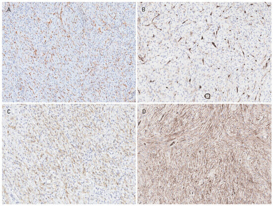
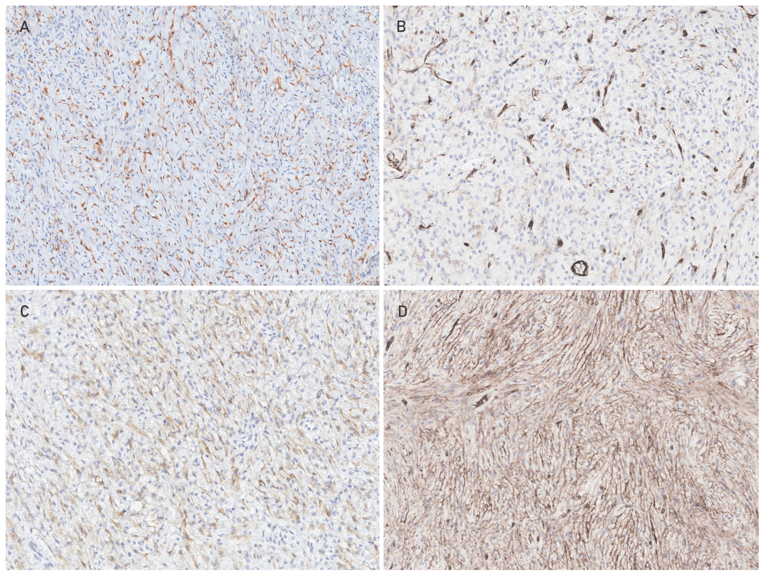
CD34 was variably expressed in 3/6 cases, S100 showed a patchy reaction in 1/6 cases and EMA was positive in 2/6 lesions. GLUT1 positivity was seen in all three neoplasms tested (Figure 2). Negative markers were SMA (n = 4), desmin (n = 2), pan-keratin (CKAE1/3) (n = 4), MUC4 (n = 3), SOX10 (n = 4), STAT6 (n = 2), ALK (n = 1), and panTRK (n = 1).
Figure 2.
Immunohistochemical results showing variable expression of S100 (case 6) (A), CD34 (case 3) (B), EMA (case 3) (C), and GLUT1 (case 3) (D).
2.4. Molecular Results (Table 1)
Using RNA sequencing (n = 4) or anchored multiplex PCR-based targeted RNA sequencing (n = 2), in all cases, a GAB1 (exon 6)::ABL1 (exon 2) fusion gene was detected.
In two cases (cases 1 and 3), our RNA expression classifier (version 5.9) was applied, classifying them as dermatofibrosarcoma protuberans (score 0.96).
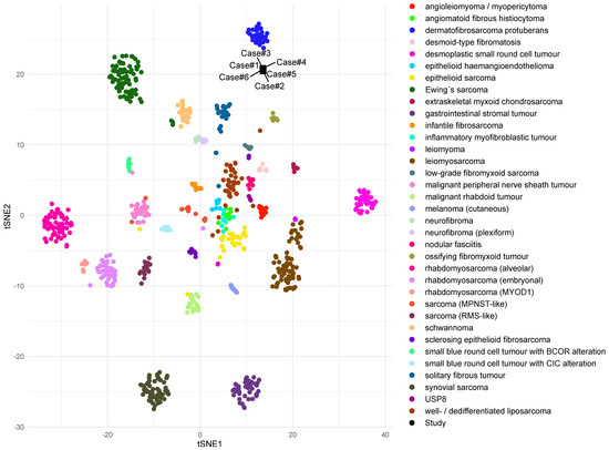
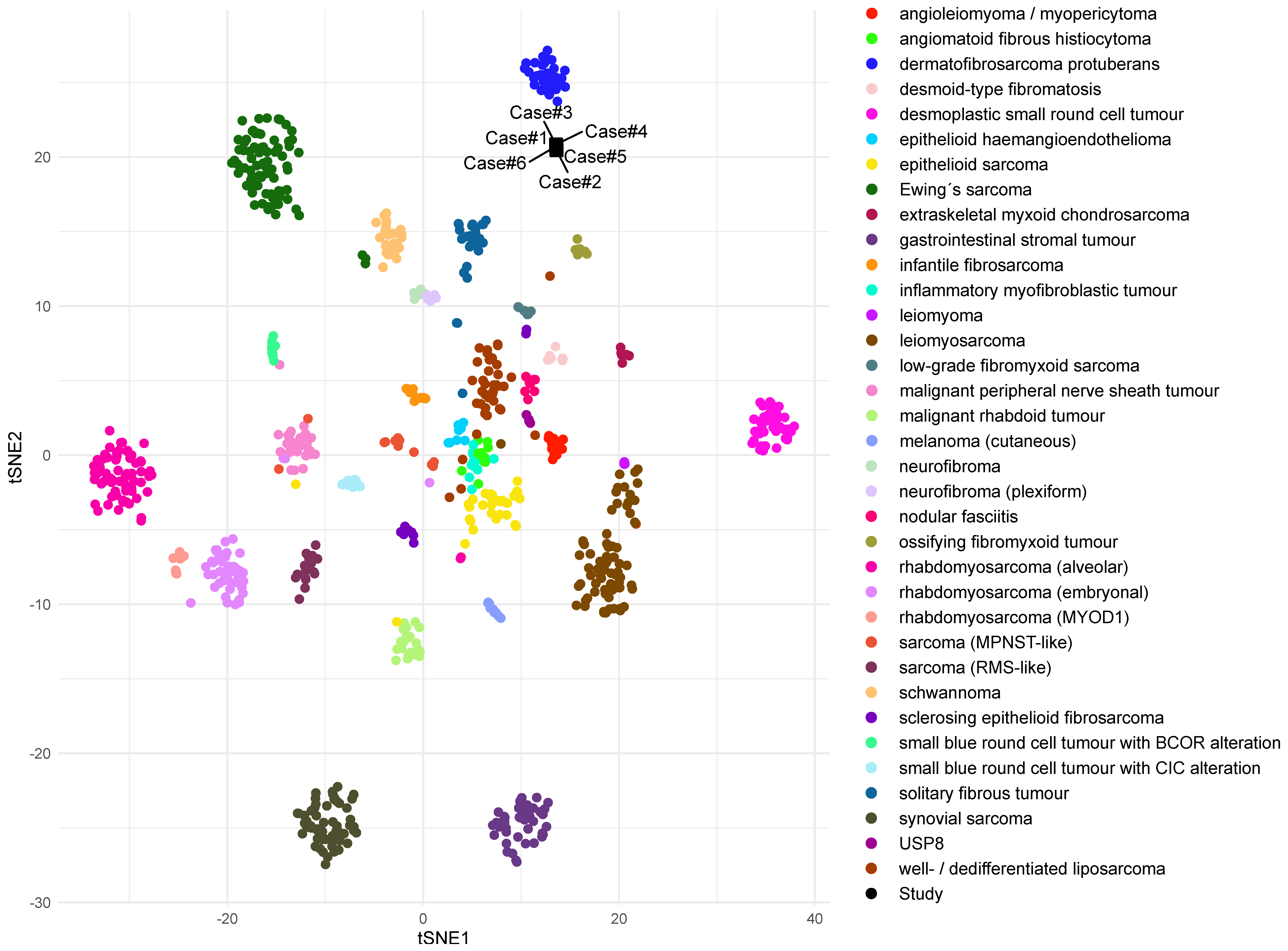
Applying the published version of the Heidelberg DNA-methylation sarcoma classifier, all cases showed the highest similarity to the DFSP methylation class, although with low calibrated scores. Using the most recent Heidelberg sarcoma classifier version (v13.1) for diagnostic purposes, 4/6 cases were assigned to the NTRK-rearranged spindle cell neoplasm class; however the calibration scores were low and did not support a reliable classification. In the t-SNE analysis, all cases formed a distinct cluster located in close proximity to DFSP (Figure 3).
Figure 3.
tSNE plot of the study cohort (n = 6, in black) compared to morphological mimics.
All cases had relatively stable genomes reflected by their flat CNV profiles; nevertheless, case 1 had partial loss of chromosome 10q, case 2 partial loss of chromosome 2p and complete loss of chromosome 10, focal deletions on chromosomes 9q and 15q were found in case 5, and gain of EGFR was detected in case 6.
3. Discussion
The ABL proto-oncogene 1 (ABL1) is named after Herbert Abelson, who isolated, together with Louise Rabstein, a retrovirus that induced B-cell leukemia in murine lymphocytes by a protein with tyrosine kinase activity [15]. The human counterpart was mapped to chromosome 9 and found to be part of the Philadelphia chromosome (chromosome 22), fusing upon a reciprocal translocation (t9;22) (q34;q11) BCR and ABL1 [16,17,18]. This fusion oncogene was primarily attributed to chronic myeloid leukemia (CML)—a major breakthrough in understanding cancer—and later on, to other hematological malignancies [19,20,21,22].
Diverse other ABL1 fusion partners have been detected in hematological and epithelial malignancies and rarely in mesenchymal tumors with the specific GAB1::ABL1, reported so far in 15 benign spindle cell tumors with morphologic similarities with other PK-related mesenchymal tumors [9,10,11,12,13,22,23,24].
The oncogenic ABL1 fusions lead to constitutive kinase activation by retaining almost the entire ABL1 gene beginning at exon 2, thereby preserving the SH3, SH2, and kinase domain while deleting the N-terminal myristoylated cap that mediates physiological autoinhibition. The consequently disrupted autoinhibition as well as fusion-partner-derived functions, like oligomerization, aberrant subcellular localization, or assembly into signaling complexes that facilitate trans-autophosphorylation and sustained downstream signaling, are thought to be the combined effects. The relative contribution of these mechanisms appears to be fusion-partner-dependent [25]. As the GAB1::ABL1 fusion product retains the same part of ABL1, it is likely that the activating mechanism is similar to other well-studied fusions such as BCR::ABL1 resulting in activation of ABL1’s downstream pathways including the MAPK, AKT, and JAK/STAT, similar to other PK-related tumors [4,10]. It is not clear why GAB1 (GRB2-associated binding protein1), a multi-substrate docking protein with a prominent role in signal transduction, is the constantly recurrent 5’ fusion partner in these mesenchymal tumors. However, the GAB1::ABL1 fusion consistently retains exons 1 through 6 of GAB1. Therefore, one could hypothesize that the N-terminal PH domain and GRB2-binding region of GAB1 are under positive selective pressure. Unlike BCR, GAB1 does not contribute a canonical oligomerization motif, raising the possibility that its primary role is to target the fusion kinase to membrane-associated RTK signaling complexes. Given the established role of GAB1 as a central adaptor downstream of MET and other RTKs in fibroblasts and mesenchymal tissues, this architecture may preferentially confer a growth advantage in mesenchymal cells, potentially contributing to the apparent restriction of GAB1::ABL1 fusions to spindle cell neoplasms [26]. Although this hypothesis remains speculative, it provides a biologically plausible explanation for the pathogenesis of these tumors.
By methylation profiling, all cases formed a cluster in close proximity to the group of PK-related tumors, mainly DFSP as well as spindle cell neoplasms with a NTRK rearrangement. In addition, on the RNA level, lesions had a similar expression profile to DFSP. These findings underscore the close relationship of neoplasms with activating alterations in protein kinases, better classified by their driver mutation instead of morphological and immunohistochemical features because of the overlap and treatment modalities based on the activated kinase.
The neoplasms mainly occur in children and young adults, although there is a broad age range. Reported sites are the soft tissue of the extremities (proximal and distal), limb girdles, trunk, and neck [9,10,11,12,13,23,24]. Lesions may arise superficially (subcutaneously) or deeply (intramuscular), and bone erosion has been observed in an individual case [9,10]. Our results are in concert with these findings.
So far, it seems that all reported lesions including ours follow a benign clinical course, with recurrences arising exceptionally [9,10,11,12,13,23,24]. This is in accordance with the flat CNV profile shown here. One patient was treated with imatinib and the tumor showed regression by MRI, which indicates an alternative when surgery is not feasible [10].
From a histomorphological perspective, spindle cell neoplasms with a GAB1::ABL1 gene fusion show overlap with other PK-related lesions. They are composed of haphazardly arranged bland spindle cells forming vague bundles, storiform formations, and focal whorls within a fibrous to fibromyxoid matrix with staghorn-like and small (branching) vessels. A sclerotic/hyaline background and thick bands of collagen are variably present [9,10,11,12]. Furthermore, lesions show variable expression of S100, CD34, EMA, GLUT1, and claudin. As such, it is not surprising that these neoplasms were previously designated as perineurioma, soft tissue angiofibroma (STAF), or solitary fibrous tumor (SFT) [14,23,24]. However, co-expression of S100 and CD34 without SOX10 positivity is strongly suggestive of a PK-related lesion [2,3,8]. Perineurioma shows characteristic whorls like meningeomas, due to the function of perineurial cells of protective encasement of nerve fascicles, and immunohistochemical expression of EMA, CD34, GLUT1, and claudin [9,23,27]. Soft tissue angiofibroma is most recognizable by its prominent branching vasculature of small vessels, these being far less obvious in GAB1::ABL1 fused lesions. The immunophenotype of STAF is not specific; however, CD34 and EMA positivity can be misleading, and the detected fusion gene involves NCOA2 in most of the cases [27]. SFTs commonly do not have the smooth nuclear contours as seen in GAB1::ABL-positive cases, and STAT6 is a perfect surrogate marker for the underlying fusion gene [27].
Other differential diagnoses include other PK-related lesions like infantile fibrosarcoma and malignant peripheral nerve sheath tumor (MPNST)-like tumors harboring, as mentioned in the introduction, other hyperactivated kinases like NTRK1,2,3, (B)RAF, MET, RET, and EGFR. Molecular analysis would be relevant for therapeutic purposes, mainly in aggressive lesions [6,28,29].
Other differential diagnoses are low-grade fibromyxoid sarcoma, cellular myofibroma, fat-poor spindle-cell lipoma, desmoid-type fibromatosis, and schwannoma [27,30,31]. All differential diagnoses are listed in Table 2.
Table 2.
Differential diagnoses [6,27,31,32].
In conclusion, spindle cell lesions with a GAB1::ABL1 fusion activating the tyrosine kinase of ABL1 are a family member within the group of protein-kinase-related mesenchymal neoplasms. Imatinib has already been an effective treatment in an individual case and is therefore a promising therapeutic option for patients with such tumors when surgery is not feasible. However, tumors pursue an indolent clinical course (so far).
4. Materials and Methods
The six cases were retrieved from the author’s (referral) files and hematoxilin and eosin (H&E) and immunohistochemical slides were reviewed. Clinical information was available in all cases. Case 2 was already published by Bekers et al. in 2017 [14].
For the Dutch cases, residual tissue was used in accordance with the Dutch Code of Conduct for Responsible Secondary Use of Human Tissue. Patients are informed that leftover tissue may be stored and used for scientific research unless they object (opt-out procedure). The international collaborating centers contributed coded archival material in accordance with their respective institutional and national regulations.
4.1. Immunohistochemistry
Sections of four µm thickness were cut from formalin-fixed paraffin-embedded (FFPE) blocks, mounted on pre-coated slides, and dried for at least 10 min at 56 °C. After deparaffinization, the slides were stained using an automated Ventana tissue stainer (BenchMark Ultra, Roche, Tucson, AZ, USA). The following antibodies were used: S100 (ready to use, 4C4.9, Roche), SOX10 (ready to use, EP268, Roche), CD34 (ready to use, QBEND/10, Leica, Wetzlar, Germany), EMA (ready to use, GP1.4, Leica), CKAE1/3 (ready to use, CKAE1/AE3, Leica), GLUT1 (ready to use, polyclonal Cell Marque, Rocklin, CA, USA), SMA (ready to use, Alph-sm-1, Leica), Desmin (ready to use, DE-R-11, Leica), MUC4 (ready to use, 8G7, Cell Marque), STAT6 (ready to use, EP325, Cell Marque), panTRK (1:40, RM423, Sanbio, Uden, The Netherlands), and ALK (ready to use, D5F3, Ventana, Tucson, AZ, USA). Pretreatment was performed according to standard protocols.
4.2. Molecular Analyses
4.2.1. Targeted mRNA Sequencing
RNA was isolated from FFPE tissue sections with the Reliaprep FFPE Total RNA Miniprep system (Promega, Madison, WI, USA) according to the manufacturers’ protocol. Up to 250 ng RNA was used for preparing cDNA. Target-enriched NGS libraries were subsequently prepared using the Archer FusionPlex kit (ArcherDX, Coralville, IA, USA) with a custom designed gene panel (Radboudv1) for Illumina, which includes primers for detecting gene fusions upstream of ABL1 exon 1, 2, 3, 4, 5, and downstream of exon 5 and 6 (RefSeq: NM_005157.6). Sequencing was performed with the NextSeq 550 system (Illumina, San Diego, CA, USA), and data were analyzed using Archer Analysis software (ArcherDX, Boulder, CO, USA, 6.2.7 or 7.3.2).
4.2.2. Whole Transcriptome Sequencing (mRNA Sequencing)
Total RNA was isolated using the AllPrep DNA/RNA/Protein Mini Kit (Qiagen, Hilden, Germany) in keeping with the standard protocol on the QiaCube (Qiagen, Hilden, Germany). For case 6, fresh frozen tissue was used, while for cases 1–5, only FFPE was available. RNA-seq libraries were generated with 300 ng RNA using the KAPA RNA HyperPrep Kit with RiboErase (Roche) and subsequently sequenced on a NovaSeq 6000 system (2 × 150 bp) (Illumina). The RNA sequencing data were processed as per GATK 4.0 best practices workflow for variant calling, using a wdl and Cromwell-based workflow (https://gatk.broadinstitute.org/hc/en-us/sections/360007226651-Best-Practices-Workflows, accessed on 1 June 2026). This included performing quality control with Fastqc (version 0.11.5) to calculate the number of sequencing reads and the insert size Picard (version 2.20.1) for RNA metrics output and MarkDuplicates [33]. The raw sequencing reads were aligned using Star (version 2.7.0f) to GRCh38 and gencode version 29 [34].
4.2.3. DNA Methylation Profiling and Copy Number Variation (CNV) Analyses
DNA was isolated by NorDiag Arrow using the DiaSorin DNA extraction kit (NL) or GeneRead DNA FFPE Kit (Qiagen) according to the respective manufacturer’s instructions. DNA concentration was measured using the Qubit 2.0 fluorometer. Per sample, we used 200 ng (NL) of DNA. Bisulphite conversion was performed with EZ DNA Methylation™ Kit (Zymo Research, Irvine, CA, USA). All methylation data were generated using the Illumina® MethylationEPIC (850 k) or EPICv2 (935K) BeadChip platforms as previously described [35]. Classification of the samples was performed by the Heidelberg sarcoma classifier using both the published version (v12.2) and the most recent version available, v13.1. Further computational analyses were performed employing R version 4.2.0 (https://www.R-project.org, accessed on 1 June 2026). Specifically, raw DNA methylation data were processed using a modified version of the minfi workflow [36] and a t-distributed stochastic neighbor embedding (t-SNE) analysis was carried out using selected potential histologic mimics from a published cohort as a reference. Regarding CNV analyses, the high-density DNA methylation arrays allow for determining CNVs.
Author Contributions
L.S.H.-J. and U.F. conducted the study and drafted the manuscript. L.A.K., S.P., E.M., S.B. and S.v.H. contributed to interpretation of the molecular data. All other authors provided cases and revised the manuscript. All authors have read and agreed to the published version of the manuscript.
Funding
This research received no external funding.
Institutional Review Board Statement
The study was conducted in accordance with the Declaration of Helsinki and the Dutch Code of Conduct for Responsible Secondary Use of Human Tissue. The study used coded (pseudonymized) archival human tissue obtained from leftover diagnostic material stored in institutional biobanks. Under the Dutch Medical Research Involving Human Subjects Act (WMO), research using coded leftover human tissue does not constitute research involving human subjects and is therefore exempt from review by a Medical Research Ethics Committee (METC).
Informed Consent Statement
Patient consent was waived due to the study used coded (pseudonymized) archival leftover human tissue obtained after routine diagnostic procedures and stored in institutional biobanks. Patients had provided general consent for the use of residual tissue for research according to institutional procedures.
Data Availability Statement
The original contributions presented in this study are included in the article. Further inquiries can be directed to the corresponding author(s).
Conflicts of Interest
All authors were employed by their respective hospitals. The authors declare that the research was conducted in the absence of any commercial or financial relationships that could be construed as potential conflicts of interest.
References
- Xu, B.; Suurmeijer, A.J.H.; Agaram, N.P.; Antonescu, C.R. Head and Neck Mesenchymal Tumors with Kinase Fusions: A Report of 15 Cases with Emphasis on Wide Anatomic Distribution and Diverse Histologic Appearance. Am. J. Surg. Pathol. 2023, 47, 248–258. [Google Scholar]
- Antonescu, C.R. Emerging soft tissue tumors with kinase fusions: An overview of the recent literature with an emphasis on diagnostic criteria. Genes Chromosomes Cancer 2020, 59, 437–444. [Google Scholar] [CrossRef] [Scilit]
- Davis, J.L.; Al-Ibraheemi, A.; Rudzinski, E.R.; Surrey, L.F. Mesenchymal neoplasms with NTRK and other kinase gene alterations. Histopathology 2022, 80, 4–18. [Google Scholar] [CrossRef] [Scilit]
- Mertens, F.; Antonescu, C.R.; Mitelman, F. Gene fusions in soft tissue tumors: Recurrent and overlapping pathogenetic themes. Genes Chromosomes Cancer 2016, 55, 291–310. [Google Scholar] [CrossRef] [Scilit]
- Kumar, H.; Chen, Z.; Adegunlehin, A.; Trowbridge, L.; Aguilar, L.; Kim, P. KinaseFusionDB: An integrative knowledge of kinase fusion proteins in multi-scales. Brief. Bioinform. 2025, 26, bbaf259. [Google Scholar] [CrossRef] [Scilit]
- Flucke, U.; Versleijen-Jonkers, Y.M.H.; Mentzel, T.; Mueller, A.M.; Hiemcke-Jiwa, L.S.; Alaggio, R. Protein kinase-related tumors in the pediatric population: Updated review on an emerging group with emphasis on the more rarely involved kinases. Pathologie 2026, 47, 105–115. [Google Scholar]
- Cicenas, J.; Zalyte, E.; Bairoch, A.; Gaudet, P. Kinases and Cancer. Cancers 2018, 10, 63. [Google Scholar] [CrossRef] [Scilit]
- Suurmeijer, A.J.H.; Dickson, B.C.; Swanson, D.; Zhang, L.; Sung, Y.S.; Cotzia, P.; Fletcher, C.D.M.; Antonescu, C.R. A novel group of spindle cell tumors defined by S100 and CD34 co-expression shows recurrent fusions involving RAF1, BRAF, and NTRK1/2 genes. Genes Chromosomes Cancer 2018, 57, 611–621. [Google Scholar] [CrossRef] [Scilit]
- Agaimy, A.; Perret, R.; Demicco, E.G.; Gross, J.; Liu, Y.J.; Azmani, R.; Engelmann, C.; Schubart, C.; Seppet, J.; Stoehr, R.; et al. GAB1::ABL1 fusions define a distinctive soft tissue neoplasm, with variable perineurial differentiation, and a predilection for children and young adults. Genes Chromosomes Cancer 2023, 62, 449–459. [Google Scholar] [CrossRef] [Scilit]
- Choo, F.; Rakheja, D.; Davis, L.E.; Davare, M.; Park, J.Y.; Timmons, C.F.; Neff, T.; Beadling, C.; Corless, C.L.; Davis, J.L. GAB1-ABL1 fusions in tumors that have histologic overlap with NTRK-rearranged spindle cell tumors. Genes Chromosomes Cancer 2021, 60, 623–630. [Google Scholar] [CrossRef] [Scilit]
- Panagopoulos, I.; Gorunova, L.; Andersen, K.; Tafjord, S.; Lund-Iversen, M.; Lobmaier, I.; Micci, F.; Heim, S. Recurrent Fusion of the GRB2 Associated Binding Protein 1 (GAB1) Gene with ABL Proto-oncogene 1 (ABL1) in Benign Pediatric Soft Tissue Tumors. Cancer Genom. Proteom. 2020, 17, 499–508. [Google Scholar] [CrossRef] [Scilit]
- Dorwal, P.; Currie, A.; Bennett, J.; Wallwork, B.; Song, L.; Joy, C.; Walsh, M. GAB1::ABL1 fusion-associated spindle cell neoplasm: Expanding our knowledge of kinase-rearranged spindle cell tumours. Pathology 2025, 57, 802–805. [Google Scholar] [CrossRef] [Scilit]
- Kimura, E.; Komuta, M.; Agaimy, A.; Ito, K.; Nagashima, A.; Norimatsu, Y.; Morimura, S.; Shiomi, T.; Sugaya, M.; Hamada, T. The First Case of GAB1::ABL1 Fusion-Positive Spindle Cell Soft Tissue Neoplasm in a Japanese Patient. J. Dermatol. 2026, 53, e41–e42. [Google Scholar] [CrossRef] [Scilit]
- Bekers, E.M.; Groenen, P.; Verdijk, M.A.J.; Raaijmakers-van Geloof, W.L.; Roepman, P.; Vink, R.; Gilhuijs, N.D.; van Gorp, J.M.; Bovée, J.V.; Creytens, D.H.; et al. Soft tissue angiofibroma: Clinicopathologic, immunohistochemical and molecular analysis of 14 cases. Genes Chromosomes Cancer 2017, 56, 750–757. [Google Scholar] [CrossRef] [Scilit]
- Abelson, H.T.; Rabstein, L.S. Lymphosarcoma: Virus-induced thymic-independent disease in mice. Cancer Res. 1970, 30, 2213–2222. [Google Scholar]
- Heisterkamp, N.; Groffen, J.; Stephenson, J.R.; Spurr, N.K.; Goodfellow, P.N.; Solomon, E.; Carritt, B.; Bodmer, W.F. Chromosomal localization of human cellular homologues of two viral oncogenes. Nature 1982, 299, 747–749. [Google Scholar] [CrossRef] [Scilit]
- de Klein, A.; van Kessel, A.G.; Grosveld, G.; Bartram, C.R.; Hagemeijer, A.; Bootsma, D.; Spurr, N.K.; Heisterkamp, N.; Groffen, J.; Stephenson, J.R. A cellular oncogene is translocated to the Philadelphia chromosome in chronic myelocytic leukaemia. Nature 1982, 300, 765–767. [Google Scholar] [CrossRef] [Scilit]
- Heisterkamp, N.; Stephenson, J.R.; Groffen, J.; Hansen, P.F.; de Klein, A.; Bartram, C.R.; Grosveld, G. Localization of the c-ab1 oncogene adjacent to a translocation break point in chronic myelocytic leukaemia. Nature 1983, 306, 239–242. [Google Scholar] [CrossRef] [Scilit]
- Bernt, K.M.; Hunger, S.P. Current concepts in pediatric Philadelphia chromosome-positive acute lymphoblastic leukemia. Front. Oncol. 2014, 4, 54. [Google Scholar] [CrossRef] [Scilit]
- Wang, Y.; Liang, Z.-J.; Gale, R.-P.; Liao, H.-Z.; Ma, J.; Gong, T.-J.; Shao, Y.-Q.; Liang, Y. Chronic myeloid leukaemia: Biology and therapy. Blood Rev. 2024, 65, 101196. [Google Scholar] [CrossRef] [Scilit]
- Wang, J.; Pendergast, A.M. The Emerging Role of ABL Kinases in Solid Tumors. Trends Cancer 2015, 1, 110–123. [Google Scholar] [CrossRef] [Scilit]
- Mitelman, F. Mitelman Database of Chromosome Aberrations and Gene Fusions in Cancer. 2026. Available online: https://mitelmandatabase.isb-cgc.org (accessed on 1 June 2026).
- Duff, D.J.; Guzman, M.A.; Batanian, J.R. ABL1 gene involvement within a complex three-way translocation (2;9;4) in perineurioma characterized by molecular cytogenetic methods. Cancer Genet. 2014, 207, 263–267. [Google Scholar] [CrossRef] [Scilit]
- Rakheja, D.; Wilson, K.S.; Meehan, J.J.; Schultz, R.A.; Maale, G.E.; Timmons, C.F. Extrapleural benign solitary fibrous tumor in the shoulder of a 9-year-old girl: Case report and review of the literature. Pediatr. Dev. Pathol. 2004, 7, 653–660. [Google Scholar] [CrossRef] [Scilit]
- Hantschel, O.; Superti-Furga, G. Regulation of the c-Abl and Bcr-Abl tyrosine kinases. Nat. Rev. Mol. Cell Biol. 2004, 5, 33–44. [Google Scholar] [CrossRef] [Scilit]
- Mood, K.; Saucier, C.; Bong, Y.S.; Lee, H.S.; Park, M.; Daar, I.O. Gab1 is required for cell cycle transition, cell proliferation, and transformation induced by an oncogenic met receptor. Mol. Biol. Cell. 2006, 17, 3717–3728. [Google Scholar] [CrossRef] [Scilit]
- WHO Classification of Tumours Editorial Board. Soft Tissue and Bone Tumours; International Agency for Research on Cancer: Lyon, France, 2020. [Google Scholar]
- van Spronsen, R.; Kester, L.A.; Knops, R.R.G.; van de Sande, M.A.J.; van Leenders, G.; de Laat, P.C.J.; Stortelder, E.; Korpershoek, E.; van Noesel, M.; Meister, M.; et al. Infantile fibrosarcoma with an EGFR kinase domain duplication: Underlining a close relationship with congenital mesoblastic nephroma and highlighting a similar morphological spectrum. Ann. Diagn. Pathol. 2022, 57, 151885. [Google Scholar] [CrossRef] [Scilit]
- Vallese, S.; Barresi, S.; Hiemcke-Jiwa, L.; Patrizi, S.; Kester, L.; Giovannoni, I.; Cardoni, A.; Pedace, L.; Nardini, C.; Tancredi, C.; et al. Spindle Cell Lesions with Oncogenic EGFR Kinase Domain Aberrations: Expanding the Spectrum of Protein Kinase-Related Mesenchymal Tumors. Mod. Pathol. 2024, 37, 100539. [Google Scholar] [CrossRef] [Scilit]
- Karanian, M.; Pissaloux, D.; Gomez-Brouchet, A.; Chevenet, C.; Le Loarer, F.; Fernandez, C.; Minard, V.; Corradini, N.; Castex, M.-P.; Duc-Gallet, A.M.; et al. SRF-FOXO1 and SRF-NCOA1 Fusion Genes Delineate a Distinctive Subset of Well-Differentiated Rhabdomyosarcoma. Am. J. Surg. Pathol. 2020, 44, 607–616. [Google Scholar] [CrossRef] [Scilit]
- Karanian, M.; Kelsey, A.; Paindavoine, S.; Duc, A.; Vanacker, H.; Hook, L.; Weinbreck, N.; Delfour, C.; Minard, V.; Baillard, P.; et al. SRF Fusions Other Than with RELA Expand the Molecular Definition of SRF-Fused Perivascular Tumors. Am. J. Surg. Pathol. 2020, 44, 1725–1735. [Google Scholar] [CrossRef] [Scilit]
- Thway, K.; Fisher, C.; Debiec-Rychter, M.; Calonje, E. Claudin-1 is expressed in perineurioma-like low-grade fibromyxoid sarcoma. Hum. Pathol. 2009, 40, 1586–1590. [Google Scholar] [CrossRef] [Scilit]
- Wingett, S.W.; Andrews, S. FastQ Screen: A tool for multi-genome mapping and quality control. F1000Research 2018, 7, 1338. [Google Scholar] [CrossRef] [Scilit]
- Frankish, A.; Diekhans, M.; Jungreis, I.; Lagarde, J.; Loveland, J.E.; Mudge, J.M.; Sisu, C.; Wright, J.C.; Armstrong, J.; Barnes, I.; et al. GENCODE 2021. Nucleic Acids Res. 2021, 49, D916–D923. [Google Scholar] [CrossRef] [Scilit]
- Koelsche, C.; Schrimpf, D.; Stichel, D.; Sill, M.; Sahm, F.; Reuss, D.E.; Blattner, M.; Worst, B.; Heilig, C.E.; Beck, K.; et al. Sarcoma classification by DNA methylatiom profiling. Nat. Commun. 2021, 12, 498. [Google Scholar] [CrossRef] [Scilit]
- Aryee, M.J.; Jaffe, A.E.; Corrada-Bravo, H.; Ladd-Acosta, C.; Feinberg, A.P.; Hansen, K.D.; Irizarry, R.A. Minfi: A flexible and comprehensive Bioconductor package for the analysis of Infinium DNA methylation microarrays. Bioinformatics 2014, 30, 1363–1369. [Google Scholar] [CrossRef] [Scilit]

Disclaimer/Publisher’s Note: The statements, opinions and data contained in all publications are solely those of the individual author(s) and contributor(s) and not of MDPI and/or the editor(s). MDPI and/or the editor(s) disclaim responsibility for any injury to people or property resulting from any ideas, methods, instructions or products referred to in the content. |
© 2026 by the authors. Licensee MDPI, Basel, Switzerland. This article is an open access article distributed under the terms and conditions of the Creative Commons Attribution (CC BY) license.