


Abstract
The skin functions primarily as a protective physical shield, defending the body against harmful external factors such as invading microorganisms and toxic agents. When this barrier becomes impaired and skin microbiome homeostasis is disturbed, exacerbation of chronic skin diseases may occur, including atopic dermatitis, psoriasis, and acne. This issue became particularly important during the coronavirus pandemic, when it turned out that handwashing and disinfection removed not only impurities and pathogenic microorganisms but also beneficial components of the skin microbiome. This review provides a critical overview of recent advances in understanding the skin microbiome and its influence on skin health, as well as supportive therapies for the treatment of dermatoses. Contemporary therapeutic approaches focus not only on suppressing inflammatory symptoms but also on modulating the skin microbiome as part of a causal treatment strategy, in which prebiotics and probiotics play a significant role. Moreover, probiotics are also considered a valuable component of anti-aging approaches, as they may help counteract the detrimental effects of ultraviolet (UV) radiation on the skin. By reducing oxidative stress, strengthening the epidermal barrier, and exerting immunomodulatory effects, they may complement traditional photoprotection methods. Nevertheless, further research—particularly into their long-term efficacy and safety—is essential to support their broader clinical and consumer application.
1. Introduction
Human skin constitutes a complex ecosystem inhabited by numerous microorganisms, including bacteria, fungi, viruses, and mites. These microorganisms collectively form the skin microbiota [1]. Various skin structures—such as hair follicles, sebaceous and sweat glands, as well as the stratum corneum—serve as niches for distinct microbial communities. The richness of this microbiota was first systematically characterized in 2011 by Elizabeth Grice and Julia Segre [2]. Today, researchers emphasize the important role of the skin microbiome in maintaining proper skin function: protection against pathogenic microorganisms, strengthening of the immune system, and the ability to break down and produce various compounds [3,4,5,6,7]. The skin microbiota serves as the first line of defense against harmful environmental factors. It contributes to host immune defense, and one of its mechanisms involves inhibiting the growth of pathogens by producing bactericidal substances. The microbiota also participates in the development of adaptive immunity by modulating local cytokine production, as well as enhancing innate immunity by increasing the production of antimicrobial proteins [8].
Unfortunately, improper hygiene, cleansing, and daily skincare practices may alter the composition and function of the skin microbiota and disrupt skin microbiome homeostasis, leading to excessive skin dryness or skin disorders such as atopic dermatitis. For this reason, the impact of cosmetic formulations on the skin microbiome and the need to use microbiome-friendly raw materials, as well as to enrich formulations with pro- and prebiotic ingredients, have become not only a subject of scientific research [9,10] but also one of the leading trends in the cosmetics market.
This issue became particularly important during the coronavirus pandemic, when handwashing and disinfection were key elements of sanitary regulations. While hygienic and disinfectant products effectively cleanse the skin, removing impurities and pathogenic microorganisms, they also strip away beneficial components of the skin microbiome [11].
Probiotic and prebiotic substances have long been successfully used as ingredients in dietary supplements. Existing definitions of these terms were developed primarily in the context of nutritional applications. According to FAO/WHO, probiotics are “live microorganisms which, when administered in adequate amounts, confer a health benefit on the host” [12]. In cosmetics, however, these terms are used more broadly. Live microorganisms can only be used in cosmetic products to a very limited extent, as their viability and activity are difficult to control [11,13]. Limitations also arise from the strict microbiological purity requirements of cosmetic products [14].
To include live bacteria in cosmetic formulations, preservatives should be omitted. However, from the perspective of cosmetic safety, as well as the quality requirements imposed on cosmetic products (Regulation No. 1223/2009 on cosmetic products) [15], this is not possible—especially in the case of water-based formulations.
2. Skin Microbiome
The skin is the largest organ of the human body. It forms an ecosystem with a surface area of approximately 2 m2, but when considering its complexity (about 5 million skin appendages), the total area increases significantly to around 25 m2, making it the largest organ in the body [16,17,18].
The skin primarily acts as a protective physical barrier, safeguarding the body from harmful external agents such as invading microorganisms and toxic compounds. In addition, it serves as a boundary with the surrounding environment and is inhabited by a diverse array of microorganisms, including bacteria, fungi, and viruses, as well as mites. These organisms are typically grouped into resident, temporary, and transient populations [19,20]. Both microorganisms and mites occur not only on the surface of the skin but also deeper within structures such as hair follicles and glands, collectively forming the skin microbiota. Together with their genetic material, metabolites, and interactions with the host, these microorganisms constitute the skin microbiome. The term Stratum microbium has been proposed to describe this functional microbial layer associated with the skin (Figure 1) [2].
Figure 1.
Diagram of the structure of the skin. Created in BioRender. Łętocha, A. (2026). https://BioRender.com/9lwx9lm (accessed on 3 July 2026).
Members of the skin microbiota are largely harmless, and in some cases, their presence provides essential functions to the host, making them beneficial for the organism. Symbiotic and commensal microorganisms, which coexist “in harmony” with the host, occupy numerous niches on the skin and protect against colonization by pathogenic organisms as well as harmful substances [2,21,22]. Until recently, the characterization of skin microorganisms relied primarily on culture-based techniques. While many human-associated microorganisms can be cultivated under appropriate laboratory conditions, conventional cultivation methods recover only part of the microbial diversity present on the skin and may favor fast-growing taxa. Consequently, culture-based studies often overrepresent genera such as Staphylococcus and Malassezia [23,24]. Recent advances in DNA amplification and sequencing technologies have made it possible to bypass cell culture, revolutionizing our understanding of microbial communities inhabiting the skin. DNA sequencing methodologies allow for more precise and accurate microbiome analyses. This approach, based on sequencing the 16S rRNA gene, revealed that most skin-associated bacteria belong to four dominant phyla: Actinobacteria, Firmicutes, Proteobacteria and Bacteroidetes. However, taxonomic resolution at the genus and species levels provides a more biologically relevant description of the skin microbiome. The most abundant bacteria include Cutibacterium spp. (particularly Cutibacterium acnes), Staphylococcus spp. (especially Staphylococcus epidermidis and Staphylococcus hominis), Corynebacterium spp., Micrococcus spp. and Acinetobacter spp., whereas fungi are dominated by Malassezia species, mainly M. restricta and M. globosa. The relative abundance of these microorganisms strongly depends on skin physiology and microenvironmental characteristics [2,21,25,26]. Skin can be divided into three main habitats—moist, sebaceous, and dry—each characterized by a distinct microbial composition (Figure 2A, Table 1) [2,27,28].
Figure 2.
(A) Distribution of microenvironments on human skin, (B) Internal and external factors influencing the skin microbiome [inspired by [2] and created in BioRender. Łętocha, A. (2026). https://BioRender.com/9lwx9lm (accessed on 3 July 2026)].
Table 1.
Representative microbiota of major skin microenvironments.
In addition to anatomical differences, the variability and richness of the skin’s bacterial flora are influenced by age, sex, season, and ethnic background, as well as stressors such as physical injuries and psychological stress. Environmental factors also contribute to the diversity of the skin microbiome, including climate, temperature, UV radiation, and lifestyle (Figure 2B) [21,29].
3. Early Skin Colonization—Factors Influencing Skin Colonization
The human skin microbiota is established at birth [30]. The newborn skin microbiota begins to develop at birth and is influenced by multiple maternal, neonatal, and environmental factors. Mode of delivery is one of the factors associated with early microbial colonization patterns. Infants born vaginally often show a higher relative abundance of microorganisms commonly found in the maternal vaginal and gastrointestinal microbiota, whereas infants delivered by cesarean section may initially harbour microorganisms associated with maternal skin and the hospital environment. This early colonization process is considered important for the development of immune tolerance to commensal microorganisms. However, these microbial differences can also be influenced by factors such as antibiotic exposure, feeding practices, skin-to-skin contact, hospitalization, and environmental conditions and may change substantially during infancy. Understanding the factors that modulate colonization during this critical period may help prevent potential disease states (Figure 3), as disturbances in skin barrier function and early colonization can predispose to both local and systemic immune dysregulation [31].
Figure 3.
Factors modulating the infant skin microbiome [inspired by [31] and created in BioRender. Łętocha, A. (2026). https://BioRender.com/9lwx9lm (accessed on 3 July 2026)].
The mechanisms of early skin colonization in newborns are complex and depend on interactions between microorganisms, the skin as a barrier organ, and the immune system. Colonization begins during childbirth, when microorganisms are transferred from the mother to the infant. In vaginal delivery, newborns are primarily colonized by bacteria originating from the maternal vaginal and gastrointestinal microbiota, including Lactobacillus, Prevotella, and Sneathia spp. [32,33]. In contrast, infants delivered by cesarean section are mainly colonized by microorganisms from the mother’s skin and the hospital environment, such as Staphylococcus, Corynebacterium, and Propionibacterium [33,34]. Additionally, skin-to-skin contact (SSC) and breastfeeding play a crucial role in the transfer of commensal bacteria and colonization-supporting compounds, such as human milk oligosaccharides [35]. Preterm infants represent a special case, as they exhibit distinct microbiome patterns compared to full-term newborns. In preterm infants, higher abundances of Staphylococcus, Corynebacterium (phylum Actinobacteria), and Prevotella (phylum Bacteroidetes) are observed. Immune responses to colonizing Staphylococcus species may underlie a common benign condition in newborns—erythema toxicum [36,37]. Commensal bacteria and their products, including staphylococcal lipoteichoic acid and other epitopes, can trigger skin inflammatory responses and enhance immunity. Moreover, protease secretion by commensal S. epidermidis prevents colonization by pathogenic S. aureus [37].
The newborn’s skin thus represents a dynamic environment for microbial colonization. The factors influencing colonization and their interrelationships are summarized in Table 2.
Table 2.
Factors influencing newborn skin colonization.
Newborns possess a set of innate defense mechanisms that participate in the selection and regulation of colonizing microorganisms. Recent discoveries in dermatology highlight the potential importance of antimicrobial peptides (AMPs) in combating microbial skin infections. AMPs additionally act as multifunctional immune effectors, promoting angiogenesis, wound healing, and the production of cytokines and chemokines. In human skin, AMPs such as β-defensins, S100 proteins, and cathelicidins are secreted mainly by keratinocytes, neutrophils, sebocytes, or sweat glands and act selectively against pathogens [41]. These helical peptides bind to lipid membranes and are believed to kill microbes through their ability to disrupt the normal structure of the lipid bilayer [42].
Furthermore, immunological control of microbial colonization on the skin is supported by immunoglobulin A (IgA), present in breast milk [35]. Healthy, breastfed full-term infants receive a mixture of nutrients, bacteria, and antimicrobial proteins—such as carbohydrates, fatty acids, and lactoferrin, along with secretory IgA (sIgA) from breast milk—which shapes the development of their own microbiota [35,43]. Human milk oligosaccharides, glycoproteins, and natural components are also believed to prevent attacks from enteropathogens and stimulate the growth of Bifidobacterium [35,44,45]. These compounds can bind directly to the surface of pathogenic microorganisms, while certain milk oligosaccharides prevent pathogens and their toxins from attaching to host cell receptors [46]. In addition, other constituents of human milk—including interleukin-10, epidermal growth factor, transforming growth factor β1, and erythropoietin—may play key roles as regulators of inflammatory responses directed against bacterial pathogens, for example, within the gut [35,44].
Early skin colonization provides an important immunological signal and promotes the development of tolerance to commensals. Commensal skin bacteria, such as Staphylococcus epidermidis, induce the differentiation of regulatory T cells (Tregs), which suppress excessive inflammatory responses and protect against the development of inflammatory skin diseases, such as atopic dermatitis [47]. Moreover, S. epidermidis contributes to maintaining the skin barrier through the secretion of sphingomyelinase, which is utilized by the host for ceramide synthesis [48,49]. Commensal microorganisms can also enhance barrier functions by stimulating keratinocytes to produce differentiation markers, including filaggrin [50,51], and by increasing AMP expression [50].
Microbial interactions and selective pressures also play a crucial role. Bacteria colonizing the skin participate in biological selection by competing for space and resources. S. epidermidis produces bacteriocins—substances acting antagonistically against pathogens such as Staphylococcus aureus [47]. Microorganisms also form biofilms, which increase colony stability and favor survival [52]. Life in a shared niche involves not only competition but also cooperation and mutual protection. Some bacteria act synergistically—for example, Corynebacterium and Staphylococcus have complementary enzymatic abilities, allowing them to survive together on the skin. Studies by Kwaszewska et al. [53] indicate that the lipolytic properties of staphylococci and corynebacteria are clearly complementary. Lipophilic corynebacteria are fatty acid auxotrophs due to their inability to synthesize fatty acids. In most species, these fatty acids are incorporated directly into cellular structures and transformed into corynomycolic acids of the cell wall. Coexistence with active lipolytic staphylococci may be beneficial for them. Moreover, glycerol released after triacylglycerol hydrolysis cannot be utilized by lipophilic corynebacteria, although it serves as a good substrate for staphylococci. According to James et al. [54], this substrate, along with lactic, acetic, or propionic acids, serves as a precursor for volatile fatty acids in non-lipophilic corynebacteria. Additionally, Kwaszewska et al. [53] reported the inability of corynebacteria to utilize macromolecules such as proteins or polysaccharides, whereas staphylococci are highly efficient in this regard, supplying themselves and corynebacteria with products of their enzymatic activity. Although Corynebacterium is less tolerant to low pH and salt conditions, cohabitation with Staphylococcus provides mutual protection and population stabilization, promoting the development of commensal flora.
Microorganisms colonizing the skin play a key role in the development of immune tolerance and skin barrier functions. Lack of proper colonization is associated with an increased risk of atopy, atopic dermatitis, and other diseases.
4. The Influence of the Microbiome on Skin Diseases
The skin is a dynamic organ characterized by continuous renewal processes [55,56]. Alterations in the composition of the stratum corneum can contribute to dysbiosis, affecting both the richness and the relative abundance of commensal microorganisms [31]. Such disturbances in the skin microbiota may, in turn, lead to the development of various dermatological conditions (Figure 4) [29,57].
Figure 4.
The effects of dysbiosis on human skin [created in BioRender. Łętocha, A. (2026). https://BioRender.com/9lwx9lm (accessed on 03 July 2026)].
When the skin barrier function becomes impaired, exacerbation of chronic skin diseases may occur, including atopic dermatitis [16,17,21,27,29,58,59,60], psoriasis [16,17,21,29,59], and acne [16,17,21,29,59,60].
4.1. Atopic Dermatitis
Atopic dermatitis (AD) is a long-lasting, relapsing inflammatory condition of the skin, marked by severe itching, dryness, and disruption of the epidermal barrier function [61,62]. The pathogenesis of AD is multifactorial and includes genetic, immunological, and environmental factors, as well as skin microbiome dysregulation. It is noteworthy that the prevalence of AD is significantly higher in industrialized countries and has been systematically increasing for several decades. This suggests that excessive hygiene may disrupt the natural microbial environment of the body, limiting the development of beneficial immune mechanisms and impairing the proper response to pathogen exposure [61,63]. A growing body of research indicates that the skin microbiome plays a key role in both the development and exacerbation of this dermatosis [27,58].
In AD, genetic defects lead to both physical and Th2-dependent immunological impairments of the skin barrier, resulting in increased susceptibility to infections and allergens [27,64]. One of the key epidermal proteins is filaggrin, essential for the production of natural moisturizing factor and maintenance of proper hydration of the stratum corneum. Mutations in the filaggrin gene occur in approximately half of patients with moderate-to-severe AD [65] and are often associated with early-onset disease [66]. Filaggrin deficiency leads to abnormal keratinocyte differentiation and reduced lipid content in the skin [67]. Epidermal lipids, such as ceramides, free fatty acids, and cholesterol, play a critical role in maintaining skin barrier integrity by preventing excessive transepidermal water loss (TEWL) and protecting against the penetration of irritants, allergens, and microbes. Both alterations in composition and reduced levels of these lipids are closely linked to the development of AD [61,68,69].
Skin barrier dysfunction is further exacerbated by mechanical damage resulting from chronic scratching of dry and itchy skin [70,71]. Th2 cytokines can inhibit the production of antimicrobial peptides (AMPs) by keratinocytes, such as human beta-defensin-3 and cathelicidins. There may be a link between the sensation of itch in atopic dermatitis and cathelicidins, which can stimulate mast cell degranulation and reduce IL-10 levels in keratinocytes [41,72]. In addition to strengthening the skin barrier, cathelicidins promote cutaneous immunity by increasing the expression and distribution of tight junction components on cell membranes [41,73,74]. Evidence shows that cathelicidin levels are lower in lesional skin of patients with AD and that these levels are correlated with both infection severity in atopic dermatitis and the ability to protect against Staphylococcus aureus skin infections. Cathelicidins play an essential role in defense against pathogen colonization, particularly S. aureus, thereby contributing to the maintenance of a healthy skin microbiome balance [75]. As the disease progresses, a persistent shift in the skin microbial composition occurs, promoting chronic inflammation and disturbance of homeostasis (Figure 5).
Figure 5.
Variability of skin microbiota and biofilm production in the pathogenesis of atopic dermatitis. Arrows indicate increase (↑) and decrease (↓) [inspired by [58] and created in BioRender. Łętocha, A. (2026). https://BioRender.com/anqy4x3 (accessed on 30 June 2026)].
In atopic dermatitis (AD), skin microbiota is notably disrupted, with reduced overall diversity. This imbalance is reflected in diminished levels of genera such as Cutibacterium, Streptococcus, Acinetobacter, Corynebacterium, and Prevotella, accompanied by a marked expansion of Staphylococcus, particularly S. aureus [76,77,78,79]. Under normal conditions, in healthy skin, coagulase-negative staphylococci (CoNS) occupy the same ecological niche and limit S. aureus colonization. However, in AD, elevated production of pro-inflammatory cytokines facilitates the proliferation of S. aureus, enabling biofilm formation and allowing it to dominate over other commensal microbes. Consequently, S. aureus further sustains persistent inflammation in eczematous lesions [58].
Reduced microbial diversity is particularly evident during severe disease exacerbations, with reports showing that the Staphylococcus composition often decreases to a single S. aureus strain [25]. As an opportunistic pathogen, S. aureus exhibits a strong ability to adhere to the skin, disrupt epithelial barrier integrity, and activate the host immune system, ultimately driving inflammation [80,81]. While the carriage rate of S. aureus in the general population is approximately 20%, significantly higher colonization rates are observed in patients with AD, ranging from 30 to 100% depending on age, disease severity, and sampling and analysis methods [82]. This pathogen produces numerous proteolytic enzymes and toxins and stimulates keratinocytes to increase the expression of endogenous proteases, thereby damaging the skin barrier and facilitating penetration through the stratum corneum [83,84]. Additionally, certain S. aureus toxins can trigger type 2 immune responses by activating immune cells and inducing inflammatory mediators such as IL-4, IL-13, IL-22, and TSLP.
It is important to emphasize that in the pathogenesis of AD, not only Staphylococcus strains are involved. In a study by Kobayashi et al. [85], Adam17ΔSox9 mice developed eczematous inflammation accompanied by a marked alteration in the skin microbiome, with dominance of S. aureus and Corynebacterium species. Further bacterial inoculation experiments confirmed that inflammation could be enhanced by both S. aureus and C. bovis. Moreover, Cutibacterium acnes (formerly Propionibacterium acnes), one of the most common skin commensals and a component of its defense mechanisms [86], may also contribute to disease development. This bacterium increases the cytolytic activity of S. aureus by enhancing pro-inflammatory cytokine production [87]. In addition, coproporphyrin III (CIII) produced by C. acnes promotes S. aureus aggregation and biofilm formation, suggesting synergistic interactions between these two species [88].
On the other hand, there are also reports indicating a lack of direct or even negative correlation between the presence of certain microbes and inflammatory lesion development. Scharschmidt et al. confirmed that early skin colonization in newborns is essential for establishing immune tolerance to commensal microbes [89]. Other studies have shown that coagulase-negative staphylococci (S. epidermidis, Staphylococcus hominis, and Staphylococcus lugdunensis) effectively inhibit S. aureus colonization and biofilm formation. In studies by Iwase et al. [90] and Sugimoto et al. [91], serine protease Esp secreted by commensal S. epidermidis was shown to inhibit S. aureus biofilm formation and colonization. Furthermore, Zipperer et al. [92] reported that nasal isolates of S. lugdunensis produce lugdunin, a novel cyclic thiazolidine-containing peptide antibiotic, which prevents S. aureus colonization and represents a rare example of a bioactive compound nonribosomally synthesized by human-associated bacteria. A similar effect was demonstrated in a study by Nakatsuji et al. [93], which evaluated both the safety profile and mechanisms of action of Staphylococcus hominis A9 (ShA9), a strain derived from healthy human skin, as a candidate for topical treatment of AD. ShA9 was shown to eliminate S. aureus from mouse skin and suppress the expression of the S. aureus toxin (psmα) responsible for inducing inflammation.
In addition to bacterial commensals, it has also been shown that the protease MgSAP1 secreted by Malassezia globosa hydrolyzes S. aureus protein A, thereby hindering its biofilm formation [94].
4.2. Psoriasis
Psoriasis is a long-term inflammatory disorder with a complex, multifactorial origin, in which impaired immune tolerance to cutaneous microorganisms plays an important role in disease development. It is estimated that approximately 125 million individuals worldwide are affected. The condition substantially influences patients’ quality of life as well as psychosocial well-being. Pathologically, it is characterized by excessive proliferation of epidermal cells alongside inflammation within the dermis. The disease typically begins with an acute inflammatory phase and may subsequently progress into a chronic state [95,96]. Histologically, psoriasis presents with three key features: (1) epidermal thickening resulting from keratinocyte overgrowth, (2) dilation and increased formation of dermal blood vessels, and (3) infiltration and accumulation of inflammatory cells, predominantly within the dermal layer. Clinically, these processes manifest as visible skin lesions, such as scaling plaques, and may also be accompanied by joint or tendon involvement [97].
The pathogenesis of psoriasis involves a complex network of cytokines and their receptors, some of which also represent potential therapeutic targets (Figure 6) [98]. The inflammatory cascade is initiated when plasmacytoid dendritic cells (pDCs) are activated by complexes formed between self-DNA and the antimicrobial peptide cathelicidin (LL-37), released from damaged keratinocytes [98,99]. In parallel, pDCs can present antigens to CD8+ T cells via MHC–TCR interactions, promoting their activation and subsequent migration to the epidermis. In response, pDCs produce IFN-α and IFN-β, which are key mediators in the early phase of the disease. These cytokines subsequently activate myeloid dendritic cells (mDCs) and promote their migration to regional lymph nodes [100]. Once activated, mDCs secrete cytokines such as TNF-α, IL-12, and IL-23, driving the differentiation and expansion of T cells into effector subsets including Th1, Th17, and Th22. IL-1 further contributes to Th17 polarization. Th1 cells produce TNF-α and IFN-γ, whereas Th22 cells secrete IL-22, a cytokine implicated in keratinocyte hyperproliferation and altered epidermal differentiation. These cells enter the circulation and gain the capacity to home to the skin [101,102].
Figure 6.
Mechanisms underlying psoriasis and immune system interactions [inspired by [98,101] and created in BioRender. Łętocha, A. (2026). https://BioRender.com/9lwx9lm (accessed on 3 July 2026)].
The IL-23/IL-17 signaling pathway plays a central role in psoriatic inflammation. Activated Th17 cells, CD8+ T lymphocytes, and natural killer (NK) cells are major sources of IL-17A, which binds to IL-17 receptors expressed on keratinocytes. This interaction promotes excessive keratinocyte proliferation and contributes to epidermal barrier dysfunction. In the presence of TNF-α, IL-17 further stimulates keratinocytes to produce pro-inflammatory mediators, including TNF-α and the chemokine CCL20, thereby amplifying local inflammation and promoting the recruitment of additional T cells to the skin. IL-22 released by Th22 cells acts synergistically with IL-17 to induce keratinocyte hyperproliferation and abnormal epidermal differentiation, ultimately leading to the formation of characteristic psoriatic plaques. This process is further associated with reduced expression of adhesion molecules and increased production of antimicrobial peptides and chemokines, which facilitate the recruitment of additional immune cells—including Th17 cells, neutrophils, dendritic cells, and macrophages—to the site of inflammation [95].
Psoriasis is the result of interplay between genetic and external factors, wherein the cutaneous microbiota may play a crucial role by responding to external triggers and interacting with the immune system [95,96]. Recent studies have revealed a strong association between skin microbiota and psoriasis. In psoriatic lesions, in contrast to healthy skin, microorganisms are predominantly localized within the epidermal layer. Mechanical damage caused by itch-related scratching may disrupt the skin barrier, allowing certain bacteria—both typical epidermal colonizers and others—to penetrate into deeper dermal layers. In this environment, they can more easily interact with immune cells and trigger both innate and adaptive inflammatory responses. As a consequence, dysbiosis develops. Several studies have reported reduced abundance of taxa such as Cutibacterium, Lactobacillus, Burkholderia, and Corynebacterium in psoriatic lesions compared with healthy skin [103]. However, the microbial alterations reported in psoriasis remain heterogeneous across studies. Other investigations have described increased relative abundance of selected bacterial genera, including Streptococcus, Staphylococcus, and Corynebacterium, as well as members of the phyla Firmicutes and Proteobacteria, in psoriatic skin compared with healthy controls [81,104,105,106,107,108,109,110]. These discrepancies likely reflect differences in study populations, sampling strategies, anatomical sites, disease severity, and analytical methodologies.
Colonization of the skin by S. aureus plays an important role in activating the innate immune system, including the induction of antimicrobial peptides and the production of dendritic cell–derived cytokines such as IL-12 and IL-23. This process promotes the expansion of Th17 lymphocytes. These cells subsequently release pro-inflammatory cytokines, including IL-17A, TNF-α, IFN-α, and IL-22, which drive keratinocyte hyperproliferation and enhance immune cell infiltration within the skin, thereby exacerbating psoriatic inflammation [111]. Streptococcus species contain components such as M protein and peptidoglycan, which act as superantigens capable of activating T cells. Peptidoglycan can interact with MHC class II molecules and T-cell receptors, leading to a targeted immune response and cytokine secretion. Additionally, T cells in psoriasis patients may cross-react with structurally similar epitopes derived from streptococcal M protein and keratins 6 and 17, which further contributes to disease development [112].
Alterations in the skin microbiome observed in psoriasis are not limited to bacterial populations but also involve fungi. For example, Malassezia species can increase the expression of transforming growth factor-β (TGF-β), heat shock protein 70 (HSP70), and integrins in keratinocytes. These changes promote epidermal hyperplasia and cell migration, thereby worsening psoriatic lesions [113]. Furthermore, Malassezia spp. are capable of producing chemoattractants for neutrophils, which may accumulate in large numbers in psoriatic lesions and contribute to the formation of Munro’s microabscesses [114].
Candida albicans has likewise been detected in psoriatic skin, particularly in inverse psoriasis. In addition, Candida-reactive αβ T cells are known to produce IL-17, which supports the persistence of inflammation and may trigger disease exacerbations [115,116,117,118].
Advances in the study of the skin microbiome in individuals with psoriasis suggest that it may serve as a useful biomarker for predicting therapeutic outcomes and facilitating the selection of individualized treatment strategies. Nevertheless, further research is required to better understand the role of the skin microbiota in the pathogenesis of psoriasis and to evaluate the effectiveness of microbiome-targeted therapeutic approaches.
4.3. Acne Vulgaris
Acne vulgaris is a long-standing inflammatory disorder of the skin with an immunological basis. It is characterized by the presence of both non-inflammatory lesions, such as open and closed comedones, and inflammatory lesions, including papules, pustules, and nodules [119,120]. It affects up to 80% of adolescents, and according to the latest epidemiological data, its prevalence is also increasing in the adult population [119,121,122]. The development of acne is complex, and the interaction between various factors is responsible for the onset and progression of this skin condition, as illustrated in Figure 7.
Figure 7.
Diagram of acne pathogenesis [inspired by [51] and created in BioRender. Łętocha, A. (2026). https://BioRender.com/9lwx9lm (accessed on 3 July 2026)].
Excessive activity of the sebaceous glands is considered the primary mechanism in the development of acne; however, growing evidence suggests that the composition of sebum, rather than its overproduction alone, also plays a crucial role [123]. Lipid metabolism disturbances include, among others, a reduction in linoleic acid [124], which exerts a protective effect against comedogenesis, and an increased amount of squalene—a lipid particularly susceptible to oxidation [125]. Oxidized squalene initiates inflammatory cascades by activating lipoxygenase in keratinocytes and stimulating the release of pro-inflammatory cytokines, including interleukin-6 (IL-6) [126]. Excess IL-6 enhances keratinocyte proliferation, leading to hyperkeratinization. Importantly, studies have demonstrated that systemic modulation of lipoxygenase activity reduces the number of inflammatory lesions in patients with acne, further confirming the significance of IL-6 in lipid dysregulation within sebocytes [126].
Disturbances in sphingolipid homeostasis are associated with various skin disorders, including acne vulgaris, where alterations in sphingolipid metabolism contribute to barrier dysfunction and inflammation [123]. Balanced sphingolipid metabolism, particularly the relationship between ceramides and sphingosine-1-phosphate (S1P), is fundamental for maintaining epidermal integrity, regulating immune cell activity, and supporting skin regeneration. A deficiency of sphingolipids leads to excessive follicular keratinization and abnormal desquamation, which contribute to clogged pores and comedone formation in acne patients [127].
Moreover, increased sebum production combined with microcomedone hyperkeratinization creates an anaerobic, lipid-rich environment that favors the proliferation of Cutibacterium acnes [128,129]. These bacteria produce biofilm that permeates sebum and acts as a kind of “glue” for corneocytes (mature keratinocytes), facilitating comedone formation [130,131]. Additionally, lipases secreted by C. acnes hydrolyze sebum triglycerides into free fatty acids (FFA), which on one hand exacerbate follicular hyperkeratinization and on the other stimulate the production of inflammatory mediators [132,133]. Different FFAs modulate sebocyte function in distinct ways. Particularly important is palmitic acid (PA), which shows strong pro-inflammatory activity. PA interacts with sebocytes by activating TLR2 and TLR4 receptors, leading to increased production of IL-6 and IL-8. Furthermore, PA activates the NLRP3 inflammasome via enhanced reactive oxygen species (ROS) production, resulting in IL-1β release. Elevated PA levels detected in acne lesions indicate its key role in the development of local inflammation [121,134,135,136,137]. Some experimental evidence also suggests that C. acnes may stimulate lipid production in sebocytes through the release of soluble factors, contributing to a feed-forward loop between sebum production and bacterial colonization [129,138,139]. In addition, C. acnes produces various bacterial molecules and virulence factors—such as enzymes, toxins, and short-chain fatty acids (SCFA)—which amplify pathogenic processes [51,140,141]. Well-characterized virulence factors of this microorganism include lipases, polyunsaturated fatty acid isomerases, glycosidases, sialidases, and CAMP factor proteins [51]. Activation of the cutaneous immune system in response to C. acnes has also been demonstrated in vivo [142]. For example, Ashbee et al. reported that serum levels of IgG1 and IgG3 antibodies targeting C. acnes were higher in individuals with severe acne compared to those with healthy skin, while IgG2 antibodies specific to C. acnes were higher in patients with moderate-to-severe acne compared to those with mild disease [143]. The bacteria also activate pathways related to corticotropin-releasing hormone and Christie-Atkins-Munch-Peterson (CAMP) factors, leading to increased sebum production and an enhanced inflammatory response, including the release of IL-6 and IL-8 in sebaceous glands [120,144,145,146].
In addition to lipid disturbances, bacterial dysbiosis is emerging as a key factor in the pathophysiology of acne vulgaris, with changes in the skin microbiome contributing to the inflammatory phenotype of the disease [51]. In recent years, increasing attention has been paid to strain-level differences in Cutibacterium acnes and the interaction between the skin microbiota and the immune system. Despite evidence that C. acnes contributes to acne development, no consistent difference in the relative abundance of this bacterium has been detected between acne patients and healthy individuals [147,148,149]. In acne pathogenesis, the critical factor is not a quantitative increase in bacterial load but rather qualitative changes in microbiome composition, including the predominance of certain C. acnes phylotypes (particularly IA1 and IA2) and an imbalance of other commensals. C. acnes is classified into several phylogenetic subtypes: IA1, IA2, IB, IC, II, and III [150]. In a genetic analysis conducted by Shimokawa et al. [151], the distribution of phylotypes was compared between acne patients and healthy controls. The most frequently detected subtype was IA1, consistent with earlier findings [152]. Subtype IA1, isolated from acne lesions, shows high pathogenic potential [86,153,154]. In contrast, type III displays distinct characteristics: its prevalence differed significantly between groups, being present on healthy skin but absent from typical acne sites. Type III lacks toxic factors, and its morphology and metabolic pathways differ from those of types I and II [155].
The coexistence of C. acnes with other microorganisms may also be relevant to acne. C. acnes possesses various strategies for niche competition within hair follicles, including the production of bacteriocins (acnecin and cutimycin) and propionic acid [141]. Besides the extensively studied C. acnes, other microorganisms, such as the most abundant commensal skin fungi, Malassezia spp. and Staphylococcus species, are associated with acne [77,156,157]. Dagnelie et al. analyzed the impact of dysbiosis between Cutibacterium acnes and Staphylococcus epidermidis. Human skin explants were cultured with varying ratios of the two bacteria—predominance of C. acnes, predominance of S. epidermidis, or balance (1:1). Dysbiosis induced a stronger inflammatory response compared with balance. Notably, S. epidermidis strongly induced IL-6 secretion, a key mediator of innate pro-inflammatory immune responses [158]. However, Xia et al. [159] found that lipoteichoic acid (LTA) generated by S. epidermidis can inhibit C. acnes proliferation and decrease TLR2 protein expression in keratinocytes [142]. Therefore, further studies are needed to clarify the role of S. epidermidis in acne development and resolve these discrepancies. In contrast, O’Neill et al. [160] demonstrated that the Staphylococcus capitis strain (S. capitis E12) effectively and selectively inhibited C. acnes growth, acting more strongly than commonly used antibiotics. This mechanism was based on modulins secreted by S. capitis E12, which acted selectively against C. acnes without disturbing the rest of the skin microbiota. Understanding the role of Staphylococcus species in acne pathogenesis opens new possibilities for therapeutic interventions [157].
Similarly to Cutibacterium acnes, the lipophilic properties of Malassezia spp. suggest its potential involvement in acne pathogenesis. This association is supported by favorable responses to antifungal treatment in patients with acne resistant to antibiotic therapy. Hu et al. [161] demonstrated that antifungal therapy in patients with resistant papules and pustules resulted in a significant reduction in acne lesions, and in some cases, complete resolution after discontinuation of antibiotics. Moreover, Malassezia shows increased abundance in young individuals with acne [162], elevated lipase activity, and the ability to stimulate immune responses in PBMCs (peripheral blood mononuclear cells) and keratinocytes [163,164]. Despite these observations, clear evidence directly confirming a causal relationship between Malassezia and acne development is still lacking.
4.4. Adjunctive Therapies for Microbiome-Associated Skin Dermatoses
When skin barrier integrity is impaired and skin microbiome homeostasis is disturbed, several approaches, including antibiotic therapy, prebiotic and probiotic supplementation, and microbiota transplantation, have been investigated as potential strategies to support restoration of microbial balance (Figure 8).
Figure 8.
The major concepts to regenerate the human skin microbiota [created in BioRender. Łętocha, A. (2026). https://BioRender.com/9lwx9lm (accessed on 3 July 2026)].
One possible approach to combating skin microbiome-related diseases is microbiome transplantation. Myles et al. [165], for the first time ever, performed a local microbiome transplant using Roseomonas mucosa isolates from healthy donors for the treatment of atopic dermatitis. As a result, they observed improved clinical scores of atopic dermatitis (less itching, improved skin) and reduced S. aureus colonization in some patients. However, the study also had limitations, such as the small number of patients (10 adults and 5 children were included in the open-label study). In a randomized phase 1 clinical trial, Staphylococcus hominis A9 (Sh A9), a bacterium isolated from healthy human skin, was used as a topical therapy for atopic dermatitis. S. hominis was applied to the forearms of 54 patients with atopic dermatitis for 8 days and resulted in a significant reduction in S. aureus compared to vehicle [93]. Given the constant interplay between the gut and skin microbiome, the frequent association of psoriasis with inflammatory bowel disease, and the impact of gut dysbiosis on the course of psoriasis, the use of fecal microbiota transplantation (FMT) in patients with psoriasis has been considered. Yin et al. successfully performed FMT in a patient suffering from psoriasis and irritable bowel syndrome [166]. Despite promising results, there are still many challenges and limitations (risk of transmission of undesirable microorganisms, individual microbiome variability) that hinder the widespread use of skin microbiota transplantation in the treatment of dermatological diseases. Therefore, long-term clinical trials on safety and efficacy are needed before this method becomes a therapeutic option [167].
Another approach currently used is antibiotic therapy. Topical antibiotics, such as clindamycin and erythromycin, are often used to combat C. acnes and other acne-related bacteria. They inhibit bacterial protein synthesis, reducing the bacteria that contribute to acne, but their broad-spectrum action can disrupt the skin microbiome, affecting both harmful and beneficial bacteria [157]. In this case, the problem of overuse of antibiotics also arises. While antibiotics were initially, and remain, an important milestone in the treatment of bacterial infections, their widespread use has become a major public health concern due to the emergence of numerous antibiotic-resistant strains of microorganisms, making the treatment of infections increasingly difficult and, in some cases, nearly impossible. A study of Korean acne patients revealed high rates of resistance of S. epidermidis to tetracycline (31%), doxycycline (27%), clindamycin (33%), and erythromycin (58%) [168]. Another study conducted in Jordan showed that 35% of S. aureus isolates and 25% of S. epidermidis isolates were resistant to antibiotics [169]. Another study conducted in France involving 1472 hospitalized patients showed that all Cutibacterium strains remained susceptible to amoxicillin, ceftriaxone, vancomycin and moxifloxacin [170]. However, it was noted that 15% of C. acnes strains were resistant to erythromycin, 4.1% to clindamycin, and 2.2% to tetracycline. These results indicate the high adaptability of this microorganism, particularly in the context of forming biofilms on the skin surface and within the sebaceous glands [171]. In biofilm structures, C. acnes gains protection against antibiotics because its matrix limits the penetration of drugs into the colony [172]. Additionally, bacteria within the biofilm communicate through quorum sensing and exhibit reduced metabolic activity, making effective antibiotic treatment even more difficult [173]. These findings underscore the growing concern about antibiotic resistance in the treatment of skin diseases, emphasizing the need for alternative therapeutic approaches and more judicious use of antibiotics to maintain their effectiveness. In this context, when skin dysbiosis and microbiome diversity are disrupted, prebiotics and probiotics are a promising avenue, as they can act as modulators to restore microbial balance [174].
5. Pre- and Probiotics in Cosmetics
Prebiotics and probiotics are widely used in the food, dietary supplement, and pharmaceutical industries due to their attributed health benefits. In the case of food and pharmaceutical products, probiotics are applied to regulate the gut microbiome and to combat antibiotic resistance. The rise in antibiotic resistance and associated side effects has prompted the search for alternative therapies. Antibiotic resistance poses a serious threat to modern medicine and global public health. It is estimated that antibiotic resistance accounts for approximately 1.3 million deaths worldwide [175,176], and projections suggest that this number will increase to 10 million deaths globally by 2050 [177]. Recent research indicates that probiotic supplementation shows considerable therapeutic potential, as it can help reestablish gastrointestinal microbiota homeostasis and reduce disruptions caused by antibiotic use while also indirectly inhibiting the growth of Helicobacter pylori (H. pylori) [178], a bacterium responsible for gastritis and peptic ulcer disease of the stomach and duodenum. Initiated by Grice, E. A. and Segre, J. A. Reference [2] and currently continued by research groups worldwide, studies indicate that probiotics may not only serve as an alternative in the fight against antibiotic resistance or peptic ulcer disease but may also be beneficial in the treatment of skin disorders.
The growing interest in microorganisms colonizing the human body—not only those causing infections—has led to numerous studies aiming to regulate microbiome composition and to investigate how its modulation impacts human health. The global market for pre- and probiotics continues to expand, partly attributed to consumer awareness of their health benefits [19,60,179]. This also includes cosmetic applications, where the probiotic market is expected to grow by 12% over the next decade, driven largely by North America [179]. Current definitions of these terms have been developed primarily based on their use as dietary supplements [180].
5.1. Prebiotics
The definition of prebiotics has evolved considerably over the last two decades. According to the current consensus statement of the International Scientific Association for Probiotics and Prebiotics (ISAPP), a prebiotic is defined as “a substrate that is selectively utilized by host microorganisms conferring a health benefit” [181]. This updated definition broadened the original concept of prebiotics beyond non-digestible dietary carbohydrates acting exclusively in the gastrointestinal tract. It acknowledges that prebiotics may include different classes of compounds, may act at body sites other than the gut, and can be incorporated into various products, including topical formulations [181].
In the context of skin health, prebiotics are used to support beneficial members of the skin microbiota and to promote a balanced microbial ecosystem. By selectively stimulating commensal microorganisms, prebiotics may contribute to maintaining skin homeostasis, strengthening barrier function, and reducing the risk of dysbiosis-associated skin disorders [19,182]. Interest in topical prebiotics has increased together with growing recognition of the role of the skin microbiome in conditions such as atopic dermatitis, acne vulgaris, and psoriasis.
At present, fructooligosaccharides (FOS) and galactooligosaccharides (GOS) are the most commonly recognized prebiotics, as demonstrated in numerous studies evaluating their biological activity [181]. However, plant-derived polyphenols have recently been proposed as another group of compounds with potential prebiotic properties. It is estimated that the majority of dietary polyphenols (approximately 90–95%) are not absorbed in the small intestine and instead reach the colon, where they are extensively metabolized by the gut microbiota [183]. Growing evidence indicates that the beneficial health effects associated with polyphenol intake are largely mediated by microbial transformation and the resulting metabolites, rather than by the original compounds themselves [184]. These observations broaden the definition of prebiotics beyond traditional non-digestible carbohydrates such as FOS and GOS. Nevertheless, further studies are required to confirm their effectiveness and formally establish their status as prebiotic substances.
Despite increasing interest in microbiome-targeted skincare, the mechanisms through which prebiotics influence the skin microbiota remain less well characterized than those of probiotics. Nevertheless, prebiotics represent a promising strategy for supporting microbial balance and maintaining healthy skin function.
5.2. Probiotics
According to the widely accepted FAO/WHO definition, “probiotics are live microorganisms which, when administered in adequate amounts, confer a health benefit on the host” [12,28]. The most common probiotics are representatives of the genera Lactobacillus and Bifidobacterium. In contrast, components derived from Lactococcus, Streptococcus, Leuconostoc, Pediococcus, and Saccharomyces are used much less frequently [185]. It should be noted that probiotic activity is strain-specific rather than species-specific [28,186]. Moreover, different strains may exhibit distinct probiotic properties, and the parameters of one strain cannot be extrapolated to another, even if they belong to the same species [28,185]. This fact is of particular importance when selecting the appropriate strain for specific applications.
In the field of cosmetics, however, the term “probiotic” is frequently used more broadly than its scientific definition allows. Because maintaining viability, stability, and microbiological safety of live microorganisms in cosmetic products is technically challenging and often incompatible with conventional preservation systems, most cosmetic formulations do not contain live probiotic microorganisms [13,60]. Consequently, many commercially available products marketed as “probiotic cosmetics” actually contain non-viable microorganisms, lysates, fermentation products, or microbial metabolites rather than true probiotics as defined by ISAPP [60,180,187].
Therefore, it is important to distinguish between true probiotics (live microorganisms) and probiotic-derived cosmetic ingredients, which are often referred to in the cosmetics literature as “probiotic ingredients” despite not meeting the formal definition of probiotics [180,187].
Among cosmetic ingredients derived from probiotic microorganisms, four categories can be distinguished [13,185,186]:
- Fermentation products—probiotic bacteria are grown in a specific culture medium, later removed, but the solution contains their metabolites such as amino acids, vitamins, and antioxidants.
- Cell lysates—bacteria are not filtered out of the culture medium but are inactivated; the solution thus contains structural components of their cells.
- Tyndallized (heat-killed) microorganisms—bacteria are killed by heating before being introduced into the formulation. Although no longer viable, their cellular components and metabolites exert immunomodulatory effects and inhibit pathogen development.
- Live probiotic microorganisms—the only category that fully complies with the scientific definition of probiotics.
Among these categories, fermentation products are currently the most common ingredients used in cosmetic formulations [60]. However, strictly speaking, fermentation products, cell lysates, and tyndallized microorganisms should not be classified as probiotics because they do not contain viable microorganisms. According to the current ISAPP consensus, such preparations are more closely related to the concept of postbiotics.
In 2021, the International Scientific Association for Probiotics and Prebiotics (ISAPP) defined postbiotics as “a preparation of inanimate microorganisms and/or their components that confers a health benefit on the host” [187]. According to this definition, postbiotics are complex preparations containing non-viable microorganisms and their structural components. These preparations may include a wide range of bioactive substances, such as enzymes, peptides, teichoic acids, surface-associated proteins, polysaccharides, and organic acids, which can exert beneficial effects on host health [188,189,190,191].
Importantly, purified microbial metabolites alone are not considered postbiotics under the ISAPP definition, although they may contribute to the biological activity of postbiotic preparations [187]. Therefore, microbial lysates, heat-killed microorganisms, and preparations containing inactivated microbial cells can be classified as postbiotics, whereas isolated fermentation products or individual microbial metabolites should be referred to as microbial-derived compounds or microbial metabolites.
Comparing these two concepts, the distinction becomes clear: probiotics are “live microorganisms,” whereas postbiotics are “inanimate microorganisms.” However, despite its use in the food industry, this term is rarely applied in the context of cosmetic raw materials, where metabolites such as lysates, fermentation products, or tyndallized bacteria are still referred to as “probiotics” or “probiotic raw materials” [60,192]. The literature uses a range of terms for such substances: probiotic raw materials, non-viable probiotics, heat-killed probiotics, or tyndallized probiotics [180]. Research indicates that even such forms containing inanimate microorganisms may exert beneficial biological effects [193]. When used as cosmetic ingredients, they have been shown to improve skin condition by moisturizing, alleviating inflammation and UV-induced photodamage, and improving atopic dermatitis–affected skin [19,31,56,182,193,194,195,196,197,198]. Thus, both live probiotics and probiotic-derived ingredients (including lysates, fermentation products, and other postbiotic preparations)—may help prevent inflammatory and allergic skin conditions [199]. Clinical studies demonstrated that the application of Bifidobacterium longum lysate is effective in treating sensitive and dry skin [194,195]. In ex vivo studies with B. longum extract, Guéniche et al. [194] reported statistically significant improvements compared with placebo in inflammation-related parameters such as reduced vasodilation, edema, and TNF-α release. Furthermore, volunteers applying creams containing bacterial extracts showed significant decreases in skin sensitivity and dryness after 29 days of treatment. In an animal model of atopic dermatitis, Kim et al. [193] demonstrated that two weeks of treatment with Lactobacillus sakei probio 65 (both live and heat-killed forms) significantly inhibited the development of AD-like skin lesions, as evidenced by decreased serum immunoglobulin E levels compared with the control group. Studies by Jung et al. [196] on reconstructed epidermis (Keraskin™) confirmed that Lactobacillus rhamnosus lysates positively affect skin barrier function by increasing the expression of tight junction proteins (claudin, occludin) and barrier proteins such as filaggrin. Pretreatment with lysates also reduced the cytotoxicity of sodium lauryl sulfate (SLS) and mitigated desmosome damage induced by SLS. In a study by Khmaladze et al. [197], the probiotic strain L. reuteri DSM 17938 was tested in topical applications using two models: ex vivo (anti-inflammatory and barrier-supporting effects) and in vitro (antimicrobial activity). Both live and lysed forms reduced IL-6 and IL-8 pro-inflammatory activities. Moreover, the live form demonstrated antibacterial effects against skin pathogens (S. aureus, S. pyogenes M1, C. acnes AS12, and P. aeruginosa), whereas the lysate did not. These results suggest that live L. reuteri DSM 17938 may be useful in managing photoaging, bacterial overgrowth, and skin hydration, while lysates may be more suitable in contexts requiring anti-inflammatory activity. Therefore, the use of live microorganisms appears to be highly promising in the context of topically applied skin formulations [28].
Despite the fact that cosmetic products increasingly claim to contain “probiotics”, which are usually fermentation-derived products, there is great interest in the use of live bacteria for managing dry and sensitive skin or inflammatory skin diseases [197,200]. For example, in a clinical study, Peral et al. [201] demonstrated that topical application of live Lactobacillus plantarum reduced inflammation and accelerated wound healing in patients with chronic venous leg ulcers. In 14 diabetic and 20 non-diabetic patients, topical application of L. plantarum to lesions (25–60 cm2) resulted in wound cleansing, granulation tissue formation, and complete healing within 30 days in 43% of diabetic patients and 50% of non-diabetic patients. Biopsies collected after 10 days showed decreased proportions of polymorphonuclear, apoptotic, and necrotic cells, as well as modulation of IL-8 production (initially increased after 5 days, then decreased after 10 days), ultimately accelerating wound repair. In a clinical trial by Butler et al. [7], an ointment containing the L. reuteri strain DSM 17938 was shown to significantly improve the SCORAD score in adult patients with atopic dermatitis after 4 and 8 weeks of continuous use. Recent studies have also shown that topical probiotics can alleviate acne by increasing ceramide production, reducing inflammation, and protecting against pathogens [202]. A comparison of L. paracasei lotion with 2.5% benzoyl peroxide demonstrated that both effectively reduced inflammation and erythema, but adverse events were less common with the probiotic lotion [203]. Although the evidence comes from limited studies, the results are promising. Importantly, the observed benefits of microbiome modulation suggest that similar mechanisms may also play a key role in protecting skin from premature aging and UV-induced damage. Although the evidence comes from limited studies, the results are promising.
Importantly, the observed benefits of microbiome modulation indicate that similar mechanisms may also play a key role in protecting skin from premature aging and UV-induced damage.
6. The Role of Biotics in the Context of Anti-Aging
Skin aging can be divided into two main forms: chronological aging and photoaging [204,205]. Chronological aging is mainly driven by intrinsic factors, whereas photoaging results from external factors, primarily UV radiation [206]. Although the causes of these two types of skin aging differ, their molecular mechanisms show significant similarities. In the context of anti-aging interventions, strengthening the epidermal barrier, stimulating regeneration and collagen synthesis, and regulating skin pH are particularly important.
Growing evidence suggests that microbiome-targeted interventions may contribute to healthy skin aging through improved barrier function, enhanced hydration, reduced oxidative stress, and modulation of inflammatory pathways. However, it is important to distinguish between the effects of live probiotics and those of postbiotics or probiotic-derived ingredients, as many studies conducted in dermatology and cosmetic science have investigated non-viable microbial preparations rather than live microorganisms.
6.1. Postbiotics and Probiotic-Derived Ingredients in Anti-Aging Applications
Most microbiome-related anti-aging studies performed in dermatology and cosmetic science have investigated postbiotics and probiotic-derived ingredients rather than live probiotic microorganisms. These preparations include bacterial lysates, fermented extracts, heat-killed (tyndallized) microorganisms, and microbial metabolites [188].
Postbiotics and probiotic-derived ingredients support the production of ceramides and structural proteins (e.g., filaggrin), which improve epidermal barrier integrity and reduce transepidermal water loss (TEWL) [207]. Studies conducted by various research groups have demonstrated the significant anti-aging effects of postbiotics. For example, Ogawa et al. [208] in a randomized, placebo-controlled clinical trial, examined the role of heat-killed Lactobacillus brevis SBC8803 (SBL88™) in improving skin hydration and showed that oral administration of SBL88™ reduced TEWL, proving its potential to enhance skin hydration and reduce dryness [207,209]. Jung et al. [196] studied Lactobacillus rhamnosus (LR) lysates in a reconstructed human epidermal model (Keraskin™). Application of LR lysate increased the expression of tight junction proteins (claudin-1 and occludin) as well as skin barrier proteins (loricrin and filaggrin), both at the protein and mRNA levels. Moreover, LR lysate demonstrated beneficial effects on skin barrier function by reducing SLS-induced cytotoxicity and skin permeability in Keraskin™, and it also protected desmosomes from SLS-induced damage. In an in vitro study, Im et al. [210] measured hyaluronic acid (HA) content via ELISA and found that HA, a key molecule in skin hydration [211], increased after treatment with tyndallized Lactobacillus acidophilus IDCC 3302. Wang et al. [212] (in vitro study) reported that topical application of fermented Bifidobacterium lysate enhanced the expression of genes such as FLG (filaggrin), LOR (loricrin), IVL, TGM1, AQP3, and antimicrobial peptides (CAMP, hBD-2) in HaCaT keratinocytes, suggesting improved barrier integrity and immunomodulatory mechanisms. In yet another study, Kim et al. [213] observed increased sphingomyelinase activity and, consequently, higher ceramide production after treatment with L. rhamnosus IDCC 3201 lysates.
Another mechanism by which microbiome-derived preparations act on the skin involves stimulating fibroblasts to produce type I collagen. In an in vitro study by Huuskonen et al. [214], Bifidobacterium animalis ssp. lactis strains Bl-04 and B420 most effectively supported type I procollagen production and maintained a favorable MMP-1 to type I collagen ratio under pro-inflammatory (TNF-α) stimulation. Importantly, it was not the cell lysates but rather metabolites produced by these bifidobacteria that significantly reduced the levels of several pro-inflammatory cytokines (IL-6, IL-8, and TNF-α) during inflammatory provocation. These findings suggest that metabolites produced by Bl-04 and B420 may positively affect collagen homeostasis in the skin and mitigate inflammatory responses.
Collectively, these findings indicate that postbiotics and probiotic-derived ingredients may support skin hydration, barrier integrity, and collagen homeostasis, thereby contributing to anti-aging strategies.
6.2. Probiotics in Anti-Aging Applications
Probiotic microorganisms employ various mechanisms, such as lowering pH, to maintain skin health and inhibit the growth of pathogens. The acidic environment of the skin (<5.0) discourages bacterial colonization and provides a moisture barrier through the absorption of water by amino acids, salts, and other substances in the acid mantle [215]. In an in vitro study by Lambers et al. [216] demonstrated that the commensal skin species Staphylococcus epidermidis showed better growth at pH 4.7 in the presence of lactate buffer compared to neutral pH (pH 7). In contrast, the growth of the pathogenic Staphylococcus aureus was strongly inhibited under these conditions [216]. This suggests that in the natural, slightly acidic environment of the skin, S. epidermidis may gain an advantage over S. aureus due to its ability to grow in the presence of lactic acid. Furthermore, this study also showed that individuals with skin pH < 5.0 exhibited significantly less scaling and higher hydration levels compared to those with skin pH > 5.0. Another mechanism of probiotics is fermentative metabolism, which involves the production of acidic molecules (e.g., lactic acid), thereby acidifying the surrounding environment [217].
In human dermal fibroblasts and a hairless mouse model, Ra et al. [218] evaluated the effect of live Lactobacillus plantarum HY7714 on skin hydration in human dermal fibroblasts and hairless mice. In Hs68 cells, L. plantarum HY7714 not only increased mRNA levels of serine palmitoyltransferase (SPT) but also reduced ceramidase mRNA levels, leading to improved ceramide levels. Ceramides play a crucial role in maintaining skin barrier integrity and hydration [219,220,221]. Better skin hydration translates into smoother wrinkles and improved elasticity. However, studies on the effects of live probiotic bacteria and cosmetic formulations on skin condition remain limited.
Meanwhile, UV radiation plays a key role in photoaging, activating numerous signaling pathways that affect, among others, collagen synthesis and the production of matrix metalloproteinases (MMPs) [222]. In this context, the influence of bacterial forms, particularly probiotics, on the activity of kinases involved in these processes has gained increasing attention, as it may beneficially affect both types of skin aging.
Additionally, UV radiation accelerates telomere shortening, supporting the concept that chronological aging and photoaging often overlap. This results in the accumulation of DNA damage in skin cells, which is repaired through two main mechanisms: nucleotide excision repair (NER) and base excision repair (BER) [223,224]. DNA damage induced by reactive oxygen species (ROS) is mainly repaired via BER, whereas direct DNA damage caused by UV radiation is primarily repaired by NER [225].
7. The Role of Biotics Against Ultraviolet (UV) Radiation
Ultraviolet (UV) radiation, particularly the UVB (290–315 nm) and UVA (315–400 nm) ranges, is one of the most significant environmental stressors for human skin. Exposure to UV leads to DNA damage, oxidative stress, disruption of the epidermal barrier function, as well as induction of inflammation and photoaging [182]. In recent years, there has been growing interest in the use of skin probiotics as potential agents to help protect the skin against the harmful effects of UV (Figure 9).
Figure 9.
Defense mechanisms of probiotics against UV [created in BioRender. Łętocha, A. (2026). https://BioRender.com/9lwx9lm (accessed on 3 July 2026)].
7.1. Postbiotics and Probiotic-Derived Preparations Against UV-Induced Skin Damage
Ultraviolet (UV) radiation induces the generation of reactive oxygen species (ROS), leading to lipid peroxidation and damage to cellular structures. Numerous in vitro studies have demonstrated that postbiotic preparations possess antioxidant properties, providing a certain degree of protection against oxidative stress [226,227,228]. Experimental evidence of the antioxidant activity of heat-killed Lactobacillus acidophilus KCCM12625P (AL) was provided by Lim et al. [60,229]. It was found that in human keratinocytes and human dermal fibroblasts (HDF) exposed to UV light, AL regulated ROS and MMP (matrix metalloproteinase) levels. AL was also able to reduce UV-induced melanin production in B16F10 mouse melanoma cells. Furthermore, studies by Im et al. [230] showed that heat-inactivated Lactobacillus acidophilus could reverse UV-induced skin damage by lowering MMP expression and enhancing skin antioxidant mechanisms.
Prolonged exposure to UV radiation can induce not only oxidative stress but also DNA damage, inflammation, and apoptosis [231,232]. Xu et al. [233] demonstrated that heat-killed Lacticaseibacillus paracasei (PL) significantly reduced DNA damage in NHDF cells and B16F10 melanocytes after UVB exposure, which was associated with a decrease in cyclobutane pyrimidine dimer (CPD) levels and an increase in the level of XPA protein, a key component of the NER pathway. These findings indicate the potential role of postbiotic preparations in stimulating DNA repair pathways.
Postbiotic preparations may also modulate inflammatory responses induced by UV radiation. Kimoto-Nira et al. [234] reported that an extract obtained from heat-killed Lactococcus lactis H61 reduced UV-induced interleukin-8 (IL-8) levels in pretreated cells while also absorbing electromagnetic radiation in the UVB range.
Other studies suggest that postbiotics may strengthen the epidermal barrier and support skin regeneration following UV exposure. Mondadori et al. [235] investigated the potential of heat-treated probiotics (Skinbac™) as protective effects against ultraviolet radiation, specifically both UVA- and UVB-related injury, using the human keratinocyte cell line HaCaT. As expected, exposure to UVA and UVB reduced cell viability. However, 24 h treatment of cells with heat-treated Skinbac™ probiotics supported regeneration and significantly increased survival. This effect was further confirmed in in vivo studies using a three-dimensional human skin model (Phenion® Full-Thickness Skin Model). Similarly, Shaheen et al. [236] suggested significant radioprotective effects of hyaluronic acid (HA) produced by Enterococcus durans K11 and Lactiplantibacillus plantarum St3 against UVB radiation in human keratinocytes. In their study, HA obtained from strains K11 and St3 significantly improved the viability of UVB-exposed keratinocytes to 91.3% and 91.4%, respectively, compared to the control group (76%). These findings suggest significant radioprotection of human keratinocytes against UVB radiation [236]. Another study by Fu et al. [237] showed that Rhodiola rosea fermented with Lactobacillus plantarum significantly reduced cellular matrix metalloproteinase activity, thereby increasing collagen and elastin content and alleviating UVA-induced photoaging.
7.2. Probiotics Against UV-Induced Skin Damage
Studies evaluating the photoprotective effects of live probiotic microorganisms are comparatively less numerous; however, available evidence suggests that viable probiotic strains may help protect the skin through antioxidant, anti-inflammatory, and immunomodulatory mechanisms.
In an in vitro study by Wang et al. [238] pretreatment with L. plantarum ZLP001 reduced ROS production and malondialdehyde concentration in cells while increasing mitochondrial membrane potential compared to H2O2 treatment alone, suggesting that L. plantarum ZLP001 promotes the maintenance of cellular redox homeostasis. Moreover, L. plantarum ZLP001 regulated the expression and production of certain antioxidant enzymes, thereby activating the antioxidant defense system.
Both UVA and UVB radiation can cause photochemical damage by inducing ROS production and oxidative lesions [239], and ROS can directly or indirectly lead to DNA damage. DNA lesions caused by UV radiation primarily affect the electronic structure of DNA, promoting chemical reactions between bases, especially thymine dimerization (cyclobutane derivatives) [240]. Direct absorption of UV radiation by DNA can result in adducts between adjacent pyrimidine nucleotides, forming cyclobutane pyrimidine dimers (CPD) and pyrimidine (6–4)pyrimidone photoproducts [(6–4)PP] [241]. Probiotics may indirectly enhance the skin’s ability to repair UVB-induced DNA damage through the activation of nucleotide excision repair (NER) pathways and modulation of the expression of DNA damage response genes such as XPC and TP53. Although there is currently no conclusive evidence for the direct upregulation of XPC, p53, or other NER components by live probiotics, similar effects have been observed in studies involving bioactive compounds such as silibinin [242] or green tea polyphenols [243]. These substances have been shown to activate the p53-dependent DNA repair pathway, leading to increased expression of XPC and XPA genes and accelerated elimination of cyclobutane pyrimidine dimers (CPD).
Another mechanism of probiotic action is the modulation of the inflammatory and immune responses. Probiotics have the ability to suppress excessive inflammatory reactions after UV exposure, for example, by reducing levels of pro-inflammatory cytokines (IL-1β, IL-6, TNF-α) and inducing anti-inflammatory cytokines (IL-10). In a randomized, placebo-controlled clinical trial, Peguet-Navarro et al. [244] showed that oral supplementation with Lactobacillus johnsonii protected against UV-induced suppression of contact hypersensitivity, reduction in epidermal Langerhans cell density, and increased IL-10 levels in serum. In the absence of UV exposure, probiotic bacteria exerted no detectable effect on the skin immune system, acting only to restore skin homeostasis.
In an in vitro study, Khmaladze et al. [197] demonstrated that live Lactobacillus reuteri DSM 17938 reduced UVB-induced pro-inflammatory IL-6 and IL-8 production. Interestingly, the authors observed similar anti-inflammatory activity for the corresponding bacterial lysate, indicating that both probiotics and postbiotics may contribute to photoprotection through different mechanisms.
Evidence from animal studies also supports a role for probiotics in photoprotection. In an animal study, Sugimoto et al. [91] showed that application of Bifidobacterium breve following UV exposure inhibited the increase in elastase activity and IL-1β levels in the skin.
Overall, both probiotics and postbiotics demonstrate potential for protecting the skin against UV-induced damage. However, it should be emphasized that a considerable proportion of the currently available evidence relates to non-viable microbial preparations rather than true probiotics. Future studies should clearly distinguish between these categories to improve mechanistic understanding and facilitate comparison of clinical outcomes.
8. Future Directions
Although both probiotics and postbiotics show promise in protecting the skin, the current body of evidence is considerably stronger for postbiotic preparations and probiotic-derived ingredients than for live probiotic microorganisms. This disparity is largely attributable to the technological challenges associated with incorporating viable microorganisms into topical formulations.
The use of live probiotics in cosmetic products remains particularly challenging because it requires maintaining microbial viability during manufacturing, storage, and application, while simultaneously ensuring compliance with strict microbiological safety requirements. Furthermore, conventional preservative systems used in cosmetic formulations may negatively affect probiotic survival and functionality.
One of the most promising strategies to overcome these limitations is microencapsulation, which enables the stabilization and protection of live microorganisms by enclosing them within protective carrier materials. Such systems may improve bacterial viability, protect probiotics from environmental stresses and preservative exposure, and facilitate their controlled release upon application to the skin. The selection of appropriate encapsulation materials remains crucial, as it directly influences particle stability, bacterial survival, and the effectiveness of delivery.
Consequently, future research should focus not only on elucidating the mechanisms underlying microbiome–skin interactions but also on the development of innovative formulation technologies that enable the safe and effective use of live probiotics in dermatological and cosmetic products. Well-designed clinical studies comparing probiotics, postbiotics, and other microbiome-derived ingredients are also needed to establish their relative efficacy in photoprotection and anti-aging applications.
9. Conclusions
The skin microbiome plays a crucial role in maintaining skin homeostasis, barrier integrity, immune regulation, and protection from environmental stressors. Dysbiosis is associated with the development and progression of several inflammatory skin conditions, including atopic dermatitis, psoriasis, and acne vulgaris, underscoring the growing interest in therapeutic strategies targeting the microbiome. Prebiotics, probiotics, and postbiotics represent promising tools for modulating the microbiome in both dermatological and cosmetic applications. Among these approaches, postbiotics and probiotic-derived ingredients currently constitute the majority of available scientific evidence, demonstrating beneficial effects on skin hydration, barrier function, inflammation, oxidative stress, and protection against UV damage. On the other hand, although live probiotics have shown encouraging results in selected experimental and clinical studies, the available evidence remains relatively limited.
All of this highlights the need for further research into the potential of the skin microbiome, with particular emphasis on the role of probiotics, as well as the development of innovative strategies for microbiome modulation and safe, effective dermatological and cosmetic formulations.
Author Contributions
Conceptualization, A.Ł., M.M., E.S.; formal analysis, A.Ł.; investigation, A.Ł.; writing—original draft preparation, A.Ł., M.M., E.S.; writing—review and editing, A.Ł., M.M., E.S.; visualization, A.Ł. All authors have read and agreed to the published version of the manuscript.
Funding
This research received no external funding.
Data Availability Statement
No new data were created or analyzed in this study. Data sharing is not applicable to this article.
Conflicts of Interest
The authors declare no conflicts of interest.
References
- Berg, G.; Rybakova, D.; Fischer, D.; Cernava, T.; Vergès, M.C.C.; Charles, T.; Chen, X.; Cocolin, L.; Eversole, K.; Corral, G.H.; et al. Microbiome Definition Re-Visited: Old Concepts and New Challenges. Microbiome 2020, 8, 103, Correction in Microbiome 2020, 8, 119. [Google Scholar] [CrossRef] [Scilit] [PubMed]
- Grice, E.A.; Segre, J.A. The Skin Microbiome. Nat. Rev. Microbiol. 2011, 9, 244–253, Correction in 2011, 9, 626. [Google Scholar] [CrossRef] [Scilit] [PubMed]
- Belkaid, Y.; Segre, J.A. Dialogue between Skin Microbiota and Immunity. Science 2014, 346, 954–959. [Google Scholar] [CrossRef] [Scilit] [PubMed]
- Erin Chen, Y.; Fischbach, M.A.; Belkaid, Y. Skin Microbiota-Host Interactions. Nature 2018, 553, 427–436, Erratum in Nature 2018, 555, 543. [Google Scholar] [CrossRef] [Scilit] [PubMed]
- Grice, E.A. The Intersection of Microbiome and Host at the Skin Interface: Genomic- and Metagenomic-Based Insights. Genome Res. 2015, 25, 1514–1520. [Google Scholar] [CrossRef] [Scilit] [PubMed]
- Scharschmidt, T.C.; Fischbach, M.A. What Lives on Our Skin: Ecology, Genomics and Therapeutic Opportunities of the Skin Microbiome. Drug Discov. Today Dis. Mech. 2013, 10, e83–e89. [Google Scholar] [CrossRef] [Scilit] [PubMed]
- Butler, É.; Lundqvist, C.; Axelsson, J. Lactobacillus reuteri DSM 17938 as a Novel Topical Cosmetic Ingredient: A Proof of Concept Clinical Study in Adults with Atopic Dermatitis. Microorganisms 2020, 8, 1026. [Google Scholar] [CrossRef] [Scilit] [PubMed]
- Stanford, J.A.; Gallo, R.L. Functions of the Skin Microbiota in Health and Disease. Semin. Immunol. 2013, 25, 370–377. [Google Scholar] [CrossRef] [Scilit] [PubMed]
- Paetzold, B.; Willis, J.R.; Pereira De Lima, J.; Knödlseder, N.; Brüggemann, H.; Quist, S.R.; Gabaldón, T.; Güell, M. Skin Microbiome Modulation Induced by Probiotic Solutions. Microbiome 2019, 7, 95. [Google Scholar] [CrossRef] [Scilit] [PubMed]
- Lee, H.J.; Jeong, S.E.; Lee, S.; Kim, S.; Han, H.; Jeon, C.O. Effects of Cosmetics on the Skin Microbiome of Facial Cheeks with Different Hydration Levels. Microbiologyopen 2018, 7, e00557. [Google Scholar] [CrossRef] [Scilit] [PubMed]
- Yan, Y.; Chen, H.; Chen, L.; Cheng, B.; Diao, P.; Dong, L.; Gao, X.; Gu, H.; He, L.; Ji, C.; et al. Consensus of Chinese Experts on Protection of Skin and Mucous Membrane Barrier for Health-Care Workers Fighting against Coronavirus Disease 2019. Dermatol. Ther. 2020, 33, e13310. [Google Scholar] [CrossRef] [Scilit] [PubMed]
- Hill, C.; Guarner, F.; Reid, G.; Gibson, G.R.; Merenstein, D.J.; Pot, B.; Morelli, L.; Canani, R.B.; Flint, H.J.; Salminen, S.; et al. Expert Consensus Document: The International Scientific Association for Probiotics and Prebiotics Consensus Statement on the Scope and Appropriate Use of the Term Probiotic. Nat. Rev. Gastroenterol. Hepatol. 2014, 11, 506–514. [Google Scholar] [CrossRef] [Scilit] [PubMed]
- Sfriso, R.; Egert, M.; Gempeler, M.; Voegeli, R.; Campiche, R. Revealing the Secret Life of Skin-with the Microbiome You Never Walk Alone. Int. J. Cosmet. Sci. 2020, 42, 116–126. [Google Scholar] [CrossRef] [Scilit] [PubMed]
- PN-EN ISO 17516:2014-11; Kosmetyki—Mikrobiologia—Limity Mikrobiologiczne. Polski Komitet Normalizacyjny: Warsaw, Poland, 2014.
- Regulation (Ec) No 1223/2009 of the European Parliament and of the Council. Off. J. Eur. Union L 2009, 342, 59.
- Fernandes, A.; Rodrigues, P.M.; Pintado, M.; Tavaria, F.K. A Systematic Review of Natural Products for Skin Applications: Targeting Inflammation, Wound Healing, and Photo-Aging. Phytomedicine 2023, 115, 154824. [Google Scholar] [CrossRef] [Scilit] [PubMed]
- Haykal, D.; Cartier, H.; Dréno, B. Dermatological Health in the Light of Skin Microbiome Evolution. J. Cosmet. Dermatol. 2024, 23, 3836–3846. [Google Scholar] [CrossRef] [Scilit] [PubMed]
- Gallo, R.L. Human Skin Is the Largest Epithelial Surface for Interaction with Microbes. J. Investig. Dermatol. 2017, 137, 1213–1214. [Google Scholar] [CrossRef] [Scilit] [PubMed]
- Bustamante, M.; Oomah, B.D.; Oliveira, W.P.; Burgos-Díaz, C.; Rubilar, M.; Shene, C. Probiotics and Prebiotics Potential for the Care of Skin, Female Urogenital Tract, and Respiratory Tract. Folia Microbiol. 2020, 65, 245–264. [Google Scholar] [CrossRef] [Scilit] [PubMed]
- Holland, K.T.; Bojar, R.A. Cosmetics: What Is Their Influence on the Skin Microflora? Am. J. Clin. Dermatol. 2002, 3, 445–449. [Google Scholar] [CrossRef] [Scilit] [PubMed]
- Dréno, B.; Araviiskaia, E.; Berardesca, E.; Gontijo, G.; Sanchez Viera, M.; Xiang, L.F.; Martin, R.; Bieber, T. Microbiome in Healthy Skin, Update for Dermatologists. J. Eur. Acad. Dermatol. Venereol. 2016, 30, 2038–2047. [Google Scholar] [CrossRef] [Scilit] [PubMed]
- Singh, P.; Teal, T.K.; Marsh, T.L.; Tiedje, J.M.; Mosci, R.; Jernigan, K.; Zell, A.; Newton, D.W.; Salimnia, H.; Lephart, P.; et al. Intestinal Microbial Communities Associated with Acute Enteric Infections and Disease Recovery. Microbiome 2015, 3, 45. [Google Scholar] [CrossRef] [Scilit] [PubMed]
- Chen, Y.E.; Tsao, H. The Skin Microbiome: Current Perspectives and Future Challenges. J. Am. Acad. Dermatol. 2013, 69, 143–155. [Google Scholar] [CrossRef] [Scilit] [PubMed]
- Walker, A.W.; Hoyles, L. Human Microbiome Myths and Misconceptions. Nat. Microbiol. 2023, 8, 1392–1396. [Google Scholar] [CrossRef] [Scilit] [PubMed]
- Byrd, A.L.; Belkaid, Y.; Segre, J.A. The Human Skin Microbiome. Nat. Rev. Microbiol. 2018, 16, 143–155. [Google Scholar] [CrossRef] [Scilit] [PubMed]
- Ye, H.; Wong, A.; Chen, X.; Sun, W.; He, H.; Li, X.; Wu, J. Dynamic Remodeling of Skin Microbiota during Healthy Homeostatic and Wound Repair Conditions. Front. Microbiol. 2026, 17, 1781606. [Google Scholar] [CrossRef] [Scilit] [PubMed]
- Ellis, S.R.; Nguyen, M.; Vaughn, A.R.; Notay, M.; Burney, W.A.; Sandhu, S.; Sivamani, R.K. The Skin and Gut Microbiome and Its Role in Common Dermatologic Conditions. Microorganisms 2019, 7, 550. [Google Scholar] [CrossRef] [Scilit] [PubMed]
- Ouwehand, A.C.; Lahtinen, S.; Tiihonen, K. The Potential of Probiotics and Prebiotics for Skin Health. In Textbook of Aging Skin; Springer: Berlin/Heidelberg, Germany, 2017; pp. 1299–1313. [Google Scholar] [CrossRef] [Scilit]
- Skowron, K.; Bauza-Kaszewska, J.; Kraszewska, Z.; Wiktorczyk-Kapischke, N.; Grudlewska-Buda, K.; Kwiecińska-Piróg, J.; Wałecka-Zacharska, E.; Radtke, L.; Gospodarek-Komkowska, E. Human Skin Microbiome: Impact of Intrinsic and Extrinsic Factors on Skin Microbiota. Microorganisms 2021, 9, 543. [Google Scholar] [CrossRef] [Scilit] [PubMed]
- Power, S.E.; O’Toole, P.W.; Stanton, C.; Ross, R.P.; Fitzgerald, G.F. Intestinal Microbiota, Diet and Health. Br. J. Nutr. 2014, 111, 387–402. [Google Scholar] [CrossRef] [Scilit] [PubMed]
- Prescott, S.L.; Larcombe, D.; Logan, A.C.; West, C.; Burks, W.; Caraballo, L.; Levin, M.; Van Etten, E.; Horwitz, P.; Kozyrskyj, A.; et al. The Skin Microbiome: Impact of Modern Environments on Skin Ecology, Barrier Integrity, and Systemic Immune Programming. World Allergy Organ. J. 2017, 10, 29. [Google Scholar] [CrossRef] [Scilit] [PubMed]
- Dominguez-Bello, M.G.; Costello, E.K.; Contreras, M.; Magris, M.; Hidalgo, G.; Fierer, N.; Knight, R. Delivery Mode Shapes the Acquisition and Structure of the Initial Microbiota across Multiple Body Habitats in Newborns. Proc. Natl. Acad. Sci. USA 2010, 107, 11971–11975. [Google Scholar] [CrossRef] [Scilit] [PubMed]
- Darlenski, R.; Fluhr, J.W. How Do the Skin Barrier and Microbiome Adapt to the Extra-uterine Environment. Int. J. Cosmet. Sci. 2023, 45, 288–298. [Google Scholar] [CrossRef] [Scilit] [PubMed]
- Chu, D.M.; Ma, J.; Prince, A.L.; Antony, K.M.; Seferovic, M.D.; Aagaard, K.M. Maturation of the Infant Microbiome Community Structure and Function across Multiple Body Sites and in Relation to Mode of Delivery. Nat. Med. 2017, 23, 314–326. [Google Scholar] [CrossRef] [Scilit] [PubMed]
- Gritz, E.C.; Bhandari, V. The Human Neonatal Gut Microbiome: A Brief Review. Front. Pediatr. 2015, 3, 1–12, Correction in Front. Pediatr. 2015, 3, 60. [Google Scholar] [CrossRef] [Scilit] [PubMed]
- Larralde, M.; Abad, M.E. Transient Skin Disorders in the Neonate and Young Infant. In Harper’s Textbook of Pediatric Dermatology; Wiley: Hoboken, NJ, USA, 2020. [Google Scholar]
- Pammi, M.; O’Brien, J.L.; Ajami, N.J.; Wong, M.C.; Versalovic, J.; Petrosino, J.F. Development of the Cutaneous Microbiome in the Preterm Infant: A Prospective Longitudinal Study. PLoS ONE 2017, 12, e0176669. [Google Scholar] [CrossRef] [Scilit] [PubMed]
- Fluhr, J.W.; Darlenski, R.; Lachmann, N.; Baudouin, C.; Msika, P.; De Belilovsky, C.; Hachem, J.P. Infant Epidermal Skin Physiology: Adaptation after Birth. Br. J. Dermatol. 2012, 166, 483–490. [Google Scholar] [CrossRef] [Scilit] [PubMed]
- Oranges, T.; Dini, V.; Romanelli, M. Skin Physiology of the Neonate and Infant: Clinical Implications. Adv. Wound Care 2015, 4, 587–595. [Google Scholar] [CrossRef] [Scilit] [PubMed]
- Capone, K.A.; Dowd, S.E.; Stamatas, G.N.; Nikolovski, J. Diversity of the Human Skin Microbiome Early in Life. J. Investig. Dermatol. 2011, 131, 2026–2032. [Google Scholar] [CrossRef] [Scilit] [PubMed]
- Hsu, C.Y.; Yousif, A.M.; Abullah, K.A.; Abbas, H.H.; Ahmad, H.; Eldesoky, G.E.; Adil, M.; Hussein, Z. Antimicrobial Peptides (AMPs): New Perspectives on Their Function in Dermatological Diseases. Int. J. Pept. Res. Ther. 2024, 30, 33. [Google Scholar] [CrossRef] [Scilit]
- Cogen, A.L.; Yamasaki, K.; Sanchez, K.M.; Dorschner, R.A.; Lai, Y.; MacLeod, D.T.; Torpey, J.W.; Otto, M.; Nizet, V.; Kim, J.E.; et al. Selective Antimicrobial Action Is Provided by Phenol-Soluble Modulins Derived from Staphylococcus epidermidis, a Normal Resident of the Skin. J. Investig. Dermatol. 2010, 130, 192–200. [Google Scholar] [CrossRef] [Scilit] [PubMed]
- Jain, N.; Walker, W.A. Diet and Host-Microbial Crosstalk in Postnatal Intestinal Immune Homeostasis. Nat. Rev. Gastroenterol. Hepatol. 2015, 12, 14–25. [Google Scholar] [CrossRef] [Scilit] [PubMed]
- Putignani, L.; Del Chierico, F.; Petrucca, A.; Vernocchi, P.; Dallapiccola, B. The Human Gut Microbiota: A Dynamic Interplay with the Host from Birth to Senescence Settled during Childhood. Pediatr. Res. 2014, 76, 2–10. [Google Scholar] [CrossRef] [Scilit] [PubMed]
- Oozeer, R.; Van Limpt, K.; Ludwig, T.; Ben Amor, K.; Martin, R.; Wind, R.D.; Boehm, G.; Knol, J. Intestinal Microbiology in Early Life: Specific Prebiotics Can Have Similar Functionalities as Human-Milk Oligosaccharides. Am. J. Clin. Nutr. 2013, 98, 561S–571S. [Google Scholar] [CrossRef] [Scilit] [PubMed]
- Musilova, S.; Rada, V.; Vlkova, E.; Bunesova, V. Beneficial Effects of Human Milk Oligosaccharides on Gut Microbiota. Benef. Microbes 2014, 5, 273–283. [Google Scholar] [CrossRef] [Scilit] [PubMed]
- Naik, S.; Bouladoux, N.; Wilhelm, C.; Molloy, M.J.; Kastenmuller, W.; Deming, C.; Quinones, M.; Koo, L.; Spencer, S.; Hall, J.A.; et al. Compartmentalized Control of Skin Immunity by Resident Commensals. Science 2013, 337, 1115–1119. [Google Scholar] [CrossRef] [Scilit] [PubMed]
- Zheng, Y.; Hunt, R.L.; Villaruz, A.E.; Fisher, E.L.; Liu, R.; Liu, Q.; Cheung, G.Y.C.; Li, M.; Otto, M. Commensal Staphylococcus epidermidis Contributes to Skin Barrier Homeostasis by Generating Protective Ceramides. Cell Host Microbe 2022, 30, 301–313.e9. [Google Scholar] [CrossRef] [Scilit] [PubMed]
- Almoughrabie, S.; Cau, L.; Cavagnero, K.; O’Neill, A.M.; Li, F.; Roso-Mares, A.; Mainzer, C.; Closs, B.; Kolar, M.J.; Williams, K.J.; et al. Commensal Cutibacterium Acnes Induce Epidermal Lipid Synthesis Important for Skin Barrier Function. Sci. Adv. 2023, 9, eadg6262. [Google Scholar] [CrossRef] [Scilit] [PubMed]
- Loomis, K.H.; Wu, S.K.; Ernlund, A.; Zudock, K.; Reno, A.; Blount, K.; Karig, D.K. A Mixed Community of Skin Microbiome Representatives Influences Cutaneous Processes More than Individual Members. Microbiome 2021, 9, 22. [Google Scholar] [CrossRef] [Scilit] [PubMed]
- Szabó, K.; Bolla, B.S.; Erdei, L.; Balogh, F.; Kemény, L. Are the Cutaneous Microbiota a Guardian of the Skin’s Physical Barrier? The Intricate Relationship between Skin Microbes and Barrier Integrity. Int. J. Mol. Sci. 2023, 24, 15962. [Google Scholar] [CrossRef] [Scilit] [PubMed]
- Percival, S.L.; Emanuel, C.; Cutting, K.F.; Williams, D.W. Microbiology of the Skin and the Role of Biofilms in Infection. Int. Wound J. 2012, 9, 14–32. [Google Scholar] [PubMed]
- Kwaszewska, A.; Sobiś-Glinkowska, M.; Szewczyk, E.M. Cohabitation—Relationships of Corynebacteria and Staphylococci on Human Skin. Folia Microbiol. 2014, 59, 495–502. [Google Scholar] [CrossRef] [Scilit] [PubMed]
- James, A.G.; Casey, J.; Hyliands, D.; Mycock, G. Fatty Acid Metabolism by Cutaneous Bacteria and Its Role in Axillary Malodour. World J. Microbiol. Biotechnol. 2004, 20, 787–793. [Google Scholar] [CrossRef] [Scilit]
- Salem, I.; Ramser, A.; Isham, N.; Ghannoum, M.A. The Gut Microbiome as a Major Regulator of the Gut-Skin Axis. Front. Microbiol. 2018, 9, 1459. [Google Scholar] [CrossRef] [Scilit] [PubMed]
- Lopes, E.G.; Moreira, D.A.; Gullón, P.; Gullón, B.; Cardelle-Cobas, A.; Tavaria, F.K. Topical Application of Probiotics in Skin: Adhesion, Antimicrobial and Antibiofilm in Vitro Assays. J. Appl. Microbiol. 2017, 122, 450–461. [Google Scholar] [CrossRef] [Scilit] [PubMed]
- Suri, H.; Suri, H.; Nagda, N.; Misra, T.; Vuppu, S. Current Perspectives on the Human Skin Microbiome: Functional Insights and Strategies for Therapeutic Modulation. Biomed. Pharmacother. 2025, 193, 118655. [Google Scholar] [CrossRef] [Scilit] [PubMed]
- Di Domenico, E.G.; Cavallo, I.; Capitanio, B.; Ascenzioni, F.; Pimpinelli, F.; Morrone, A.; Ensoli, F. Staphylococcus aureus and the Cutaneous Microbiota Biofilms in the Pathogenesis of Atopic Dermatitis. Microorganisms 2019, 7, 301. [Google Scholar] [CrossRef] [Scilit] [PubMed]
- Hyseni, E.; Glavas Dodov, M. Probiotics in Dermatological and Cosmetic Products—Application and Efficiency. Maced. Pharm. Bull. 2022, 68, 9–26. [Google Scholar] [CrossRef] [Scilit]
- Dou, J.; Feng, N.; Guo, F.; Chen, Z.; Liang, J.; Wang, T.; Guo, X.; Xu, Z. Applications of Probiotic Constituents in Cosmetics. Molecules 2023, 28, 6765. [Google Scholar] [CrossRef] [Scilit] [PubMed]
- Hrestak, D.; Matijašić, M.; Paljetak, H.Č.; Drvar, D.L.; Hadžavdić, S.L.; Perić, M. Skin Microbiota in Atopic Dermatitis. Int. J. Mol. Sci. 2022, 23, 3503. [Google Scholar] [CrossRef] [Scilit] [PubMed]
- Osei-Karikari, K.; Jones, K.; Erickson, A.; Myles, I.A. Emerging Microbiome-Based Therapies for Atopic Dermatitis: Clinical Insights and Future Development. Expert Opin. Emerg. Drugs 2026, 31, 17–28. [Google Scholar] [CrossRef] [Scilit] [PubMed]
- Kantor, R.; Silverberg, J.I. Environmental Risk Factors and Their Role in the Management of Atopic Dermatitis. Expert Rev. Clin. Immunol. 2017, 13, 15–26. [Google Scholar] [CrossRef] [Scilit] [PubMed]
- Ong, P.Y.; Ohtake, T.; Brandt, C.; Strickland, I.; Boguniewicz, M.; Ganz, T.; Gallo, R.L.; Leung, D.Y. Endogenous Antimicrobial Peptides and Skin Infections in Atopic Dermatitis. N. Engl. J. Med. 2002, 347, 1151–1160. [Google Scholar] [CrossRef] [Scilit] [PubMed]
- Nedoszytko, B.; Reszka, E.; Gutowska-Owsiak, D.; Trzeciak, M.; Lange, M.; Jarczak, J.; Niedoszytko, M.; Jablonska, E.; Romantowski, J.; Strapagiel, D.; et al. Genetic and Epigenetic Aspects of Atopic Dermatitis. Int. J. Mol. Sci. 2020, 21, 6484. [Google Scholar] [CrossRef] [Scilit] [PubMed]
- Weidinger, S.; Illig, T.; Baurecht, H.; Irvine, A.D.; Rodriguez, E.; Diaz-Lacava, A.; Klopp, N.; Wagenpfeil, S.; Zhao, Y.; Liao, H.; et al. Loss-of-Function Variations within the Filaggrin Gene Predispose for Atopic Dermatitis with Allergic Sensitizations. J. Allergy Clin. Immunol. 2006, 118, 214–219, Correction in J. Allergy Clin. Immunol. 2006, 118, 724. Correction in J. Allergy Clin. Immunol. 2006, 118, 922. [Google Scholar] [CrossRef] [Scilit] [PubMed]
- Kawasaki, H.; Nagao, K.; Kubo, A.; Hata, T.; Shimizu, A.; Mizuno, H.; Yamada, T.; Amagai, M. Altered Stratum Corneum Barrier and Enhanced Percutaneous Immune Responses in Filaggrin-Null Mice. J. Allergy Clin. Immunol. 2012, 129, 1538–1546.e6. [Google Scholar] [CrossRef] [Scilit] [PubMed]
- Jurakic Toncic, R.; Jakasa, I.; Sun, Y.; Hurault, G.; Ljubojevic Hadzavdic, S.; Tanaka, R.J.; Pavicic, B.; Balic, A.; Zuzul, K.; Petkovic, M.; et al. Stratum Corneum Markers of Innate and T Helper Cell-Related Immunity and Their Relation to the Disease Severity in Croatian Patients with Atopic Dermatitis. J. Eur. Acad. Dermatol. Venereol. 2021, 35, 1186–1196. [Google Scholar] [CrossRef] [Scilit] [PubMed]
- Van Smeden, J.; Bouwstra, J.A. Stratum Corneum Lipids: Their Role for the Skin Barrier Function in Healthy Subjects and Atopic Dermatitis Patients. Curr. Probl. Dermatol. 2016, 49, 8–26. [Google Scholar] [CrossRef] [Scilit] [PubMed]
- Williams, M.R.; Gallo, R.L. The Role of the Skin Microbiome in Atopic Dermatitis. Curr. Allergy Asthma Rep. 2015, 15, 65. [Google Scholar] [CrossRef] [Scilit] [PubMed]
- Alia, K.; Khan, H.; Muzaffar, H.; Perveen, N.; Alrashedi, S.; Al Dhaheri, Y.; Waheed, Y.; Alam, M.T.; Naseem, M.; Muhammad, K. Gut Dysbiosis and Microbial Metabolites in Atopic Dermatitis: Implications for Immune Regulation along Gut-Skin Axis. Front. Microbiol. 2026, 17, 1829876. [Google Scholar] [CrossRef] [Scilit] [PubMed]
- Kahlenberg, J.M.; Kaplan, M.J. Little Peptide, Big Effects: The Role of LL-37 in Inflammation and Autoimmune Disease. J. Immunol. 2013, 191, 4895–4901. [Google Scholar] [CrossRef] [Scilit] [PubMed]
- Akiyama, T.; Niyonsaba, F.; Kiatsurayanon, C.; Nguyen, T.T.; Ushio, H.; Fujimura, T.; Ueno, T.; Okumura, K.; Ogawa, H.; Ikeda, S. The Human Cathelicidin LL-37 Host Defense Peptide Upregulates Tight Junction-Related Proteins and Increases Human Epidermal Keratinocyte Barrier Function. J. Innate Immun. 2014, 6, 739–753. [Google Scholar] [CrossRef] [Scilit] [PubMed]
- Nguyen, H.L.T.; Trujillo-Paez, J.V.; Umehara, Y.; Yue, H.; Peng, G.; Kiatsurayanon, C.; Chieosilapatham, P.; Song, P.; Okumura, K.; Ogawa, H.; et al. Role of Antimicrobial Peptides in Skin Barrier Repair in Individuals with Atopic Dermatitis. Int. J. Mol. Sci. 2020, 21, 7607. [Google Scholar] [CrossRef] [Scilit] [PubMed]
- Howell, M.D. The Role of Human Beta Defensins and Cathelicidins in Atopic Dermatitis. Curr. Opin. Allergy Clin. Immunol. 2007, 7, 413–417. [Google Scholar] [CrossRef] [Scilit] [PubMed]
- Kong, H.H.; Oh, J.; Deming, C.; Conlan, S.; Grice, E.A.; Beatson, M.A.; Nomicos, E.; Polley, E.C.; Komarow, H.D.; Mullikin, J.; et al. Temporal Shifts in the Skin Microbiome Associated with Disease Flares and Treatment in Children with Atopic Dermatitis. Genome Res. 2012, 22, 850–859. [Google Scholar] [CrossRef] [Scilit] [PubMed]
- Rademacher, F.; Gläser, R.; Harder, J. Antimicrobial Peptides and Proteins: Interaction with the Skin Microbiota. Exp. Dermatol. 2021, 30, 1496–1508. [Google Scholar] [CrossRef] [Scilit] [PubMed]
- Bjerre, R.D.; Bandier, J.; Skov, L.; Engstrand, L.; Johansen, J.D. The Role of the Skin Microbiome in Atopic Dermatitis: A Systematic Review. Br. J. Dermatol. 2017, 177, 1272–1278. [Google Scholar] [CrossRef] [Scilit] [PubMed]
- Zhong, L.; Zhou, X.; Su, J.; Zhang, Y.; Zhang, D.; Wan, H. Microbiome Dysbiosis and Therapeutic Restoration in Atopic Dermatitis. Front. Cell. Infect. Microbiol. 2026, 16, 1693905. [Google Scholar] [CrossRef] [Scilit] [PubMed]
- Williams, M.R.; Nakatsuji, T.; Gallo, R.L. Staphylococcus aureus: Master Manipulator of the Skin. Cell Host Microbe 2017, 22, 579–581. [Google Scholar] [CrossRef] [Scilit] [PubMed]
- Fyhrquist, N.; Muirhead, G.; Prast-Nielsen, S.; Jeanmougin, M.; Olah, P.; Skoog, T.; Jules-Clement, G.; Feld, M.; Barrientos-Somarribas, M.; Sinkko, H.; et al. Microbe-Host Interplay in Atopic Dermatitis and Psoriasis. Nat. Commun. 2019, 10, 4703. [Google Scholar] [CrossRef] [Scilit] [PubMed]
- Totté, J.E.E.; van der Feltz, W.T.; Hennekam, M.; van Belkum, A.; van Zuuren, E.J.; Pasmans, S.G.M.A. Prevalence and Odds of Staphylococcus aureus Carriage in Atopic Dermatitis: A Systematic Review and Meta-Analysis. Br. J. Dermatol. 2016, 175, 687–695. [Google Scholar] [CrossRef] [Scilit] [PubMed]
- Nakatsuji, T.; Chen, T.H.; Two, A.M.; Chun, K.A.; Narala, S.; Geha, R.S.; Hata, T.R.; Gallo, R.L. Staphylococcus aureus Exploits Epidermal Barrier Defects in Atopic Dermatitis to Trigger Cytokine Expression. J. Investig. Dermatol. 2016, 136, 2192–2200. [Google Scholar] [CrossRef] [Scilit] [PubMed]
- Williams, M.R.; Nakatsuji, T.; Sanford, J.A.; Vrbanac, A.F.; Gallo, R.L. Staphylococcus aureus Induces Increased Serine Protease Activity in Keratinocytes. J. Investig. Dermatol. 2017, 137, 377–384. [Google Scholar] [CrossRef] [Scilit] [PubMed]
- Kobayashi, T.; Glatz, M.; Horiuchi, K.; Kawasaki, H.; Akiyama, H.; Kaplan, D.H.; Kong, H.H.; Amagai, M.; Nagao, K. Dysbiosis and Staphylococcus aureus Colonization Drives Inflammation in Atopic Dermatitis HHS Public Access Microbiota-Host Immunity Axis as a Possible Target for Future Therapeutics in Eczematous Dermatitis. Immunity 2015, 21, 756–766. [Google Scholar] [CrossRef] [Scilit] [PubMed]
- Fitz-Gibbon, S.; Tomida, S.; Chiu, B.H.; Nguyen, L.; Du, C.; Liu, M.; Elashoff, D.; Erfe, M.C.; Loncaric, A.; Kim, J.; et al. Propionibacterium Acnes Strain Populations in the Human Skin Microbiome Associated with Acne. J. Investig. Dermatol. 2013, 133, 2152–2160. [Google Scholar] [CrossRef] [Scilit] [PubMed]
- Lo, C.W.; Lai, Y.K.; Liu, Y.T.; Gallo, R.L.; Huang, C.M. Staphylococcus aureus Hijacks a Skin Commensal to Intensify Its Virulence: Immunization Targeting Β-Hemolysin and CAMP Factor. J. Investig. Dermatol. 2011, 131, 401–409. [Google Scholar] [CrossRef] [Scilit] [PubMed]
- Wollenberg, M.S.; Claesen, J.; Escapa, I.F.; Aldridge, K.L.; Fischbach, M.A.; Lemon, K.P. Propionibacterium-Produced Coproporphyrin III Induces Staphylococcus aureus Aggregation and Biofilm Formation. mBio 2014, 5, 1–10. [Google Scholar] [CrossRef] [Scilit] [PubMed]
- Scharschmidt, T.C.; Vasquez, K.S.; Truong, H.A.; Gearty, S.V.; Pauli, M.L.; Nosbaum, A.; Gratz, I.K.; Otto, M.; Moon, J.J.; Liese, J.; et al. A Wave of Regulatory T Cells into Neonatal Skin Mediates Tolerance to Commensal Microbes. Immunity 2015, 43, 1011–1021. [Google Scholar] [CrossRef] [Scilit] [PubMed]
- Iwase, T.; Uehara, Y.; Shinji, H.; Tajima, A.; Seo, H.; Takada, K.; Agata, T.; Mizunoe, Y. Staphylococcus epidermidis Esp Inhibits Staphylococcus aureus Biofilm Formation and Nasal Colonization. Nature 2010, 465, 346–349. [Google Scholar] [CrossRef] [Scilit] [PubMed]
- Sugimoto, S.; Ishii, Y.; Izawa, N.; Masuoka, N.; Kano, M.; Sone, T.; Chiba, K.; Miyazaki, K.; Ishikawa, F. Photoprotective Effects of Bifidobacterium Breve Supplementation against Skin Damage Induced by Ultraviolet Irradiation in Hairless Mice. Photodermatol. Photoimmunol. Photomed. 2012, 28, 312–319. [Google Scholar] [CrossRef] [Scilit] [PubMed]
- Zipperer, A.; Konnerth, M.C.; Laux, C.; Berscheid, A.; Janek, D.; Weidenmaier, C.; Burian, M.; Schilling, N.A.; Slavetinsky, C.; Marschal, M.; et al. Human Commensals Producing a Novel Antibiotic Impair Pathogen Colonization. Nature 2016, 535, 511–516, Correction in Nature 2016, 539, 314. [Google Scholar] [CrossRef] [Scilit] [PubMed]
- Nakatsuji, T.; Hata, T.R.; Tong, Y.; Cheng, J.Y.; Shafiq, F.; Butcher, A.M.; Salem, S.S.; Brinton, S.L.; Rudman Spergel, A.K.; Johnson, K.; et al. Development of a Human Skin Commensal Microbe for Bacteriotherapy of Atopic Dermatitis and Use in a Phase 1 Randomized Clinical Trial. Nat. Med. 2021, 27, 700–709. [Google Scholar] [CrossRef] [Scilit] [PubMed]
- Li, H.; Goh, B.N.; Teh, W.K.; Jiang, Z.; Goh, J.P.Z.; Goh, A.; Wu, G.; Hoon, S.S.; Raida, M.; Camattari, A.; et al. Skin Commensal Malassezia Globosa Secreted Protease Attenuates Staphylococcus aureus Biofilm Formation. J. Investig. Dermatol. 2018, 138, 1137–1145. [Google Scholar] [CrossRef] [Scilit] [PubMed]
- Fyhrquist, N.; Lauerma, A. Skin Microbiome in Psoriasis. In The Skin Microbiome Manual; Lauerma, A., Fyhrquist, N., Maibach, H.I., Homey, B., Eds.; Springer: Cham, Switzerland, 2025. [Google Scholar]
- Celoria, V.; Rosset, F.; Pala, V.; Dapavo, P.; Ribero, S.; Quaglino, P.; Mastorino, L. The Skin Microbiome and Its Role in Psoriasis: A Review. Psoriasis Targets Ther. 2023, 13, 71–78. [Google Scholar] [CrossRef] [Scilit] [PubMed]
- Bielecka, E.; Zubrzycka, N.; Marzec, K.; Maksylewicz, A.; Sochalska, M.; Kulawik-Pióro, A.; Lasoń, E.; Śliwa, K.; Malinowska, M.; Sikora, E.; et al. Ursolic Acid Formulations Effectively Induce Apoptosis and Limit Inflammation in the Psoriasis Models In Vitro. Biomedicines 2024, 12, 732. [Google Scholar] [CrossRef] [Scilit] [PubMed]
- Vicić, M.; Kaštelan, M.; Brajac, I.; Sotošek, V.; Prpić Massari, L. Current Concepts of Psoriasis Immunopathogenesis. Int. J. Mol. Sci. 2021, 22, 11574. [Google Scholar] [CrossRef] [Scilit] [PubMed]
- Rønholt, K.; Iversen, L. Old and New Biological Therapies for Psoriasis. Int. J. Mol. Sci. 2017, 18, 2297. [Google Scholar] [CrossRef] [Scilit] [PubMed]
- Georgescu, S.R.; Tampa, M.; Caruntu, C.; Sarbu, M.I.; Mitran, C.I.; Mitran, M.I.; Matei, C.; Constantin, C.; Neagu, M. Advances in Understanding the Immunological Pathways in Psoriasis. Int. J. Mol. Sci. 2019, 20, 739. [Google Scholar] [CrossRef] [Scilit] [PubMed]
- Sieminska, I.; Pieniawska, M.; Grzywa, T.M. The Immunology of Psoriasis—Current Concepts in Pathogenesis. Clin. Rev. Allergy Immunol. 2024, 66, 164–191. [Google Scholar] [CrossRef] [Scilit] [PubMed]
- Conrad, C.; Gilliet, M. Psoriasis: From Pathogenesis to Targeted Therapies. Clin. Rev. Allergy Immunol. 2018, 54, 102–113. [Google Scholar] [CrossRef] [Scilit] [PubMed]
- Hsu, D.K.; Fung, M.A.; Chen, H.L. Role of Skin and Gut Microbiota in the Pathogenesis of Psoriasis, an Inflammatory Skin Disease. Med. Microecol. 2020, 4, 100016. [Google Scholar] [CrossRef] [Scilit]
- Chang, H.W.; Yan, D.; Singh, R.; Liu, J.; Lu, X.; Ucmak, D.; Lee, K.; Afifi, L.; Fadrosh, D.; Leech, J.; et al. Alteration of the Cutaneous Microbiome in Psoriasis and Potential Role in Th17 Polarization. Microbiome 2018, 6, 154. [Google Scholar] [CrossRef] [Scilit] [PubMed]
- Alekseyenko, A.V.; Perez-Perez, G.I.; De Souza, A.; Strober, B.; Gao, Z.; Bihan, M.; Li, K.; Methé, B.A.; Blaser, M.J. Community Differentiation of the Cutaneous Microbiota in Psoriasis. Microbiome 2013, 1, 31. [Google Scholar] [CrossRef] [Scilit] [PubMed]
- Fahlén, A.; Engstrand, L.; Baker, B.S.; Powles, A.; Fry, L. Comparison of Bacterial Microbiota in Skin Biopsies from Normal and Psoriatic Skin. Arch. Dermatol. Res. 2012, 304, 15–22. [Google Scholar] [CrossRef] [Scilit] [PubMed]
- Gao, Z.; Tseng, C.H.; Strober, B.E.; Pei, Z.; Blaser, M.J. Substantial Alterations of the Cutaneous Bacterial Biota in Psoriatic Lesions. PLoS ONE 2008, 3, e2719. [Google Scholar] [CrossRef] [Scilit] [PubMed]
- Thio, H.B. The Microbiome in Psoriasis and Psoriatic Arthritis: The Skin Perspective. J. Rheumatol. 2018, 94, 30–31. [Google Scholar] [CrossRef] [Scilit] [PubMed]
- Visser, M.J.E.; Kell, D.B.; Pretorius, E. Bacterial Dysbiosis and Translocation in Psoriasis Vulgaris. Front. Cell. Infect. Microbiol. 2019, 9, 7. [Google Scholar] [CrossRef] [Scilit] [PubMed]
- Quan, C.; Chen, X.Y.; Li, X.; Xue, F.; Chen, L.H.; Liu, N.; Wang, B.; Wang, L.Q.; Wang, X.P.; Yang, H.; et al. Psoriatic Lesions Are Characterized by Higher Bacterial Load and Imbalance between Cutibacterium and Corynebacterium. J. Am. Acad. Dermatol. 2020, 82, 955–961. [Google Scholar] [CrossRef] [Scilit] [PubMed]
- Bettelli, E.; Oukka, M.; Kuchroo, V.K. TH-17 Cells in the Circle of Immunity and Autoimmunity. Nat. Immunol. 2007, 8, 345–350. [Google Scholar] [CrossRef] [Scilit] [PubMed]
- Sigmundsdottir, H.; Sigurgeirsson, B.; Troye-Blomberg, M.; Good, M.F.; Valdimarsson, H.; Jonsdottir, I. Circulating T Cells of Patients with Active Psoriasis Respond to Streptococcal M-Peptides Sharing Sequences with Human Epidermal Keratins. Scand. J. Immunol. 1997, 45, 688–697. [Google Scholar] [CrossRef] [Scilit] [PubMed]
- Baroni, A.; Paoletti, I.; Ruocco, E.; Agozzino, M.; Tuffano, A.M.; Donnarumma, G. Possible Role of Malassezia Furfur in Psoriasis: Modulation of TGF-B1, Integrin, and HSP70 Expression in Human Keratinocytes and in the Skin of Psoriasis-Affected Patients. J. Cutan. Pathol. 2004, 31, 35–42. [Google Scholar] [PubMed]
- Mazur, M.; Tomczak, H.; Lodyga, M.; Czajkowski, R.; Zaba, R.; Adamski, Z. The Microbiome of the Human Skin and Its Variability in Psoriasis and Atopic Dermatitis. Postep. Dermatol. I Alergol. 2021, 38, 205–209. [Google Scholar] [CrossRef] [Scilit] [PubMed]
- Hurabielle, C.; Link, V.M.; Bouladoux, N.; Han, S.J.; Merrill, E.D.; Lightfoot, Y.L.; Seto, N.; Bleck, C.K.E.; Smelkinson, M.; Harrison, O.J.; et al. Immunity to Commensal Skin Fungi Promotes Psoriasiform Skin Inflammation. Proc. Natl. Acad. Sci. USA 2020, 117, 16465–16474. [Google Scholar] [CrossRef] [Scilit] [PubMed]
- Saunte, D.M.; Mrowietz, U.; Puig, L.; Zachariae, C. Candida Infections in Patients with Psoriasis and Psoriatic Arthritis Treated with Interleukin-17 Inhibitors and Their Practical Management. Br. J. Dermatol. 2017, 177, 47–62. [Google Scholar] [CrossRef] [Scilit] [PubMed]
- Waldman, A.; Gilhar, A.; Duek, L.; Berdicevsky, I. Incidence of Candida in Psoriasis—A Study on the Fungal Flora of Psoriatic Patients. Mycoses 2001, 44, 77–81. [Google Scholar] [CrossRef] [Scilit] [PubMed]
- Radaschin, D.S.; Tatu, A.; Iancu, A.V.; Beiu, C.; Popa, L.G. The Contribution of the Skin Microbiome to Psoriasis Pathogenesis and Its Implications for Therapeutic Strategies. Medicina 2024, 60, 1619. [Google Scholar] [CrossRef] [Scilit] [PubMed]
- Tuchayi, S.; Makrantonaki, E.; Ganceviciene, R. Acne Vulgaris. Nat. Rev. Dis. Prim. 2015, 1, 15029. [Google Scholar] [CrossRef] [Scilit]
- Del Rosso, J.Q.; Kircik, L. The Primary Role of Sebum in the Pathophysiology of Acne Vulgaris and Its Therapeutic Relevance in Acne Management. J. Dermatol. Treat. 2024, 35, 2296855. [Google Scholar] [CrossRef] [Scilit] [PubMed]
- Mosca, S.; Ottaviani, M.; Briganti, S.; Di Nardo, A.; Flori, E. The Sebaceous Gland: A Key Player in the Balance Between Homeostasis and Inflammatory Skin Diseases. Cells 2025, 14, 747. [Google Scholar] [CrossRef] [Scilit] [PubMed]
- Warp, P.V.; Bilik, S.M.; Ferrari, L.M.; Keri, J.E. Prebiotics, Probiotics, and Postbiotics for Acne Vulgaris: A Systematic Review. Dermatol. Ther. 2026, 16, 1531–1550. [Google Scholar] [CrossRef] [Scilit] [PubMed]
- Miner, K.; Murphy, R.; Steiss, S.; Burian, M.; Welp, H.; Loperfito, A.; O’Malley, A.; Misra, R.; Frasier, K.M. Lipid Dysregulation in Sebaceous Gland Disorders and the Impact of Sphingolipid Metabolism on Acne Pathogenesis. Cureus 2025, 17, e82463. [Google Scholar] [CrossRef] [Scilit] [PubMed]
- Wang, X.; Jia, Y.; He, H. The Role of Linoleic Acid in Skin and Hair Health: A Review. Int. J. Mol. Sci. 2025, 26, 246. [Google Scholar] [CrossRef] [Scilit] [PubMed]
- Shi, V.Y.; Leo, M.; Hassoun, L.; Chahal, D.S.; Maibach, H.I.; Sivamani, R.K. Role of Sebaceous Glands in Inflammatory Dermatoses. J. Am. Acad. Dermatol. 2015, 73, 856–863. [Google Scholar] [CrossRef] [Scilit] [PubMed]
- Ottaviani, M.; Alestas, T.; Flori, E.; Mastrofrancesco, A.; Zouboulis, C.C.; Picardo, M. Peroxidated Squalene Induces the Production of Inflammatory Mediators in HaCaT Keratinocytes: A Possible Role in Acne Vulgaris. J. Investig. Dermatol. 2006, 126, 2430–2437. [Google Scholar] [CrossRef] [Scilit] [PubMed]
- Lesiak, A.; Paprocka, P.; Wnorowska, U.; Mańkowska, A.; Król, G.; Głuszek, K.; Piktel, E.; Spałek, J.; Okła, S.; Fiedoruk, K.; et al. Significance of Host Antimicrobial Peptides in the Pathogenesis and Treatment of Acne Vulgaris. Front. Immunol. 2024, 15, 1502242. [Google Scholar] [CrossRef] [Scilit] [PubMed]
- Borrel, V.; Gannesen, A.V.; Barreau, M.; Gaviard, C.; Duclairoir-Poc, C.; Hardouin, J.; Konto-Ghiorghi, Y.; Lefeuvre, L.; Feuilloley, M.G. Adaptation of Acneic and Non Acneic Strains of Cutibacterium Acnes to Sebum-like Environment. Microbiologyopen 2019, 8, e841. [Google Scholar] [CrossRef] [Scilit]
- Josse, G.; Mias, C.; Le Digabel, J.; Filiol, J.; Ipinazar, C.; Villaret, A.; Gomiero, C.; Bevilacqua, M.; Redoules, D.; Nocera, T.; et al. High Bacterial Colonization and Lipase Activity in Microcomedones. Exp. Dermatol. 2020, 29, 168–176. [Google Scholar] [CrossRef] [Scilit] [PubMed]
- Burkhart, C.G.; Burkhart, C.N. Expanding the Microcomedone Theory and Acne Therapeutics: Propionibacterium Acnes Biofilm Produces Biological Glue That Holds Corneocytes Together to Form Plug. J. Am. Acad. Dermatol. 2007, 57, 722–724. [Google Scholar] [CrossRef] [Scilit] [PubMed]
- Platsidaki, E.; Dessinioti, C. Recent Advances in Understanding Propionibacterium Acnes (Cutibacterium Acnes) in Acne. F1000Research 2018, 7, 1953. [Google Scholar] [CrossRef] [Scilit]
- Li, W.H.; Zhang, Q.; Flach, C.R.; Mendelsohn, R.; Southall, M.D.; Parsa, R. In Vitro Modeling of Unsaturated Free Fatty Acid-Mediated Tissue Impairments Seen in Acne Lesions. Arch. Dermatol. Res. 2017, 309, 529–540. [Google Scholar] [CrossRef] [Scilit] [PubMed]
- Kim, Y.J.; Lee, S.-B.; Lee, H.B. Oleic Acid Enhances Keratinocytes Differentiation via the Upregulation of MiR-203 in Human Epidermal Keratinocytes. J. Cosmet. Dermatol. 2018, 18, 383–389. [Google Scholar] [CrossRef] [Scilit] [PubMed]
- Flori, E.; Mastrofrancesco, A.; Ottaviani, M.; Maiellaro, M.; Zouboulis, C.C.; Camera, E. Desaturation of Sebaceous-type Saturated Fatty Acids through the SCD1 and The. Exp. Ther. Med. 2023, 32, 808–821. [Google Scholar] [CrossRef] [Scilit]
- Jung, Y.R.; Shin, J.M.; Kim, C.H.; Kim, S.; Kim, C.D.; Seo, Y.J.; Lee, J.H.; Im, M.; Lee, Y.; Lee, Y.H. Activation of NLRP3 Inflammasome by Palmitic Acid in Human Sebocytes. Ann. Dermatol. 2021, 33, 541–548. [Google Scholar] [CrossRef] [Scilit] [PubMed]
- Törőcsik, D.; Fazekas, F.; Póliska, S.; Gregus, A.; Janka, E.A.; Dull, K.; Szegedi, A.; Zouboulis, C.C.; Kovács, D. Epidermal Growth Factor Modulates Palmitic Acid-Induced Inflammatory and Lipid Signaling Pathways in SZ95 Sebocytes. Front. Immunol. 2021, 12, 600017. [Google Scholar] [CrossRef] [Scilit] [PubMed]
- Choi, K.O.; Jin, M.; Zouboulis, C.C.; Lee, Y.J. Increased Lipid Accumulation under Hypoxia in SZ95 Human Sebocytes. Dermatology 2021, 237, 131–141. [Google Scholar] [CrossRef] [Scilit] [PubMed]
- Zouboulis, C.C. Propionibacterium Acnes and Sebaceous Lipogenesis: A Love-Hate Relationship? J. Investig. Dermatol. 2009, 129, 2093–2096. [Google Scholar] [CrossRef] [Scilit] [PubMed]
- Iinuma, K.; Sato, T.; Akimoto, N.; Noguchi, N.; Sasatsu, M.; Nishijima, S.; Kurokawa, I.; Ito, A. Involvement of Propionibacterium Acnes in the Augmentation of Lipogenesis in Hamster Sebaceous Glands in Vivo and in Vitro. J. Investig. Dermatol. 2009, 129, 2113–2119. [Google Scholar] [CrossRef] [Scilit] [PubMed]
- Tax, G.; Urbán, E.; Palotás, Z.; Puskás, R.; Kónya, Z.; Bíró, T.; Kemény, L.; Szabó, K. Propionic Acid Produced by Propionibacterium Acnes Strains Contributes to Their Pathogenicity. Acta Derm. Venereol. 2016, 96, 43–49. [Google Scholar] [CrossRef] [Scilit] [PubMed]
- Brüggemann, H.; Salar-Vidal, L.; Gollnick, H.P.M.; Lood, R. A Janus-Faced Bacterium: Host-Beneficial and -Detrimental Roles of Cutibacterium Acnes. Front. Microbiol. 2021, 12, 673845. [Google Scholar] [CrossRef] [Scilit] [PubMed]
- Jin, Z.; Song, Y.; He, L. A Review of Skin Immune Processes in Acne. Front. Immunol. 2023, 14, 1324930. [Google Scholar] [CrossRef] [Scilit] [PubMed]
- Ashbee, H.R.; Muir, S.R.; Cunliffe, W.J.; Ingham, E. IgG Subclasses Specific to Staphylococcus epidermidis and Propionibacterium acnes in Patients with Acne Vulgaris. Br. J. Dermatol. 1997, 136, 730–733. [Google Scholar] [CrossRef] [Scilit]
- Krause, K.; Schnitger, A.; Fimmel, S.; Glass, E.; Zouboulis, C.C. Corticotropin-Releasing Hormone Skin Signaling Is Receptor-Mediated and Is Predominant in the Sebaceous Glands. Horm. Metab. Res. 2007, 39, 166–170. [Google Scholar] [CrossRef] [Scilit] [PubMed]
- Wang, Y.; Hata, T.R.; Tong, Y.L.; Kao, M.S.; Zouboulis, C.C.; Gallo, R.L.; Huang, C.M. The Anti-Inflammatory Activities of Propionibacterium acnes CAMP Factor-Targeted Acne Vaccines. J. Investig. Dermatol. 2018, 138, 2355–2364. [Google Scholar] [CrossRef] [Scilit] [PubMed]
- Huang, L.; Yang, S.; Yu, X.; Fang, F.; Zhu, L.; Wang, L.; Zhang, X.; Yang, C.; Qian, Q.; Zhu, T. Association of Different Cell Types and Inflammation in Early Acne Vulgaris. Front. Immunol. 2024, 15, 1275269. [Google Scholar] [CrossRef] [Scilit] [PubMed]
- Dreno, B.; Martin, R.; Moyal, D.; Henley, J.B.; Khammari, A.; Seité, S. Skin Microbiome and Acne vulgaris: Staphylococcus, a New Actor in Acne. Exp. Dermatol. 2017, 26, 798–803. [Google Scholar] [CrossRef] [Scilit] [PubMed]
- Dréno, B.; Dagnelie, M.A.; Khammari, A.; Corvec, S. The Skin Microbiome: A New Actor in Inflammatory Acne. Am. J. Clin. Dermatol. 2020, 21, 18–24. [Google Scholar] [CrossRef] [Scilit] [PubMed]
- Barnard, E.; Shi, B.; Kang, D.; Craft, N.; Li, H. The Balance of Metagenomic Elements Shapes the Skin Microbiome in Acne and Health. Sci. Rep. 2016, 6, 1–12, Correction in Sci. Rep. 2020, 10, 6037. [Google Scholar] [CrossRef] [Scilit] [PubMed]
- Mayslich, C.; Grange, P.A.; Dupin, N. Cutibacterium Acnes as an Opportunistic Pathogen: An Update of Its Virulence-Associated Factors. Microorganisms 2021, 9, 303. [Google Scholar] [CrossRef] [Scilit] [PubMed]
- Shimokawa, Y.; Funatsu, O.; Ohata, K.; Inoue, F.; Tachibana, K.; Dekio, I. Detailed Microbiome Analysis of Sticker-Stripped Surface Materials of Acne Lesions Revealed Acne-Related Cutibacterium Acnes Subtypes: A Pilot Study. bioRXiv 2024. [Google Scholar] [CrossRef] [Scilit]
- Dagnelie, M.A.; Corvec, S.; Saint-Jean, M.; Bourdès, V.; Nguyen, J.M.; Khammari, A.; Dréno, B. Decrease in Diversity of Propionibacterium acnes Phylotypes in Patients with Severe Acne on the Back. Acta Derm. Venereol. 2018, 98, 262–267. [Google Scholar] [CrossRef] [Scilit] [PubMed]
- McLaughlin, J.; Watterson, S.; Layton, A.M.; Bjourson, A.J.; Barnard, E.; McDowell, A. Propionibacterium acnes and Acne vulgaris: New Insights from the Integration of Population Genetic, Multi-Omic, Biochemical and Host-Microbe Studies. Microorganisms 2019, 7, 128. [Google Scholar] [CrossRef] [Scilit] [PubMed]
- McDowell, A.; Nagy, I.; Magyari, M.; Barnard, E.; Patrick, S. The Opportunistic Pathogen Propionibacterium acnes: Insights into Typing, Human Disease, Clonal Diversification and CAMP Factor Evolution. PLoS ONE 2013, 8, e70897. [Google Scholar] [CrossRef] [Scilit] [PubMed]
- McDowell, A.; Perry, A.L.; Lambert, P.A.; Patrick, S. A New Phylogenetic Group of Propionibacterium acnes. J. Med. Microbiol. 2008, 57, 218–224. [Google Scholar] [CrossRef] [Scilit] [PubMed]
- Dessinioti, C.; Katsambas, A. The Microbiome and Acne: Perspectives for Treatment. Dermatol. Ther. 2024, 14, 31–44. [Google Scholar] [CrossRef] [Scilit] [PubMed]
- Niedźwiedzka, A.; Micallef, M.P.; Biazzo, M.; Podrini, C. The Role of the Skin Microbiome in Acne: Challenges and Future Therapeutic Opportunities. Int. J. Mol. Sci. 2024, 25, 11422. [Google Scholar] [CrossRef] [Scilit] [PubMed]
- Dagnelie, M.A.; Corvec, S.; Timon-David, E.; Khammari, A.; Dréno, B. Cutibacterium Acnes and Staphylococcus epidermidis: The Unmissable Modulators of Skin Inflammatory Response. Exp. Dermatol. 2022, 31, 406–412. [Google Scholar] [CrossRef] [Scilit] [PubMed]
- Xia, X.; Li, Z.; Liu, K.; Wu, Y.; Jiang, D.; Lai, Y. Staphylococcal LTA-Induced MiR-143 Inhibits Propionibacterium acnes-Mediated Inflammatory Response in Skin. J. Investig. Dermatol. 2016, 136, 621–630. [Google Scholar] [CrossRef] [Scilit] [PubMed]
- O’Neill, A.M.; Nakatsuji, T.; Hayachi, A.; Williams, M.R.; Mills, R.H.; Gonzalez, D.J.; Gallo, R.L. Identification of a Human Skin Commensal Bacterium That Selectively Kills Cutibacterium Acnes. J. Investig. Dermatol. 2020, 140, 1619–1628.e2. [Google Scholar] [CrossRef] [Scilit] [PubMed]
- Hu, G.; Wei, Y.P.; Feng, J. Malassezia Infection: Is There Any Chance or Necessity in Refractory Acne? Chin. Med. J. 2010, 123, 628–632. [Google Scholar] [CrossRef] [Scilit]
- Akaza, N.; Akamatsu, H.; Numata, S.; Yamada, S.; Yagami, A.; Nakata, S.; Matsunaga, K. Microorganisms Inhabiting Follicular Contents of Facial Acne Are Not Only Propionibacterium but Also Malassezia spp. J. Dermatol. 2016, 43, 906–911. [Google Scholar] [CrossRef] [Scilit] [PubMed]
- Xu, H.; Li, H. Acne, the Skin Microbiome, and Antibiotic Treatment. Am. J. Clin. Dermatol. 2019, 20, 335–344. [Google Scholar] [CrossRef] [Scilit] [PubMed]
- Akaza, N.; Akamatsu, H.; Takeoka, S.; Mizutani, H.; Nakata, S.; Matsunaga, K. Increased Hydrophobicity in Malassezia Species Correlates with Increased Proinflammatory Cytokine Expression in Human Keratinocytes. Med. Mycol. 2012, 87, 802–810. [Google Scholar] [CrossRef] [Scilit] [PubMed]
- Myles, I.A.; Earland, N.J.; Anderson, E.D.; Moore, I.N.; Kieh, M.D.; Williams, K.W.; Saleem, A.; Fontecilla, N.M.; Welch, P.A.; Darnell, D.A.; et al. First-in-Human Topical Microbiome Transplantation with Roseomonas Mucosa for Atopic Dermatitis. JCI Insight 2018, 3, e120608. [Google Scholar] [CrossRef] [Scilit] [PubMed]
- Yin, G.; Li, J.F.; Sun, Y.F.; Ding, X.; Zeng, J.Q.; Zhang, H. Fecal Microbiota Transplantation as a Novel Therapy for Severe Psoriasis. Zhonghua Nei KE ZA Zhi 2019, 58, 782–785. [Google Scholar] [PubMed]
- Wu, M.; Chen, X.; Lu, Q.; Yao, X. Fecal Microbiota Transplantation for the Treatment of Chronic Inflammatory Skin Diseases. Heliyon 2024, 10, e37432. [Google Scholar] [CrossRef] [Scilit] [PubMed]
- Moon, S.H.; Roh, H.S.; Kim, Y.H.; Kim, J.E.; Ko, J.Y.; Ro, Y.S. Antibiotic Resistance of Microbial Strains Isolated from Korean Acne Patients. J. Dermatol. 2012, 39, 833–837. [Google Scholar] [CrossRef] [Scilit] [PubMed]
- Alkhawaja, E.; Hammadi, S.; Abdelmalek, M.; Mahasneh, N.; Alkhawaja, B.; Abdelmalek, S.M. Antibiotic Resistant Cutibacterium Acnes among Acne Patients in Jordan: A Cross Sectional Study. BMC Dermatol. 2020, 20, 17. [Google Scholar] [CrossRef] [Scilit] [PubMed]
- Broly, M.; Ruffier d’Epenoux, L.; Guillouzouic, A.; Le Gargasson, G.; Juvin, M.E.; Leroy, A.G.; Bémer, P.; Corvec, S. Propionibacterium/Cutibacterium Species–Related Positive Samples, Identification, Clinical and Resistance Features: A 10-Year Survey in a French Hospital. Eur. J. Clin. Microbiol. Infect. Dis. 2020, 39, 1357–1364. [Google Scholar] [CrossRef] [Scilit] [PubMed]
- Jahns, A.C.; Alexeyev, O.A. Three Dimensional Distribution of Propionibacterium acnes Biofilms in Human Skin. Exp. Dermatol. 2014, 23, 687–689. [Google Scholar] [CrossRef] [Scilit] [PubMed]
- Coenye, T.; Spittaels, K.J.; Achermann, Y. The Role of Biofilm Formation in the Pathogenesis and Antimicrobial Susceptibility of Cutibacterium Acnes. Biofilm 2022, 4, 100063. [Google Scholar] [CrossRef] [Scilit] [PubMed]
- Sionov, R.V.; Steinberg, D. Targeting the Holy Triangle of Quorum Sensing, Biofilm Formation, and Antibiotic Resistance in Pathogenic Bacteria. Microorganisms 2022, 10, 1239. [Google Scholar] [CrossRef] [Scilit] [PubMed]
- Knackstedt, R.; Knackstedt, T.; Gatherwright, J. The Role of Topical Probiotics on Wound Healing: A Review of Animal and Human Studies. Int. Wound J. 2020, 17, 1687–1694. [Google Scholar] [CrossRef] [Scilit] [PubMed]
- Murray, C.J.; Ikuta, K.S.; Sharara, F.; Swetschinski, L.; Robles Aguilar, G.; Gray, A.; Han, C.; Bisignano, C.; Rao, P.; Wool, E.; et al. Global Burden of Bacterial Antimicrobial Resistance in 2019: A Systematic Analysis. Lancet 2022, 399, 629–655, Erratum in Lancet 2022, 400, 1102. [Google Scholar] [CrossRef] [Scilit] [PubMed]
- Lamberte, L.E.; van Schaik, W. Antibiotic Resistance in the Commensal Human Gut Microbiota. Curr. Opin. Microbiol. 2022, 68, 102150. [Google Scholar] [CrossRef] [Scilit] [PubMed]
- Montassier, E.; Valdés-Mas, R.; Batard, E.; Zmora, N.; Dori-Bachash, M.; Suez, J.; Elinav, E. Probiotics Impact the Antibiotic Resistance Gene Reservoir along the Human GI Tract in a Person-Specific and Antibiotic-Dependent Manner. Nat. Microbiol. 2021, 6, 1043–1054. [Google Scholar] [CrossRef] [Scilit] [PubMed]
- Bai, X.; Zhu, M.; He, Y.; Wang, T.; Tian, D.; Shu, J. The Impacts of Probiotics in Eradication Therapy of Helicobacter Pylori. Arch. Microbiol. 2022, 204, 692. [Google Scholar] [CrossRef] [Scilit] [PubMed]
- Puebla-Barragan, S.; Reid, G. Probiotics in Cosmetic and Personal Care Products: Trends and Challenges. Molecules 2021, 26, 1249. [Google Scholar] [CrossRef] [Scilit] [PubMed]
- Vinderola, G.; Sanders, M.E.; Salminen, S. The Concept of Postbiotics. Foods 2022, 11, 1077. [Google Scholar] [CrossRef] [Scilit] [PubMed]
- Gibson, G.R.; Hutkins, R.; Sanders, M.E.; Prescott, S.L.; Reimer, R.A.; Salminen, S.J.; Scott, K.; Stanton, C.; Swanson, K.S.; Cani, P.D.; et al. Expert Consensus Document: The International Scientific Association for Probiotics and Prebiotics (ISAPP) Consensus Statement on the Definition and Scope of Prebiotics. Nat. Rev. Gastroenterol. Hepatol. 2017, 14, 491–502. [Google Scholar] [CrossRef] [Scilit] [PubMed]
- Patra, V.; Sérézal, I.G.; Wolf, P. Potential of Skin Microbiome, pro-and/or Pre-Biotics to Affect Local Cutaneous Responses to UV Exposure. Nutrients 2020, 12, 1795. [Google Scholar] [CrossRef] [Scilit] [PubMed]
- Clifford, M.N. Diet-Derived Phenols in Plasma and Tissues and Their Implications for Health. Planta Med. 2004, 70, 1103–1114. [Google Scholar] [CrossRef] [Scilit] [PubMed]
- Dueñas, M.; Muñoz-González, I.; Cueva, C.; Jiménez-Girón, A.; Sánchez-Patán, F.; Santos-Buelga, C.; Moreno-Arribas, M.V.; Bartolomé, B. A Survey of Modulation of Gut Microbiota by Dietary Polyphenols. BioMed Res. Int. 2015, 2015, 850902. [Google Scholar] [CrossRef] [Scilit] [PubMed]
- Łętocha, A.; Sikora, E. Pre- and Probiotics as Raw Materials for Cosmetic Products. In EYEC Monograph: 9th European Young Engineers Conference; Warsaw University of Technology: Warsaw, Poland, 2021; pp. 47–51. [Google Scholar]
- Ministry of Environment and Food of Denmark. Survey of Cosmetic Products with “Probiotic” or “Prebiotic” Claims; Warming, M., Lassen, C., Christensen, F., Eds.; The Danish Environmental Protection Agency: Odense, Denmark, 2016. [Google Scholar]
- Salminen, S.; Collado, M.C.; Endo, A.; Hill, C.; Lebeer, S.; Quigley, E.M.M.; Sanders, M.E.; Shamir, R.; Swann, J.R.; Szajewska, H.; et al. The International Scientific Association of Probiotics and Prebiotics (ISAPP) Consensus Statement on the Definition and Scope of Postbiotics. Nat. Rev. Gastroenterol. Hepatol. 2021, 18, 649–667, Correction in Nat. Rev. Gastroenterol. Hepatol. 2021, 18, 671. Correction in Nat. Rev. Gastroenterol. Hepatol. 2022, 19, 551. [Google Scholar] [CrossRef] [Scilit] [PubMed]
- Aguilar-Toalá, J.E.; Garcia-Varela, R.; Garcia, H.S.; Mata-Haro, V.; González-Córdova, A.F.; Vallejo-Cordoba, B.; Hernández-Mendoza, A. Postbiotics: An Evolving Term within the Functional Foods Field. Trends Food Sci. Technol. 2018, 75, 105–114. [Google Scholar] [CrossRef] [Scilit]
- Koleilat, A. Beyond Probiotics the Postbiotics. Gastroenterol. Hepatol. Open Access 2019, 10, 324–326. [Google Scholar] [CrossRef] [Scilit]
- Majeed, M.; Majeed, S.; Nagabhushanam, K.; Mundkur, L.; Rajalakshmi, H.R.; Shah, K.; Beede, K. Novel Topical Application of a Postbiotic, Lactosporin®, in Mild to Moderate Acne: A Randomized, Comparative Clinical Study to Evaluate Its Efficacy, Tolerability and Safety. Cosmetics 2020, 7, 70, Correction in Cosmetics 2022, 9, 65. [Google Scholar] [CrossRef] [Scilit]
- Zolkiewicz, J.; Marzec, A.; Ruszczynski, M.; Feleszko, W. Postbiotics—A Step Beyond Pre- and Probiotics. Nutrients 2020, 12, 2189. [Google Scholar] [CrossRef] [Scilit] [PubMed]
- Chia, J.; Carma, A.; Alwyn, A.; Cho, R.; Hill, D.S.; Borrello, M.T. The Skin Microbiome Revolution: The Science and Challenges of Prebiotics, Probiotics, and Postbiotics in Skincare. Cosmetics 2026, 13, 43. [Google Scholar] [CrossRef] [Scilit]
- Kim, J.Y.; Park, B.K.; Park, H.J.; Park, Y.H.; Kim, B.O.; Pyo, S. Atopic Dermatitis-Mitigating Effects of New Lactobacillus Strain, Lactobacillus Sakei Probio 65 Isolated from Kimchi. J. Appl. Microbiol. 2013, 115, 517–526. [Google Scholar] [CrossRef] [Scilit] [PubMed]
- Guéniche, A.; Bastien, P.; Ovigne, J.M.; Kermici, M.; Courchay, G.; Chevalier, V.; Breton, L.; Castiel-Higounenc, I. Bifidobacterium Longum Lysate, a New Ingredient for Reactive Skin. Exp. Dermatol. 2010, 19, e1–e8. [Google Scholar] [CrossRef] [Scilit] [PubMed]
- Szöllősi, A.G.; Gueniche, A.; Jammayrac, O.; Szabó-Papp, J.; Blanchard, C.; Vasas, N.; Andrási, M.; Juhász, I.; Breton, L.; Bíró, T. Bifidobacterium Longum Extract Exerts Pro-differentiating Effects on Human Epidermal Keratinocytes, In Vitro. Exp. Dermatol. 2017, 26, 92–94. [Google Scholar] [CrossRef] [Scilit] [PubMed]
- Jung, Y.-O.; Jeong, H.; Cho, Y.; Lee, E.-O.; Jang, H.-W.; Kim, J.; Nam, K.T.; Lim, K.-M. Lysates of a Probiotic, Lactobacillus Rhamnosus, Can Improve Skin Barrier Function in a Reconstructed Human Epidermis Model. Int. J. Mol. Sci. 2019, 20, 4289. [Google Scholar] [CrossRef] [Scilit] [PubMed]
- Khmaladze, I.; Butler, É.; Fabre, S.; Gillbro, J.M. Lactobacillus reuteri DSM 17938—A Comparative Study on the Effect of Probiotics and Lysates on Human Skin. Exp. Dermatol. 2019, 28, 822–828. [Google Scholar] [CrossRef] [Scilit] [PubMed]
- Rong, J.; Shan, C.; Liu, S.; Zheng, H.; Liu, C.; Liu, M.; Jin, F.; Wang, L. Skin Resistance to UVB-Induced Oxidative Stress and Hyperpigmentation by the Topical Use of Lactobacillus Helveticus NS8-Fermented Milk Supernatant. J. Appl. Microbiol. 2017, 123, 511–523. [Google Scholar] [CrossRef] [Scilit] [PubMed]
- Adams, C.A. The Probiotic Paradox: Live and Dead Cells Are Biological Response Modifiers. Nutr. Res. Rev. 2010, 23, 37–46. [Google Scholar] [CrossRef] [Scilit] [PubMed]
- Notay, M.; Saric-Bosanac, S.; Vaughn, A.R.; Dhaliwal, S.; Trivedi, M.; Reiter, P.N.; Rybak, I.; Li, C.C.; Weiss, L.B.; Ambrogio, L.; et al. The Use of Topical Nitrosomonas Eutropha for Cosmetic Improvement of Facial Wrinkles. J. Cosmet. Dermatol. 2020, 19, 689–693. [Google Scholar] [CrossRef] [Scilit] [PubMed]
- Peral, M.C.; Rachid, M.M.; Gobbato, N.M.; Huaman Martinez, M.A.; Valdez, J.C. Interleukin-8 Production by Polymorphonuclear Leukocytes from Patients with Chronic Infected Leg Ulcers Treated with Lactobacillus plantarum. Clin. Microbiol. Infect. 2010, 16, 281–286. [Google Scholar] [CrossRef] [Scilit] [PubMed]
- Chilicka, K.; Dzieńdziora-Urbińska, I.; Szyguła, R.; Asanova, B.; Nowicka, D. Microbiome and Probiotics in Acne vulgaris—A Narrative Review. Life 2022, 12, 422. [Google Scholar] [CrossRef] [Scilit] [PubMed]
- Sathikulpakdee, S.; Kanokrungsee, S.; Vitheejongjaroen, P.; Kamanamool, N. Efficacy of Probiotic-Derived Lotion from Lactobacillus Paracasei MSMC 39-1 in Mild to Moderate Acne vulgaris, Randomized Controlled Trial. J. Cosmet. Dermatol. 2022, 21, 5092–5097. [Google Scholar] [CrossRef] [Scilit] [PubMed]
- Gao, T.; Wang, X.; Li, Y.; Ren, F. The Role of Probiotics in Skin Health and Related Gut–Skin Axis: A Review. Nutrients 2023, 15, 3123. [Google Scholar] [CrossRef] [Scilit] [PubMed]
- Cho, S. The Role of Functional Foods in Cutaneous Anti-Aging. J. Lifestyle Med. 2014, 4, 8–16. [Google Scholar] [CrossRef] [Scilit] [PubMed]
- Trojahn, C.; Dobos, G.; Lichterfeld, A.; Blume-Peytavi, U.; Kottner, J. Characterizing Facial Skin Ageing in Humans: Disentangling Extrinsic from Intrinsic Biological Phenomena. BioMed Res. Int. 2015, 2015, 318586. [Google Scholar] [CrossRef] [Scilit] [PubMed]
- Yu, J.; Ma, X.; Wang, X.; Cui, X.; Ding, K.; Wang, S.; Han, C. Application and Mechanism of Probiotics in Skin Care: A Review. J. Cosmet. Dermatol. 2022, 21, 886–894. [Google Scholar] [CrossRef] [Scilit] [PubMed]
- Ogawa, M.; Saiki, A.; Matsui, Y.; Tsuchimoto, N.; Nakakita, Y.; Takata, Y.; Nakamura, T. Effects of Oral Intake of Heat-Killed Lactobacillus Brevis SBC8803 (SBL88TM) on Dry Skin Conditions: A Randomized, Double-Blind, Placebo-Controlled Study. Exp. Ther. Med. 2016, 12, 3863–3872. [Google Scholar] [CrossRef] [Scilit] [PubMed]
- Horii, Y.; Kaneda, H.; Fujisaki, Y.; Fuyuki, R.; Nakakita, Y.; Shigyo, T.; Nagai, K. Effect of Heat-Killed Lactobacillus Brevis SBC8803 on Cutaneous Arterial Sympathetic Nerve Activity, Cutaneous Blood Flow and Transepidermal Water Loss in Rats. J. Appl. Microbiol. 2014, 116, 1274–1281. [Google Scholar] [CrossRef] [Scilit] [PubMed]
- Im, A.R.; Lee, B.; Kang, D.J.; Chae, S. Skin Moisturizing and Antiphotodamage Effects of Tyndallized Lactobacillus Acidophilus IDCC 3302. J. Med. Food 2018, 21, 1016–1023. [Google Scholar] [CrossRef] [Scilit] [PubMed]
- Papakonstantinou, E.; Roth, M.; Karakiulakis, G. Hyaluronic Acid: A Key Molecule in Skin Aging. Dermato-endocrinology 2012, 4, 253–258. [Google Scholar] [CrossRef] [Scilit] [PubMed]
- Wang, R.; Yan, S.; Ma, X.; Zhao, J.; Han, Y.; Pan, Y.; Zhao, H. The Pivotal Role of Bifida Ferment Lysate on Reinforcing the Skin Barrier Function and Maintaining Homeostasis of Skin Defenses In Vitro. J. Cosmet. Dermatol. 2023, 22, 3427–3435. [Google Scholar] [CrossRef] [Scilit] [PubMed]
- Kim, M.S.; Lee, M.; Oh, H.; Seo, W.; Kim, G.S.; Ban, O.H.; Shin, M.; Jung, Y.H.; Yang, J. Enhanced Ceramides Production by Lactobacillus Rhamnosus IDCC 3201 and Its Proposed Mechanism. Appl. Biol. Chem. 2021, 64, 50. [Google Scholar] [CrossRef] [Scilit]
- Huuskonen, L.; Anglenius, H.; Ahonen, I.; Tiihonen, K. Effects of Bacterial Lysates and Metabolites on Collagen Homeostasis in TNF-α-Challenged Human Dermal Fibroblasts. Microorganisms 2023, 11, 1465. [Google Scholar] [CrossRef] [Scilit] [PubMed]
- Cinque, B.; La Torre, C.; Melchiorre, E.; Marchesani, G.; Zoccali, G.; Palumbo, P.; Di Marzio, L.; Masci, A.; Mosca, L.; Mastromarino, P.; et al. Use of Probiotics for Dermal Applications; Springer: Berlin/Heidelberg, Germany, 2011; pp. 221–241. [Google Scholar] [CrossRef] [Scilit]
- Lambers, H.; Piessens, S.; Bloem, A.; Pronk, H.; Finkel, P. Natural Skin Surface PH Is on Average below 5, Which Is Beneficial for Its Resident Flora. Int. J. Cosmet. Sci. 2006, 28, 359–370. [Google Scholar] [CrossRef] [Scilit] [PubMed]
- Krutmann, J. Pre- and Probiotics for Human Skin. J. Dermatol. Sci. 2009, 54, 1–5. [Google Scholar] [CrossRef] [Scilit] [PubMed]
- Ra, J.; Lee, D.E.; Kim, S.H.; Jeong, J.W.; Ku, H.K.; Kim, T.Y.; Choi, I.D.; Jeung, W.; Sim, J.H.; Ahn, Y.T. Effect of Oral Administration of Lactobacillus plantarum HY7714 on Epidermal Hydration in Ultraviolet B-Irradiated Hairless Mice. J. Microbiol. Biotechnol. 2014, 24, 1736–1743. [Google Scholar] [CrossRef] [Scilit] [PubMed]
- Schild, J.; Kalvodová, A.; Zbytovská, J.; Farwick, M.; Pyko, C. The Role of Ceramides in Skin Barrier Function and the Importance of Their Correct Formulation for Skincare Applications. Int. J. Cosmet. Sci. 2024, 46, 526–543. [Google Scholar] [CrossRef] [Scilit] [PubMed]
- Uchida, Y.; Park, K. Ceramides in Skin Health and Disease: An Update. Am. J. Clin. Dermatol. 2021, 22, 853–866. [Google Scholar] [CrossRef] [Scilit] [PubMed]
- Li, Q.; Fang, H.; Dang, E.; Wang, G. The Role of Ceramides in Skin Homeostasis and Inflammatory Skin Diseases. J. Dermatol. Sci. 2020, 97, 2–8. [Google Scholar] [CrossRef] [Scilit] [PubMed]
- Kim, H.M.; Lee, D.E.; Park, S.D.; Kim, Y.T.; Kim, Y.J.; Jeong, J.W.; Jang, S.S.; Ahn, Y.T.; Sim, J.H.; Huh, C.S.; et al. Oral Administration of Lactobacillus plantarum HY7714 Protects Hairless Mouse against Ultraviolet B-Induced Photoaging. J. Microbiol. Biotechnol. 2014, 24, 1583–1591. [Google Scholar] [CrossRef] [Scilit] [PubMed]
- Hooda, R.; Madke, B.; Choudhary, A. Photoaging: Reversal of the Oxidative Stress Through Dietary Changes and Plant-Based Products. Cureus 2023, 15, 4–9. [Google Scholar] [CrossRef] [Scilit] [PubMed]
- Kosmadaki, M.G.; Gilchrest, B.A. The Role of Telomeres in Skin Aging/Photoaging. Micron 2004, 35, 155–159. [Google Scholar] [CrossRef] [Scilit] [PubMed]
- Pandel, R.; Poljšak, B.; Godic, A.; Dahmane, R. Skin Photoaging and the Role of Antioxidants in Its Prevention. ISRN Dermatol. 2013, 2013, 930164. [Google Scholar] [CrossRef] [Scilit] [PubMed]
- Wang, B.G.; Xu, H.B.; Xu, F.; Zeng, Z.L.; Wei, H. Efficacy of Oral Bifidobacterium Bifidum ATCC 29521 on Microflora and Antioxidant in Mice. Can. J. Microbiol. 2015, 62, 249–262. [Google Scholar] [CrossRef] [Scilit] [PubMed]
- Qian, Y.; Zhang, J.; Zhou, X.; Yi, R.; Mu, J.; Long, X.; Pan, Y.; Zhao, X.; Liu, W. Lactobacillus plantarum CQPC11 Isolated from Sichuan Pickled Cabbages Antagonizes D-Galactose-Induced Oxidation and Aging in Mice. Molecules 2018, 23, 3026. [Google Scholar] [CrossRef] [Scilit] [PubMed]
- Feng, T.; Wang, J. Oxidative Stress Tolerance and Antioxidant Capacity of Lactic Acid Bacteria as Probiotic: A Systematic Review. Gut Microbes 2020, 12, 1801944. [Google Scholar] [CrossRef] [Scilit] [PubMed]
- Lim, H.Y.; Jeong, D.; Park, S.H.; Shin, K.K.; Hong, Y.H.; Kim, E.; Yu, Y.G.; Kim, T.R.; Kim, H.; Lee, J.; et al. Antiwrinkle and Antimelanogenesis Effects of Tyndallized Lactobacillus Acidophilus KCCM12625P. Int. J. Mol. Sci. 2020, 21, 1620. [Google Scholar] [CrossRef] [Scilit] [PubMed]
- Im, A.R.; Lee, B.; Kang, D.J.; Chae, S. Protective Effects of Tyndallized Lactobacillus Acidophilus IDCC 3302 against UVB-induced Photodamage to Epidermal Keratinocytes Cells. Int. J. Mol. Med. 2019, 43, 2499–2506. [Google Scholar] [CrossRef] [Scilit] [PubMed]
- Wei, M.; He, X.; Liu, N.; Deng, H. Role of Reactive Oxygen Species in Ultraviolet-Induced Photodamage of the Skin. Cell Div. 2024, 19, 1. [Google Scholar] [CrossRef] [Scilit] [PubMed]
- Panich, U.; Sittithumcharee, G.; Rathviboon, N.; Jirawatnotai, S. Ultraviolet Radiation-Induced Skin Aging: The Role of DNA Damage and Oxidative Stress in Epidermal Stem Cell Damage Mediated Skin Aging. Stem Cells Int. 2016, 2016, 7370642. [Google Scholar] [CrossRef] [Scilit] [PubMed]
- Xu, J.; Zhang, X.; Song, Y.; Zheng, B.; Wen, Z.; Gong, M.; Meng, L. Heat-Killed Lacticaseibacillus Paracasei Ameliorated UVB-Induced Oxidative Damage and Photoaging and Its Underlying Mechanisms. Antioxidants 2022, 11, 1875. [Google Scholar] [CrossRef] [Scilit] [PubMed]
- Kimoto-Nira, H.; Sekiyama, Y.; Moriya, N. Towards Application of Water Extract from Heat-Killed Lactococcus Lactis H61 as a Cosmetic Ingredient. Lett. Appl. Microbiol. 2019, 68, 530–536. [Google Scholar] [CrossRef] [Scilit] [PubMed]
- Mondadori, G.; Amoruso, A.; Visciglia, A.; Deusebio, G.; Pinto, D.; Pane, M.; Rinaldi, F. Heat-Treated Probiotics’ Role in Counteraction of Skin UVs-Induced Damage In Vitro. Cosmetics 2025, 12, 121. [Google Scholar] [CrossRef] [Scilit]
- Shaheen, A.E.; Gebreel, H.M.; Moussa, L.A.; Zakaria, A.E.; Nemr, W.A. Photoprotection Against UV-Induced Skin Damage Using Hyaluronic Acid Produced by Lactiplantibacillus Plantarum and Enterococcus Durans. Curr. Microbiol. 2023, 80, 262. [Google Scholar] [CrossRef] [Scilit] [PubMed]
- Fu, H.; Zhang, Y.; An, Q.; Wang, D.; You, S.; Zhao, D.; Zhang, J.; Wang, C.; Li, M. Anti-Photoaging Effect of Rhodiola Rosea Fermented by Lactobacillus plantarum on UVA-Damaged Fibroblasts. Nutrients 2022, 14, 2324. [Google Scholar] [CrossRef] [Scilit] [PubMed]
- Wang, J.; Zhang, W.; Wang, S.; Wang, Y.; Chu, X.; Ji, H. Lactobacillus plantarum Exhibits Antioxidant and Cytoprotective Activities in Porcine Intestinal Epithelial Cells Exposed to Hydrogen Peroxide. Oxid. Med. Cell. Longev. 2021, 2021, 8936907. [Google Scholar] [CrossRef] [Scilit] [PubMed]
- McDaniel, D.; Farris, P.; Valacchi, G. Atmospheric Skin Aging—Contributors and Inhibitors. J. Cosmet. Dermatol. 2018, 17, 124–137. [Google Scholar] [CrossRef] [Scilit] [PubMed]
- Honda, T.; Yamamoto, O.; Sawada, Y.; Egawa, G.; Kitoh, A.; Otsuka, A.; Dainichi, T.; Nakajima, S.; Miyachi, Y.; Kabashima, K. Receptor-Interacting Protein Kinase 3 Controls Keratinocyte Activation in a Necroptosis-Independent Manner and Promotes Psoriatic Dermatitis in Mice. J. Allergy Clin. Immunol. 2017, 140, 619–622.e6. [Google Scholar] [CrossRef] [Scilit] [PubMed]
- Barbatti, M.; Ullrich, S.; Borin, A.C. Photoinduced Phenomena in Nucleic Acids II DNA Fragments and Phenomenological Aspects; Springer: Berlin/Heidelberg, Germany, 2015; Volume 356. [Google Scholar]
- Guillermo-Lagae, R.; Deep, G.; Ting, H.; Agarwal, C.; Agarwal, R. Silibinin Enhances the Repair of Ultraviolet B-Induced DNA Damage by Activating P53-Dependent Nucleotide Excision Repair Mechanism in Human Dermal Fibroblasts. Oncotarget 2015, 6, 39594–39606. [Google Scholar] [CrossRef] [Scilit] [PubMed]
- Katiyar, S.K.; Vaid, M.; Van Steeg, H.; Meeran, S.M. Green Tea Polyphenols Prevent Uv-Induced Immunosuppression by Rapid Repair of DNA Damage and Enhancement of Nucleotide Excision Repair Genes. Cancer Prev. Res. 2010, 3, 179–189. [Google Scholar] [CrossRef] [Scilit] [PubMed]
- Peguet-Navarro, J.; Dezutter-Dambuyant, C.; Buetler, T.; Leclaire, J.; Smola, H.; Blum, S.; Bastien, P.; Breton, L.; Gueniche, A. Supplementation with Oral Probiotic Bacteria Protects Human Cutaneous Immune Homeostasis after UV Exposure-Double Blind, Randomized, Placebo Controlled Clinical Trial. Eur. J. Dermatol. 2008, 18, 504–511. [Google Scholar] [PubMed]
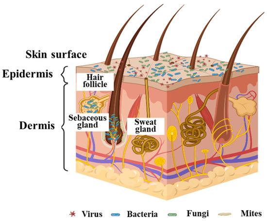
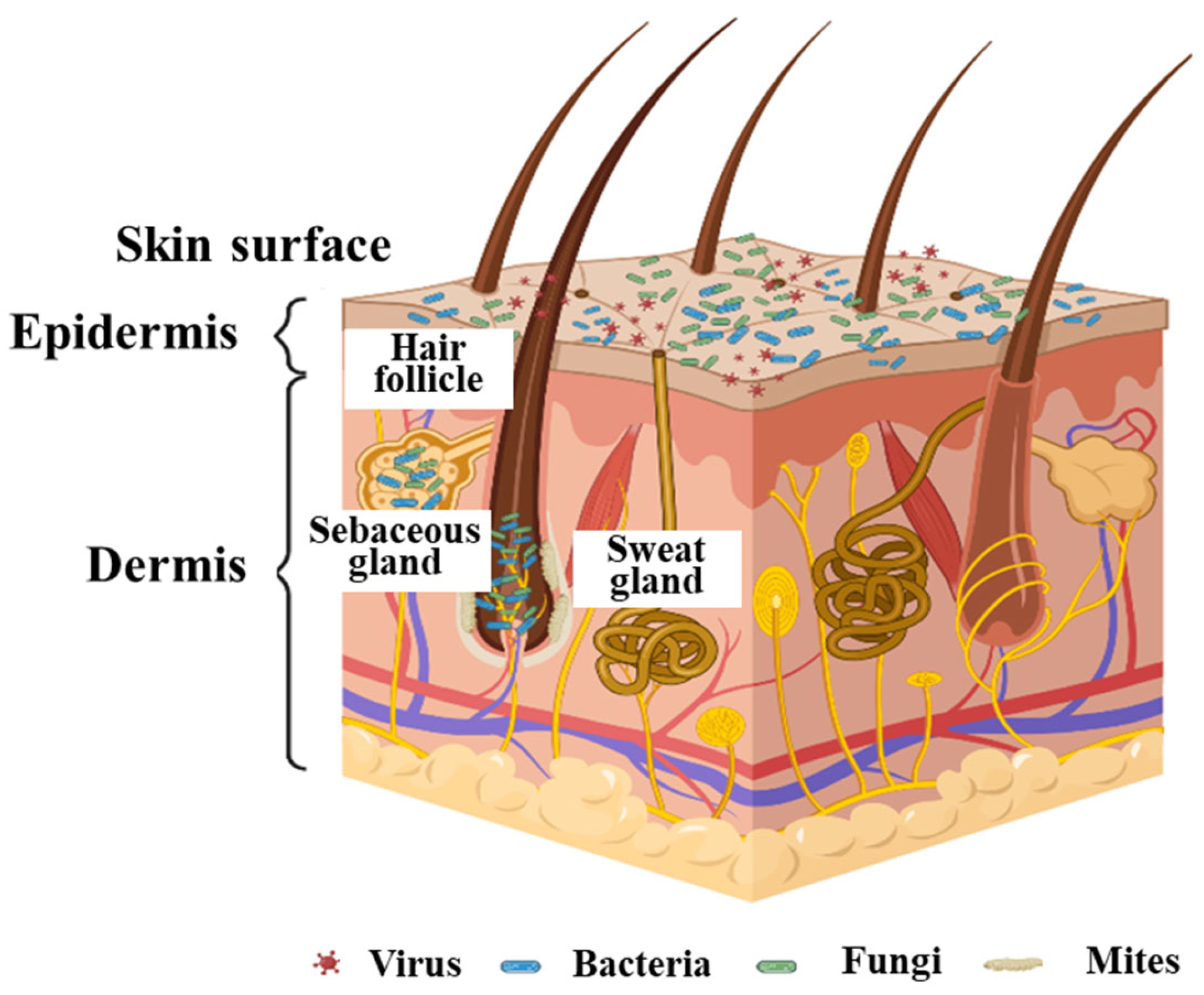
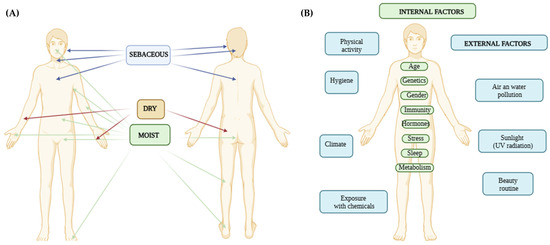
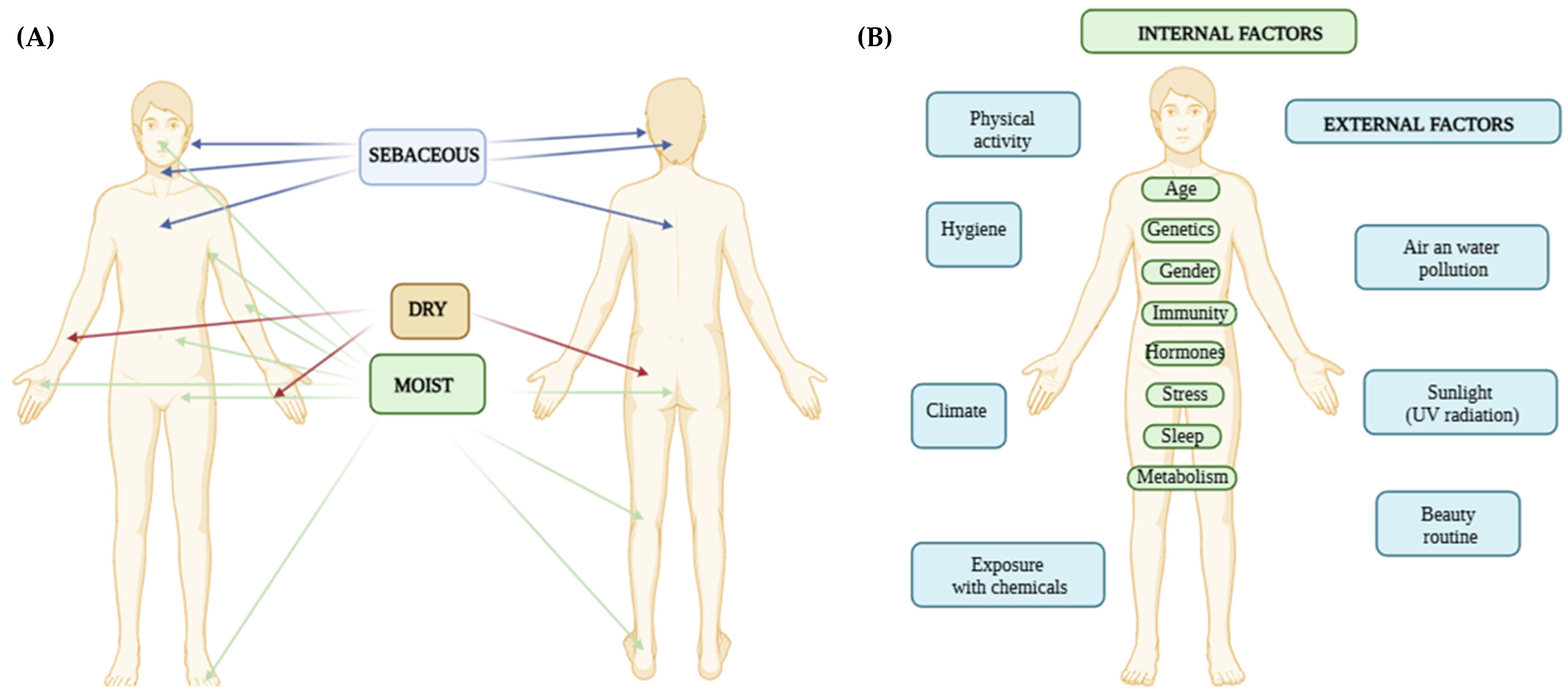
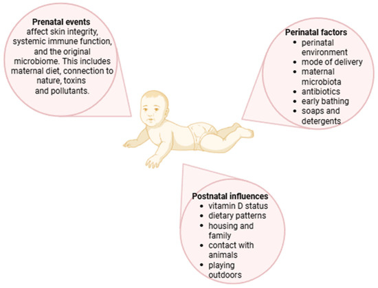
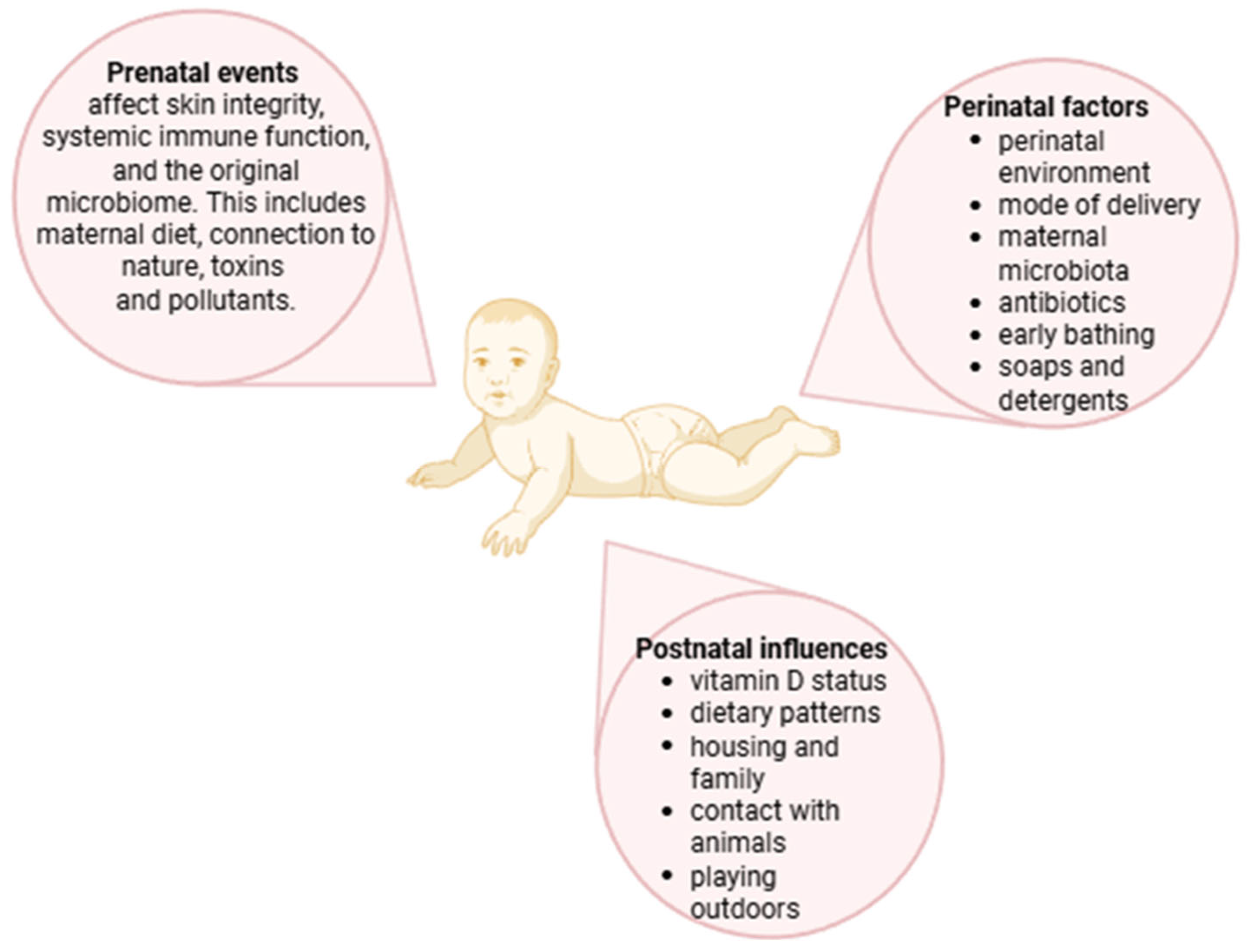
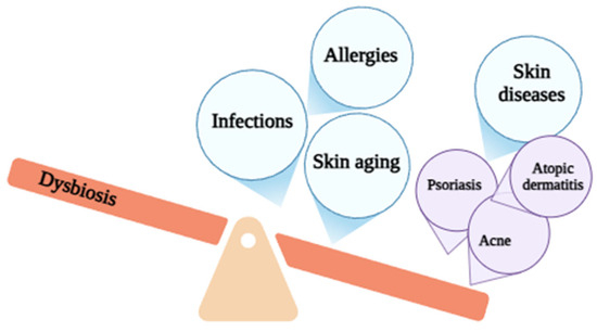
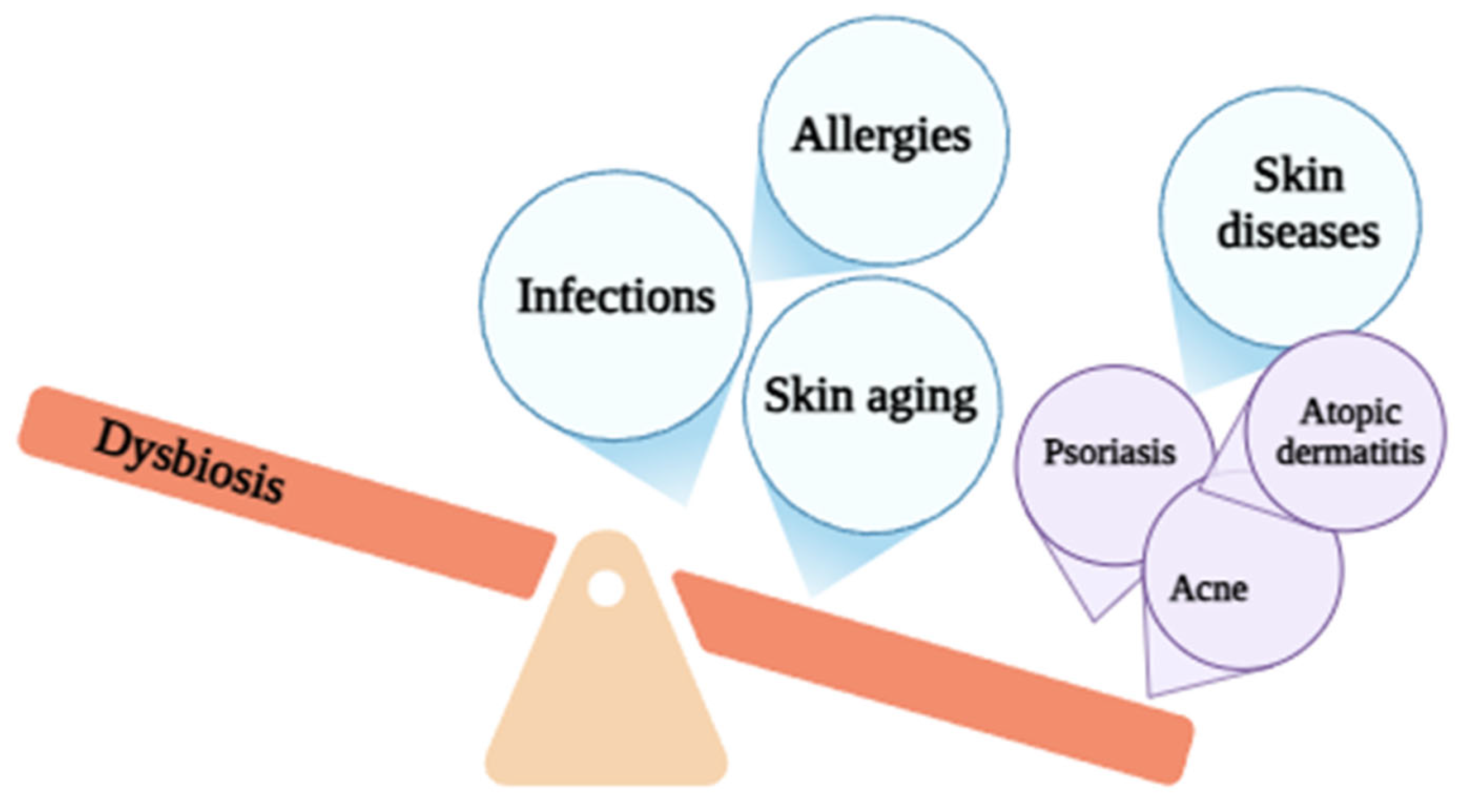
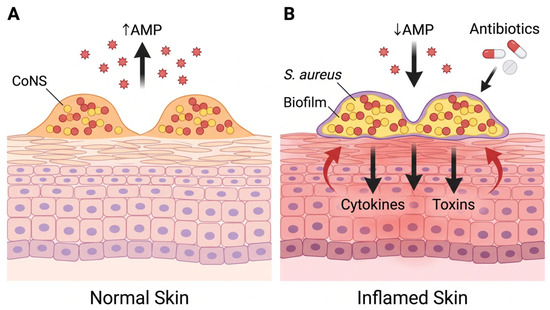
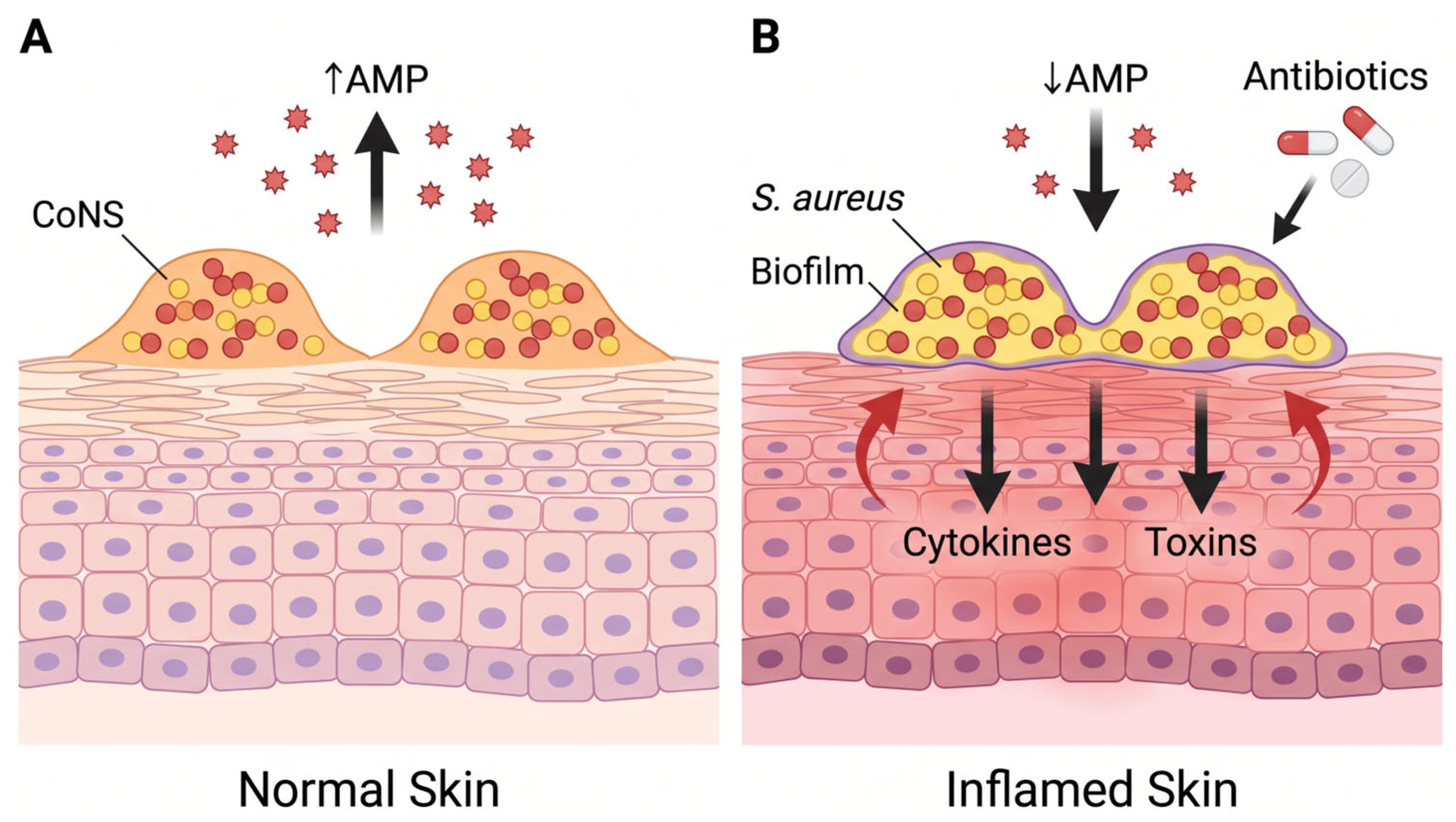
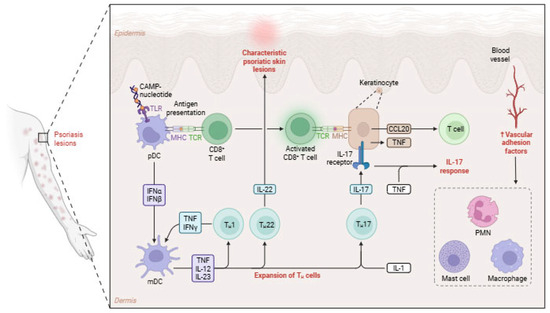
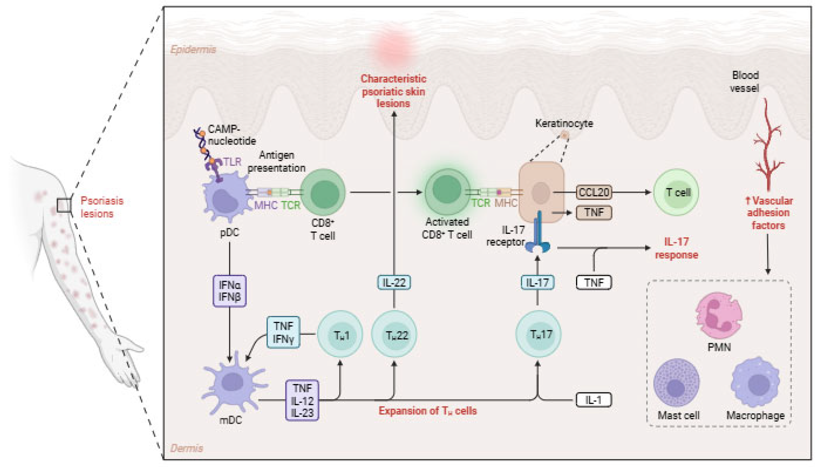
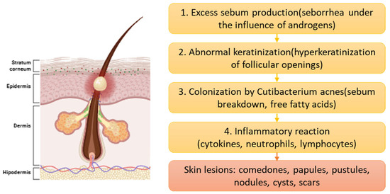
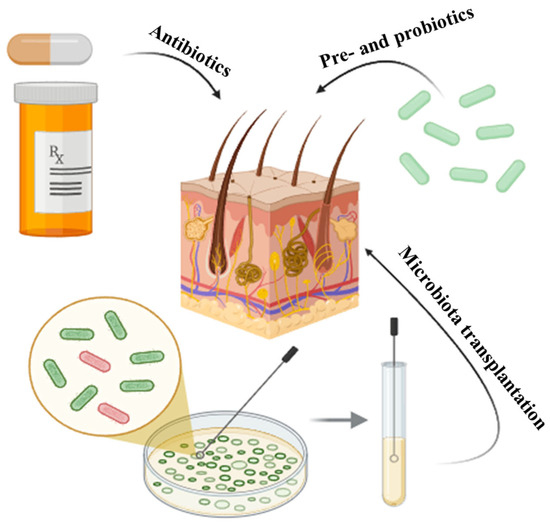
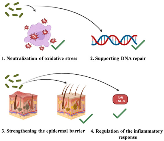
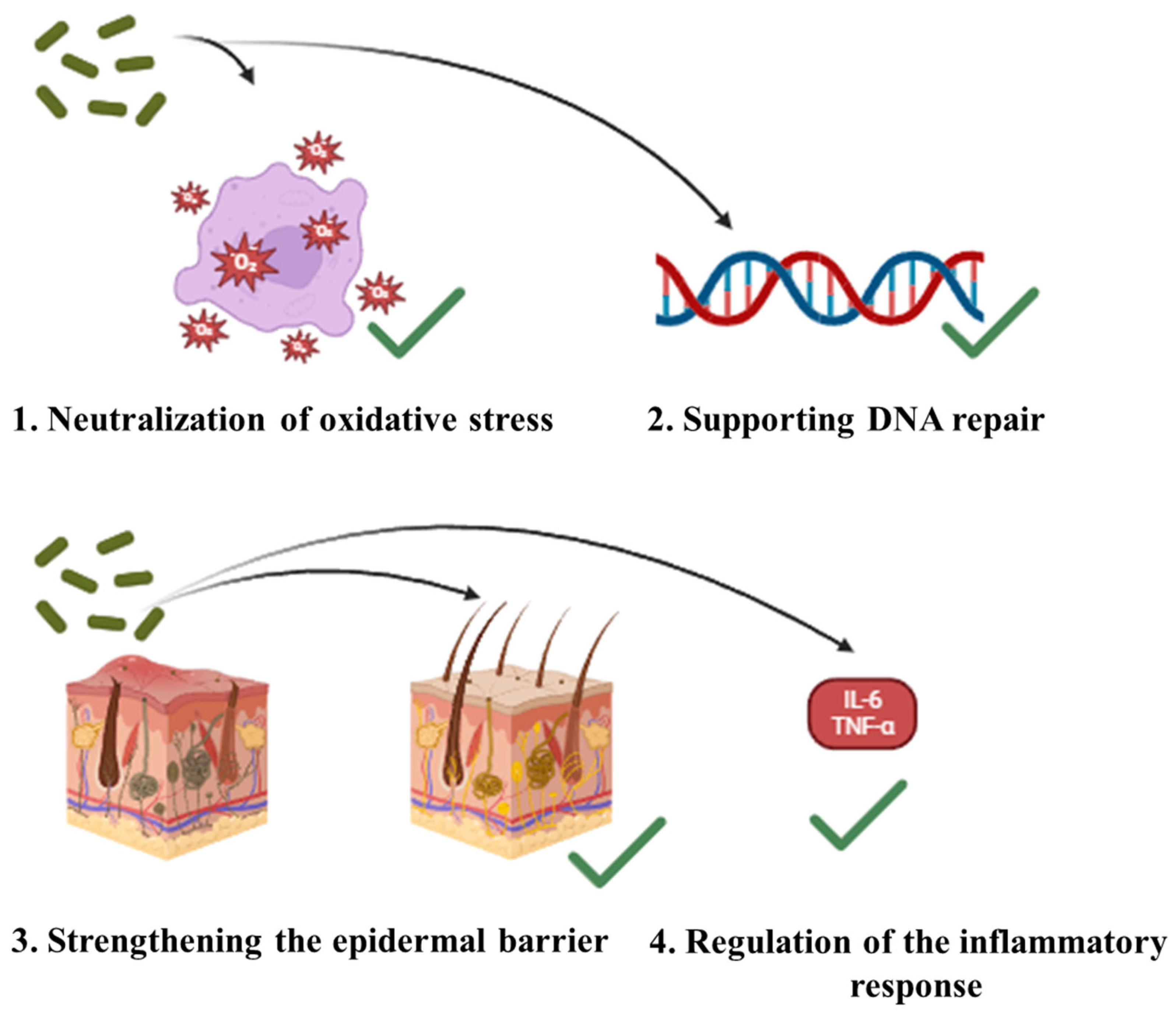

Disclaimer/Publisher’s Note: The statements, opinions and data contained in all publications are solely those of the individual author(s) and contributor(s) and not of MDPI and/or the editor(s). MDPI and/or the editor(s) disclaim responsibility for any injury to people or property resulting from any ideas, methods, instructions or products referred to in the content. |
© 2026 by the authors. Licensee MDPI, Basel, Switzerland. This article is an open access article distributed under the terms and conditions of the Creative Commons Attribution (CC BY) license.