


Abstract
Poly(ADP-ribose) polymerase inhibitors (PARPis) have demonstrated remarkable efficacy in tumors carrying BRCA1/2 pathogenic variants (PVs) through the mechanism of synthetic lethality. PVs in other homologous recombination (HR) genes may also impair homologous recombination repair and confer sensitivity to PARPis; however, their predictive value remains uncertain and appears to vary according to the affected gene and tumor type. We review and critically discuss the current evidence regarding the predictive value of non-BRCA HR gene pathogenic variants as biomarkers of PARPi sensitivity across ovarian, breast, prostate, and pancreatic cancers. A narrative review was conducted between October 2023 and December 2025, first identifying pivotal clinical trials of PARP inhibitors across ovarian, breast, prostate, and pancreatic cancers, followed by a targeted search of PubMed, Embase, Web of Science, and Google Scholar for relevant preclinical and clinical studies. Seventeen clinical studies and multiple preclinical reports were analyzed regarding genomic frequency, HRD association, and treatment response. Preclinical studies consistently demonstrated increased PARPi sensitivity in models with alterations in several non-BRCA HR genes. Clinical evidence, however, was heterogeneous. PALB2 demonstrated the strongest and most consistent association with PARPi benefit across tumor types, while RAD51C and RAD51D also showed clinically meaningful activity, particularly in ovarian cancer. In contrast, evidence supporting PARPi sensitivity in tumors harboring ATM, CHEK2, CDK12, and several other HR gene alterations remained limited or inconsistent. Differences in gene function, biallelic inactivation, variant type, and current limitations of HRD companion diagnostic assays likely contribute to the observed variability in clinical response. Non-BRCA HR gene alterations represent promising predictive biomarkers for PARPi therapy but should not be considered a homogeneous group. Future biomarker-driven studies integrating comprehensive genomic profiling and functional assessment of homologous recombination deficiency are needed to refine patient selection and optimize the clinical application of PARPis beyond BRCA1/2-associated cancers.
1. Introduction
The homologous recombination (HR) pathway is a high-fidelity DNA repair mechanism that enables accurate repair of DNA double-strand breaks and maintenance of genomic stability. Among the key genes involved in this pathway, BRCA1 and BRCA2 genes are the most extensively characterized and are frequently altered in hereditary and sporadic cancers [1]. Germline pathogenic variants (PVs) affecting one of these two genes increase the risk of developing cancer, particularly ovarian, breast, prostate and pancreatic cancers [2]. The proportion of HR-deficient homologous recombination deficiency (HRD) ovarian cancers is close to 50%: 20% with BRCA1/2 pathogenic variants, 10% with BRCA1 promoter methylation and 20% for other reasons [3]. The proportion of breast, prostate and pancreatic HRD cancers is about 21%, 10% and about 15%, respectively. To perform DNA repair, BRCA1 and BRCA2 and other HR genes, such as PALB2, RAD51C/D, ATM, CHEK2, are involved in a common pathway [4,5]. PVs in these genes may also lead to an HRD phenotype. PVs in other HR genes are also associated with cancer, but to a lesser extent [1,6,7,8,9,10,11]. The frequency of somatic HR gene PVs other than BRCA1/2 in ovarian, breast, prostate and pancreatic cancers ranges from 5 to 10% [12,13]. In addition, germline PVs affecting HR genes have been identified in ~30% of patients with familial breast and ovarian cancer [14] and in 11% and 16% of metastatic prostate and pancreatic cancer patients, respectively [15,16]. Promoter methylation could also explain the HRD phenotype. Methylation of the BRCA1 promoter remains significant only in breast and ovarian cancer (10–15%), and a few rare cases of methylation of the RAD51C promoter have also been reported in breast and ovarian cancers [17,18,19,20,21,22]. However, methylation is a rare event in the other cancers [10,17,19]. On the remaining proportion, PVs in other HR genes could explain the HRD phenotype.
It is widely known that HRD tumors exhibit increased sensitivity to platinum agents because of the accumulation of unrepaired DNA damage leading to apoptosis [23]. PARPs are a family of enzymes involved in different cellular pathways, including DNA single-strand break repair. They trigger a cascade of events in response to single-stranded DNA breaks, leading to the recruitment of repair factors. It has been shown that HRD tumors rely on PARPs for survival [24]. Therefore, concurrent inhibition of PARP enzymes along with HRD results in an accumulation of DNA damage and genomic instability, which is lethal to cancer cells (Figure 1). This has been confirmed in preclinical studies as well as in clinical trials and has led to the development of PARPis as promising targeted therapy [25] and FDA approval of Olaparib, Niraparib, Talozaparib and Rucaparib for treatment of breast, ovarian, prostate and pancreatic cancers. However, most clinical trials to date have primarily focused on BRCA1/2 PVs, and functional and clinical evidence regarding PARPi efficacy in tumors with PVs in non-BRCA HR genes remains limited (Figure 1).
Figure 1.
Synthetic lethality induced by PARP inhibition in homologous recombination deficient tumors. PARP inhibition leads to the accumulation of DNA damage that cannot be efficiently repaired in homologous recombination deficient cells, resulting in synthetic lethality, while homologous recombination proficient cells survive through effective DNA repair.
Biomarker testing is critical to identify patients most likely to benefit from PARPis and to guide treatment decisions. For ovarian cancers in addition to BRCA1/2 screening, determining HRD status (signature) is equally important to guide treatment decisions as PARPi maintenance therapy showed the greatest benefit for patients with a BRCA1/2 PV or for those who tested positive for HRD. Two FDA-approved commercial tests are available for determining ovarian cancer HRD status: Myriad MyChoice® CDx tests for the presence of a BRCA1/2 PV and/or genomic instability (LOH, telomeric allelic imbalance, and large-scale state transitions), and FoundationOne CDxTM tests for the presence of a BRCA1/2 PV and LOH. Academic-laboratory-developed tests are also an alternative to commercial assays [26,27,28,29,30,31,32]. However, for breast, prostate and pancreatic cancers HRD phenotype analysis is not part of the daily routine, and the choice of using PARPis is still limited to cases with identified BRCA1/2 PVs [33]. The predictive potential of non-BRCA HR PVs (e.g., PVs in RAD51C, RAD51D, BRIP1, PALB2, NBN, ATM, CHEK2, CDK12) has been evaluated as exploratory analysis of clinical trial data, but no definitive conclusion can be reached so far.
The review aims to present the current evidence regarding the potential role of non-BRCA HR gene PVs as predictive biomarkers of response to PARPi therapy for ovarian, breast, prostate, and pancreatic cancers.
2. Methodology
2.1. Literature Search Strategy
This study was conducted as a narrative review aimed at summarizing and critically discussing the available evidence regarding the predictive value of pathogenic variants in non-BRCA homologous recombination (HR) genes for response to poly(ADP-ribose) polymerase inhibitors (PARPis) across ovarian, breast, prostate, and pancreatic cancers.
A structured literature search was performed between October 2023 and December 2025 using PubMed, Embase, Web of Science, and Google Scholar. The search combined Medical Subject Headings (MeSH) and free-text terms related to PARP inhibitors, homologous recombination deficiency (HRD), DNA damage response (DDR), and homologous recombination genes. Search terms included combinations of “PARP inhibitors”, “homologous recombination deficiency”, “DNA damage repair”, “non-BRCA”, “PALB2”, “RAD51C”, “RAD51D”, “BRIP1”, “ATM”, “CHEK2”, “CDK12”, “NBN”, “ovarian cancer”, “breast cancer”, “prostate cancer”, and “pancreatic cancer”. Reference lists of relevant publications and recent review articles were also manually screened to identify additional eligible studies (Figure 2).
Figure 2.
Summary of the literature review used in this report. Selection of clinical phase I, II or III studies investigating PARPi monotherapy for treatment or ovarian, breast, prostate and pancreatic cancer patients harboring non-BRCA HR gene pathogenic variants.
2.2. Study Selection
Publications were selected according to their relevance to the objectives of this review. Eligible studies included preclinical investigations, prospective and retrospective clinical studies, translational research, biomarker analyses, and clinical trials evaluating the efficacy of PARPis in tumors harboring pathogenic variants in non-BRCA HR genes. High-quality review articles, consensus statements, and current clinical guidelines were also consulted to provide background information and contextualize the available evidence.
When multiple publications reported results from the same clinical trial, the most recent or comprehensive report was preferentially included. Greater emphasis was placed on studies reporting clinically meaningful outcomes, including objective response rate (ORR), progression-free survival (PFS), overall survival (OS), duration of response (DoR), and clinical benefit rate (CBR), whenever available. Considering the inclusion criteria, 17 clinical studies were selected for inclusion (Figure 2 and Figure 3). We will initially present the frequency of genomic alterations and preclinical data in the Section 3, followed by the presentation of clinical data available so far.
Figure 3.
Description of clinical trials according to the primary tumor. Trials are organized according to primary tumor type, PARP inhibitor, trial phase, target population, and main clinical characteristics.
2.3. Data Synthesis
The evidence was synthesized narratively and organized according to individual HR genes and tumor type. Preclinical and clinical evidence were evaluated separately to distinguish experimental findings from clinically validated outcomes. Particular attention was given to the consistency of evidence supporting each non-BRCA HR gene as a predictive biomarker of PARPi sensitivity, as well as to factors potentially contributing to heterogeneous treatment responses across studies, including differences in study design, patient populations, molecular alterations, and HRD assessment methods.
As this work was designed as a narrative review, no formal systematic review protocol, prospective registration, or PRISMA-guided study selection process was performed [34].
3. Gene Review and Preclinical Studies
This section summarizes the principal genes (Figure 4) identified across the included studies, most of which are related to HR, and lists them alphabetically. Table 1 and Figure 5 present the frequency of somatic alterations in HR genes. Preclinical evidence and the functional impact of PVs on the HR pathway are presented in Table 2.
Figure 4.
Overview of DNA damage repair pathways relevant to PARPi sensitivity and HRD. DNA damage is repaired through two major pathways: base excision repair (BER), responsible for repairing single-strand breaks in a PARP1-dependent manner, and homologous recombination repair (HRR), which repairs double-strand breaks through the coordinated action of DNA damage sensors (ATM, ATR, CHEK1/2), core HR proteins (BRCA1, BRCA2, PALB2, BARD1, RAD51 paralogs, FANC complex, MRN complex, and RPA1), and regulatory helicases (ATRX, BLM, and BRIP1). Defects in HRR components result in homologous recombination deficiency (HRD), impairing DNA repair and increasing tumor susceptibility to PARP inhibitors through synthetic lethality. The figure highlights the principal molecular components involved in both repair pathways and their contribution to the maintenance of genomic stability.
Table 1.
Frequency of somatic HR-gene alterations in prostate, ovarian, breast and pancreatic cancers [https://www.cbioportal.org/ (accessed on 25 March 2026)].
Figure 5.
Frequency of homologous recombination repair (HRR) gene alterations across prostate, ovarian, breast, and pancreatic cancers. Higher frequencies were observed in key genes such as BRCA1, BRCA2, and ATM, with variability across tumor types, reflecting differences in molecular profiles and HRR pathway dependency.
Table 2.
Selected preclinical studies of the association between HR gene defect and PARPi sensitivity of cancer cells.
ATM
Ataxia telangiectasia mutated (ATM) is a protein kinase with a key role in cell signaling in response to DNA double-stranded breaks and in recruiting proteins involved in repair [63,64,65,66]. ATM gene is associated with autosomal dominant predisposition to breast, pancreatic and prostate cancers [67,68,69,70]. For ovarian cancer, lifetime absolute risk is less prominent, ranging from 2 to 3%. Preclinical models have shown that ATM depletion sensitizes breast cancer cells to PARP inhibition, suggesting a potential role of PARPis for treatment of breast cancers low in ATM protein expression, such as those arising in mutant ATM heterozygous carriers [40,71]. In agreement with this, synthetic lethality between PARPis and ATM PVs has been reported in cells of patients with ataxia telangiectasia [72]. Furthermore, it was shown that olaparib induces significant killing of ATM-deficient cells with a significant correlation between reduction in ATM expression and sensitivity to the PARPis [73,74]. Despite this, ATM pathogenic variants are not consistently associated with an HRD genomic signature in ovarian cancer and, similarly, this non-association has also been observed in breast and prostate cancers, where clinical benefit from PARP inhibition remains heterogeneous [35,75,76].
ATR
ATR is a serine/threonine kinase and a DNA damage sensor. It plays a role in the HR pathway by phosphorylating and activating several proteins (BRCA1, CHEK1, MCM2, RAD17, RPA2, SMC1 and p53/TP53) responsible for the inhibition of mitosis and replication [77,78]. No association between hereditary cancers and ATR PVs has been validated to date. Somatic PVs have been reported in 0.5% of breast and prostate cancers and in 0.2% and 0.6% in ovarian and pancreatic cancers, respectively [79,80]. Preclinical studies have shown that loss of function of this gene is associated with higher sensitivity to PARPis [36,38,44,45,46]. Despite this, the HRD status is not associated with ATR PVs in ovarian cancer [35].
ATRX
The ATRX protein is a chromatin remodeling factor, initially described as a putative helicase protein due to sequence homology with the DNA repair and recombination RAD54 protein. ATRX is a member of the SNF2 subgroup of the SWI/SNF protein superfamily. It plays a role in suppressing proteins of the ATM pathway by decreasing histone H3K9me3 [77,81]. No association between any hereditary cancer and this gene has been validated to date. However, somatic PVs have been reported in 0.4% of breast and prostate cancers, in 0.7% and 0.1% of ovarian and pancreatic cancers, respectively [79,80]. CRISPR-based studies revealed that loss of ATRX confers sensitivity to PARP inhibitors [82]. ATRX PVs were not associated with HRD status in ovarian cancer [35].
BARD1
BARD1 is an E3 ubiquitin-protein ligase which interacts with BRCA1 via its N-terminal RING finger domains to form a heterodimer essential for BRCA1 stability [77,83]. Germline PVs in BARD1 confer a twofold higher risk of breast cancer development [84,85,86]. Somatic PVs have been reported in 0.4% and 0.3% of breast and prostate cancers, respectively, and in 0.1% of ovarian and pancreatic cancers [79,80]. Preclinical studies have shown that loss of function of BARD1 confers higher sensitivity of tumor cells to PARPis [47]. The HRD status is not associated with BARD1 PVs in ovarian cancer sample alone [35].
BLM
BLM is an ATP-dependent DNA helicase that unwinds and resects the 5′ end of DNA during double-stranded break repair [77,87]. No definitive association between any hereditary cancer and this gene has been validated to date [84]. Somatic PVs have been described in 0.3% and 0.2% of breast and prostate cancers, respectively, and in 0.4% of ovarian and pancreatic cancers [79,80]. PVs in this gene conferred higher sensitivity of tumor cells to PARPis in two preclinical studies [39,48]. The HRD status is associated with BLM PVs in ovarian cancer [35].
BRIP1
BRIP1 (BACH1) is a 5′ to 3′ DNA helicase and DNA-dependent ATPase. It has an important function in the HR pathway by binding BRCA1 by its BRCT repeat domain [77,88,89]. Germline PVs affecting BRIP1 may increase the risk of breast cancer development 1.5-fold and ovarian cancer 5-fold compared to the normal population [84]. Somatic PVs have been reported in 0.3% of breast and pancreatic cancers, and in 0.2% of ovarian and prostate cancers [79,80]. Preclinical studies have shown that loss of function of BRIP1 confers higher sensitivity of cancer cells to PARPis [49,90]. BRIP1 PVs have been associated with HRD status in ovarian cancer [35].
CDK12
CDK12 is a cyclin-dependent kinase and a transcription factor involved in the HR pathway. It regulates the expression of HR genes by activating RNA polymerase II (POLR2A) through phosphorylation of its C-terminal domain [77,91]. It regulates gene transcription, RNA splicing, translation, cell cycle, cell proliferation, DDR and maintenance of genomic stability [92,93]. No definitive association between any hereditary cancer and CDK12 has been described or validated in the literature. However, a case–control study which included patients of Tatar descent suggested a possible association between a variant of CDK12 (c.1047-2A>G) and hereditary breast and ovarian cancer [77]. Somatic PVs have been described in breast, ovarian, prostate and pancreatic cancer with a frequency of 0.8%, 2.7%, 4% and 0.1%, respectively [79,80]. In ovarian cancer, clinical trial studies reported around 1,5% [35]. Preclinical studies have shown that CDK12 PVs confer higher sensitivity of tumor cells to PARPis [50,51]. The HRD status is not associated with CDK12 PVs [35].
CHEK2-CHEK1
CHEK2 and CHEK1 are serine/threonine-protein kinases implicated in cell-cycle arrest in response to DNA damage, interacting with several proteins in the HR pathway (ATM, ATR, BRCA2) [77]. No association between any hereditary cancer and CHEK1 has been validated to date. Germline PV affecting CHEK2 may increase the risk of breast cancer 1.5-fold [84]. Emerging evidence suggests an association with increased prostate cancer risk. Although the CHEK2 c.1100del variant has also been proposed to be associated with colorectal cancer (CRC) [94], this association was not confirmed in a subsequent population-based study. Two meta-analysis reported a modestly increased CRC risk of about twofold [95,96]. Somatic PVs in CHEK1 have been reported in 0.6%, 0.7% and 0.5% of breast, ovarian and prostate cancers, respectively. Somatic alterations affecting CHEK2 have been described in 0.3%, 1%, 1.5% and 0.5% of breast, ovarian, prostate and pancreatic cancers, respectively [79,80,97]. Preclinical studies have shown that loss of function of CHEK1 and CHEK2 confers higher sensitivity of cancer cells to PARPis [36,46]. The association between CHEK2 PVs and HRD status was inconsistent [35].
FANCs
The Fanconi anemia (FA) family includes 22 distinct functional complementation genes: FANCA, FANCB, FANCC, FANCD1 (BRCA2), FANCD2, FANCE, FANCF, FANCI, FANCJ (BRIP1), FANCL, FANCM, FANCN (PALB2), FANCO (RAD51C), FANCP (SLX4), FANCQ (ERCC4), FANCR (RAD51), FANCS (BRCA1), FANCT (UBE2T), FANCU (XRCC2), FANCV (REV7). The FA pathway, also called the FA-BRCA pathway, is a fundamental DNA repair pathway that recognizes DNA damage and orchestrates DNA damage responses, especially for DNA interstrand crosslink (ICL) repair [98]. Roles in the repair of other types of DNA damage and in the regulation of replication stress responses have been additionally ascribed. One FA gene subset encodes nine proteins of the FA core complex (FANCA/B/C/E/F/G/L/M/T), which activates the FANCI–FANCD2 heterodimer through monoubiquitination. The remaining eight FA proteins (FANCD1/J/N/O/P/Q/R/S) mediate recombinational and nucleolytic reactions to complete repair [77,99]. For example, excision repair cross-complementation group 4 (FANCQ/ERCC4/XPF) is a gene that encodes a protein that is involved in nucleotide excision repair. The most common alterations in ERCC4 are ERCC4 PV (1.37%), ERCC4 Amplification (0.15%), ERCC4 E527D (0.08%), ERCC4 Loss (0.03%), and ERCC4 S540L (0.02%). The evidence available so far suggests that ERCC4 is not a cancer susceptibility gene. ERCC4 is altered in 1.51% of breast carcinoma patients, 0.85% of ovarian carcinoma tumor, 0.75% of prostate adenocarcinoma patients, and is altered in 0.7% of pancreatic carcinoma [100]. Of note, increased hereditary risk for cancer has been associated with some of other FA genes (FAND1 (BRCA2), FANCS (BRCA1), FANCN (PALB2), FANCO (RAD51C)). Somatic PVs in the FA-BRCA pathway have been reported in around 0.3–3% of breast, ovarian, prostate and pancreatic cancers [79,80]. Preclinical studies have shown that loss of function of FANCA, FANCC, FANCD2 and FANCG confers higher sensitivity of cancer cells to PARPis [36,39,52,53]. The association between FANCA PVs and HRD status was inconsistent [35]. The HRD status is not associated with FANCM, FANCI, or FANCL PVs in ovarian cancer [35].
HDAC2
Histone deacetylase 2 (HDAC2) is an enzyme encoded by the HDAC2 gene. This protein is involved in chromatin remodeling through deacetylation of core histones and plays a role in transcriptional regulation and cell-cycle control. Following the identification of inactivating HDAC2 PVs in cancer, studies demonstrated that HDAC inhibition can suppress tumor growth in colorectal cancer cell lines [101]. Although HDAC2 is not a canonical homologous recombination (HR) gene and HDAC2 PVs have not been validated as predictive biomarkers of PARP inhibitor response, preclinical data indicate that HDAC inhibition can induce a functional “BRCAness” phenotype by downregulating HR-related proteins such as BRCA1 and RAD51 [102]. More recently, dual PARP/HDAC inhibitors have been shown to restore synthetic lethality and enhance antitumor activity through combined DNA damage and immune signaling activation in triple-negative breast cancer models [103].
MRE11
MRE11 has exonuclease and endonuclease activity. It is a component of the MRN complex (MRE11-RAD50-NBN) which is implicated in activating DNA damage sensors in the HR pathway [77,104]. No association between any hereditary cancer and this gene has been validated to date [84]. Somatic PVs have been reported in 0.5% of breast and prostate cancers, and in 0.6% and 0.1% of ovarian and pancreatic cancers, respectively [79,80]. Preclinical studies have shown that loss of function of this gene is associated with higher sensitivity to PARPis [54,55,56,57,105].
NBN
NBN is a component of the MRN complex (MRE11-RAD50-NBN) which is implicated in double-stranded break repair. It activates and recruits signal check-point sensors (ATM, ATR) and other proteins like MRE11 and RAD50 to the site of DNA damage [77,104]. Germline PVs in the NBN gene have been described and confer a relative risk of ovarian cancer development nearly twice that of the normal population [61,84]. Women with NBN PVs are also at increased risk of developing breast cancer [106]. Somatic PVs have been described in 0.1%, 0.5%, 0.3% and 0.4% of breast, ovarian, prostate and pancreatic cancers, respectively [79,80]. Preclinical studies have shown that loss of function of NBN is associated with higher sensitivity to PARPis [36,55]. NBN pathogenic variants were not associated with HRD status in ovarian cancer [35].
PALB2
PALB2 plays a critical role in the HR pathway through different mechanisms [107]. It binds DNA with high affinity for the D loop and recruits BRCA2 and RAD51 to DNA break sites [108]. It promotes stability of BRCA2 and is essential for its checkpoint functions. It also forms a complex with BRCA1 and BRCA2, serving as a bridge of these two proteins, which is an essential step for HR repair [77,109]. Germline PVs in PALB2 confer a 5-times-higher risk of breast cancer and 2-times-higher risk of ovarian cancer compared to the normal population [84]. Estimated lifetime risk is 53% for breast cancer, 5% for ovarian cancer and 2–3% for pancreatic cancer [110,111]. Somatic PVs have been reported in 0.2%, 0.5%, 0.4% and 0.2% of breast, ovarian, prostate and pancreatic cancers, respectively [79,80]. Preclinical studies have shown that loss of function of PALB2 confers higher sensitivity of cancer cells to PARPis [53,58]. The HRD status is associated with PALB2 PV [35].
PPP2R2A
Protein phosphatase 2A (PP2A) exhibits tumor suppressor function, and the reduced expression of PPP2R2A promotes tumor pathogenesis [112]. PPP2R2A, located at chromosome 8p21.2, is deleted at high frequencies in prostate, ovarian, and luminal type B breast cancers [113]. PPP2R2A is altered in 1.42% of breast carcinoma patients [83,100] and is associated with poor prognosis in prostate adenocarcinomas [112]. PP2A genes are heterozygous-lost in the majority of HGSOC (high-grade serous ovarian cancer) and this loss correlates with worse survival. Preclinical studies showed that SMAP-061-induced stabilization of PP2A inhibits the HR output by targeting RAD51, leading to chronic accumulation of DNA damage and ultimately apoptosis. This evidence emphasize the potential of PP2A modulators to overcome PARPi insensitivity, given that targeting RAD51 presents benefits in overcoming PARPi resistance driven by BRCA1/2 mutation reversions [114]. In HGSOC, PPP2R2A is frequently deleted/downregulated, and ovarian cancer cells with low PPP2R2A expression showed increased sensitivity to the PARP inhibitor niraparib in vitro; however, this evidence remains limited and requires validation in larger preclinical models and clinically annotated cohorts [115].
PTEN
PTEN encodes a phosphatase whose role in the control of the phosphoinositide 3 kinase (PI3K) signaling pathway is well established [116]. Mutations in the phosphatase and tensin homolog (PTEN) gene and loss of PTEN expression have both been associated with a wide range of human tumors. A new functional role for PTEN was recently suggested by the observation that mouse embryonic Pten−/− cells exhibit genomic instability, a phenotype ascribed to either defects in RAD51-mediated DNA double-strand break repair (DSBR) [117] or defects in cell-cycle checkpoints [118]. Germline PTEN pathogenic variant confers a lifetime risk of 60% for breast cancers. The risk of thyroid, colorectal, endometrial, and renal cancers is also improved [119,120]. Concerning somatic alterations, TCGA data shows that PVs in PTEN occur across a wide range of cancers, with uterine cancer and glioblastoma multiforme having the highest percentages of PTEN PVs and homozygous loss. Thus far, no in vitro correlation has been detected between a PTEN mutational status and PARPi response [119,120].
RAD50
RAD50 is a member of the MRN complex, essential to initiate double-stranded DNA repair. This protein plays a role in binding and holding DNA ends in close proximity [77,104]. No association between any hereditary cancer and this gene has been validated. Somatic PVs have been reported in 0.1% and 0.5% of breast and ovarian cancers, respectively, and in 0.7% of prostate and pancreatic cancers [79,80]. Preclinical studies have shown that loss of function of this gene is associated with higher sensitivity to PARPis [55,121].
RAD51
The RAD51 recombinase has long been considered a major guardian of genomic stability but multifaceted roles that RAD51 plays in carcinogenesis, cancer progression and anticancer drug resistance [77,122,123]. No association between any hereditary cancer and this gene has been validated to date. Somatic PVs have been reported in 0.5%, 2.5%, 0.6% and 0.1% of breast, ovarian, prostate and pancreatic cancers, respectively [79,80]. Preclinical studies have shown that loss of function of RAD51 confers higher sensitivity of tumor cells to PARPis [36,44,59,60].
RAD51B/C/D/XRCC2/3
RAD51B/C/D paralogs are members of the RAD51 complex (RAD51B, RAD51C, RAD51D, XRCC2 and XRCC3) involved in HR DNA repair. They form distinct complexes that act as mediators of RAD51 presynaptic filament assembly, and they are required for loading of RAD51 at sites of DNA damage [77,124,125,126,127]. X-ray repair cross-complementing group 3 (XRCC3) is responsible for maintaining the integrity of the genome, playing a critical role in protecting it against mutations which lead to cancer [128]. Germline PVs in RAD51C/D have been associated with a four- and sevenfold higher risk of developing ovarian cancer compared to the normal population, respectively [84,129]. No association between any hereditary cancer and RAD51B has been validated to date. There is emerging evidence of an increased risk for breast cancer for RAD51B PV/VPL carriers [130,131,132]. Somatic PVs have been reported with a frequency of 0.6%, 1.2%, 0.4% and 0.2% in breast, ovarian, prostate and pancreatic cancer, respectively, for RAD51B; of 0.4%, 0.2%, 0.5% and 0.5% in breast, ovarian, prostate and pancreatic cancers, respectively, for RAD51C; and of 0.2%, 1.6%, 0.4% and 0.1% in breast and ovarian cancers, respectively, for RAD51D [79,80]. The genetic defects in XRCC2 and XRCC3 genes can affect repairing efficiency as the genes are highly polymorphic, which could lead to the progression of ovarian cancer [133].
Preclinical studies have shown that loss of function of RAD51C [61] and RAD51D [62] confers higher sensitivity of cancer cells to PARPis. The HRD status is associated with RAD51C, RAD51D PVs. The HRD status is inconsistently associated with RAD51B PVs [35].
RAD52
RAD52 was first identified along with a large group of homologous recombination proteins in a screen for DNA-repair-deficient S. cerevisiae mutants following ionizing radiation. Nonetheless, RAD52 is critically important for most, if not all, recombination events in yeast including meiotic recombination, homologous DNA integration, and mating-type switching [134,135]. As RAD52 PV cause no discernible homologous recombination phenotype in humans, the synthetically lethal BRCA/RAD52 relationship makes RAD52 an attractive therapeutic target. It is important to develop more work to create truly drug-like RAD52 inhibitors that can be used in clinic [136,137]. Thus far, no in vitro correlation has been detected between a RAD52 PV and PARPi response [119,120].
RAD54L
RAD54L (RAD54L gene) is a member of the DEAD-like helicase superfamily and plays a role in dissociating RAD51 from nucleoprotein filaments on double-stranded DNA [124]. No association between any hereditary cancer and this gene has been validated to date, while somatic PVs affecting RAD54L have been reported in the literature. Conflicting preclinical data have been published concerning the association of genomic alterations in RAD54L with the sensitivity of tumor cells to PARPis. Some studies have reported increased sensitivity after RAD54L knock down whilst others have showed the opposite [36,48,53]. The HRD status is not associated with RAD54L PVs [35].
WRN
WRN, a member of the RECQ family of DNA helicases, plays essential roles at stalled forks to counteract replication stress, thereby ensuring genomic stability. Biallelic PVs of the WRN gene are associated with a rare syndrome (Werner syndrome) characterized by shot statures, signs of premature aging and an increased risk of developing several specific types of cancer, mostly thyroid epithelial neoplasms, melanoma, meningiomas, soft tissue sarcomas, leukemia and preleukemic conditions, and primary bone neoplasms. Thus far, no in vitro correlation has been detected between WRN mutations, and PARPi response has been published. In addition, genetic or pharmacologic inhibition of WRN selectively induces DNA damage and apoptosis in microsatellite instability-high (MSI-H) tumor cells with sparing microsatellite-stable cells, supporting WRN as a promising context-dependent therapeutic target, although clinical validation remains limited [138].
4. Clinical Trials Results
Following the literature review, 17 articles were identified reporting use of PARPis in patients with ovarian, breast, prostate and pancreatic cancers harboring PVs in homologous recombination genes other than BRCA1/2 (Figure 3 and Table 3).
Table 3.
Clinical trials evaluating PARP inhibitor efficacy in tumors with homologous recombination gene alterations/pathogenic variants beyond BRCA1/2.
Below are descriptions of each clinical trial by tumor type that include study design, PARPis in test, type of primary tumor, eligibility requirements, number of patients, and primary and secondary endpoint results.
4.1. Ovarian Cancer Clinical Studies
STUDY 19 Trial ((NCT00753545)
STUDY 19 was a randomized, double-blind, placebo-controlled phase II trial published in 2012 that evaluated olaparib as maintenance therapy in patients with platinum-sensitive recurrent high-grade ovarian, fallopian tube, or primary peritoneal cancer. Eligibility criteria included completion of at least two prior lines of platinum-based chemotherapy and achievement of a complete or partial response to the most recent platinum regimen. Patients were randomized to receive olaparib 400 mg twice daily (capsule formulation) or placebo. The primary endpoint was progression-free survival (PFS), while secondary endpoints included time to progression (TTP), overall response rate (ORR), and overall survival (OS) [154].
A total of 265 patients were enrolled, with 136 assigned to olaparib and 129 to placebo. In a retrospective exploratory analysis, 21 of the 136 patients treated with olaparib were identified as harboring pathogenic variants (PVs) in homologous recombination (HR) genes other than BRCA1/2. These PVs included BRIP1 (n = 5; 4 germline and 1 somatic), CDK12 (n = 3), RAD54L (n = 3), RAD51B (n = 2), RAD51C (n = 1, germline), RAD52 (n = 1), ATM (n = 1), FANCA (n = 1), FANCL (n = 1), FANCD2 (n = 1, somatic), FANCI (n = 1), and XRCC3 (n = 1) [154].
In this subgroup, olaparib maintenance was associated with a significant improvement in PFS, with a hazard ratio (HR) of 0.21 (95% CI, 0.04–0.86) compared with placebo. The overall survival (OS) analysis showed an HR of 0.77 (95% CI, 0.28–2.28), although this result was not statistically significant [154].
ENGOT-OV16/NOVA Trial (NCT01847274)
The ENGOT-OV16/NOVA was a randomized, double-blind, phase III trial evaluating niraparib as maintenance therapy in patients with platinum-sensitive recurrent high-grade serous ovarian, fallopian-tube, or primary peritoneal cancer [140]. Eligible patients had received at least two prior lines of platinum-based chemotherapy and were randomized within 8 weeks after completion of their last platinum regimen to receive niraparib (300 mg once daily) or placebo until disease progression or unacceptable toxicity. The primary endpoint was progression-free survival (PFS).
Patients were stratified according to the presence of a germline BRCA1/2 pathogenic variant (gBRCA cohort) or absence thereof (non-gBRCA cohort), with a predefined HRD analysis performed in the non-gBRCA population. Among the 553 enrolled patients, 203 were classified as gBRCA and 350 as non-gBRCA. Of the 345 non-gBRCA, 115 were HRD-positive (without somatic PVs in BRCA). In the gBRCA cohort, niraparib significantly prolonged median PFS compared with placebo (21.0 vs. 5.5 months), corresponding to a hazard ratio (HR) of 0.27 (95% CI, 0.17–0.41). In the non-gBRCA HRD-positive subgroup, median PFS was 12.9 months with niraparib versus 3.8 months with placebo (HR 0.38; 95% CI, 0.24–0.59). In the overall non-gBRCA population, niraparib also conferred a significant PFS benefit (9.3 vs. 3.9 months; HR 0.45; 95% CI, 0.34–0.61) [140]. Niraparib significantly improved progression-free survival in non-gBRCA ovarian cancer, particularly in HRD-positive tumors, but the study did not report outcomes according to individual non-BRCA HR genes.
ARIEL2 Trial (NCT01891344)
The ARIEL2 trial was an open-label, phase II study that evaluated rucaparib monotherapy in patients with relapsed, platinum-sensitive high-grade ovarian cancer [25]. Eligible patients had high-grade serous or endometrioid ovarian, fallopian tube, or primary peritoneal carcinoma and had received at least one prior platinum-based chemotherapy regimen. Patients were treated with rucaparib at a dose of 600 mg twice daily until disease progression or treatment discontinuation.
The primary endpoint was progression-free survival (PFS). Secondary endpoints included objective response rate (ORR), defined as a complete or partial response according to RECIST criteria, and duration of response. Tumors were prospectively classified into three homologous recombination deficiency (HRD) subgroups based on tumor BRCA status and genomic loss of heterozygosity (LOH): BRCA-mutant, BRCA wild-type with high LOH (LOH-high), and BRCA wild-type with low LOH (LOH-low).
Among the 204 enrolled patients, all received rucaparib and were allocated to the following three subgroups: 40 patients with tumor BRCA pathogenic variants, 82 with LOH-high tumors, and 70 with LOH-low tumors; LOH status could not be determined in 12 BRCA wild-type tumors. Median PFS was 12.8 months in the BRCA-mutant subgroup, 5.7 months in the LOH-high subgroup, and 5.2 months in the LOH-low subgroup. Corresponding ORRs were 80%, 29%, and 10%, respectively.
An exploratory analysis was performed in BRCA wild-type tumors harboring pathogenic variants in other homologous recombination genes. Patients with RAD51C pathogenic variants (n = 4) demonstrated an ORR of 75% (three partial responses and one stable disease). Two patients with ATM pathogenic variants were included; only one had measurable disease and achieved stable disease. No objective responses were observed in patients with pathogenic variants in BRIP1 (n = 2), RAD51D (n = 2), CHEK2 (n = 2), FANCM (n = 2), FANCA (n = 1), FANCI (n = 1), RAD51B (n = 1), or RAD54L (n = 1). One patient with an NBN pathogenic variant achieved a complete response, while the second had stable disease, corresponding to an ORR of 50% in this small subgroup.
Overall, ARIEL2 demonstrated that rucaparib activity extends beyond BRCA-mutant tumors, particularly in BRCA wild-type, HRD-positive cancers. However, responses among non-BRCA HR gene alterations were heterogeneous and limited by small sample sizes [25]. Rucaparib showed heterogeneous activity in BRCA-wild-type tumors harboring pathogenic variants in RAD51C, ATM, BRIP1, RAD51D, CHEK2, FANCM, FANCA, FANCI, RAD51B, RAD54L, and NBN. The most notable responses were observed in tumors with RAD51C and NBN pathogenic variants, whereas activity was limited or absent in several other genes.
ARIEL3 Trial (NCT01968213)
Of the two LOH-low patients, the PFL for Rucaparib and placebo groups were 6.7 and 5.4 months, respectively (HR 0.58; (95% CI 0.40–0.85); p = 0.0049).
The ARIEL3 trial was a randomized, double-blind, phase III study evaluating rucaparib as maintenance therapy in patients with platinum-sensitive, recurrent high-grade ovarian cancer [141]. Eligible patients had high-grade serous or endometrioid ovarian, fallopian tube, or primary peritoneal carcinoma, had received at least two prior platinum-based chemotherapy regimens, and had achieved a complete or partial response to their most recent platinum-based doublet, which was administered for a minimum of four cycles and assessed according to RECIST criteria.
Patients were randomized in a 2:1 ratio to receive rucaparib (600 mg twice daily) or placebo until disease progression, death, or unacceptable toxicity. The primary endpoint was investigator-assessed progression-free survival (PFS), with secondary endpoints including PFS assessed by blinded independent central review (BICR).
Tumors were prospectively stratified according to homologous recombination deficiency (HRD) status based on tumor BRCA1/2 pathogenic variant and genomic loss of heterozygosity (LOH), defining three subgroups: BRCA-mutant, BRCA wild-type with high LOH (LOH-high), and BRCA wild-type with low LOH (LOH-low). HRD-positive tumors were defined as either BRCA-mutant or BRCA wild-type with high LOH. The LOH-high category included PVs across a broad panel of homologous recombination-related genes.
A total of 564 patients were enrolled. In the BRCA wild-type HRD-positive subgroup (n = 158), median PFS was 9.7 months in the rucaparib group compared with 5.4 months in the placebo group (hazard ratio (HR) 0.44; 95% CI, 0.29–0.66; p < 0.0001). When assessed by BICR, median PFS was 11.1 months with rucaparib versus 5.6 months with placebo (HR 0.55; 95% CI, 0.35–0.89; p = 0.0135).
30 genes were associated with patients with HRD and LOH-high: ATM; ATR; ATRX; BARD1; BLM; BRIP1; CHEK1; CHEK2; FANCA; FANCC; FANCD2; FANCE; FANCF; FANCG; FANCI; FANCL; FANCM; MRE11A; NBN; PALB2; RAD50; RAD51; RAD51B; RAD51C; RAD51D; RAD52; RAD54L; and RPA1.
In the BRCA wild-type LOH-low subgroup, rucaparib also demonstrated a statistically significant but more modest PFS benefit, with median PFS of 6.7 months compared with 5.4 months in the placebo group (HR 0.58; 95% CI, 0.40–0.85; p = 0.0049). Overall, ARIEL3 demonstrated that rucaparib maintenance therapy provides a clinically meaningful PFS benefit not only in BRCA-mutant tumors but also in BRCA wild-type, HRD-positive ovarian cancers, while the benefit in HRD-negative tumors was limited [141]. The predictive value appeared to be driven mainly by LOH/HRD status rather than by any individual gene.
PAOLA trial ((ENGOT-ov25/PAOLA-1, NCT02477644))
The PAOLA-1 trial was a randomized, double-blind, phase III study evaluating the efficacy of maintenance therapy with olaparib in combination with bevacizumab in patients with newly diagnosed, advanced, high-grade ovarian, fallopian tube, or primary peritoneal cancer who had achieved a complete or partial response after first-line platinum–taxane chemotherapy plus bevacizumab [155]. Patients were eligible regardless of surgical outcome or BRCA pathogenic variant status.
Tumor homologous recombination deficiency (HRD) status was assessed using the myChoice® HRD Plus assay (Myriad Genetic Laboratories), which incorporates BRCA1/2 pathogenic variant status and a genomic instability score (GIS); a GIS ≥ 42 defines HRD positivity [155]. The primary endpoint was investigator-assessed progression-free survival (PFS), and overall survival (OS) was a key secondary endpoint.
A total of 806 patients underwent randomization: 537 were assigned to receive olaparib plus bevacizumab and 269 to receive placebo plus bevacizumab. As expected, a substantial benefit was observed in patients with BRCA-mutated tumors, with a median PFS of 37.2 months in the olaparib group compared with 21.7 months in the placebo group (HR 0.31; 95% CI, 0.20–0.47) [155]. Importantly, a clinically meaningful benefit was also demonstrated in patients with HRD-positive tumors without BRCA pathogenic variants, with a median PFS of 28.1 months versus 16.6 months, respectively (HR 0.43; 95% CI, 0.28–0.66) [155].
A subsequent overall survival analysis showed a trend toward improved OS in the HRD-positive/BRCA-wild-type subgroup treated with olaparib plus bevacizumab. At 5 years, 54.7% of patients in the olaparib group were alive compared with 44.2% in the placebo group (median OS not reached vs. 52.0 months; HR 0.71; 95% CI, 0.45–1.13), although this difference did not reach statistical significance [142].
More recently, an exploratory analysis of PAOLA-1 investigated whether PVs in non-BRCA1/2 homologous recombination repair (HRR) genes could independently predict benefit from olaparib plus bevacizumab, irrespective of HRD status [35]. Progression-free survival was evaluated across six different non-BRCA HRR gene panels. Non-BRCA HRR pathogenic variants were not enriched in HRD-positive tumors (25/806; 3.1%) compared with HRD-negative tumors (33/806; 4.1%) or tumors with unknown HRD status (14/806; 1.7%) [35]. Moreover, most tumors harboring non-BRCA HRR pathogenic variants exhibited low median GIS values, although higher GIS values were observed in tumors with PVs in BLM, BRIP1, RAD51C, PALB2, and RAD51D [35]. Importantly, none of the evaluated non-BRCA HRR gene panels predicted a differential PFS benefit from maintenance olaparib plus bevacizumab compared with bevacizumab alone. The authors concluded that current non-BRCA HRR gene panels should not be considered a substitute for HRD assessment based on BRCA mutation status and genomic instability testing in newly diagnosed advanced ovarian cancer [35].
OPINION Trial l (NCT03402841)
The OPINION trial was a phase IIIb, single-arm study evaluating olaparib maintenance monotherapy in patients without a pathogenic germline BRCA1/BRCA2 variant (gBRCAwt) who had platinum-sensitive relapsed ovarian cancer (PSROC) and had received at least two prior lines of platinum-based chemotherapy [143]. Patients with somatic BRCA pathogenic variants (sBRCAm) were eligible for enrollment. Retrospective centralized tumor testing was performed in all enrolled patients to assess sBRCA pathogenic variant status and homologous recombination deficiency (HRD) using the myChoice® HRD Plus assay [143].
The primary endpoint was progression-free survival (PFS). A key secondary endpoint was PFS according to HRD status and sBRCA pathogenic variant status. A total of 279 patients were enrolled and received olaparib.
Median PFS was 16.4 months in patients with sBRCAm, 11.1 months in the overall HRD-positive population (including sBRCAm), 9.7 months in HRD-positive patients excluding sBRCAm, and 7.3 months in HRD-negative patients, respectively [143]. These results indicate that olaparib maintenance therapy provides clinical benefit in patients without a germline BRCA pathogenic variant, with the greatest benefit observed in tumors harboring sBRCA mutations and in HRD-positive disease, although activity was also observed in HRD-negative tumors [143]. Because efficacy was not reported by individual non-BRCA HR genes, the study supports HRD status rather than specific gene alterations.
LIGHT Trial (NCT02983799)
This was a phase II, multicohort study (2022) that evaluated olaparib monotherapy in patients with platinum-sensitive relapsed ovarian cancer (PSROC) who had received ≥1 prior line of platinum-based chemotherapy [156]. Patients were prospectively assigned to four cohorts according to tumor genomic status: germline BRCA1/2 pathogenic variants (gBRCAm), somatic BRCA1/2 pathogenic variants (sBRCAm), HRD-positive tumors without BRCA pathogenic variants, or HRD-negative tumors.
The primary endpoint was objective response rate (ORR). Secondary endpoints included disease control rate (DCR) and PFS. Tumor molecular characterization was performed using the Myriad BRCAAnalysis CDx and myChoice® HRD assays; HRD positivity was defined by a genomic instability score ≥ 42 [156].
Of the 272 patients enrolled, 271 received olaparib, and 270 were included in the efficacy analysis. The ORRs in the gBRCAm, sBRCAm, HRD-positive (BRCA wild-type), and HRD-negative cohorts were 69.3%, 64.0%, 29.4%, and 10.1%, respectively. Corresponding DCRs were 96.0%, 100.0%, 79.4%, and 75.3%. Median PFS was 11.0 months in the gBRCAm cohort, 10.8 months in the sBRCAm cohort, 7.2 months in the HRD-positive cohort, and 5.4 months in the HRD-negative cohort [156]. In the final overall survival analysis, OS rates remained highest in the germline and somatic BRCA cohorts, and among BRCA–wild-type tumors, outcomes continued to favor HRD-positive over HRD-negative disease, supporting the clinical relevance of HRD beyond BRCA alterations [144].
Overall, the LIGHT trial demonstrated that olaparib has clinical activity across all biomarker-defined cohorts, with the greatest efficacy observed in patients harboring BRCA pathogenic variants, irrespective of germline or somatic origin. Among patients without BRCA PVs, improved outcomes were observed in those with HRD-positive tumors compared with HRD-negative disease, supporting the biological relevance of HRD beyond BRCA alterations [144,156]. As individual non-BRCA HR genes were not analyzed separately, the study again favors HRD positivity over gene-level interpretation.
4.2. Breast Cancer Clinical Studies
TBRC-048 (NCT03344965)
The TBCRC 048 trial was a phase II, multicenter, single-arm study that evaluated olaparib monotherapy in patients with metastatic breast cancer harboring either germline pathogenic variants (PVs) in non-BRCA1/2 homologous recombination-related genes or somatic pathogenic variants in BRCA1/2 or other HR-related genes [145]. Eligible patients had received up to two prior lines of chemotherapy for metastatic disease and were excluded if they had platinum-refractory disease. Olaparib was administered at a dose of 300 mg twice daily until disease progression or unacceptable toxicity.
Patients were assigned to two cohorts. Cohort 1 included patients with germline PVs in non-BRCA HR genes (ATM, ATR, BAP1, BARD1, BLM, BRIP1, CHEK1, CHEK2, CDK12, FANCA, FANCC, FANCD2, FANCF, MRE11A, NBN, PALB2, RAD50, RAD51C, RAD51D, or WRN), whereas cohort 2 included patients with somatic BRCA1/2 pathogenic variants. The primary endpoint was ORR according to RECIST v1.1. Secondary endpoints included PFS, clinical benefit rate (CBR; defined as complete or partial response or stable disease ≥18 weeks), duration of response, and safety.
In cohort 1 (n = 27), the distribution of HR gene PVs was as follows: PALB2 (n = 10), CHEK2 (n = 8), ATM (n = 4), BARD1 (n = 1), RAD50 (n = 1), dual CHEK2/ATM (n = 2), and dual PALB2/ATM (n = 1). In this cohort, the ORR was 33%, the clinical benefit rate was 50%, the median duration of response was 9 months, and the median PFS was 4.5 months.
Gene-level exploratory analyses demonstrated marked heterogeneity in response. Among CHEK2 carriers, no objective responses were observed, with best responses limited to stable disease. Patients with ATM PVs showed minimal activity, with three cases of progressive disease and one stable disease. The single patient with a BARD1 pathogenic variant achieved prolonged stable disease (>6 months). Two patients harboring concurrent CHEK2 and ATM alterations experienced disease progression. In contrast, the patient with combined PALB2 and ATM PVs achieved a partial response.
Notably, patients with germline PALB2 pathogenic variants demonstrated substantially greater benefit, with an ORR of 82%, 100% clinical benefit rate, median duration of response of 9 months, and median PFS of 13.3 months [145]. An updated analysis of an expanded cohort including 24 patients with germline PALB2 pathogenic variants reported a confirmed ORR of 75% and a median duration of response of 12.4 months, reinforcing PALB2 as a strong predictive biomarker of PARP inhibitor sensitivity in metastatic breast cancer [157]. Olaparib demonstrated meaningful activity in metastatic breast cancer with germline or somatic alterations in non-BRCA HR-related genes, especially PALB2, while limited or no responses were observed with ATM and CHEK2; other genes represented included BARD1 and RAD50. Among non-BRCA genes, PALB2 emerged as the strongest predictor of PARP inhibitor sensitivity.
GRUBER ET AL. (NCT02401347)
Gruber et al. conducted a phase II, open-label study evaluating talazoparib monotherapy in patients with advanced solid tumors harboring homologous recombination deficiency (HRD)-associated gene alterations other than BRCA1/2 [146]. Patients were allocated to two cohorts: cohort A, comprising patients with triple-negative breast cancer (TNBC) with high HRD scores, and cohort B, including patients with any solid tumor carrying germline or somatic pathogenic variants in HRD-associated genes other than BRCA1/2.
Eligible patients had metastatic or recurrent HER2-negative breast cancer or another advanced solid tumor, with disease progression after at least one prior line of systemic therapy for advanced disease; there was no upper limit on the number of previous treatment lines. The primary endpoint was objective response rate (ORR) according to RECIST v1.1, and secondary endpoints included clinical benefit rate (CBR), progression-free survival (PFS), and safety [146].
A total of 20 patients were enrolled. Thirteen patients had HER2-negative breast cancer (eleven hormone receptor–positive and two triple-negative), and seven patients had other tumor types, including pancreatic, colorectal, mixed Müllerian uterine, testicular, and parotid acinic cell carcinomas [146].
In cohort A, responses were modest and heterogeneous. Among patients with germline PALB2 pathogenic variants (n = 5), no objective responses were observed; four patients achieved stable disease and one had progressive disease as best response. Patients with somatic PTEN alterations (n = 3) showed limited benefit, with two cases of progressive disease and one stable disease. Single patients harboring somatic ATR or ATM alterations and germline BRIP1 pathogenic variants achieved stable disease, whereas the patient with a germline CHEK2 pathogenic variant had progressive disease. One patient carrying multiple HRD-associated alterations (CHEK2, FANCA, and PTEN) achieved stable disease as best response. The median PFS in cohort A was 5.6 months [146].
Overall, this study demonstrated limited clinical activity of talazoparib monotherapy in breast cancers harboring non-BRCA HRD-associated gene alterations, reinforcing that not all HR gene pathogenic variants confer PARP inhibitor sensitivity and highlighting the need for more refined biomarkers beyond HRD gene panels [146].
4.3. Pancreatic Cancer Clinical Studies
REISS ET AL. (NCT03140670)
Reiss et al. conducted a phase II, single-arm study evaluating rucaparib monotherapy in patients with locally advanced or metastatic pancreatic cancer harboring a germline or somatic pathogenic variant in BRCA1 or BRCA2, or a somatic pathogenic variant in PALB2 [147]. Eligible patients were required to have received at least 16 weeks of prior platinum-based chemotherapy without evidence of platinum-resistant disease. Rucaparib was administered at 600 mg twice daily in continuous 28-day cycles until disease progression or unacceptable toxicity. The primary endpoint was PFS, and secondary endpoints included ORR and safety [147].
A total of 46 patients were enrolled, of whom 42 were evaluable for efficacy. Among these, six patients carried germline pathogenic variants in PALB2. In this subgroup, the median PFS was 14.0 months (95% CI, 0.7–28.3), and the ORR was 50%, including one complete response, two partial responses, and three cases of stable disease as best response. Notably, the complete response occurred in a patient with pancreatic squamous cell carcinoma [147].
Overall, this study provided clinical evidence supporting the activity of PARP inhibition in PALB2-mutated pancreatic cancer, extending the benefit of PARP inhibitors beyond BRCA1/2-mutated disease in carefully selected, platinum-sensitive patients [147]. Rucaparib demonstrated encouraging activity in platinum-sensitive pancreatic cancer with PALB2 pathogenic variants, including durable responses in a small subgroup. These findings provide strong clinical support for PALB2 as a non-BRCA predictor of PARP inhibitor benefit in pancreatic cancer.
SWOG S1513 Trial SWOG S1513 Trial (NCT02890355)
The SWOG S1513 was a randomized phase II trial that evaluated the addition of the PARP inhibitor veliparib to chemotherapy as second-line treatment for patients with metastatic pancreatic ductal adenocarcinoma (PDAC) [148]. Prior exposure to PARP inhibitors was not allowed. Eligible patients were required to have either disease progression after one prior line of systemic therapy for metastatic PDAC or progression to metastatic disease within 3 months after completion of gemcitabine plus nab-paclitaxel administered for localized disease.
Patients were randomized to receive either veliparib plus modified FOLFIRI (mFOLFIRI) or FOLFIRI alone. In the experimental arm, veliparib was administered at 200 mg twice daily on days 1–7 of each 14-day cycle, in combination with mFOLFIRI (irinotecan 180 mg/m2, folinic acid 400 mg/m2, and continuous-infusion 5-fluorouracil [5-FU] 2400 mg/m2, without bolus). The control arm received standard FOLFIRI in 14-day cycles, consisting of irinotecan 180 mg/m2, folinic acid 400 mg/m2, 5-FU bolus 400 mg/m2, followed by 5-FU infusion 2400 mg/m2. The primary endpoint was OS, while secondary endpoints included ORR, PFS, and safety [148].
Patients were stratified according to molecular PVs identified by BROCA-HR sequencing into four predefined categories: BRCA1/2; core non-BRCA1/2 HR genes (PALB2, ATM, RAD51C/D, BRIP1, BARD1); non-core DNA repair genes (FANCA–M, CDK12, CHEK2, BLM, SLX4, ERCC1/4); and wild-type or non-HR DDR genes, including EZH2, MSH2, MSH6, POLD1, POLE, RIF1, and WRN [148].
Among 117 enrolled patients, 115 (59 in the veliparib arm and 56 in the control arm) underwent successful BROCA-HR testing. In the veliparib arm, nine patients harbored non-BRCA homologous recombination-related PVs. Clinical outcomes in this subgroup were heterogeneous: one patient with a germline ATM pathogenic variant experienced disease progression as best response, one patient with a somatic FANCI PV achieved a partial response, one patient with a somatic CDK12 PV had progressive disease, and one patient with a somatic ERCC4 alteration achieved stable disease. Two patients with BLM PVs (one germline and one somatic) experienced disease progression. Additionally, one patient with dual alterations (FANCC germline plus BLM somatic) achieved stable disease, whereas another patient with FANCM germline and BLM somatic PVs had progressive disease. A patient harboring a germline ERCC3 PV and a somatic SLX4 PV also experienced disease progression as best response [148].
Overall, the SWOG S1513 trial did not demonstrate a survival benefit with the addition of veliparib to FOLFIRI in an unselected metastatic PDAC population. Moreover, exploratory molecular analyses did not identify a clear subgroup of patients with non-BRCA homologous recombination gene alterations who derived consistent clinical benefit from veliparib, highlighting the challenges of translating preclinical PARP inhibitor sensitivity into meaningful clinical outcomes in pancreatic cancer [148].
In metastatic pancreatic cancer, multiple genes have been evaluated in tumors with non BRCA HR or DDR alterations, including ATM, FANCI, CDK12, ERCC4, BLM, FANCC, FANCM, ERCC3, and SLX4. Exploratory analyses did not identify any non-BRCA gene subgroup with consistent clinical benefit.
4.4. Prostate Cancer Clinical Studies
TOPARP B Trial (NCT01682772)
The TOPARP-B trial was a multicenter, randomized phase II study that evaluated olaparib in patients with metastatic castration-resistant prostate cancer (mCRPC) harboring DNA damage repair (DDR) gene aberrations [76]. Eligible patients were required to have received at least one and no more than two prior taxane-based chemotherapy regimens, irrespective of previous exposure to novel hormonal agents. Prior treatment with PARP inhibitors, mitoxantrone, cyclophosphamide, or platinum-based chemotherapy was not permitted.
Patients were randomized to receive olaparib 300 mg twice daily (bid) or 400 mg bid, administered continuously until disease progression, unacceptable toxicity, or treatment discontinuation at patient request. Patients initially assigned to 300 mg bid were allowed to escalate to 400 mg bid after confirmed radiographic progression [76].
The primary endpoint was confirmed response, defined as a composite of at least one of the following:
- (i)
- Radiological objective response according to RECIST criteria;
- (ii)
- A ≥50% decline in prostate-specific antigen (PSA50);
- (iii)
- Conversion of circulating tumor cell count to <5 cells per 7.5 mL of blood.
Secondary endpoints included radiographic progression-free survival (rPFS), time to radiographic progression, disease-free survival, and overall survival [76].
Patients were categorized into five non–mutually exclusive DDR gene subgroups based on PVs: BRCA1/2, ATM, CDK12, PALB2, and Other DDR genes (genes potentially associated with DNA repair deficiency or PARP inhibitor sensitivity). Patients harboring PVs in more than one gene were included in all relevant subgroup analyses.
Among 592 patients with evaluable tumor tissue, 161 (27%) harbored DDR gene aberrations. Of these, 92 patients were evaluable for the primary endpoint analysis. Twenty-five patients had PVs in BRCA1/2 and showed the highest ORR, consistent with prior TOPARP-A findings [76].
In the non-BRCA subgroups, clinical activity varied by gene. Patients with ATM PVs (n = 21) achieved an ORR of 36.8% and a median rPFS of 5.8 months. Those with CDK12 PVs (n = 7) had an ORR of 25% and a median rPFS of 2.9 months. Patients with PALB2 PVs (n = 7) demonstrated a notably higher activity, with an ORR of 57.1% and a median rPFS of 5.3 months. The heterogeneous “Other DDR” gene subgroup (n = 20) had a more modest benefit, with an ORR of 20% and a median rPFS of 2.8 months [76].
The “Other DDR” subgroup included PVs in the following genes: WRN (n = 7), FANCA (n = 5), CHEK2 (n = 5), FANCF (n = 2), FANCM (n = 2), ARID1A (n = 1), ATRX (n = 1), FANCG (n = 1), FANCI (n = 1), CHEK1 (n = 1), RAD50 (n = 1), MSH2 (n = 1), and NBN (n = 1). Responses within this subgroup were inconsistent, underscoring the biological heterogeneity of non-BRCA DDR alterations [76].
Overall, TOPARP-B confirmed the robust efficacy of olaparib in BRCA1/2-altered mCRPC and demonstrated variable but clinically relevant activity in selected non-BRCA DDR gene alterations, particularly PALB2. In contrast, responses associated with ATM, CDK12, and Other DDR genes were less consistent, highlighting the need for refined biomarkers beyond gene panels alone to predict PARP inhibitor benefit in prostate cancer [76].
Olaparib showed variable activity across non-BRCA DDR genes in mCRPC, with the most promising results in PALB2, more modest benefit in ATM and CDK12, and heterogeneous activity in other genes including WRN, FANCA, CHEK2, FANCF, FANCM, ARID1A, ATRX, FANCG, FANCI, CHEK1, RAD50, MSH2, and NBN. These findings suggest that only selected non-BRCA PVs may predict sensitivity to PARP inhibition in pancreatic cancers.
TRITON-2 Trial (NCT02952534)
The TRITON2 trial was a multicenter, open-label phase II study that evaluated rucaparib in patients with metastatic castration-resistant prostate cancer (mCRPC) harboring pathogenic non-BRCA DDR gene alterations [158]. Eligible patients were required to have received at least one prior taxane-based chemotherapy regimen and one to two lines of next-generation androgen receptor (AR)-directed therapy. Patients previously treated with PARP inhibitors, mitoxantrone, cyclophosphamide, or platinum-based chemotherapy were excluded.
Patients received rucaparib 600 mg twice daily (bid) until disease progression, death, or treatment discontinuation.
The primary endpoints were radiographic objective response rate (rORR)—defined as complete or partial response per RECIST criteria—and PSA response rate, defined as a ≥50% decline from baseline (PSA50) confirmed by a second consecutive measurement at least three weeks later. Secondary endpoints included clinical benefit rate, time to PSA progression (PSA TTP), and safety [158].
A total of 78 patients with non-BRCA DDR PVs were included and grouped into four non–mutually exclusive gene cohorts: ATM, CDK12, CHEK2, and Other DDR genes, which included BARD1, BRIP1, FANCA, NBN, PALB2, RAD51, RAD51B, RAD51C, RAD51D, and RAD54L. Patients with co-occurring PVs were included in multiple cohorts.
In the ATM cohort (n = 49), 19 patients had measurable disease, with an rORR of 10.5%, including 2 partial responses, 9 cases of stable disease, and 7 cases of progressive disease; one patient was non-evaluable. The PSA response rate was 4.1% (4/49), the median PSA TTP was 3.1 months, and the clinical benefit rate at 6 and 12 months was 28.6% and 16.7%, respectively [158].
In the CDK12 cohort (n = 15), 10 patients had measurable disease, with no radiographic responses observed (rORR 0%). Best responses included six cases of stable disease and three of progressive disease, with one non-evaluable patient. The PSA response rate was 6.7%, median PSA TTP was 3.2 months, and the clinical benefit rate at 6 and 12 months was 20% and 7.1%, respectively [158].
In the CHEK2 cohort (n = 12), nine patients had measurable disease, with an rORR of 11.1%, corresponding to one partial response, alongside six cases of stable disease and two of progressive disease. The PSA response rate was 16.7%, the median PSA TTP was 7.4 months, and the clinical benefit rate at 6 months was 37.5%, decreasing to 0% at 12 months [158].
The Other DDR gene cohort (n = 14) demonstrated the most favorable outcomes among non-BRCA groups. All patients had measurable disease, with an rORR of 28.6%, including three partial responses and one complete response, eight cases of stable disease, and one case of progressive disease; one patient was non-evaluable. The PSA response rate was 35.7%, the median PSA TTP was 11.1 months, and the clinical benefit rate at 6 and 12 months was 54.5% and 37.5%, respectively. This cohort included alterations in FANCA (n = 4), NBN (n = 4), BRIP1 (n = 2), PALB2 (n = 2), RAD51 (n = 1), and RAD51B/RAD54L (n = 1) [158].
Overall, TRITON2 demonstrated limited activity of rucaparib in mCRPC patients with isolated ATM, CDK12, or CHEK2 alterations, while more meaningful responses were observed in a subset of patients with other non-BRCA DDR gene alterations, particularly those involving PALB2, FANCA, and NBN. These findings highlight the heterogeneity of PARP inhibitor sensitivity across non-BRCA DDR alterations and underscore the need for refined molecular stratification beyond single-gene categorization [158].
In this assay, rucaparib showed limited activity in patients with ATM, CDK12, and CHEK2 alterations, whereas some responses were observed in the more heterogeneous “Other DDR genes” group, including PALB2, FANCA, NBN, BRIP1, RAD51, RAD51B, and RAD54L. This trial highlights the strong heterogeneity of PARP inhibitor sensitivity across non-BRCA genes.
PROfound Trial (NCT02987543)
The PROfound trial was a randomized, open-label, phase III study that evaluated olaparib in patients with metastatic castration-resistant prostate cancer (mCRPC) harboring alterations in DNA damage repair (DDR) genes, whose disease had progressed during prior treatment with a novel androgen receptor-targeted agent (enzalutamide or abiraterone) [75].
Eligible patients were randomized in a 2:1 ratio to receive either olaparib 300 mg twice daily (bid) until disease progression or unacceptable toxicity, or physician’s choice of hormonal therapy, consisting of enzalutamide (160 mg once daily) or abiraterone (1000 mg once daily plus prednisone 5 mg bid).
Patients were stratified into two predefined cohorts based on tumor genomic profiling:
- Cohort A included patients with alterations (PVs) in BRCA1, BRCA2, or ATM, regardless of co-occurring PVs in other genes.
- Cohort B included patients with PVs in any of 12 other prespecified DDR genes: BRIP1, BARD1, CDK12, CHEK1, CHEK2, FANCL, PALB2, PPP2R2A, RAD51B, RAD51C, RAD51D, or RAD54L.
The primary endpoint was radiographic progression-free survival (rPFS), assessed by blinded independent central review (BICR). Secondary endpoints included ORR, time to pain progression, and overall survival (OS) [75].
A total of 387 patients were enrolled; 245 were assigned to Cohort A and 142 to Cohort B. Importantly, PROfound was not powered to detect gene-specific effects within Cohort B, and analyses for individual non-BRCA genes were exploratory.
In Cohort A, olaparib significantly improved radiographic progression-free survival compared with control (median 7.4 vs. 3.6 months; HR 0.34), as well as overall survival (19.1 vs. 14.7 months; HR 0.69), with a markedly higher objective response rate (~33% vs. ~2%).
In Cohort B, the median rPFS was 5.6 months in the olaparib arm versus 3.5 months in the control arm, with an HR of 0.88 by BICR, indicating no statistically significant benefit. Median overall survival in Cohort B was 14.2 months in the olaparib group versus 11.5 months in the control group (HR 0.73) [75].
Exploratory subgroup analyses showed heterogeneous outcomes across individual non-BRCA DDR genes. Patients with ATM PVs had a median rPFS of 5.36 months with olaparib (HR 1.04) and a median OS of 17.3 months (HR 0.82), indicating minimal benefit. Patients with CDK12 PVs had a median rPFS of 5.1 months (HR 0.74) and a median OS of 14.2 months (HR 0.65). For other individual genes, rPFS by BICR was 5.59 months for CHEK2, 2.69 months for PPP2R2A, 10.89 months for RAD51B, and 7.2 months for RAD54L. However, patient numbers were very small, precluding definitive conclusions [75].
Overall, the PROfound trial demonstrated a clear clinical benefit of olaparib in mCRPC patients with BRCA1/2 PVs, while patients with non-BRCA DDR gene PVs—particularly ATM and CDK12—derived limited or no benefit. These findings underscore that not all DDR gene PVs confer PARP inhibitor sensitivity, highlighting the need for refined biomarker strategies beyond inclusion in broad DDR gene panels [75].
In Cohort B of PROfound, olaparib did not show a clear overall benefit across non-BRCA DDR genes, including BRIP1, BARD1, CDK12, CHEK1, CHEK2, FANCL, PALB2, PPP2R2A, RAD51B, RAD51C, RAD51D, and RAD54L. Exploratory analyses suggested that inclusion in a broad DDR panel alone is insufficient to predict PARP inhibitor benefit.
TALAPRO-1 Trial (NCT03148795)
The TALAPRO-1 trial was a multicenter, open-label, phase II study that evaluated talazoparib monotherapy in patients with metastatic castration-resistant prostate cancer (mCRPC) harboring alterations in DDR genes involved in HRR [150].
Eligible patients had evidence of progressive disease, defined by at least one of the following: a minimum of three rising PSA values obtained at least one week apart, soft-tissue progression according to RECIST, or bone disease progression defined by the appearance of two or more new metastatic lesions on bone scan. Patients were required to have received one to two prior systemic chemotherapy regimens (including at least one taxane) in either the castration-sensitive or castration-resistant setting.
Enrollment was restricted to patients with pathogenic alterations in one of the following 11 DDR/HRR genes: ATM, ATR, BRCA1, BRCA2, CHEK2, FANCA, MLH1, MRE11A, NBN, PALB2, or RAD51C. Patients received talazoparib 1 mg once daily until disease progression or unacceptable toxicity.
The primary endpoint was ORR, defined as complete or partial response according to RECIST in patients with measurable disease. Secondary endpoints included radiographic progression-free survival (rPFS), OS, and PSA time to progression (PSA-TTP) [150].
A total of 127 patients were enrolled, of whom 104 had measurable disease and were included in the efficacy analysis. Among these, 43 patients harbored non-BRCA DDR alterations, including 17 with ATM alterations, 4 with PALB2 alterations, and 22 with alterations in Other DDR genes (ATR, CHEK2, FANCA, MLH1, MRE11A, NBN, or RAD51C).
In the ATM subgroup (n = 17), best responses included six cases of stable disease, eight cases of progressive disease, one partial response, one complete response, and one non-evaluable patient, resulting in an ORR of 12%. Median rPFS was 3.5 months, OS was 12.2 months, and PSA-TTP was 3.7 months [150].
In the PALB2 subgroup (n = 4), responses included two cases of stable disease, one partial response, and one non-evaluable patient, yielding an ORR of 25%. Median rPFS was 5.6 months, OS was 16.0 months, and PSA-TTP was 5.5 months [150]. In the Other DDR gene subgroup (n = 22), which included ATR, CHEK2, FANCA, MLH1, MRE11A, NBN, and RAD51C, no objective responses were observed (ORR 0%). Median rPFS was 1.8 months, OS was 10.1 months, and PSA-TTP was 3.7 months [150].
Overall, the TALAPRO-1 trial demonstrated that talazoparib has meaningful antitumor activity primarily in mCRPC patients with BRCA1/2 alterations, while clinical benefit in non-BRCA DDR gene alterations—particularly ATM and other HRR genes—was limited and heterogeneous. These findings further support the concept that not all HRR gene alterations confer equivalent sensitivity to PARP inhibition in prostate cancer, reinforcing the need for gene-specific and functional biomarkers [150]. Talazoparib showed limited activity in mCRPC with non-BRCA DDR alterations, with modest signal in PALB2 and little benefit in most other genes, including ATM, ATR, CHEK2, FANCA, MLH1, MRE11A, NBN, and RAD51C. These results support a selective, gene-specific approach rather than broad inclusion of all HRR genes.
GALAHAD Trial (NCT02854436)
Phase II study (2022) evaluated Niraparib in mCRPC with DNA repair gene defects (DRD) [151]. Eligibility included disease progression on an androgen signaling inhibitor and taxane chemotherapy, as well as a deleterious germline or somatic alteration found in at least one of the following genes: ATM, BRCA1, BRCA2, BRIP1, CHEK2, FANCA, HDAC2, and PALB2. Patients received niraparib 300 mg once daily until treatment discontinuation, death, or study termination.
The two cohorts were based on germline pathogenic or biallelic pathogenic alterations in BRCA1 or BRCA2 (BRCA cohort) and other prespecified non-BRCA genes (non-BRCA cohort). The primary endpoint was ORR (partial or complete response) in the measurable BRCA cohort. Secondary endpoints were OS, rPFS, and PSA TTP, and ORR in the non-BRCA cohort.
The Non-BRCA cohort had 81 patients; 12 had mutations in multiple genes, while the remaining 69 had single gene PVs as follows: 37 ATM, 18 FANCA, 8 HDAC2, 5 CHECK2, and 1 BRIP1. Of the 81 patients, 47 had measurable disease.
Secondary endpoints for total non-BRCA (n = 69) were 9.63-month (8.05–13.44) OS, 3.71-month (1.97–5.49) rPFS, and 3.65-month (2.83–3.71) PSA TTP. Among the measurable non-BRCA cohort (n = 81), the ORR was 10.6% (five patients) with no complete responses.
Niraparib showed low response rates in the non-BRCA cohort, which included ATM, FANCA, HDAC2, CHEK2, and BRIP1, suggesting that these alterations as a group are weak predictors of PARP inhibitor benefit in prostate cancer. Clinical activity was clearly lower than that observed in BRCA1/2-altered disease.
PROpel study (NCT03732820)
The PROpel trial was a randomized, double-blind, phase III study that evaluated first-line treatment with abiraterone plus prednisone/prednisolone combined with either olaparib (300 mg orally twice daily) or placebo in patients with metastatic castration-resistant prostate cancer (mCRPC), unselected for HRRm status [152,153]. Eligible patients had not received prior treatment for mCRPC and were allowed regardless of prior therapies in the castration-sensitive setting.
Patients were prospectively classified into three biomarker subgroups based on tumor tissue and/or circulating tumor DNA (ctDNA) testing: HRRm, non-HRRm, and HRRm-unknown. The HRRm subgroup included patients with at least one pathogenic variant detected in a predefined panel of homologous recombination repair genes, while the non-HRRm subgroup comprised patients without detectable HRR gene pathogenic variants. BRCA-mutated subgroups (BRCA1 and/or BRCA2) were analyzed both within the HRRm population and as a distinct prespecified subgroup.
The primary endpoint was rPFS assessed by investigator review. Overall survival (OS) was a key secondary endpoint, along with safety and exploratory biomarker analyses.
In biomarker subgroup analyses, abiraterone plus olaparib consistently favored improved rPFS compared with abiraterone plus placebo, with a greater magnitude of benefit observed in patients harboring HRR pathogenic variants, particularly BRCA1/2. In the HRRm subgroup, the hazard ratio (HR) for rPFS was 0.50 (95% CI, 0.34–0.73), whereas in the non-HRRm subgroup, the HR was 0.76 (95% CI, 0.60–0.97), indicating benefit across biomarker-defined populations, albeit less pronounced in HRR-wild-type tumors [152].
After a median follow-up of approximately 36 months, overall survival did not reach statistical significance between treatment arms. Median OS was 42.1 months in the olaparib plus abiraterone group versus 34.7 months in the placebo plus abiraterone group (HR 0.81; 95% CI, 0.67–1.00; p = 0.054) [153].
Overall, the PROpel trial demonstrated that upfront combination therapy with abiraterone and olaparib improves rPFS in unselected mCRPC patients, with the greatest benefit observed in HRRm and BRCA-mutated tumors, while also showing a modest but consistent benefit in non-HRRm patients. These findings support the biological rationale for PARP inhibition beyond BRCA alterations, although the lack of a statistically significant OS benefit highlights the need for further refinement of biomarker-driven patient selection.
Olaparib plus abiraterone improved radiographic progression-free survival in mCRPC regardless of HRR status, with greater benefit observed in HRR-mutated tumors. However, as the study did not provide robust gene-specific analyses for non-BRCA alterations, definitive conclusions for individual genes beyond BRCA1/2 cannot be drawn.
5. Discussion
Clinical trials suggest that the benefit of PARPis is not limited to BRCA-mutated tumors but also extends to the BRCA wild-type group with an HRD phenotype. Different genomic markers have been suggested as potential biomarkers of sensitivity to PARPis including mutational signatures able to detect HRD [159,160,161], as well as genomic scores based on available tests (academic or commercial). However, to our knowledge, there is still no standard clinically applicable approach that can establish unequivocally whether the HR pathway is deficient or not, allowing for a binary phenotypic classification (similar to PCR-MSI which is a surrogate for DNA mismatch repair deficiency).
This review synthesizes current evidence regarding PARPi sensitivity associated with selected non-BRCA HR genes across different cancer types. Available data from published preclinical and clinical studies, together with ongoing clinical trials, were reviewed up to 2025. As shown in Table 2, defects affecting most of the listed genes were shown to increase cancer cell sensitivity to PARPis in the preclinical setting, including both in vitro and in vivo models. The choice of the genes are wide and are not strictly related to the HR gene core pathways. For instance, ATM is in a related pathway to the HR, whereas RAD51C is a core HR gene.
For example, a recent meta-analysis including 13 trials, published by Yi et al. [162], investigated the role of genomic methods in predicting the sensitivity of ovarian and non-ovarian cancer to PARPis. Patients were classified into two subgroups: HRD (n = 1175) and non-HRD (n = 1417). Patients classified as HRD had one of the following characteristics: germline or somatic BRCA1/2 PVs (n = 697), high HRD or HRD-LOH scores (n = 294), low ATM (n = 125), high-level MSI (n = 13), loss of PTEN (n = 30) or aberrations in other HR DNA repair genes (n = 16). Patients classified as non-HRD (n = 1417) did not have any of the aforementioned characteristics. Pooled analysis showed that both subgroups derived some benefit from PARPi which was more pronounced in patients with BRCA PVs, followed by those with BRCA1/2wt in the HRD subgroup and finally by non-HRD patients. However, only 16 patients with aberrations in non-BRCA HR repair genes were included in this meta-analysis, and all were derived from the study of Mateo et al. [163]. This could have under- or overestimated the significance of PARPi efficacy in this patient category. On the other hand, Kondroshova et al. reported several cases of ovarian cancer patients with RAD51C and RAD51D alterations in whom resistance to rucaparib was due to the acquisition of secondary PV at the same gene level, capable of restoring the activity of the affected protein [164]. This finding is proof of concept that non-BRCA HR gene defects could affect tumor sensitivity to PARPis. However, not all gene defects have the same impact on DNA repair, nor do they have potentially the same impact across different cancer types [159].
With regard to clinical data, this review suggests that patients with tumors harboring aberrations in some non-BRCA homologous recombination genes may derive some benefit from PARP inhibitors. However, the magnitude of benefit appears to be lower than that observed in BRCA1/2-mutated tumors, and not all DNA repair gene defects have the same impact across different cancer types. Alterations in ATM, CDK12, and PALB2 were most commonly reported in prostate and breast cancer trials, whereas alterations in RAD51C/D, CHEK2, RAD54L, and FANCM were more frequently described in ovarian cancer studies (Supplementary Table S1). Exploratory gene-by-gene analyses were not always feasible, and comparisons may be confounded by multiple methodological and biological factors.
Regarding homologous recombination deficiency (HRD), recurrent genomic instability signatures following gene inactivation have been consistently observed for a limited subset of genes—namely, PALB2, BRIP1, RAD51C, RAD51D, and BLM—primarily in ovarian cancer. The evaluation of whether similar HRD-associated signatures can reliably predict PARP inhibitor sensitivity in other tumor types, such as breast, prostate, and pancreatic cancers, is currently under active investigation and remains to be fully established.
The present review identified several challenges that complicate the evaluation of the impact of HR gene defects on PARPi sensitivity. These include the rarity of events and limiting large meta-analysis. One of the limitations is the small number of these variant defects in each trial, especially as individual data related to the subgroup of patients harboring gene defect is scarce and not always clearly reported. A solution is to systematically and clearly report data related to these subgroups and all future trials related to PARPis. Another limitation found was that the extent of gene panels used in clinical trials were not standardized and were not all related to the HR core genes.
The same issue was found for variant interpretation; in fact, very few articles have reported clearly the criteria that were used in variant interpretation (multifactorial model, ACMG/AMP classification or experts variant database) [165,166,167,168,169,170,171]. Finally, the status of the BRCA1/2 gene is sometimes partial: lack of the large rearrangement detection, mobile elements exploration and, above all, no epigenetic status [18,172,173,174,175,176,177]. The association with the HRD status should always be validated with the absence of any methylation either on the BRCA1 or RAD51C promoting region [18] as they can fully explain the HRD status. Some association could be misled, for instance, in case of the methylation of BRCA1. Thus, future trials should clearly state this issue in the methodology knowing that variant interpretation could differ between databases. Expert gene level review is required, as HRD status alone should not currently be used as standalone evidence for variant classification [174]. This initiative is just beginning and is being launched exclusively for ovarian cancer [178].
5.1. Biological Determinants of Heterogeneous PARP Inhibitor Sensitivity
The heterogeneous clinical activity of PARP inhibitors (PARPis) among tumors harboring non-BRCA homologous recombination (HR) gene alterations likely reflects biological differences that extend beyond the presence of a pathogenic variant alone. Clinical trials reviewed in this study consistently demonstrated greater benefit in tumors with PALB2, RAD51C, and RAD51D pathogenic variants than in those harboring alterations in ATM, CHEK2, or CDK12, suggesting that not all HR genes confer equivalent PARPi sensitivity (Table 4). One important determinant is biallelic inactivation, as complete loss of gene function is generally required to establish a clinically relevant HR-deficient phenotype. Consequently, tumors carrying monoallelic pathogenic variants may retain sufficient HR activity to repair DNA damage, reducing susceptibility to PARPis. Because most clinical trials did not systematically assess loss of heterozygosity (LOH) or second-allele inactivation, this may partially explain the heterogeneous responses observed across studies [179].
Table 4.
Level of clinical evidence for PARP inhibitor sensitivity according to non-BRCA homologous recombination gene and tumor type.
The biological function of individual HR genes also influences treatment response. PALB2, RAD51C, and RAD51D are core components of the homologous recombination machinery and are directly involved in DNA repair, whereas ATM, CHEK2, and CDK12 primarily regulate DNA damage signaling, cell-cycle checkpoints, or transcription of DNA repair genes. Consequently, pathogenic variants in these latter genes may not consistently generate the profound HR deficiency required for synthetic lethality, which is consistent with the more modest clinical activity reported in trials such as PROfound, TOPARP-B, TALAPRO-1, GALAHAD, and TRITON2 [74,75,149,150].
The functional consequences of individual variants further contribute to response variability. Protein-truncating variants are generally more likely to impair HR than missense variants, although variant-specific functional effects are not routinely evaluated in clinical studies. Moreover, acquired resistance mechanisms—including secondary reversion mutations that restore HR function, replication fork protection, and activation of alternative DNA repair pathways—have been increasingly recognized as important causes of PARPi resistance following an initial response [180,181]. Collectively, these observations indicate that the predictive value of non-BRCA HR gene alterations depends not only on the affected gene but also on allele status, variant type, and functional consequences, reinforcing the need for integrated genomic and functional biomarkers, such as RAD51 foci assays or others, to improve patient selection.
5.2. Limitations of Current HRD Companion Diagnostic Assays
Although homologous recombination deficiency (HRD) assays have expanded the use of PARP inhibitors (PARPis) beyond BRCA1/2-mutated tumors, several limitations remain, particularly when evaluating pathogenic variants in non-BRCA homologous recombination (HR) genes. Most clinically validated companion diagnostic assays were originally developed using ovarian cancer cohorts enriched for BRCA1/2 alterations, and their performance in tumors carrying defects in other HR genes is less well established [31,182].
The two assays most frequently used in clinical practice, MyChoice CDx and FoundationOne CDx, estimate HRD through different genomic approaches. MyChoice CDx integrates genomic instability parameters, including loss of heterozygosity (LOH), telomeric allelic imbalance, and large-scale state transitions into a genomic instability score (GIS), whereas FoundationOne CDx combines genome-wide LOH assessment with comprehensive genomic profiling. Because these platforms rely on different analytical methods and positivity thresholds, direct comparison of HRD status across clinical studies remains challenging [31,182,183].
Another important limitation is that genomic scar assays measure the genomic consequences of past HR deficiency rather than the current functional status of the repair pathway. Consequently, tumors that have restored homologous recombination through resistance mechanisms may remain HRD-positive, whereas tumors harboring pathogenic variants in non-BRCA HR genes may not accumulate sufficient genomic instability to be classified as HRD-positive. Moreover, most commercially available assays do not determine whether pathogenic variants are associated with biallelic inactivation, a key determinant of PARPi sensitivity. Future biomarker strategies will likely require integration of genomic scar assays with comprehensive genomic profiling and functional biomarkers, such as RAD51 foci formation, to improve prediction of PARPi benefit in tumors with non-BRCA HR gene alterations [53].
6. New Perspectives
The clinical benefit of PARPis in HRD tumors remains incompletely characterized. In many clinical trials, homologous recombination deficiency has been defined using composite genomic scores or loss-of-heterozygosity metrics, without systematically dissecting the individual contribution of PVs in specific HR genes. As a result, HRD is frequently treated as a binary phenotype, potentially obscuring biologically distinct mechanisms of genomic instability and heterogeneous responses to PARPis. Ongoing clinical studies aim to refine this framework by improving molecular stratification and better defining which HR alterations truly confer therapeutic vulnerability to PARPis (Tables S1–S6).
Among non-BRCA homologous recombination genes, PALB2 has consistently emerged as the most promising candidate for clinical translation. Across multiple tumor types, including breast, ovarian, prostate, and pancreatic cancers, PALB2 alterations have been associated with reproducible responses to PARPis, positioning this gene as the most likely next actionable target beyond BRCA1/2. This makes biological sense. These observations suggest that PALB2 may represent an intermediate step toward expanding PARPi indications beyond classical HBOC-associated tumors [184].
In this context, combination strategies involving PARPis and immune checkpoint blockade are also being actively explored. A recent phase II study evaluated the combination of the PARPi niraparib with the anti-PD-1 antibody dostarlimab in patients with metastatic pancreatic cancer harboring germline or somatic HRR gene alterations, including BRCA1/2, PALB2, RAD51C/D, and BARD1 (NCT04493060—Table S5). Although the study did not meet its primary endpoint of disease control rate at 12 weeks, exploratory analyses suggested differential activity according to mutation type, with higher disease control observed in patients with germline alterations and non-BRCA2 pathogenic variants, particularly BRCA1. These results highlight both the biological complexity of HRD tumors and the current limitations of combining PARPis with immunotherapy in unselected or heavily pretreated populations, reinforcing the need for improved molecular stratification and predictive biomarkers [185].
Similar combination strategies are also being explored in breast cancer. A phase II pilot study evaluated the combination of olaparib with the anti-PD-1 antibody pembrolizumab in patients with advanced breast cancer harboring germline BRCA mutations or HRD (NCT03025035—Table S2). In this heavily pretreated population, the combination achieved an objective response rate of 33.3%, with manageable toxicity, including responses in patients previously exposed to PARP inhibitors. Although limited by small sample size and preliminary follow-up, these findings further support the biological rationale for combining PARP inhibition with immune checkpoint blockade in HRD tumors and underscore the need for larger, biomarker-driven studies to better define the patients most likely to benefit from this approach [186].
In line with this evolving strategy, additional phase II studies are being initiated to explore PARPi combinations in HRD-defined breast cancer populations, including an open-label trial evaluating fluzoparib in combination with the anti-PD-L1 antibody adebrelimab in HRD-positive HR+/HER2− advanced breast cancer (NCT06254066—Table S2).
Beyond PARPis, alternative therapeutic strategies targeting homologous recombination deficiency are also being explored. For instance, the phase Ib expansion study of CX-5461, a G-quadruplex-stabilizing agent, is currently investigating its activity in solid tumors harboring BRCA1/2 or PALB2 pathogenic variants or an HRD phenotype, while integrating exploratory biomarkers such as mutational signatures, circulating tumor DNA, and plasma DNA methylome profiling (NCT04890613—Table S6) [187].
Advances in sequencing technologies are also expected to significantly refine patient selection for PARP inhibitor therapy. Long-read sequencing approaches may allow the detection of complex genomic alterations, including structural variants and deep intronic pathogenic variants affecting homologous recombination genes, which are frequently missed by conventional short-read sequencing panels. Improved detection of such alterations may clarify currently unexplained HRD phenotypes and enhance the biological interpretation of HRD-positive tumors [176,188]. In addition, the integration of polygenic risk scores (PRSs) with tumor-based homologous recombination deficiency markers may further refine patient stratification for PARP inhibitor therapy, particularly in individuals without single high-penetrance pathogenic variants [189].
Notably, PARPis have also been tested for treatment of tumors beyond those classically related to HBOC syndrome harboring HR gene PVs, including lung, gastric, esophageal, urothelial, and endometrial cancers [2]. In the near future, as HRD assessment becomes more standardized and clinically validated, PARPi use may increasingly shift toward a biomarker-driven approach, in which treatment decisions are guided by HRD status rather than tumor type alone. However, this evolution is limited in fact as the threshold of HRD can be variable between tumors. In reviewing emerging data of PARPis and positive HRD tumors, several ongoing clinical trials were identified that are specifically evaluating this setting. Expansion of these cohorts will be essential to clarify the true clinical impact of PARPis in HDR tumors. Full transparency on the list of genes identified and the screening methodology will also be crucial.
Moreover, part of these ongoing trials evaluated new PARPis, such as Pamiparib, Fluzoparib, PARPis conjugated with microtubule polymerization inhibitor (e.g., drug AMXI-5001), and PARPis in association with a tankyrase inhibitor (e.g., drug JPI-547). Other combinations being evaluated are PARPis and immunotherapy, PARPis and different chemotherapy, and PARPis concomitant with radiotherapy treatment, such as the combination of Olaparib and radium-223 (Table S4). Notably, the majority of the trials consider response rate and progression-free survival as primary outcome, but also safety and tolerability, adverse events and dose limiting toxicity. Detailed characteristics of these ongoing studies, including tumor type, molecular selection criteria, and therapeutic strategies, are summarized in the Supplementary Material (Supplementary Tables S2–S6).
7. Conclusions
In conclusion, pathogenic variants in non-BRCA homologous recombination (HR) genes may serve as predictive biomarkers of response to PARP inhibitors (PARPis), although their clinical relevance varies according to the affected gene and tumor type. Among the non-BRCA HR genes, PALB2 currently has the strongest clinical evidence supporting PARPi sensitivity, while RAD51C and RAD51D also demonstrate clinically meaningful benefit in selected settings. In contrast, the available evidence for ATM, CHEK2, CDK12, and other HR genes remains limited or inconsistent. Future prospective biomarker-driven studies integrating comprehensive genomic and functional HRD assessment are needed to refine patient selection and optimize the use of PARPis beyond BRCA1/2-associated cancers.
Supplementary Materials
The following supporting information can be downloaded at: https://www.mdpi.com/article/10.3390/ijms27156754/s1.
Author Contributions
Conceptualization, M.B. and E.R.; methodology, M.B. and E.R.; validation, M.B., E.R., E.S.d.S., A.L.C., M.F.E.S. and S.M.C.; resources; data curation, M.B., A.L.C. and E.S.d.S.; writing—original draft preparation, M.B. and E.R.; writing—review and editing, E.S.d.S., A.L.C., M.F.E.S. and S.M.C.; supervision, E.R. All authors have read and agreed to the published version of the manuscript.
Funding
This research received no external funding.
Data Availability Statement
Data sharing is not applicable to this article as no new data were created or analyzed in this study. All data supporting the findings of this study are derived from previously published studies and are appropriately cited within the article.
Conflicts of Interest
Rouleau E declares travel funding by AstraZeneca and Board participation for AstraZeneca, BMS, Roche. The other authors declare that there are no conflicts of interest. There was no funding or sponsorship of any kind for this project.
References
- Romeo, M.; Pardo, J.C.; Martínez-Cardús, A.; Martínez-Balibrea, E.; Quiroga, V.; Martínez-Román, S.; Solé, F.; Margelí, M.; Mesía, R. Translational Research Opportunities Regarding Homologous Recombination in Ovarian Cancer. Int. J. Mol. Sci. 2018, 19, 3249. [Google Scholar] [CrossRef] [Scilit] [PubMed]
- Li, H.; Engel, C.; de la Hoya, M.; Peterlongo, P.; Yannoukakos, D.; Livraghi, L.; Radice, P.; Thomassen, M.; Hansen, T.V.O.; Gerdes, A.-M.; et al. Risks of Breast and Ovarian Cancer for Women Harboring Pathogenic Missense Variants in BRCA1 and BRCA2 Compared with Those Harboring Protein Truncating Variants. Genet. Med. 2022, 24, 119–129, Erratum in Genet. Med. 2022, 24, 2208. [Google Scholar] [CrossRef] [Scilit] [PubMed]
- Konstantinopoulos, P.A.; Ceccaldi, R.; Shapiro, G.I.; D’Andrea, A.D. Homologous Recombination Deficiency: Exploiting the Fundamental Vulnerability of Ovarian Cancer. Cancer Discov. 2015, 5, 1137–1154. [Google Scholar] [CrossRef] [Scilit] [PubMed]
- Casolino, R.; Paiella, S.; Azzolina, D.; Beer, P.A.; Corbo, V.; Lorenzoni, G.; Gregori, D.; Golan, T.; Braconi, C.; Froeling, F.E.M.; et al. Homologous Recombination Deficiency in Pancreatic Cancer: A Systematic Review and Prevalence Meta-Analysis. J. Clin. Oncol. 2021, 39, 2617–2631. [Google Scholar] [CrossRef] [Scilit] [PubMed]
- Batalini, F.; Madison, R.W.; Sokol, E.S.; Jin, D.X.; Chen, K.-T.; Decker, B.; Pavlick, D.C.; Frampton, G.M.; Wulf, G.M.; Garber, J.E.; et al. Homologous Recombination Deficiency Landscape of Breast Cancers and Real-World Effectiveness of Poly ADP-Ribose Polymerase Inhibitors in Patients with Somatic BRCA1/2, Germline PALB2, or Homologous Recombination Deficiency Signature. JCO Precis. Oncol. 2023, 7, e2300091. [Google Scholar] [CrossRef] [Scilit] [PubMed]
- Cerrato, A.; Morra, F.; Celetti, A. Use of Poly ADP-Ribose Polymerase [PARP] Inhibitors in Cancer Cells Bearing DDR Defects: The Rationale for Their Inclusion in the Clinic. J. Exp. Clin. Cancer Res. CR 2016, 35, 179. [Google Scholar] [CrossRef] [Scilit] [PubMed]
- Auguste, A.; Leary, A. Abnormalities of DNA repair and gynecological cancers. Bull. Cancer 2017, 104, 971–980. [Google Scholar] [CrossRef] [Scilit] [PubMed]
- Kawahara, N.; Ogawa, K.; Nagayasu, M.; Kimura, M.; Sasaki, Y.; Kobayashi, H. Candidate Synthetic Lethality Partners to PARP Inhibitors in the Treatment of Ovarian Clear Cell Cancer. Biomed. Rep. 2017, 7, 391–399. [Google Scholar] [CrossRef] [Scilit] [PubMed]
- Riaz, N.; Blecua, P.; Lim, R.S.; Shen, R.; Higginson, D.S.; Weinhold, N.; Norton, L.; Weigelt, B.; Powell, S.N.; Reis-Filho, J.S. Pan-Cancer Analysis of Bi-Allelic Alterations in Homologous Recombination DNA Repair Genes. Nat. Commun. 2017, 8, 857. [Google Scholar] [CrossRef] [Scilit] [PubMed]
- Kalachand, R.D.; Stordal, B.; Madden, S.; Chandler, B.; Cunningham, J.; Goode, E.L.; Ruscito, I.; Braicu, E.I.; Sehouli, J.; Ignatov, A.; et al. BRCA1 Promoter Methylation and Clinical Outcomes in Ovarian Cancer: An Individual Patient Data Meta-Analysis. J. Natl. Cancer Inst. 2020, 112, 1190–1203. [Google Scholar] [CrossRef] [Scilit] [PubMed]
- Rodler, E.; Sharma, P.; Barlow, W.E.; Gralow, J.R.; Puhalla, S.L.; Anders, C.K.; Goldstein, L.; Tripathy, D.; Brown-Glaberman, U.A.; Huynh, T.-T.; et al. Cisplatin with Veliparib or Placebo in Metastatic Triple-Negative Breast Cancer and BRCA Mutation-Associated Breast Cancer (S1416): A Randomised, Double-Blind, Placebo-Controlled, Phase 2 Trial. Lancet Oncol. 2023, 24, 162–174. [Google Scholar] [CrossRef] [Scilit] [PubMed]
- Heeke, A.L.; Pishvaian, M.J.; Lynce, F.; Xiu, J.; Brody, J.R.; Chen, W.-J.; Baker, T.M.; Marshall, J.L.; Isaacs, C. Prevalence of Homologous Recombination-Related Gene Mutations Across Multiple Cancer Types. JCO Precis. Oncol. 2018, 2018, PO.17.00286. [Google Scholar] [CrossRef] [Scilit] [PubMed]
- Romey, M.; Rodepeter, F.; Hattesohl, A.; Kaiser, K.; Teply-Szymanski, J.; Heitz, F.; Staebler, A.; Serra, V.; Grass, A.; Marmé, F.; et al. Systematic Analysis of Homologous Recombination Deficiency Testing in Ovarian Cancer-Development of Recommendations for Optimal Assay Performance. Mod. Pathol. 2024, 37, 100445. [Google Scholar] [CrossRef] [Scilit] [PubMed]
- Hoang, L.N.; Gilks, B.C. Hereditary Breast and Ovarian Cancer Syndrome: Moving Beyond BRCA1 and BRCA2. Adv. Anat. Pathol. 2018, 25, 85–95. [Google Scholar] [CrossRef] [Scilit] [PubMed]
- Pritchard, C.C.; Mateo, J.; Walsh, M.F.; De Sarkar, N.; Abida, W.; Beltran, H.; Garofalo, A.; Gulati, R.; Carreira, S.; Eeles, R.; et al. Inherited DNA-Repair Gene Mutations in Men with Metastatic Prostate Cancer. N. Engl. J. Med. 2016, 375, 443–453. [Google Scholar] [CrossRef] [Scilit] [PubMed]
- Bertelsen, B.; Tuxen, I.V.; Yde, C.W.; Gabrielaite, M.; Torp, M.H.; Kinalis, S.; Oestrup, O.; Rohrberg, K.; Spangaard, I.; Santoni-Rugiu, E.; et al. High Frequency of Pathogenic Germline Variants within Homologous Recombination Repair in Patients with Advanced Cancer. npj Genom. Med. 2019, 4, 13. [Google Scholar] [CrossRef] [Scilit] [PubMed]
- Abdallah, R.; Zhao, S.; Garinet, S.; Hormigos, K.; Le Corre, D.; Cros, J.; Perez Toralla, K.; Bats, A.S.; Augustin, J.; Bachet, J.-B.; et al. BRCA1 and RAD51C Promotor Methylation in Human Resectable Pancreatic Adenocarcinoma. Clin. Res. Hepatol. Gastroenterol. 2022, 46, 101880. [Google Scholar] [CrossRef] [Scilit] [PubMed]
- Blanc-Durand, F.; Tang, R.; Pommier, M.; Nashvi, M.; Cotteret, S.; Genestie, C.; Le Formal, A.; Pautier, P.; Michels, J.; Kfoury, M.; et al. Clinical Relevance of BRCA1 Promoter Methylation Testing in Patients with Ovarian Cancer. Clin. Cancer Res. 2023, 29, 3124–3129. [Google Scholar] [CrossRef] [Scilit] [PubMed]
- Schwartz, M.; Ibadioune, S.; Chansavang, A.; Vacher, S.; Caputo, S.M.; Delhomelle, H.; Wong, J.; Abidallah, K.; Moncoutier, V.; Becette, V.; et al. Mosaic BRCA1 Promoter Methylation Contribution in Hereditary Breast/Ovarian Cancer Pedigrees. J. Med. Genet. 2024, 61, 284–288. [Google Scholar] [CrossRef] [Scilit] [PubMed]
- Schwartz, M.; Ibadioune, S.; Vacher, S.; Villy, M.-C.; Trabelsi-Grati, O.; Le Gall, J.; Caputo, S.M.; Delhomelle, H.; Warcoin, M.; Moncoutier, V.; et al. Male Breast Cancer: No Evidence for Mosaic BRCA1 Promoter Methylation Involvement. Breast Edinb. Scotl. 2024, 73, 103620. [Google Scholar] [CrossRef] [Scilit] [PubMed]
- Schwartz, M.; Ibadioune, S.; Delhomelle, H.; Barraud, S.; Caputo, S.M.; Trabelsi-Grati, O.; Villy, M.-C.; Laugé, A.; Tang, R.; Rouleau, E.; et al. High Prevalence of Constitutional BRCA1 Epimutation in Patients with Early-Onset Triple-Negative Breast Cancer. Clin. Epigenet. 2025, 17, 91. [Google Scholar] [CrossRef] [Scilit] [PubMed]
- Blons, H.; Abdelli, J.; Landman, S.; Taly, V.; Mulot, C.; Laurent-Puig, P.; You, B.; Harter, P.; Lorusso, D.; García-García, Y.; et al. Predictive Value of BRCA1/RAD51C Methylation in HGSOC—An Ancillary Study of the PAOLA-1/ENGOT-Ov25 Phase 3 Trial. Eur. J. Cancer 1990, 225, 115534. [Google Scholar] [CrossRef] [Scilit] [PubMed]
- Pennington, K.P.; Walsh, T.; Harrell, M.I.; Lee, M.K.; Pennil, C.C.; Rendi, M.H.; Thornton, A.; Norquist, B.M.; Casadei, S.; Nord, A.S.; et al. Germline and Somatic Mutations in Homologous Recombination Genes Predict Platinum Response and Survival in Ovarian, Fallopian Tube, and Peritoneal Carcinomas. Clin. Cancer Res. 2014, 20, 764–775. [Google Scholar] [CrossRef] [Scilit] [PubMed]
- Ganguly, B.; Dolfi, S.C.; Rodriguez-Rodriguez, L.; Ganesan, S.; Hirshfield, K.M. Role of Biomarkers in the Development of PARP Inhibitors. Biomark. Cancer 2016, 8, 15–25. [Google Scholar] [CrossRef] [Scilit] [PubMed]
- Swisher, E.M.; Lin, K.K.; Oza, A.M.; Scott, C.L.; Giordano, H.; Sun, J.; Konecny, G.E.; Coleman, R.L.; Tinker, A.V.; O’Malley, D.M.; et al. Rucaparib in Relapsed, Platinum-Sensitive High-Grade Ovarian Carcinoma (ARIEL2 Part 1): An International, Multicentre, Open-Label, Phase 2 Trial. Lancet Oncol. 2017, 18, 75–87. [Google Scholar] [CrossRef] [Scilit] [PubMed]
- Eeckhoutte, A.; Houy, A.; Manié, E.; Reverdy, M.; Bièche, I.; Marangoni, E.; Goundiam, O.; Vincent-Salomon, A.; Stoppa-Lyonnet, D.; Bidard, F.-C.; et al. ShallowHRD: Detection of Homologous Recombination Deficiency from Shallow Whole Genome Sequencing. Bioinformatics 2020, 36, 3888–3889. [Google Scholar] [CrossRef] [Scilit] [PubMed]
- Schouten, P.C.; Richters, L.; Vis, D.J.; Kommoss, S.; van Dijk, E.; Ernst, C.; Kluin, R.J.C.; Marmé, F.; Lips, E.H.; Schmidt, S.; et al. Ovarian Cancer-Specific BRCA-like Copy-Number Aberration Classifiers Detect Mutations Associated with Homologous Recombination Deficiency in the AGO-TR1 Trial. Clin. Cancer Res. 2021, 27, 6559–6569. [Google Scholar] [CrossRef] [Scilit] [PubMed]
- Callens, C.; Rodrigues, M.; Briaux, A.; Frouin, E.; Eeckhoutte, A.; Pujade-Lauraine, E.; Renault, V.; Stoppa-Lyonnet, D.; Bieche, I.; Bataillon, G.; et al. Shallow Whole Genome Sequencing Approach to Detect Homologous Recombination Deficiency in the PAOLA-1/ENGOT-OV25 Phase-III Trial. Oncogene 2023, 42, 3556–3563. [Google Scholar] [CrossRef] [Scilit] [PubMed]
- Christinat, Y.; Ho, L.; Clément, S.; Genestie, C.; Sehouli, J.; Cinieri, S.; Martin, A.G.; Denison, U.; Fujiwara, K.; Vergote, I.; et al. Normalized LST Is an Efficient Biomarker for Homologous Recombination Deficiency and Olaparib Response in Ovarian Carcinoma. JCO Precis. Oncol. 2023, 7, e2200555, Erratum in JCO Precis. Oncol. 2023, 7, e2300472. [Google Scholar] [CrossRef] [Scilit] [PubMed]
- Leman, R.; Muller, E.; Legros, A.; Goardon, N.; Chentli, I.; Atkinson, A.; Tranchant, A.; Castera, L.; Krieger, S.; Ricou, A.; et al. Validation of the Clinical Use of GIScar, an Academic-Developed Genomic Instability Score Predicting Sensitivity to Maintenance Olaparib for Ovarian Cancer. Clin. Cancer Res. 2023, 29, 4419–4429. [Google Scholar] [CrossRef] [Scilit] [PubMed]
- Loverix, L.; Vergote, I.; Busschaert, P.; Vanderstichele, A.; Venken, T.; Boeckx, B.; Harter, P.; Brems, H.; Nieuwenhuysen, E.V.; Pignata, S.; et al. PARP Inhibitor Predictive Value of the Leuven HRD Test Compared with Myriad MyChoice CDx PLUS HRD on 468 Ovarian Cancer Patients from the PAOLA-1/ENGOT-Ov25 Trial. Eur. J. Cancer 2023, 188, 131–139. [Google Scholar] [CrossRef] [Scilit] [PubMed]
- Willing, E.-M.; Vollbrecht, C.; Vössing, C.; Weist, P.; Schallenberg, S.; Herbst, J.M.; Schatz, S.; Jóri, B.; Bataillon, G.; Harter, P.; et al. Development of the NOGGO GIS v1 Assay, a Comprehensive Hybrid-Capture-Based NGS Assay for Therapeutic Stratification of Homologous Repair Deficiency Driven Tumors and Clinical Validation. Cancers 2023, 15, 3445. [Google Scholar] [CrossRef] [Scilit] [PubMed]
- Adel Jensen, L.; Baekgaard, C.H.; Larsen, M.B.; Boonen, S.E.; Bak Jylling, A.M.; Hikmat, Z.; Hao, Q.; van Overeem Hansen, T.; Pedersen, I.S.; Larsen, M.J.; et al. Preliminary Evaluation of ShallowHRD Performance Compared to HRDetect in Familial Breast Cancer Tumors. Sci. Rep. 2025, 15, 29442. [Google Scholar] [CrossRef] [Scilit] [PubMed]
- Moher, D.; Liberati, A.; Tetzlaff, J.; Altman, D.G.; PRISMA Group. Preferred Reporting Items for Systematic Reviews and Meta-Analyses: The PRISMA Statement. PLoS Med. 2009, 6, e1000097. [Google Scholar] [CrossRef] [Scilit] [PubMed]
- Pujade-Lauraine, E.; Brown, J.; Barnicle, A.; Wessen, J.; Lao-Sirieix, P.; Criscione, S.W.; du Bois, A.; Lorusso, D.; Romero, I.; Petru, E.; et al. Homologous Recombination Repair Gene Mutations to Predict Olaparib Plus Bevacizumab Efficacy in the First-Line Ovarian Cancer PAOLA-1/ENGOT-Ov25 Trial. JCO Precis. Oncol. 2023, 7, e2200258. [Google Scholar] [CrossRef] [Scilit] [PubMed]
- McCabe, N.; Turner, N.C.; Lord, C.J.; Kluzek, K.; Białkowska, A.; Swift, S.; Giavara, S.; O’Connor, M.J.; Tutt, A.N.; Zdzienicka, M.Z.; et al. Deficiency in the Repair of DNA Damage by Homologous Recombination and Sensitivity to Poly(ADP-Ribose) Polymerase Inhibition. Cancer Res. 2006, 66, 8109–8115. [Google Scholar] [CrossRef] [Scilit] [PubMed]
- Aguilar-Quesada, R.; Muñoz-Gámez, J.A.; Martín-Oliva, D.; Peralta, A.; Valenzuela, M.T.; Matínez-Romero, R.; Quiles-Pérez, R.; Menissier-de Murcia, J.; de Murcia, G.; Ruiz de Almodóvar, M.; et al. Interaction between ATM and PARP-1 in Response to DNA Damage and Sensitization of ATM Deficient Cells through PARP Inhibition. BMC Mol. Biol. 2007, 8, 29. [Google Scholar] [CrossRef] [Scilit] [PubMed]
- Turner, N.C.; Lord, C.J.; Iorns, E.; Brough, R.; Swift, S.; Elliott, R.; Rayter, S.; Tutt, A.N.; Ashworth, A. A Synthetic Lethal siRNA Screen Identifying Genes Mediating Sensitivity to a PARP Inhibitor. EMBO J. 2008, 27, 1368–1377. [Google Scholar] [CrossRef] [Scilit] [PubMed]
- Murai, J.; Huang, S.N.; Das, B.B.; Renaud, A.; Zhang, Y.; Doroshow, J.H.; Ji, J.; Takeda, S.; Pommier, Y. Trapping of PARP1 and PARP2 by Clinical PARP Inhibitors. Cancer Res. 2012, 72, 5588–5599. [Google Scholar] [CrossRef] [Scilit] [PubMed]
- Gilardini Montani, M.S.; Prodosmo, A.; Stagni, V.; Merli, D.; Monteonofrio, L.; Gatti, V.; Gentileschi, M.P.; Barilà, D.; Soddu, S. ATM-Depletion in Breast Cancer Cells Confers Sensitivity to PARP Inhibition. J. Exp. Clin. Cancer Res. CR 2013, 32, 95. [Google Scholar] [CrossRef] [Scilit] [PubMed]
- Kubota, E.; Williamson, C.T.; Ye, R.; Elegbede, A.; Peterson, L.; Lees-Miller, S.P.; Bebb, D.G. Low ATM Protein Expression and Depletion of P53 Correlates with Olaparib Sensitivity in Gastric Cancer Cell Lines. Cell Cycle 2014, 13, 2129–2137. [Google Scholar] [CrossRef] [Scilit] [PubMed]
- Subhash, V.V.; Tan, S.H.; Yeo, M.S.; Yan, F.L.; Peethala, P.C.; Liem, N.; Krishnan, V.; Yong, W.P. ATM Expression Predicts Veliparib and Irinotecan Sensitivity in Gastric Cancer by Mediating P53-Independent Regulation of Cell Cycle and Apoptosis. Mol. Cancer Ther. 2016, 15, 3087–3096. [Google Scholar] [CrossRef] [Scilit] [PubMed]
- Schmitt, A.; Knittel, G.; Welcker, D.; Yang, T.-P.; George, J.; Nowak, M.; Leeser, U.; Büttner, R.; Perner, S.; Peifer, M.; et al. ATM Deficiency Is Associated with Sensitivity to PARP1- and ATR Inhibitors in Lung Adenocarcinoma. Cancer Res. 2017, 77, 3040–3056. [Google Scholar] [CrossRef] [Scilit] [PubMed]
- Lord, C.J.; McDonald, S.; Swift, S.; Turner, N.C.; Ashworth, A. A High-Throughput RNA Interference Screen for DNA Repair Determinants of PARP Inhibitor Sensitivity. DNA Repair 2008, 7, 2010–2019. [Google Scholar] [CrossRef] [Scilit] [PubMed]
- Yazinski, S.A.; Comaills, V.; Buisson, R.; Genois, M.-M.; Nguyen, H.D.; Ho, C.K.; Todorova Kwan, T.; Morris, R.; Lauffer, S.; Nussenzweig, A.; et al. ATR Inhibition Disrupts Rewired Homologous Recombination and Fork Protection Pathways in PARP Inhibitor-Resistant BRCA-Deficient Cancer Cells. Genes Dev. 2017, 31, 318–332. [Google Scholar] [CrossRef] [Scilit] [PubMed]
- Kim, H.; George, E.; Ragland, R.; Rafail, S.; Zhang, R.; Krepler, C.; Morgan, M.; Herlyn, M.; Brown, E.; Simpkins, F. Targeting the ATR/CHK1 Axis with PARP Inhibition Results in Tumor Regression in BRCA-Mutant Ovarian Cancer Models. Clin. Cancer Res. 2017, 23, 3097–3108. [Google Scholar] [CrossRef] [Scilit] [PubMed]
- Ozden, O.; Bishehsari, F.; Bauer, J.; Park, S.-H.; Jana, A.; Baik, S.H.; Sporn, J.C.; Staudacher, J.J.; Yazici, C.; Krett, N.; et al. Expression of an Oncogenic BARD1 Splice Variant Impairs Homologous Recombination and Predicts Response to PARP-1 Inhibitor Therapy in Colon Cancer. Sci. Rep. 2016, 6, 26273. [Google Scholar] [CrossRef] [Scilit] [PubMed]
- Gottipati, P.; Vischioni, B.; Schultz, N.; Solomons, J.; Bryant, H.E.; Djureinovic, T.; Issaeva, N.; Sleeth, K.; Sharma, R.A.; Helleday, T. Poly(ADP-Ribose) Polymerase Is Hyperactivated in Homologous Recombination-Defective Cells. Cancer Res. 2010, 70, 5389–5398, Erratum in Cancer Res. 2011, 71, 2803. [Google Scholar] [CrossRef] [Scilit] [PubMed]
- Ciccone, M.A.; Ricker, C.; Culver, J.; Maoz, A.; Melas, M.; Idos, G.E.; Gruber, S.B.; McDonnell, K. Inactivation of the Tumor Suppressor BRCA1 Interacting Protein C-Terminal Helicase 1 (BRIP1) Gene Confers Increased Susceptibility to Platinum Antineoplastic Agents and Augments the Synergistic Response to Poly (ADP-Ribose) Polymerase (PARP) Inhibition in Ovarian Epithelial Cells. Gynecol. Oncol. 2016, 143, 196. [Google Scholar] [CrossRef] [Scilit]
- Bajrami, I.; Frankum, J.R.; Konde, A.; Miller, R.E.; Rehman, F.L.; Brough, R.; Campbell, J.; Sims, D.; Rafiq, R.; Hooper, S.; et al. Genome-Wide Profiling of Genetic Synthetic Lethality Identifies CDK12 as a Novel Determinant of PARP1/2 Inhibitor Sensitivity. Cancer Res. 2014, 74, 287–297. [Google Scholar] [CrossRef] [Scilit] [PubMed]
- Joshi, P.M.; Sutor, S.L.; Huntoon, C.J.; Karnitz, L.M. Ovarian Cancer-Associated Mutations Disable Catalytic Activity of CDK12, a Kinase That Promotes Homologous Recombination Repair and Resistance to Cisplatin and Poly(ADP-Ribose) Polymerase Inhibitors. J. Biol. Chem. 2014, 289, 9247–9253. [Google Scholar] [CrossRef] [Scilit] [PubMed]
- Duan, W.; Gao, L.; Aguila, B.; Kalvala, A.; Otterson, G.A.; Villalona-Calero, M.A. Fanconi Anemia Repair Pathway Dysfunction, a Potential Therapeutic Target in Lung Cancer. Front. Oncol. 2014, 4, 368. [Google Scholar] [CrossRef] [Scilit] [PubMed]
- Castroviejo-Bermejo, M.; Cruz, C.; Llop-Guevara, A.; Gutiérrez-Enríquez, S.; Ducy, M.; Ibrahim, Y.H.; Gris-Oliver, A.; Pellegrino, B.; Bruna, A.; Guzmán, M.; et al. A RAD51 Assay Feasible in Routine Tumor Samples Calls PARP Inhibitor Response beyond BRCA Mutation. EMBO Mol. Med. 2018, 10, e9172. [Google Scholar] [CrossRef] [Scilit] [PubMed]
- Vilar, E.; Bartnik, C.M.; Stenzel, S.L.; Raskin, L.; Ahn, J.; Moreno, V.; Mukherjee, B.; Iniesta, M.D.; Morgan, M.A.; Rennert, G.; et al. MRE11 Deficiency Increases Sensitivity to Poly(ADP-Ribose) Polymerase Inhibition in Microsatellite Unstable Colorectal Cancers. Cancer Res. 2011, 71, 2632–2642. [Google Scholar] [CrossRef] [Scilit] [PubMed]
- Oplustilova, L.; Wolanin, K.; Mistrik, M.; Korinkova, G.; Simkova, D.; Bouchal, J.; Lenobel, R.; Bartkova, J.; Lau, A.; O’Connor, M.J.; et al. Evaluation of Candidate Biomarkers to Predict Cancer Cell Sensitivity or Resistance to PARP-1 Inhibitor Treatment. Cell Cycle 2012, 11, 3837–3850. [Google Scholar] [CrossRef] [Scilit] [PubMed]
- Koppensteiner, R.; Samartzis, E.P.; Noske, A.; von Teichman, A.; Dedes, I.; Gwerder, M.; Imesch, P.; Ikenberg, K.; Moch, H.; Fink, D.; et al. Effect of MRE11 Loss on PARP-Inhibitor Sensitivity in Endometrial Cancer in Vitro. PLoS ONE 2014, 9, e100041. [Google Scholar] [CrossRef] [Scilit] [PubMed]
- Lajud, S.A.; Nagda, D.A.; Yamashita, T.; Zheng, J.; Tanaka, N.; Abuzeid, W.M.; Civantos, A.; Bezpalko, O.; O’Malley, B.W.; Li, D. Dual Disruption of DNA Repair and Telomere Maintenance for the Treatment of Head and Neck Cancer. Clin. Cancer Res. 2014, 20, 6465–6478. [Google Scholar] [CrossRef] [Scilit] [PubMed]
- Buisson, R.; Dion-Côté, A.-M.; Coulombe, Y.; Launay, H.; Cai, H.; Stasiak, A.Z.; Stasiak, A.; Xia, B.; Masson, J.-Y. Cooperation of Breast Cancer Proteins PALB2 and Piccolo BRCA2 in Stimulating Homologous Recombination. Nat. Struct. Mol. Biol. 2010, 17, 1247–1254. [Google Scholar] [CrossRef] [Scilit] [PubMed]
- Sakogawa, K.; Aoki, Y.; Misumi, K.; Hamai, Y.; Emi, M.; Hihara, J.; Shi, L.; Kono, K.; Horikoshi, Y.; Sun, J.; et al. Involvement of Homologous Recombination in the Synergism between Cisplatin and Poly (ADP-Ribose) Polymerase Inhibition. Cancer Sci. 2013, 104, 1593–1599. [Google Scholar] [CrossRef] [Scilit] [PubMed]
- Liu, Y.; Burness, M.L.; Martin-Trevino, R.; Guy, J.; Bai, S.; Harouaka, R.; Brooks, M.D.; Shang, L.; Fox, A.; Luther, T.K.; et al. RAD51 Mediates Resistance of Cancer Stem Cells to PARP Inhibition in Triple-Negative Breast Cancer. Clin. Cancer Res. 2017, 23, 514–522. [Google Scholar] [CrossRef] [Scilit] [PubMed]
- Min, A.; Im, S.-A.; Yoon, Y.-K.; Song, S.-H.; Nam, H.-J.; Hur, H.-S.; Kim, H.-P.; Lee, K.-H.; Han, S.-W.; Oh, D.-Y.; et al. RAD51C-Deficient Cancer Cells Are Highly Sensitive to the PARP Inhibitor Olaparib. Mol. Cancer Ther. 2013, 12, 865–877. [Google Scholar] [CrossRef] [Scilit] [PubMed]
- Loveday, C.; Turnbull, C.; Ramsay, E.; Hughes, D.; Ruark, E.; Frankum, J.R.; Bowden, G.; Kalmyrzaev, B.; Warren-Perry, M.; Snape, K.; et al. Germline Mutations in RAD51D Confer Susceptibility to Ovarian Cancer. Nat. Genet. 2011, 43, 879–882. [Google Scholar] [CrossRef] [Scilit] [PubMed]
- Biankin, A.V.; Waddell, N.; Kassahn, K.S.; Gingras, M.-C.; Muthuswamy, L.B.; Johns, A.L.; Miller, D.K.; Wilson, P.J.; Patch, A.-M.; Wu, J.; et al. Pancreatic Cancer Genomes Reveal Aberrations in Axon Guidance Pathway Genes. Nature 2012, 491, 399–405. [Google Scholar] [CrossRef] [Scilit] [PubMed]
- Collisson, E.A.; Campbell, J.D.; Brooks, A.N.; Berger, A.H.; Lee, W.; Chmielecki, J.; Beer, D.G.; Cope, L.; Creighton, C.J.; Danilova, L.; et al. Comprehensive Molecular Profiling of Lung Adenocarcinoma. Nature 2014, 511, 543–550, Erratum in Nature 2014, 514, 262. Erratum in Nature 2018, 559, E12. [Google Scholar] [CrossRef] [Scilit] [PubMed]
- Forbes, S.A.; Beare, D.; Boutselakis, H.; Bamford, S.; Bindal, N.; Tate, J.; Cole, C.G.; Ward, S.; Dawson, E.; Ponting, L.; et al. COSMIC: Somatic Cancer Genetics at High-Resolution. Nucleic Acids Res. 2017, 45, D777–D783. [Google Scholar] [CrossRef] [Scilit] [PubMed]
- Jette, N.R.; Kumar, M.; Radhamani, S.; Arthur, G.; Goutam, S.; Yip, S.; Kolinsky, M.; Williams, G.J.; Bose, P.; Lees-Miller, S.P. ATM-Deficient Cancers Provide New Opportunities for Precision Oncology. Cancers 2020, 12, 687. [Google Scholar] [CrossRef] [Scilit] [PubMed]
- van Os, N.J.; Roeleveld, N.; Weemaes, C.M.; Jongmans, M.C.; Janssens, G.O.; Taylor, A.M.; Hoogerbrugge, N.; Willemsen, M.A.A.P. Health Risks for Ataxia-Telangiectasia Mutated Heterozygotes: A Systematic Review, Meta-Analysis and Evidence-Based Guideline. Clin. Genet. 2016, 90, 105–117. [Google Scholar] [CrossRef] [Scilit] [PubMed]
- Hsu, F.-C.; Roberts, N.J.; Childs, E.; Porter, N.; Rabe, K.G.; Borgida, A.; Ukaegbu, C.; Goggins, M.G.; Hruban, R.H.; Zogopoulos, G.; et al. Risk of Pancreatic Cancer Among Individuals with Pathogenic Variants in the ATM Gene. JAMA Oncol. 2021, 7, 1664–1668. [Google Scholar] [CrossRef] [Scilit] [PubMed]
- Karlsson, Q.; Brook, M.N.; Dadaev, T.; Wakerell, S.; Saunders, E.J.; Muir, K.; Neal, D.E.; Giles, G.G.; MacInnis, R.J.; Thibodeau, S.N.; et al. Rare Germline Variants in ATM Predispose to Prostate Cancer: A PRACTICAL Consortium Study. Eur. Urol. Oncol. 2021, 4, 570–579. [Google Scholar] [CrossRef] [Scilit] [PubMed]
- Lesueur, F.; Easton, D.F.; Renault, A.-L.; Tavtigian, S.V.; Bernstein, J.L.; Kote-Jarai, Z.; Eeles, R.A.; Plaseska-Karanfia, D.; Feliubadaló, L.; Spanish ATM Working Group; et al. First International Workshop of the ATM and Cancer Risk Group (4–5 December 2019). Fam. Cancer 2022, 21, 211–227. [Google Scholar] [CrossRef] [Scilit] [PubMed]
- Kurian, A.W.; Hughes, E.; Handorf, E.A.; Gutin, A.; Allen, B.; Hartman, A.-R.; Hall, M.J. Breast and Ovarian Cancer Penetrance Estimates Derived from Germline Multiple-Gene Sequencing Results in Women. JCO Precis. Oncol. 2017, 1, 1–12. [Google Scholar] [CrossRef] [Scilit] [PubMed]
- Muvaffak, A.; Coleman, K.G. PARP Inhibitor Synthetic Lethality in ATM Biallelic Mutant Cancer Cell Lines Is Associated with BRCA1/2 and RAD51 Downregulation. Front. Oncol. 2024, 14, 1380633. [Google Scholar] [CrossRef] [Scilit] [PubMed]
- Williamson, C.T.; Muzik, H.; Turhan, A.G.; Zamò, A.; O’Connor, M.J.; Bebb, D.G.; Lees-Miller, S.P. ATM Deficiency Sensitizes Mantle Cell Lymphoma Cells to Poly(ADP-Ribose) Polymerase-1 Inhibitors. Mol. Cancer Ther. 2010, 9, 347–357. [Google Scholar] [CrossRef] [Scilit] [PubMed]
- Wang, C.; Jette, N.; Moussienko, D.; Bebb, D.G.; Lees-Miller, S.P. ATM-Deficient Colorectal Cancer Cells Are Sensitive to the PARP Inhibitor Olaparib. Transl. Oncol. 2017, 10, 190–196. [Google Scholar] [CrossRef] [Scilit] [PubMed]
- De Bono, J.; Mateo, J.; Fizazi, K.; Saad, F.; Shore, N.; Sandhu, S.; Chi, K.N.; Sartor, O.; Agarwal, N.; Olmos, D.; et al. Olaparib for Metastatic Castration-Resistant Prostate Cancer. N. Engl. J. Med. 2020, 382, 2091–2102. [Google Scholar] [CrossRef] [Scilit] [PubMed]
- Mateo, J.; Porta, N.; Bianchini, D.; McGovern, U.; Elliott, T.; Jones, R.; Syndikus, I.; Ralph, C.; Jain, S.; Varughese, M.; et al. Olaparib in Patients with Metastatic Castration-Resistant Prostate Cancer with DNA Repair Gene Aberrations (TOPARP-B): A Multicentre, Open-Label, Randomised, Phase 2 Trial. Lancet Oncol. 2020, 21, 162–174. [Google Scholar] [CrossRef] [Scilit] [PubMed]
- Stelzer, G.; Rosen, N.; Plaschkes, I.; Zimmerman, S.; Twik, M.; Fishilevich, S.; Stein, T.I.; Nudel, R.; Lieder, I.; Mazor, Y.; et al. The GeneCards Suite: From Gene Data Mining to Disease Genome Sequence Analyses. Curr. Protoc. Bioinform. 2016, 54, 1.30.1–1.30.33. [Google Scholar] [CrossRef] [Scilit] [PubMed]
- Brandsma, I.; Fleuren, E.D.G.; Williamson, C.T.; Lord, C.J. Directing the Use of DDR Kinase Inhibitors in Cancer Treatment. Expert Opin. Investig. Drugs 2017, 26, 1341–1355. [Google Scholar] [CrossRef] [Scilit] [PubMed]
- Cerami, E.; Gao, J.; Dogrusoz, U.; Gross, B.E.; Sumer, S.O.; Aksoy, B.A.; Jacobsen, A.; Byrne, C.J.; Heuer, M.L.; Larsson, E.; et al. The cBio Cancer Genomics Portal: An Open Platform for Exploring Multidimensional Cancer Genomics Data. Cancer Discov. 2012, 2, 401–404, Erratum in Cancer Discov. 2012, 2, 960. [Google Scholar] [CrossRef] [Scilit] [PubMed]
- Gao, J.; Aksoy, B.A.; Dogrusoz, U.; Dresdner, G.; Gross, B.; Sumer, S.O.; Sun, Y.; Jacobsen, A.; Sinha, R.; Larsson, E.; et al. Integrative Analysis of Complex Cancer Genomics and Clinical Profiles Using the cBioPortal. Sci. Signal. 2013, 6, pl1. [Google Scholar] [CrossRef] [Scilit] [PubMed]
- Watson, L.A.; Goldberg, H.; Bérubé, N.G. Emerging Roles of ATRX in Cancer. Epigenomics 2015, 7, 1365–1378. [Google Scholar] [CrossRef] [Scilit] [PubMed]
- Garbarino, J.; Eckroate, J.; Sundaram, R.K.; Jensen, R.B.; Bindra, R.S. Loss of ATRX Confers DNA Repair Defects and PARP Inhibitor Sensitivity. Transl. Oncol. 2021, 14, 101147. [Google Scholar] [CrossRef] [Scilit] [PubMed]
- Alenezi, W.M.; Fierheller, C.T.; Recio, N.; Tonin, P.N. Literature Review of BARD1 as a Cancer Predisposing Gene with a Focus on Breast and Ovarian Cancers. Genes 2020, 11, 856. [Google Scholar] [CrossRef] [Scilit] [PubMed]
- Suszynska, M.; Klonowska, K.; Jasinska, A.J.; Kozlowski, P. Large-Scale Meta-Analysis of Mutations Identified in Panels of Breast/Ovarian Cancer-Related Genes—Providing Evidence of Cancer Predisposition Genes. Gynecol. Oncol. 2019, 153, 452–462. [Google Scholar] [CrossRef] [Scilit] [PubMed]
- Breast Cancer Association Consortium; Dorling, L.; Carvalho, S.; Allen, J.; González-Neira, A.; Luccarini, C.; Wahlström, C.; Pooley, K.A.; Parsons, M.T.; Fortuno, C.; et al. Breast Cancer Risk Genes—Association Analysis in More than 113,000 Women. N. Engl. J. Med. 2021, 384, 428–439. [Google Scholar] [CrossRef] [Scilit] [PubMed]
- Lesueur, F.; Eon-Marchais, S.; Bonnet-Boissinot, S.; Beauvallet, J.; Dondon, M.-G.; Golmard, L.; Rouleau, E.; Garrec, C.; Martinez, M.; Toulas, C.; et al. TUMOSPEC: A Nation-Wide Study of Hereditary Breast and Ovarian Cancer Families with a Predicted Pathogenic Variant Identified through Multigene Panel Testing. Cancers 2021, 13, 3659. [Google Scholar] [CrossRef] [Scilit] [PubMed]
- Cunniff, C.; Bassetti, J.A.; Ellis, N.A. Bloom’s Syndrome: Clinical Spectrum, Molecular Pathogenesis, and Cancer Predisposition. Mol. Syndromol. 2017, 8, 4–23. [Google Scholar] [CrossRef] [Scilit] [PubMed]
- Ouhtit, A.; Gupta, I.; Shaikh, Z. BRIP1, a Potential Candidate Gene in Development of Non-BRCA1/2 Breast Cancer. Front. Biosci. Elite Ed. 2016, 8, 289–298. [Google Scholar] [CrossRef] [Scilit] [PubMed]
- Petitalot, A.; Dardillac, E.; Jacquet, E.; Nhiri, N.; Guirouilh-Barbat, J.; Julien, P.; Bouazzaoui, I.; Bonte, D.; Feunteun, J.; Schnell, J.A.; et al. Combining Homologous Recombination and Phosphopeptide-Binding Data to Predict the Impact of BRCA1 BRCT Variants on Cancer Risk. Mol. Cancer Res. MCR 2019, 17, 54–69. [Google Scholar] [CrossRef] [Scilit] [PubMed]
- Ciccone, M.A.; Adams, C.L.; Bowen, C.; Thakur, T.; Ricker, C.; Culver, J.O.; Maoz, A.; Melas, M.; Idos, G.E.; Jeyasekharan, A.D.; et al. Inhibition of Poly(ADP-Ribose) Polymerase Induces Synthetic Lethality in BRIP1 Deficient Ovarian Epithelial Cells. Gynecol. Oncol. 2020, 159, 869–876. [Google Scholar] [CrossRef] [Scilit] [PubMed]
- Liang, S.; Hu, L.; Wu, Z.; Chen, Z.; Liu, S.; Xu, X.; Qian, A. CDK12: A Potent Target and Biomarker for Human Cancer Therapy. Cells 2020, 9, 1483. [Google Scholar] [CrossRef] [Scilit] [PubMed]
- Malumbres, M.; Barbacid, M. Cell Cycle, CDKs and Cancer: A Changing Paradigm. Nat. Rev. Cancer 2009, 9, 153–166. [Google Scholar] [CrossRef] [Scilit] [PubMed]
- Blazek, D.; Kohoutek, J.; Bartholomeeusen, K.; Johansen, E.; Hulinkova, P.; Luo, Z.; Cimermancic, P.; Ule, J.; Peterlin, B.M. The Cyclin K/Cdk12 Complex Maintains Genomic Stability via Regulation of Expression of DNA Damage Response Genes. Genes Dev. 2011, 25, 2158–2172. [Google Scholar] [CrossRef] [Scilit] [PubMed]
- Meijers-Heijboer, H.; Wijnen, J.; Vasen, H.; Wasielewski, M.; Wagner, A.; Hollestelle, A.; Elstrodt, F.; van den Bos, R.; de Snoo, A.; Tjon, A.; et al. The CHEK2 1100delC Mutation Identifies Families with a Hereditary Breast and Colorectal Cancer Phenotype. Am. J. Hum. Genet. 2003, 72, 1308–1314. [Google Scholar] [CrossRef] [Scilit] [PubMed]
- Weischer, M.; Bojesen, S.E.; Tybjaerg-Hansen, A.; Axelsson, C.K.; Nordestgaard, B.G. Increased Risk of Breast Cancer Associated with CHEK2*1100delC. J. Clin. Oncol. 2007, 25, 57–63. [Google Scholar] [CrossRef] [Scilit] [PubMed]
- Katona, B.W.; Yang, Y.-X. Colorectal Cancer Risk Associated with the CHEK2 1100delC Variant. Eur. J. Cancer 2017, 83, 103–105. [Google Scholar] [CrossRef] [Scilit] [PubMed]
- Hanson, H.; Astiazaran-Symonds, E.; Amendola, L.M.; Balmaña, J.; Foulkes, W.D.; James, P.; Klugman, S.; Ngeow, J.; Schmutzler, R.; Voian, N.; et al. Management of Individuals with Germline Pathogenic/Likely Pathogenic Variants in CHEK2: A Clinical Practice Resource of the American College of Medical Genetics and Genomics (ACMG). Genet. Med. 2023, 25, 100870. [Google Scholar] [CrossRef] [Scilit] [PubMed]
- Fang, C.-B.; Wu, H.-T.; Zhang, M.-L.; Liu, J.; Zhang, G.-J. Fanconi Anemia Pathway: Mechanisms of Breast Cancer Predisposition Development and Potential Therapeutic Targets. Front. Cell Dev. Biol. 2020, 8, 160. [Google Scholar] [CrossRef] [Scilit] [PubMed]
- Michl, J.; Zimmer, J.; Tarsounas, M. Interplay between Fanconi Anemia and Homologous Recombination Pathways in Genome Integrity. EMBO J. 2016, 35, 909–923. [Google Scholar] [CrossRef] [Scilit] [PubMed]
- AACR Project GENIE Consortium. AACR Project GENIE: Powering Precision Medicine through an International Consortium. Cancer Discov. 2017, 7, 818–831. [Google Scholar] [CrossRef] [Scilit] [PubMed]
- Ropero, S.; Fraga, M.F.; Ballestar, E.; Hamelin, R.; Yamamoto, H.; Boix-Chornet, M.; Caballero, R.; Alaminos, M.; Setien, F.; Paz, M.F.; et al. A Truncating Mutation of HDAC2 in Human Cancers Confers Resistance to Histone Deacetylase Inhibition. Nat. Genet. 2006, 38, 566–569. [Google Scholar] [CrossRef] [Scilit] [PubMed]
- Min, A.; Im, S.-A.; Kim, D.K.; Song, S.-H.; Kim, H.-J.; Lee, K.-H.; Kim, T.-Y.; Han, S.-W.; Oh, D.-Y.; Kim, T.-Y.; et al. Histone Deacetylase Inhibitor, Suberoylanilide Hydroxamic Acid (SAHA), Enhances Anti-Tumor Effects of the Poly (ADP-Ribose) Polymerase (PARP) Inhibitor Olaparib in Triple-Negative Breast Cancer Cells. Breast Cancer Res. BCR 2015, 17, 33. [Google Scholar] [CrossRef] [Scilit] [PubMed]
- Zhu, Q.; Dai, Q.; Zhao, L.; Zheng, C.; Li, Q.; Yuan, Z.; Li, L.; Xie, Z.; Qiu, Z.; Huang, W.; et al. Novel Dual Inhibitors of PARP and HDAC Induce Intratumoral STING-Mediated Antitumor Immunity in Triple-Negative Breast Cancer. Cell Death Dis. 2024, 15, 10. [Google Scholar] [CrossRef] [Scilit] [PubMed]
- Bian, L.; Meng, Y.; Zhang, M.; Li, D. MRE11-RAD50-NBS1 Complex Alterations and DNA Damage Response: Implications for Cancer Treatment. Mol. Cancer 2019, 18, 169. [Google Scholar] [CrossRef] [Scilit] [PubMed]
- Gaymes, T.J.; Mohamedali, A.M.; Patterson, M.; Matto, N.; Smith, A.; Kulasekararaj, A.; Chelliah, R.; Curtin, N.; Farzaneh, F.; Shall, S.; et al. Microsatellite Instability Induced Mutations in DNA Repair Genes CtIP and MRE11 Confer Hypersensitivity to Poly (ADP-Ribose) Polymerase Inhibitors in Myeloid Malignancies. Haematologica 2013, 98, 1397–1406. [Google Scholar] [CrossRef] [Scilit] [PubMed]
- Steffen, J.; Nowakowska, D.; Niwińska, A.; Czapczak, D.; Kluska, A.; Piątkowska, M.; Wiśniewska, A.; Paszko, Z. Germline Mutations 657del5 of the NBS1 Gene Contribute Significantly to the Incidence of Breast Cancer in Central Poland. Int. J. Cancer 2006, 119, 472–475. [Google Scholar] [CrossRef] [Scilit] [PubMed]
- Wu, S.; Zhou, J.; Zhang, K.; Chen, H.; Luo, M.; Lu, Y.; Sun, Y.; Chen, Y. Molecular Mechanisms of PALB2 Function and Its Role in Breast Cancer Management. Front. Oncol. 2020, 10, 301. [Google Scholar] [CrossRef] [Scilit] [PubMed]
- Foo, T.K.; Xia, B. BRCA1-Dependent and Independent Recruitment of PALB2-BRCA2-RAD51 in the DNA Damage Response and Cancer. Cancer Res. 2022, 82, 3191–3197. [Google Scholar] [CrossRef] [Scilit] [PubMed]
- Caputo, S.M.; Léone, M.; Damiola, F.; Ehlen, A.; Carreira, A.; Gaidrat, P.; Martins, A.; Brandão, R.D.; Peixoto, A.; Vega, A.; et al. Full In-Frame Exon 3 Skipping of BRCA2 Confers High Risk of Breast and/or Ovarian Cancer. Oncotarget 2018, 9, 17334–17348. [Google Scholar] [CrossRef] [Scilit] [PubMed]
- Jones, S.; Hruban, R.H.; Kamiyama, M.; Borges, M.; Zhang, X.; Parsons, D.W.; Lin, J.C.-H.; Palmisano, E.; Brune, K.; Jaffee, E.M.; et al. Exomic Sequencing Identifies PALB2 as a Pancreatic Cancer Susceptibility Gene. Science 2009, 324, 217. [Google Scholar] [CrossRef] [Scilit] [PubMed]
- Yang, X.; Leslie, G.; Doroszuk, A.; Schneider, S.; Allen, J.; Decker, B.; Dunning, A.M.; Redman, J.; Scarth, J.; Plaskocinska, I.; et al. Cancer Risks Associated with Germline PALB2 Pathogenic Variants: An International Study of 524 Families. J. Clin. Oncol. 2020, 38, 674–685. [Google Scholar] [CrossRef] [Scilit] [PubMed]
- Zhao, Z.; Kurimchak, A.; Nikonova, A.S.; Feiser, F.; Wasserman, J.S.; Fowle, H.; Varughese, T.; Connors, M.; Johnson, K.; Makhov, P.; et al. PPP2R2A Prostate Cancer Haploinsufficiency Is Associated with Worse Prognosis and a High Vulnerability to B55α/PP2A Reconstitution That Triggers Centrosome Destabilization. Oncogenesis 2019, 8, 72. [Google Scholar] [CrossRef] [Scilit] [PubMed]
- Cheng, Y.; Liu, W.; Kim, S.-T.; Sun, J.; Lu, L.; Sun, J.; Zheng, S.L.; Isaacs, W.B.; Xu, J. Evaluation of PPP2R2A as a Prostate Cancer Susceptibility Gene: A Comprehensive Germline and Somatic Study. Cancer Genet. 2011, 204, 375–381. [Google Scholar] [CrossRef] [Scilit] [PubMed]
- Avelar, R.A.; Armstrong, A.J.; Carvette, G.; Gupta, R.; Puleo, N.; Colina, J.A.; Joseph, P.; Sobeck, A.M.; O’Connor, C.M.; Raines, B.; et al. Small-Molecule-Mediated Stabilization of PP2A Modulates the Homologous Recombination Pathway and Potentiates DNA Damage-Induced Cell Death. Mol. Cancer Ther. 2023, 22, 599–615. [Google Scholar] [CrossRef] [Scilit] [PubMed]
- Manasa, P.; Krishnapriya, S.; Sidhanth, C.; Vasudevan, S.; Murhekar, K.; Ganesan, T.S. Characterization of RNF144B and PPP2R2A Identified by a Novel Approach Using TCGA Data in Ovarian Cancer. Sci. Rep. 2025, 15, 5414. [Google Scholar] [CrossRef] [Scilit] [PubMed]
- Salmena, L.; Carracedo, A.; Pandolfi, P.P. Tenets of PTEN Tumor Suppression. Cell 2008, 133, 403–414. [Google Scholar] [CrossRef] [Scilit] [PubMed]
- Shen, W.H.; Balajee, A.S.; Wang, J.; Wu, H.; Eng, C.; Pandolfi, P.P.; Yin, Y. Essential Role for Nuclear PTEN in Maintaining Chromosomal Integrity. Cell 2007, 128, 157–170. [Google Scholar] [CrossRef] [Scilit] [PubMed]
- Gupta, A.; Yang, Q.; Pandita, R.K.; Hunt, C.R.; Xiang, T.; Misri, S.; Zeng, S.; Pagan, J.; Jeffery, J.; Puc, J.; et al. Cell Cycle Checkpoint Defects Contribute to Genomic Instability in PTEN Deficient Cells Independent of DNA DSB Repair. Cell Cycle 2009, 8, 2198–2210. [Google Scholar] [CrossRef] [Scilit] [PubMed]
- Schrager, C.A.; Schneider, D.; Gruener, A.C.; Tsou, H.C.; Peacocke, M. Clinical and Pathological Features of Breast Disease in Cowden’s Syndrome: An Underrecognized Syndrome with an Increased Risk of Breast Cancer. Hum. Pathol. 1998, 29, 47–53. [Google Scholar] [CrossRef] [Scilit] [PubMed]
- Pilarski, R.; Stephens, J.A.; Noss, R.; Fisher, J.L.; Prior, T.W. Predicting PTEN Mutations: An Evaluation of Cowden Syndrome and Bannayan-Riley-Ruvalcaba Syndrome Clinical Features. J. Med. Genet. 2011, 48, 505–512. [Google Scholar] [CrossRef] [Scilit] [PubMed]
- Zhang, M.; Liu, G.; Xue, F.; Edwards, R.; Sood, A.K.; Zhang, W.; Yang, D. Copy Number Deletion of RAD50 as Predictive Marker of BRCAness and PARP Inhibitor Response in BRCA Wild Type Ovarian Cancer. Gynecol. Oncol. 2016, 141, 57–64. [Google Scholar] [CrossRef] [Scilit] [PubMed]
- Rass, E.; Grabarz, A.; Bertrand, P.; Lopez, B.-S. Double strand break repair, one mechanism can hide another: Alternative non-homologous end joining. Cancer Radiothérapie J. Société Fr. Radiothérapie Oncol. 2012, 16, 1–10. [Google Scholar] [CrossRef] [Scilit] [PubMed]
- Role of Rad51 and DNA Repair in Cancer: A Molecular Perspective. Pharmacol. Ther. 2020, 208, 107492. [CrossRef] [Scilit] [PubMed]
- Godin, S.K.; Sullivan, M.R.; Bernstein, K.A. Novel Insights into RAD51 Activity and Regulation during Homologous Recombination and DNA Replication. Biochem. Cell Biol. 2016, 94, 407–418. [Google Scholar] [CrossRef] [Scilit] [PubMed]
- Sullivan, M.R.; Bernstein, K.A. RAD-Ical New Insights into RAD51 Regulation. Genes 2018, 9, 629. [Google Scholar] [CrossRef] [Scilit] [PubMed]
- Greenhough, L.A.; Liang, C.-C.; Belan, O.; Kunzelmann, S.; Maslen, S.; Rodrigo-Brenni, M.C.; Anand, R.; Skehel, M.; Boulton, S.J.; West, S.C. Structure and Function of the RAD51B-RAD51C-RAD51D-XRCC2 Tumour Suppressor. Nature 2023, 619, 650–657. [Google Scholar] [CrossRef] [Scilit] [PubMed]
- Rawal, Y.; Jia, L.; Meir, A.; Zhou, S.; Kaur, H.; Ruben, E.A.; Kwon, Y.; Bernstein, K.A.; Jasin, M.; Taylor, A.B.; et al. Structural Insights into BCDX2 Complex Function in Homologous Recombination. Nature 2023, 619, 640–649. [Google Scholar] [CrossRef] [Scilit] [PubMed]
- Qin, X.-P.; Zhou, Y.; Chen, Y.; Li, N.-N.; Wu, X.-T. XRCC3 Thr241Met Polymorphism and Gastric Cancer Susceptibility: A Meta-Analysis. Clin. Res. Hepatol. Gastroenterol. 2014, 38, 226–234. [Google Scholar] [CrossRef] [Scilit] [PubMed]
- Yang, X.; Song, H.; Leslie, G.; Engel, C.; Hahnen, E.; Auber, B.; Horváth, J.; Kast, K.; Niederacher, D.; Turnbull, C.; et al. Ovarian and Breast Cancer Risks Associated with Pathogenic Variants in RAD51C and RAD51D. J. Natl. Cancer Inst. 2020, 112, 1242–1250. [Google Scholar] [CrossRef] [Scilit] [PubMed]
- Golmard, L.; Caux-Moncoutier, V.; Davy, G.; Al Ageeli, E.; Poirot, B.; Tirapo, C.; Michaux, D.; Barbaroux, C.; d’Enghien, C.D.; Nicolas, A.; et al. Germline Mutation in the RAD51B Gene Confers Predisposition to Breast Cancer. BMC Cancer 2013, 13, 484. [Google Scholar] [CrossRef] [Scilit] [PubMed]
- Golmard, L.; Castéra, L.; Krieger, S.; Moncoutier, V.; Abidallah, K.; Tenreiro, H.; Laugé, A.; Tarabeux, J.; Millot, G.A.; Nicolas, A.; et al. Contribution of Germline Deleterious Variants in the RAD51 Paralogs to Breast and Ovarian Cancers. Eur. J. Hum. Genet. EJHG 2017, 25, 1345–1353. [Google Scholar] [CrossRef] [Scilit] [PubMed]
- Setton, J.; Selenica, P.; Mukherjee, S.; Shah, R.; Pecorari, I.; McMillan, B.; Pei, I.X.; Kemel, Y.; Ceyhan-Birsoy, O.; Sheehan, M.; et al. Germline RAD51B Variants Confer Susceptibility to Breast and Ovarian Cancers Deficient in Homologous Recombination. npj Breast Cancer 2021, 7, 135. [Google Scholar] [CrossRef] [Scilit] [PubMed]
- Gowtham Kumar, G.; Paul, S.F.D.; Martin, J.; Manickavasagam, M.; Sundersingh, S.; Ganesan, N.; Ramya, R.; Usha Rani, G.; Andrea Mary, F. Association between RAD51, XRCC2 and XRCC3 Gene Polymorphisms and Risk of Ovarian Cancer: A Case Control and an in Silico Study. Mol. Biol. Rep. 2021, 48, 4209–4220. [Google Scholar] [CrossRef] [Scilit] [PubMed]
- Symington, L.S. Role of RAD52 Epistasis Group Genes in Homologous Recombination and Double-Strand Break Repair. Microbiol. Mol. Biol. Rev. MMBR 2002, 66, 630–670. [Google Scholar] [CrossRef] [Scilit] [PubMed]
- Bhat, D.S.; Spies, M.A.; Spies, M. A Moving Target for Drug Discovery: Structure Activity Relationship and Many Genome (de)Stabilizing Functions of the RAD52 Protein. DNA Repair 2022, 120, 103421. [Google Scholar] [CrossRef] [Scilit] [PubMed]
- Rossi, M.J.; DiDomenico, S.F.; Patel, M.; Mazin, A.V. RAD52: Paradigm of Synthetic Lethality and New Developments. Front. Genet. 2021, 12, 780293. [Google Scholar] [CrossRef] [Scilit] [PubMed]
- Balboni, B.; Rinaldi, F.; Previtali, V.; Ciamarone, A.; Girotto, S.; Cavalli, A. Novel Insights into RAD52’s Structure, Function, and Druggability for Synthetic Lethality and Innovative Anticancer Therapies. Cancers 2023, 15, 1817. [Google Scholar] [CrossRef] [Scilit] [PubMed]
- Chen, S.; Wang, Z.; Cao, Z.; Xu, M.; Zhang, Y. Targeting the Werner Syndrome Protein in Microsatellite Instability Cancers: Mechanisms and Therapeutic Potential. Clin. Exp. Med. 2025, 25, 278. [Google Scholar] [CrossRef] [Scilit] [PubMed]
- Ledermann, J.A.; Harter, P.; Gourley, C.; Friedlander, M.; Vergote, I.; Rustin, G.; Scott, C.; Meier, W.; Shapira-Frommer, R.; Safra, T.; et al. Overall Survival in Patients with Platinum-Sensitive Recurrent Serous Ovarian Cancer Receiving Olaparib Maintenance Monotherapy: An Updated Analysis from a Randomised, Placebo-Controlled, Double-Blind, Phase 2 Trial. Lancet Oncol. 2016, 17, 1579–1589. [Google Scholar] [CrossRef] [Scilit] [PubMed]
- Mirza, M.R.; Monk, B.J.; Herrstedt, J.; Oza, A.M.; Mahner, S.; Redondo, A.; Fabbro, M.; Ledermann, J.A.; Lorusso, D.; Vergote, I.; et al. Niraparib Maintenance Therapy in Platinum-Sensitive, Recurrent Ovarian Cancer. N. Engl. J. Med. 2016, 375, 2154–2164. [Google Scholar] [CrossRef] [Scilit] [PubMed]
- Coleman, R.L.; Oza, A.M.; Lorusso, D.; Aghajanian, C.; Oaknin, A.; Dean, A.; Colombo, N.; Weberpals, J.I.; Clamp, A.; Scambia, G.; et al. Rucaparib Maintenance Treatment for Recurrent Ovarian Carcinoma after Response to Platinum Therapy (ARIEL3): A Randomised, Double-Blind, Placebo-Controlled, Phase 3 Trial. Lancet 2017, 390, 1949–1961, Erratum in Lancet 2017, 390, 1948. [Google Scholar] [CrossRef] [Scilit] [PubMed]
- Ray-Coquard, I.; Leary, A.; Pignata, S.; Cropet, C.; González-Martín, A.; Marth, C.; Nagao, S.; Vergote, I.; Colombo, N.; Mäenpää, J.; et al. Olaparib plus Bevacizumab First-Line Maintenance in Ovarian Cancer: Final Overall Survival Results from the PAOLA-1/ENGOT-Ov25 Trial. Ann. Oncol. 2023, 34, 681–692. [Google Scholar] [CrossRef] [Scilit] [PubMed]
- Poveda, A.; Lheureux, S.; Colombo, N.; Cibula, D.; Lindemann, K.; Weberpals, J.; Bjurberg, M.; Oaknin, A.; Sikorska, M.; González-Martín, A.; et al. Olaparib Maintenance Monotherapy in Platinum-Sensitive Relapsed Ovarian Cancer Patients without a Germline BRCA1/BRCA2 Mutation: OPINION Primary Analysis. Gynecol. Oncol. 2022, 164, 498–504. [Google Scholar] [CrossRef] [Scilit] [PubMed]
- Liu, Y.L.; Mathews, C.A.; Simpkins, F.; Cadoo, K.A.; Provencher, D.; McCormick, C.C.; ElNaggar, A.C.; Altman, A.D.; Gilbert, L.; Black, D.; et al. Olaparib as Treatment for Platinum-Sensitive Relapsed Ovarian Cancer by BRCA Mutation and Homologous Recombination Deficiency: Phase 2 LIGHT Study Final Overall Survival Analysis. Cancer 2025, 131, e35707. [Google Scholar] [CrossRef] [Scilit] [PubMed]
- Tung, N.M.; Robson, M.E.; Ventz, S.; Santa-Maria, C.A.; Nanda, R.; Marcom, P.K.; Shah, P.D.; Ballinger, T.J.; Yang, E.S.; Vinayak, S.; et al. TBCRC 048: Phase II Study of Olaparib for Metastatic Breast Cancer and Mutations in Homologous Recombination-Related Genes. J. Clin. Oncol. 2020, 38, 4274–4282. [Google Scholar] [CrossRef] [Scilit] [PubMed]
- Gruber, J.J.; Afghahi, A.; Timms, K.; DeWees, A.; Gross, W.; Aushev, V.N.; Wu, H.-T.; Balcioglu, M.; Sethi, H.; Scott, D.; et al. A Phase II Study of Talazoparib Monotherapy in Patients with Wild-Type BRCA1 and BRCA2 with a Mutation in Other Homologous Recombination Genes. Nat. Cancer 2022, 3, 1181–1191. [Google Scholar] [CrossRef] [Scilit] [PubMed]
- Reiss, K.A.; Mick, R.; O’Hara, M.H.; Teitelbaum, U.; Karasic, T.B.; Schneider, C.; Cowden, S.; Southwell, T.; Romeo, J.; Izgur, N.; et al. Phase II Study of Maintenance Rucaparib in Patients with Platinum-Sensitive Advanced Pancreatic Cancer and a Pathogenic Germline or Somatic Variant in BRCA1, BRCA2, or PALB2. J. Clin. Oncol. 2021, 39, 2497–2505. [Google Scholar] [CrossRef] [Scilit] [PubMed]
- Chiorean, E.G.; Guthrie, K.A.; Philip, P.A.; Swisher, E.M.; Jalikis, F.; Pishvaian, M.J.; Berlin, J.; Noel, M.S.; Suga, J.M.; Garrido-Laguna, I.; et al. Randomized Phase II Study of PARP Inhibitor ABT-888 (Veliparib) with Modified FOLFIRI versus FOLFIRI as Second-Line Treatment of Metastatic Pancreatic Cancer: SWOG S1513. Clin. Cancer Res. 2021, 27, 6314–6322. [Google Scholar] [CrossRef] [Scilit] [PubMed]
- Abida, W.; Campbell, D.; Patnaik, A.; Shapiro, J.D.; Sautois, B.; Vogelzang, N.J.; Voog, E.G.; Bryce, A.H.; McDermott, R.; Ricci, F.; et al. Non-BRCA DNA Damage Repair Gene Alterations and Response to the PARP Inhibitor Rucaparib in Metastatic Castration-Resistant Prostate Cancer: Analysis from the Phase II TRITON2 Study. Clin. Cancer Res. 2020, 26, 2487–2496. [Google Scholar] [CrossRef] [Scilit] [PubMed]
- de Bono, J.S.; Mehra, N.; Scagliotti, G.V.; Castro, E.; Dorff, T.; Stirling, A.; Stenzl, A.; Fleming, M.T.; Higano, C.S.; Saad, F.; et al. Talazoparib Monotherapy in Metastatic Castration-Resistant Prostate Cancer with DNA Repair Alterations (TALAPRO-1): An Open-Label, Phase 2 Trial. Lancet Oncol. 2021, 22, 1250–1264, Erratum in Lancet Oncol. 2022, 23, e207. Erratum in Lancet Oncol. 2022, 23, e249. [Google Scholar] [CrossRef] [Scilit] [PubMed]
- Smith, M.R.; Scher, H.I.; Sandhu, S.; Efstathiou, E.; Lara, P.N.; Yu, E.Y.; George, D.J.; Chi, K.N.; Saad, F.; Ståhl, O.; et al. Niraparib in Patients with Metastatic Castration-Resistant Prostate Cancer and DNA Repair Gene Defects (GALAHAD): A Multicentre, Open-Label, Phase 2 Trial. Lancet Oncol. 2022, 23, 362–373. [Google Scholar] [CrossRef] [Scilit] [PubMed]
- Clarke, N.W.; Armstrong, A.J.; Thiery-Vuillemin, A.; Oya, M.; Shore, N.; Loredo, E.; Procopio, G.; de Menezes, J.; Girotto, G.; Arslan, C.; et al. Abiraterone and Olaparib for Metastatic Castration-Resistant Prostate Cancer. NEJM Evid. 2022, 1, EVIDoa2200043. [Google Scholar] [CrossRef] [Scilit] [PubMed]
- Clarke, N.W.; Armstrong, A.J.; Thiery-Vuillemin, A.; Oya, M.; Shore, N.D.; Procopio, G.; Guedes, J.D.C.; Arslan, C.; Mehra, N.; Parnis, F.; et al. Final Overall Survival (OS) in PROpel: Abiraterone (Abi) and Olaparib (Ola) versus Abiraterone and Placebo (Pbo) as First-Line (1L) Therapy for Metastatic Castration-Resistant Prostate Cancer (mCRPC). J. Clin. Oncol. 2023, 41, LBA16. [Google Scholar] [CrossRef] [Scilit]
- Ledermann, J.; Harter, P.; Gourley, C.; Friedlander, M.; Vergote, I.; Rustin, G.; Scott, C.; Meier, W.; Shapira-Frommer, R.; Safra, T.; et al. Olaparib Maintenance Therapy in Platinum-Sensitive Relapsed Ovarian Cancer. N. Engl. J. Med. 2012, 366, 1382–1392. [Google Scholar] [CrossRef] [Scilit] [PubMed]
- Ray-Coquard, I.; Pautier, P.; Pignata, S.; Pérol, D.; González-Martín, A.; Berger, R.; Fujiwara, K.; Vergote, I.; Colombo, N.; Mäenpää, J.; et al. Olaparib plus Bevacizumab as First-Line Maintenance in Ovarian Cancer. N. Engl. J. Med. 2019, 381, 2416–2428. [Google Scholar] [CrossRef] [Scilit] [PubMed]
- Cadoo, K.; Simpkins, F.; Mathews, C.; Liu, Y.L.; Provencher, D.; McCormick, C.; ElNaggar, A.C.; Altman, A.D.; Gilbert, L.; Black, D.; et al. Olaparib Treatment for Platinum-Sensitive Relapsed Ovarian Cancer by BRCA Mutation and Homologous Recombination Deficiency Status: Phase II LIGHT Study Primary Analysis. Gynecol. Oncol. 2022, 166, 425–431. [Google Scholar] [CrossRef] [Scilit] [PubMed]
- Tung, N.M.; Robson, M.E.; Li, T.; Nanda, R.; Shah, P.D.; Khoury, K.; Kimmick, G.; Santa-Maria, C.; Brufsky, A.; DeMeo, M.; et al. TBCRC 048 (Olaparib Expanded) Expansion Cohorts: Phase II Study of Olaparib Monotherapy for Patients with Metastatic Breast Cancer with Germline Mutations in PALB2 or Somatic Mutations in BRCA1 or BRCA2. J. Clin. Oncol. 2026, 44, 653–661. [Google Scholar] [CrossRef] [Scilit] [PubMed]
- Abida, W.; Patnaik, A.; Campbell, D.; Shapiro, J.; Bryce, A.H.; McDermott, R.; Sautois, B.; Vogelzang, N.J.; Bambury, R.M.; Voog, E.; et al. Rucaparib in Men with Metastatic Castration-Resistant Prostate Cancer Harboring a BRCA1 or BRCA2 Gene Alteration. J. Clin. Oncol. 2020, 38, 3763–3772. [Google Scholar] [CrossRef] [Scilit] [PubMed]
- Polak, P.; Kim, J.; Braunstein, L.Z.; Karlic, R.; Haradhavala, N.J.; Tiao, G.; Rosebrock, D.; Livitz, D.; Kübler, K.; Mouw, K.W.; et al. A Mutational Signature Reveals Alterations Underlying Deficient Homologous Recombination Repair in Breast Cancer. Nat. Genet. 2017, 49, 1476–1486. [Google Scholar] [CrossRef] [Scilit] [PubMed]
- Gulhan, D.C.; Lee, J.J.-K.; Melloni, G.E.M.; Cortés-Ciriano, I.; Park, P.J. Detecting the Mutational Signature of Homologous Recombination Deficiency in Clinical Samples. Nat. Genet. 2019, 51, 912–919. [Google Scholar] [CrossRef] [Scilit] [PubMed]
- Póti, Á.; Gyergyák, H.; Németh, E.; Rusz, O.; Tóth, S.; Kovácsházi, C.; Chen, D.; Szikriszt, B.; Spisák, S.; Takeda, S.; et al. Correlation of Homologous Recombination Deficiency Induced Mutational Signatures with Sensitivity to PARP Inhibitors and Cytotoxic Agents. Genome Biol. 2019, 20, 240. [Google Scholar] [CrossRef] [Scilit] [PubMed]
- Yi, T.; Feng, Y.; Sundaram, R.; Tie, Y.; Zheng, H.; Qian, Y.; You, D.; Yi, T.; Wang, P.; Zhao, X. Antitumor Efficacy of PARP Inhibitors in Homologous Recombination Deficient Carcinomas. Int. J. Cancer 2019, 145, 1209–1220. [Google Scholar] [CrossRef] [Scilit] [PubMed]
- Mateo, J.; Carreira, S.; Sandhu, S.; Miranda, S.; Mossop, H.; Perez-Lopez, R.; Nava Rodrigues, D.; Robinson, D.; Omlin, A.; Tunariu, N.; et al. DNA-Repair Defects and Olaparib in Metastatic Prostate Cancer. N. Engl. J. Med. 2015, 373, 1697–1708. [Google Scholar] [CrossRef] [Scilit] [PubMed]
- Kondrashova, O.; Nguyen, M.; Shield-Artin, K.; Tinker, A.V.; Teng, N.N.H.; Harrell, M.I.; Kuiper, M.J.; Ho, G.-Y.; Barker, H.; Jasin, M.; et al. Secondary Somatic Mutations Restoring RAD51C and RAD51D Associated with Acquired Resistance to the PARP Inhibitor Rucaparib in High-Grade Ovarian Carcinoma. Cancer Discov. 2017, 7, 984–998. [Google Scholar] [CrossRef] [Scilit] [PubMed]
- Richards, S.; Aziz, N.; Bale, S.; Bick, D.; Das, S.; Gastier-Foster, J.; Grody, W.W.; Hegde, M.; Lyon, E.; Spector, E.; et al. Standards and Guidelines for the Interpretation of Sequence Variants: A Joint Consensus Recommendation of the American College of Medical Genetics and Genomics and the Association for Molecular Pathology. Genet. Med. 2015, 17, 405–424. [Google Scholar] [CrossRef] [Scilit] [PubMed]
- Landrum, M.J.; Lee, J.M.; Benson, M.; Brown, G.; Chao, C.; Chitipiralla, S.; Gu, B.; Hart, J.; Hoffman, D.; Hoover, J.; et al. ClinVar: Public Archive of Interpretations of Clinically Relevant Variants. Nucleic Acids Res. 2016, 44, D862–D868. [Google Scholar] [CrossRef] [Scilit] [PubMed]
- Cline, M.S.; Liao, R.G.; Parsons, M.T.; Paten, B.; Alquaddoomi, F.; Antoniou, A.; Baxter, S.; Brody, L.; Cook-Deegan, R.; Coffin, A.; et al. BRCA Challenge: BRCA Exchange as a Global Resource for Variants in BRCA1 and BRCA2. PLoS Genet. 2018, 14, e1007752. [Google Scholar] [CrossRef] [Scilit] [PubMed]
- Parsons, M.T.; Tudini, E.; Li, H.; Hahnen, E.; Wappenschmidt, B.; Feliubadaló, L.; Aalfs, C.M.; Agata, S.; Aittomäki, K.; Alducci, E.; et al. Large Scale Multifactorial Likelihood Quantitative Analysis of BRCA1 and BRCA2 Variants: An ENIGMA Resource to Support Clinical Variant Classification. Hum. Mutat. 2019, 40, 1557–1578. [Google Scholar] [CrossRef] [Scilit] [PubMed]
- Parsons, M.T.; de la Hoya, M.; Richardson, M.E.; Tudini, E.; Anderson, M.; Berkofsky-Fessler, W.; Caputo, S.M.; Chan, R.C.; Cline, M.S.; Feng, B.-J.; et al. Evidence-Based Recommendations for Gene-Specific ACMG/AMP Variant Classification from the ClinGen ENIGMA BRCA1 and BRCA2 Variant Curation Expert Panel. Am. J. Hum. Genet. 2024, 111, 2044–2058. [Google Scholar] [CrossRef] [Scilit] [PubMed]
- Caputo, S.M.; Golmard, L.; Léone, M.; Damiola, F.; Guillaud-Bataille, M.; Revillion, F.; Rouleau, E.; Derive, N.; Buisson, A.; Basset, N.; et al. Classification of 101 BRCA1 and BRCA2 Variants of Uncertain Significance by Cosegregation Study: A Powerful Approach. Am. J. Hum. Genet. 2021, 108, 1907–1923. [Google Scholar] [CrossRef] [Scilit] [PubMed]
- Castera, L.; Caputo, S.; Baert-Desurmont, S.; Costil, A.; Ghezali, L.; Baron, C.; Lavolé, T.; Atkinson, A.; Coutant, S. Consortium FrOG. FrOG—The French OncoGenetics database. Innov. Thérapeutiques Oncol. 2024, 10, 49–56. [Google Scholar] [CrossRef] [Scilit]
- Rouleau, E.; Jesson, B.; Briaux, A.; Nogues, C.; Chabaud, V.; Demange, L.; Sokolowska, J.; Coulet, F.; Barouk-Simonet, E.; Bignon, Y.J.; et al. Rare Germline Large Rearrangements in the BRCA1/2 Genes and Eight Candidate Genes in 472 Patients with Breast Cancer Predisposition. Breast Cancer Res. Treat. 2012, 133, 1179–1190. [Google Scholar] [CrossRef] [Scilit] [PubMed]
- Ben Aissa-Haj, J.; Pinheiro, H.; Cornelis, F.; Sebai, M.; Meseure, D.; Briaux, A.; Berteaux, P.; Lefol, C.; Des Guetz, G.; Trassard, M.; et al. The Identification of Large Rearrangements Involving Intron 2 of the CDH1 Gene in BRCA1/2 Negative and Breast Cancer Susceptibility. Genes 2022, 13, 2213. [Google Scholar] [CrossRef] [Scilit] [PubMed]
- Santana Dos Santos, E.; Spurdle, A.B.; Carraro, D.M.; Briaux, A.; Southey, M.; Torrezan, G.; Petitalot, A.; Leman, R.; Lafitte, P.; kConFab Investigators; et al. Value of the Loss of Heterozygosity to BRCA1 Variant Classification. npj Breast Cancer 2022, 8, 9. [Google Scholar] [CrossRef] [Scilit] [PubMed]
- Aucouturier, C.; Soirat, N.; Castéra, L.; Bertrand, D.; Atkinson, A.; Lavolé, T.; Goardon, N.; Quesnelle, C.; Levilly, J.; Barbachou, S.; et al. Fine Mapping of RNA Isoform Diversity Using an Innovative Targeted Long-Read RNA Sequencing Protocol with Novel Dedicated Bioinformatics Pipeline. BMC Genom. 2024, 25, 909. [Google Scholar] [CrossRef] [Scilit] [PubMed]
- Schwartz, M.; Filser, M.; Merchadou, K.; Lemaitre, E.; Abidallah, K.; Tenreiro, H.; Dubois D’enghien, C.; Rapinat, A.; Pierre-Noel, E.; Suybeng, V.; et al. A Founder BRCA1 Exonic Duplication Involving Breakpoint in T2T Reference Genome-Specific Region Results in Constitutional Fusion Transcript. npj Genom. Med. 2025, 10, 62, Erratum in npj Genom. Med. 2025, 10, 58. [Google Scholar] [CrossRef] [Scilit] [PubMed]
- Pasanisi, J.; Lamy, C.; Lecompte, L.; Vacher, S.; Schwartz, M.; Hamza, A.; Caputo, S.M.; Ibadioune, S.; Courtois, L.; Berger, F.; et al. Constitutional BRCA1 Methylation Is Associated with High Level of Tumoral BRCA1 Methylation and Homologous Recombination Deficiency in Triple-Negative Breast Cancer. npj Breast Cancer 2026, 12, 39. [Google Scholar] [CrossRef] [Scilit] [PubMed]
- Schnaiter, S.; Santer, F.R.; Csanaky, K.A.; Jensen, L.A.; Zeimet, A.G.; Michailidou, K.; Spurdle, A.B.; Thomassen, M. Homologous Recombination Deficiency-Driven Genomic Instability in Ovarian Cancer as an Indicator of BRCA1 and BRCA2 Variant Pathogenicity. Am. J. Hum. Genet. 2026, 113, 1570–1577. [Google Scholar] [CrossRef] [Scilit] [PubMed]
- Lord, C.J.; Ashworth, A. PARP Inhibitors: Synthetic Lethality in the Clinic. Science 2017, 355, 1152–1158. [Google Scholar] [CrossRef] [Scilit] [PubMed]
- Lord, C.J.; Ashworth, A. Mechanisms of Resistance to Therapies Targeting BRCA-Mutant Cancers. Nat. Med. 2013, 19, 1381–1388. [Google Scholar] [CrossRef] [Scilit] [PubMed]
- Dilmac, S.; Ozpolat, B. Mechanisms of PARP-Inhibitor-Resistance in BRCA-Mutated Breast Cancer and New Therapeutic Approaches. Cancers 2023, 15, 3642. [Google Scholar] [CrossRef] [Scilit] [PubMed]
- Zhao, Q.; Bai, L.; Tan, Y.; Qie, M. Limitations of Homologous Recombination Status Testing and Poly (ADP-Ribose) Polymerase Inhibitor Treatment in the Current Management of Ovarian Cancer. Front. Oncol. 2024, 14, 1435029. [Google Scholar] [CrossRef] [Scilit] [PubMed]
- Miller, R.E.; Leary, A.; Scott, C.L.; Serra, V.; Lord, C.J.; Bowtell, D.; Chang, D.K.; Garsed, D.W.; Jonkers, J.; Ledermann, J.A.; et al. ESMO Recommendations on Predictive Biomarker Testing for Homologous Recombination Deficiency and PARP Inhibitor Benefit in Ovarian Cancer. Ann. Oncol. 2020, 31, 1606–1622. [Google Scholar] [CrossRef] [Scilit] [PubMed]
- Lu, X.; Sauter, B.; Keller, A.; Zhanybekova, S.; Gillingham, D. Exploring the Potential of Homologous Recombination Protein PALB2 in Synthetic Lethal Combinations. ACS Chem. Biol. 2025, 20, 1099–1106. [Google Scholar] [CrossRef] [Scilit] [PubMed]
- Jazieh, K.; Babiker, H.M.; Foster, N.R.; Fruth, B.; Carr, R.M.; Alberts, S.R.; Conboy, C.; Tran, N.H.; Hartgers, M.L.; Halfdanarson, T.R.; et al. Phase II Study of Niraparib and Dostarlimab for the Treatment of Germline or Somatic Homologous Recombination Repair Mutated (HRD) Metastatic Pancreatic Cancer. J. Clin. Oncol. 2025, 43, 727. [Google Scholar] [CrossRef] [Scilit]
- Bitar, J.S.; Mita, M.; McAndrew, P.; Park, D.; Basho, R.; Di Padova, J.; Lauzon, M.; Yuan, Y. Abstract P3-10-19: Phase II Trial of Pembrolizumab in Combination with Olaparib in Advanced Breast Cancer with BRCA-Mutation or Homologous Recombination Defect (HRD). Clin. Cancer Res. 2025, 31, P3-10-19. [Google Scholar] [CrossRef] [Scilit]
- Simoneau, A.; Pratt, C.B.; Wu, H.-J.; Rajeswaran, S.S.; Comer, C.G.; Sudsakorn, S.; Zhang, W.; Liu, S.; Meier, S.R.; Choi, A.H.; et al. Characterization of TNG348: A Selective, Allosteric USP1 Inhibitor That Synergizes with PARP Inhibitors in Tumors with Homologous Recombination Deficiency. Mol. Cancer Ther. 2025, 24, 678–691. [Google Scholar] [CrossRef] [Scilit] [PubMed]
- Olivucci, G.; Iovino, E.; Innella, G.; Turchetti, D.; Pippucci, T.; Magini, P. Long Read Sequencing on Its Way to the Routine Diagnostics of Genetic Diseases. Front. Genet. 2024, 15, 1374860. [Google Scholar] [CrossRef] [Scilit] [PubMed]
- Jiao, Y.; Truong, T.; Eon-Marchais, S.; Mebirouk, N.; Caputo, S.M.; Dondon, M.-G.; Karimi, M.; Le Gal, D.; Beauvallet, J.; Le Floch, É.; et al. Association and Performance of Polygenic Risk Scores for Breast Cancer among French Women Presenting or Not a Familial Predisposition to the Disease. Eur. J. Cancer 2023, 179, 76–86. [Google Scholar] [CrossRef] [Scilit] [PubMed]
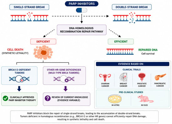
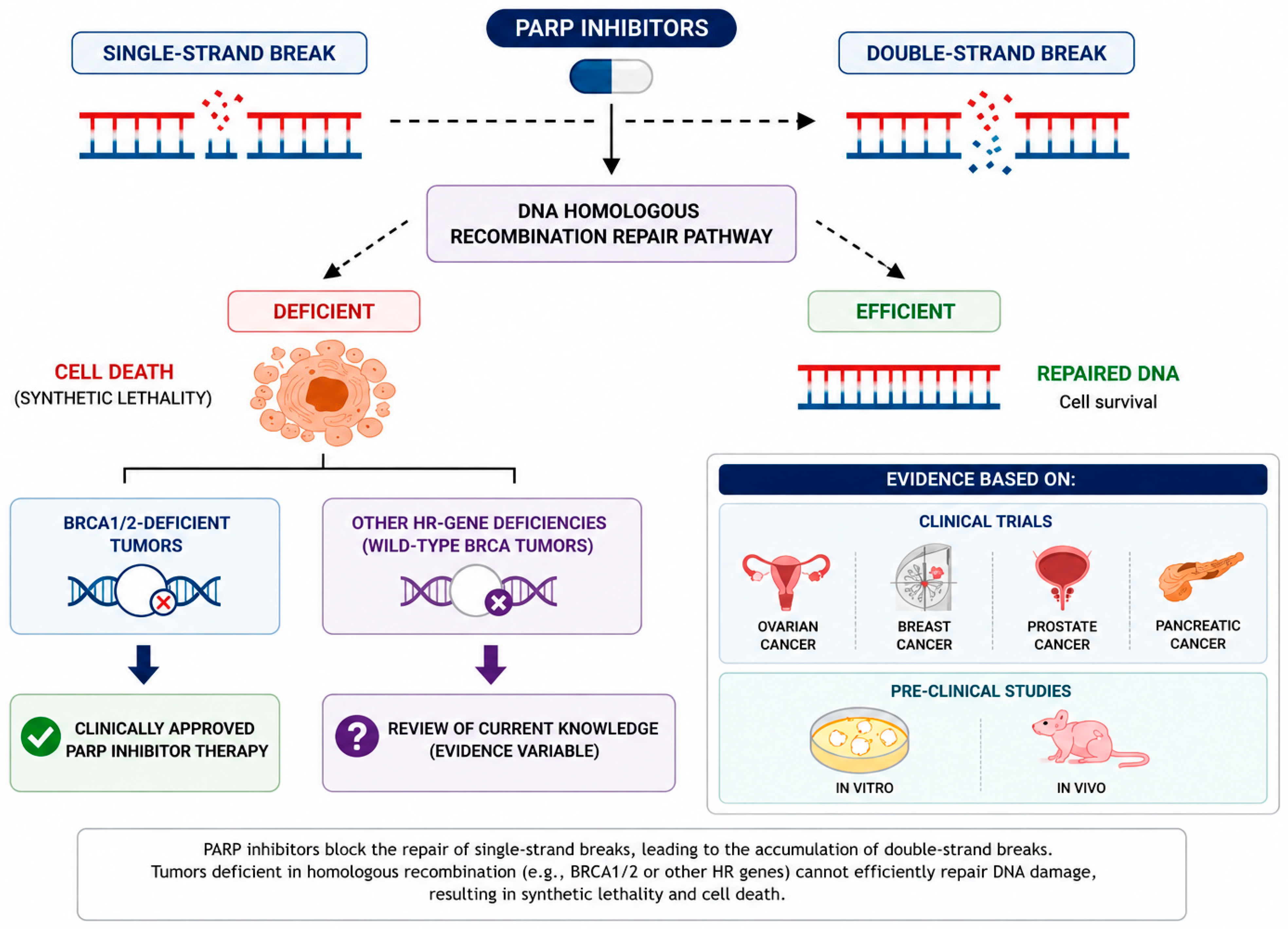
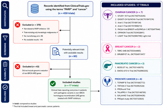
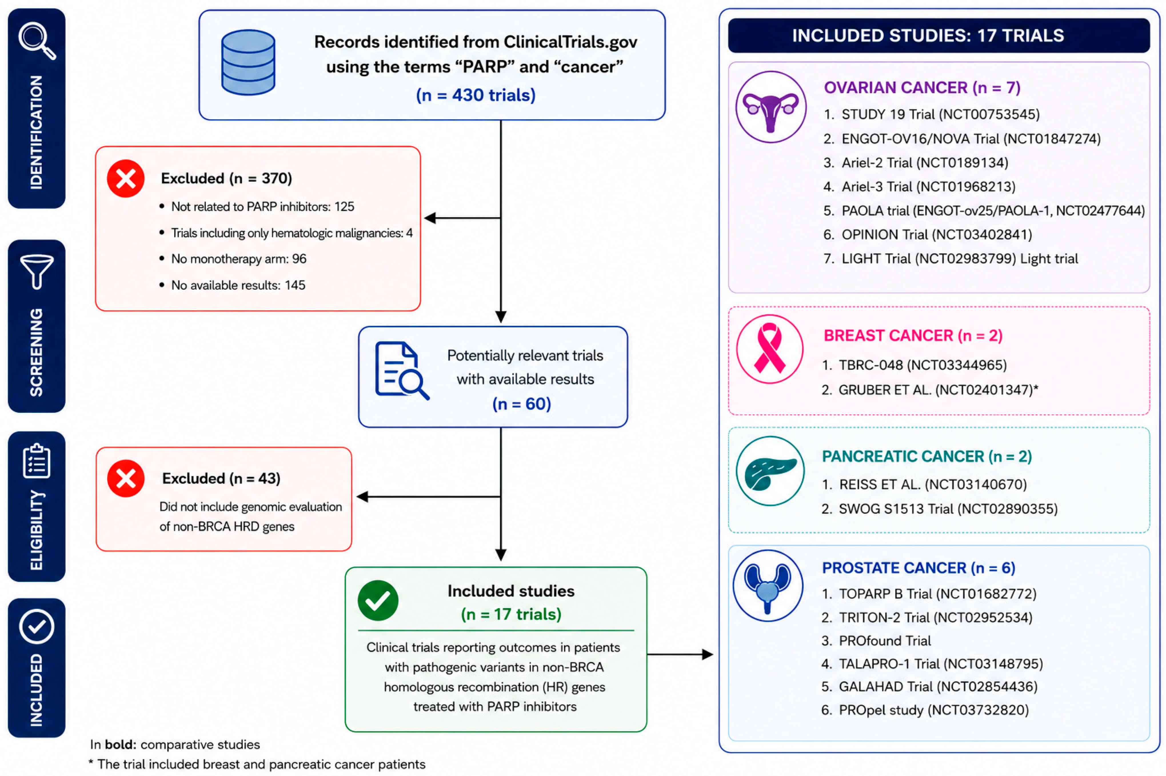
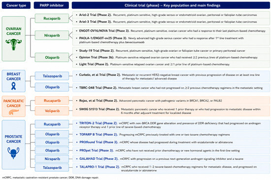
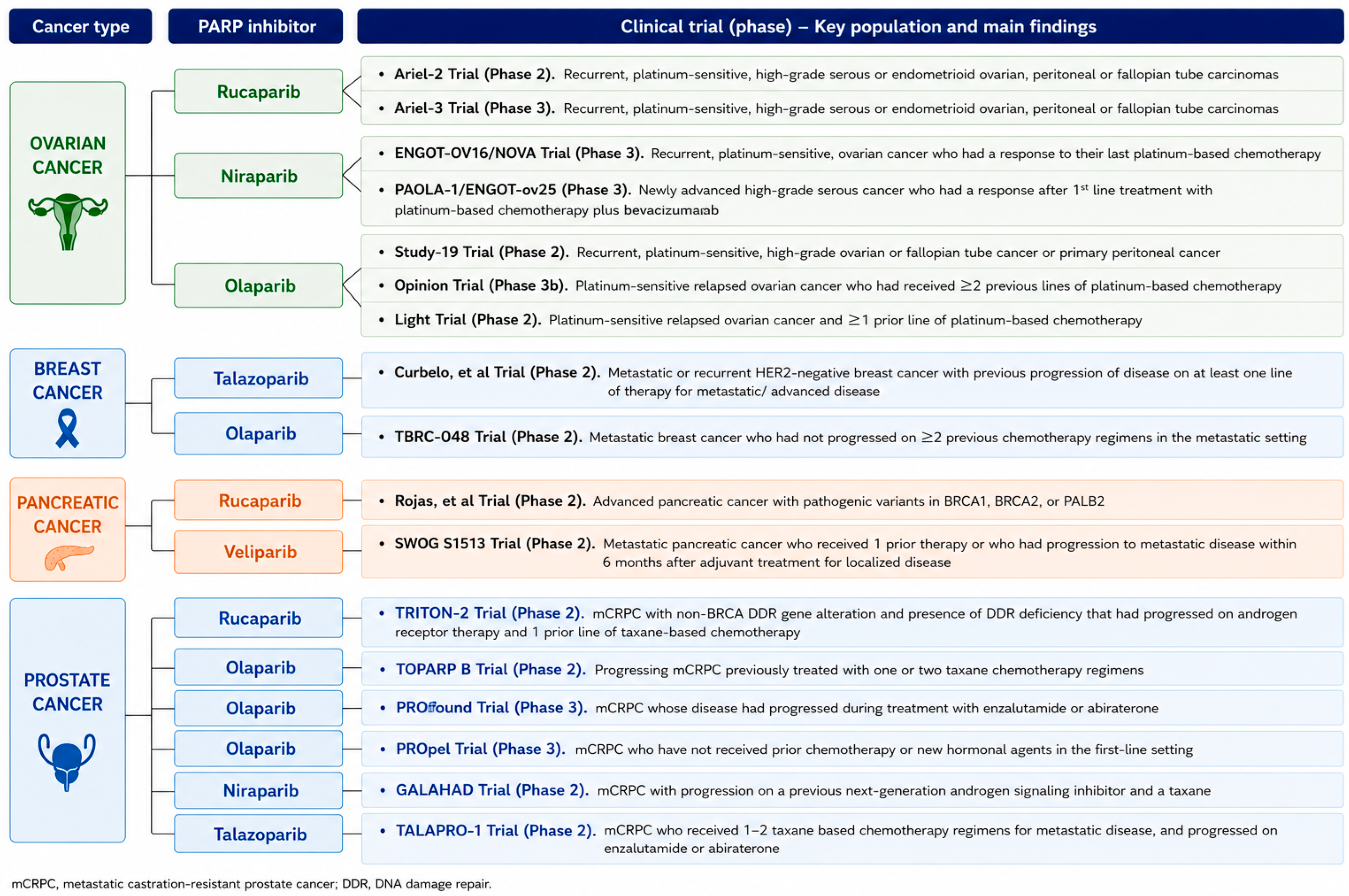
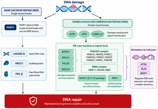
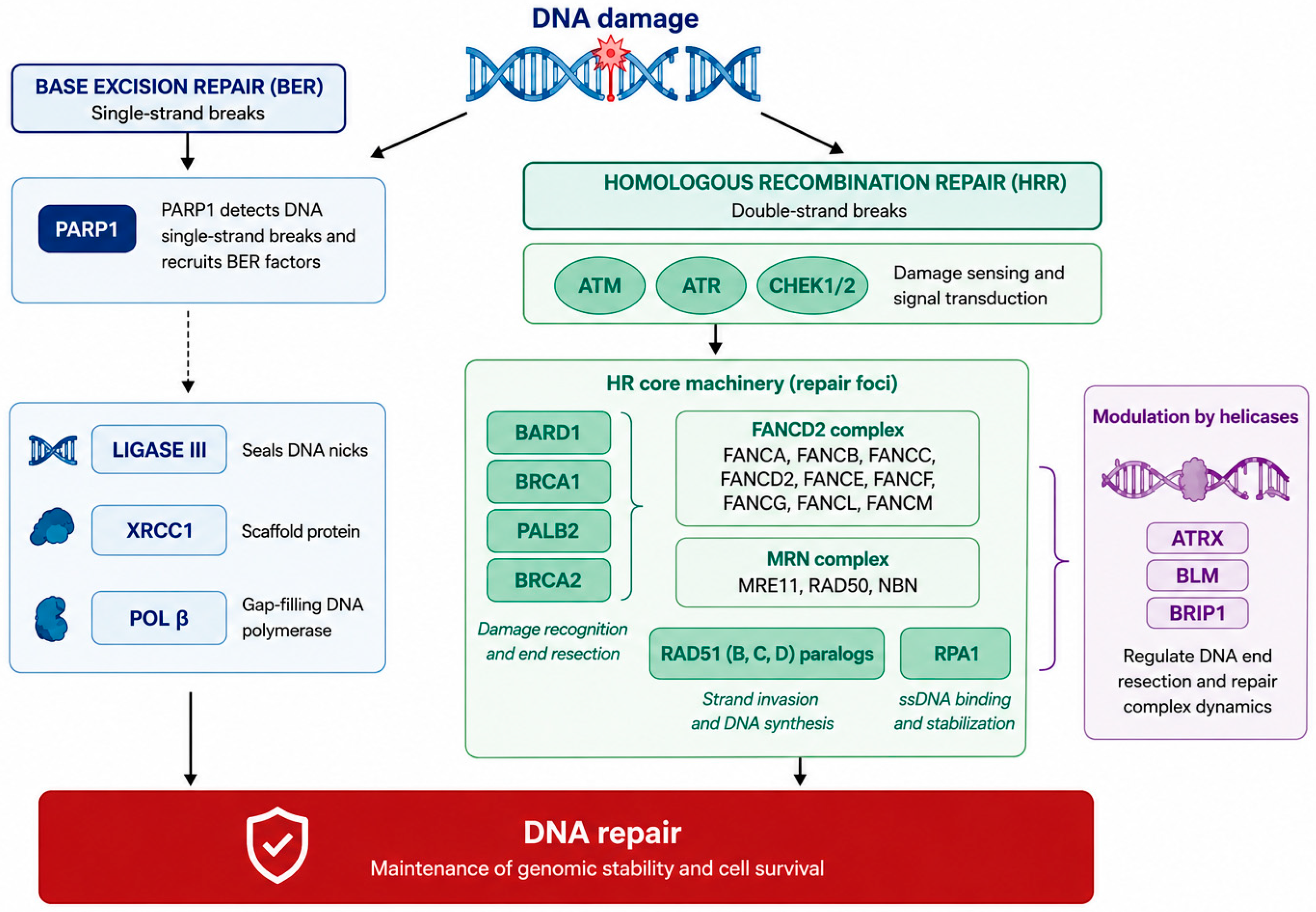
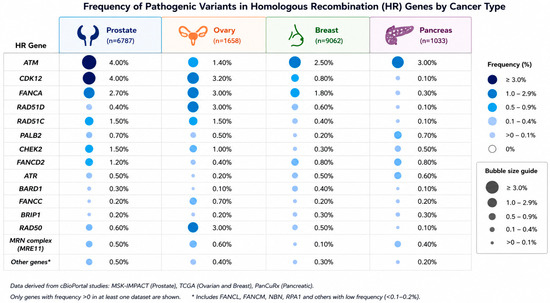
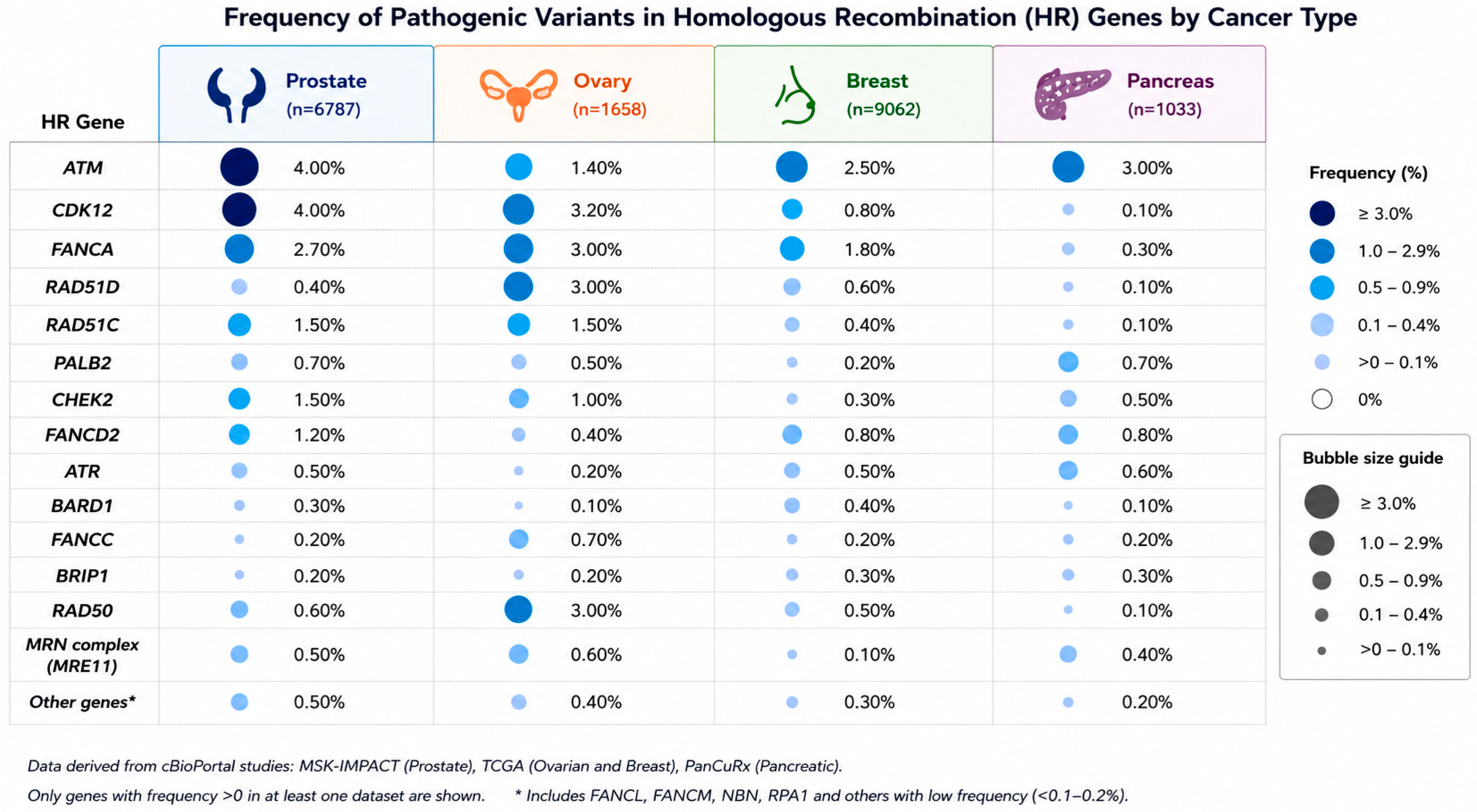

Disclaimer/Publisher’s Note: The statements, opinions and data contained in all publications are solely those of the individual author(s) and contributor(s) and not of MDPI and/or the editor(s). MDPI and/or the editor(s) disclaim responsibility for any injury to people or property resulting from any ideas, methods, instructions or products referred to in the content. |
© 2026 by the authors. Licensee MDPI, Basel, Switzerland. This article is an open access article distributed under the terms and conditions of the Creative Commons Attribution (CC BY) license.