


Abstract
Thymic epithelial tumors (TETs), comprising thymomas and thymic carcinomas, are rare, biologically heterogeneous thoracic malignancies with limited therapeutic advances since platinum-based regimens were adopted decades ago. Recent genomic and immunophenotypic profiling has uncovered recurrent genomic alterations and high programmed death-ligand 1 (PD-L1) expression, particularly in thymic carcinoma, yet few targeted or immunotherapies have achieved regulatory approval. This mini-review synthesizes current knowledge on TET heterogeneity and the genomic landscape, evaluates the clinical evidence supporting targeted agents and immune checkpoint inhibitors, and examines emerging biomarkers, including circulating tumor DNA. We also address practical barriers to precision oncology in TETs: challenges of next-generation sequencing implementation and cost in resource-limited settings; scarcity of large, biomarker-driven trials; and safety concerns unique to TETs (immune-related toxicity). Finally, we discuss the translational hurdles for antibody–drug conjugates and cellular therapies, limited validated cell surface targets, antigen heterogeneity, and preclinical model gaps and outline strategic paths forward to enable rational, safe, and equitable precision therapeutics for TET patients.
1. Background
The thymus is an immune organ located in the anterior mediastinum and plays an essential role in T-cell maturation and selection. Thymic epithelial tumors (TETs) are rare malignancies, with reported incidence rates ranging from approximately 0.13 to 0.32 per 100,000 person-years across population-based cohorts. Geographic variation has been reported, with higher incidence rates in East Asian populations and lower rates in some African populations [1,2,3]. TETs arise from thymic epithelial cells and include thymomas, thymic carcinomas (TCs), and thymic neuroendocrine tumors [4]. Despite their shared origin, these entities differ markedly in biology, clinical behavior, and prognosis.
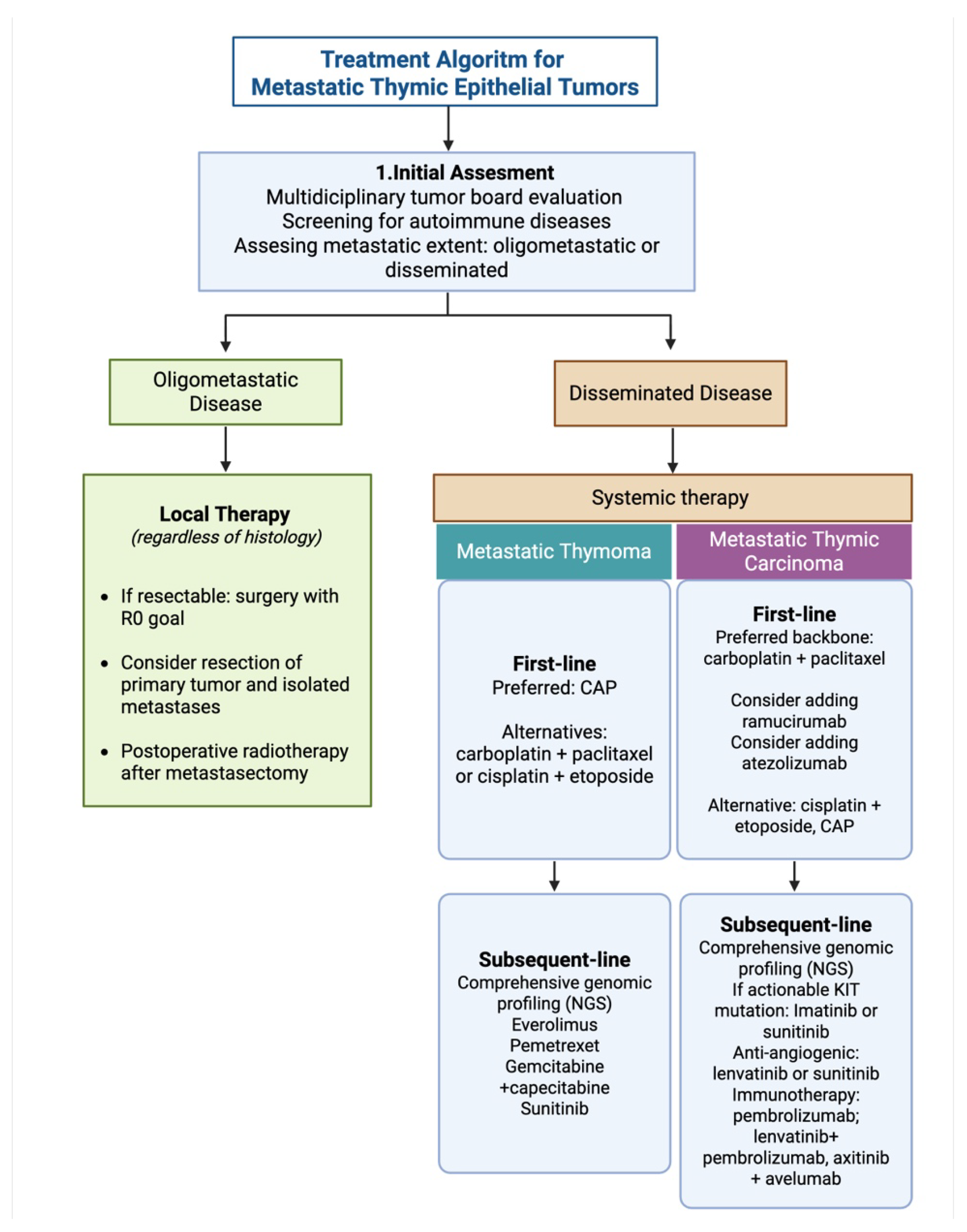
Platinum-based chemotherapy remains the standard first-line systemic treatment for advanced or unresectable TETs. In TC, recent data support the addition of antiangiogenic inhibitors (vascular endothelial growth factor receptor 2 (VEGFR2) monoclonal antibody, ramucirumab) to carboplatin and paclitaxel. However, unlike lung cancer, in which next-generation sequencing (NGS) and immunotherapy have reshaped treatment, therapeutic progress in TETs has been limited. No molecularly targeted therapy is currently approved specifically for TETs by the United States Food and Drug Administration (FDA). Nevertheless, agents such as multikinase inhibitor lenvatinib [5], mechanistic target of rapamycin (mTOR) inhibitor everolimus [6], and KIT inhibitor imatinib [7] have demonstrated clinical activity in selected settings. The current systemic treatment approach for metastatic TETs is summarized in Figure 1.
The development of individualized therapy in TETs is constrained by disease rarity, histologic heterogeneity, low frequency of canonical actionable drivers, limited prospective clinical trials, immune-related toxicity, and unequal access to molecular testing and off-label therapies [8]. Therefore, the central challenge is not simply identifying molecular abnormalities but demonstrating that biomarker-directed treatment improves clinically meaningful outcomes.
Figure 1.
Current landscape of treatment of metastatic TETs. The algorithm outlines a disease extent- and histology-based approach to metastatic thymic epithelial tumors according to current National Comprehensive Cancer Network (NCCN) and European Society for Medical Oncology (ESMO) guideline recommendations. Initial evaluation includes multidisciplinary assessment, autoimmune disease screening, and classification as oligometastatic or disseminated disease. Oligometastatic disease may be managed with local therapies when feasible, whereas disseminated disease requires systemic treatment selected according to histology. CAP-based therapy is commonly preferred for metastatic thymoma, while carboplatin plus paclitaxel is a preferred backbone for metastatic thymic carcinoma, with selected use of ramucirumab, immunotherapy, antiangiogenic therapy, targeted therapy, and genomic profiling in subsequent-line settings. CAP, cyclophosphamide, doxorubicin, and cisplatin; NGS, next-generation sequencing. Treatment options were summarized based on NCCN-2026 [9] and ESMO practice guideline [10,11] guideline recommendations [9,10]. Created in BioRender. Canaslan, K. (2026). https://BioRender.com/903go6z accessed on 19 July 2026.
This review focuses on the gap between biological target identification and clinical actionability in TETs. It examines why frequently detected molecular alterations or protein expression do not consistently predict treatment benefit. It also addresses the practical implementation of precision oncology, including genomic testing and cost-related barriers. Emerging biomarkers and imaging-based technologies are also evaluated in the context of perioperative decision-making and postoperative surveillance.
2. Biological and Clinical Heterogeneity of Thymic Epithelial Tumors
TETs represent a spectrum of diseases characterized by histologic and clinical heterogeneity [12]. The World Health Organization (WHO) classifies thymomas (types A, AB, B1, B2, and B3) based on epithelial cell morphology and the proportion of non-tumoral lymphocytes. In contrast to thymomas, TCs are more aggressive, lack an organoid architecture, and closely resemble extrathymic squamous cell carcinomas [4,13].
This heterogeneity extends beyond conventional histologic classification. A recent integrative genomic and transcriptomic analysis classified TETs into three molecular groups: GTF2I-mutant thymomas, copy-number-altered thymomas characterized by Insulin Receptor Substrate 4 (IRS4) activation, and thymic carcinomas. These groups displayed distinct transcriptional programs, immune phenotypes, and putative cells of origin, indicating that histologic classification alone may not fully capture the biological diversity of TETs [14]. Although this framework may support future biomarker development and patient stratification, its predictive value for treatment selection remains to be validated.
These histologic and molecular differences have direct clinical implications. Thymomas are often more indolent and are frequently associated with autoimmune paraneoplastic disorders, especially myasthenia gravis, pure red cell aplasia, and Good’s syndrome (associated with hypogammaglobulinemia and phenocopies of inborn errors of immunity). In contrast, TC is generally more aggressive, more likely to metastasize early, less frequently associated with autoimmunity, and associated with poorer survival [15].
Recent multi-omics clustering has further dissected TC into immunologically “hot” and “cold” subsets [16]. The “hot” TC cluster exhibits significantly higher CD3+ and CD4+ T-cell infiltration, which translates clinically into a longer median overall survival (OS) and higher progression-free survival (PFS) ratios when treated with immune checkpoint inhibitors (ICIs) compared to the “cold” TC subset [16]. Additionally, programmed death-ligand 1 (PD-L1) [17,18] and lymphocyte-activation gene 3 (LAG-3) [12] were reported to be highly expressed in single-cell analyses, and both are known targets for immunotherapy.
3. Genomic Landscape of Thymic Epithelial Tumors
Tumor mutational burden (TMB) varies according to TET histology. In the The Cancer Genome Atlas (TCGA) cohort, the overall mean TMB was low at 0.48 mutations/Mb; however, the cohort was predominantly composed of thymomas, and histology-stratified analysis demonstrated significantly higher TMB in TC than in thymoma subtypes [19]. Despite this, comprehensive genomic profiling has elucidated distinct molecular drivers between subtypes. The indolent thymomas are characterized primarily by point mutations in GTF2I, most commonly p.L424H or p.L404H, depending on transcript annotation. These variants affect the second conserved general transcription factor II-I (TFII-I) repeat domain near the DNA-binding region [20]. These alterations have been reported in up to 58% of tested thymoma cohorts, appear highly specific to thymoma, and are considered thymoma-associated genomic alteration [21].
TC harbors distinct mutational profiles involving tumor suppressors and epigenetic regulators such as TP53 (30–36%), CDKN2A/B, BAP1, CYLD, and SETD2 [16,22,23,24,25]. In a recent comprehensive profiling cohort, alterations were identified in cell-cycle-related genes in 13% of cases, DNA repair pathways in 8%, and chromatin-remodeling genes in 19%. A distinct subset of cases (8%) demonstrated mismatch repair deficiency (dMMR) or high microsatellite instability (MSI-H) status [25]. Furthermore, TCs demonstrate significantly higher frequencies of gene amplifications in ERBB2, MDM4, and MYC [16,23].
While immunohistochemical overexpression of the c-KIT protein is observed in only 2% to 4% of thymomas, it is a hallmark feature detected in 46% to 86% of TC cases [23,26]. However, despite this widespread and strong protein-level overexpression, true gain-of-function KIT gene variants (e.g., p.V560del and p.L576P in exon 11, p.D820E in exon 17) are found in only approximately 7 to 11% of TCs [19,27].
Precision oncology studies have identified pathogenic germline variants in up to 18% of patients with advanced TETs, particularly in DNA damage repair-associated genes such as BRCA1/2, BAP1, MUTYH, and CHEK2 [16,28]. This high rate of pathogenic germline variants (PGVs) leads to Homologous Recombination Deficiency (HRD) signatures in up to 53% of patients, uncovering vulnerabilities that could potentially be exploited by poly ADP-ribose polymerase (PARP) inhibitors [16].
4. Targeted Therapy in TETs: Clinical Evidence and Limitations
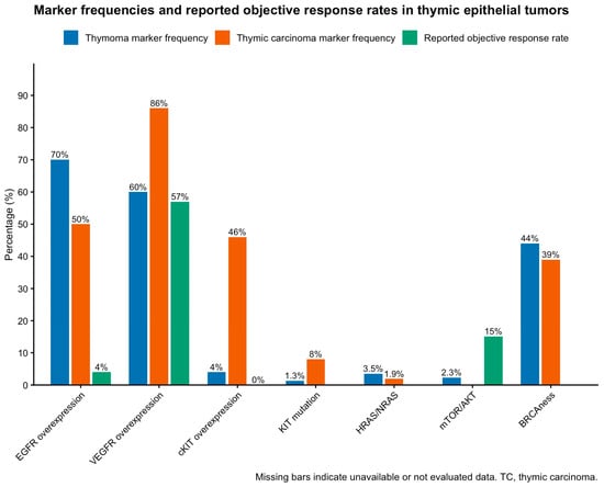
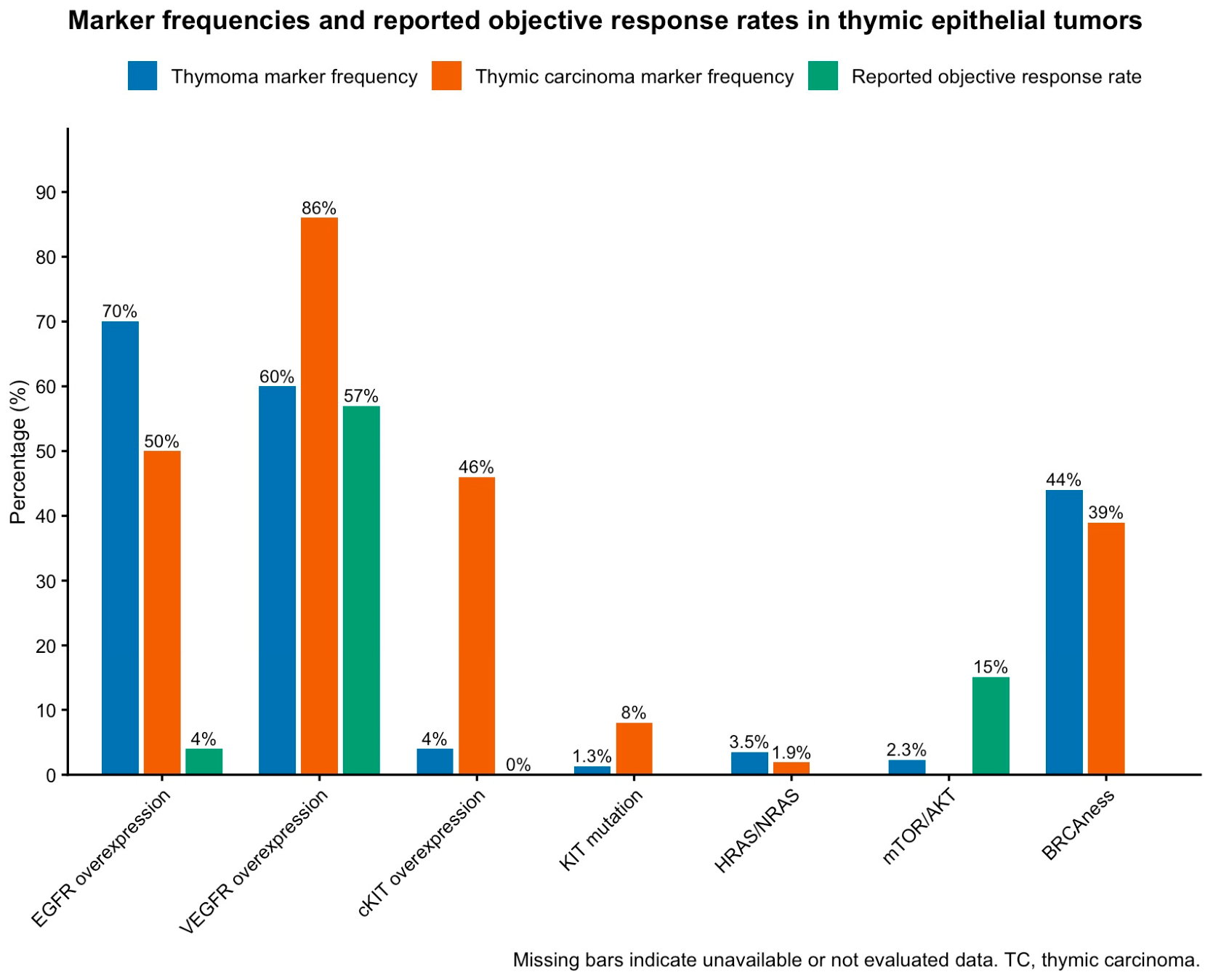
While cytotoxic chemotherapy remains the standard frontline treatment for advanced TETs [10], precision oncology has paved the way for evaluating targeted agents. However, translating molecular discoveries into clinical efficacy has proven challenging, yielding mixed results that are heavily dependent on the specific pathway being targeted (Figure 2). The main targeted agents evaluated in clinical studies of TETs are summarized in Table 1.
Figure 2.
Comparative frequencies of selected molecular markers and reported objective response rates in thymic epithelial tumors. The bar plot compares selected target expression or genomic alteration frequencies in thymoma and thymic carcinoma with reported objective response rates (ORRs) for corresponding targeted or pathway-directed therapeutic strategies. ORR was defined as the proportion of patients achieving a complete or partial response. For each marker, the plotted alteration or expression frequency represents the highest reported value from the cited published study or review. EGFR [29], VEGFR [30], and cKIT [31] represent protein overexpression or pathway-level target expression, whereas KIT mutation [23], HRAS/NRAS [23], mTOR/AKT [23], and BRCAness [16] represent genomic or pathway alterations. ORRs were derived from clinical studies evaluating the corresponding therapeutic class, including anti-EGFR therapy [32], antiangiogenic therapy [33], KIT-directed therapy [7,34], and mTOR inhibition [6], where available. Missing bars indicate unavailable or unevaluated data. This figure was independently created based entirely on aggregate data extracted from previously published studies and reviews; it does not include unpublished original data and was not reproduced or adapted from any single publication. ORR, objective response rate; TC, thymic carcinoma. Created in BioRender. Canaslan, K. (2026). https://BioRender.com/4a2tfw9 accessed on 19 July 2026.
4.1. Antiangiogenic Strategies
Currently, multi-kinase inhibitors, mainly tyrosine kinase inhibitors (TKIs), with potent anti-angiogenic properties represent the most clinically active class of targeted drugs for refractory tumors [35,36]. Their biological rationale is supported by vascular endothelial growth factor (VEGF) overexpression in 86–88% of TETs and high VEGFR2 expression in 88.5% of aggressive subtypes, including TC and type B3 thymoma [30].
In a phase II trial of heavily pretreated patients, sunitinib yielded an objective response rate (ORR) of 26% and disease control rate (DCR) of 91% in the TC cohort, establishing it as an off-label later-line option [37]. In the multicenter phase II REMORA trial, lenvatinib demonstrated an ORR of 38% and a median PFS of 9.3 months in previously treated TC, leading to its recognition as a highly promising salvage therapy [38]. Long-term follow-up of REMORA further supported the durability of lenvatinib activity, with a median OS of 28.3 months and a 36-month OS rate of 35.7% [5]. Although lenvatinib has not received FDA or European Medicines Agency (EMA) approval specifically for TC, it is approved in Japan for unresectable TC and it has been recommended in NCCN guideline [39].
Moving anti-angiogenics to the frontline, the phase II RELEVENT trial recently evaluated the VEGFR2 monoclonal antibody ramucirumab combined with carboplatin and paclitaxel in treatment-naive advanced TC. The ORR was 80% by investigator assessment and 57.6% by central review, with a median PFS of 18.1 months. These results indicate substantial first-line activity, although confirmation in randomized studies is required [33].
4.2. mTOR, AKT, and PI3K Pathway Inhibitors
Given the frequent activation of the PI3K/mTOR pathway, the mTOR inhibitor everolimus was evaluated in a phase II trial. Everolimus achieved an overall DCR of 88%, with a median PFS of 16.6 months in thymoma and 5.6 months in TC. However, this efficacy was counterbalanced by a high incidence of severe toxicities, including a 6% incidence of fatal pneumonitis [6]. The pan-PI3K inhibitor buparlisib showed only modest activity with a 7.1% ORR while carrying substantial toxicities [40].
4.3. EGFR and KIT: Lesson in Target Selection
One of the most notable disappointments in targeted therapy involves the epidermal growth factor receptor (EGFR) pathway. EGFR protein is frequently overexpressed, found in approximately 70% of thymomas and 50% of TCs [29,35,36]. Despite this robust and widespread overexpression, anti-EGFR targeted therapies have been largely unsuccessful. In a phase II trial evaluating the EGFR-TKI gefitinib in heavily pretreated patients, the ORR was a 4%, with no complete responses [32]. Similarly, a phase II trial combining the EGFR-TKI erlotinib with bevacizumab yielded no objective responses, leaving 60% of patients with stable disease and 40% with progressive disease as their best response [41]. The primary reason for this clinical failure is that EGFR overexpression almost never correlates with activating EGFR gene variants, which are the actual biological predictors of sensitivity to EGFR-directed TKI therapy [42].
A similar paradigm is observed with the c-KIT expression and KIT mutations. Early phase II trials testing the KIT inhibitor imatinib in unselected cohorts based on c-KIT overexpression reported no objective responses [7,34]. However, in TCs with activating somatic KIT variants such as exon 11 (p.V560del), patients have demonstrated impressive and occasionally long-lasting tumor shrinkage with imatinib [43,44]. Therefore, routine screening for c-KIT followed by mutational sequencing is critical to identify the minority of patients who may benefit from KIT inhibitors. As a result, EGFR overexpression and c-KIT overexpression without pathogenic gain-of-function mutation should be considered non-predictive.
4.4. Somatostatin Receptor-Targeted Therapy
Immunohistochemical evaluations demonstrate that approximately 78% of TETs express at least one somatostatin receptor (SSTR) subtype, predominantly expressing SSTR2A on malignant neoplastic epithelial cells and SSTR3 on infiltrating reactive thymocytes [45]. This high level of expression provides a biological rationale for utilizing somatostatin analogs, such as octreotide, particularly in pretreated patients whose tumors exhibit positive uptake on SSTR-based functional imaging [46,47].
Octreotide monotherapy demonstrated modest clinical activity, yielding an ORR of 10.5% and a median PFS of 2.0 months. However, the subsequent addition of prednisone was associated with an ORR of 31.6% and a median PFS of 9.2 months [46]. Activity appeared to differ by histology: responses were observed primarily in thymoma, whereas no objective responses were reported in patients with TC. The combination therapy requires strict safety monitoring. However, prolonged high-dose corticosteroid treatment may worsen thymoma-associated immunodeficiency and increase the risk of infection. [46]. Therefore, while SSTR-targeted therapy offers a viable chemo-free alternative, its efficacy is largely confined to thymomas and must be balanced against the risk of toxicity.
4.5. IGF-1R, Histone Deacetylase, and DNA Damage Response Strategies
Cell-cycle inhibition has been evaluated with the cyclin-dependent kinases 4 and 6 (CDK4/6) inhibitor palbociclib. In a phase II trial of recurrent or refractory advanced TETs, palbociclib achieved an ORR of 12.5% and a median PFS of 11.0 months [48]. Trials targeting the insulin-like growth factor 1 receptor (IGF-1R) pathway with the monoclonal antibody cixutumumab showed a 14% response rate in thymomas, but no responses in TC. New or worsening autoimmune disorders occurred in 24% of patients, limiting its clinical use [49]. Belinostat, a Histone Deacetylase (HDAC) inhibitor, has shown limited objective activity in recurrent or refractory TETs, with disease stabilization in some patients [50].
Table 1.
Targeted therapies with clinical evaluation in thymic epithelial tumors.
5. Immune Checkpoint Inhibition: Efficacy Versus Autoimmune Toxicity
Immunotherapy in TETs is primarily focused on immune checkpoint inhibitors (ICIs). Phase II trials of pembrolizumab (anti-PD-1) monotherapy have demonstrated ORRs of approximately 19.2–22.5% in pretreated TC and 28.6% in thymoma, providing durable clinical benefits for a subset of patients [52,53]. Results with nivolumab (anti-PD-1) have been less consistent. The NIVOTHYM trial reported an ORR of approximately 12% in a mixed cohort of patients with type B3 thymoma and TC, whereas no objective responses were observed in the PRIMER trial of unselected patients with recurrent TC [54].
Despite these encouraging signals, immunotherapy in TETs is called a “double-edged sword” due to a prohibitive risk of severe immune-related adverse events (irAEs) [55,56]. Because the thymus is the central organ for T-cell maturation and immune tolerance, ICIs can trigger life-threatening systemic autoimmunity. In thymoma, disrupted thymic architecture, reduced autoimmune regulator (AIRE)-dependent self-antigen expression, defective negative selection, and impaired regulatory T-cell generation allow autoreactive T cells to escape into the circulation. PD-1/PD-L1 or cytotoxic T-lymphocyte-associated protein 4 (CTLA-4) blockade may weaken residual peripheral tolerance and activate these pre-existing autoreactive clones, leading to autoantibody production and T-cell-mediated tissue injury [57]. Accordingly, irAE rates are substantially higher in thymoma than in TC, reaching 71.6% and 15.4%, respectively. Severe manifestations include myocarditis, polymyositis, myasthenia gravis, and hepatitis, which may require treatment discontinuation and can be fatal [36,56,58]. Consequently, ICIs are generally discouraged in thymoma and require rigorous monitoring in TC. To enhance efficacy while managing toxicity, current research is shifting toward combinatorial strategies, such as combining ICIs with anti-angiogenic TKI agents like axitinib (CAVEATT trial, 34% ORR) or lenvatinib (PECATI trial) and integrating ICIs with frontline platinum-based chemotherapy [13,59,60]. The key immune checkpoint inhibitor studies in TETs, including single-agent PD-1/PD-L1 blockade and antiangiogenic–immunotherapy combinations, are summarized in Table 2.
Table 2.
Immune and antibody-based therapies with clinical evaluation or active TET-directed investigation.
6. Antibody-Drug Conjugate Therapy, Vaccines, and Cellular Therapy
The development of antibody–drug conjugates (ADCs) and antigen-directed cellular therapies depends on identifying cell-surface targets that are widely expressed on tumor cells but absent or minimally expressed in normal tissues [18,69]. In rare malignancies like TETs, the discovery of such targeted cell surface antigens has been limited, which has delayed the clinical translation of these advanced therapeutic modalities. Recent comprehensive tissue microarray studies have evaluated various potential surface targets, such as HER2, PSMA, ROS1, and ALK, which have demonstrated little or no expression in most TETs [18]. In contrast, trophoblast cell-surface antigen 2 (Trop-2) demonstrated consistently high membranous expression across all histologic subtypes of TETs and has emerged as a promising therapeutic target [18]. Broader surface-antigen profiling has also identified high expression of additional candidate targets, including NECTIN-4 and mesothelin [25]. This robust expression of Trop-2 has paved the way for clinical trials evaluating sacituzumab govitecan, an anti-Trop-2 ADC, in patients with advanced TETs [67].
Mesothelin, another cell-surface antigen typically found on cells lining the pleura and pericardium, is frequently and strongly expressed in TC but is less common in thymoma [70]. This expression profile has led to clinical trials exploring anetumab ravtansine, a mesothelin-directed ADC, specifically in patients with TC [68]. Furthermore, the expression of mesothelin provides a strong biological rationale for the future development and evaluation of mesothelin-directed chimeric antigen receptor T cell (CAR-T cell) therapies for this patient population [71]. Another novel ADC targeting the B7-H3 surface antigen is also currently being investigated in early-phase clinical trials that include TC patients [72].
CD70 represents another appealing target for cellular therapies. This transmembrane protein contributes to B- and T-cell interactions and has limited expression in most normal tissues [73]. Clinical development of CD70-directed cellular products, including CTX130, has primarily focused on hematologic malignancies [66,74,75] and renal cell carcinoma [76]. The high prevalence of CD70 expression supports its investigation in TC, although no CD70-directed cellular therapy has yet demonstrated clinical efficacy in TETs.
The Wilms’ tumor 1 (WT1) protein is responsible for promoting oncological development and tumor vascularization and is overexpressed in approximately 80–85% of both thymomas and TCs [66]. A phase II clinical trial evaluating a WT1 peptide vaccine in advanced, pretreated patients demonstrated an ability to induce WT1-specific immune responses. However, reflecting the unique immunological risks associated with the thymus, some thymoma patients developed severe irAEs, including pure red cell aplasia and myasthenia gravis [66]. Antibody-based and vaccine-based strategies are summarized in Table 2.
7. Therapeutic Strategies for Autoimmune Diseases Associated with TET
Autoimmunity is one of the defining common features of TETs and reflects disruption of thymus-mediated central immune tolerance. Under physiological conditions, medullary thymic epithelial cells, major histocompatibility complex (MHC)-dependent antigen presentation, and AIRE-mediated expression of tissue-restricted antigens eliminate autoreactive T cells. In thymoma, architectural disorganization, reduced AIRE expression, impaired antigen presentation, and abnormal thymocyte selection allow autoreactive T-cell escape, contributing to autoimmune manifestations such as myasthenia gravis, pure red cell aplasia, and other immune-mediated disorders [77,78,79,80].
Recent transcriptomic and spatial profiling studies further suggest that thymoma-associated autoimmunity is sustained by abnormal medullary niches, ectopic germinal center formation, autoreactive B-cell responses, and reduced regulatory T-cell populations. This immune dysregulation may persist even after thymectomy [81,82,83,84,85,86].
Management depends on the specific autoimmune manifestation and requires multidisciplinary assessment. Thymectomy remains central to oncologic treatment when feasible and may improve some associated autoimmune disorders, although it does not uniformly restore immune tolerance. Corticosteroids and steroid-sparing agents, including azathioprine, mycophenolate mofetil, tacrolimus, and cyclosporine, remain standard options. In refractory antibody-mediated disease cases, targeted immune therapies including rituximab, complement inhibitors (eculizumab, ravulizumab, and zilucoplan), and neonatal Fc receptor (FcRn) antagonists (efgartigimod, rozanolixizumab, and nipocalimab) may be considered. Other emerging approaches, including cytokine-directed therapies targeting interleukin (IL)-6, IL-17, or IL-23 and experimental cellular strategies such as CAR-T therapy, remain investigational in TET-associated autoimmunity [83,87,88].
From an oncologic standpoint, the presence of underlying autoimmunity plays a key role in guiding treatment decisions. ICIs, while effective in some cancers, can provoke severe or even life-threatening immune-related adverse events in patients with TETs, particularly those with thymoma. For this reason, their use should be approached with great caution, especially in individuals with active or prior autoimmune disease. Overall, careful assessment of autoimmune status is essential when tailoring treatment strategies for patients with TETs.
8. Liquid Biopsy, Tumor Microenvironment, and ctDNA-Based Monitoring
8.1. Liquid Biopsy and ctDNA-Based Monitoring
Liquid biopsy techniques, including cell-free DNA (cfDNA) and circulating tumor DNA (ctDNA), are emerging as complementary tools to address the limitations of single-site tissue biopsies. The incorporation of liquid biopsy (ctDNA analysis) into the clinical workflow holds substantial promise. These tools not only enhance diagnostic accuracy and subtyping but also provide a mechanism to identify actionable alterations, capture molecular heterogeneity, and enable longitudinal assessment of treatment response or resistance. Thereby expanding therapeutic options and enabling more targeted, personalized treatment, particularly for patients in later lines of therapy [89,90]. In prospective TET cohorts, cfDNA levels are significantly higher in TCs compared to thymomas and healthy controls [90]. This disparity likely reflects histology-specific ctDNA shedding. Aggressive TCs, with greater metastatic potential, generally show increased tumor DNA shedding into circulation, whereas indolent thymomas may have lower tumor burden and reduced plasma ctDNA detectability [91]. Consequently, cfDNA levels correlate with metastatic burden rather than primary tumor volume or local stage [22,90]. However, because the current evidence remains limited, a negative ctDNA result should not be considered sufficient to exclude residual disease or recurrence, especially in thymoma or low-volume disease.
ctDNA assays have identified tumor-associated alterations (TP53, CYLD, GTF2I) that mirror those found in primary tumor tissues [91]. Longitudinal ctDNA monitoring has shown the potential to detect minimal residual disease (MRD) following surgery [92]. In reported clinical instances, ctDNA analysis tracked a decline in tumor-specific mutations post-resection and flagged molecular recurrence a month before radiographic progression became visible [90,93]. Nevertheless, these findings remain preliminary, and ctDNA has not yet been validated for routine surveillance, postoperative decision-making, or treatment selection in TETs. Furthermore, complementary platforms utilizing Circulating Tumor Cell (CTC)-derived organoids have demonstrated high sensitivity in matching ex vivo drug sensitivity to actual patient clinical responses, laying the groundwork for real-time therapeutic monitoring [94].
8.2. Tumor Microenvironment and Spatial Profiling
Single-cell RNA sequencing and spatial profiling techniques, including high-resolution spatial transcriptomics and multiplex immunofluorescence, can characterize the cellular composition and interactions within the TET microenvironment [82]. In myasthenia gravis-associated thymomas, spatial analyses have localized polygenic risk signals and disease-specific ectopic germinal centers predominantly to expanded medullary regions. Mapping these physical interactions offers critical clues for treatment, distinguishing between therapy-responsive “hot” microenvironments enriched with tertiary lymphoid structures and non-responsive tumors that exhibit localized thymic epithelial cell (TEC)-driven immunosuppression [82].
Spatial profiling recently demonstrated that major pathological responses to neoadjuvant immunochemotherapy in TC are closely associated with distinct tumor microenvironments. Tumors that respond well are characterized by “hot” microenvironments with extensive immune infiltration and tertiary lymphoid structure (TLS) formation, whereas non-responsive tumors exhibit localized immunosuppression driven by TECs that exhaust infiltrating CD8+ T cells [95]. These findings may contribute to future biomarker-based patient selection and identification of mechanisms of treatment resistance, although prospective validation is required.
8.3. Perioperative and Surgical Implications of Emerging Precision Technologies
In TETs, multidisciplinary treatment decisions are strongly influenced by resectability, histologic subtype, local invasion, completeness of resection, and postoperative recurrence risk [9]. Therefore, ctDNA, radiomics, and digital pathology should not be viewed only as tools for systemic treatment selection but also as potential adjuncts for surgical planning and postoperative surveillance. Preoperatively, computed tomography (CT) remains the reference imaging modality for evaluating disease extent, response to induction therapy, and potential resectability after systemic treatment [96]. In this context, radiomics may provide additional quantitative information beyond conventional imaging. CT-based radiomic models have shown potential for predicting resectability and tumor–node–metastasis (TNM) stage in TETs [97]. Additionally, magnetic resonance imaging (MRI)-based radiomics has been explored for predicting pathologic classification and staging [98]. However, radiomics remains investigational and should be considered complementary to expert radiologic assessment rather than a replacement for multidisciplinary review [99].
Induction treatment followed by surgery has produced encouraging complete resection rates in initially unresectable TETs, and pathologic response and completeness of resection have been associated with prognosis [100]. Standardized assessment of viable tumor and treatment-related changes is therefore important for postoperative risk stratification and clinical trial design [101]. Digital pathology may improve the reproducibility of these assessments, although it has not been validated to guide adjuvant treatment decisions.
In the postoperative setting, cfDNA and ctDNA may eventually serve as adjuncts for MRD assessment and recurrence monitoring. Early TET-specific studies suggest that cfDNA correlates with metastatic burden and that ctDNA can detect tumor-associated alterations during longitudinal follow-up, but current evidence is insufficient to guide routine postoperative management [90,91,93]. These findings raise the possibility of risk-adapted surveillance or treatment, but current evidence is insufficient to guide routine postoperative management.
9. Barriers to Individualized Therapy in Rare Thymic Epithelial Tumors
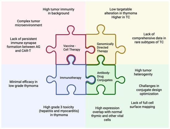
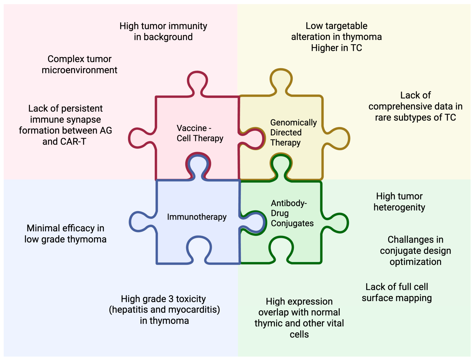
When evaluating the impact of actionable genomic alterations on patient survival in thymic epithelial tumors, the absence of high-level evidence is primarily driven by the unique biological and clinical challenges associated with these rare malignancies (Figure 3). Comprehensive genomic profiling identifies potentially relevant alterations more frequently in TC than in thymoma [22]. However, alterations with established predictive value and a clinically validated matched therapy remain uncommon, particularly in thymoma [102].
Figure 3.
Treatment for thymic epithelial tumors: current landscapes and emerging therapeutic challenges. This conceptual framework summarizes the main biological and clinical challenges limiting individualized treatment strategies in thymic epithelial tumors. Genomically directed therapy is constrained by the low frequency of targetable alterations in thymoma, higher but heterogeneous genomic alterations in thymic carcinoma, and limited comprehensive data in rare thymic carcinoma subtypes. Immunotherapy is limited by modest efficacy in low-grade thymoma and a high risk of grade ≥3 immune-related toxicities, particularly hepatitis and myocarditis. Vaccine and cellular therapy development is challenged by the complex tumor microenvironment, high baseline tumor-associated immunity, and limited persistence of antigen–CAR-T immune synapses. Antibody–drug conjugate development is restricted by tumor heterogeneity, incomplete cell-surface target mapping, challenges in conjugate design optimization, and overlap of target expression with normal thymic or other vital tissues. AG, antigen; CAR-T, chimeric antigen receptor T-cell therapy; TC, thymic carcinoma. Created in BioRender. Canaslan, K. (2026). https://BioRender.com/48dw9sd accessed on 19 July 2026.
In the prospective MASTER trial, comprehensive multi-omics profiling enabled molecularly informed treatment recommendations for 86% of patients with advanced TETs [103]. However, this figure should not be interpreted as the prevalence of validated actionable drivers. Recommendations included off-label and investigational therapies supported by different levels of molecular and clinical evidence. Only 45% of patients for whom a recommendation was made ultimately received the proposed treatment, highlighting the gap between theoretical molecular actionability and treatment implementation. Among evaluable patients who received molecularly informed therapy, the reported ORR was 23% and the DCR was 62% [16]. Precision oncology programs match patients to a wide variety of off-label drugs depending on their unique tumor profile. Evaluating a single survival metric, such as overall survival, across a molecularly heterogeneous cohort treated with dozens of different substances is statistically challenging and prevents traditional meta-analyses [16]. Similarly, a clinicogenomic study demonstrated that 60% of patients with metastatic TETs harbored molecular alterations with known or potential therapeutic implications. In their cohort, 54% of patients subsequently received at least one targeted therapy. For patients whose treatment was eventually stopped due to disease progression rather than toxicity, the mean time to progression was 9.9 months. However, nearly half of the treated patients discontinued targeted therapy early because of toxicity [22].
Furthermore, because of the rarity of the disease, conducting large-scale randomized controlled trials comparing targeted therapy against standard chemotherapy requires years of trial accrual. While the literature lacks level 1 evidence proving an OS benefit, the MATCH trial showed that matching patients to their actionable genomic alterations can slow disease progression and provide a therapeutic advantage [16]. The integration of precision medicine into the management of TETs faces significant economic and logistical hurdles, particularly in developing countries. While NGS is essential for identifying actionable targets, the high cost of whole-genome sequencing (WGS), panel-based genomic testing and the complex computational demands required for analysis remain major limitations [21]. The cost of the therapies themselves represents a formidable barrier to individualized care. Data from the prospective trial highlight a stark gap; although molecularly informed therapy was recommended for 86% of advanced TET patients, it was actually administered to only 45%, frequently due to the challenges of accessing expensive, off-label drugs [16]. Consequently, while genomic profiling offers a theoretical blueprint for personalized treatment, the reality of therapeutic delivery is strictly constrained by a patient’s socioeconomic status and the healthcare infrastructure of their country.
Because organizing large-scale, randomized phase III trials is virtually impossible in this population, there are currently no targeted therapies or immunotherapies specifically approved by the FDA or EMA for TETs. Later-line use of agents such as sunitinib, lenvatinib, and pembrolizumab therefore often depends on guideline recommendations, off-label access, and local reimbursement policies.
10. Future Directions
The management of TETs is often complicated by a lack of specialized expertise in histological diagnosis and subtyping, which is critical given the significant divergence in systemic treatment strategies between thymoma and thymic carcinoma. To address this diagnostic ambiguity and the necessity for more precise, biologically driven therapy, integrating advanced diagnostic modalities is essential.
Artificial intelligence (AI) and machine learning methods may facilitate this multimodal integration [13]. CT-based radiomic features have differentiated thymoma from the more aggressive TC, with an area under the curve (AUC) of 0.882, increasing to 0.921 when combined with patient sex [104]. Furthermore, machine learning applied to non-contrast CT scans has successfully predicted WHO subtypes with an AUC of 0.935 [105]. Similarly, utilizing CT-derived radiomic features to train machine learning classifiers has yielded good discriminatory performance for predicting histologic subtypes (AUC 0.876) and early versus advanced TNM stages (AUC 0.838) [106]. However, the radiomics model yielded poor predictive performance for detecting the presence of myasthenia gravis (AUC 0.639) [106]. These findings demonstrate the feasibility of noninvasive tumor phenotyping but do not establish an ability to predict benefit from a specific treatment.
AI-assisted digital pathology may provide complementary information. Zullo et al. developed a multiple-instance learning model based on digitized hematoxylin–eosin–saffron-stained whole-slide images from the French RYTHMIC network [107]. This pathomics model successfully classified the major WHO histological subtypes (A, AB, B1, B2, B3, C), achieving a mean AUC of 0.94 during internal validation and 0.89 in a prospective test cohort. The model identified regions with varying proportions of epithelial cells and lymphocytes, demonstrating the potential of AI-assisted digital pathology to characterize TET heterogeneity and support histologic classification [107]. Computational analysis of multi-omics data has also predicted candidate neoantigens associated with CYLD and KMT2C alterations, providing a potential basis for personalized vaccines or adoptive cellular therapies [108]. Future studies should move beyond diagnostic accuracy and determine whether multimodal models improve biopsy selection, treatment-response prediction, therapeutic stratification, and clinical outcomes.
11. Conclusions
TETs are biologically heterogeneous malignancies that remain challenging candidates for individualized therapy. Although TC is enriched for potentially relevant alterations such as TP53, CDKN2A/B, KIT, CYLD, BAP1, and PI3K/AKT/mTOR pathway abnormalities, most molecular findings remain prognostic or biologically informative rather than directly actionable. To date, antiangiogenic strategies, particularly lenvatinib, sunitinib, and ramucirumab-based chemotherapy, provide the strongest targeted-therapy evidence in TC. In contrast, approaches targeting mTOR, EGFR, KIT, IGF-1R, HDAC, and DNA damage response pathways have shown limited or highly selected activity. ICIs may induce durable responses in selected patients with TC, but their use is constrained by severe immune-related toxicity, including myocarditis, particularly in thymoma. ctDNA monitoring, ADCs, vaccines, and cellular therapies are promising but remain investigational. Progress in this rare disease will require molecularly annotated international trials, systematic evaluation of matched therapy, validated liquid biopsy approaches, surface-target discovery, and equitable access to genomic testing and targeted treatments. Emerging precision technologies may also support perioperative decision-making, response assessment after induction therapy, and postoperative surveillance, although prospective validation is required.
Author Contributions
K.C.: Investigation, Writing—Original Draft, Visualization. Ö.Y.: Investigation, Writing—Original Draft. Y.T.: Writing—Review & Editing. A.S.: Writing—Review & Editing. D.E.M.: Writing—Review & Editing. A.E.: Writing—Review & Editing. H.A.: Writing—Review & Editing. F.A.-L.: Conceptualization, Writing—Original Draft, Writing—Review & Editing, Supervision. All authors have read and agreed to the published version of the manuscript.
Funding
This research received no external funding.
Institutional Review Board Statement
Not applicable.
Informed Consent Statement
Not applicable.
Data Availability Statement
No new data were created or analyzed in this study. Data sharing is not applicable to this article.
Acknowledgments
This review was developed as a joint international academic project involving experts with complementary expertise in thymic epithelial tumors, thoracic oncology, molecular oncology, and rare tumor therapeutics. The collaboration was established to provide a multidisciplinary perspective on precision treatment strategies for thymic epithelial tumors. We also thank the Precision Medicine Lab, Endocrinology & Metabolism Research Institute, TUMS, for its support of this project.
Conflicts of Interest
The authors declare no conflicts of interest.
References
- Gatta, G.; Capocaccia, R.; Botta, L.; Mallone, S.; De Angelis, R.; Ardanaz, E.; Comber, H.; Dimitrova, N.; Leinonen, M.K.; Siesling, S.; et al. Burden and centralised treatment in Europe of rare tumours: Results of RARECAREnet—A population-based study. Lancet Oncol. 2017, 18, 1022–1039, Erratum in Lancet Oncol. 2017, 18, e433. [Google Scholar] [CrossRef] [Scilit] [PubMed]
- Rich, A.L. Epidemiology of thymoma. J. Thorac. Dis. 2020, 12, 7531. [Google Scholar] [CrossRef] [Scilit] [PubMed]
- Jiang, B.; Gu, Z.; Kann, C.S.; Mao, T.; Fang, W. Global Patterns and Trends in Thymic Epithelial Tumor Incidence: Worldwide Health Care Disparity on Detection. JCO Glob. Oncol. 2026, 12, e2500542. [Google Scholar] [CrossRef] [Scilit] [PubMed]
- World Health Organization. Thoracic Tumours. In WHO Classification of Tumours, 5th ed.; IARC Publications: Lyon, France, 2021. [Google Scholar]
- Niho, S.; Sato, J.; Satouchi, M.; Itoh, S.; Okuma, Y.; Mizugaki, H.; Murakami, H.; Fujisaka, Y.; Kozuki, T.; Nakamura, K.; et al. Long-term follow-up and exploratory analysis of lenvatinib in patients with metastatic or recurrent thymic carcinoma: Results from the multicenter, phase 2 REMORA trial. Lung Cancer 2024, 191, 107557. [Google Scholar] [CrossRef] [Scilit] [PubMed]
- Zucali, P.A.; De Pas, T.; Palmieri, G.; Favaretto, A.; Chella, A.; Tiseo, M.; Caruso, M.; Simonelli, M.; Perrino, M.; De Vincenzo, F.; et al. Phase II study of everolimus in patients with thymoma and thymic carcinoma previously treated with cisplatin-based chemotherapy. J. Clin. Oncol. 2018, 36, 342–349. [Google Scholar] [CrossRef] [Scilit] [PubMed]
- Giaccone, G.; Rajan, A.; Ruijter, R.; Smit, E.; Van Groeningen, C.; Hogendoorn, P.C.W. Imatinib mesylate in patients with WHO B3 thymomas and thymic carcinomas. J. Thorac. Oncol. 2009, 4, 1270–1273. [Google Scholar] [CrossRef] [Scilit] [PubMed]
- Imbimbo, M.; Salfi, G.; Borgeaud, M.; Ottaviano, M.; Froesch, P.; Bouchaab, H.; Cafarotti, S.; Addeo, A. Thymic epithelial tumors: What’s new and what’s next? ESMO Rare Cancers 2025, 4, 100024. [Google Scholar] [CrossRef] [Scilit]
- Riely, G.J.; Wood, D.E.; Loo, B.W., Jr.; Aisner, D.L.; Akerley, W.; Bauman, J.R.; Bharat, A.; Chang, J.Y.; Chirieac, L.R.; DeCamp, M.; et al. Thymomas and Thymic Carcinomas, Version 2.2025. J. Natl. Compr. Cancer Netw. 2025, 23, 255–269. [Google Scholar] [CrossRef] [Scilit] [PubMed]
- Girard, N.; Ruffini, E.; Marx, A.; Faivre-Finn, C.; Peters, S. Thymic epithelial tumours: ESMO Clinical Practice Guidelines for diagnosis, treatment and follow-up. Ann. Oncol. 2015, 26, v40–v55. [Google Scholar] [CrossRef] [Scilit] [PubMed]
- Baudin, E.; Caplin, M.; Garcia-Carbonero, R.; Fazio, N.; Ferolla, P.; Filosso, P.L.; Frilling, A.; de Herder, W.W.; Hörsch, D.; Knigge, U.; et al. Lung and thymic carcinoids: ESMO Clinical Practice Guidelines for diagnosis, treatment and follow-up. Ann. Oncol. 2021, 32, 439–451, Erratum in Ann. Oncol. 2021, 32, 1453–1455. [Google Scholar] [CrossRef] [Scilit] [PubMed]
- Liu, X.; Wang, C.; Huang, Y.; Lv, Q.; Yu, C.; Ying, J.; Duan, L.; Guo, Y.; Huang, G.; Shen, W.; et al. Abnormal Cellular Populations Shape Thymic Epithelial Tumor Heterogeneity and Anti-Tumor by Blocking Metabolic Interactions in Organoids. Adv. Sci. 2024, 11, e2406653. [Google Scholar] [CrossRef] [Scilit] [PubMed]
- Ardeshir-Larinani, F.; Ashrafian, L.; Chiappetta, M.; Mansilla, D.; Mariani, A.W.; Menis, J.; Rimner, A.; Sharma, A.; Szolkowska, M.; Upadhya, S.; et al. Challenges and Controversies in the Diagnosis and Management of Thymic Epithelial Tumors from the Thymic Tumor Subcommittee of the International Association for the Study of Lung Cancer Rare Tumors Committee. J. Thorac. Oncol. 2026, 21, 103611. [Google Scholar] [CrossRef] [Scilit] [PubMed]
- Park, S.; Yi, K.; Lee, J.; Yang, M.J.; Song, J.H.; Lim, J.; Kim, T.; Kim, S.; Kim, S.Y.; Kim, H.D.; et al. Integrative Multiomic Classification Reveals Distinct Origins and Evolutionary Trajectories of Thymic Epithelial Tumors. Cancer Res. 2026, 86, 1496–1513. [Google Scholar] [CrossRef] [Scilit] [PubMed]
- Pavese, V.; Carfì, F.M.; Capelletto, E.; Tabbò, F.; Leo, F.; Passiglia, F.; Righi, L.; Novello, S.; Merlini, A.; Bironzo, P. Therapeutic management of patients with advanced thymic malignancies: A review for clinicians. Lung Cancer 2025, 204, 108554. [Google Scholar] [CrossRef] [Scilit] [PubMed]
- Möhrmann, L.; Rostock, L.; Werner, M.; Oleś, M.; Arnold, J.S.; Paramasivam, N.; Jöhrens, K.; Rupp, L.; Schmitz, M.; Richter, D.; et al. Genomic landscape and molecularly informed therapy in thymic carcinoma and other advanced thymic epithelial tumors. Med. 2025, 6, 100612. [Google Scholar] [CrossRef] [Scilit] [PubMed]
- Weissferdt, A.; Fujimoto, J.; Kalhor, N.; Rodriguez, J.; Bassett, R.; Wistuba, I.I.; Moran, C.A. Expression of PD-1 and PD-L1 in thymic epithelial neoplasms. Mod. Pathol. 2017, 30, 826–833. [Google Scholar] [CrossRef] [Scilit] [PubMed]
- Ardeshir-Larijani, F.; Maniar, R.; Goyal, S.; Loehrer, P.J.; Hou, T.; DeBrock, V.; Mesa, H. Trop-2 Expression and Its Impact on Survival in Thymic Epithelial Tumors: Brief Report. Clin. Lung Cancer 2024, 25, 180–185.e1. [Google Scholar] [CrossRef] [Scilit] [PubMed]
- Radovich, M.; Pickering, C.R.; Felau, I.; Ha, G.; Zhang, H.; Jo, H.; Hoadley, K.A.; Anur, P.; Zhang, J.; McLellan, M.; et al. The Integrated Genomic Landscape of Thymic Epithelial Tumors. Cancer Cell 2018, 33, 244–258.e10. [Google Scholar] [CrossRef] [Scilit] [PubMed]
- Müllauer, L. GTF2I gene mutation—A driver of thymoma pathogenesis. Mediastinum 2017, 1, 18. [Google Scholar] [CrossRef] [Scilit]
- Kostic Peric, J.; Cirkovic, A.; Srzentic Drazilov, S.; Samardzic, N.; Skodric Trifunovic, V.; Jovanovic, D.; Pavlovic, S. Molecular profiling of rare thymoma using next-generation sequencing: Meta-analysis. Radiol. Oncol. 2023, 57, 12–19. [Google Scholar] [CrossRef] [Scilit] [PubMed]
- Ardeshir-Larijani, F.; Schneider, B.P.; Althouse, S.K.; Radovich, M.; Masood, A.; Perna, F.; Salman, H.; Loehrer, P.J. Clinicogenomic Landscape of Metastatic Thymic Epithelial Tumors. JCO Precis. Oncol. 2023, 7, e2200465. [Google Scholar] [CrossRef] [Scilit] [PubMed]
- Kurokawa, K.; Shukuya, T.; Greenstein, R.A.; Kaplan, B.G.; Wakelee, H.; Ross, J.S.; Miura, K.; Furuta, K.; Kato, S.; Suh, J.; et al. Genomic characterization of thymic epithelial tumors in a real-world dataset. ESMO Open 2023, 8, 101627. [Google Scholar] [CrossRef] [Scilit] [PubMed]
- Tagliamento, M.; Morfouace, M.; Loizides, C.; Oliveira, J.; Greillier, L.; Raimbourg, J.; Toffart, A.C.; Chatellier, T.; Cloarec, N.; Sullivan, I.; et al. EORTC-SPECTA Arcagen study, comprehensive genomic profiling and treatment adaptation of rare thoracic cancers. npj Precis. Oncol. 2024, 8, 37. [Google Scholar] [CrossRef] [Scilit] [PubMed]
- Kim, C.; Adeyelu, T.T.; Maniar, R.; Farrell, A.P.; Zaemes, J.P.; Solzak, J.P.; Hancock, B.A.; Elliott, A.; Micaily, I.; Nagasaka, M.; et al. Comprehensive molecular characterization of thymic epithelial tumors. J. Clin. Oncol. 2024, 42, 8113. [Google Scholar] [CrossRef] [Scilit]
- Pan, C.C.; Chen, P.C.H.; Chiang, H. KIT (CD117) is frequently overexpressed in thymic carcinomas but is absent in thymomas. J. Pathol. 2004, 202, 375–381. [Google Scholar] [CrossRef] [Scilit] [PubMed]
- Sakane, T.; Sakamoto, Y.; Masaki, A.; Murase, T.; Okuda, K.; Nakanishi, R.; Inagaki, H. Mutation Profile of Thymic Carcinoma and Thymic Neuroendocrine Tumor by Targeted Next-generation Sequencing. Clin. Lung Cancer 2021, 22, 92–99.e4. [Google Scholar] [CrossRef] [Scilit] [PubMed]
- Tettamanzi, F.; De Carlo, C.; Cappadona, C.; Giuliano, D.; Rimoldi, V.; Piombo, C.; Cecchi, L.G.; Zanella, S.; Marulli, G.; Voulaz, E.; et al. Somatic and germline genomic variation in thymic carcinomas. ESMO Open 2026, 11, 106305. [Google Scholar] [CrossRef] [Scilit] [PubMed]
- Prays, J.; Ortiz-Villalón, C. Molecular landscape of thymic epithelial tumors. Semin. Diagn. Pathol. 2022, 39, 131–136. [Google Scholar] [CrossRef] [Scilit] [PubMed]
- Chiba, K.; Murase, T.; Yokota, K.; Tatematsu, T.; Oda, R.; Nakamura, R.; Yobita, S.; Takano, T.; Okuda, K. Expression of vascular endothelial growth factor receptor in thymic epithelial tumors. Oncol. Lett. 2024, 28, 383. [Google Scholar] [CrossRef] [Scilit] [PubMed]
- Petrini, I.; Zucali, P.A.; Lee, H.S.; Pineda, M.A.; Meltzer, P.S.; Walter-Rodriguez, B.; Roncalli, M.; Santoro, A.; Wang, Y.; Giaccone, G. Expression and Mutational Status of c-kit in Thymic Epithelial Tumors. J. Thorac. Oncol. 2010, 5, 1447. [Google Scholar] [CrossRef] [Scilit] [PubMed]
- Kurup, A.; Burns, M.; Dropcho, S.; Pao, W.; Loehrer, P.J. Phase II study of gefitinib treatment in advanced thymic malignancies. J. Clin. Oncol. 2005, 23, 7068. [Google Scholar] [CrossRef] [Scilit]
- Proto, C.; Ganzinelli, M.; Manglaviti, S.; Imbimbo, M.; Galli, G.; Marabese, M.; Zollo, F.; Alvisi, M.F.; Perrino, M.; Cordua, N.; et al. Efficacy and safety of ramucirumab plus carboplatin and paclitaxel in untreated metastatic thymic carcinoma: RELEVENT phase II trial (NCT03921671). Ann. Oncol. 2024, 35, 817–826. [Google Scholar] [CrossRef] [Scilit] [PubMed]
- Salter, J.T.; Lewis, D.; Yiannoutsos, C.; Loehrer PJSr Risley, L.; Chiorean, E.G. Imatinib for the treatment of thymic carcinoma. J. Clin. Oncol. 2008, 26, 8116. [Google Scholar] [CrossRef] [Scilit]
- Maniar, R.; Loehrer, P.J.S. What Have We Learned from Molecularly Informed Clinical Trials on Thymomas and Thymic Carcinomas-Current Status and Future Directions? Cancers 2024, 16, 416. [Google Scholar] [CrossRef] [Scilit] [PubMed]
- Agrafiotis, A.C.; Brandão, M.; Berghmans, T.; Durieux, V.; Jungels, C. Immunotherapy and Targeted Therapies Efficacy in Thymic Epithelial Tumors: A Systematic Review. Biomedicines 2023, 11, 2722. [Google Scholar] [CrossRef] [Scilit] [PubMed]
- Thomas, A.; Rajan, A.; Berman, A.; Tomita, Y.; Brzezniak, C.; Lee, M.J.; Lee, S.; Ling, A.; Spittler, A.J.; Carter, C.A.; et al. Sunitinib in patients with chemotherapy-refractory thymoma and thymic carcinoma: An open-label phase 2 trial. Lancet Oncol. 2015, 16, 177–186, Erratum in Lancet Oncol. 2015, 16, e105. [Google Scholar] [CrossRef] [Scilit] [PubMed]
- Sato, J.; Satouchi, M.; Itoh, S.; Okuma, Y.; Niho, S.; Mizugaki, H.; Murakami, H.; Fujisaka, Y.; Kozuki, T.; Nakamura, K.; et al. Lenvatinib in patients with advanced or metastatic thymic carcinoma (REMORA): A multicentre, phase 2 trial. Lancet Oncol. 2020, 21, 843–850. [Google Scholar] [CrossRef] [Scilit] [PubMed]
- Eisai Co., Ltd. LENVIMA® (Lenvatinib) Approved for Additional Indication of Unresectable Thymic Carcinoma in Japan. 23 March 2021. Available online: https://www.eisai.com/news/2021/news202120.html (accessed on 17 May 2026).
- Abu Zaid, M.I.; Radovich, M.; Althouse, S.; Liu, H.; Spittler, A.J.; Solzak, J.; Badve, S.; Loehrer, P.J.S. A phase II study of buparlisib in relapsed or refractory thymomas. Front. Oncol. 2022, 12, 891383. [Google Scholar] [CrossRef] [Scilit] [PubMed]
- Bedano, F.M.; Perkins, S.; Burns, M.; Kessler, K.; Nelson, R.; Schneider, B.P.; Risley, L.; Dropcho, S.; Loehrer, P.J. A phase II trial of erlotinib plus bevacizumab in patients with recurrent thymoma or thymic carcinoma. J. Clin. Oncol. 2008, 26, 19087. [Google Scholar] [CrossRef] [Scilit]
- Kuhn, E.; Pescia, C.; Mendogni, P.; Nosotti, M.; Ferrero, S. Thymic Epithelial Tumors: An Evolving Field. Life 2023, 13, 314. [Google Scholar] [CrossRef] [Scilit] [PubMed]
- Buti, S.; Donini, M.; Sergio, P.; Garagnani, L.; Schirosi, L.; Passalacqua, R.; Rossi, G. Impressive response with imatinib in a heavily pretreated patient with metastatic c-KIT mutated thymic carcinoma. J. Clin. Oncol. 2011, 29, e803–e805. [Google Scholar] [CrossRef] [Scilit] [PubMed]
- Ströbel, P.; Hartmann, M.; Jakob, A.; Mikesch, K.; Brink, I.; Dirnhofer, S.; Marx, A. Thymic carcinoma with overexpression of mutated KIT and the response to imatinib. N. Engl. J. Med. 2004, 350, 2625–2626. [Google Scholar] [CrossRef] [Scilit] [PubMed]
- Ferone, D.; Montella, L.; De Chiara, A.; Hofland, L.J.; Lamberts, S.W.; Palmieri, G. Somatostatin receptor expression in thymic tumors. Front. Biosci. (Landmark Ed.) 2009, 14, 3304–3309. [Google Scholar] [CrossRef] [Scilit] [PubMed]
- Loehrer, P.J.S.; Wang, W.; Johnson, D.H.; Aisner, S.C.; Ettinger, D.S. Eastern Cooperative Oncology Group Phase II Trial. Octreotide alone or with prednisone in patients with advanced thymoma and thymic carcinoma: An Eastern Cooperative Oncology Group Phase II Trial. J. Clin. Oncol. 2004, 22, 293–299. [Google Scholar] [CrossRef] [Scilit] [PubMed]
- Montella, L.; Ottaviano, M.; Morra, R.; Pietroluongo, E.; De Placido, P.; Tortora, M.; Sorrentino, C.; Facchini, G.; De Placido, S.; Giuliano, M.; et al. The Never-Ending History of Octreotide in Thymic Tumors: A Vintage or A Contemporary Drug? Cancers 2022, 14, 774. [Google Scholar] [CrossRef] [Scilit] [PubMed]
- Jung, H.A.; Kim, M.; Kim, H.S.; Kim, J.H.; Choi, Y.H.; Cho, J.; Park, J.H.; Park, K.U.; Ku, B.M.; Park, S.; et al. A Phase 2 Study of Palbociclib for Recurrent or Refractory Advanced Thymic Epithelial Tumors (KCSG LU17-21). J. Thorac. Oncol. 2023, 18, 223–231. [Google Scholar] [CrossRef] [Scilit] [PubMed]
- Rajan, A.; Carter, C.A.; Berman, A.; Cao, L.; Kelly, R.J.; Thomas, A.; Khozin, S.; Chavez, A.L.; Bergagnini, I.; Scepura, B.; et al. Cixutumumab for patients with recurrent or refractory advanced thymic epithelial tumours: A multicentre, open-label, phase 2 trial. Lancet Oncol. 2014, 15, 191–200. [Google Scholar] [CrossRef] [Scilit] [PubMed]
- Giaccone, G.; Rajan, A.; Berman, A.; Kelly, R.J.; Szabo, E.; Lopez-Chavez, A.; Trepel, J.; Lee, M.J.; Cao, L.; Espinoza-Delgado, I.; et al. Phase II study of belinostat in patients with recurrent or refractory advanced thymic epithelial tumors. J. Clin. Oncol. 2011, 29, 2052–2059. [Google Scholar] [CrossRef] [Scilit] [PubMed]
- Besse, B.; Garassino, M.C.; Rajan, A.; Novello, S.; Mazieres, J.; Weiss, G.J.; Ciomei, M.; Martignoni, M.; Petroccione, A.; Davite, C.; et al. A phase II study of milciclib (PHA-848125AC) in patients (pts) with thymic carcinoma (TC). J. Clin. Oncol. 2014, 32, 7526. [Google Scholar] [CrossRef] [Scilit]
- Cho, J.; Kim, H.S.; Ku, B.M.; Choi, Y.L.; Cristescu, R.; Han, J.; Sun, J.M.; Lee, S.H.; Ahn, J.S.; Park, K.; et al. Pembrolizumab for patients with refractory or relapsed thymic epithelial tumor: An open-label phase II trial. J. Clin. Oncol. 2019, 37, 2162–2170. [Google Scholar] [CrossRef] [Scilit] [PubMed]
- Giaccone, G.; Kim, C.; Thompson, J.; McGuire, C.; Kallakury, B.; Chahine, J.J.; Manning, M.; Mogg, R.; Blumenschein, W.M.; Tan, M.T.; et al. Pembrolizumab in patients with thymic carcinoma: A single-arm, single-centre, phase 2 study. Lancet Oncol. 2018, 19, 347–355. [Google Scholar] [CrossRef] [Scilit] [PubMed]
- Girard, N.; Besse, B.; Duruisseaux, M.; Greillier, L.; Berghmans, T.; Pardo, N.; Popat, S.; Gervais, R.; Ponce Aix, S.; Janssens, A.; et al. Efficacy and safety of nivolumab plus ipilimumab for patients with pre-treated type B3 thymoma and thymic carcinoma: Results from the EORTC-ETOP NIVOTHYM phase II trial. J. Clin. Oncol. 2025, 43, 8016. [Google Scholar] [CrossRef] [Scilit]
- Ballman, M.; Zhao, C.; McAdams, M.J.; Rajan, A. Immunotherapy for Management of Thymic Epithelial Tumors: A Double-Edged Sword. Cancers 2022, 14, 2060. [Google Scholar] [CrossRef] [Scilit] [PubMed]
- Canaslan, K.; Nia, F.M.; Baez, M.; Abolhassani, H.; Ridge, N.; Zerdan, M.B.; Marks, J.; Rangoonwala, H.; Jahanbin, B.; Emami, A.H.; et al. Thymic epithelial tumors at the crossroads of immunity, autoimmunity, and immunotherapy. Cancer Immunol. Immunother. 2026, 75, 110, Erratum in Cancer Immunol. Immunother. 2026, 75, 164. [Google Scholar] [CrossRef] [Scilit] [PubMed]
- Abikenari, M.; Choi, J.; Enayati, I.; Tucker, A.; Bhatnagar, K.; Chen, Y.; Himic, V.; Liu, J.; Nageeb, G.; Poe, J.; et al. When Central Tolerance Fails: Thymic Malignancies at the Intersection of Cancer Immunity and Autoimmunity. Cancers 2026, 18, 747. [Google Scholar] [CrossRef] [Scilit] [PubMed]
- Ao, Y.Q.; Gao, J.; Wang, S.; Jiang, J.H.; Deng, J.; Wang, H.K.; Xu, B.; Ding, J.Y. Immunotherapy of thymic epithelial tumors: Molecular understandings and clinical perspectives. Mol. Cancer 2023, 22, 70. [Google Scholar] [CrossRef] [Scilit] [PubMed]
- Remon Masip, J.; Bironzo, P.; Girard, N.; Bigay-Game, L.; Juan Vidal, O.J.; de Castro Carpeño, J.; Reguart Aransay, N.; Greillier, L.; Cousin, S.; Dansin, E.; et al. LBA83 PECATI: A phase II trial to evaluate the efficacy and safety of lenvatinib in combination with pembrolizumab in pretreated advanced B3-thymoma and thymic carcinoma. Ann. Oncol. 2024, 35, S1270. [Google Scholar] [CrossRef] [Scilit]
- Conforti, F.; Zucali, P.A.; Pala, L.; Catania, C.; Bagnardi, V.; Sala, I.; Della Vigna, P.; Perrino, M.; Zagami, P.; Corti, C.; et al. Avelumab plus axitinib in unresectable or metastatic type B3 thymomas and thymic carcinomas (CAVEATT): A single-arm, multicentre, phase 2 trial. Lancet Oncol. 2022, 23, 1287–1296. [Google Scholar] [CrossRef] [Scilit] [PubMed]
- Katsuya, Y.; Horinouchi, H.; Seto, T.; Umemura, S.; Hosomi, Y.; Satouchi, M.; Nishio, M.; Kozuki, T.; Hida, T.; Sukigara, T.; et al. Single-arm, multicentre, phase II trial of nivolumab for unresectable or recurrent thymic carcinoma: PRIMER study. Eur. J. Cancer 2019, 113, 78–86. [Google Scholar] [CrossRef] [Scilit] [PubMed]
- Girard, N.; Ponce Aix, S.; Cedres, S.; Berghmans, T.; Burgers, S.; Toffart, A.C.; Popat, S.; Janssens, A.; Gervais, R.; Hochstenbag, M.; et al. Efficacy and safety of nivolumab for patients with pre-treated type B3 thymoma and thymic carcinoma: Results from the EORTC-ETOP NIVOTHYM phase II trial. ESMO Open 2023, 8, 101576. [Google Scholar] [CrossRef] [Scilit] [PubMed]
- Rajan, A.; Heery, C.R.; Thomas, A.; Mammen, A.L.; Perry, S.; O’Sullivan Coyne, G.; Guha, U.; Berman, A.; Szabo, E.; Madan, R.A.; et al. Efficacy and tolerability of anti-programmed death-ligand 1 (PD-L1) antibody (Avelumab) treatment in advanced thymoma. J. Immunother. Cancer 2019, 7, 269. [Google Scholar] [CrossRef] [Scilit] [PubMed]
- Tabernero, J.; Andre, F.; Blay, J.Y.; Bustillos, A.; Fear, S.; Ganta, S.; Jaeger, D.; Maio, M.; Mileshkin, L.; Melero, I. Phase II multicohort study of atezolizumab monotherapy in multiple advanced solid cancers. ESMO Open 2022, 7, 100419. [Google Scholar] [CrossRef] [Scilit] [PubMed]
- Shukuya, T.; Asao, T.; Goto, Y.; Mimori, T.; Takayama, K.; Kaira, K.; Tanaka, H.; Ko, R.; Amano, Y.; Tachihara, M.; et al. Activity and safety of atezolizumab plus carboplatin and paclitaxel in patients with advanced or recurrent thymic carcinoma (MARBLE): A multicentre, single-arm, phase 2 trial. Lancet Oncol. 2025, 26, 331–342. [Google Scholar] [CrossRef] [Scilit] [PubMed]
- Oji, Y.; Inoue, M.; Takeda, Y.; Hosen, N.; Shintani, Y.; Kawakami, M.; Harada, T.; Murakami, Y.; Iwai, M.; Fukuda, M.; et al. WT1 peptide-based immunotherapy for advanced thymic epithelial malignancies. Int. J. Cancer 2018, 142, 2375–2382. [Google Scholar] [CrossRef] [Scilit] [PubMed]
- Marks, J.A.; Ahn, J.; Reuss, J.E.; Barbie, D.; Altan, M.; Gutierrez, M.E.; Garassino, M.C.; Riely, G.J.; Wakelee, H.; Liu, S.V.; et al. Phase II Parallel Arm Study of Sacituzumab Govitecan-Hziy in Patients With Advanced Thymoma or Thymic Carcinoma. Clin. Lung Cancer 2025, 26, 165–167. [Google Scholar] [CrossRef] [Scilit] [PubMed]
- Adjei, A.A.; Walter, A.; Cupit, L.; Siegel, J.; Holynskyj, A.; Childs, B.H.; Elbi, C. Phase 1b multi-indication study of the antibody drug conjugate anetumab ravtansine in patients with mesothelin-expressing advanced or recurrent malignancies. J. Clin. Oncol. 2018, 36, TPS2607. [Google Scholar] [CrossRef] [Scilit]
- Goydel, R.S.; Rader, C. Antibody-based cancer therapy. Oncogene 2021, 40, 3655–3664. [Google Scholar] [CrossRef] [Scilit] [PubMed]
- Thomas, A.; Chen, Y.; Berman, A.; Schrump, D.S.; Giaccone, G.; Pastan, I.; Venzon, D.J.; Liewehr, D.J.; Steinberg, S.M.; Miettinen, M.; et al. Expression of mesothelin in thymic carcinoma and its potential therapeutic significance. Lung Cancer 2016, 101, 104–110. [Google Scholar] [CrossRef] [Scilit] [PubMed]
- Hassan, R.; Thomas, A.; Alewine, C.; Le, D.T.; Jaffee, E.M.; Pastan, I. Mesothelin Immunotherapy for Cancer: Ready for Prime Time? J. Clin. Oncol. 2016, 34, 4171–4179. [Google Scholar] [CrossRef] [Scilit] [PubMed]
- Ma, Y.; Yang, Y.; Huang, Y.; Fang, W.; Xue, J.; Meng, X.; Fan, Y.; Fu, S.; Wu, L.; Zheng, Y.; et al. A B7H3-targeting antibody–drug conjugate in advanced solid tumors: A phase 1/1b trial. Nat. Med. 2025, 31, 1949–1957. [Google Scholar] [CrossRef] [Scilit] [PubMed]
- Shaffer, D.R.; Savoldo, B.; Yi, Z.; Chow, K.K.; Kakarla, S.; Spencer, D.M.; Dotti, G.; Wu, M.F.; Liu, H.; Kenney, S.; et al. T cells redirected against CD70 for the immunotherapy of CD70-positive malignancies. Blood 2011, 117, 4304–4314. [Google Scholar] [CrossRef] [Scilit] [PubMed]
- Kashima, J.; Hishima, T.; Okuma, Y.; Ogawa, M.; Hayashi, Y.; Horiguchi, S.; Tonooka, A.; Motoi, T.; Horio, H. AB007. OA01.07: CD70 in thymic carcinoma: A promising diagnostic marker. Mediastinum 2018, 2, AB007. [Google Scholar] [CrossRef] [Scilit]
- Silva, H.J.; Martin, G.; Birocchi, F.; Wehrli, M.; Kann, M.C.; Supper, V.; Parker, A.; Graham, C.; Bratt, A.; Bouffard, A.; et al. CD70 CAR T cells secreting an anti-CD33/anti-CD3 dual-targeting antibody overcome antigen heterogeneity in AML. Blood 2025, 145, 720–731. [Google Scholar] [CrossRef] [Scilit] [PubMed]
- Pal, S.K.; Tran, B.; Haanen, J.B.A.G.; Hurwitz, M.E.; Sacher, A.; Tannir, N.M.; Budde, L.E.; Harrison, S.J.; Klobuch, S.; Patel, S.S.; et al. CD70-Targeted Allogeneic CAR T-Cell Therapy for Advanced Clear Cell Renal Cell Carcinoma. Cancer Discov. 2024, 14, 1176–1189. [Google Scholar] [CrossRef] [Scilit] [PubMed]
- Cheng, M.; Anderson, M.S. Thymic tolerance as a key brake on autoimmunity. Nat. Immunol. 2018, 19, 659. [Google Scholar] [CrossRef] [Scilit] [PubMed]
- Shelly, S.; Agmon-Levin, N.; Altman, A.; Shoenfeld, Y. Thymoma and autoimmunity. Cell. Mol. Immunol. 2011, 8, 199–202. [Google Scholar] [CrossRef] [Scilit] [PubMed]
- Sipos, F.; Műzes, G. Good’s syndrome: Brief overview of an enigmatic immune deficiency. APMIS 2023, 131, 698–704. [Google Scholar] [CrossRef] [Scilit] [PubMed]
- Ströbel, P.; Chuang, W.Y.; Chuvpilo, S.; Zettl, A.; Katzenberger, T.; Kalbacher, H.; Rieckmann, P.; Nix, W.; Schalke, B.; Gold, R.; et al. Common cellular and diverse genetic basis of thymoma-associated myasthenia gravis: Role of MHC class II and AIRE genes and genetic polymorphisms. Ann. N. Y. Acad. Sci. 2008, 1132, 143–156. [Google Scholar] [CrossRef] [Scilit] [PubMed]
- Yasumizu, Y.; Ohkura, N.; Murata, H.; Kinoshita, M.; Funaki, S.; Nojima, S.; Kido, K.; Kohara, M.; Motooka, D.; Okuzaki, D.; et al. Myasthenia gravis-specific aberrant neuromuscular gene expression by medullary thymic epithelial cells in thymoma. Nat. Commun. 2022, 13, 4230. [Google Scholar] [CrossRef] [Scilit] [PubMed]
- Yasumizu, Y.; Kinoshita, M.; Zhang, M.J.; Motooka, D.; Suzuki, K.; Nojima, S.; Koizumi, N.; Okuzaki, D.; Funaki, S.; Shintani, Y.; et al. Spatial transcriptomics elucidates medulla niche supporting germinal center response in myasthenia gravis-associated thymoma. Cell Rep. 2024, 43, 114677. [Google Scholar] [CrossRef] [Scilit] [PubMed]
- Rossini, E.; Leonardi, L.; Morino, S.; Antonini, G.; Fionda, L. Immunological Targets in Generalized Myasthenia Gravis Treatment: Where Are We Going Now? Brain Sci. 2025, 15, 978. [Google Scholar] [CrossRef] [Scilit] [PubMed]
- Huijbers, M.G.; Marx, A.; Plomp, J.J.; Le Panse, R.; Phillips, W.D. Advances in the understanding of disease mechanisms of autoimmune neuromuscular junction disorders. Lancet Neurol. 2022, 21, 163–175. [Google Scholar] [CrossRef] [Scilit] [PubMed]
- Marx, A.; Willcox, N.; Leite, M.I.; Chuang, W.Y.; Schalke, B.; Nix, W.; Ströbel, P. Thymoma and paraneoplastic myasthenia gravis. Autoimmunity 2010, 43, 413–427. [Google Scholar] [CrossRef] [Scilit] [PubMed]
- Binks, S.N.M.; Morse, I.M.; Ashraghi, M.; Vincent, A.; Waters, P.; Leite, M.I. Myasthenia gravis in 2025: Five new things and four hopes for the future. J. Neurol. 2025, 272, 226. [Google Scholar] [CrossRef] [Scilit] [PubMed]
- Evoli, A.; Padua, L. Diagnosis and therapy of myasthenia gravis with antibodies to muscle-specific kinase. Autoimmun. Rev. 2013, 12, 931–935. [Google Scholar] [CrossRef] [Scilit] [PubMed]
- Ren, Z.; Ma, X.; Liu, Y.; Zhang, Y.; Chen, Y.; Sun, C.; Qiu, F. Alterations in plasma cytokine profiles in generalized myasthenia gravis following different immunotherapeutic regimens. Front. Neurol. 2025, 16, 1728767. [Google Scholar] [CrossRef] [Scilit] [PubMed]
- Krebs, M.G.; Malapelle, U.; André, F.; Paz-Ares, L.; Schuler, M.; Thomas, D.M.; Vainer, G.; Yoshino, T.; Rolfo, C. Practical Considerations for the Use of Circulating Tumor DNA in the Treatment of Patients With Cancer: A Narrative Review. JAMA Oncol. 2022, 8, 1830–1839. [Google Scholar] [CrossRef] [Scilit] [PubMed]
- Ottaviano, M.; Giuliano, M.; Tortora, M.; La Civita, E.; Liotti, A.; Longo, M.; Bruzzese, D.; Cennamo, M.; Riccio, V.; De Placido, P. A New Horizon of Liquid Biopsy in Thymic Epithelial Tumors: The Potential Utility of Circulating Cell-Free DNA. Front. Oncol. 2021, 10, 602153. [Google Scholar] [CrossRef] [Scilit] [PubMed]
- Rocco, R.; Antaramian, A.; Krishna, V.; Moonsamy, P.; Brownlee, A.R.; Soukiasian, H.J. Noninvasive biomarkers in thymic epithelial tumors: A systematic review of cfDNA/ctDNA detection, molecular profiling, and organoid-based monitoring. J. Thorac. Dis. 2026, 18, 171. [Google Scholar] [CrossRef] [Scilit] [PubMed]
- Ardeshir-Larijani, F.; Ravichandran, K.R.; Schneider, B.P.; Radovich, M.; Maniar, R.; Masood, A.; Kesler, K.; Loehrer, P.J. 8598 Poster Session Utilization of circulating tumor DNA (ctDNA) analysis to detect minimal residual disease post-surgery and disease progression in metastatic thymic tumors. J. Clin. Oncol. 2023, 41, 8598. [Google Scholar] [CrossRef] [Scilit]
- Lagarde, A.; Le Collen, L.; Boulagnon, C.; Brixi, H.; Durlach, A.; Mougel, G.; Cuny, T.; Delemer, B.; Barlier, A.; Romanet, P. Early Detection of Relapse by ctDNA Sequencing in a Patient with Metastatic Thymic Tumor and MEN1 Mosaicism. J. Clin. Endocrinol. Metab. 2022, 107, E4154–E4158. [Google Scholar] [CrossRef] [Scilit] [PubMed]
- Wu, Y.H.; Chao, H.S.; Chiang, C.L.; Luo, Y.H.; Chiu, C.H.; Yen, S.H.; Liu, C.Y.; Chiou, J.F.; Burnouf, T.; Chen, Y.J. Personalized cancer avatars for patients with thymic malignancies: A pilot study with circulating tumor cell-derived organoids. Thorac. Cancer 2023, 14, 2591–2600. [Google Scholar] [CrossRef] [Scilit] [PubMed]
- Kim, H.; Kim, J.; Jung, H.A.; Sun, J.M.; Lee, S.H.; Ahn, J.S.; Ahn, M.J.; Jeon, Y.J.; Lee, J.; Cho, J.H.; et al. Abstract 6197: Spatial transcriptomics reveal distinct tumor microenvironment associated with clinical response in thymic epithelial tumors after neoadjuvant therapy. Cancer Res. 2026, 86, 6197. [Google Scholar] [CrossRef] [Scilit]
- Yamada, Y.; Hamaji, M.; Okada, H.; Takahagi, A.; Ajimizu, H.; Koyasu, S.; Sakamori, Y.; Aoyama, A. Re-evaluation and operative indications after induction therapy for thymic epithelial tumors. Mediastinum 2024, 8, 43. [Google Scholar] [CrossRef] [Scilit] [PubMed]
- Araujo-Filho, J.A.B.; Mayoral, M.; Zheng, J.; Tan, K.S.; Gibbs, P.; Shepherd, A.F.; Rimner, A.; Simone, C.B., II; Riely, G.; Huang, J.; et al. CT Radiomic Features for Predicting Resectability and TNM Staging in Thymic Epithelial Tumors. Ann. Thorac. Surg. 2022, 113, 957–965. [Google Scholar] [CrossRef] [Scilit] [PubMed]
- Xiao, G.; Rong, W.C.; Hu, Y.C.; Shi, Z.Q.; Yang, Y.; Ren, J.L.; Cui, G.B. MRI Radiomics Analysis for Predicting the Pathologic Classification and TNM Staging of Thymic Epithelial Tumors: A Pilot Study. AJR Am. J. Roentgenol. 2020, 214, 328–340. [Google Scholar] [CrossRef] [Scilit] [PubMed]
- Wang, X.; Huang, P.; Wang, Z.; Liu, Y.; Fan, B.; Dong, W. Radiomics in thymic epithelial tumors: A scoping review of current status and advances. BMC Cancer 2025, 25, 493. [Google Scholar] [CrossRef] [Scilit] [PubMed]
- Wang, S.; Jiang, J.; Gao, J.; Chen, G.; Fan, Y.; Xu, B.; Dong, J.; Du, S.; Liu, J.; Ding, J. Induction Therapy Followed by Surgery for Unresectable Thymic Epithelial Tumours. Front. Oncol. 2022, 11, 791647. [Google Scholar] [CrossRef] [Scilit] [PubMed]
- Roden, A.C. Pathologic responses to neoadjuvant therapy in thymic epithelial tumors: Extended abstract. Mediastinum 2024, 8, 12. [Google Scholar] [CrossRef] [Scilit]
- Kiesewetter, B.; Melhorn, P.; Fuereder, T. Thymic malignancies: Role of immunotherapy and novel approaches. Curr. Opin. Oncol. 2025, 37, 84–94. [Google Scholar] [PubMed]
- Jahn, A.; Rump, A.; Widmann, T.J.; Heining, C.; Horak, P.; Hutter, B.; Paramasivam, N.; Uhrig, S.; Gieldon, L.; Drukewitz, S.; et al. Comprehensive cancer predisposition testing within the prospective MASTER trial identifies hereditary cancer patients and supports treatment decisions for rare cancers. Ann. Oncol. 2022, 33, 1186–1199. [Google Scholar] [CrossRef] [Scilit] [PubMed]
- Ohira, R.; Yanagawa, M.; Suzuki, Y.; Hata, A.; Miyata, T.; Kikuchi, N.; Yoshida, Y.; Yamagata, K.; Doi, S.; Ninomiya, K.; et al. CT-based radiomics analysis for differentiation between thymoma and thymic carcinoma. J. Thorac. Dis. 2022, 14, 1342–1352. [Google Scholar] [CrossRef] [Scilit] [PubMed]
- Tian, D.; Yan, H.J.; Shiiya, H.; Sato, M.; Shinozaki-Ushiku, A.; Nakajima, J. Machine learning-based radiomic computed tomography phenotyping of thymic epithelial tumors: Predicting pathological and survival outcomes. J. Thorac. Cardiovasc. Surg. 2023, 165, 502–516.e9. [Google Scholar] [CrossRef] [Scilit] [PubMed]
- Blüthgen, C.; Patella, M.; Euler, A.; Baessler, B.; Martini, K.; von Spiczak, J.; Schneiter, D.; Opitz, I.; Frauenfelder, T. Computed tomography radiomics for the prediction of thymic epithelial tumor histology, TNM stage and myasthenia gravis. PLoS ONE 2021, 16, e0261401. [Google Scholar] [CrossRef] [Scilit] [PubMed]
- Zullo, L.; Bateson, M.; Benitez-Montanez, J.C.; Mansuet-Lupo, A.; Sizaret, D.; Florez-Arango, J.; Lopez-Gutierrez, A.; Miliziano, D.; Missy, P.; De Montpreville, V.T.; et al. Pathomics-based prediction of thymic epithelial tumor subtypes within the French RYTHMIC network. J. Clin. Oncol. 2025, 43, 8112. [Google Scholar] [CrossRef] [Scilit]
- Fang, W.; Wu, C.H.; Sun, Q.L.; Gu, Z.T.; Zhu, L.; Mao, T.; Zhang, X.F.; Xu, N.; Lu, T.P.; Tsai, M.H.; et al. Novel Tumor-Specific Antigens for Immunotherapy Identified From Multi-omics Profiling in Thymic Carcinomas. Front. Immunol. 2021, 12, 748820. [Google Scholar] [CrossRef] [Scilit] [PubMed]

Disclaimer/Publisher’s Note: The statements, opinions and data contained in all publications are solely those of the individual author(s) and contributor(s) and not of MDPI and/or the editor(s). MDPI and/or the editor(s) disclaim responsibility for any injury to people or property resulting from any ideas, methods, instructions or products referred to in the content. |
© 2026 by the authors. Licensee MDPI, Basel, Switzerland. This article is an open access article distributed under the terms and conditions of the Creative Commons Attribution (CC BY) license.