


Abstract
Neutrophils are essential for antimicrobial defense through reactive oxygen species (ROS) production, tumor necrosis factor-alpha (TNF-α) release, and neutrophil extracellular trap formation. Lipoic acid, a redox-active antioxidant, modulates activation in human neutrophils. Neutrophils isolated from healthy donors were pretreated with lipoic acid and then exposed to lipopolysaccharide (LPS) or whole Escherichia coli, according to the specific assay. ROS production, NET formation, TNF-α release, bacterial killing, metabolic activity, and cell death were assessed using fluorometric assays, enzyme-linked immunosorbent assay, colony-forming unit assays, MTT reduction, and Annexin V-FITC/propidium iodide flow cytometry. Lipoic acid significantly reduced ROS production and NET formation induced by LPS and Escherichia coli. at 0.5 mM, lipoic acid also reduced E. coli-induced NET formation by approximately 50% and attenuated TNF-α release at early stimulation times. In colony-forming unit assays, lipoic acid did not significantly reduce neutrophil-mediated killing of Escherichia coli or Staphylococcus aureus. Although only neutrophil preparations with high baseline viability were used, Escherichia coli challenge markedly reduced cell viability during the assay; under this condition, lipoic acid pretreatment limited bacteria-induced necrosis and preserved a higher proportion of viable neutrophils. These findings indicate that lipoic acid dampens excessive oxidative and inflammatory neutrophil responses while maintaining measurable bactericidal capacity in vitro.
1. Introduction
Neutrophils are the most abundant leukocytes in human peripheral blood [1] and constitute one of the main effector cells of the innate immune system [2]. During early responses to bacterial and fungal infections, they function as professional phagocytes and deploy coordinated antimicrobial mechanisms, including chemotaxis, phagocytosis, degranulation, production of reactive oxygen species (ROS) [3], and neutrophil extracellular trap (NET) formation [4].
Beyond direct microbial killing, bacterial recognition and phagocytosis can also induce neutrophils to release inflammatory mediators, including tumor necrosis factor-alpha (TNF-α) and chemokines [5,6]. These mediators contribute to endothelial cell activation, increase leukocyte adhesion and transmigration, recruit additional neutrophils through chemotactic gradients, and promote monocyte/macrophage activation at the inflammatory sites [7].
Microbial recognition occurs through receptors that detect pathogen-associated molecular patterns and that bind microorganisms opsonized by complement or immunoglobulins [8]. In Gram-negative bacteria, lipopolysaccharide (LPS) is a structural component of the outer membrane and is primarily recognized by the TLR4/CD14/MD-2 complex [9]. However, stimulation with purified LPS is not equivalent to exposure to whole bacteria, since intact microorganisms such as Escherichia coli can simultaneously activate multiple receptors and functional responses [6].
In addition, opsonization promotes efficient interactions between neutrophils and bacteria through complement receptors, such as CR1 and CR3, which recognize particles coated with C3b or iC3b, and through Fc-gamma receptors, which recognize microorganisms opsonized with IgG [10]. During phagocytosis, assembly of the NADPH oxidase/NOX2 complex enables ROS generation, contributing to microbial elimination [8]. However, when this response is excessive or sustained, ROS can also participate in neutrophil cell-death pathways, including NET formation, apoptosis, and necrosis, depending on stimulus intensity [11].
Neutrophils have been reported to internalize approximately 47–50 bacteria per cell in vitro conditions, suggesting that a high bacterial burden can impose considerable oxidative stress on these cells [12]. An excessive or dysregulated neutrophil activity can promote inflammatory damage and contribute to endothelial and vascular injury, thrombosis, organ dysfunction, and related pathological processes [13,14]. In this context, regulating neutrophil activity without compromising its microbicidal capacity represents a relevant strategy to limit inflammatory damage. Therefore, compounds capable of modulating excessive ROS production may offer potential therapeutic benefit.
Lipoic acid, also known as alpha-lipoic acid (1,2-dithiolane-3-pentanoic acid), is found in dietary sources and endogenous mitochondrial synthesis. It is a natural organosulfur compound with antioxidant, anti-inflammatory, and redox-modulating properties [15]. Both lipoic acid and its reduced form, dihydrolipoic acid (DHLA), can participate in species scavenging, regeneration of endogenous antioxidants, and regulation of intracellular redox balance [16]. One of the main biochemical features is its ability to function as a lipoic acid/dihydrolipoic acid redox couple, which has a low standard reduction potential of approximately −0.32 V [17]. This property allows it to participate in the regulation of cellular redox balance and to contribute to the regeneration or restoration of endogenous antioxidants, including glutathione, vitamin C, and vitamin E [16].
Despite these relevant properties, evidence regarding the direct effects of lipoic acid on functionally activated neutrophils remains limited. Some studies have shown that it can modulate neutrophil differentiation, metabolism, or neutrophil-associated functions, both in vitro and in vivo [18,19,20]. In addition, other studies have reported that it reduces inflammation in tissues characterized by marked neutrophilic infiltration [21]. However, only one study has evaluated its ability to reduce reactive oxygen species production in human neutrophils stimulated with PMA, a pharmacological activator of the respiratory burst [22].
In this context, it is important to determine whether alpha lipoic acid can decrease reactive oxygen species production induced by whole bacteria, particularly under conditions in which neutrophils approach the limit of their phagocytic capacity. It is also essential to assess whether this redox modulation compromises key neutrophil functions, such as bacterial killing. This approach allows the antioxidant potential of lipoic acid to be evaluated in a more biologically relevant model, considering that neutrophils are among the major cellular sources of reactive oxygen species during the antimicrobial response.
2. Results
2.1. Lipoic Acid Does Not Significantly Reduce Neutrophil Metabolic Activity, as Assessed by the MTT Assay
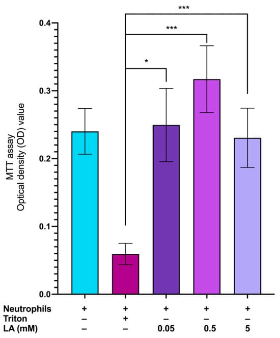
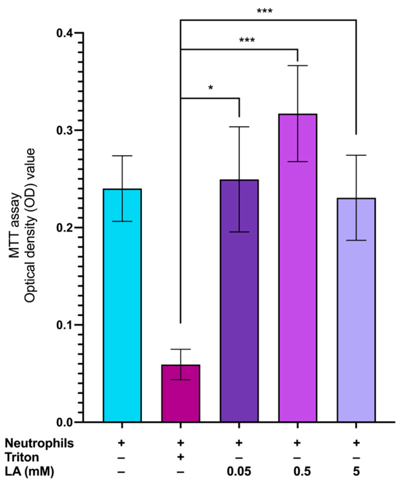
Neutrophil metabolic activity was assessed by measuring MTT reduction. Treatment with 0.05, 0.5, or 5 mM lipoic acid for 60 min did not significantly decrease MTT reduction compared with untreated neutrophils (Figure 1), indicating that lipoic acid does not impair metabolic activity. In contrast, the positive control (10% Triton X-100) caused a marked decrease in MTT reduction. These results demonstrate that lipoic acid, across the concentration range tested, does not compromise basal neutrophil metabolic activity.
Figure 1.
Lipoic acid (LA) does not reduce neutrophil metabolic activity at the concentrations evaluated. Lipoic acid at 0.05, 0.5, and 5 mM yielded MTT reduction levels comparable to untreated controls (left column), whereas the detergent Triton X-100 markedly reduced the MTT signal. * p < 0.05 and *** p < 0.001.
2.2. Lipoic Acid Does Not Significantly Affect Neutrophil Viability or Induce Apoptosis/Necrosis
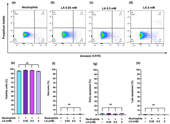
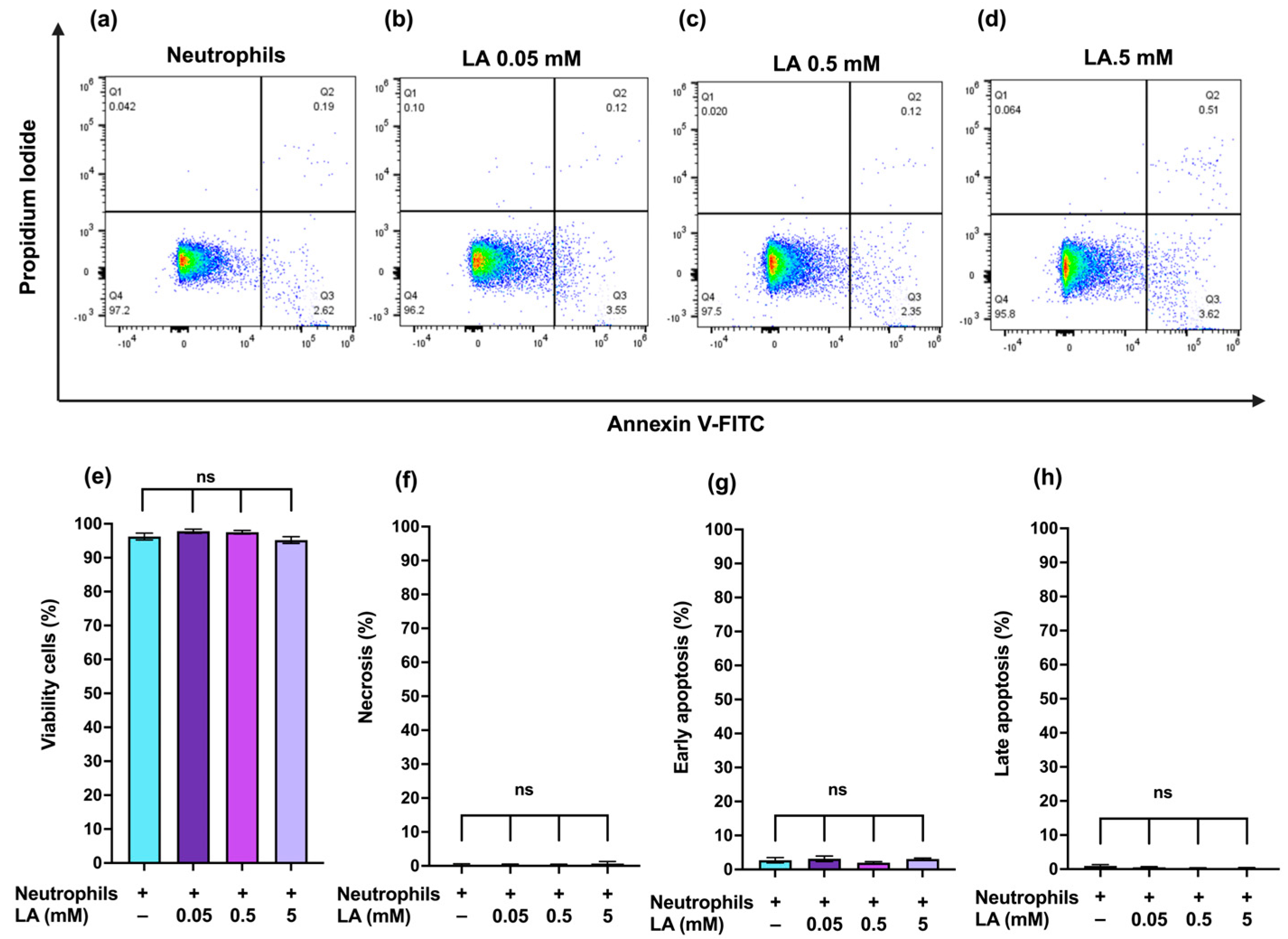
To determine whether lipoic acid induces cell death in neutrophils, cells were incubated with lipoic acid at 0.05, 0.5, or 5 mM for 60 min and analyzed by Annexin V-FITC/PI staining. As shown in Figure 2, lipoic acid treatment did not significantly affect the percentage of viable cells or significantly increase the percentages of necrotic, early apoptotic, or late apoptotic cells compared with untreated neutrophils. In all conditions, the viable cell population remained predominant, indicating that lipoic acid did not induce detectable apoptosis or necrosis under the experimental conditions tested.
Figure 2.
Lipoic acid does not induce apoptosis or necrosis in neutrophils at the concentrations tasted. Dot plots (a–d) and corresponding quantification (e–h) show comparable distributions of viable, early apoptotic, late apoptotic, and necrotic populations between untreated and LA-treated (0.05–5 mM) neutrophils. ns, no significant.
2.3. Lipoic Acid Reduces NET Formation Induced by LPS and E. coli
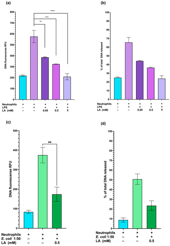
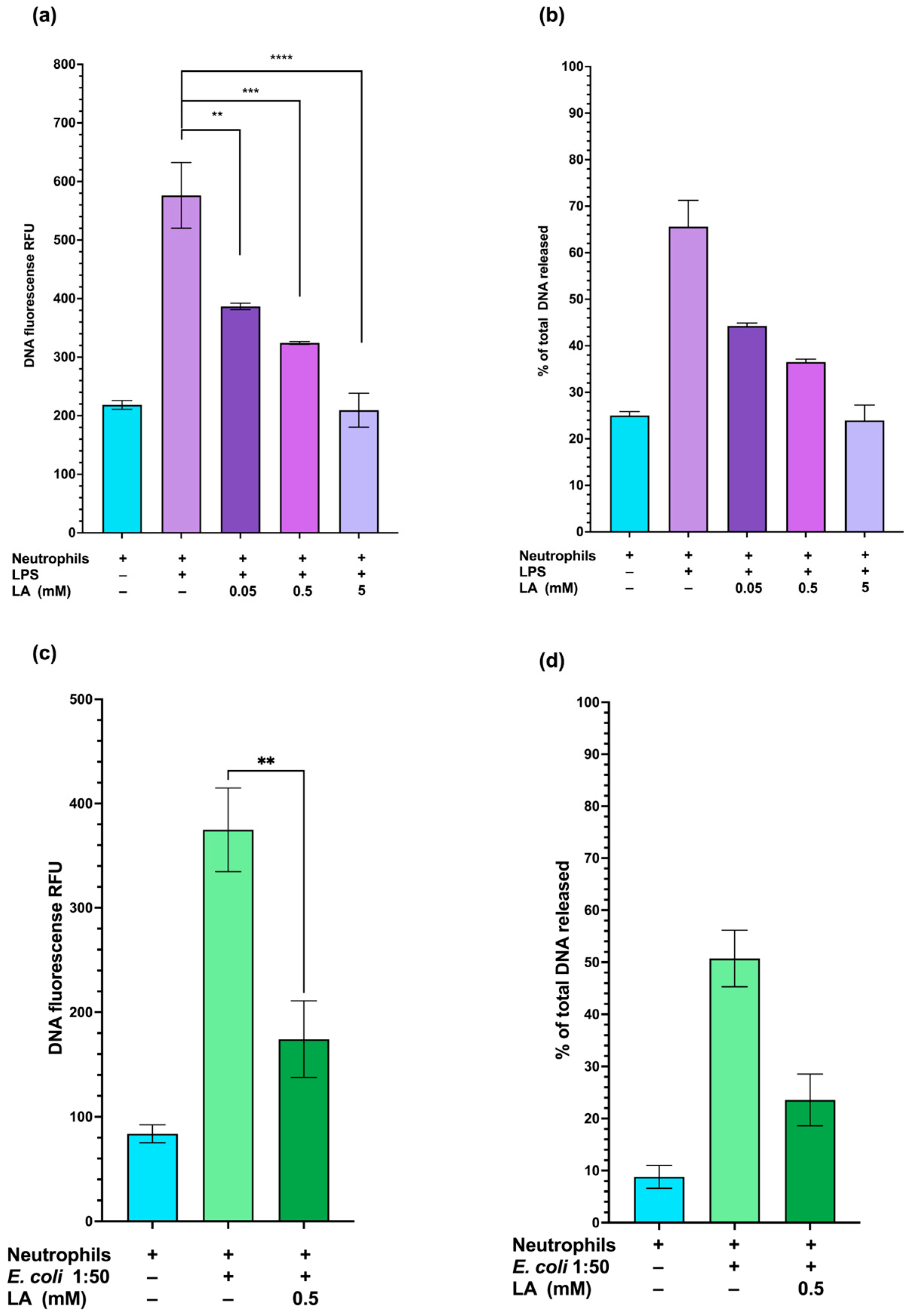
As shown in Figure 3a,b, pretreatment with lipoic acid at 0.05 mM, 0.5 mM, and 5 mM significantly reduced LPS-induced NET formation. Based on these findings, 0.5 mM lipoic acid was selected for subsequent experiments as an intermediate concentration that showed a marked inhibitory effect on NET formation. Because LPS is a major structural component of the outer membrane of Gram-negative bacteria, we next examined whether lipoic acid also inhibits NET formation induced by whole bacteria. Neutrophils were stimulated with E. coli at a 1:50 neutrophil-to-bacteria ratio, and pretreatment with 0.5 mM of lipoic acid significantly reduced NET formation (Figure 3c,d).
Figure 3.
Lipoic acid reduces NET formation in neutrophils with 10 μg/mL LPS or E. coli. Panels (a,c) show NET-associated extracellular DNA fluorescence for LPS and E. coli stimulation, respectively; panels (b,d) provide the corresponding quantitative comparisons. LA treatment lowers the DNA signal relative to stimulation alone in both settings. ** p < 0.01, *** p < 0.001, and **** p < 0.0001.
2.4. Lipoic Acid Attenuates ROS Production Induced by LPS and Escherichia coli Stimulation in Human Neutrophils
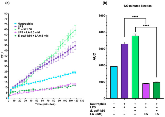
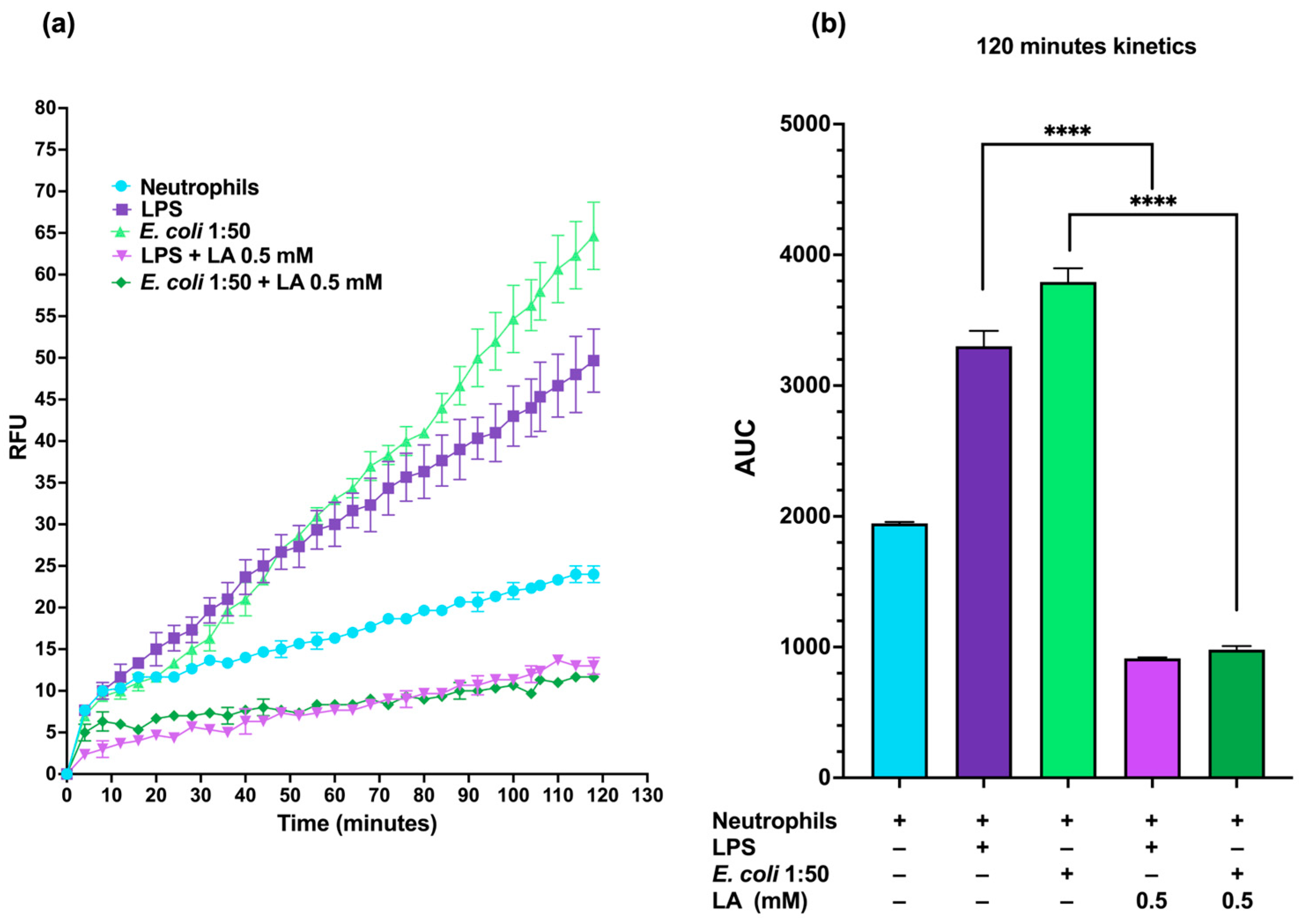
In neutrophils stimulated without lipoic acid, ROS production increased markedly after 30 min of stimulation with LPS or E. coli. Neutrophils pretreated with lipoic acid and then stimulated with LPS or E. coli exhibited lower fluorescence intensity than untreated stimulated neutrophils throughout the assay (Figure 4a). Consistent with this finding, the area under the curve (AUC) over 120 min was lower in lipoic acid-pretreated neutrophils than in untreated stimulated cells (Figure 4b). These results indicate that lipoic acid attenuates ROS production induced by LPS and E. coli stimulation in human neutrophils.
Figure 4.
Lipoic acid attenuates ROS production in stimulated neutrophils. Real-time ROS kinetics (a) and the corresponding area under the curve (b) are shown for neutrophils stimulated with 10 μg/mL LPS or E. coli. Lipoic acid reduces the overall ROS response compared with stimulated controls. **** p < 0.0001.
2.5. Lipoic Acid Does Not Impair Neutrophil-Mediated Bacterial Killing
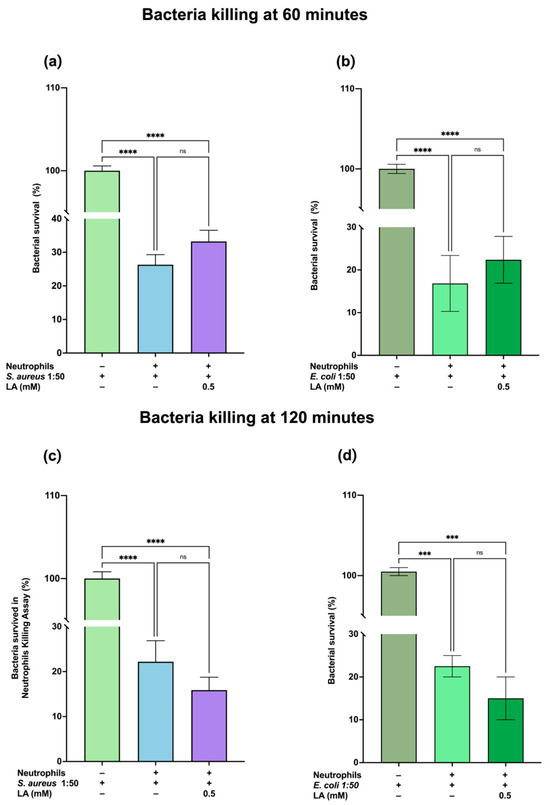
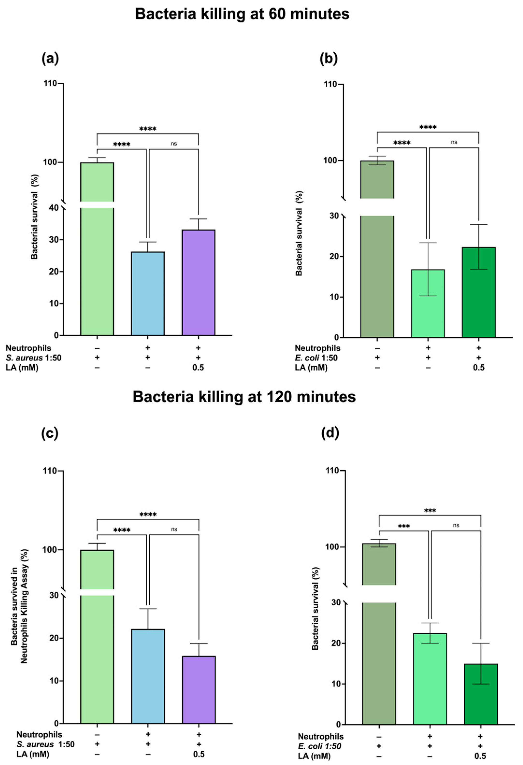
We performed CFU assays with Staphylococcus aureus and Escherichia coli, including bacteria-only controls for each species. Neutrophils were pretreated with lipoic acid for 1 h or left untreated and were then challenged with bacteria at a 1:50 neutrophil-to-bacteria ratio. CFU counts did not differ significantly between lipoic acid-treated and untreated neutrophils, indicating that lipoic acid does not impair bactericidal activity under the conditions tested.
2.6. Lipoic Acid Attenuates Early TNF-α Secretion in E. coli-Stimulated Neutrophils
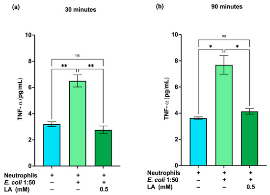
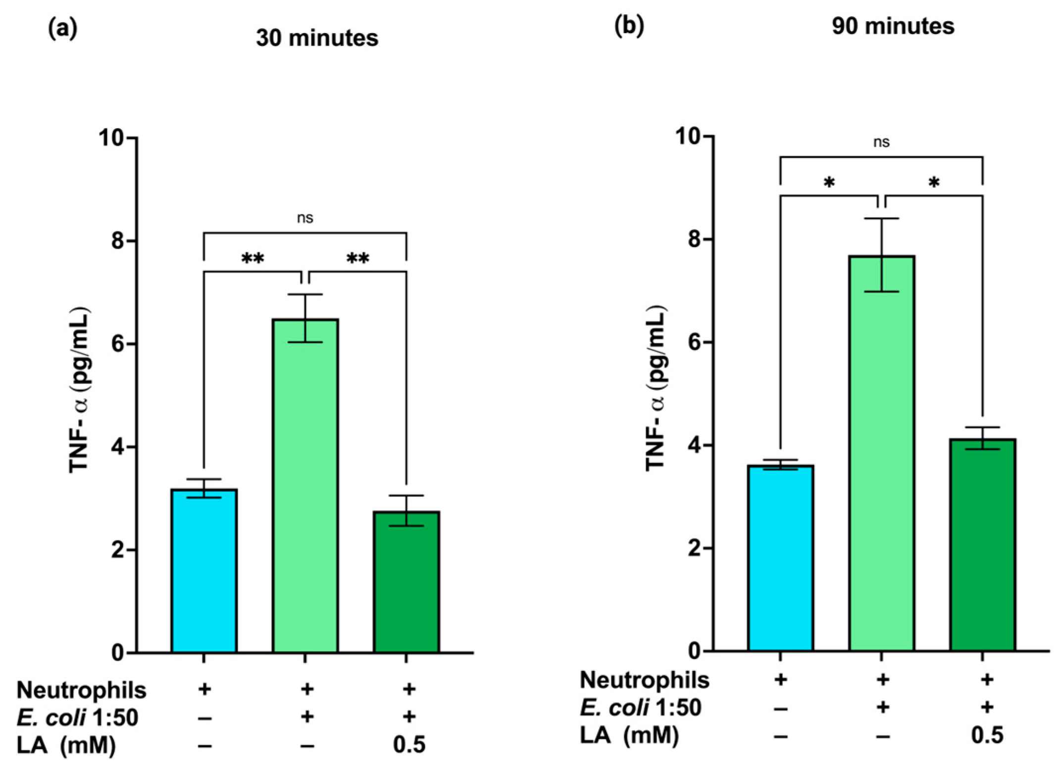
TNF-α is a key pro-inflammatory cytokine that promotes neutrophil activation and amplifies inflammatory responses. We therefore measured TNF-α levels in neutrophil culture supernatants 30 and 90 min after bacterial stimulation Figure 5. E. coli stimulation increased TNF-α release compared with untreated neutrophils. Pretreatment with lipoic acid significantly reduced TNF-α secretion relative to stimulated neutrophils alone, although levels did not fully return to basal values at 90 min. Although TNF-α levels at 90 min were slightly higher than at 30 min, the Figure 6 response remained attenuated compared with stimulated neutrophils without lipoic acid. These results indicate that lipoic acid dampens the early TNF-α response induced by E. coli.
Figure 5.
Lipoic acid does not impair neutrophil bactericidal activity. Bacterial survival (relative to bacteria-only controls) for S. aureus (a,c) and E. coli (b,d) at 60 min (a,b) and 120 min (c,d). LA-treated neutrophils maintain killing efficiency similar to that of untreated neutrophils across all conditions. *** p < 0.001, and **** p < 0.0001.
Figure 6.
Lipoic acid reduces early TNF-α release in E. coli-stimulated neutrophils. TNF-α levels were measured at 30 min (a) and 90 min (b) following E. coli stimulation. LA-treated neutrophils show lower TNF-α levels than stimulated untreated controls. * p < 0.05, ** p < 0.01.
2.7. Lipoic Acid Preserves Neutrophil Viability After Bacterial Stimulation
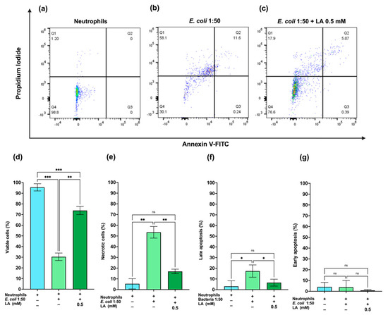
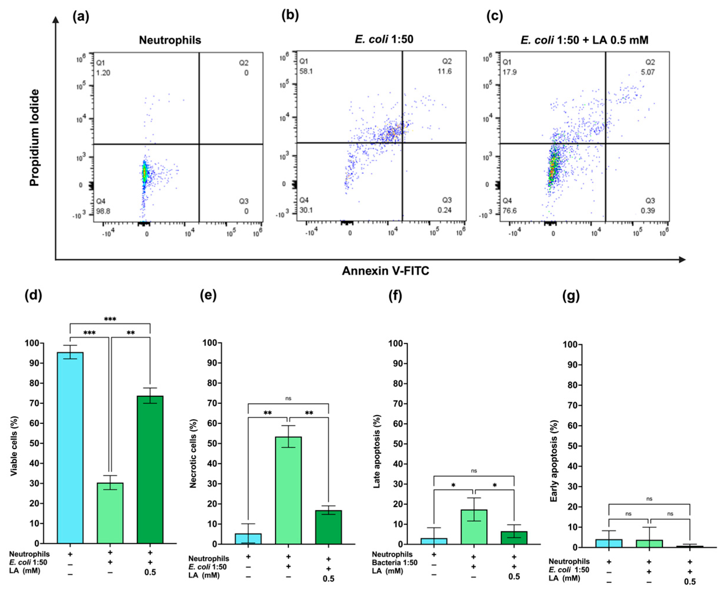
Neutrophil viability after 2 h of bacterial stimulation was assessed by Annexin V-FITC/PI staining Figure 7. Stimulation with bacteria at a 1:50 neutrophil-to-bacteria ratio markedly reduced viability to 30%, accompanied by a predominance of necrotic cells (58%) and a minor fraction of apoptotic cells (11.6%) (Figure 7b). Under the same conditions, pretreatment with 0.5 mM lipoic acid maintained viability (76.5%), with a substantial reduction in necrotic cells (18%) and late apoptotic cells (4.92%); early apoptosis remained minimal in both conditions (Figure 7c). These results demonstrate that LA preserves neutrophil viability and reduces bacterial stimulation-induced cell death.
Figure 7.
Lipoic acid preserves neutrophil viability during E. coli stimulation. Dot plots (a–c) and corresponding quantification (d–g) of viable, necrotic, early apoptotic, and late apoptotic populations. The addition of LA to E. coli-stimulated neutrophils restores the population distribution toward that of unstimulated controls. * p < 0.05, ** p < 0.01, *** p < 0.001.
3. Discussion
The MTT assay showed that lipoic acid did not significantly affect neutrophil metabolic activity under the experimental conditions tested. Specifically, lipoic acid at 0.05 mM, 0.5 mM and 5 mM maintained MTT reduction levels comparable to those of untreated controls, suggesting that lipoic acid does not compromise basal metabolic activity.
The concentrations of lipoic acid used in this study are consistent with concentrations previously evaluated in other cell types. Huerta-Olvera et al. used HepG2 liver cells, whereas Mozaffarian et al. evaluated mitochondrial metabolic activity in isolated rat liver mitochondria [23,24].
To further confirm the safety of neutrophil exposure to lipoic acid, we performed flow cytometry with Annexin V-FITC/PI. The absence of significant changes in Annexin V- or PI-positive populations confirmed that short-term exposure to lipoic acid alone does not exert a cytotoxic effect.
Nevertheless, constitutive death, or spontaneous neutrophil apoptosis, is a central mechanism for an essential component of neutrophil homeostasis and inflammation resolution. Therefore, longer lipoic acid-only incubations could potentially influence neutrophil lifespan, possibly by reducing spontaneous apoptosis through redox-dependent mechanisms [25]. However, the biological implications of this effect would likely depend on the physiological or pathological context, such as sepsis, neutropenia, or healthy conditions. This question should be addressed in future studies specifically designed to evaluate the effect of lipoic acid on spontaneous neutrophil apoptosis over longer incubation periods.
In this study, we used both LPS, a defined Gram-negative stimulus and canonical TLR4 agonist, and whole E. coli, a more physiologically relevant bacterial challenge. Both stimuli induced ROS production and NET formation [4,26]. Under these conditions, lipoic acid reduced LPS-induced NET formation in a dose-dependent manner across concentrations ranging from 0.05 to 5 mM. These findings are consistent with previous reports showing that antioxidants such as vitamin C, vitamin E, glutathione, N-acetylcysteine, and resveratrol attenuate NET formation in activated neutrophils [27,28,29]. NET formation can also be induced by intact bacteria such as E. coli. In our model, 0.5 mM lipoic acid significantly reduced E. coli-induced NET formation by approximately 50%.
Alternative explanations for the decrease in extracellular DNA should also be considered. One possibility is that the lower extracellular DNA signal could reflect enhanced degradation of released NETs rather than reduced NET formation. NET degradation is mainly mediated by extracellular DNases, particularly DNase1 and DNase1-like 3, and NET clearance can also involve macrophage-dependent uptake and lysosomal degradation. However, this explanation is less likely in our experimental setting because the assays were performed with isolated neutrophils, without macrophages or other major cellular sources of NET clearance. In addition, neutrophils are not considered the principal source of the DNase1/DNase1L3-dependent extracellular degradation system involved in NET removal [30]. Therefore, although altered extracellular DNA degradation cannot be completely excluded without directly measuring DNase activity, the most plausible interpretation is that lipoic acid reduced NET formation by limiting neutrophil oxidative activation and the downstream redox-sensitive pathways required for chromatin decondensation and NET release.
Our results show that lipoic acid significantly reduced ROS levels, restoring baseline levels despite neutrophil stimulation. This finding is consistent with the observations of O’Nell et al., who demonstrated that preincubation of neutrophils with lipoic acid suppresses the respiratory burst of PMA-stimulated neutrophils without affecting glucose-6-phosphate dehydrogenase activity [22]. This suggests that its effect may occur at the level of the intracellular cascade involved in NADPH oxidase assembly or activation, rather than through a nonspecific impairment of cellular metabolism. One proposed mechanism is that reduction of lipoic acid to DHLA consumes reducing equivalents, thereby limiting NADPH availability for NOX2-dependent superoxide generation. However, in their study, they did not directly measure the NADPH/NADP+ ratio or NOX2 activity to confirm this hypothesis.
An alternative explanation is that LA/DHLA may directly influence the DCFDA-based ROS readout through its antioxidant activity. Because DCFDA does not distinguish among individual ROS and depends on the oxidation of intracellular DCFH2 to fluorescent DCF, LA/DHLA could reduce the fluorescence signal by scavenging ROS or by competing with DCFH2 for oxidizing intermediates before DCF formation. Therefore, the decrease in DCFDA fluorescence should be interpreted as a reduction in detectable oxidative activity rather than as definitive evidence of decreased ROS generation alone. Nevertheless, this possibility is unlikely to fully explain the biological effect observed. In a previous study by Muñoz-Sánchez et al. using human neutrophils, antioxidants with redox properties comparable to LA/DHLA, including GSH and NAC, also decreased DCFDA fluorescence; importantly, this reduction was accompanied by parallel decreases in independent oxidative-stress endpoints, including nitrite/nitrate levels and malondialdehyde (MDA), a marker of lipid peroxidation [28]. Thus, the concordance between reduced DCFDA fluorescence and decreased downstream oxidative damage supports the biological relevance of the ROS measurement and suggests that the effect is not merely due to a direct artifact on the DCFDA probe.
Additionally, lipoic acid reduced ROS levels in unstimulated neutrophils, as shown in Supplementary Figure S1. This suggests that lipoic acid can also modulate basal oxidative activity. This observation is relevant because neutrophils are highly reactive cells and may increase basal ROS production as a consequence of isolation, handling, and in vitro manipulation. The inclusion of vitamin C as an antioxidant reference control further supports the interpretation that this basal ROS signal is sensitive to antioxidant modulation.
One of the most noteworthy findings of this study is that lipoic acid treatment did not significantly impair the ability of neutrophils to eliminate S. aureus or E. coli, as determined by CFU assays after 60 and 120 min of co-incubation at a neutrophil-to-bacteria ratio of 1:50. The preservation of bactericidal activity at 120 min is particularly relevant because neutrophils are highly reactive and short-lived effector cells, and their functional lifespan may be further shortened under infectious or intense inflammatory conditions. Therefore, the finding that LA-treated neutrophils retained antimicrobial activity up to 2 h suggests that LA does not simply suppress neutrophil function or accelerate functional exhaustion.
This observation is also important because neutrophil-mediated bacterial clearance depends on both neutrophil concentration and bacterial burden [31]. In our study, neutrophils were challenged under high bacterial-load conditions using 6 × 10⁶ neutrophils/mL and a 1:50 neutrophil-to-bacteria ratio, representing a demanding setting in which antimicrobial capacity may become functionally saturated [31]. Under these conditions, our findings show that lipoic acid reduces ROS production and NET formation while preserving the capacity of neutrophils to continue eliminating bacteria over time.
Consistent with this modulatory profile, lipoic acid also significantly decreased TNF-α levels in culture supernatants at 30 and 90 min, suggesting attenuation of early cytokine-mediated inflammatory signaling. Thus, lipoic acid appears to dampen excessive neutrophil activation while preserving bactericidal capacity under the experimental conditions evaluated.
A plausible explanation for this preserved bactericidal activity is that lipoic acid did not induce apoptosis or necrotic loss of neutrophils during the experimental period, allowing the cells to maintain essential antimicrobial functions. This interpretation is supported by the viability assays showing no significant increase in Annexin V- or PI-positive populations after lipoic acid treatment.
An alternative, non-mutually exclusive explanation is that lipoic acid may help preserve phagocytosis-dependent bacterial clearance by limiting excessive oxidative stress and maintaining neutrophil functional integrity. However, because phagocytosis was not directly measured in the present study, this possibility should be addressed in future experiments using specific phagocytosis assays, intracellular bacterial killing assays, and compartment-specific ROS measurements. Nevertheless, future studies should determine how ROS and redox-related molecules are distributed among different cellular compartments and how this compartmentalization influences neutrophil function.
Taken together, these results demonstrate that lipoic acid attenuates key features of excessive neutrophil activation, including ROS production, NET formation, and TNF-α release, while maintaining bactericidal capacity in vitro. These findings support the concept that redox modulation can selectively dampen inflammatory responses without abolishing essential antimicrobial functions.
However, several limitations should be considered. ROS measurements were performed using DCFH-DA, which detects general oxidative activity rather than specific ROS, and extracellular DNA quantification does not exclusively distinguish NET-derived DNA from other sources of extracellular DNA. The experiments were conducted in isolated neutrophils under in vitro conditions, which do not fully reproduce the complexity of inflammatory environments involving endothelial cells, complement activation, hormonal regulation and interactions with other immune cells. Another limitation is that this study was not designed to evaluate the influence of donor sex or hormonal status on neutrophil responses. Since sex-related and hormonal factors, particularly estrogen levels, may modulate inflammatory and immune responses, future studies should include a larger donor cohort and assess hormonal profiles to determine whether lipoic-acid-mediated effects on ROS production, NET formation, TNF-α release, and bactericidal activity differ according to donor sex.
Therefore, further in vivo studies and mechanistic analyses are required to confirm these effects and define the molecular pathways involved, including NADPH oxidase activity, NF-kB signaling, MAPK pathways, and redox-dependent regulation of neutrophil activation.
4. Materials and Methods
4.1. Neutrophil Isolation
Neutrophils were obtained from a total of six healthy fasting donors, including three males and three females, aged 18–30 years. Donors had no known chronic disease, no regular medication or vitamin supplementation, and no acute infection within the preceding two weeks. All donors provided written informed consent for the exclusive use of their blood samples for experimental purposes. The study was approved by the Ethics Committee of the University Center for Health Sciences of the Universidad de Guadalajara (protocol number: 23-04).
Blood samples were processed immediately after collection. During handling, each sample was identified using a unique alphanumeric code. A double-density gradient was prepared in a 15 mL conical tube by layering blood onto Histopaque® 1119 and Histopaque® 1077 (Sigma-Aldrich, St. Louis, MO, USA). Samples were centrifuged at 700× g for 30 min at room temperature without brake to allow phase separation and recovery of polymorphonuclear cells (PMNs). PMNs were resuspended in 2 mL of PBS and used immediately for the different experiments; therefore, isolated cells were not stored. Cell viability was determined by trypan blue exclusion using a Neubauer chamber at a 1:10 dilution, and only preparations with viability greater than 95% were used.
4.2. Culture Conditions
Neutrophils were seeded at 2.5 × 10⁵ cells/well in 24-well plates (Costar 3526, Corning, NY, USA) and incubated in 1 mL of phenol-red-free RPMI 1640 medium (Sigma-Aldrich, R8755) supplemented with 2% sodium bicarbonate and 2% autologous serum.
4.3. Reagent Preparation
A fresh stock solution of lipoic acid (Sigma-Aldrich, 62320, St. Louis, MO, USA) was prepared at 24.4 mM in physiological saline containing 0.05% DMSO and protected from light. This stock solution was freshly diluted in culture medium to obtain final lipoic acid concentrations of 0.05, 0.5, and 5 mM in the wells.
Lipopolysaccharide (LPS) from Escherichia coli O127:B8 (Sigma-Aldrich, L3129) was reconstituted according to the manufacturer’s instructions by adding 2 mL of sterile distilled water to obtain a 5 mg/mL stock solution. The LPS stock was diluted in culture medium to obtain the final concentration used for neutrophil stimulation. LPS was used as an independent stimulus and was not replaced by whole bacteria. Whole E. coli and S. aureus were used in separate experiments, as described in the bacterial culture and bactericidal assay sections.
4.4. Bacterial Culture
Escherichia coli (ATCC 11229, strain AMC 198, Manassas, VA, USA) and Staphylococcus aureus (ATCC 6538, strain FDA 209, Manassas, VA, USA) were cultured in tryptic soy broth (TSB) at 37 °C for 12 h. Bacterial cells were harvested by centrifugation (1250× g, 10 min, room temperature), resuspended in physiological saline, and the concentration (CFU/mL) was estimated by measuring the optical density at 600 nm (OD600) using a previously established growth curve. Viable counts were confirmed by plating serial dilutions on standard count agar and incubating overnight at 37 °C.
4.5. ROS Production Assay
Intracellular ROS production was measured using the DCFDA/H2DCFDA Cellular ROS Assay Kit (catalog no. ab113851, Abcam, Cambridge, UK) according to the manufacturer’s protocol. Neutrophils were resuspended in PBS at 5 × 10⁵ cells/mL and pretreated with 0.5 mM lipoic acid for 60 min at 37 °C. Cells were washed three times with PBS and loaded with DCFDA for 30 min at 37 °C, washed twice with cold PBS, and resuspended in fresh phenol-red-free RPMI 1640 medium. Neutrophils were seeded into black opaque 96-well plates at 1.5 × 10⁵ cells/well in 300 µL and stimulated with 10 µg/mL LPS or E. coli at a neutrophil-to-bacteria ratio of 1:50. Fluorescence kinetics were monitored every 2 min for 120 min using an Synergy HTX (BioTek Instruments, Inc., Winooski, VT, USA) multimode reader at excitation 485 nm, emission 535 nm. All conditions were assayed in triplicate.
4.6. NET Quantification by Fluorometry
NET formation was quantified by measuring extracellular DNA fluorescence. Neutrophils were seeded at 2.5 × 10⁵ cells/well in 24-well plates (Nunclon, Delta, catalog no. 143982, Nunc A/S, Thermo Fisher Scientific Inc., Roskilde, Denmark). For LPS-induced NET formation, neutrophils were pretreated with lipoic acid at the indicated concentrations for 60 min at 37 °C and then stimulated with 10 µg/mL LPS. For whole-bacteria stimulation, neutrophils were pretreated with 0.5 mM lipoic acid for 60 min and then stimulated with E. coli at a neutrophil-to-bacteria ratio of 1:50. Untreated neutrophils were included as negative controls, and Triton X-100-lysed (catalog no. X100; Sigma-Aldrich Inc., St. Louis, MO, USA) neutrophils were used to determine total DNA release.
After NET induction, plates were centrifuged (500× g, 10 min), and 1 µL DNase I (Sigma-Aldrich) was added to each well. Plates were incubated for 30 at 37 °C and centrifuged again at 500× g for 10 min. Then 300 µL of each supernatant was transferred to a black 96-well plate. Next, 1 µL of SYTOX Green (5 mM stock; Thermo Fisher Scientific, S7020) was added, and the plate was incubated for 15 min at room temperature in the dark. Fluorescence was recorded with an Synergy HTX (BioTek Instruments, Inc., Winooski, VT, USA) multimode reader at excitation 485 nm, emission 527 nm, and results were expressed as relative fluorescence units (RFUs). Total DNA release was normalized to that of neutrophils lysed with Triton X-100, which was set as 100% DNA release.
4.7. Bactericidal Assay
Neutrophil bactericidal activity was assessed by quantifying colony-forming units (CFUs). Neutrophils 6 × 10⁶ were pretreated with 0.5 mM lipoic acid for 60 min at 37 °C and washed twice with physiological saline to remove residual compound. E. coli or S. aureus were opsonized by incubation with 10% autologous serum for 30 min at 37 °C with agitation. Neutrophils were then challenged with either bacteria at a neutrophil-to-bacteria ratio of 1:50 and incubated at 37 °C with shaking at 200 rpm for 60 or 120 min.
Following incubation, neutrophils were lysed by adding 650 µL of alkaline water, pH 11, to each 350 µL sample and incubating for 5 min at 37 °C. Suspensions were serially diluted, plated on standard count agar, and incubated overnight at 37 °C. Bacterial survival was determined by colony-forming unit counting. Bacteria cultured without neutrophils served as growth controls, and untreated neutrophils served as killing controls.
Bacterial survival (%) was calculated as the number of CFUs recovered from wells containing neutrophils and bacteria divided by the number of CFUs from the corresponding bacteria-only control, multiplied by 100. This ratio was determined separately for untreated and lipoic acid-pretreated neutrophils.
Untreated neutrophils:
Lipoic acid -pretreated neutrophils:
4.8. MTT Assay
Metabolic activity was assessed using the CyQUANT™ MTT Cell Proliferation Assay Kit (I, catalog no. V13154Invitrogen, Life Technologies Corporation, Eugene, OR, USA). Neutrophils were pretreated with lipoic acid for 60 min, washed twice with PBS, and resuspended in phenol-red-free RPMI 1640 medium at 2 × 10⁶ cells/mL. Aliquots of 100 µL 2 × 10⁵ cells were seeded per well into flat-bottom 96-well plates (Thermo Fisher, 168055), and 10 µL of MTT reagent per well was added following the manufacturer’s instructions. After incubation for 3 h at 37 °C, plates were centrifuged (500× g, 10 min, 4 °C), supernatants were carefully removed, and formazan crystals were dissolved in 50 µL of DMSO. Absorbance was measured at 540 nm using an microplate reader (Hangzhou Allsheng Instruments Co., Ltd., Hangzhou, Zhejiang, China).
4.9. TNF-α Quantification by ELISA
TNF-α release was quantified in culture supernatants by enzyme-linked immunosorbent assay (ELISA). Neutrophils were pretreated with 0.5 mM lipoic acid for 60 min at 37 °C and then stimulated with Escherichia coli at a neutrophil-to-bacteria ratio of 1:50. Supernatants were collected after 30 and 90 min of stimulation following centrifugation to remove cells and debris. TNF-α concentrations were measured using the Quantikine HS ELISA Human TNF-α (catalog no. HSTA00E; R&D Systems, a Bio-Techne brand, Minneapolis, MN, USA) Set according to the manufacturer’s instructions. Absorbance was measured using a microplate reader, and cytokine concentrations were calculated from the standard curve and expressed as pg/mL.
4.10. Annexin V-FITC/PI Flow Cytometry
Neutrophil viability, apoptosis, and necrosis were assessed using the Dead Cell Apoptosis Kit with Annexin V-FITC and Propidium Iodide (catalog no. V13242, Invitrogen, Life Technologies Corporation, Eugene, OR, USA). To determine that lipoic acid alone does not induce cytotoxicity, neutrophils (2 × 10⁵ cells/mL) were pretreated with lipoic acid (0.05 mM, 0.5 mM and 5 mM) for 60 min, washed with PBS, resuspended in Annexin-binding buffer, and stained with Annexin V-FITC (5 µL) and propidium iodide (1 µL) for 15 min at room temperature in the dark.
To evaluate the effect of lipoic acid on bacteria-induced cell death, the same staining protocol was applied after 2 h of stimulation with Escherichia coli at a neutrophil-to-bacteria ratio of 1:50, in the presence or absence of 0.5 mM lipoic acid pretreatment. Samples were acquired using an Attune Acoustic Focusing Cytometer, with 40,000 events collected per sample, and analyzed using FlowJo™ v10.9.0.
4.11. Statistical Analysis
Data was analyzed using GraphPad Prism 10. Technical triplicates were averaged for each donor, and each donor was treated as an independent biological replicate. Results are expressed as mean ± SEM. Normality was assessed using the Shapiro–Wilk test. When data met parametric assumptions, statistical comparisons were performed using one-way repeated-measures ANOVA followed by Tukey’s or Dunnett’s multiple comparisons test, as appropriate. When assumptions were not met, the non-parametric Friedman test with Dunn’s post hoc test was applied. A p-value < 0.05 was considered statistically significant.
5. Conclusions
This study demonstrates that lipoic acid attenuates key inflammatory responses in human neutrophils, including ROS production, NET formation and early TNF-α secretion, while preserving neutrophil-mediated bacterial killing. Lipoic acid also reduced bacteria-induced neutrophil necrosis and improved the proportion of viable cells after bacterial stimulation. These findings support lipoic acid as a potential redox-modulating compound capable of limiting excessive neutrophil activation without abolishing essential antimicrobial functions. Further studies are required to define the molecular mechanisms involved, particularly NADPH oxidase activity, NADPH/NADP+ balance, and redox-sensitive inflammatory signaling pathways.
Supplementary Materials
The following supporting information can be downloaded at: https://www.mdpi.com/article/10.3390/ijms27136072/s1.
Author Contributions
Conceptualization, G.A.V.-H. and V.D.-R.; methodology, G.A.V.-H. and V.D.-R.; software, G.A.V.-H.; validation, V.D.-R.; formal analysis, G.A.V.-H. and V.D.-R.; investigation, G.A.V.-H., M.F.-M., G.M.-S., L.A.G.-M., L.Í.-G. and V.D.-R.; resources, V.D.-R.; data curation, G.A.V.-H. and V.D.-R.; writing—original draft preparation, G.A.V.-H. and V.D.-R.; writing—review and editing, G.A.V.-H., M.F.-M., L.A.G.-M., G.M.-S., M.G.M.-B., A.A.G.-C., L.Í.-G. and V.D.-R.; visualization, G.A.V.-H. and V.D.-R.; supervision, V.D.-R.; project administration and funding acquisition V.D.-R. All authors have read and agreed to the published version of the manuscript.
Funding
This research was funded by PROSNI 270455 FONDO 11485 UdeG, 1.1.2 260699 PIN2020 UdeG.
Institutional Review Board Statement
The study was conducted in accordance with the Declaration of Helsinki and approved by the Institutional Ethics Committee of Centro Universitario de Ciencias de la Salud, Universidad de Guadalajara (protocol code 23-04; 18 May 2023), for studies involving humans.
Informed Consent Statement
Informed consent was obtained from all subjects involved in the study.
Data Availability Statement
The data presented in this study are available on request from the corresponding author.
Acknowledgments
The authors would like to express their gratitude to Francisco Muñoz Valle for providing access to the flow cytometry facilities at the Instituto de Investigación en Ciencias Biomédicas-CUCS. The authors also thank the Instituto de Investigación en Odontología-CUCS for providing the facilities necessary to carry out this research.
Conflicts of Interest
The authors declare no conflicts of interest.
Abbreviations
| LA | Lipoic acid |
| DHLA | Dihydrolipoic acid |
| NET | Neutrophil extracellular trap |
| AS | Autologous serum |
| CFUs | Colony-forming units |
| DCFDA | 2′7′-Dichlorofluorescin diacetate |
| DMSO | Dimethyl sulfoxide |
| ELISA | Enzyme-linked immunosorbent assay |
| FITC | Fluorescein isothiocyanate |
| LPS | Lipopolysaccharide |
| NF-kB | Nuclear factor kappa B |
| NOX2 | NADPH oxidase 2 |
| PI | Propidium iodide |
| RFUs | Relative fluorescence units |
| ROS | Reactive oxygen species |
| RPMI | Roswell Park Memorial Institute medium |
| TNF-α | Tumor necrosis factor-alpha |
References
- Zhang, F.; Xia, Y.; Su, J.; Quan, F.; Zhou, H.; Li, Q.; Feng, Q.; Lin, C.; Wang, D.; Jiang, Z. Neutrophil diversity and function in health and disease. Signal Transduct. Target. Ther. 2024, 9, 343. [Google Scholar] [CrossRef] [Scilit] [PubMed]
- Rungelrath, V.; Kobayashi, S.D.; DeLeo, F.R. Neutrophils in innate immunity and systems biology-level approaches. WIREs Syst. Biol. Med. 2020, 12, e1458. [Google Scholar]
- Winterbourn, C.C.; Kettle, A.J.; Hampton, M.B. Reactive oxygen species and neutrophil function. Annu. Rev. Biochem. 2016, 85, 765–792. [Google Scholar] [CrossRef] [Scilit] [PubMed]
- Brinkmann, V.; Reichard, U.; Goosmann, C.; Fauler, B.; Uhlemann, Y.; Weiss, D.S.; Weinrauch, Y.; Zychlinsky, A. Neutrophil extracellular traps kill bacteria. Science 2004, 303, 1532–1535. [Google Scholar] [CrossRef] [Scilit] [PubMed]
- Tecchio, C.; Micheletti, A.; Cassatella, M.A. Neutrophil-derived cytokines: Facts beyond expression. Front. Immunol. 2014, 5, 508. [Google Scholar] [CrossRef] [Scilit] [PubMed]
- Lentini, G.; De Gaetano, G.V.; Famà, A.; Galbo, R.; Coppolino, F.; Mancuso, G.; Teti, G.; Beninati, C. Neutrophils discriminate live from dead bacteria by integrating signals initiated by Fprs and TLRs. EMBO J. 2022, 41, e109386. [Google Scholar] [CrossRef] [Scilit] [PubMed]
- Capucetti, A.; Albano, F.; Bonecchi, R. Multiple roles for chemokines in neutrophil biology. Front. Immunol. 2020, 11, 1259. [Google Scholar] [CrossRef] [Scilit] [PubMed]
- Naish, E.; Wood, A.J.; Stewart, A.P.; Routledge, M.; Morris, A.C.; Chilvers, E.R.; Lodge, K.M. The formation and function of the neutrophil phagosome. Immunol. Rev. 2023, 314, 158–180. [Google Scholar] [PubMed]
- Park, B.S.; Lee, J.O. Recognition of lipopolysaccharide pattern by TLR4 complexes. Exp. Mol. Med. 2013, 45, e66. [Google Scholar] [CrossRef] [Scilit] [PubMed]
- Vandendriessche, S.; Cambier, S.; Proost, P.; Marques, P.E. Complement receptors and their role in leukocyte recruitment and phagocytosis. Front. Cell Dev. Biol. 2021, 9, 624025. [Google Scholar] [CrossRef] [Scilit] [PubMed]
- Pérez-Figueroa, E.; Álvarez-Carrasco, P.; Ortega, E.; Maldonado-Bernal, C. Neutrophils: Many ways to die. Front. Immunol. 2021, 12, 631821. [Google Scholar] [CrossRef] [Scilit] [PubMed]
- Lázaro-Díez, M.; Chapartegui-González, I.; Redondo-Salvo, S.; Leigh, C.; Merino, D.; Segundo, D.S.; Fernández, A.; Navas, J.; Icardo, J.M.; Acosta, F.; et al. Human neutrophils phagocytose and kill Acinetobacter baumannii and A. pittii. Sci. Rep. 2017, 7, 4571, Erratum in Sci. Rep. 2020, 10, 4797. [Google Scholar] [CrossRef] [Scilit] [PubMed]
- Shirakawa, K.; Sano, M. Neutrophils and neutrophil extracellular traps in cardiovascular disease: An overview and potential therapeutic approaches. Biomedicines 2022, 10, 1850. [Google Scholar] [CrossRef] [Scilit] [PubMed]
- Papayannopoulos, V. Neutrophil extracellular traps in immunity and disease. Nat. Rev. Immunol. 2018, 18, 134–147. [Google Scholar] [PubMed]
- Shahid, A.; Nasir, K.; Bhatia, M. Therapeutic potential of alpha-lipoic acid: Unraveling its role in oxidative stress and inflammatory conditions. Curr. Issues Mol. Biol. 2025, 47, 322. [Google Scholar] [CrossRef] [Scilit] [PubMed]
- Packer, L.; Tritschler, H.J.; Wessel, K. Neuroprotection by the metabolic antioxidant α-lipoic acid. Free Radic. Biol. Med. 1997, 22, 359–378. [Google Scholar] [CrossRef] [Scilit] [PubMed]
- Moini, H.; Packer, L.; Saris, N.E.L. Antioxidant and prooxidant activities of α-lipoic acid and dihydrolipoic acid. Toxicol. Appl. Pharmacol. 2002, 182, 84–90. [Google Scholar] [CrossRef] [Scilit] [PubMed]
- Wang, X.; Liu, S.; Su, D.; Sui, J.; Yan, X.; Yang, J.; Zheng, Z.; Wang, P.; Song, W.; Jin, Z.; et al. Alpha-lipoic acid attenuates cardiac inflammation of CVB3-induced viral myocarditis via neutrophil-derived YM-1. Biochim. Biophys. Acta Mol. Basis Dis. 2026, 1872, 168034. [Google Scholar] [CrossRef] [Scilit] [PubMed]
- Zhang, Y.; Zhou, Y.; Li, X.; Pan, X.; Bai, J.; Chen, Y.; Lai, Z.; Chen, Q.; Ma, F.; Dong, Y. Small-molecule α-lipoic acid targets ELK1 to balance human neutrophil and erythrocyte differentiation. Stem Cell Res. Ther. 2024, 15, 100. [Google Scholar] [CrossRef] [Scilit] [PubMed]
- Dong, Y.; Zhang, Y.; Zhang, Y.; Pan, X.; Bai, J.; Chen, Y.; Zhou, Y.; Lai, Z.; Chen, Q.; Hu, S.; et al. Dissecting the process of human neutrophil lineage determination by using alpha-lipoic acid inducing neutrophil deficiency model. Redox Biol. 2022, 54, 102392. [Google Scholar] [CrossRef] [Scilit] [PubMed]
- Lin, Y.C.; Lai, Y.S.; Chou, T.C. The protective effect of alpha-lipoic acid in lipopolysaccharide-induced acute lung injury is mediated by heme oxygenase-1. Evid. Based Complement. Alternat. Med. 2013, 2013, 590363. [Google Scholar]
- O’Neill, H.C.; Rancourt, R.C.; White, C.W. Lipoic acid suppression of neutrophil respiratory burst: Effect of NADPH. Antioxid. Redox Signal. 2008, 10, 277–286. [Google Scholar] [CrossRef] [Scilit] [PubMed]
- Huerta-Olvera, S.G.; Macías-Barragán, J.; Ramos-Márquez, M.E.; Armendáriz-Borunda, J.; Díaz-Barriga, F.; Siller-López, F. Alpha-lipoic acid regulates heme oxygenase gene expression and nuclear Nrf2 activation as a mechanism of protection against arsenic exposure in HepG2 cells. Environ. Toxicol. Pharmacol. 2010, 29, 144–149. [Google Scholar] [CrossRef] [Scilit] [PubMed]
- Mozaffarian, F.; Dehghani, M.A.; Vanani, A.R.; Mahdavinia, M. Protective effects of alpha lipoic acid against arsenic-induced oxidative stress in isolated rat liver mitochondria. Biol. Trace Elem. Res. 2022, 200, 1190–1200. [Google Scholar] [PubMed]
- Luo, H.R.; Loison, F. Constitutive neutrophil apoptosis: Mechanisms and regulation. Am. J. Hematol. 2008, 83, 288–295. [Google Scholar] [PubMed]
- Marin-Esteban, V.; Turbica, I.; Dufour, G.; Semiramoth, N.; Gleizes, A.; Gorges, R.; Beau, I.; Servin, A.L.; Lievin-Le Moal, V.; Sandré, C.; et al. Afa/Dr diffusely adhering Escherichia coli strain C1845 induces neutrophil extracellular traps that kill bacteria and damage human enterocyte-like cells. Infect. Immun. 2012, 80, 1891–1899. [Google Scholar] [CrossRef] [Scilit] [PubMed]
- De Mattos, T.R.F.; Formiga-Jr, M.A.; Saraiva, E.M. Resveratrol prevents the release of neutrophil extracellular traps (NETs) by controlling hydrogen peroxide levels and nuclear elastase migration. Sci. Rep. 2024, 14, 9107. [Google Scholar] [CrossRef] [Scilit] [PubMed]
- Muñoz-Sánchez, G.; Godínez-Méndez, L.A.; Fafutis-Morris, M.; Delgado-Rizo, V. Effect of antioxidant supplementation on NET formation induced by LPS in vitro: The roles of vitamins E and C, glutathione, and N-acetyl cysteine. Int. J. Mol. Sci. 2023, 24, 13162. [Google Scholar] [CrossRef] [Scilit] [PubMed]
- Vorobjeva, N.V.; Pinegin, B.V. Effects of the antioxidants Trolox, Tiron and Tempol on neutrophil extracellular trap formation. Immunobiology 2016, 221, 208–219. [Google Scholar] [CrossRef] [Scilit] [PubMed]
- Jiménez-Alcázar, M.; Rangaswamy, C.; Panda, R.; Bitterling, J.; Simsek, Y.J.; Long, A.T.; Bilyy, R.; Krenn, V.; Renné, C.; Renné, T.; et al. Host DNases prevent vascular occlusion by neutrophil extracellular traps. Science 2017, 358, 1202–1206. [Google Scholar] [CrossRef] [Scilit] [PubMed]
- Li, Y.; Karlin, A.; Loike, J.D.; Silverstein, S.C. A critical concentration of neutrophils is required for effective bacterial killing in suspension. Proc. Natl. Acad. Sci. USA 2002, 99, 8289–8294. [Google Scholar] [CrossRef] [Scilit] [PubMed]
Disclaimer/Publisher’s Note: The statements, opinions and data contained in all publications are solely those of the individual author(s) and contributor(s) and not of MDPI and/or the editor(s). MDPI and/or the editor(s) disclaim responsibility for any injury to people or property resulting from any ideas, methods, instructions or products referred to in the content. |
© 2026 by the authors. Licensee MDPI, Basel, Switzerland. This article is an open access article distributed under the terms and conditions of the Creative Commons Attribution (CC BY) license.