


Abstract
Cancer-associated cachexia (CAC) is a complex metabolic syndrome characterised by progressive skeletal muscle loss, systemic inflammation, reduced treatment tolerance, and poor survival. Marked interindividual variability in CAC susceptibility suggests that host genetic factors may contribute to its development. Vitamin D plays an important role in muscle metabolism and inflammatory control through activation of the vitamin D receptor (VDR). VDR signalling influences myogenesis, mitochondrial function, insulin-like growth factor pathways, and pro-inflammatory cytokine expression, all of which are implicated in CAC pathogenesis. Hence, VDR variants, including the rs731236 (A>G) polymorphism, which modifies receptor activity, may affect CAC pathogenesis. Thus, this study investigated the association between the polymorphism and susceptibility to CAC as well as patient survival in a cohort of 140 adult cancer patients. Briefly, the rs731236 GG genotype was significantly associated with a lower prevalence of CAC across disease diagnostic approaches (chi-square tests, p < 0.05). Furthermore, GG genotype carriers demonstrated significantly improved survival compared with carriers of AA/AG genotypes (Log-rank and Tarone–Ware tests, p < 0.05). In summary, these findings suggest that the rs731236 polymorphism influences both susceptibility to CAC and survival outcomes in patients with cancer, further supporting a clinically relevant role for vitamin D signalling in supportive oncology care.
1. Introduction
Cancer-associated cachexia (CAC) is a multifactorial metabolic syndrome characterised by involuntary weight loss, progressive skeletal muscle wasting, systemic inflammation, and functional impairment [1]. It affects up to 80% of patients with advanced malignancies, including both solid and haematological tumours, and is a major contributor to treatment intolerance, diminished quality of life, and increased mortality [2,3]. Unlike simple starvation, CAC is driven by complex metabolic and inflammatory alterations that are not fully reversible by nutritional support alone [4]. Despite its high prevalence and prognostic relevance, with 20% of cancer patients dying due to this condition, CAC remains underdiagnosed, undertreated, and insufficiently stratified in routine oncology practice [2,3].
Chronic inflammation is a central driver of CAC pathophysiology, promoting muscle proteolysis, impairing protein synthesis, causing mitochondrial dysfunction, and altering overall energy metabolism. Pro-inflammatory cytokines such as interleukin-6 (IL-6) and tumour necrosis factor-α (TNF-α) activate catabolic signalling pathways, disrupt anabolic responses, and contribute to endocrine resistance, ultimately accelerating muscle loss [1,4]. However, substantial interindividual variability exists in the development and severity of CAC, suggesting that several host-related factors, including genetic determinants, may modulate susceptibility to this syndrome [5].
Vitamin D3 (or simply vitamin D) plays a fundamental role at the interface between endocrine and immune regulation, exerting biological effects that extend well beyond its traditional involvement in calcium and phosphate metabolism [6]. Namely, it has emerged as a central modulator of muscle function, metabolism and immune regulation, primarily via activation of the vitamin D receptor (VDR), a nuclear transcription factor expressed in skeletal muscle, immune cells, and adipose tissue. VDR signalling regulates myogenesis, muscle fibre composition, mitochondrial oxidative capacity, insulin-like growth factor 1 (IGF-1) pathways, and the production of inflammatory cytokines [7,8,9,10]. Consistently, vitamin D deficiency and impaired VDR signalling have been associated with sarcopenia (progressive loss of skeletal muscle mass associated with ageing), frailty, and chronic inflammatory states, all of which share pathophysiological features with CAC [11,12,13].
Worldwide, genetic polymorphisms are the most prevalent genetic variations, with implications for several clinical traits [14,15]. Genetic polymorphisms in VDR may influence the receptor’s transcriptional activity and messenger (mRNA) stability, thereby modifying downstream biological responses to vitamin D. The rs731236 (A>G) variant, also known as TaqI, is one of the most studied VDR polymorphisms due to its influence on vitamin D signalling [16,17]. It is a single-nucleotide polymorphism (SNP) located in the 3′ untranslated region (UTR) of exon 9 of the VDR gene on chromosome 12q13.11 [18,19]. It involves the substitution of adenine with guanine (or, equivalently, thymine with cytosine, depending on strand orientation) [19]. Although VDR rs731236 does not alter the amino acid sequence of the encoded protein (i.e., it is a synonymous variant), it may nonetheless affect gene regulation and expression through mechanisms such as changes in transcription, splicing, co-translational folding, and mRNA stability, among other functionally relevant alterations [20]. Given its link with altered inflammatory profiles, muscle-related phenotypes, and clinical outcomes, rs731236 has been widely studied across multiple conditions and traits, including musculoskeletal diseases [21,22,23], neurological disorders [24,25], infectious and immune-related diseases [26,27,28,29], gut microbiota composition [30], cardiometabolic conditions [31,32] and reproductive and endocrine disorders [33,34,35]. Furthermore, the influence of VDR rs731236 has been evaluated across multiple malignancies with respect to disease susceptibility and prognosis [36,37,38,39,40,41]. However, the associations are frequently inconsistent across studies, ethnic groups and disease contexts, requiring additional studies for validation. Regarding CAC, the role of VDR rs731236 remains poorly explored, with most previous studies focusing on either vitamin D levels or single tumour types, rather than the intersection of host genetics, muscle wasting, and patient survival. Given existing knowledge gaps, this study explored the association of this polymorphism with susceptibility to CAC and survival outcomes in a cohort of 140 adult cancer patients from the Northern region of Portugal.
2. Results
2.1. Distribution of VDR rs731236 Genotypes
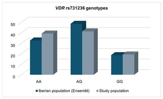
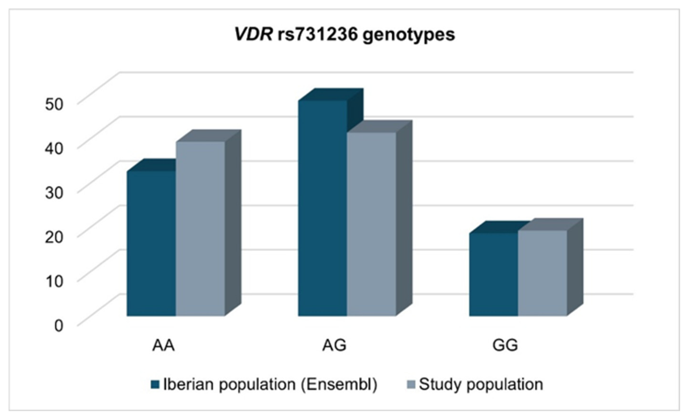
Genotype distributions for VDR rs731236 in the study population and the reference Iberian population described in the Ensembl database (https://www.ensembl.org/index.html, last accessed on the 28 December 2025) are indicated in Figure 1. Compared with the reference population, the distribution of SNP genotypes did not differ significantly [Chi-square (χ2) test, p = 0.551], indicating that the variant is consistent with Hardy–Weinberg equilibrium (HWE) expectations. Notably, the minor allele frequency (MAF) was 43% and 40% in the reference population and in the study population, respectively.
Figure 1.
Genotype distribution in the study cohort compared with the reference Iberian population (https://www.ensembl.org/index.html, last accessed on the 28 December 2025).
Notably, SNP genotype distributions did not differ significantly depending on patient demographic or clinicopathological variables (χ2 test or Fisher’s exact test, p > 0.05).
2.2. Association Between VDR rs731236 and CAC Status
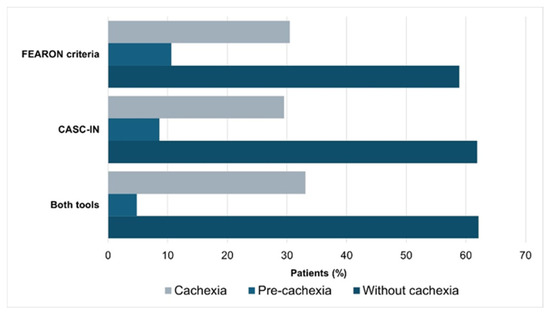
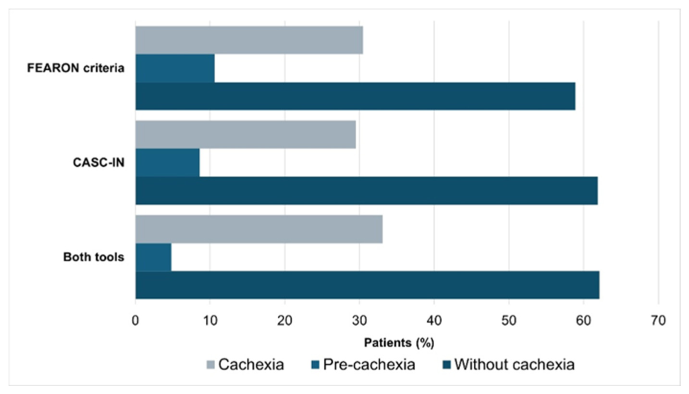
Cachexia prevalence in the study cohort was approximately 30% using Fearon criteria, CASC-IN, or the combination of both tools in disease diagnosis (Figure 2).
Figure 2.
Cachexia prevalence according to each diagnostic approach.
Associations between the VDR rs731236 SNP and CAC susceptibility were examined under codominant, dominant and recessive genetic models. Analyses were conducted using the three diagnostic approaches (Fearon criteria, CASC-IN tool and the combination of both tools), considering (i) three-category comparisons (no CAC, pre-CAC, and CAC) and (ii) dichotomised comparisons (no CAC versus pre-CAC + CAC). A significant association was observed between VDR rs731236 genotypes and CAC status (χ2 test or Fisher’s exact test, p < 0.05). Namely, across all diagnostic criteria, the SNP GG genotype was consistently more prevalent among individuals without CAC (Table 1).
Table 1.
Distribution of VDR rs731236 genotypes across groups (no cachexia, pre-cachexia, and cachexia) as defined by FEARON criteria, CASC-IN, and combined assessment tools.
Consistently, when pre-CAC and CAC patients were combined into a single entity group, the significant association between VDR rs731236 genotypes and CAC status remained (Table 2). Under the recessive genetic model (GG vs. AG/AA), for which significant results were observed, the GG genotype was associated with a significantly lower disease risk according to the Fearon criteria [odds ratio (OR) = 0.27; 95% confidence interval (CI), 0.09–0.75], the CASC-IN criteria (OR = 0.32; 95% CI, 0.11–0.90), and the combined assessment tool (OR = 0.20; 95% CI, 0.06–0.71).
Table 2.
Distribution of VDR rs731236 genotypes by disease status (no cachexia vs. pre-cachexia/cachexia) according to FEARON criteria, CASC-IN, and combined assessment tools.
2.3. Association Between VDR rs731236 and Patient Survival
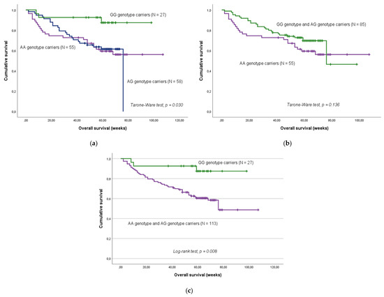
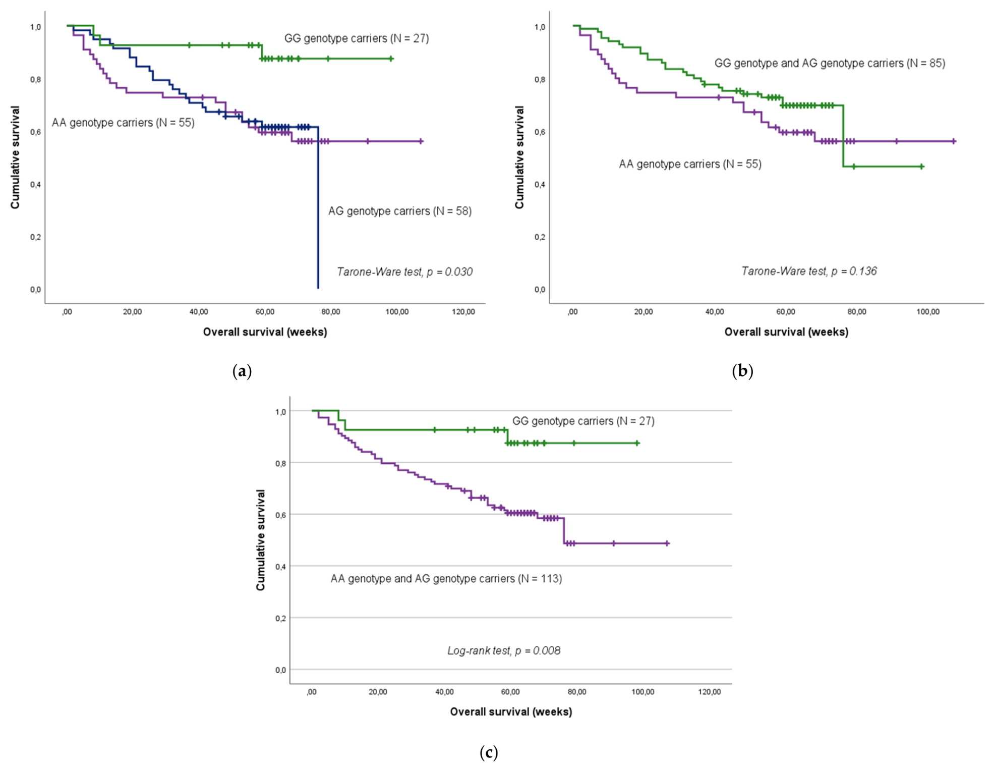
In the overall cohort (N = 140), carriers of the SNP GG genotype exhibited a significantly longer overall survival (OS) than patients with AA/AG genotypes (Figure 3). Individuals harbouring the GG genotype experienced a 76% reduction in the risk of death compared with their counterparts [hazard ratio (HR) = 0.24; 95% CI, 0.07–0.76; p = 0.016]. This association remained robust after adjustment for potential confounding variables, confirming the independent prognostic value of the VDR rs731236 GG genotype (Table 3). Notably, these analyses were based on 49 events (deaths).
Figure 3.
Overall survival (OS) according to VDR rs731236 genotypes. (a) In the codominant genetic model, patients with the GG genotype had significantly higher survival time compared to AA and AG genotype carriers (mean OS: 89.4 ± 4.8 weeks, 71.9 ± 5.8 weeks, and 57.6 ± 3.4 weeks, respectively; Tarone–Ware test, p = 0.030). (b) In the dominant genetic model, no significant differences in survival time were observed (Tarone–Ware test, p = 0.136). (c) In the recessive genetic model, patients carrying the GG genotype had significantly longer survival time than A allele carriers (mean OS = 89.4 ± 4.8 weeks and 71.2 ± 4.6 weeks, respectively; Log-rank test, p = 0.008).
Table 3.
Multivariable Cox analysis on the risk of death.
To specifically assess the impact of VDR rs731236 on survival in CAC patients, a subgroup analysis was performed. In this CAC-only population, no significant association between SNP genotypes and survival was detected, irrespective of the diagnostic criteria or genetic model applied (Log-rank and Tarone–Ware tests, p > 0.05).
3. Discussion
Muscle regeneration is a multifaceted process that requires the recovery of mitochondrial activity and the activation of satellite cells, which are the intrinsic stem cells responsible for skeletal muscle repair. In recent years, growing evidence has implicated vitamin D signalling as an important regulator of skeletal muscle metabolism, regeneration, and protein turnover, pathways that are critically disrupted in CAC and that influence patient outcomes [10]. In this context, the present study investigated the association between the VDR rs731236 polymorphism and clinical outcomes in cancer patients, focusing on CAC susceptibility and OS. Our findings indicate that GG genotype carriers have a lower risk of developing cachexia and demonstrate improved OS, independent of CAC status, compared with individuals carrying the AA or AG genotypes. To understand how the GG genotype may confer protection against CAC, it is necessary to consider the role of vitamin D in skeletal muscle.
Vitamin D is a pleiotropic hormone primarily produced in the skin through ultraviolet-dependent synthesis. It is subsequently converted in the liver into 25-hydroxyvitamin D, the primary circulating form, and then further hydroxylated to its active form, 1,25(OH)2D3. Although this final activation step primarily occurs in the kidneys, several other tissues also possess the enzymatic machinery required for local vitamin D activation [11]. In the circulation, 1,25(OH)2D3 is transported to target tissues by vitamin D-binding proteins. Its biological effects are mainly mediated by its receptor, VDR, a widely expressed nuclear receptor [42]. Upon ligand binding, the VDR heterodimerises with the retinoid X receptor and regulates gene transcription via interactions with vitamin D response elements (VDREs) within promoters of target genes involved in calcium and bone metabolism, cell growth and differentiation, and immune response and function [43,44]. In addition to these classic genomic actions, vitamin D also exerts rapid non-genomic effects via key signalling cascades, including the mitogen-activated protein kinase (MAPK) cascade, the phosphoinositide 3-kinase/protein kinase B (PI3K-Akt) pathway, and the mechanistic target of rapamycin (mTOR) signalling axis, which intersect with metabolic and anabolic networks governing muscle mass and function, namely modulation of mitochondrial function and intracellular calcium handling [10,45,46].
Within skeletal muscle, VDR expression is dynamic and stage-specific, being most abundant in proliferating myoblasts and regenerating fibres and minimal in mature myofibres. This distribution suggests that vitamin D primarily regulates muscle regeneration and satellite cell activity rather than the contractile function of fully differentiated muscle [47,48,49,50]. Consistently, 1,25(OH)2D3 modulates myogenesis by limiting early myoblast proliferation through cell-cycle arrest, while promoting later differentiation and myotube formation under specific conditions. At the molecular level, vitamin D influences the expression of key regulators of muscle mass, including Forkhead box O (FOXO) transcription factors, IGF-1 signalling components, myostatin, and integrins, thereby coordinating protein synthesis, degradation and repair processes [46,51,52]. The transcriptional activity of these pathways is further modulated by multiple coactivators and corepressors, enabling tissue- and context-specific regulation of gene expression [11,42].
Under physiological conditions, vitamin D is well recognised for its anti-inflammatory and anabolic effects, including suppression of pro-inflammatory cytokines and inhibition of the ubiquitin-proteasome pathway, which drives muscle protein breakdown, while promoting muscle regeneration via protein synthesis [10,53,54]. Salles et al. (2013) demonstrated that vitamin D enhances muscle cell responsiveness to insulin and leucine [53]. Similarly, Garcia et al. (2011) showed that vitamin D promotes muscle cell maturation, supporting muscle regeneration [54]. These studies indicate that vitamin D supports muscle homeostasis, and its deficiency may predispose to muscle atrophy.
Intriguingly, the relationship between VDR signalling and muscle homeostasis appears to be more complex in the setting of CAC, which is characterised by chronic systemic inflammation, metabolic dysregulation, and impaired regenerative capacity [1,4]. Pro-inflammatory cytokines elevated in CAC, including TNF-α and IL-6, directly promote proteolysis while suppressing anabolic signalling, thereby accelerating muscle loss. Within this inflammatory environment, dysregulation of VDR may paradoxically exacerbate muscle wasting rather than preserve muscle mass. Camperi et al. (2017) provided critical evidence of this molecular paradox using tumour-bearing animal models and human muscle biopsies [11]. They found that while circulating vitamin D levels decrease significantly in CAC, VDR expression in skeletal muscle simultaneously increases. Although this rise was initially interpreted as a compensatory response to low serum vitamin D levels, supplementation failed to reverse the muscle loss and instead further elevated VDR expression, suggesting a maladaptive feedback loop. The authors proposed that under the inflammatory conditions of CAC, vitamin D signalling is reprogrammed to act as a molecular brake on myogenic differentiation by suppressing myogenin, a key regulator of muscle fibre formation, effectively stalling regeneration. Consequently, vitamin D supplementation in this context may be counterproductive, as it can exacerbate VDR overexpression without restoring muscle repair [11,55]. Collectively, these findings suggest that the biological effects of vitamin D are highly context-dependent and that dysregulated VDR activity, rather than vitamin D deficiency alone, may drive muscle pathology in CAC. Such complexity challenges the conventional view that vitamin D supplementation is universally beneficial in cancer patients, particularly those with active muscle wasting [11].
Genetic polymorphisms in VDR may further modify individual responses to vitamin D signalling. The rs731236 polymorphism has been associated with altered receptor transcriptional activity or stability, although its functional consequences appear to be context-dependent. Tomei et al. (2020) [56] conducted an interventional study in a healthy cohort composed mainly of women of Arab ancestry to assess the association between response to vitamin D supplementation and genetic variants in vitamin D-related genes. They found that the rs731236 GG genotype was significantly associated with a “vitamin D sufficiency” state [serum 25(OH)D levels ≥30 ng/mL], and those with this genotype had a higher response to vitamin D supplementation.
Several studies have explored associations between rs731236 and musculoskeletal traits. The polymorphism has been associated with hand grip strength in schoolchildren [57]. However, other investigations did not detect significant associations with muscle strength or sarcopenic traits, suggesting that other VDR variants, such as rs1544410 (BsmI) and rs7975232 (ApaI), may contribute more strongly to these phenotypes [16,58,59,60]. Notably, rs731236 frequently occurs in linkage disequilibrium with these polymorphisms, indicating that haplotypic effects may amplify its functional consequences [16]. Among Chinese Han adults, the rs731236 variant was associated with differential adaptations to resistance training, with sex-specific effects. Namely, in women, AA genotype carriers achieved greater strength and power gains, whereas AG genotype carriers demonstrated more pronounced improvements in body composition [61]. Ovesjö et al. (2016) reported that carriers of the CC genotype (or GG genotype, depending on the DNA strand) had a fourfold increased risk of developing myopathy compared to individuals with the TT genotype (or AA genotype) [62]. A follow-up study by the same research group using in vitro experiments on primary human myoblasts suggested that the polymorphism does not directly influence the inhibitory effects of vitamin D on myoblast proliferation or differentiation. However, it has been shown that the GG genotype correlates with a higher number of VDREs, which can lead to an exaggeration of the vitamin D response, suppressing genes related to myogenic fusion, thus preventing the repair of damaged muscle fibres [63]. In the present study, the GG genotype (previously associated with vitamin D sufficiency) was linked to a protective effect against CAC development. This finding appears to contrast the cumulative evidence in musculoskeletal diseases but can be reconciled through the “molecular paradox” of vitamin D signalling in the context of CAC, whereby inflammatory and metabolic dysregulation reprograms VDR activity, altering its effects on muscle homeostasis [11].
In addition to CAC susceptibility, VDR rs731236 was also associated with cancer patient survival in the overall cohort (regardless of CAC status), independent of clinical covariates such as patient sex, age, body mass index (BMI), and inflammatory indices [prognostic nutritional index (PNI) and neutrophil-to-lymphocyte ratio (NLR)] at the time of patient recruitment (i.e., CAC diagnosis assessment). This observation suggests that the polymorphism may influence prognosis through mechanisms extending beyond muscle wasting alone. Indeed, VDR signalling exerts pleiotropic systemic effects, interacting with multiple metabolic and immune pathways, including the regulation of inflammatory cytokine profiles, oxidative stress responses, mitochondrial function, insulin sensitivity and autophagy, all of which have been independently linked to cancer progression, treatment tolerance and survival [10]. Consequently, genetic variation in VDR may affect clinical outcomes through broad host-level mechanisms, irrespective of cachexia status. By contrast, no significant associations were observed in analyses stratified by CAC status, most likely reflecting the small subgroup sizes and limited statistical power rather than a true absence of effect. Hence, these findings should also be interpreted within the broader context of vitamin D biology.
Vitamin D deficiency is highly prevalent across diverse geographic regions, particularly among older individuals, and has been associated with reduced physical performance and increased mortality risk [13,64]. This deficiency is even more common in patients with advanced cancer and correlates strongly with greater pain burden, fatigue, and increased opioid requirements [65,66,67]. Although most investigations of rs731236 have focused on cancer susceptibility, accumulating evidence suggests that this polymorphism may also influence prognosis, albeit with inconsistent findings across tumour types. In a breast cancer cohort, carriers of the GG genotype exhibited significantly higher cancer-specific mortality [36]. Similarly, in non-resectable lung cancer, rs731236 was an independent predictor of progression-free survival and OS, with GG carriers again showing poorer outcomes [68]. Conversely, a study of metastatic colorectal cancer reported longer OS among patients with the GG genotype compared with A allele carriers in a discovery cohort. Nevertheless, this association was not confirmed in an independent validation cohort [69]. In line with this heterogeneity, a large pooled colorectal cancer analysis reported no significant association between rs731236 and either cancer-specific or all-cause mortality [70], and a study in head and neck squamous cell carcinoma likewise found no statistically significant survival effects [71]. These heterogeneous results, including those from the present study, suggest that the prognostic impact of rs731236 is tumour- and context-dependent, likely influenced by factors such as inflammatory burden, tumour microenvironment, patient comorbidities, and CAC prevalence. Notably, in this study cohort, which encompassed patients with diverse tumour types, stratified analyses were not feasible due to small subgroup sizes, limiting statistical power and the ability to detect tumour-specific effects.
Overall, this study provides a personalised medicine perspective, emphasising the importance of host genetic factors when evaluating supportive or nutritional interventions in patients with cancer. Despite the promising findings, this study had some limitations that should be noted. The single-centre design and modest sample size may have limited statistical power and generalisability. Additionally, the relatively small cohort precluded subgroup analyses according to tumour type, stage and treatment modality, which are particularly relevant given the multifactorial nature of CAC. Furthermore, due to the retrospective design, circulating vitamin D levels (i.e., vitamin D status) were not available, which prevented direct assessment of genotype–phenotype relationships and their inclusion in the multivariable Cox regression analysis. Nevertheless, the study had important strengths, including the application of two cachexia classification systems and robust genotyping methodology, which enhance the reliability of the observations. Future research incorporating larger, multicentre cohorts, biochemical profiling of vitamin D status, and comprehensive assessment of cachexia-related parameters, such as skeletal muscle mass, longitudinal weight loss trajectories, C-reactive protein, and serum albumin, alongside evaluation of muscle proteolytic markers, functional assessments and mechanistic analyses of the VDR rs731236 polymorphism, is warranted. Such studies will be critical to clarify the mechanistic and clinical implications of VDR variants and to explore the potential for genotype-guided therapeutic strategies. Additionally, investigating other VDR polymorphisms may further expand understanding of how inherited variation influences muscle homeostasis and cancer outcomes. Finally, future studies should address potential sex-specific effects of rs731236 on CAC development and cancer survival, which could not be assessed in the present study due to an unbalanced sex distribution and limited statistical power in subgroup analyses.
4. Materials and Methods
4.1. Study Design and Participants
A retrospective cohort of cancer patients treated at the Portuguese Oncology Institute of Porto (IPO Porto, Portugal) was assembled, comprising individuals initiating or receiving first-line treatment, as well as those under palliative care. The cohort included participants of European descent aged 19 years or older, with an Eastern Cooperative Oncology Group Performance Status (ECOG-PS) of ≤3. Exclusion criteria comprised patients who requested a second medical opinion, were receiving medication for anorexia, had cognitive deficits, faced language barriers or refused to participate in the study. Based on these criteria, a total of 140 cancer patients were consecutively recruited between March 2023 and May 2024, with a mean follow-up of 76.3 ± 4.0 weeks. All participants provided informed consent.
At study enrolment, cachexia-related data were obtained by an experienced dietitian (A.C.L.S.) and CAC status was determined according to the Fearon criteria and CASC-IN tool, incorporating information on involuntary weight loss during the preceding six months, BMI, anorexia, and markers of systemic inflammation [72]. Anthropometric measurements were also taken, including weight on a digital scale (iHealth Nexus HS2S®, iHealth Labs®, Sunnyvale, CA, USA) and height on a stadiometer. To evaluate the participants’ nutritional health and inflammatory profiles, the study employed NLR and the PNI. The PNI is quantified by adding the serum albumin level (g/L) to five times the absolute lymphocyte count [serum albumin value (g/L) + 5X total lymphocytes (109/L)]. For NLR, it is calculated as the absolute neutrophil count divided by the absolute lymphocyte count [73]. These ratios are relevant in the study of CAC, as systemic inflammation is the fundamental mechanism driving that condition [74].
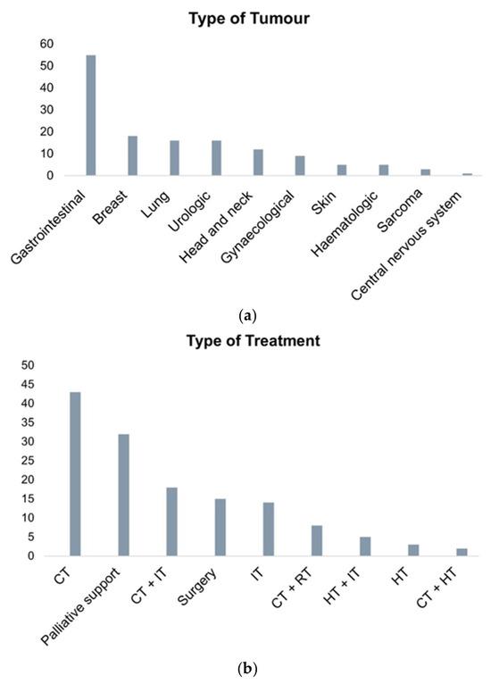
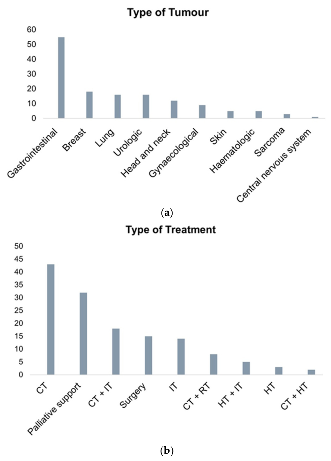
On the same day of patient recruitment and CAC diagnosis assessment, blood samples were also collected, and clinical history and demographic information were retrieved from patients’ medical records. Women comprised 54.3% (N = 76) of the study population. At enrolment, the mean age was 63.08 ± 1.1 years, with 47.1% (N = 66) of patients being over 63 years old. The mean BMI was 26.02 ± 0.44. A BMI < 26 kg/m2 was observed in 52.1% (N = 73) of participants. Concerning performance status, 48.3% (N = 57) of patients had an ECOG-PS of 0, 33.1% (N = 39) had a score of 1, 16.1% (N = 19) had a score of 2, and 2.5% (N = 3) had a score of 3. Metastatic disease was present in 64.3% (N = 90) of the cohort, while 5.7% (N = 8) reported a history of other malignancies. Details regarding tumour types and treatment modalities are presented in Figure 4.
Figure 4.
Distribution of malignant disease types (a) and cancer treatments (b) in the Study Cohort. Abbreviations: CT, chemotherapy; HT, hormonotherapy; IT, immunotherapy; RT, radiotherapy.
This study was approved by the Human Research Ethics Committee of IPO Porto (CES. 131/022, approved on 28 July 2022).
4.2. Sample Processing and Genomic DNA Extraction
At enrolment, peripheral blood was obtained from participants through standard venous blood collection. The specimens were placed in EDTA-anticoagulated tubes to prevent coagulation.
Extraction of genomic DNA was performed using the MagaBio Plus Virus DNA/RNA Purification Kit II (BSC71S1E, Bioflux®, Tokyo, Japan) in combination with the MGISP-NE32 automated nucleic acid extraction system (MGI Tech® Guangdong, China), strictly adhering to the supplier’s protocol. The yield and purity of the isolated DNA were evaluated spectrophotometrically using a NanoDrop Lite instrument (Thermo Fisher Scientific, Waltham, MA, USA). Verified DNA samples were subsequently stored at −20 °C until further analysis.
4.3. SNP Genotyping
Genotypic analysis of the VDR rs731236 polymorphism was performed using a StepOnePlus real-time PCR platform (Applied Biosystems, Carlsbad, CA, USA) with a fluorescence-based TaqMan allelic discrimination assay. The preparation of the amplification reactions was previously described [75,76,77,78,79]. Briefly, the total volume for each reaction was 6 μL, comprising 2.5 μL of TaqPath™ ProAmp™ Master Mix at working concentration (Applied Biosystems®, Foster City, CA, USA), 0.125 μL of the specific TaqMan® SNP Genotyping Assay [C___2404008_10 targeting the VDR rs731236 variant; Supplementary Table S1; (Applied Biosystems®, Foster City, CA, USA], 1 μL of template DNA, and nuclease-free water to reach the final reaction volume. Thermal cycling parameters followed a previously established protocol [77].
To monitor potential contamination, two no-template controls (i.e., negative controls) were incorporated into each run. In addition, genotyping reproducibility was validated by repeating the analysis on 20% of randomly selected samples. Interpretation of genotype calls was performed independently by three investigators who were unaware of the corresponding clinical and pathological information.
4.4. Statistical Analysis
Quantitative analyses and figure generation were carried out using IBM SPSS Statistics for Windows, version 30.0 (IBM Corp., Armonk, NY, USA) and Microsoft Excel (Microsoft Corp., Redmond, WA, USA), respectively.
Genotype frequencies observed in the study cohort were contrasted with reference data from the Iberian population available through the Ensembl database (https://www.ensembl.org/index.html, last accessed on 28 December 2025). Conformity between observed and expected genotype distributions was examined by testing for HWE using the χ2 test of independence.
The distributional properties of continuous variables were assessed using the Kolmogorov–Smirnov test. Depending on whether the variables followed a parametric or non-parametric distribution, data were categorised based on either mean or median values, respectively. Regarding PNI and NLR, initially, patients were ranked according to each marker and divided into tertiles. These three-tier classifications were subsequently combined into binary categories. For PNI, the lowest tertile (≤44.2) was designated as the low group, whereas the remaining two tertiles constituted the high group. In contrast, NLR values were considered low when falling within the first two tertiles (<3.6) and high when belonging to the upper tertile (≥3.6).
Cachexia status was assessed using the Fearon criteria, the CASC-IN classification system, and a combined approach. In the latter, patients were assigned to a group only if the Fearon and CASC-IN systems yielded the same classification. For these three diagnostic strategies, cachexia status was evaluated in two ways: firstly, using three categories (non-cachectic, pre-cachectic and cachectic); and secondly, via a binary classification where pre-cachectic and cachectic patients were grouped as cachexia-positive.
Relationships between the VDR rs731236 variant and CAC occurrence, as well as demographic and clinicopathological parameters, were evaluated using the χ2 test or Fisher’s exact test, as appropriate based on expected cell frequencies. Logistic regression analyses were conducted to estimate the CAC risk (OR and 95% CIs) associated with each SNP genotype.
The clinical outcome evaluated in this study was OS, which was defined as the time elapsed from study inclusion (corresponding to cachexia assessment) to death from any cause or to the most recent follow-up for censored cases. Survival outcomes related to VDR genetic variation were explored using Kaplan–Meier methodology. Group comparisons were performed with either the log-rank test or the Tarone–Ware test, selected according to compliance with proportional hazard assumptions and the genetic inheritance model best supported by preliminary survival curve inspection. The impact of the SNP on the risk of patient death was also examined via Cox regression analysis. Multivariable Cox analysis was conducted, adjusting for relevant demographic and clinicopathological parameters. Due to cohort size, subgroup analyses according to tumour type and cancer treatment approaches were not conducted. To evaluate data stability and reliability, bootstrap resampling (1000 iterations) was carried out.
All hypothesis testing was conducted using two-tailed procedures, and statistical significance was established at a threshold of p < 0.05.
5. Conclusions
CAC represents one of the most severe and clinically challenging complications in cancer patients and it is therefore important to identify biological determinants that influence both its susceptibility and prognosis. Hence, this real-world study examined the implications of VDR rs731236 in a cohort of cancer patients. Although these findings should be interpreted cautiously in light of the study’s limitations, they provide further evidence that this polymorphism is significantly associated with both CAC risk and patient survival. While routine genetic screening is not yet standard practice in oncology supportive care, these findings add to the growing body of evidence emphasising the importance of host genetic factors in shaping clinical outcomes. Future studies integrating VDR rs731236 with assessments of circulating vitamin D status and targeted supportive interventions are warranted to clarify the translational relevance of these associations. Moreover, further research is needed to determine the clinical effectiveness of vitamin D supplementation in preventing or mitigating active CAC. Importantly, the broader literature suggests that the biological and clinical effects of VDR rs731236 and vitamin D signalling are context-dependent. While vitamin D is widely recognised for its pro-regenerative actions in skeletal muscle under physiological conditions, these benefits may not translate directly to pathological states such as CAC. Likewise, the prognostic significance of rs731236 remains inconsistent across malignancies. Overall, these findings seem to reinforce the concept that vitamin D signalling in cancer is highly context-dependent and that genetic variation in VDR may represent a clinically relevant modifier of cancer outcomes. Pending external validation in larger cohorts, VDR rs731236 may serve as a useful biomarker for CAC risk stratification and prognostication, particularly within the evolving field of liquid biopsy.
Supplementary Materials
The following supporting information can be downloaded at: https://www.mdpi.com/article/10.3390/ijms27135816/s1.
Author Contributions
Conceptualization, V.T. and A.F.X.; methodology, V.T., A.C.L.S., I.G.d.M., T.F., M.M.P. and J.M.O.S.; formal analysis, V.T. and R.M.; investigation, V.T., A.C.L.S., I.G.d.M., T.F., M.M.P., V.R.D., C.S., M.P.S. and J.M.O.S.; resources, R.M.; writing—original draft preparation, V.T.; writing—review and editing, V.T., A.F.X., I.G.d.M., T.F., M.M.P., A.C.L.S., V.R.D., C.S., M.P.S., R.M. and J.M.O.S.; visualization, V.T.; supervision, J.M.O.S. and R.M.; project administration, R.M.; funding acquisition, V.T., I.G.d.M., T.F., J.M.O.S. and R.M. All authors have read and agreed to the published version of the manuscript.
Funding
This research was funded by Fundação para a Ciência e Tecnologia (FCT), the Portuguese Institute of Oncology of Porto (IPO Porto) (PI127-CI-IPOP-118-2019), Centre for the Research and Technology of Agro-Environmental and Biological Sciences (CITAB) [UIDB/04033/2025 (DOI: 10.54499/UIDB/04033/2025)] and Institute for Innovation, Capacity Building and Sustainability of Agri-Food Production (Inov4Agro) [LA/P/0126/2020 (DOI: 10.54499/LA/P/0126/2020)]. During the execution of the study and/or manuscript preparation, V.T., I.G.d.M. and T.F. were research fellowship holders supported by the Portuguese League Against Cancer—Northern Branch (LPCC-NRN). J.M.O.S. also contributed to this study as an LPCC-NRN 2023 Research Fellow. The institutions did not influence the writing or publishing of this article.
Institutional Review Board Statement
The study was conducted in accordance with the Declaration of Helsinki and was approved by the Human Research Ethics Committee of IPO Porto (protocol code 131/022, approved on 28 July 2022).
Informed Consent Statement
Informed consent was obtained from all subjects involved in the study.
Data Availability Statement
The data presented in this study are available on request from the corresponding author due to patient data protection.
Acknowledgments
The authors would like to thank Ministério da Saúde de Portugal, Portuguese Institute of Oncology of Porto (IPO Porto), Portuguese League Against Cancer—Northern Branch (LPCC-NRN), Centre for the Research and Technology of Agro-Environmental and Biological Sciences (CITAB), Institute for Innovation, Capacity Building and Sustainability of Agri-Food Production (Inov4Agro) and Fundação para a Ciência e Tecnologia (FCT).
Conflicts of Interest
The authors declare no conflicts of interest.
Abbreviations
The following abbreviations are used in this manuscript:
| aHR | Adjusted hazard ratio |
| BMI | Body mass index |
| CAC | Cancer-associated cachexia |
| CI | Confidence interval |
| ECOG-PS | Eastern Cooperative Oncology Group Performance Status |
| HR | Hazard ratio |
| HWE | Hardy–Weinberg equilibrium |
| IGF-1 | Insulin-like growth factor 1 |
| IL-6 | Interleukin-6 |
| mRNA | Messenger ribonucleic acid |
| NLR | Neutrophil-to-lymphocyte ratio |
| OR | Odds ratio |
| OS | Overall survival |
| PNI | Prognostic nutritional index |
| SNP | Single-nucleotide polymorphism |
| TNF-α | Tumour necrosis factor-α |
| VDR | Vitamin D receptor |
| VDREs | Vitamin D response elements |
References
- Liz-Pimenta, J.; Tavares, V.; Neto, B.V.; Santos, J.M.; Guedes, C.B.; Araújo, A.; Khorana, A.A.; Medeiros, R. Thrombosis and cachexia in cancer: Two partners in crime? Crit. Rev. Oncol. 2023, 186, 103989. [Google Scholar] [CrossRef] [Scilit]
- Mariean, C.R.; Tiucă, O.M.; Mariean, A.; Cotoi, O.S. Cancer cachexia: New insights and future directions. Cancers 2023, 15, 5590. [Google Scholar] [CrossRef] [Scilit] [PubMed]
- Alamawi, N.; Alsharawneh, A.; Abualruz, H.; AbuRuz, S.M. Cancer Cachexia in Clinical Practice: A Review of Treatment Barriers Among Cancer Patients. Eur. J. Cancer Care 2025, 2025, 6659047. [Google Scholar] [CrossRef] [Scilit]
- Peixoto da Silva, S.; Santos, J.M.; Costa e Silva, M.P.; Gil da Costa, R.M.; Medeiros, R. Cancer cachexia and its pathophysiology: Links with sarcopenia, anorexia and asthenia. J. Cachexia Sarcopenia Muscle 2020, 11, 619–635. [Google Scholar] [CrossRef] [Scilit] [PubMed]
- Johns, N.; Tan, B.; MacMillan, M.; Solheim, T.; Ross, J.; Baracos, V.; Damaraju, S.; Fearon, K. Genetic basis of interindividual susceptibility to cancer cachexia: Selection of potential candidate gene polymorphisms for association studies. J. Genet. 2014, 93, 893–916. [Google Scholar] [CrossRef] [Scilit] [PubMed]
- Liu, Q.; Li, Z.; Li, S.; Li, Y.; Pan, H.; Tao, Y. Vitamin D3 as an immunomodulatory agent: Molecular mechanisms, clinical translation, and precision therapeutic strategies. Front. Immunol. 2026, 17, 1770141. [Google Scholar] [CrossRef] [Scilit] [PubMed]
- Dzik, K.P.; Kaczor, J.J. Mechanisms of vitamin D on skeletal muscle function: Oxidative stress, energy metabolism and anabolic state. Eur. J. Appl. Physiol. 2019, 119, 825–839. [Google Scholar] [CrossRef] [Scilit] [PubMed]
- Bass, J.J.; Nakhuda, A.; Deane, C.S.; Brook, M.S.; Wilkinson, D.J.; Phillips, B.E.; Philp, A.; Tarum, J.; Kadi, F.; Andersen, D. Overexpression of the vitamin D receptor (VDR) induces skeletal muscle hypertrophy. Mol. Metab. 2020, 42, 101059. [Google Scholar] [CrossRef] [Scilit] [PubMed]
- Saponaro, F.; Saba, A.; Zucchi, R. An update on vitamin D metabolism. Int. J. Mol. Sci. 2020, 21, 6573. [Google Scholar] [CrossRef] [Scilit] [PubMed]
- Latham, C.M.; Brightwell, C.R.; Keeble, A.R.; Munson, B.D.; Thomas, N.T.; Zagzoog, A.M.; Fry, C.S.; Fry, J.L. Vitamin D promotes skeletal muscle regeneration and mitochondrial health. Front. Physiol. 2021, 12, 660498. [Google Scholar] [CrossRef] [Scilit] [PubMed]
- Camperi, A.; Pin, F.; Costamagna, D.; Penna, F.; Menduina, M.L.; Aversa, Z.; Zimmers, T.; Verzaro, R.; Fittipaldi, R.; Caretti, G. Vitamin D and VDR in cancer cachexia and muscle regeneration. Oncotarget 2017, 8, 21778. [Google Scholar] [CrossRef] [Scilit] [PubMed]
- Garcia, M.; Seelaender, M.; Sotiropoulos, A.; Coletti, D.; Lancha, A.H., Jr. Vitamin D, muscle recovery, sarcopenia, cachexia, and muscle atrophy. Nutrition 2019, 60, 66–69. [Google Scholar] [CrossRef] [Scilit] [PubMed]
- Bollen, S.E.; Bass, J.J.; Fujita, S.; Wilkinson, D.; Hewison, M.; Atherton, P.J. The Vitamin D/Vitamin D receptor (VDR) axis in muscle atrophy and sarcopenia. Cell. Signal. 2022, 96, 110355. [Google Scholar] [CrossRef] [Scilit] [PubMed]
- de Melo, I.G.; Tavares, V.; Pereira, D.; Medeiros, R. Contribution of endothelial dysfunction to Cancer susceptibility and progression: A comprehensive narrative review on the genetic risk component. Curr. Issues Mol. Biol. 2024, 46, 4845–4873. [Google Scholar] [CrossRef] [Scilit] [PubMed]
- Chiarella, P.; Capone, P.; Sisto, R. Contribution of genetic polymorphisms in human health. Int. J. Environ. Res. Public Health 2023, 20, 912. [Google Scholar] [CrossRef] [Scilit] [PubMed]
- Krasniqi, E.; Boshnjaku, A.; Wagner, K.-H.; Wessner, B. Association between polymorphisms in vitamin D pathway-related genes, vitamin D status, muscle mass and function: A systematic review. Nutrients 2021, 13, 3109. [Google Scholar] [CrossRef] [Scilit] [PubMed]
- Bollen, S.E.; Bass, J.J.; Wilkinson, D.J.; Hewison, M.; Atherton, P.J. The impact of genetic variation within the vitamin D pathway upon skeletal muscle function: A systematic review. J. Steroid Biochem. Mol. Biol. 2023, 229, 106266. [Google Scholar] [CrossRef] [Scilit] [PubMed]
- Taşkın, E.; Coşkun, M. Are vitamin D receptor gene rs731236, rs2228570 and NOS3 gene rs3138808 polymorphisms associated with diabetic retinopathy? Ophthalmic Genet. 2025, 46, 447–453. [Google Scholar] [CrossRef] [Scilit] [PubMed]
- Yates, A.D.; Austine-Orimoloye, O.; Azov, A.G.; Barba, M.; Barnes, I.; Barrera-Enriquez, V.P.; Becker, A.; Bennett, R.; Berry, A.; Bhai, J. Ensembl 2026. Nucleic Acids Res. 2026, 54, D1053–D1060. [Google Scholar] [PubMed]
- Zeng, Z.; Bromberg, Y. Predicting functional effects of synonymous variants: A systematic review and perspectives. Front. Genet. 2019, 10, 914. [Google Scholar] [CrossRef] [Scilit] [PubMed]
- Jiang, L.L.; Zhang, C.; Zhang, Y.; Ma, F.; Guan, Y. Associations between polymorphisms in VDR gene and the risk of osteoporosis: A meta-analysis. Arch. Physiol. Biochem. 2022, 128, 1637–1644. [Google Scholar] [PubMed]
- Şerifoğlu, L.; Yılmaz, S.G.; Karaaslanlı, A.; Düzkalır, A.H.; Etli, M.U.; Özdoğan, S. Association of TaqI (rs731236) Polymorphism of Vitamin D Receptor Gene with Lumbar Degenerative Disc Disease. World Neurosurg. 2024, 188, e419–e423. [Google Scholar] [CrossRef] [Scilit] [PubMed]
- Arellano Pérez Vertti, R.D.; Arellano Ramírez, D.O.; González Galarza, F.F.; Prieto Hinojosa, A.I.; Méndez Hernández, A.; Muñoz Flores, M.V.; Arellano Ramírez, D.F.; Argüello Beltrán, C.S.; Argüello Astorga, R. Association of vitamin D blood deficiency and the rs731236 polymorphism vitamin D receptor with primary knee osteoarthritis in subjects from Mexico. Clin. Rheumatol. 2025, 44, 1329–1335. [Google Scholar] [CrossRef] [Scilit] [PubMed]
- Cieślińska, A.; Kostyra, E.; Chwała, B.; Moszyńska-Dumara, M.; Fiedorowicz, E.; Teodorowicz, M.; Savelkoul, H.F. Vitamin D receptor gene polymorphisms associated with childhood autism. Brain Sci. 2017, 7, 115. [Google Scholar] [CrossRef] [Scilit] [PubMed]
- Wang, X.; Shen, N.; Lu, Y.; Tan, K. Vitamin D receptor polymorphisms and the susceptibility of Parkinson’s disease. Neurosci. Lett. 2019, 699, 206–211. [Google Scholar] [CrossRef] [Scilit] [PubMed]
- Han, J.-C.; Du, J.; Zhang, Y.-J.; Qi, G.-B.; Li, H.-B.; Zhang, Y.J.; Yu, X.-l. Vitamin D receptor polymorphisms may contribute to asthma risk. J. Asthma 2016, 53, 790–800. [Google Scholar] [CrossRef] [Scilit] [PubMed]
- Mahto, H.; Tripathy, R.; Das, B.K.; Panda, A.K. Association between vitamin D receptor polymorphisms and systemic lupus erythematosus in an Indian cohort. Int. J. Rheum. Dis. 2018, 21, 468–476. [Google Scholar] [PubMed]
- Xu, X.; Shen, M. Associations between vitamin D receptor genetic variants and tuberculosis: A meta-analysis. Innate Immun. 2019, 25, 305–313. [Google Scholar] [CrossRef] [Scilit] [PubMed]
- Tentolouris, N.; Achilla, C.; Anastasiou, I.A.; Eleftheriadou, I.; Tentolouris, A.; Basoulis, D.; Kosta, O.; Lambropoulos, A.; Yavropoulou, M.P.; Chatzikyriakidou, A. The association of vitamin D receptor polymorphisms with COVID-19 severity. Nutrients 2024, 16, 727. [Google Scholar] [CrossRef] [Scilit] [PubMed]
- Gonzalez-Soltero, R.; Tabone, M.; Larrosa, M.; Bailen, M.; Bressa, C. VDR gene TaqI (rs731236) polymorphism affects gut microbiota diversity and composition in a Caucasian population. Front. Nutr. 2024, 11, 1423472. [Google Scholar] [CrossRef] [Scilit] [PubMed]
- Raljević, D.; Peršić, V.; Markova-Car, E.; Cindrić, L.; Miškulin, R.; Žuvić, M.; Kraljević Pavelić, S. Study of vitamin D receptor gene polymorphisms in a cohort of myocardial infarction patients with coronary artery disease. BMC Cardiovasc. Disord. 2021, 21, 188. [Google Scholar] [CrossRef] [Scilit] [PubMed]
- Alkhedaide, A.; Mergani, A.; Aldhahrani, A.; Sabry, A.; Soliman, M.; Nassan, M.; Ismail, T. Association of vitamin D receptor gene polymorphisms with type 2 diabetes mellitus in Taif population: A case-control study. Braz. J. Biol. 2021, 84, e250739. [Google Scholar] [PubMed]
- Feng, M.; Li, H.; Chen, S.-F.; Li, W.-F.; Zhang, F.-B. Polymorphisms in the vitamin D receptor gene and risk of autoimmune thyroid diseases: A meta-analysis. Endocrine 2013, 43, 318–326. [Google Scholar] [PubMed]
- Donayeva, A.; Amanzholkyzy, A.; Nurgaliyeva, R.; Gubasheva, G.; Saparbayev, S.; Ayaganov, D.; Kaldybayeva, A.; Abdelazim, I.A.; Farghali, M.M. Vitamin D and vitamin D receptor polymorphism in Asian adolescents with primary dysmenorrhea. BMC Women’s Health 2023, 23, 414. [Google Scholar] [CrossRef] [Scilit] [PubMed]
- Moradkhani, A.; Azami, M.; Assadi, S.; Ghaderi, M.; Azarnezhad, A.; Moradi, Y. Association of vitamin D receptor genetic polymorphisms with the risk of infertility: A systematic review and meta-analysis. BMC Pregnancy Childbirth 2024, 24, 398. [Google Scholar] [CrossRef] [Scilit] [PubMed]
- Perna, L.; Butterbach, K.; Haug, U.; Schöttker, B.; Müller, H.; Arndt, V.; Holleczek, B.; Burwinkel, B.; Brenner, H. Vitamin D receptor genotype rs731236 (Taq1) and breast cancer prognosis. Cancer Epidemiol. Biomark. Prev. 2013, 22, 437–442. [Google Scholar] [CrossRef] [Scilit]
- Liu, S.; Cai, H.; Cheng, W.; Zhang, H.; Pan, Z.; Wang, D. Association of VDR polymorphisms (Taq I and Bsm I) with prostate cancer: A new meta-analysis. J. Int. Med. Res. 2017, 45, 3–10. [Google Scholar] [CrossRef] [Scilit] [PubMed]
- Yu, Z.-H.; Chen, M.; Zhang, Q.-Q.; Hu, X. The association of vitamin D receptor gene polymorphism with lung cancer risk: An update meta-analysis. Comb. Chem. High Throughput Screen. 2018, 21, 704–710. [Google Scholar] [PubMed]
- Aristizábal-Pachón, A.F.; González-Giraldo, Y.; Garcia, A.Y.; Suarez, D.X.; Rodríguez, A.; Gonzalez-Santos, J. Association between VDR gene polymorphisms and melanoma susceptibility in a colombian population. Asian Pac. J. Cancer Prev. APJCP 2022, 23, 79. [Google Scholar] [CrossRef] [Scilit] [PubMed]
- Guan, M.; Wang, Y. Common variants of vitamin D receptor gene polymorphisms and risk of gastric cancer: A meta-analysis. Medicine 2024, 103, e39527. [Google Scholar] [CrossRef] [Scilit] [PubMed]
- Lyu, C.; Yin, X.; Li, Z.; Wang, T.; Xu, R. Vitamin D receptor gene polymorphisms and multiple myeloma: A meta-analysis. Clin. Exp. Med. 2024, 24, 118. [Google Scholar] [CrossRef] [Scilit] [PubMed]
- Rosen, C.J.; Adams, J.S.; Bikle, D.D.; Black, D.M.; Demay, M.B.; Manson, J.E.; Murad, M.H.; Kovacs, C.S. The nonskeletal effects of vitamin D: An Endocrine Society scientific statement. Endocr. Rev. 2012, 33, 456–492. [Google Scholar] [CrossRef] [Scilit] [PubMed]
- Carlberg, C. Vitamin D and its target genes. Nutrients 2022, 14, 1354. [Google Scholar] [CrossRef] [Scilit] [PubMed]
- Voltan, G.; Cannito, M.; Ferrarese, M.; Ceccato, F.; Camozzi, V. Vitamin D: An overview of gene regulation, ranging from metabolism to genomic effects. Genes 2023, 14, 1691. [Google Scholar] [CrossRef] [Scilit] [PubMed]
- Zhao, G.; Simpson, R.U. Membrane localization, Caveolin-3 association and rapid actions of vitamin D receptor in cardiac myocytes. Steroids 2010, 75, 555–559. [Google Scholar] [CrossRef] [Scilit] [PubMed]
- Bollen, S.E.; Atherton, P.J. Myogenic, genomic and non-genomic influences of the vitamin D axis in skeletal muscle. Cell Biochem. Funct. 2021, 39, 48–59. [Google Scholar] [PubMed]
- Sandgren, M.E.; Brönnegärd, M.; DeLuca, H.F. Tissue distribution of the 1,25-dihydroxyvitamin D3 receptor in the male rat. Biochem. Biophys. Res. Commun. 1991, 181, 611–616. [Google Scholar] [CrossRef] [Scilit] [PubMed]
- Wang, Y.; DeLuca, H.F. Is the vitamin D receptor found in muscle? Endocrinology 2011, 152, 354–363. [Google Scholar] [CrossRef] [Scilit] [PubMed]
- Girgis, C.M.; Clifton-Bligh, R.J.; Mokbel, N.; Cheng, K.; Gunton, J.E. Vitamin D signaling regulates proliferation, differentiation, and myotube size in C2C12 skeletal muscle cells. Endocrinology 2014, 155, 347–357. [Google Scholar] [CrossRef] [Scilit] [PubMed]
- Braga, M.; Simmons, Z.; Norris, K.C.; Ferrini, M.G.; Artaza, J.N. Vitamin D induces myogenic differentiation in skeletal muscle derived stem cells. Endocr. Connect. 2017, 6, 139–150. [Google Scholar] [CrossRef] [Scilit] [PubMed]
- Polly, P.; Tan, T.C. The role of vitamin D in skeletal and cardiac muscle function. Front. Physiol. 2014, 5, 145. [Google Scholar] [CrossRef] [Scilit] [PubMed]
- Li, A.; Shen, P.; Liu, S.; Wang, J.; Zeng, J.; Du, C. Vitamin D alleviates skeletal muscle loss and insulin resistance by inducing vitamin D receptor expression and regulating the AMPK/SIRT1 signaling pathway in mice. Food Sci. Technol. 2021, 42, e47921. [Google Scholar]
- Salles, J.; Chanet, A.; Giraudet, C.; Patrac, V.; Pierre, P.; Jourdan, M.; Luiking, Y.C.; Verlaan, S.; Migné, C.; Boirie, Y. 1,25(OH)2-vitamin D3 enhances the stimulating effect of leucine and insulin on protein synthesis rate through Akt/PKB and mTOR mediated pathways in murine C2C12 skeletal myotubes. Mol. Nutr. Food Res. 2013, 57, 2137–2146. [Google Scholar] [CrossRef] [Scilit] [PubMed]
- Garcia, L.A.; King, K.K.; Ferrini, M.G.; Norris, K.C.; Artaza, J.N. 1,25(OH)2 vitamin D3 stimulates myogenic differentiation by inhibiting cell proliferation and modulating the expression of promyogenic growth factors and myostatin in C2C12 skeletal muscle cells. Endocrinology 2011, 152, 2976–2986. [Google Scholar] [CrossRef] [Scilit] [PubMed]
- Yang, W.; Huang, J.; Wu, H.; Wang, Y.; Du, Z.; Ling, Y.; Wang, W.; Wu, Q.; Gao, W. Molecular mechanisms of cancer cachexia-induced muscle atrophy. Mol. Med. Rep. 2020, 22, 4967–4980. [Google Scholar] [CrossRef] [Scilit] [PubMed]
- Tomei, S.; Singh, P.; Mathew, R.; Mattei, V.; Garand, M.; Alwakeel, M.; Sharif, E.; Al Khodor, S. The role of polymorphisms in vitamin D-related genes in response to vitamin D supplementation. Nutrients 2020, 12, 2608. [Google Scholar] [CrossRef] [Scilit] [PubMed]
- Bozsodi, A.; Boja, S.; Szilagyi, A.; Somhegyi, A.; Varga, P.P.; Lazary, A. Muscle strength is associated with vitamin D receptor gene variants. J. Orthop. Res. 2016, 34, 2031–2037. [Google Scholar] [CrossRef] [Scilit] [PubMed]
- Wang, P.; Ma, L.; Wang, H.; Zhang, W.; Tian, Q.; Cao, D.; Zheng, G.; Sun, Y. Association between polymorphisms of vitamin D receptor gene ApaI, BsmI and TaqI and muscular strength in young Chinese women. Int. J. Sports Med. 2006, 27, 182–186. [Google Scholar] [PubMed]
- Bahat, G.; Saka, B.; Erten, N.; Ozbek, U.; Coskunpinar, E.; Yildiz, S.; Sahinkaya, T.; Akif Karan, M. BsmI polymorphism in the vitamin D receptor gene is associated with leg extensor muscle strength in elderly men. Aging Clin. Exp. Res. 2010, 22, 198–205. [Google Scholar] [CrossRef] [Scilit] [PubMed]
- Walsh, S.; Ludlow, A.T.; Metter, E.J.; Ferrucci, L.; Roth, S.M. Replication study of the vitamin D receptor (VDR) genotype association with skeletal muscle traits and sarcopenia. Aging Clin. Exp. Res. 2016, 28, 435–442. [Google Scholar] [PubMed]
- Yang, C.; Li, Y. VDR Gene Polymorphisms and Inter-Individual Variability in Response to Resistance Training. Genes 2026, 17, 137. [Google Scholar] [CrossRef] [Scilit] [PubMed]
- Ovesjö, M.L.; Skilving, I.; Bergman, P.; Rane, A.; Ekström, L.; Björkhem-Bergman, L. Low Vitamin D Levels and Genetic Polymorphism in the Vitamin D Receptor are Associated with Increased Risk of Statin-Induced Myopathy. Basic Clin. Pharmacol. Toxicol. 2016, 118, 214–218. [Google Scholar] [PubMed]
- Saini, A.; Björkhem-Bergman, L.; Boström, J.; Lilja, M.; Melin, M.; Olsson, K.; Ekström, L.; Bergman, P.; Altun, M.; Rullman, E. Impact of vitamin D and vitamin D receptor TaqI polymorphism in primary human myoblasts. Endocr. Connect. 2019, 8, 1070–1081. [Google Scholar] [CrossRef] [Scilit] [PubMed]
- Wong, Y.Y.; McCaul, K.A.; Yeap, B.B.; Hankey, G.J.; Flicker, L. Low vitamin D status is an independent predictor of increased frailty and all-cause mortality in older men: The Health in Men Study. J. Clin. Endocrinol. Metab. 2013, 98, 3821–3828. [Google Scholar] [CrossRef] [Scilit] [PubMed]
- Martínez-Alonso, M.; Dusso, A.; Ariza, G.; Nabal, M. Vitamin D deficiency and its association with fatigue and quality of life in advanced cancer patients under palliative care: A cross-sectional study. Palliat. Med. 2016, 30, 89–96. [Google Scholar] [PubMed]
- Habib, A.M.; Nagi, K.; Thillaiappan, N.B.; Sukumaran, V.; Akhtar, S. Vitamin D and its potential interplay with pain signaling pathways. Front. Immunol. 2020, 11, 820. [Google Scholar] [CrossRef] [Scilit] [PubMed]
- Wu, J.; Chi, J.; Zhang, Y. Vitamin D and cancer-related fatigue in elderly patients: Mechanisms and therapeutic insights—A narrative review. Front. Nutr. 2025, 12, 1642166. [Google Scholar] [CrossRef] [Scilit] [PubMed]
- Pineda Lancheros, L.E.; Perez Ramirez, C.; Sanchez Martin, A.; Galvez Navas, J.M.; Martinez Martinez, F.; Ramirez Tortosa, M.d.C.; Jimenez Morales, A. Impact of genetic polymorphisms on the metabolic pathway of vitamin D and survival in non-small cell lung cancer. Nutrients 2021, 13, 3783. [Google Scholar] [CrossRef] [Scilit] [PubMed]
- Berger, M.D.; Stintzing, S.; Heinemann, V.; Cao, S.; Yang, D.; Sunakawa, Y.; Matsusaka, S.; Ning, Y.; Okazaki, S.; Miyamoto, Y. A polymorphism within the vitamin D transporter gene predicts outcome in metastatic colorectal cancer patients treated with FOLFIRI/bevacizumab or FOLFIRI/cetuximab. Clin. Cancer Res. 2018, 24, 784–793. [Google Scholar] [CrossRef] [Scilit] [PubMed]
- Perna, L.; Hoffmeister, M.; Schöttker, B.; Arndt, V.; Haug, U.; Holleczek, B.; Burwinkel, B.; Ordóñez-Mena, J.M.; Brenner, H. Vitamin D receptor polymorphism and colorectal cancer-specific and all-cause mortality. Cancer Epidemiol. 2013, 37, 905–907. [Google Scholar] [CrossRef] [Scilit] [PubMed]
- Hama, T.; Norizoe, C.; Suga, H.; Mimura, T.; Kato, T.; Moriyama, H.; Urashima, M. Prognostic significance of vitamin D receptor polymorphisms in head and neck squamous cell carcinoma. PLoS ONE 2011, 6, e29634. [Google Scholar] [CrossRef] [Scilit] [PubMed]
- Moreira Pires, M.; Guerra de Melo, I.; Silva, A.C.L.; Rocha Dias, V.; Silva, C.; Silva, M.P.; MO Santos, J.; Ferreira, T.; Tavares, V.; Medeiros, R. Genetic Variability in the IGF-1 Axis Modulates Cancer-Associated Cachexia and Prognosis. Cancers 2026, 18, 1822. [Google Scholar] [CrossRef] [Scilit] [PubMed]
- Silva, A.L.; Santos, J.; Tavares, V.; Ferreira, T.; Dias, V.; da Silva, C.; e Silva, M.; Medeiros, R. The association of the Prognostic Nutritional Index (PNI) and Neutrophil-Lymphocyte Ratio (NLR) with cachexia in cancer patients. Clin. Nutr. ESPEN 2024, 63, 1052. [Google Scholar] [CrossRef] [Scilit]
- Zhang, Q.; Song, M.M.; Zhang, X.; Ding, J.S.; Ruan, G.T.; Zhang, X.W.; Liu, T.; Yang, M.; Ge, Y.Z.; Tang, M. Association of systemic inflammation with survival in patients with cancer cachexia: Results from a multicentre cohort study. J. Cachexia Sarcopenia Muscle 2021, 12, 1466–1476. [Google Scholar] [CrossRef] [Scilit] [PubMed]
- Tavares, V.; Pinto, R.; Assis, J.; Coelho, S.; Brandao, M.; Alves, S.; Pereira, D.; Medeiros, R. Implications of venous thromboembolism GWAS reported genetic makeup in the clinical outcome of ovarian cancer patients. Pharmacogenomics J. 2021, 21, 222–232. [Google Scholar] [PubMed]
- Silva, H.-H.; Tavares, V.; Silva, M.-R.G.; Neto, B.V.; Cerqueira, F.; Medeiros, R. FAAH rs324420 Polymorphism Is Associated with Performance in Elite Rink-Hockey Players. Biology 2022, 11, 1076. [Google Scholar] [CrossRef] [Scilit] [PubMed]
- Tavares, V.; Assis, J.; Pinto, R.; Freitas-Silva, M.; Medeiros, R. Venous thromboembolism-related genetic determinant F11 rs4253417 is a potential prognostic factor in ischaemic stroke. Mol. Cell. Probes 2023, 70, 101917. [Google Scholar] [CrossRef] [Scilit] [PubMed]
- Marques, I.S.; Tavares, V.; Vieira Neto, B.; Lopes, L.R.; Goes, R.A.; Guimarães, J.A.M.; Perini, J.A.; Medeiros, R. Genetic variations in susceptibility to traumatic muscle injuries and muscle pain among Brazilian high-performance athletes. Int. J. Mol. Sci. 2024, 25, 3300. [Google Scholar] [CrossRef] [Scilit] [PubMed]
- de Melo, I.G.; Tavares, V.; Savva-Bordalo, J.; Rei, M.; Liz-Pimenta, J.; Pereira, D.; Medeiros, R. Endothelial Dysfunction Markers in Ovarian Cancer: VTE Risk and Tumour Prognostic Outcomes. Life 2024, 14, 1630. [Google Scholar] [CrossRef] [Scilit] [PubMed]

Disclaimer/Publisher’s Note: The statements, opinions and data contained in all publications are solely those of the individual author(s) and contributor(s) and not of MDPI and/or the editor(s). MDPI and/or the editor(s) disclaim responsibility for any injury to people or property resulting from any ideas, methods, instructions or products referred to in the content. |
© 2026 by the authors. Licensee MDPI, Basel, Switzerland. This article is an open access article distributed under the terms and conditions of the Creative Commons Attribution (CC BY) license.